Embed Size (px)
Citation preview
RADIOGRAPHIC PARAMETERS
Presented By : Celjhon ArinoBS RadTech 2nd year
Qualities of the Radiograph
• The image on a radiograph must have all of the same qualities as an image you visually see – intensity, contrast, noise, sharpness of detail, magnification and distortion of shape.
Visibility of Detail
• A visible image is completely worthless unless the information in it is capable of being recognized. If the image is blurry, or if it’s size or shape is grossly distorted, we may not be able to tell what it is, even though it is visible.
Sharpness of Image Detail
• It may be described as the abruptness with which the edges of a particular object stop.
• Detail describes the sharpness of appearance of small structures on the radiograph with adequate detail, even the smallest parts of the anatomy are visible.
• It is also refers to the structural lines or borders of tissues in the image and the amount of blur of the image.
Sharpness of Image Detail
• Factors that generally control the sharpness of image detail are the geometric factors : focal spot size, SID, and object-to-image receptor distance
Focal Spot Size
• The term focal spot is normally used to refer to the area on the x-ray tube anode from which x-rays are emitted, as seen from the viewpoint of the film.
• The influence of the focal spot on detail is confined to image sharpness.
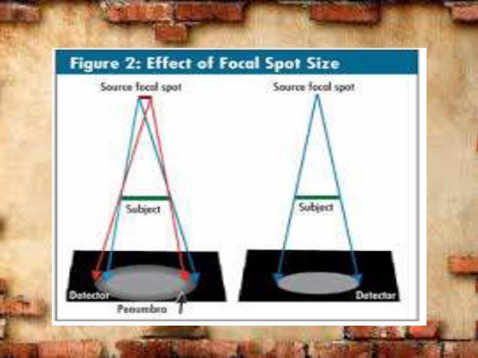
• = the smaller the focal spot, the sharper the recorded detail. Because the size of the focal spot is inversely proportional to image sharpness.
Focal Spot Size
• Most machines limited to two focal spot sizes.
• Common office focal spots are 1.0 mm for the small and 2.0 mm for large.
• Highly detailed radiography such as mammography use micro-focus tubes with 0.1 mm and 0.3 mm focal spot sizes.
Focal Spot Size
• Focal spot size has no relation to distortion of the shape of the image, because it is not related to alignment or angles.
Filtration
• All x-ray beams are affected by the filtration of the tube. The tube housing provides about 0.5 mm of filtration.
• Additional filtration is added in the collimator to meet the 2.5 mm of aluminum minimum filtration required by law.
• 2.5 mm is required for 70 kVp.
Filtration
• 3.0 mm is required for at 100 kVp.
• 3.2 mm is required for operations at 120 kVp.
• Most machines now are capable of over 100 kVp operation.
• We have no control on these filters.
Radiographic Quality
• Radiographic Quality refers to the fidelity with which the anatomic structures being examined are images on the film.
Radiographic Quality
• Characteristic of radiographic quality:
– Spatial Resolution (Recorded Detail)
– Contrast Resolution (Visibility of Detail)
– Noise (Visibility of Detail)
– Artifacts
Spatial Resolution• Spatial Resolution is the ability to image small
structures that have high subject contrast such as bone-soft tissue interface.
• When all of the factors are correct, conventional radiography has excellent spatial resolution.
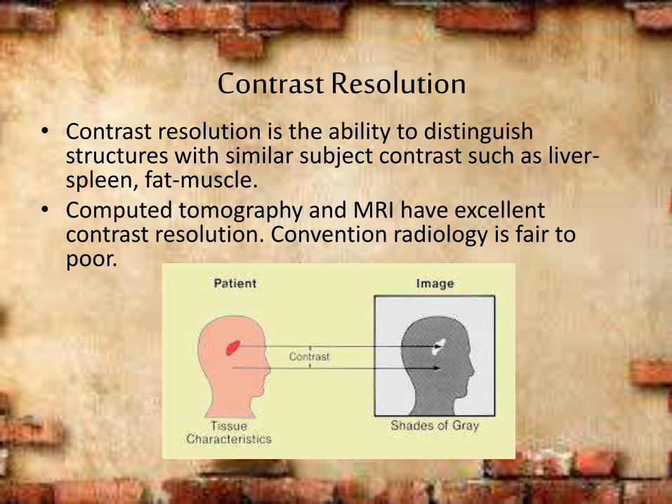
Contrast Resolution• Contrast resolution is the ability to distinguish
structures with similar subject contrast such as liver-spleen, fat-muscle.
• Computed tomography and MRI have excellent contrast resolution. Convention radiology is fair to poor.
Noise
• Noise is an undesirable fluctuation in optical density of the image. Two major types:
– Film Graininess- no control over
– Quantum Mottle- some control over
Film Graininess
• Film graininess refers to the distribution in size and space of the silver halide grains in the film emulsion.
• Similar to photographic film. 400 ASA film is more graininess than 100 ASA film.
• Similar to structure mottle that refers to the size and shape of the phosphors in the intensifying screens.
Quantum Mottle
• Quantum mottle refers to the random nature of how the x-rays interact with the image receptor.
• It is the primary form of radiographic noise.
• The use of high mAs and low kVp reduced quantum mottle.
Quantum Mottle
• Very fast screens have higher quantum mottle because it takes fewer x-rays to make the image.
Speed
• Resolution and noise are intimately connected with speed.
• While the speed of the images receptor is not apparent on the image, it influences both resolution and noise.
Film Processing
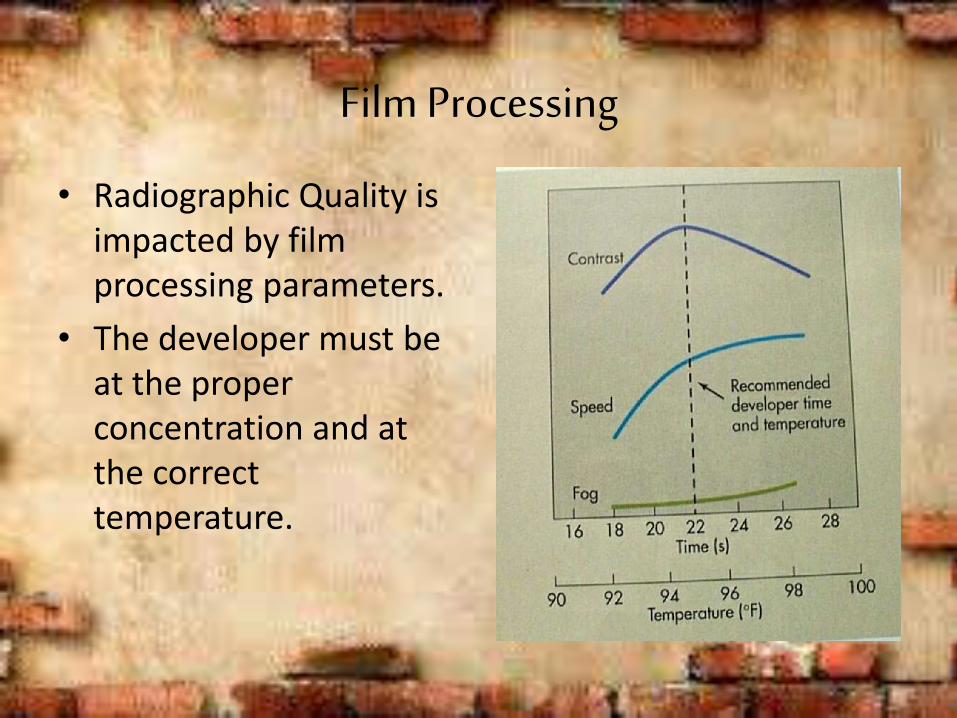
• Radiographic Quality is impacted by film processing parameters.
• The developer must be at the proper concentration and at the correct temperature.
Film Processing
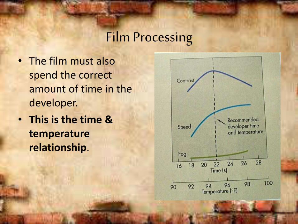
• The film must also spend the correct amount of time in the developer.
• This is the time & temperature relationship.