Embed Size (px)
Citation preview
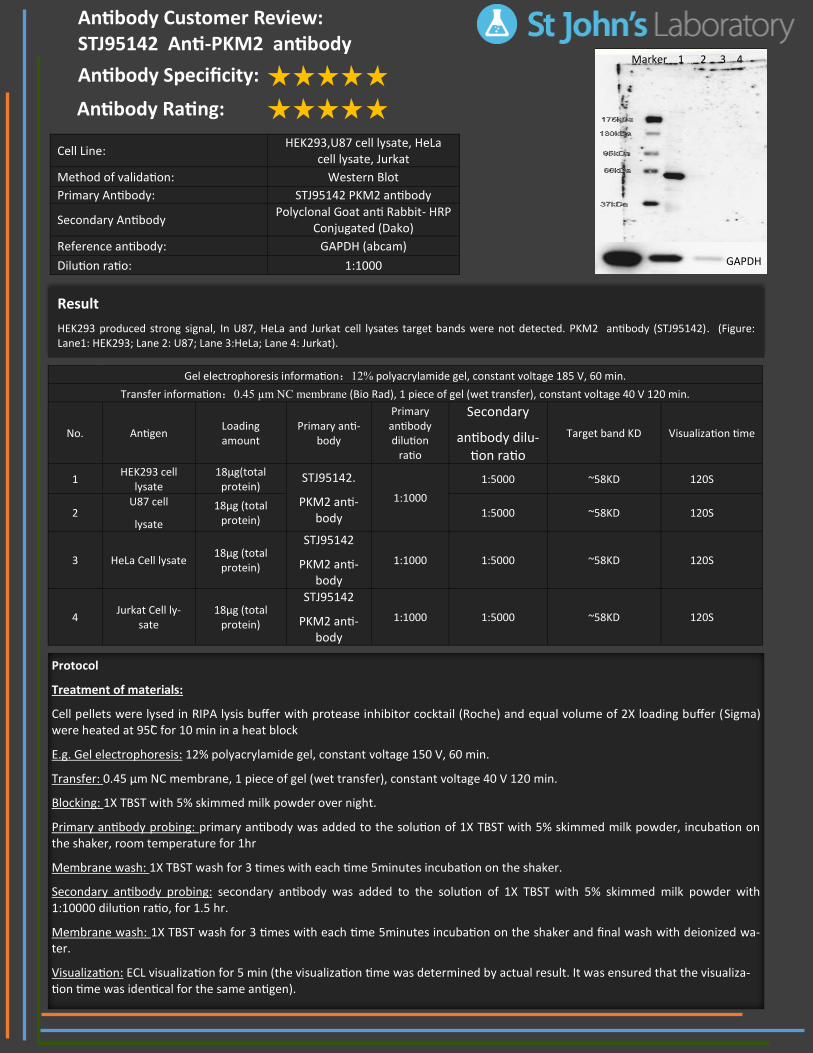
Result
HEK293 produced strong signal, In U87, HeLa and Jurkat cell lysates target bands were not detected. PKM2 antibody (STJ95142). (Figure: Lane1: HEK293; Lane 2: U87; Lane 3:HeLa; Lane 4: Jurkat).
Cell Line: HEK293,U87 cell lysate, HeLa
cell lysate, Jurkat
Method of validation: Western Blot
Primary Antibody: STJ95142 PKM2 antibody
Secondary Antibody Polyclonal Goat anti Rabbit- HRP
Conjugated (Dako)
Reference antibody: GAPDH (abcam)
Dilution ratio: 1:1000
Protocol
Treatment of materials:
Cell pellets were lysed in RIPA lysis buffer with protease inhibitor cocktail (Roche) and equal volume of 2X loading buffer (Sigma) were heated at 95C̊ for 10 min in a heat block
E.g. Gel electrophoresis: 12% polyacrylamide gel, constant voltage 150 V, 60 min.
Transfer: 0.45 µm NC membrane, 1 piece of gel (wet transfer), constant voltage 40 V 120 min.
Blocking: 1X TBST with 5% skimmed milk powder over night.
Primary antibody probing: primary antibody was added to the solution of 1X TBST with 5% skimmed milk powder, incubation on the shaker, room temperature for 1hr
Membrane wash: 1X TBST wash for 3 times with each time 5minutes incubation on the shaker.
Secondary antibody probing: secondary antibody was added to the solution of 1X TBST with 5% skimmed milk powder with 1:10000 dilution ratio, for 1.5 hr.
Membrane wash: 1X TBST wash for 3 times with each time 5minutes incubation on the shaker and final wash with deionized wa-ter.
Visualization: ECL visualization for 5 min (the visualization time was determined by actual result. It was ensured that the visualiza-tion time was identical for the same antigen).
Antibody Customer Review: STJ95142 Anti-PKM2 antibody Antibody Specificity:
Antibody Rating:
Gel electrophoresis information:12% polyacrylamide gel, constant voltage 185 V, 60 min.
Transfer information:0.45 µm NC membrane (Bio Rad), 1 piece of gel (wet transfer), constant voltage 40 V 120 min.
No. Antigen Loading amount
Primary anti-body
Primary antibody dilution
ratio
Secondary
antibody dilu-tion ratio
Target band KD Visualization time
1 HEK293 cell
lysate 18µg(total
protein) STJ95142.
PKM2 anti-body
1:1000
1:5000 ~58KD 120S
2 U87 cell
lysate
18µg (total protein)
1:5000 ~58KD 120S
3 HeLa Cell lysate 18µg (total
protein)
STJ95142
PKM2 anti-body
1:1000 1:5000 ~58KD 120S
4 Jurkat Cell ly-
sate 18µg (total
protein)
STJ95142
PKM2 anti-body
1:1000 1:5000 ~58KD 120S
GAPDH
Marker 1 2 3 4