Embed Size (px)
Citation preview
植物生理 光合作用 photosynthesis
李澤民 Tse-Min Lee
海洋植物研究室 Marine Botany Laboratory 海洋生物科技暨資源學系
Department of Marine Biotechnology and Resources 國立中山大學
National Sun Yat-sen University Kaohsiung, TAIWAN
Definition Physiology
principle for life Plant
A life that is Autotrophic Photosynthesis Cell wall
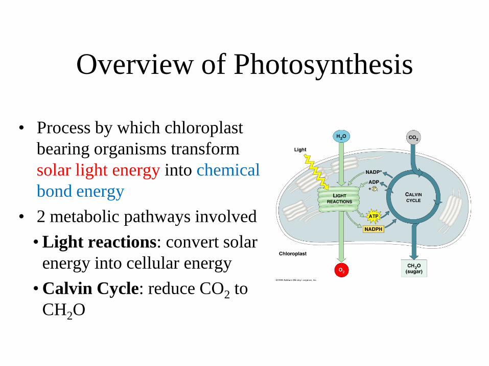
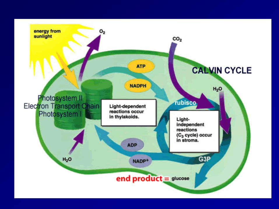
Overview of Photosynthesis
• Process by which chloroplast bearing organisms transform solar light energy into chemical bond energy
• 2 metabolic pathways involved • Light reactions: convert solar
energy into cellular energy • Calvin Cycle: reduce CO2 to
CH2O
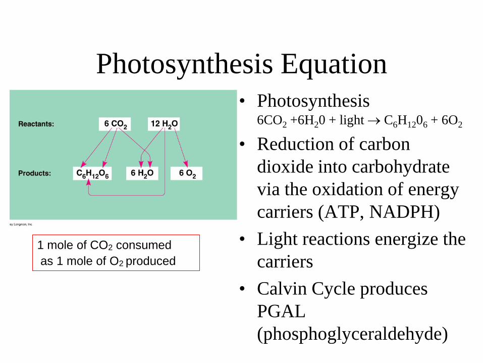
Photosynthesis Equation • Photosynthesis
6CO2 +6H20 + light → C6H1206 + 6O2
• Reduction of carbon dioxide into carbohydrate via the oxidation of energy carriers (ATP, NADPH)
• Light reactions energize the carriers
• Calvin Cycle produces PGAL (phosphoglyceraldehyde)
1 mole of CO2 consumed as 1 mole of O2 produced
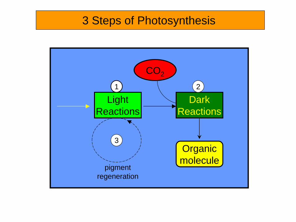
3 Steps of Photosynthesis
CO2
Dark Reactions
Organic molecule
Light Reactions
1 2
3
pigment regeneration
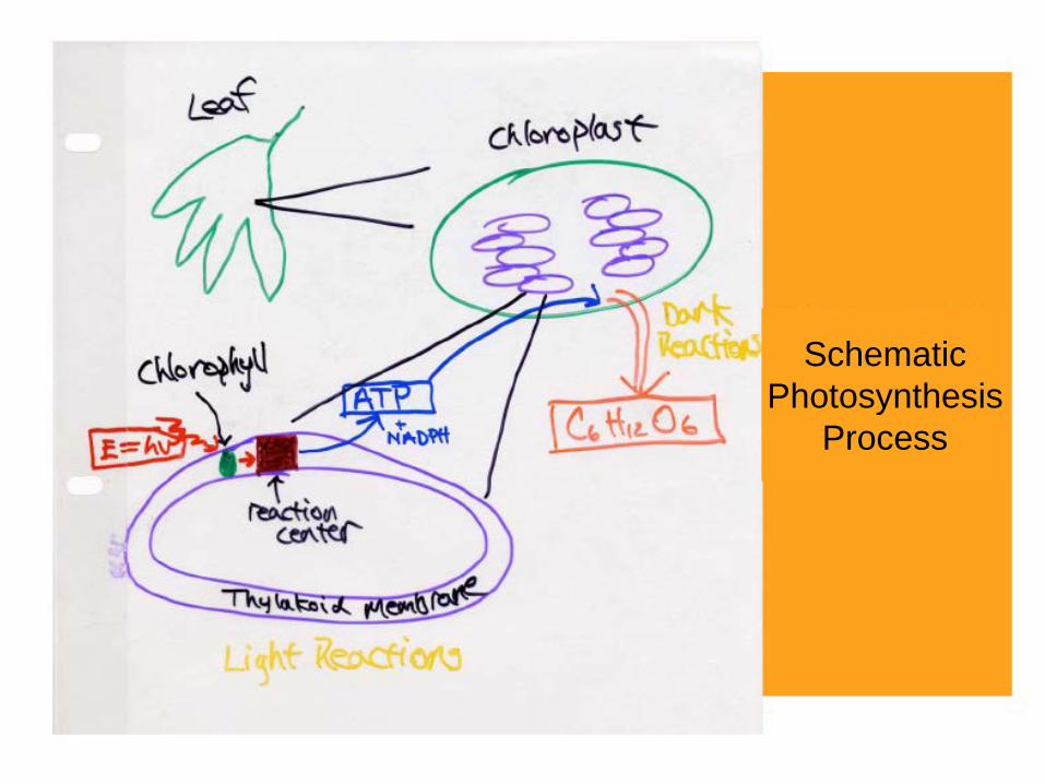
Schematic Photosynthesis
Process
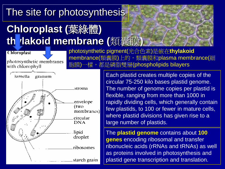
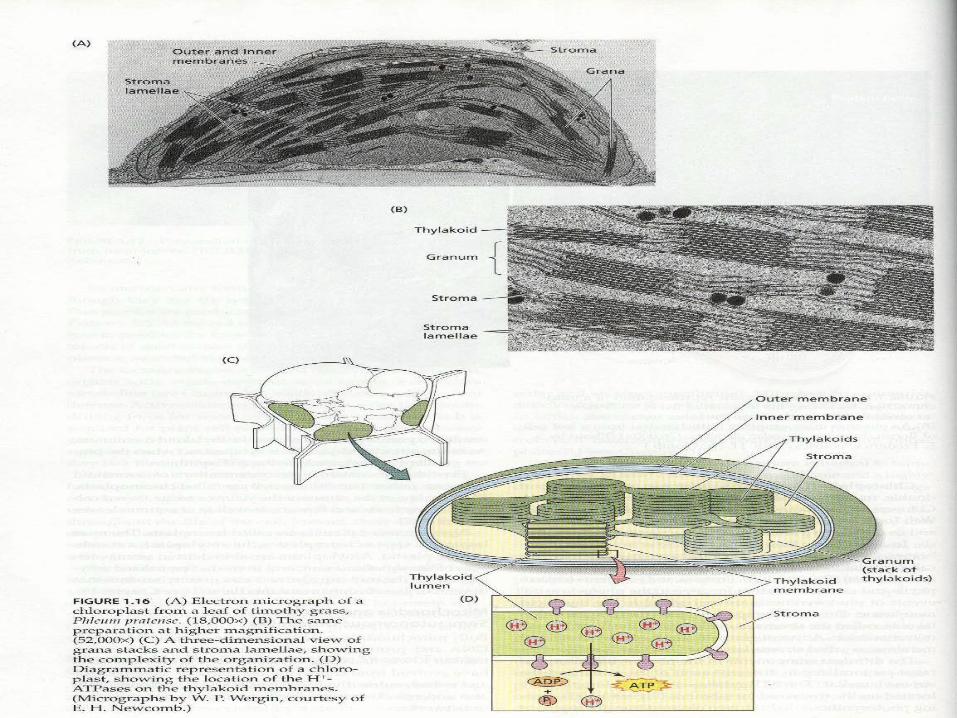
Each plastid creates multiple copies of the circular 75-250 kilo bases plastid genome. The number of genome copies per plastid is flexible, ranging from more than 1000 in rapidly dividing cells, which generally contain few plastids, to 100 or fewer in mature cells, where plastid divisions has given rise to a large number of plastids.
The plastid genome contains about 100 genes encoding ribosomal and transfer ribonucleic acids (rRNAs and tRNAs) as well as proteins involved in photosynthesis and plastid gene transcription and translation.
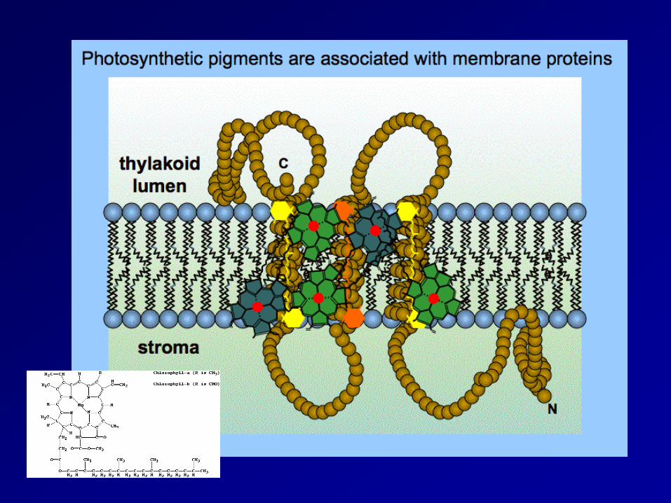
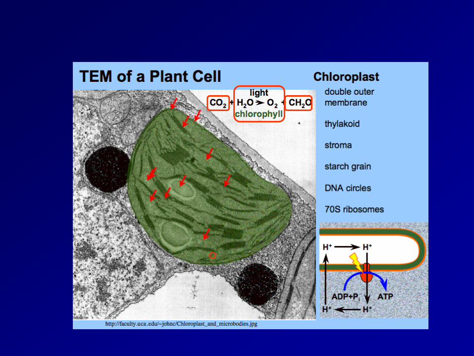
Chloroplast (葉綠體) thylakoid membrane (類囊膜)
The site for photosynthesis
photosynthetic pigment(光合色素)是嵌在thylakoid membrance(類囊膜)上的,類囊膜和plasma membrance(細胞膜)一樣,都是磷脂雙層(phospholipids bilayers
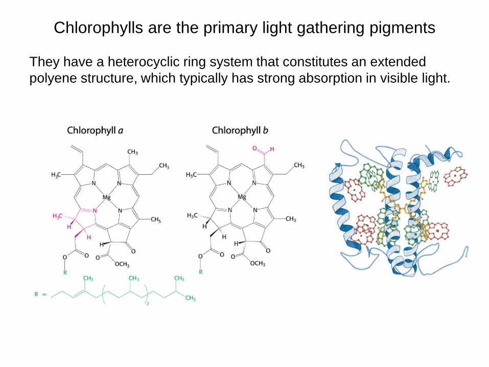
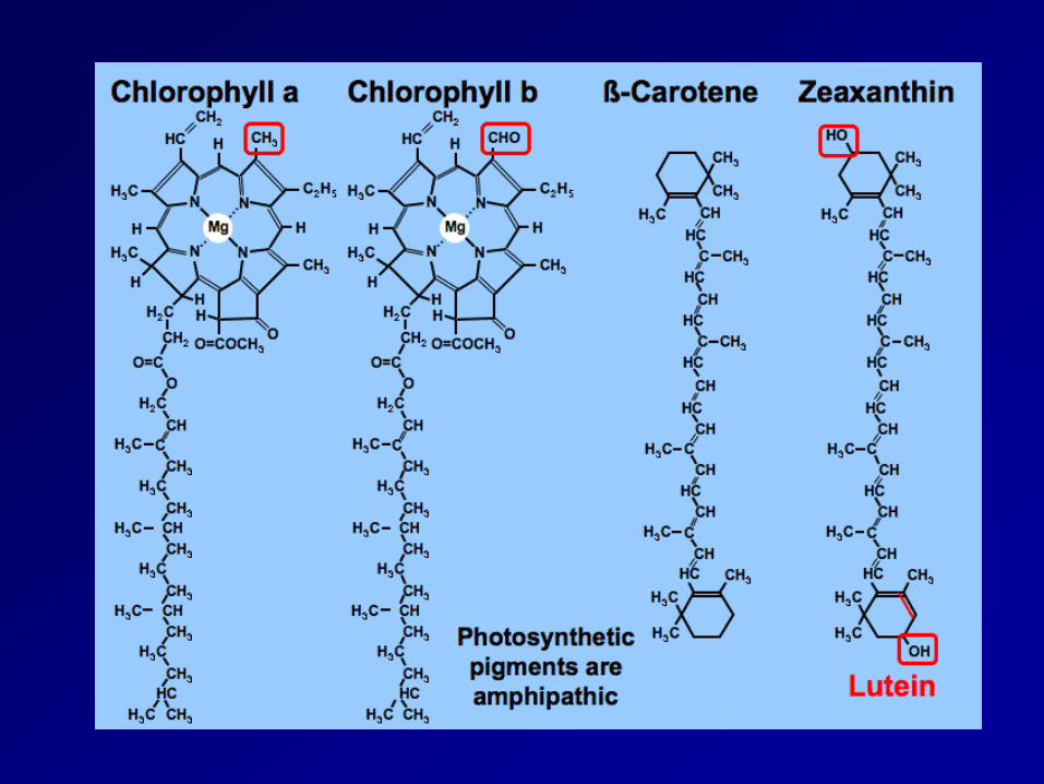
Chlorophylls are the primary light gathering pigments They have a heterocyclic ring system that constitutes an extended polyene structure, which typically has strong absorption in visible light.
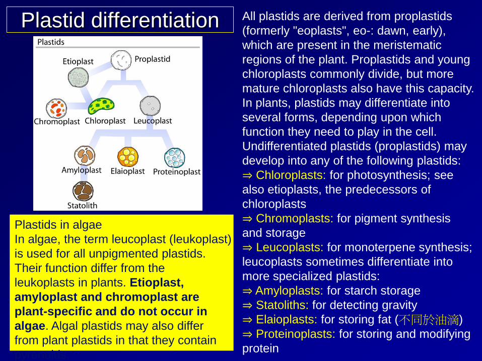
Plastid differentiation All plastids are derived from proplastids (formerly "eoplasts", eo-: dawn, early), which are present in the meristematic regions of the plant. Proplastids and young chloroplasts commonly divide, but more mature chloroplasts also have this capacity. In plants, plastids may differentiate into several forms, depending upon which function they need to play in the cell. Undifferentiated plastids (proplastids) may develop into any of the following plastids: ⇒ Chloroplasts: for photosynthesis; see also etioplasts, the predecessors of chloroplasts ⇒ Chromoplasts: for pigment synthesis and storage ⇒ Leucoplasts: for monoterpene synthesis; leucoplasts sometimes differentiate into more specialized plastids: ⇒ Amyloplasts: for starch storage ⇒ Statoliths: for detecting gravity ⇒ Elaioplasts: for storing fat (不同於油滴) ⇒ Proteinoplasts: for storing and modifying protein
Plastids in algae In algae, the term leucoplast (leukoplast) is used for all unpigmented plastids. Their function differ from the leukoplasts in plants. Etioplast, amyloplast and chromoplast are plant-specific and do not occur in algae. Algal plastids may also differ from plant plastids in that they contain pyrenoids.
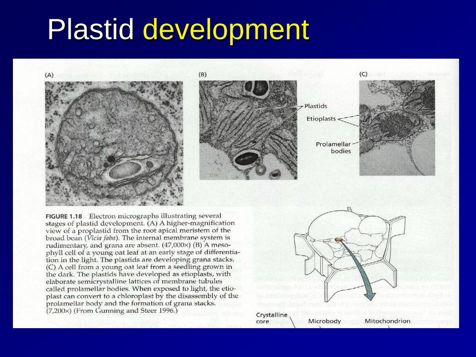
Plastid development
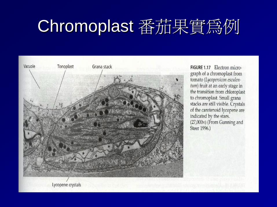
Chromoplast 番茄果實為例
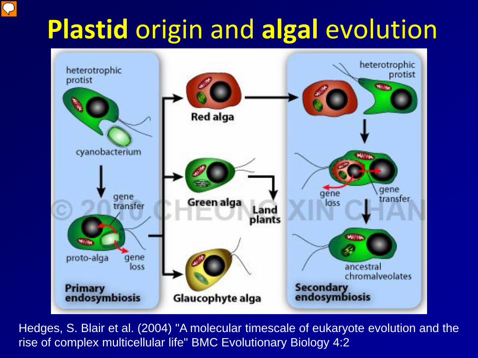
Plastid origin and algal evolution
Hedges, S. Blair et al. (2004) "A molecular timescale of eukaryote evolution and the rise of complex multicellular life" BMC Evolutionary Biology 4:2

Photosynthesis
photochemical processes enzymatic processes
2n CO2 + 2n H2O + photons → 2(CH2O)n + 2n O2
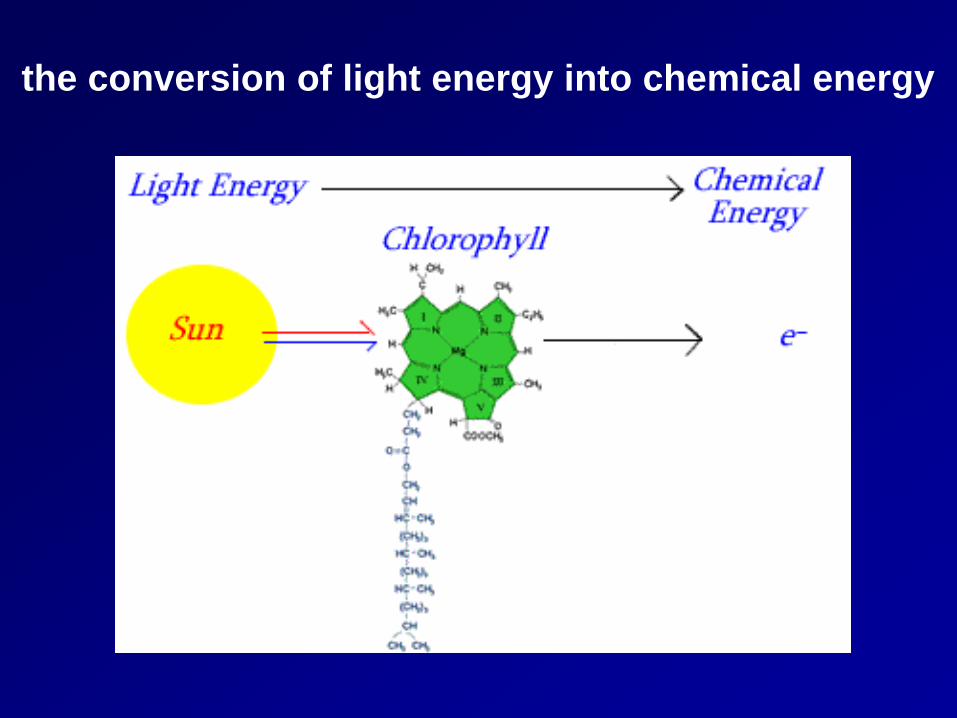
the conversion of light energy into chemical energy
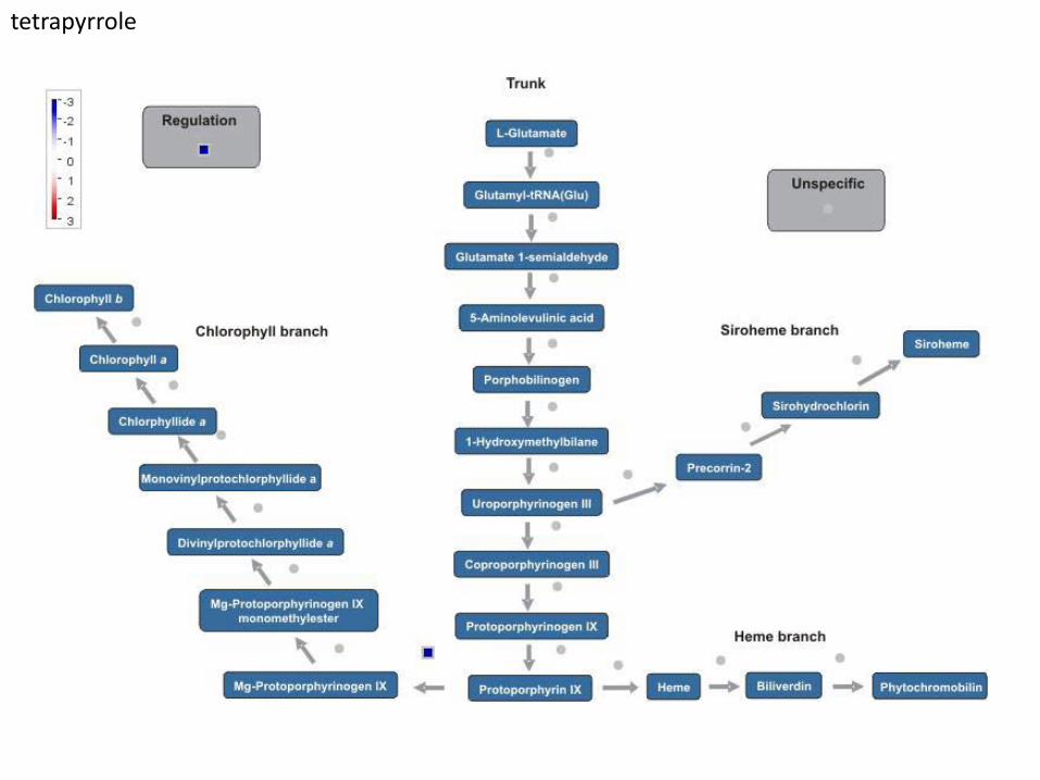
tetrapyrrole
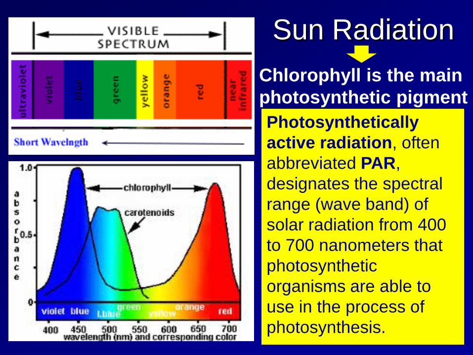
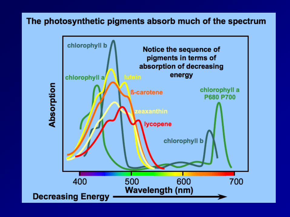
Sun Radiation Chlorophyll is the main photosynthetic pigment Photosynthetically active radiation, often abbreviated PAR, designates the spectral range (wave band) of solar radiation from 400 to 700 nanometers that photosynthetic organisms are able to use in the process of photosynthesis.
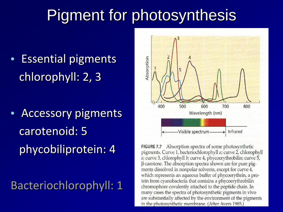
Pigment for photosynthesis
• Essential pigments
chlorophyll: 2, 3
• Accessory pigments
carotenoid: 5
phycobiliprotein: 4
Bacteriochlorophyll: 1
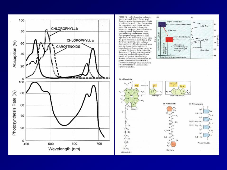
absorption spectrum
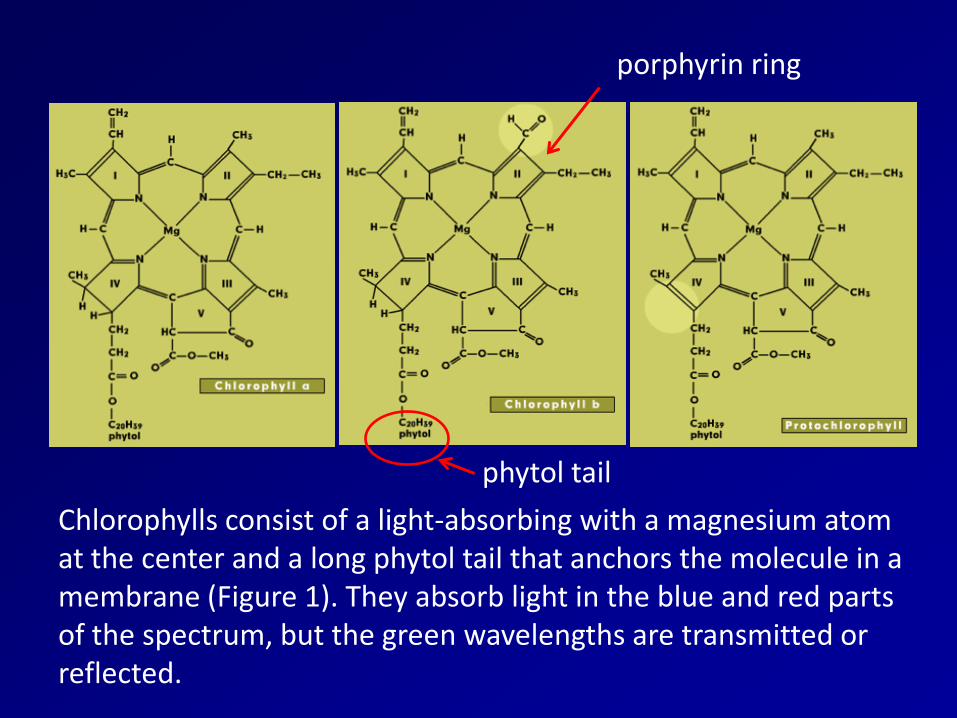
Chlorophylls consist of a light-absorbing with a magnesium atom at the center and a long phytol tail that anchors the molecule in a membrane (Figure 1). They absorb light in the blue and red parts of the spectrum, but the green wavelengths are transmitted or reflected.
porphyrin ring
phytol tail
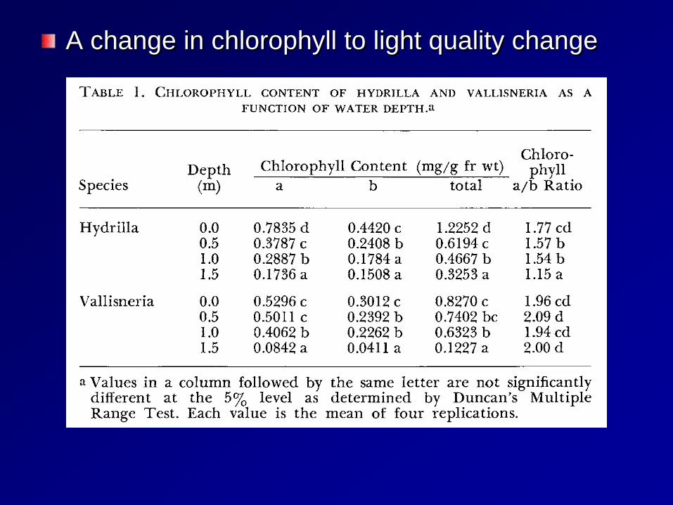
A change in chlorophyll to light quality change
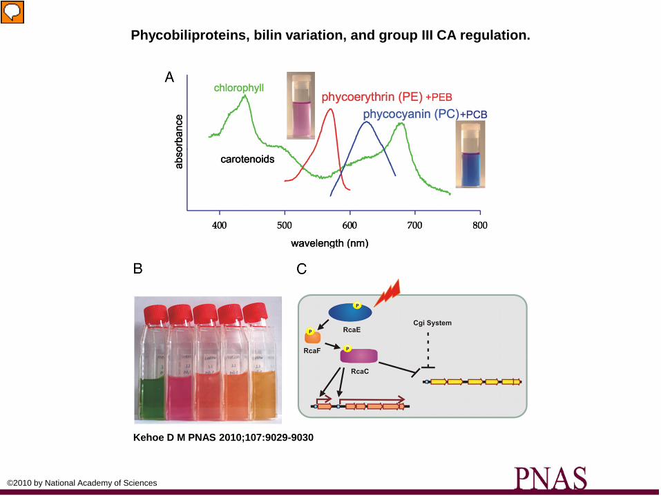
Phycobiliproteins, bilin variation, and group III CA regulation.
Kehoe D M PNAS 2010;107:9029-9030
©2010 by National Academy of Sciences
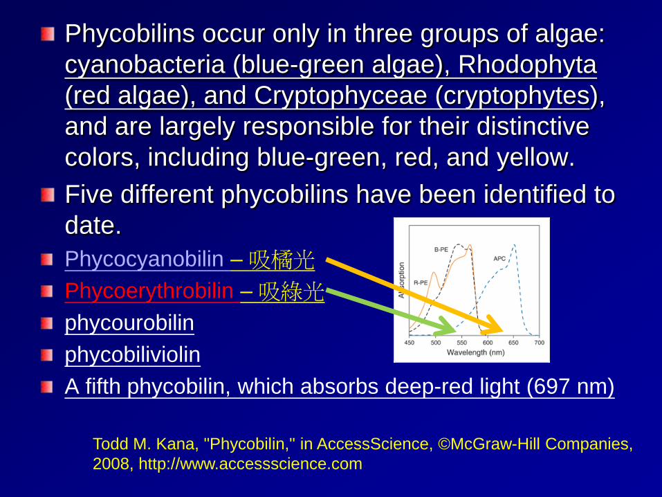
Phycobilins occur only in three groups of algae: cyanobacteria (blue-green algae), Rhodophyta (red algae), and Cryptophyceae (cryptophytes), and are largely responsible for their distinctive colors, including blue-green, red, and yellow. Five different phycobilins have been identified to date. Phycocyanobilin – 吸橘光 Phycoerythrobilin – 吸綠光 phycourobilin phycobiliviolin A fifth phycobilin, which absorbs deep-red light (697 nm)
Todd M. Kana, "Phycobilin," in AccessScience, ©McGraw-Hill Companies, 2008, http://www.accessscience.com
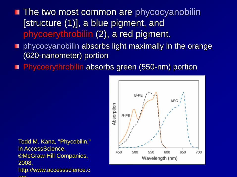
The two most common are phycocyanobilin [structure (1)], a blue pigment, and phycoerythrobilin (2), a red pigment. phycocyanobilin absorbs light maximally in the orange (620-nanometer) portion Phycoerythrobilin absorbs green (550-nm) portion
Todd M. Kana, "Phycobilin," in AccessScience, ©McGraw-Hill Companies, 2008, http://www.accessscience.com
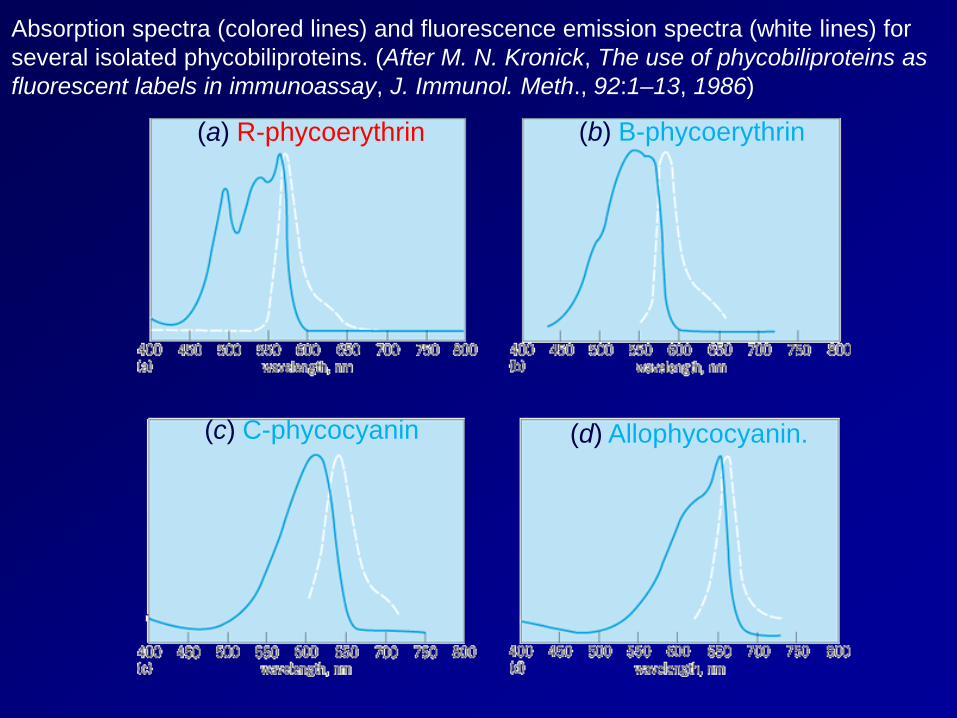
Absorption spectra (colored lines) and fluorescence emission spectra (white lines) for several isolated phycobiliproteins. (After M. N. Kronick, The use of phycobiliproteins as fluorescent labels in immunoassay, J. Immunol. Meth., 92:1–13, 1986)
(a) R-phycoerythrin (b) B-phycoerythrin
(d) Allophycocyanin. (c) C-phycocyanin
A blue-green light (495-nm) absorbing pigment, phycourobilin, is found in some cyanobacteria and red algae. A yellow light (575-nm) absorbing pigment, phycobiliviolin (also called cryptoviolin), is apparently found in all cryptophytes, but in only a few cyanobacteria. A fifth phycobilin, which absorbs deep-red light (697 nm), has been identified spectrally in some cryptophytes, but its chemical properties are unknown
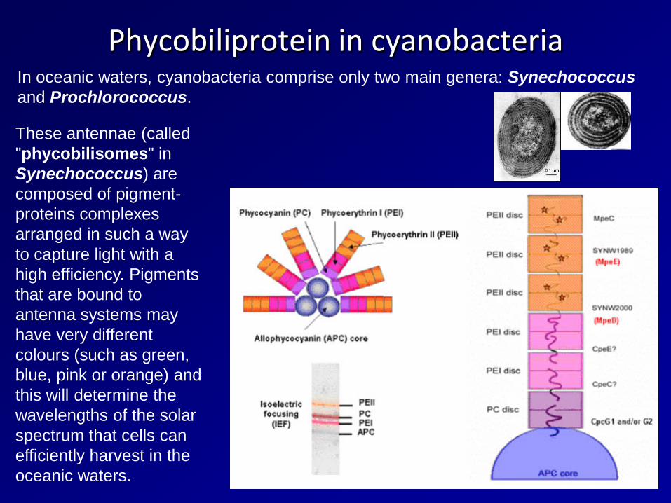
Phycobiliprotein in cyanobacteria In oceanic waters, cyanobacteria comprise only two main genera: Synechococcus and Prochlorococcus.
These antennae (called "phycobilisomes" in Synechococcus) are composed of pigment-proteins complexes arranged in such a way to capture light with a high efficiency. Pigments that are bound to antenna systems may have very different colours (such as green, blue, pink or orange) and this will determine the wavelengths of the solar spectrum that cells can efficiently harvest in the oceanic waters.
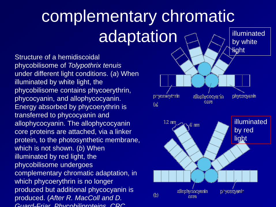
complementary chromatic adaptation
Structure of a hemidiscoidal phycobilisome of Tolypothrix tenuis under different light conditions. (a) When illuminated by white light, the phycobilisome contains phycoerythrin, phycocyanin, and allophycocyanin. Energy absorbed by phycoerythrin is transferred to phycocyanin and allophycocyanin. The allophycocyanin core proteins are attached, via a linker protein, to the photosynthetic membrane, which is not shown. (b) When illuminated by red light, the phycobilisome undergoes complementary chromatic adaptation, in which phycoerythrin is no longer produced but additional phycocyanin is produced. (After R. MacColl and D. Guard-Friar Phycobiliproteins CRC
illuminated by white light
illuminated by red light
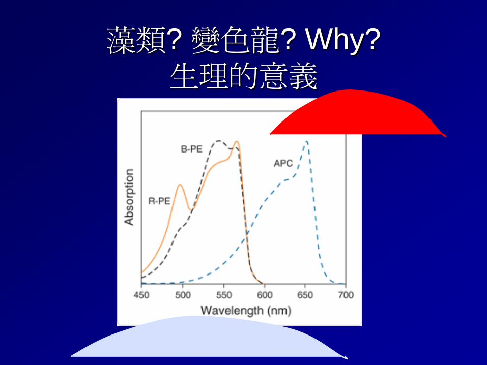
藻類? 變色龍? Why? 生理的意義
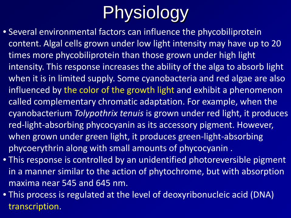
Physiology • Several environmental factors can influence the phycobiliprotein
content. Algal cells grown under low light intensity may have up to 20 times more phycobiliprotein than those grown under high light intensity. This response increases the ability of the alga to absorb light when it is in limited supply. Some cyanobacteria and red algae are also influenced by the color of the growth light and exhibit a phenomenon called complementary chromatic adaptation. For example, when the cyanobacterium Tolypothrix tenuis is grown under red light, it produces red-light-absorbing phycocyanin as its accessory pigment. However, when grown under green light, it produces green-light-absorbing phycoerythrin along with small amounts of phycocyanin .
• This response is controlled by an unidentified photoreversible pigment in a manner similar to the action of phytochrome, but with absorption maxima near 545 and 645 nm.
• This process is regulated at the level of deoxyribonucleic acid (DNA) transcription.
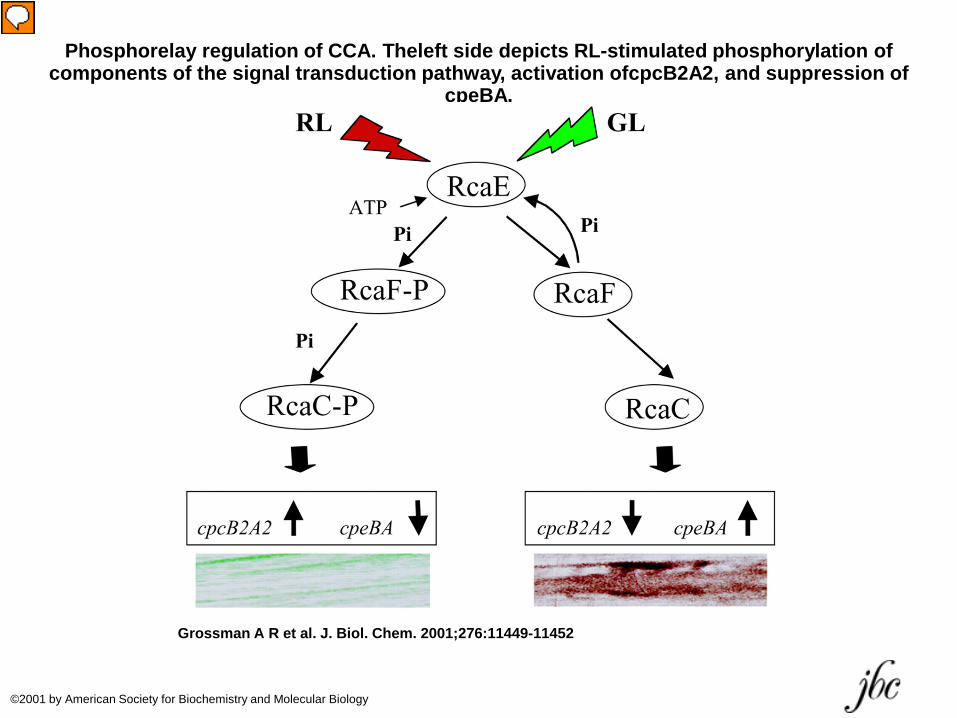
Phosphorelay regulation of CCA. Theleft side depicts RL-stimulated phosphorylation of components of the signal transduction pathway, activation ofcpcB2A2, and suppression of
cpeBA.
Grossman A R et al. J. Biol. Chem. 2001;276:11449-11452
©2001 by American Society for Biochemistry and Molecular Biology
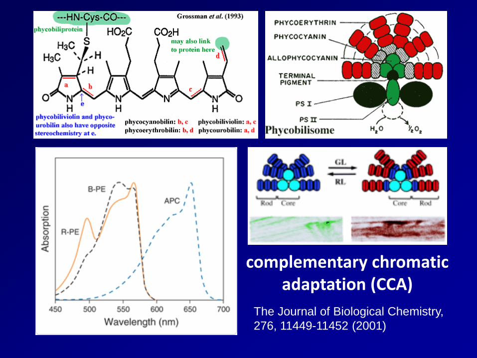
complementary chromatic adaptation (CCA)
The Journal of Biological Chemistry, 276, 11449-11452 (2001)
Regulation of phycobiliprotein concentration
Excdept light quality and intensity, phycobiliprotein concentration also depends on the availability of nutrients, including nitrogen, carbon dioxide, phosphorus, sulfur, and iron. Nutrient starvation generally causes a loss of phycobiliprotein, with the rod proteins of the phycobilisomes being lost more rapidly than the core proteins. In cyanobacteria, this reduction is due to specific proteolytic degradation of biliprotein present in the cell and repression of synthesis of new biliprotein.
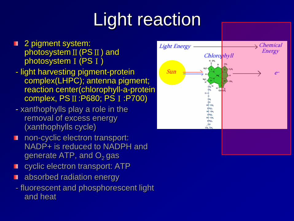
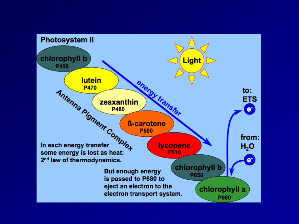
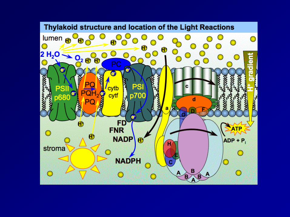
Light reaction 2 pigment system: photosystemⅡ(PSⅡ) and photosystemⅠ(PSⅠ)
- light harvesting pigment-protein complex(LHPC); antenna pigment; reaction center(chlorophyll-a-protein complex, PSⅡ:P680; PSⅠ:P700)
- xanthophylls play a role in the removal of excess energy (xanthophylls cycle) non-cyclic electron transport: NADP+ is reduced to NADPH and generate ATP, and O2 gas cyclic electron transport: ATP absorbed radiation energy
- fluorescent and phosphorescent light and heat
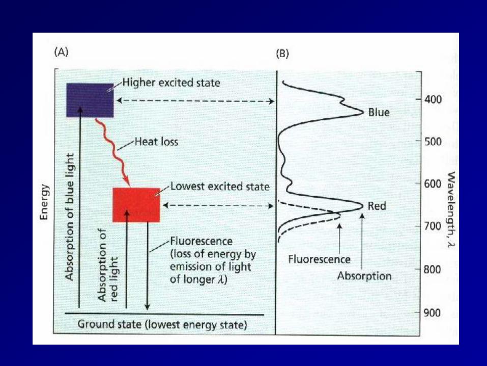
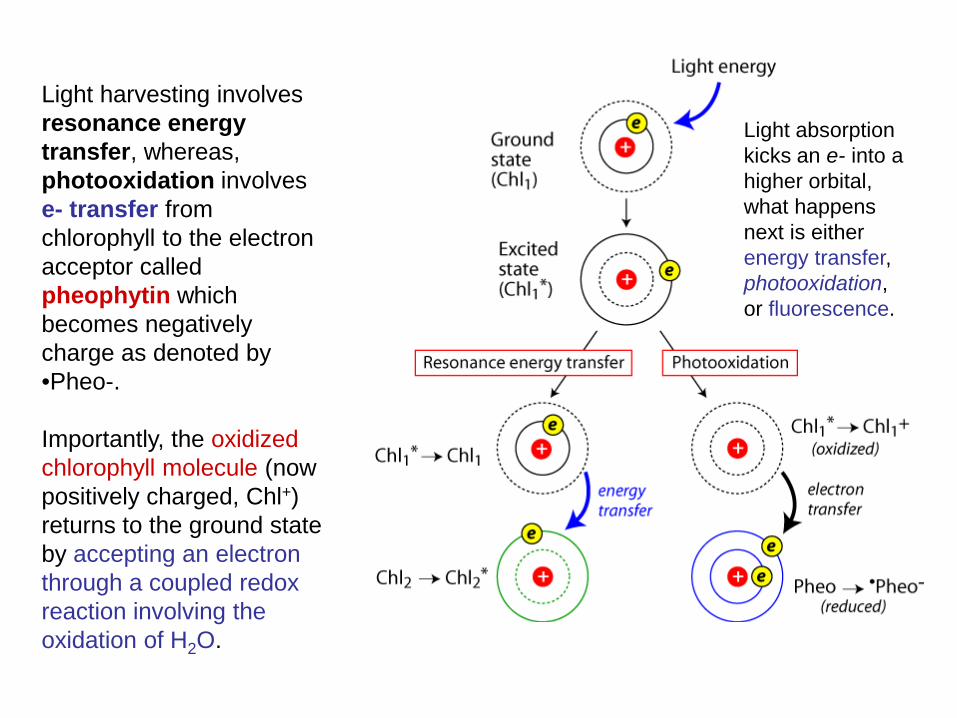
Light harvesting involves resonance energy transfer, whereas, photooxidation involves e- transfer from chlorophyll to the electron acceptor called pheophytin which becomes negatively charge as denoted by •Pheo-. Importantly, the oxidized chlorophyll molecule (now positively charged, Chl+) returns to the ground state by accepting an electron through a coupled redox reaction involving the oxidation of H2O.
Light absorption kicks an e- into a higher orbital, what happens next is either energy transfer, photooxidation, or fluorescence.
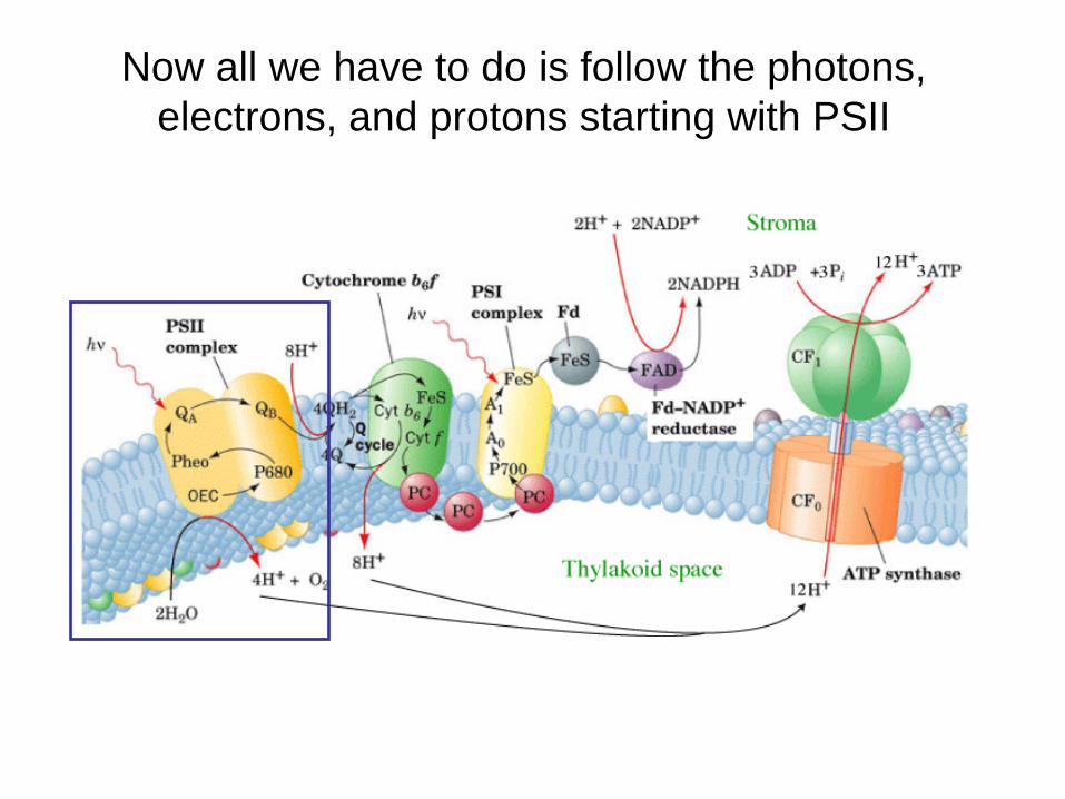
Now all we have to do is follow the photons, electrons, and protons starting with PSII
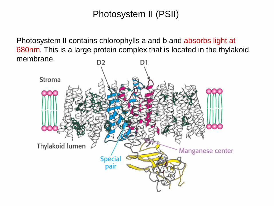
Photosystem II (PSII)
Photosystem II contains chlorophylls a and b and absorbs light at 680nm. This is a large protein complex that is located in the thylakoid membrane.
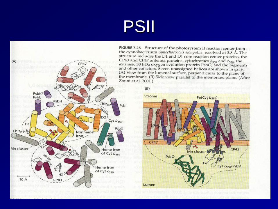
PSII
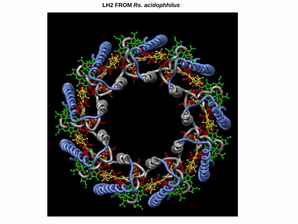
LH2 FROM Rs. acidophhilus
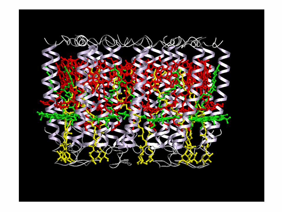
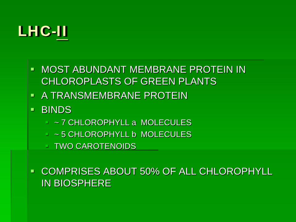
LHC-II
MOST ABUNDANT MEMBRANE PROTEIN IN CHLOROPLASTS OF GREEN PLANTS
A TRANSMEMBRANE PROTEIN BINDS
~ 7 CHLOROPHYLL a MOLECULES ~ 5 CHLOROPHYLL b MOLECULES TWO CAROTENOIDS
COMPRISES ABOUT 50% OF ALL CHLOROPHYLL IN BIOSPHERE
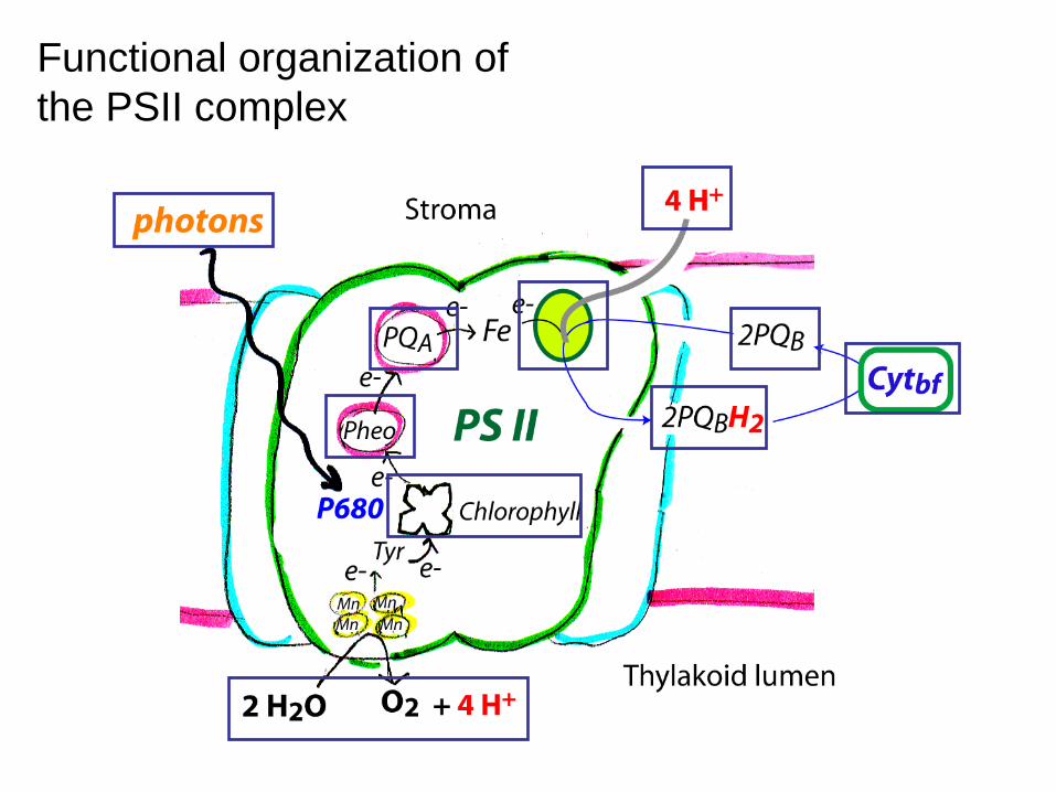
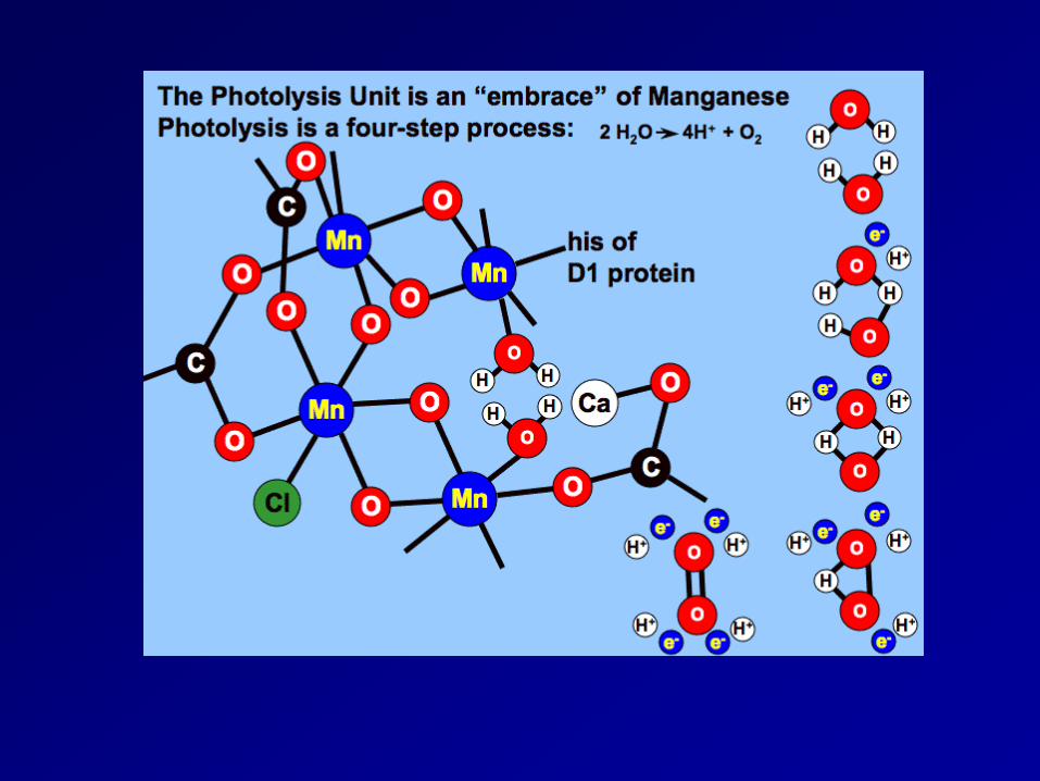
Functional organization of the PSII complex
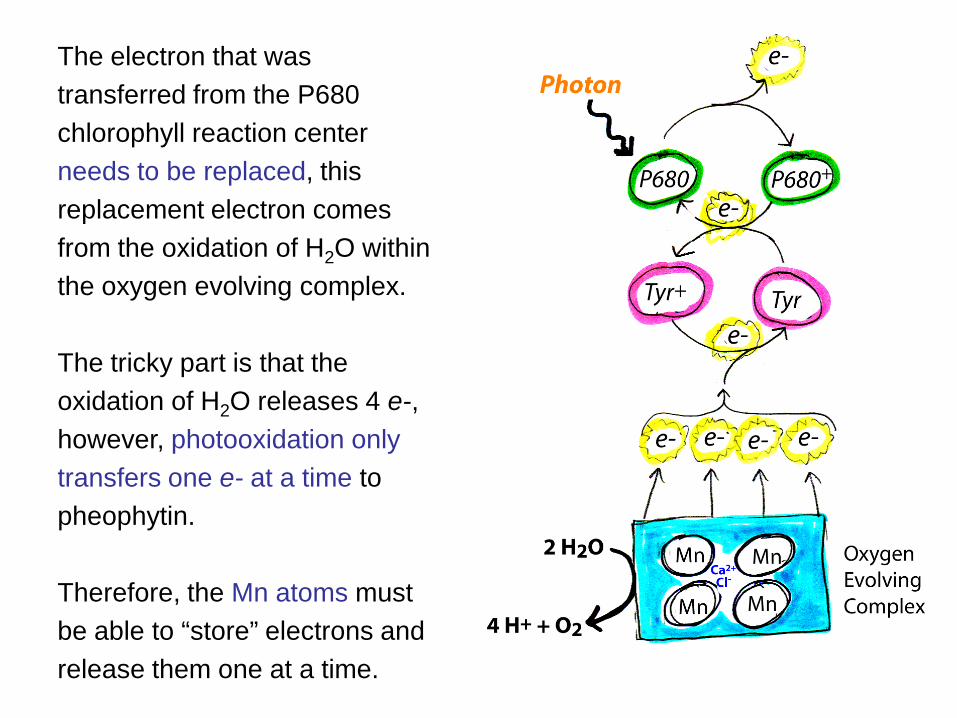
The electron that was transferred from the P680 chlorophyll reaction center needs to be replaced, this replacement electron comes from the oxidation of H2O within the oxygen evolving complex. The tricky part is that the oxidation of H2O releases 4 e-, however, photooxidation only transfers one e- at a time to pheophytin. Therefore, the Mn atoms must be able to “store” electrons and release them one at a time.
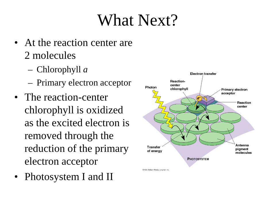
What Next? • At the reaction center are
2 molecules – Chlorophyll a – Primary electron acceptor
• The reaction-center chlorophyll is oxidized as the excited electron is removed through the reduction of the primary electron acceptor
• Photosystem I and II
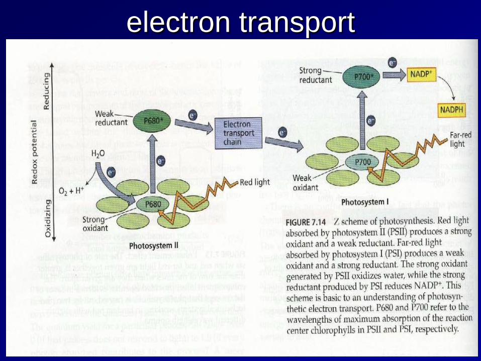
electron transport
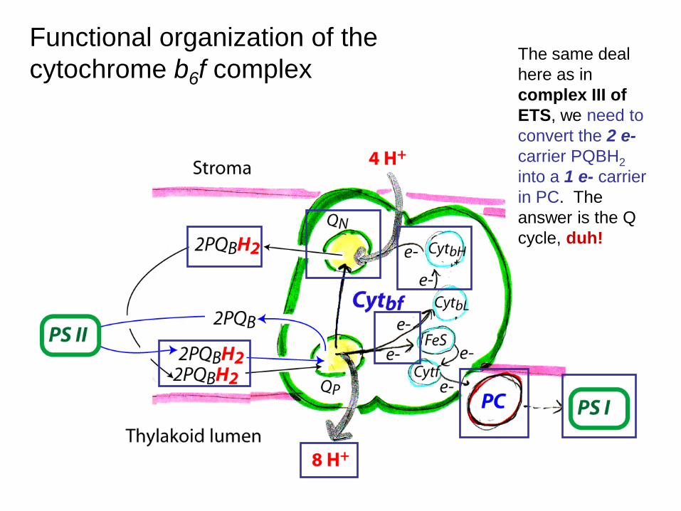
Functional organization of the cytochrome b6f complex
The same deal here as in complex III of ETS, we need to convert the 2 e- carrier PQBH2 into a 1 e- carrier in PC. The answer is the Q cycle, duh!
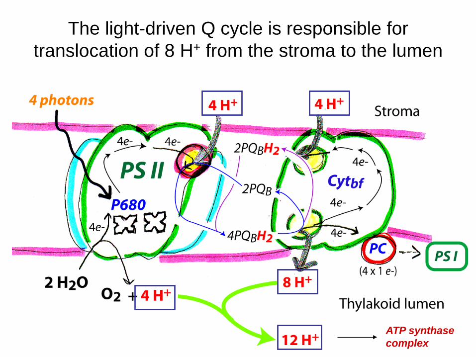
The light-driven Q cycle is responsible for translocation of 8 H+ from the stroma to the lumen
ATP synthase complex
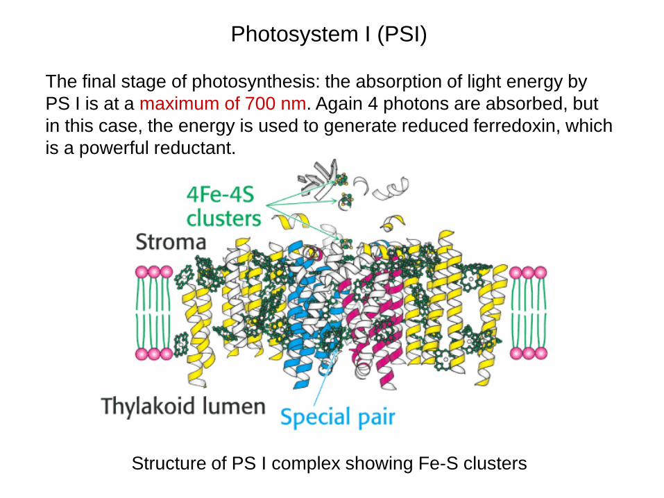
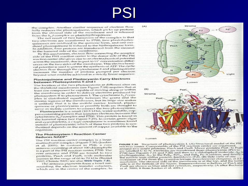
Photosystem I (PSI)
The final stage of photosynthesis: the absorption of light energy by PS I is at a maximum of 700 nm. Again 4 photons are absorbed, but in this case, the energy is used to generate reduced ferredoxin, which is a powerful reductant.
Structure of PS I complex showing Fe-S clusters
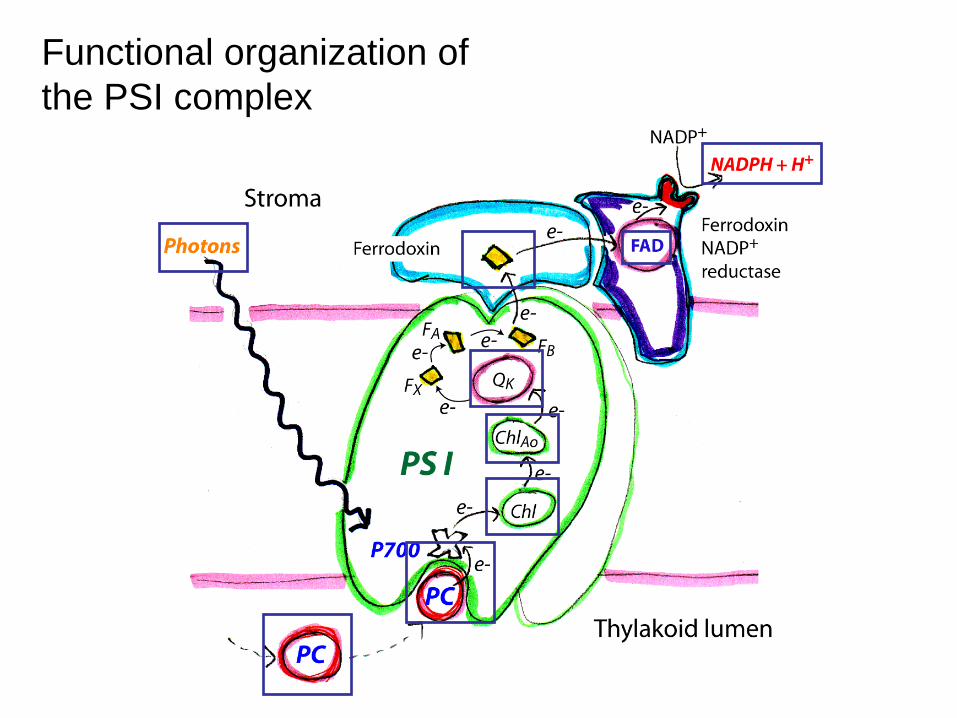
Functional organization of the PSI complex
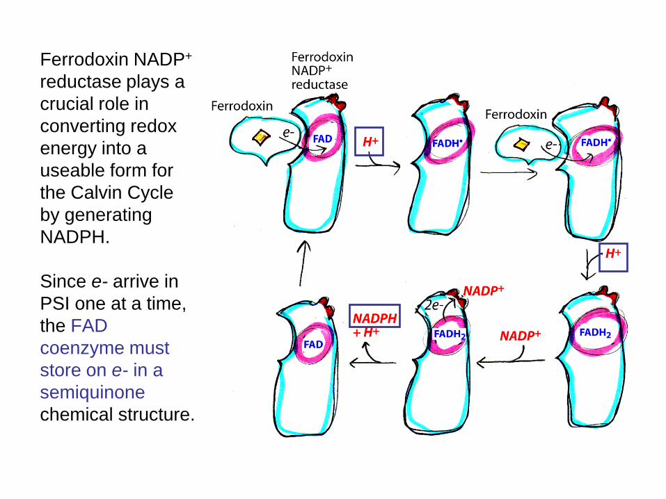
Ferrodoxin NADP+ reductase plays a crucial role in converting redox energy into a useable form for the Calvin Cycle by generating NADPH. Since e- arrive in PSI one at a time, the FAD coenzyme must store on e- in a semiquinone chemical structure.
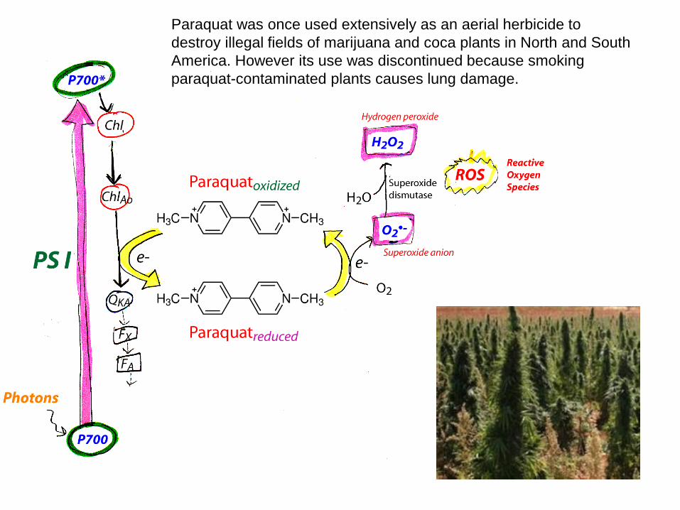
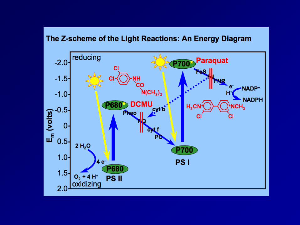
Paraquat was once used extensively as an aerial herbicide to destroy illegal fields of marijuana and coca plants in North and South America. However its use was discontinued because smoking paraquat-contaminated plants causes lung damage.
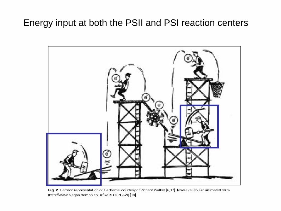
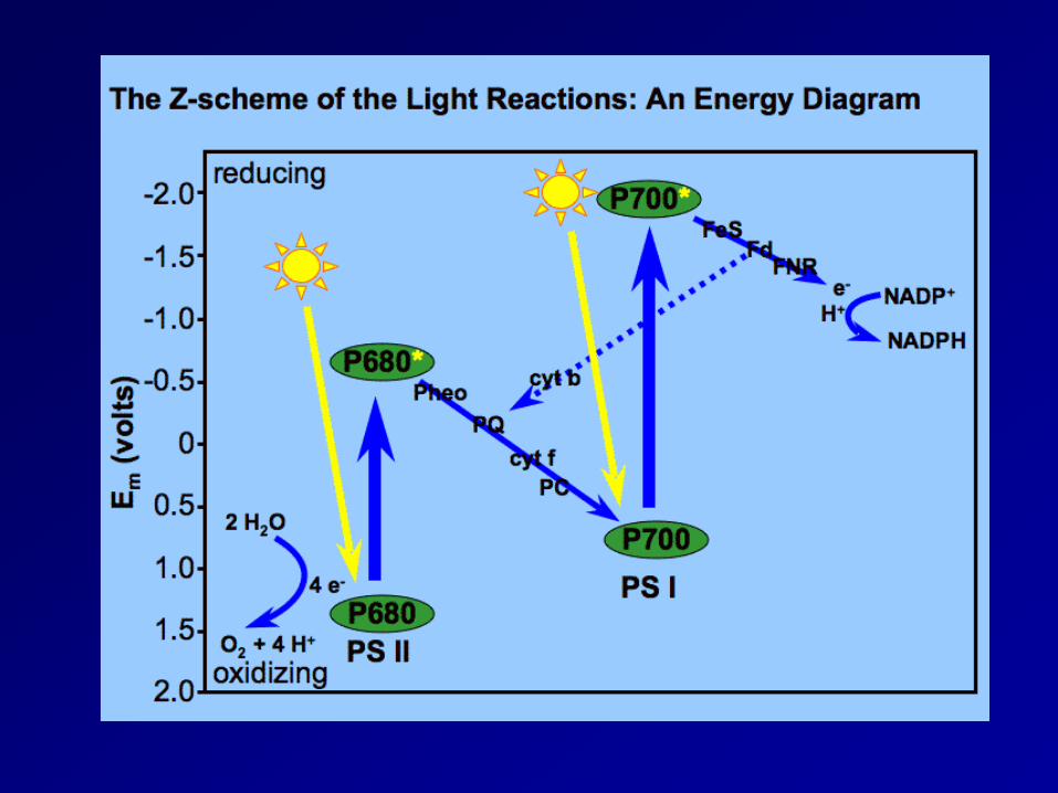
Energy input at both the PSII and PSI reaction centers
Electron Flow • Two routes for the path of electrons stored in the primary
electron acceptors • Both pathways
– begin with the capturing of photon energy – utilize an electron transport chain with cytochromes for chemiosmosis
• Noncyclic electron flow – uses both photosystem II and I – electrons from photosystem II are removed and replaced by electrons
donated from water – synthesizes ATP and NADPH – electron donation converts water into O2 and 2H+
• Cyclic electron flow – Uses photosystem I only – electrons from photosystem I are recycled – synthesizes ATP only
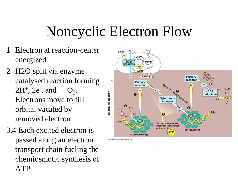
Noncyclic Electron Flow 1 Electron at reaction-center
energized 2 H2O split via enzyme
catalysed reaction forming 2H+, 2e-, and O2. Electrons move to fill orbital vacated by removed electron
3,4 Each excited electron is passed along an electron transport chain fueling the chemiosmotic synthesis of ATP
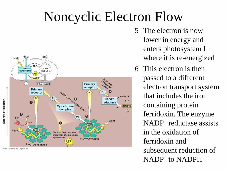
5 The electron is now lower in energy and enters photosystem I where it is re-energized
6 This electron is then passed to a different electron transport system that includes the iron containing protein ferridoxin. The enzyme NADP+ reductase assists in the oxidation of ferridoxin and subsequent reduction of NADP+ to NADPH
Noncyclic Electron Flow
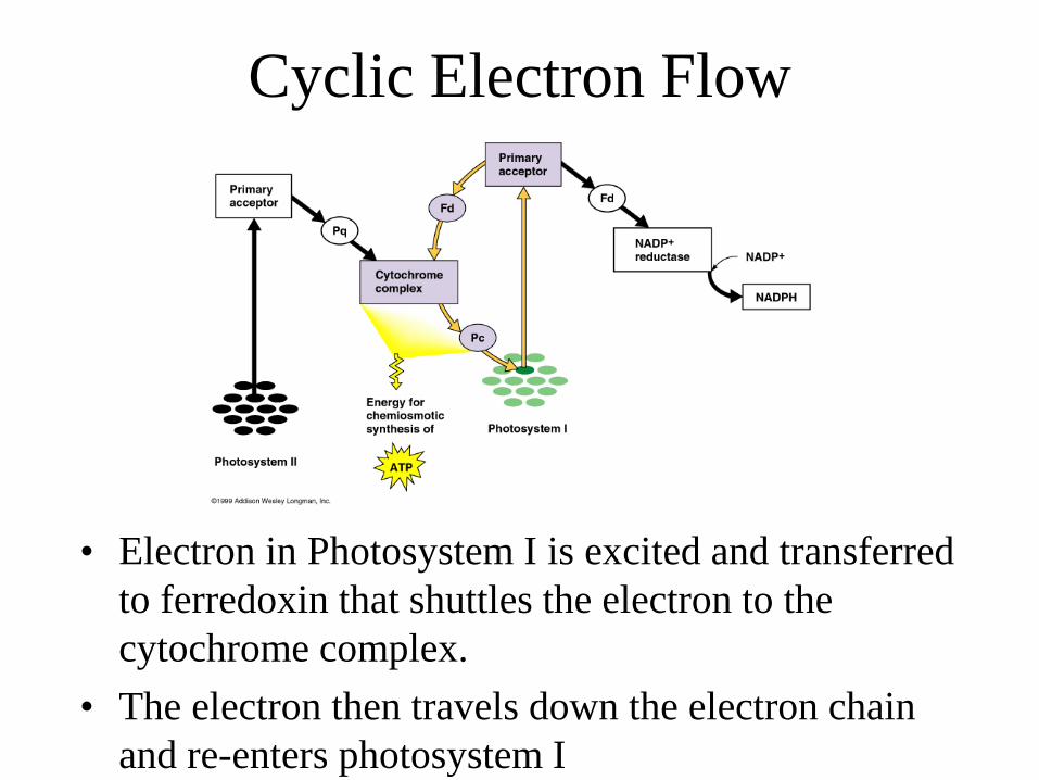
Cyclic Electron Flow
• Electron in Photosystem I is excited and transferred to ferredoxin that shuttles the electron to the cytochrome complex.
• The electron then travels down the electron chain and re-enters photosystem I
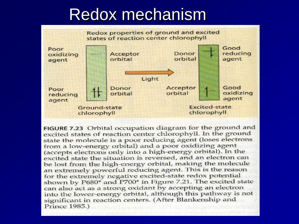
Redox mechanism
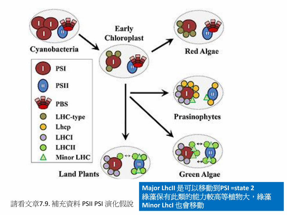
請看文章7.9. 補充資料 PSII PSI 演化假說
Major LhcII 是可以移動到PSI =state 2 綠藻保有此類的能力較高等植物大,綠藻Minor LhcI 也會移動
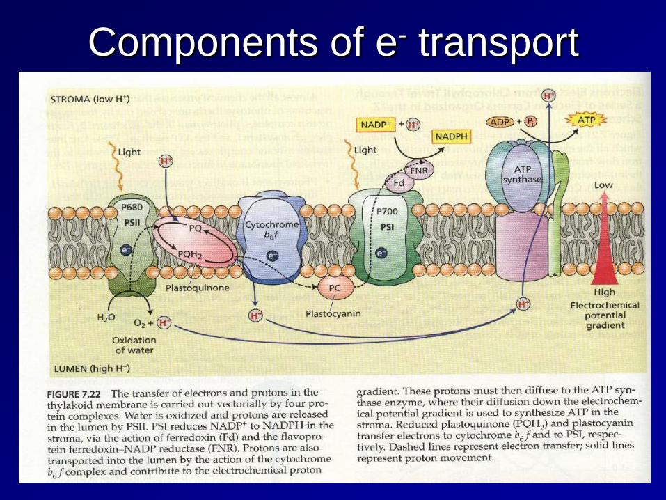
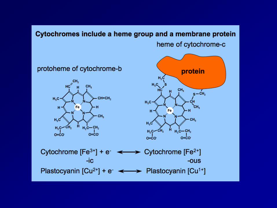
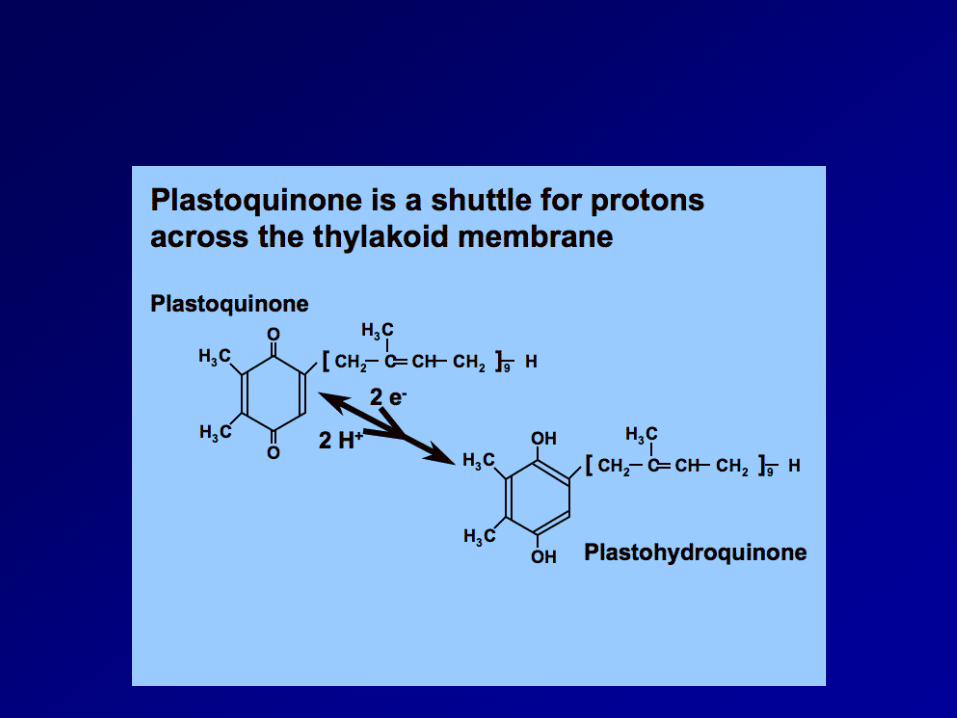
Components of e- transport
PSI
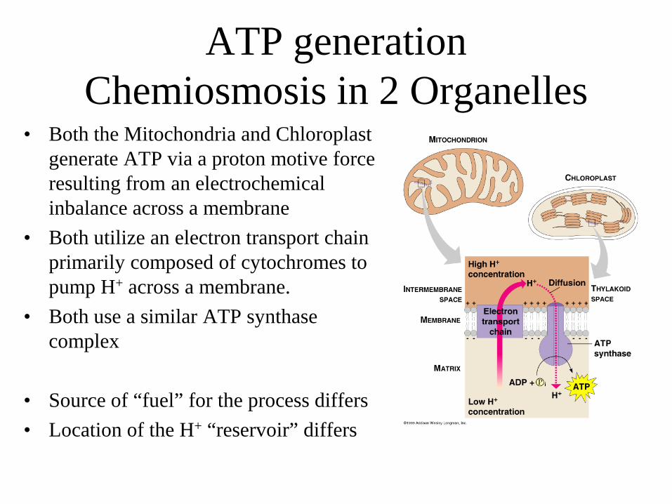
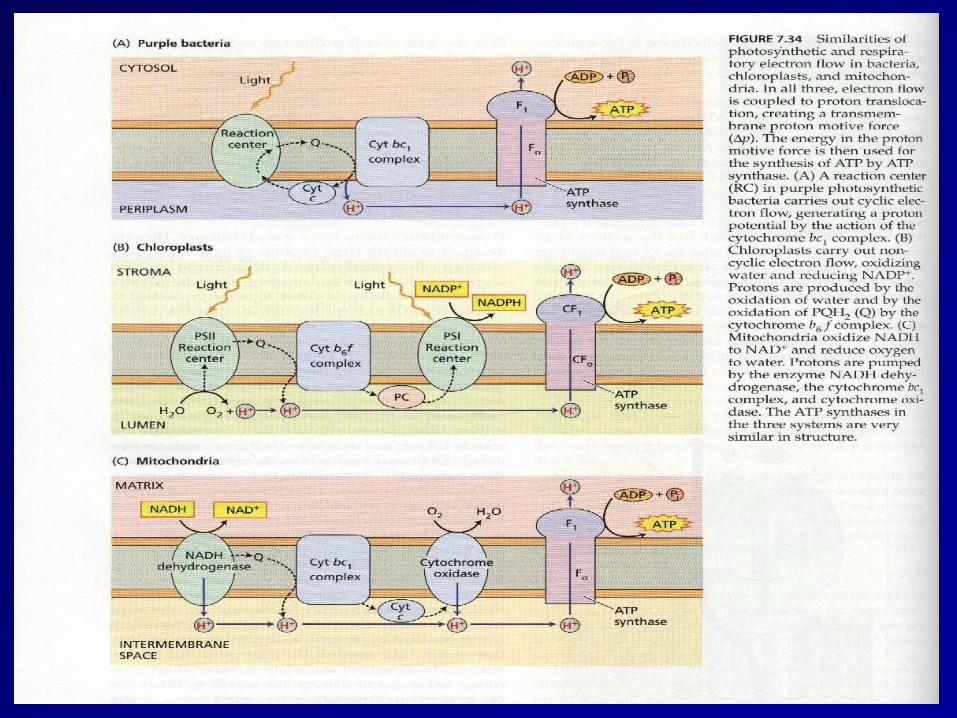
ATP generation Chemiosmosis in 2 Organelles
• Both the Mitochondria and Chloroplast generate ATP via a proton motive force resulting from an electrochemical inbalance across a membrane
• Both utilize an electron transport chain primarily composed of cytochromes to pump H+ across a membrane.
• Both use a similar ATP synthase complex
• Source of “fuel” for the process differs • Location of the H+ “reservoir” differs
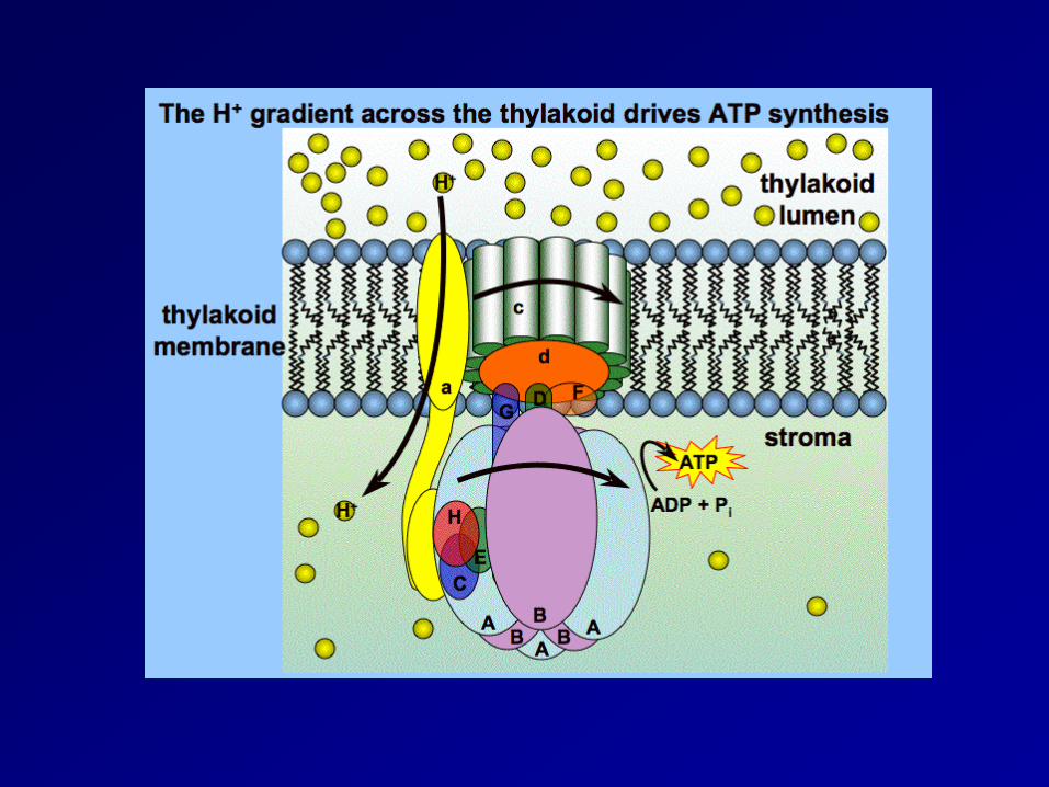
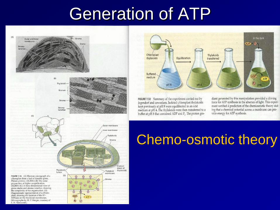
Generation of ATP
Chemo-osmotic theory
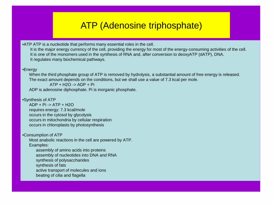
ATP (Adenosine triphosphate)
•ATP ATP is a nucleotide that performs many essential roles in the cell. It is the major energy currency of the cell, providing the energy for most of the energy-consuming activities of the cell. It is one of the monomers used in the synthesis of RNA and, after conversion to deoxyATP (dATP), DNA. It regulates many biochemical pathways. •Energy When the third phosphate group of ATP is removed by hydrolysis, a substantial amount of free energy is released. The exact amount depends on the conditions, but we shall use a value of 7.3 kcal per mole. ATP + H2O -> ADP + Pi ADP is adenosine diphosphate. Pi is inorganic phosphate. •Synthesis of ATP ADP + Pi -> ATP + H2O requires energy: 7.3 kcal/mole occurs in the cytosol by glycolysis occurs in mitochondria by cellular respiration occurs in chloroplasts by photosynthesis •Consumption of ATP Most anabolic reactions in the cell are powered by ATP. Examples: assembly of amino acids into proteins assembly of nucleotides into DNA and RNA synthesis of polysaccharides synthesis of fats active transport of molecules and ions beating of cilia and flagella
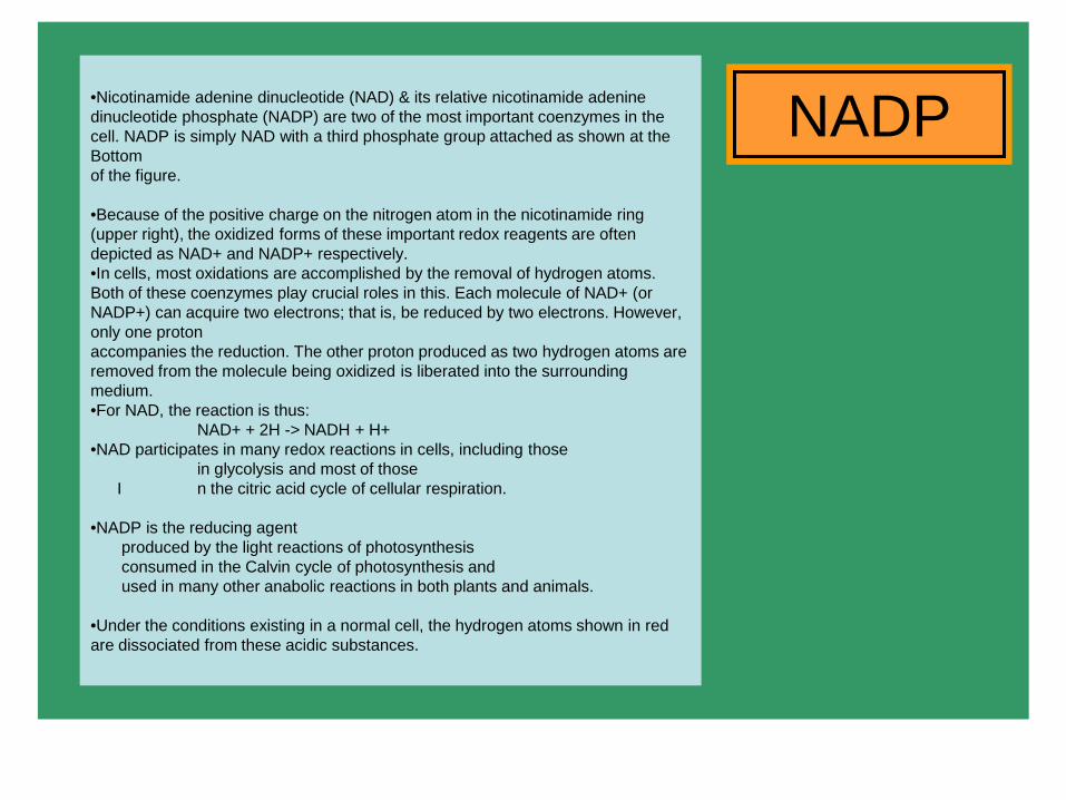
NADP •Nicotinamide adenine dinucleotide (NAD) & its relative nicotinamide adenine dinucleotide phosphate (NADP) are two of the most important coenzymes in the cell. NADP is simply NAD with a third phosphate group attached as shown at the Bottom of the figure. •Because of the positive charge on the nitrogen atom in the nicotinamide ring (upper right), the oxidized forms of these important redox reagents are often depicted as NAD+ and NADP+ respectively. •In cells, most oxidations are accomplished by the removal of hydrogen atoms. Both of these coenzymes play crucial roles in this. Each molecule of NAD+ (or NADP+) can acquire two electrons; that is, be reduced by two electrons. However, only one proton accompanies the reduction. The other proton produced as two hydrogen atoms are removed from the molecule being oxidized is liberated into the surrounding medium. •For NAD, the reaction is thus: NAD+ + 2H -> NADH + H+ •NAD participates in many redox reactions in cells, including those in glycolysis and most of those I n the citric acid cycle of cellular respiration. •NADP is the reducing agent produced by the light reactions of photosynthesis consumed in the Calvin cycle of photosynthesis and used in many other anabolic reactions in both plants and animals. •Under the conditions existing in a normal cell, the hydrogen atoms shown in red are dissociated from these acidic substances.
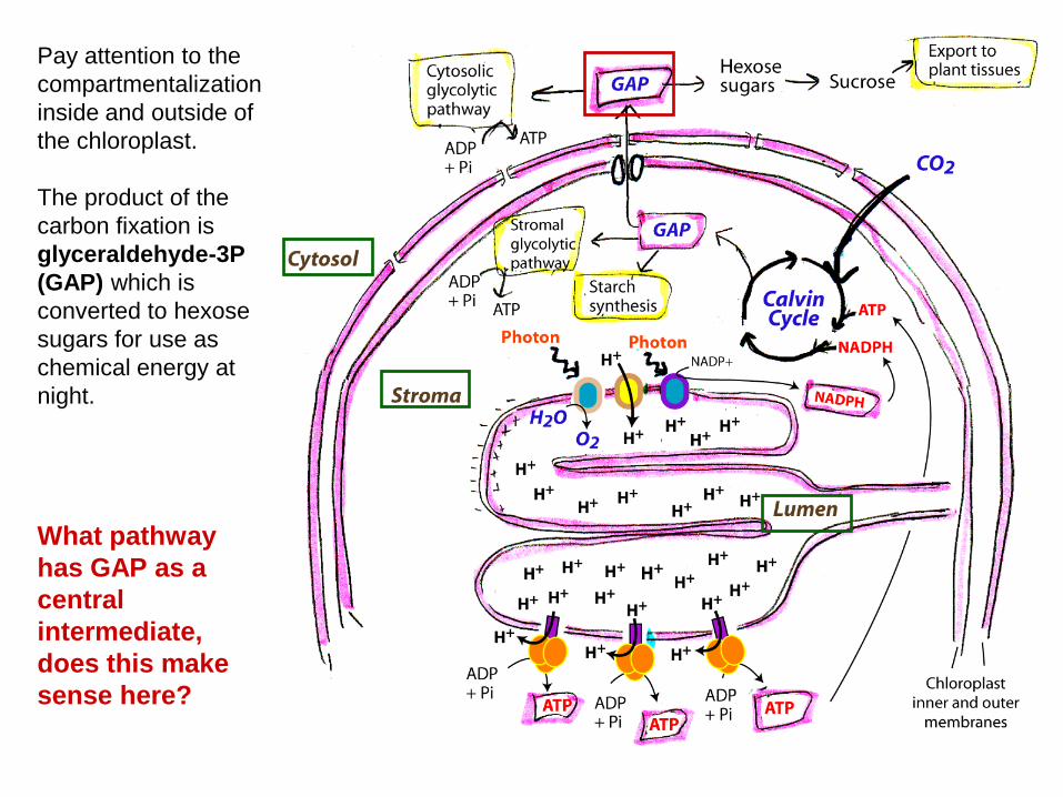
What pathway has GAP as a central intermediate, does this make sense here?
Pay attention to the compartmentalization inside and outside of the chloroplast. The product of the carbon fixation is glyceraldehyde-3P (GAP) which is converted to hexose sugars for use as chemical energy at night.
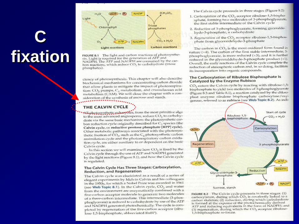
C fixation
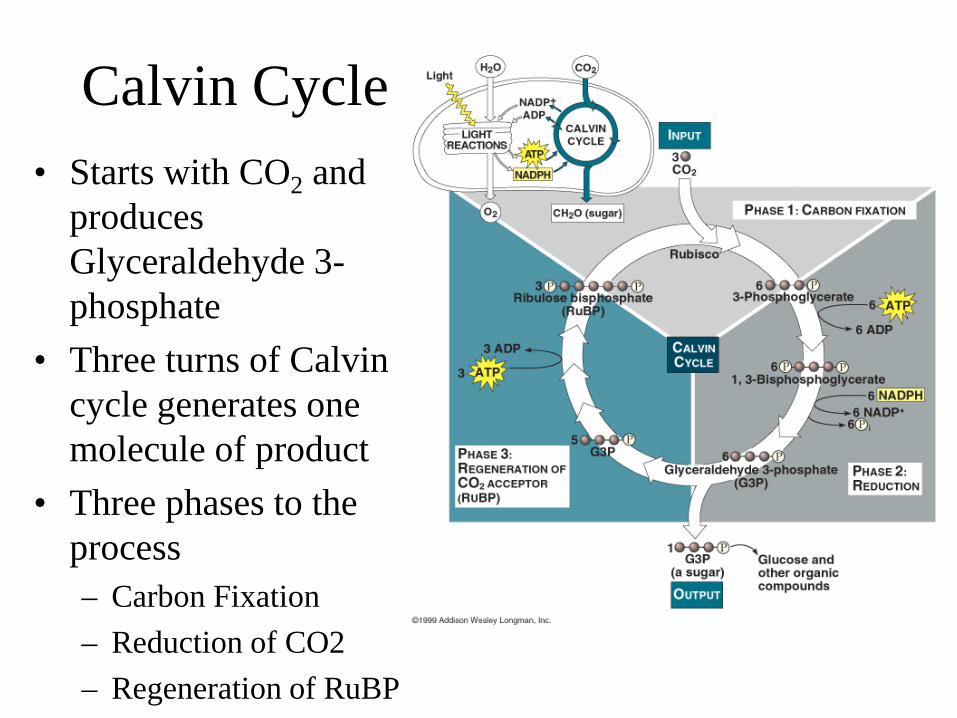
Calvin Cycle • Starts with CO2 and
produces Glyceraldehyde 3-phosphate
• Three turns of Calvin cycle generates one molecule of product
• Three phases to the process – Carbon Fixation – Reduction of CO2 – Regeneration of RuBP
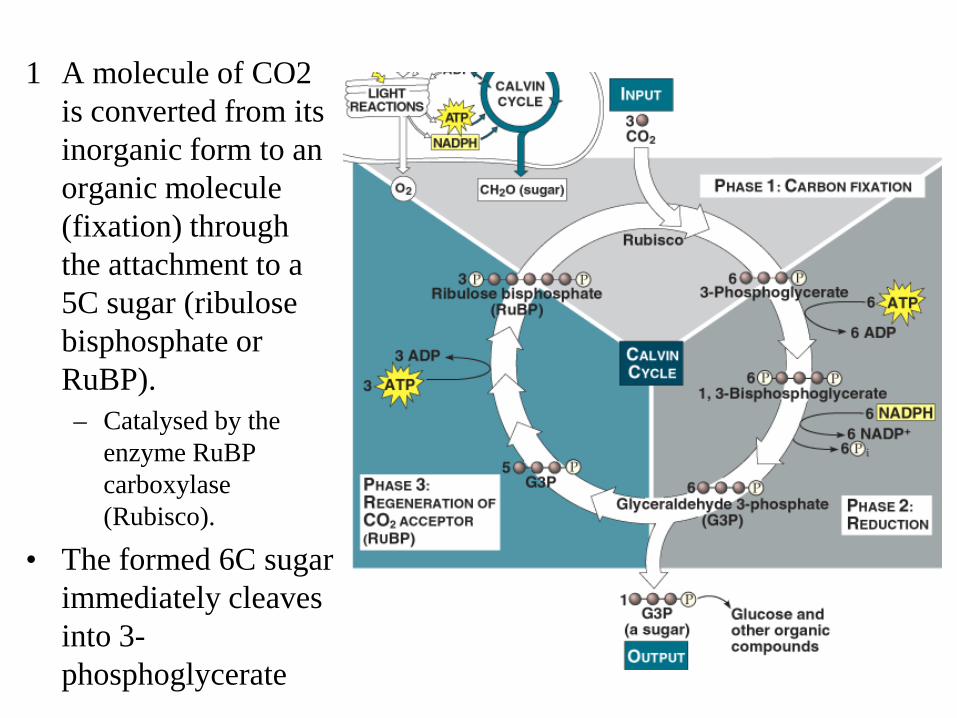
1 A molecule of CO2 is converted from its inorganic form to an organic molecule (fixation) through the attachment to a 5C sugar (ribulose bisphosphate or RuBP). – Catalysed by the
enzyme RuBP carboxylase (Rubisco).
• The formed 6C sugar immediately cleaves into 3-phosphoglycerate
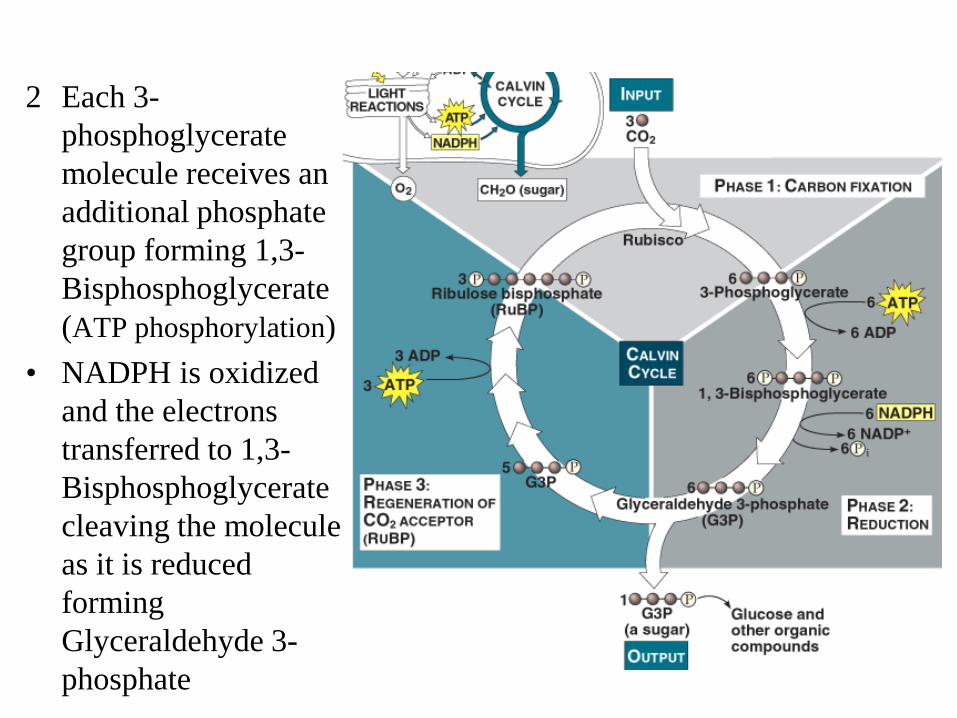
2 Each 3-phosphoglycerate molecule receives an additional phosphate group forming 1,3-Bisphosphoglycerate (ATP phosphorylation)
• NADPH is oxidized and the electrons transferred to 1,3-Bisphosphoglycerate cleaving the molecule as it is reduced forming Glyceraldehyde 3-phosphate
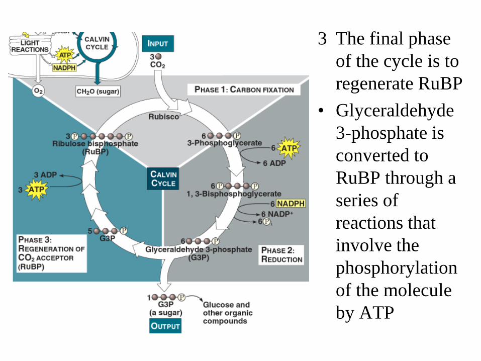
3 The final phase of the cycle is to regenerate RuBP
• Glyceraldehyde 3-phosphate is converted to RuBP through a series of reactions that involve the phosphorylation of the molecule by ATP
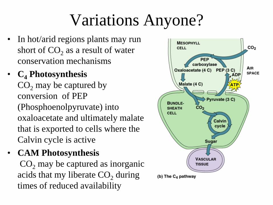
Variations Anyone? • In hot/arid regions plants may run
short of CO2 as a result of water conservation mechanisms
• C4 Photosynthesis CO2 may be captured by conversion of PEP (Phosphoenolpyruvate) into oxaloacetate and ultimately malate that is exported to cells where the Calvin cycle is active
• CAM Photosynthesis CO2 may be captured as inorganic acids that my liberate CO2 during times of reduced availability
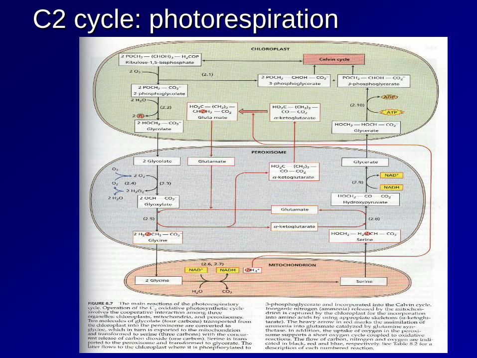
C2 cycle: photorespiration

Thank You!