Embed Size (px)
Citation preview

B The Author(s), 2009Published Online: 2 October 2009 DOI: 10.1007/s11307-009-0267-3
Mol Imaging Biol (2010) 12:259Y268
RESEARCH ARTICLE
68Ga-Chloride PET Reveals Human PancreaticAdenocarcinoma Xenograftsin Rats—Comparison with FDGTiina Ujula,1 Satu Salomäki,1 Anu Autio,1 Pauliina Luoto,1 Tuula Tolvanen,1
Pertti Lehikoinen,2 Tapio Viljanen,2 Hannu Sipilä,1 Pirkko Härkönen,3 Anne Roivainen1,4
1Turku PET Centre, Turku University Hospital, 20521 Turku, Finland2Radiopharmaceutical Chemistry Laboratory, Turku PET Centre, Turku, Finland3Institute of Biomedicine and MediCity Research Laboratory, University of Turku, Turku, Finland4Turku Centre for Disease Modeling, University of Turku, Turku, Finland
AbstractPurpose: The aim of the study was to compare 68Ga-chloride with 2-[18F]fluoro-2-deoxy-D-glucose (FDG) for the imaging of pancreatic xenografts.Procedures: Rats with subcutaneous human pancreatic adenocarcinoma xenografts wereevaluated in vivo by dynamic positron emission tomography (PET) and ex vivo by measuringradioactivity of excised tissues and by digital autoradiography of tumor cryosections.Results: Both tracers were capable of delineating all subcutaneous tumors from surroundingtissues by PET. The standardized uptake values of tumors by PET were 0.9±0.3 (mean±SD) for68Ga-chloride (n=13) and 1.8±1.2 for FDG (n=11). Ex vivo studies showed tumor-to-muscleratio of 4.0±0.3 for 68Ga-chloride (n=4) and 7.9±3.2 for FDG (n=4).Conclusions: 68Ga-chloride delineated subcutaneously implanted pancreatic adenocarcinomaxenografts by PET, but the uptake was lower than FDG. Further studies to clarify the value of68Ga-chloride for PET imaging of tumors are warranted.
Key words: Gallium-68, Fluorine-18 FDG, PET, Tumor xenografts
Introduction
G allium-67 citrate (67Ga; T1/2=78 h) has been used asan imaging agent in single-photon emission computed
tomography (SPECT) for decades for the purpose ofdetecting certain tumors and infection/inflammation, in spiteof its disadvantages resulting mainly from such unfavorablephysical characteristics as high radiation exposure and longexamination time. The first tumor uptake studies with apositron-emitting gallium-68 (68Ga; T1/2=68 min) wereperformed in the 1960s [1, 2]. For a long period of time,
68Ga was almost forgotten, but during the last years, it hasgone through a renaissance. 68Ga has been applied for thelabeling of peptides and oligonucleotides with the macro-cyclic chelating agent 1,4,7,10-tetraazacyclododecane-N,N′,N″,N‴-tetraacetic acid (DOTA) in order to target tumors bypositron emission tomography (PET) [3, 4]. Some 68Ga-labeled tumor targeting radiopharmaceuticals, e.g., 68Ga-DOTATOC, 68Ga-DOTANOC, and 68Ga-DOTA-Ianreotidebinding to somatostatin receptors, 68Ga-DOTA-bombesinbinding to bombesin receptors, and 68Ga-DOTA-D-Glu-gastrin binding to gastrin-releasing peptide receptors, havebeen reported to be potential imaging agents in clinicalstudies [5, 6]. Preclinical evaluation of interesting 68Ga-labeled biomolecules, e.g., 68Ga-deferoxamine-folate and68Ga-DOTA-hEGF, has also been reported [7, 8].
Significance: This is the first study to evaluate 68Ga-chloride for PETimaging of pancreatic xenografts in comparison with FDG.
Correspondence to: Anne Roivainen; e-mail: [email protected]
2-[18F]Fluoro-2-deoxy-D-glucose (FDG) is the most widelyused PET imaging agent. FDG is a glucose analog that reflectsthe level of glucose metabolism. Glucose consumption isincreased in tumors because of rapid cell proliferation. Glucoseand FDG uses GLUT-1 and GLUT-3 cell surface transporters,which are overexpressed in cancer cells [9]. FDG, like glucose,is phosphorylated by hexokinases (particularly hexokinase II)into 2-[18F]fluoro-2-deoxy-D-glucose 6-phosphate (FDG 6-P).Subsequently, FDG 6-P is trapped inside the cell because it isnot metabolized any further.
Very recently, we have been able to establish a relationshipbetween the 68Ga-chloride uptake with PET at the site of localbone infection and underlying structural changes determinedwith peripheral quantitative computed tomography [10]. Inaddition, when using 68Ga-chloride as a control in our 68Ga-oligonucleotide studies, we noticed uptake in tumors during 2 hPET imaging [4]. The purpose of this study was to prelimi-narily evaluate if gallium-68 (68Ga) could be used for PETimaging of tumors in a similar fashion as 67Ga is used forSPECT. The advantage of 68Ga-chloride over FDG would bethe fast and simple production, i.e., the cyclotron and labeling-free production. 68Ga-chloride is readily available at a PETlaboratory. We tested 68Ga-chloride by PET imaging ofexperimental tumors in comparison with FDG. The resultswere also verified ex vivo. Compared with the use of 67Ga inSPECT, 68Ga-chloride PET would have the following advan-tages: better spatial resolution, higher sensitivity, possibility forquantitative assessment of tracer accumulation in tissues, andlower absorbed radiation dose for the study subject.
Materials and MethodsPreparation of Tracers68Ga-chloride was eluted with 0.1 M HCl from a 68Ge/68Gagenerator (Cyclotron Co., Obninsk, Russia). The radioactive elutionpeak, monitored online with a positron-sensitive photodiodedetector (Hamamatsu S5591, Hamamatsu Photonics K.K. SolidState Division, Hamatsu City, Japan), was collected, and the 68Ga-chloride was neutralized with 1.0 M NaOH. FDG was prepared asdescribed earlier [11].
Animal ModelThe human pancreatic ductal adenocarcinoma cell line, BxPC-3, waspurchased from American Type Culture Collection (Rockville, MD,USA). The cells were grown in RPMI 1640 culture mediumsupplemented (10%) with heat-inactivated fetal bovine serum, 2 mML-glutamine, 100 U/mL penicillin, and streptomycin (100µg/mL) in ahumidified atmosphere of 5% CO2 at 37°C. The cells were detachedfrom the culture bottle by trypsin–ethylenediaminetetraacetic acid andresuspended (5×10⁷cells/mL) in medium for implantation.
Twenty-nine athymic male Hsd/RH-rnu/rnu rats were obtainedfrom Harlan (The Netherlands) at the age of 6 weeks. Tumor cells(107/rat) were injected subcutaneously into the neck, and tumors wereallowed to grow to a size of 1 cm in diameter. All animal studies hadbeen approved by the Lab-Animal Care & Use Committee of theUniversity of Turku.
PET ImagingTo detect the tumor uptake of tracers and to quantify their organdistribution and kinetics, 2 h dynamic PET imaging was performed.Nineteen rats with BxPC-3 xenografts (weight 336±50 g) wereanesthetized intraperitoneally with sodium pentobarbital (60 mg/kg;Mebunat, Orion, Finland) or with a mixture of fentanyl citrate plusfluanisone (0.25 and 8.0 mg/kg, respectively; Hypnorm® = fentanylcitrate 0.315mg/mL and fluanisone 10mg/mL, Janssen Pharmaceutica,Beerse, Belgium) and midazolam (4.0 mg/kg; Dormicum 5 mg/mL,Roche, Espoo, Finland). The rats were placed in the center of thescanner gantry to obtain the best resolution for small objects and kepton a warm pallet during the imaging procedure. PET imaging wasperformed either with Advance scanner (GE Medical Systems,Milwaukee, WI, USA) or with HRRT camera (Siemens MedicalSolutions, Knoxville, TN, USA) [12, 13].
After transmission scan for attenuation correction, a tracer wasinjected directly as a bolus intravenously via a tail vein. PET imagingstarted at the time of injection. Themean±SD dose was 16±3MBq for68Ga-chloride (n=13) and 16±9 MBq for FDG (n=11). Prior to tracerinjection, rats fasted for at least 2 h. Of a total of 19 rats, five wereimaged with both tracers on subsequent days. The acquisition timeswere 5×60 and 23×300 s, with the total imaging duration being120 min.
PET data were iteratively reconstructed with the ordered-subsetsexpectation maximization (OS-EM) algorithm. All PET images werereviewed on computer screen in the transaxial, coronal, and sagittalplanes along with maximum-intensity-projection images. Tworeviewers aware of subcutaneously implanted tumor xenografts inthe neck area independently evaluated tracer uptake both visually andsemiquantitatively. The radiotracer uptake in the tumor was visuallycompared to the surrounding tissue (skin, fat, and muscle) uptakeas negative, less-than-surrounding tissue uptake or equivalent-to-surrounding tissue uptake, and positive, uptake which is higher thanthe surrounding tissue uptake. In case of discrepancies between thetwo reviewers, a consensus was reached in a joint reading session.Semiquantitative analysis was performed by drawing a standardizedcircular region of interest (ROI) to the tumor (ROI Ø 5 mm), heart(ROI Ø 6 mm), kidney (ROI Ø 4 mm), liver (ROI Ø 6 mm), andurinary bladder (ROI Ø 4 mm) areas. The heart ROI was drawn tocover the whole heart including both myocardium and the blood poolradioactivity. The average radioactivity concentration in a ROI(kiloBecquerels per milliliter) was used for further analyses. Thetracer uptake was reported as mean standardized uptake values (SUV),which was calculated as the radioactivity of the ROI divided by theinjected dose per animal body weight. Biokinetic/time–activity curves(TAC), representing the relative radioactivity concentration in theorgan of interest (SUV) vs. time after injection, were determinedaccordingly. TACs were decay corrected to the time of injection.
Ex Vivo MeasurementsThe uptake of 68Ga-chloride and FDG in tumors was studied ex vivo intumor-bearing animals. Eight rats (weight 280±69 g) were anesthetizedwith a mixture of Hypnorm–Dormicum, as described above andintravenously administered with 12±3 MBq of 68Ga-chloride (n=4) or15±3 MBq of FDG (n=4). Prior to tracer injection, rats fasted for atleast 2 h. Ninety minutes after tracer distribution, a blood sample wasobtained via intracardiac puncture, and the animals were killed byintracardiac administration of sodium pentobarbital (Mebunat®, Orion,Finland). The time point for ex vivo measurements was based on our
260 T. Ujula, et al.: 68Ga-Chloride PET vs. FDG

previous studies [10]. Samples of blood, tumor, liver, lung, muscle, andskin were excised, weighed, and measured for total radioactivity in anautomated gamma counter (1480Wizard 3″Gamma Counter; EG & GWallac, Turku, Finland) cross-calibrated with a dose calibrator (VDC-202, Veenstra Instruments, Joure, The Netherlands) and PET cameras.The tail was also measured for radioactive content to determine theaccuracy of the injections. The radioactivity concentration was decaycorrected to the time of injection, the radioactivity remaining in the tailwas compensated, and the results were expressed as SUV (organradioactivity/organ weight)/(total given radioactivity/rat body weight).The radioactivity ratios between the target (tumor) and nontarget(blood, liver, lung, muscle, and skin) organs were also calculated.
Tumor Autoradiography, Histology,and Immunohistochemical StainingTwo rats (weight 224 g and 213 g) were injected with 19MBq of 68Ga-chloride or 24 MBq of FDG, respectively. After tracer distribution
(90 min), the tumors were excised, frozen in dry ice, and cut with acryomicrotome into 10–20-μm sections. Tumor sections were thaw-mounted onto microscope slides, briefly air dried, and exposed to animaging plate (Fujifilm BAS TR, Fuji Photo Film Co, Japan) for twohalf-lives of radio-isotope in question. The distribution of radioactivityin the sections was digitally scanned using a Fuji BAS-5000 device(Fuji Tokyo, Japan) with the image resolution of 25μm.
After autoradiography, the same sections were stained withhematoxylin and eosin (HE) or using an immunohistochemicalmethod for light microscopy to obtain corresponding histologicalinformation. In addition, some tumor samples were fixed with 4%formaldehyde, embedded in paraffin, and cut into 10-μm sections, andthe sections were stained with HE. For immunohistochemical staining,DakoCytomation EnVision-system-HRP (K4001, Dako, Glostrup,Denmark) two-step immunohistochemical technique was used.
After 68Ga-chloride autoradiography, the sections were stainedwith mouse antirat CD68 monoclonal antibody (MCA341GA; AbDSerotec, Oxford, UK; optimal dilution 1:2,000) to examine if the
c
Fig. 1. Coronal PET images of a rat injected with a 68Ga-chloride and b FDG on subsequent days. Tumor formed bysubcutaneously implanted BxPC-3 human pancreatic adenocarcinoma cells is marked with a white arrow. Urinary bladder ismarked with a black arrow. Images (a summation of a 60- to 70-min period after injection) are color-coded according to thelevel of radioactivity, from dark blue (the lowest) to hot red (the highest). c The tumor uptake (standardized uptake value, SUV)of 68Ga-chloride and FDG as a function of time.
T. Ujula, et al.: 68Ga-Chloride PET vs. FDG 261

radioactivity originates from macrophage uptake. Antibody waslocated with 3,3′-diaminobenzidine tetrahydrochloride (LiquidDAB Substrate, K3468; Dako, Glostrup, Denmark). Finally,immunohistochemical sections were slightly counterstained withMayer's hematoxylin, washed, and mounted.
The digital autoradiographs were combined with digital histo-logical and immunohistological images using GIMP 2.4.5 (GNUImage Manipulation Program, authored by Peter Mattis andSpencer Kimball; http://www.gimp.org/) and Hugin 0.7 beta 3hugin (Hugin, authored by Andrew Mihal, Pablo d'Angelo, MaxLyons, Erik Krause, Konstantin Rotkvich, and Christoph Spiel;http://hugin.sourceforge.net/) softwares. The intratumoral tracerdistribution was reviewed on computer screen by two observers.
Statistical MethodsAll the results are expressed as mean±SD. After testing ofnormality and variance, an analysis of variance test was appliedto study the significance of differences between the tracers. A t testwas used for the comparison of PET and ex vivo data. A logtransformation because of skeweness was used for all data, exceptfor comparison of ex vivo measurements. A P value of less than0.05 was considered statistically significant. Statistical analyseswere conducted using SAS 9.1.3 statistical software (SAS InstituteInc., Cary, NC, USA).
ResultsPET Imaging
Both tracers were capable of delineating all subcutaneousBxPC-3 tumor xenografts from surrounding tissues by PET(Fig. 1a, b). Based on 2 h dynamic PET imaging, the peak
radioactivity of 68Ga-chloride in the tumor was reached fasterin comparison to FDG. The uptake of 68Ga-chloride at the siteof tumors showed very fast initial uptake and moderatedecline with a plateau at 10 min after injection. In contrast,uptake of FDG accumulated slowly and reached a plateau at60 min (Fig. 1c). According to PET imaging, the tumoruptake expressed as SUV was 0.9±0.3 for 68Ga-chloride(n=13) and 1.8±1.2 for FDG (n=11; Table 1). The FDG SUVof tumor was significantly higher than that of 68Ga-chloride(P=0.003; Fig. 2). The results obtained by using GE Advancescanner were in accordance with those obtained by usingSiemens HRRT camera (P>0.05 for both tracers).
In case of 68Ga-chloride, the excess of radioactivity wasdistributed into heart, liver, and urinary bladder. The criticalorgans for FDG distribution were heart and urinary bladder(Fig. 1a, b). The corresponding tissue TACs are shown inFig. 3.
Ex Vivo Biodistribution
The results of the ex vivo measurements are presented inTable 2, and they are in accordance with the results of in vivoPET imaging (P>0.05; Fig. 2). The tumor SUV was 0.9±0.2for 68Ga-chloride and 2.2±1.2 for FDG, and the difference wasstatistically insignificant (P>0.05).
Target-to-nontarget ratios, i.e., tumor-to-blood, tumor-to-liver, tumor-to-lung, tumor-to-muscle, and tumor-to-skinratios, are presented in Table 2 and Fig. 4. The tumor-to-bloodratio of 68Ga-chloride was significantly lower compared toFDG (PG0.0001). Both tracers gave tumor-to-muscle ratioshigh enough to differentiate the tumor from surrounding tissues(range 3.7–4.3 with 68Ga-chloride and 5.7–12.6 with FDG).
Table 1. Uptake of 68Ga-chloride and FDG in human pancreatic adeno-carcinoma xenografts in rats by using two different PET devices, i.e., GEAdvance and Siemens HRRT
68Ga-chloride (n=13) FDG (n=11)
GE Advance SiemensHRRT
GE Advance SiemensHRRT
Rat # 1 1.0 1.6Rat # 2 0.9 1.8Rat # 3 1.3 4.9Rat # 4 0.7 0.7Rat # 5 0.6 1.2Rat # 6 1.7Rat # 7 0.7Rat # 8 0.7Rat # 9 0.7Rat # 10 0.7Rat # 11 1.8Rat # 12 1.9Rat # 13 1.3Rat # 14 0.5Rat # 15 0.9Rat # 16 0.6Rat # 17 0.6Rat # 18 3.0Rat # 19 1.5Mean±SD 0.9±0.4 0.7±0.2 1.7±1.3 2.3±1.0Mean±SD (all) 0.9±0.3 1.8±1.2
Results are expressed as standardized uptake value
Fig. 2. Standardized uptake values (SUV) at tumor sitecalculated from PET and ex vivo biodistribution data.The results of the ex vivo measurements were in accordancewith the results of in vivo PET imaging. ***PG0.005.
262 T. Ujula, et al.: 68Ga-Chloride PET vs. FDG

Tumor Autoradiography, Histology,and Immunohistochemical Staining
Representative digital autoradiographs of 68Ga-chloride andFDG distribution in the tumor tissue are shown in Fig. 5.The imaging agents showed different biodistribution within
the tumor. The 68Ga radioactivity was quite homogeneouslydistributed, while distribution of FDG was more hetero-genous.
The histological sections of BxPC-3 tumors showedmainly homogenous, viable tumor tissue without any majornecrotic areas (Fig. 6). 68Ga-chloride uptake by the tumor
a b
c d
Fig. 3. In vivo biokinetics (amount of radioactivity vs. time after injection) of intravenously administered 68Ga-chloride and FDGin the rat’s a heart, b liver, c kidney, and d urinary bladder. Radioactivity concentrations are expressed as standardized uptakevalues (SUV) vs. time after injection (minute). Mean value of 11 to 13 experiments.
Table 2. Ex vivo biodistribution of intravenously administered 68Ga-chloride and FDG in tumor-bearing rats
Blood Liver Lung Muscle Skin Tumor T/B T/Li T/Lu T/M T/S
68Ga-chloride (n=4)Rat # 20 2.7 6.7 3.0 0.3 0.5 1.0 0.4 0.2 0.4 4.0 2.2Rat # 21 2.4 8.6 1.5 0.2 0.5 0.9 0.4 0.1 0.6 3.8 1.8Rat # 22 2.5 7.1 1.9 0.2 0.7 1.0 0.4 0.1 0.6 4.3 1.5Rat # 23 3.0 1.1 1.0 0.2 0.5 0.7 0.2 0.7 0.7 3.7 1.5Mean ± SD 2.7±0.3 5.9±3.3 1.9±0.8 0.2±0.03 0.5±0.1 0.9±0.2 0.3±0.1 0.3±0.3 0.5±0.1 4.0±0.3 1.8±0.4Range 2.4–3.0 1.1–8.6 1.0–3.0 0.2–0.3 0.5–0.7 0.7–1.0 0.2–0.4 0.1–0.7 0.4–0.7 3.7–4.3 1.5–2.2FDG (n=4)Rat # 24 0.5 0.7 2.3 0.4 1.6 3.2 5.8 4.8 1.4 7.2 1.9Rat # 25 0.8 1.1 2.2 0.5 1.5 3.1 3.6 2.9 1.4 6.0 2.1Rat # 26 0.4 0.5 1.0 0.3 1.1 1.9 5.1 4.0 1.8 5.7 1.8Rat # 27 0.1 0.1 0.2 0.04 0.2 0.6 8.8 7.4 3.4 12.6 3.4Mean ± SD 0.5±0.3 0.6±0.4 1.4±1.0 0.3±0.2 1.1±0.7 2.2±1.2 5.8±2.2 4.8±1.9 2.0±0.9 7.9±3.2 2.3±0.7Range 0.1–0.8 0.1–1.1 0.2–2.3 0.0–0.5 0.2–1.6 0.6–3.2 3.6–8.8 2.9–7.4 1.4–3.4 5.7–12.6 1.8–3.4
Results are expressed as standardized uptake valueT/B tumor/blood, T/Li tumor/liver, T/Lu tumor/lung, T/M tumor/muscle, T/S tumor/skin
T. Ujula, et al.: 68Ga-Chloride PET vs. FDG 263

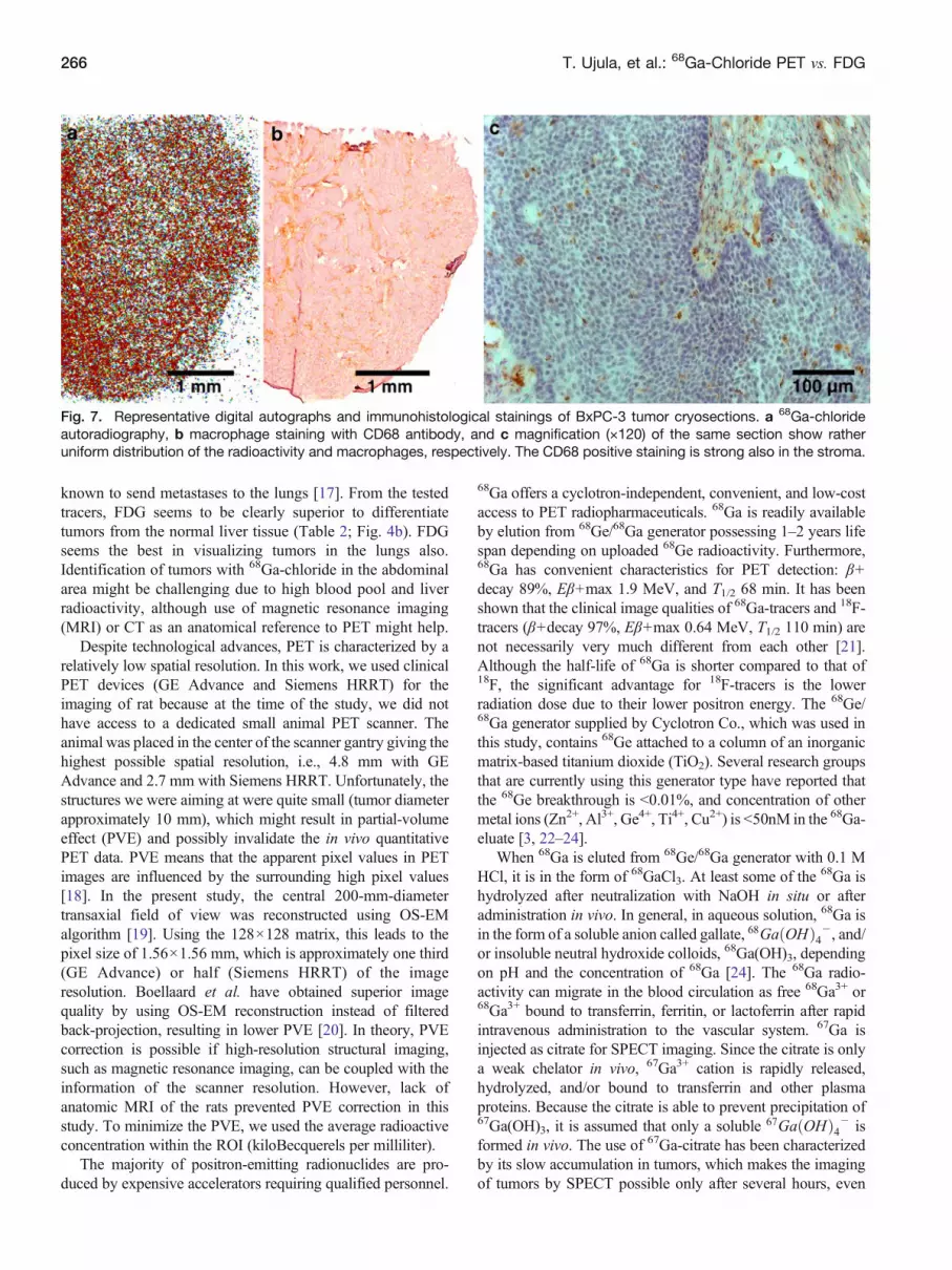
was mostly by viable cancer cell islets. Immunohistochem-ical staining with mouse antirat CD68 recognizing ratmacrophages revealed homogenous distribution in the tumortissue (Fig. 7).
DiscussionThis experimental study was designed to investigate thefeasibility of 68Ga-chloride for PET imaging of tumors bycomparing it with FDG. Although FDG is a widely usedimaging agent in PET and 67Ga-citrate in SPECT, compar-ison with 68Ga-chloride has not been performed earlier. Ourresults revealed that 68Ga-chloride was able to differentiateexperimental tumors from surrounding tissues (Fig. 1). Thetumor uptake of 68Ga-chloride was lower than that of FDG(Table 1; Fig. 2).
Human pancreatic adenocarcinoma xenograft in rat waschosen for an experimental tumor model, since imaging and
early diagnosis of pancreatic cancer still remains a challenge[14, 15]. One problem that hinders early diagnosis is thatonly symptomatic patients with suspected malignancy arePET imaged. PET is not the primary method for diagnosis,even though it has been reported to be the most accurate one[15]. Since the production of 68Ga-chloride is cyclotron-free,68Ga-chloride PET might facilitate the availability of PET incenters where no cyclotron is available.
Both tested tracers were capable of visualizing subcuta-neous tumors (Fig. 1). According to dynamic PET imaging,the tumor uptake of 68Ga-chloride was much faster than theuptake of FDG. Blood clearance of 68Ga radioactivityestimated from TAC of heart was slower compared withFDG (Fig. 3a), which is in accordance to the fact that Ga3+
binds to transferrin. The slow clearance was seen also as thelow tumor-to-blood ratio of 68Ga-chloride as noticed by exvivo studies (Table 2; Fig. 4a). In our previous studies, the68Ga-chloride uptake to healthy rat pancreas was low (SUV
a b
dc
e
Fig. 4. a Tumor-to-blood, b tumor-to-liver, c tumor-to-lung, d tumor-to-muscle, and e tumor-to-skin ratios calculated from theex vivo measurements of 68Ga-chloride and FDG at 90 min after injection in tumor bearing athymic rats. *PG0.05; ***PG0.005.
264 T. Ujula, et al.: 68Ga-Chloride PET vs. FDG

0.4±0.1 at 2 h after injection) [16]. When tumor uptake wascompared to the muscle, both 68Ga-chloride and FDG hadratio ≥4 (Table 2; Fig. 4d). However, there was a greatvariation in FDG accumulation in tumor xenografts betweenanimals. We used skin as another background tissue becausetumor cells were subcutaneously transplanted. In general, thedetermination of true skin radioactivity is difficult, since hair
and subcutaneous fat may interfere with the results if notcarefully removed from the skin sample. Tumor-to-lung andtumor-to-liver ratios are valuable when evaluating thepotential of a new imaging agent to differentiate tumors/metastases from these tissues. Pancreatic cancer cells easilysend metastases, in particular, to the liver after surgicaloperation [14], and many cancers, like breast cancer, are
Fig. 5. Representative digital autoradiographs of BxPC-3 tumor cryosections and corresponding hematoxylin and eosinstainings. a 68Ga-chloride autoradiography at 90 min postinjection (p.i.) and b the HE-staining of the same section. c FDGautoradiography at 90 min p.i. and d the corresponding HE staining. The dark blue represents the lowest and hot red thehighest amount of radioactivity.
Fig. 6. Hematoxylin and eosin stained paraffin sections of BxPC-3 tumor. a Tumor tissue is viable and without major necroticareas. Stroma is marked with an arrow. Bar=100μm. b BxPC-3 pancreatic adenocarcinoma cells form follicular structures(arrows). Bar=50μm.
T. Ujula, et al.: 68Ga-Chloride PET vs. FDG 265
known to send metastases to the lungs [17]. From the testedtracers, FDG seems to be clearly superior to differentiatetumors from the normal liver tissue (Table 2; Fig. 4b). FDGseems the best in visualizing tumors in the lungs also.Identification of tumors with 68Ga-chloride in the abdominalarea might be challenging due to high blood pool and liverradioactivity, although use of magnetic resonance imaging(MRI) or CT as an anatomical reference to PET might help.
Despite technological advances, PET is characterized by arelatively low spatial resolution. In this work, we used clinicalPET devices (GE Advance and Siemens HRRT) for theimaging of rat because at the time of the study, we did nothave access to a dedicated small animal PET scanner. Theanimal was placed in the center of the scanner gantry giving thehighest possible spatial resolution, i.e., 4.8 mm with GEAdvance and 2.7 mm with Siemens HRRT. Unfortunately, thestructures we were aiming at were quite small (tumor diameterapproximately 10 mm), which might result in partial-volumeeffect (PVE) and possibly invalidate the in vivo quantitativePET data. PVE means that the apparent pixel values in PETimages are influenced by the surrounding high pixel values[18]. In the present study, the central 200-mm-diametertransaxial field of view was reconstructed using OS-EMalgorithm [19]. Using the 128×128 matrix, this leads to thepixel size of 1.56×1.56 mm, which is approximately one third(GE Advance) or half (Siemens HRRT) of the imageresolution. Boellaard et al. have obtained superior imagequality by using OS-EM reconstruction instead of filteredback-projection, resulting in lower PVE [20]. In theory, PVEcorrection is possible if high-resolution structural imaging,such as magnetic resonance imaging, can be coupled with theinformation of the scanner resolution. However, lack ofanatomic MRI of the rats prevented PVE correction in thisstudy. To minimize the PVE, we used the average radioactiveconcentration within the ROI (kiloBecquerels per milliliter).
The majority of positron-emitting radionuclides are pro-duced by expensive accelerators requiring qualified personnel.
68Ga offers a cyclotron-independent, convenient, and low-costaccess to PET radiopharmaceuticals. 68Ga is readily availableby elution from 68Ge/68Ga generator possessing 1–2 years lifespan depending on uploaded 68Ge radioactivity. Furthermore,68Ga has convenient characteristics for PET detection: β+decay 89%, Eβ+max 1.9 MeV, and T1/2 68 min. It has beenshown that the clinical image qualities of 68Ga-tracers and 18F-tracers (β+decay 97%, Eβ+max 0.64 MeV, T1/2 110 min) arenot necessarily very much different from each other [21].Although the half-life of 68Ga is shorter compared to that of18F, the significant advantage for 18F-tracers is the lowerradiation dose due to their lower positron energy. The 68Ge/68Ga generator supplied by Cyclotron Co., which was used inthis study, contains 68Ge attached to a column of an inorganicmatrix-based titanium dioxide (TiO2). Several research groupsthat are currently using this generator type have reported thatthe 68Ge breakthrough is G0.01%, and concentration of othermetal ions (Zn2+, Al3+, Ge4+, Ti4+, Cu2+) is G50nM in the 68Ga-eluate [3, 22–24].
When 68Ga is eluted from 68Ge/68Ga generator with 0.1 MHCl, it is in the form of 68GaCl3. At least some of the 68Ga ishydrolyzed after neutralization with NaOH in situ or afteradministration in vivo. In general, in aqueous solution, 68Ga isin the form of a soluble anion called gallate, 68Ga OHð Þ4�, and/or insoluble neutral hydroxide colloids, 68Ga(OH)3, dependingon pH and the concentration of 68Ga [24]. The 68Ga radio-activity can migrate in the blood circulation as free 68Ga3+ or68Ga3+ bound to transferrin, ferritin, or lactoferrin after rapidintravenous administration to the vascular system. 67Ga isinjected as citrate for SPECT imaging. Since the citrate is onlya weak chelator in vivo, 67Ga3+ cation is rapidly released,hydrolyzed, and/or bound to transferrin and other plasmaproteins. Because the citrate is able to prevent precipitation of67Ga(OH)3, it is assumed that only a soluble 67Ga OHð Þ4� isformed in vivo. The use of 67Ga-citrate has been characterizedby its slow accumulation in tumors, which makes the imagingof tumors by SPECT possible only after several hours, even
Fig. 7. Representative digital autographs and immunohistological stainings of BxPC-3 tumor cryosections. a 68Ga-chlorideautoradiography, b macrophage staining with CD68 antibody, and c magnification (×120) of the same section show ratheruniform distribution of the radioactivity and macrophages, respectively. The CD68 positive staining is strong also in the stroma.
266 T. Ujula, et al.: 68Ga-Chloride PET vs. FDG
days. In contrast, when using 68Ga-chloride for PET, weobserved rapid localization of radioactivity in the target. Theform of 68Ga, i.e., the ratio of 68Ga-gallate and 68Ga-hydroxidecolloids, was not studied in this study.
The uptake mechanisms of radiogallium into tumors arenot fully understood, and a wide variety of factors areinvolved. It is known that Ga3+ behaves in vivo similarly toFe3+ by binding to transferrin and using the same trans-porters [25]. Gallium seems to concentrate in tissues havinghigh concentration of transferrin or transferrin receptors,lactoferrin, or ferritin. All nucleated cells of the body expresstransferrin receptors at different concentrations. Malignantcells generally express high levels of transferrin receptorsbecause they divide and require iron for DNA synthesis.However, there is some evidence that Ga can also entertumor cells by transferrin-independent mechanisms. Thismay be due to a very small soluble gallate ion, Ga OHð Þ4�,or a neutral insoluble, Ga(OH)3, both hydrolysis products ofGa [25]. In this study, 68Ga radioactivity seemed to be takenup mostly by viable cancer cell islets according to 68Gaautoradiography and HE staining (Fig. 5). The comparisonof autoradiography and CD68 immunohistochemical stain-ing (Fig. 7) suggests that the uptake might also be a result ofmacrophage phagocytosis. The resolution of autoradiogra-phy is not high enough to show the radioactivity uptake ofindividual cells, but it rather shows the accumulation incertain tissue areas, like tumor cell islets. It is possible thatthe 68Ga radioactivity is taken up by both tumor cells andinfiltrating macrophages. Indeed, accumulation of Ga inmacrophages, leukocytes, and bacteria has been reported[26, 27].
Fast tumor uptake of 68Ga-chloride would facilitate PETimaging as soon as 2 h after injection vs. long-lasting studyprocedure with 67Ga-citrate in SPECT imaging, although theslow blood clearance of 68Ga-chloride suggests for a latertime point. At 2 h after injection, the high blood poolradioactivity may contribute to tumor uptake as well.
Conclusions68Ga-chloride and FDG PET imaging were able to delineatesubcutaneously implanted human pancreatic adenocarci-noma xenografts in rats. The tumor uptake of FDG wasclearly superior, but there was great variation between theanimals. 68Ga-chloride showed fast tumor uptake, highblood pool radioactivity, and low urinary excretion. Advanta-geously, 68Ga-chloride is readily available from a generatorsystem and produces good PET image quality. However,further studies are needed to clarify the value of 68Ga-chloridefor PET imaging of tumors.
Acknowledgments. We acknowledge Maija-Liisa Hoffren (SafetyCity OyLtd, Turku), Irina Lisinen (Research Centre of Applied and PreventiveCardiovascular Medicine, University of Turku), Jouko Sandholm (CellImaging Core, Turku Centre for Biotechnology), and Erica Nyman (TurkuCentre for Disease Modeling, University of Turku) for excellent technical
assistance. The study was conducted within the Finnish Centre ofExcellence in Molecular Imaging in Cardiovascular and Metabolic Researchsupported by the Academy of Finland, University of Turku, TurkuUniversity Hospital, and Abo Academy University. In addition, study wasfinancially supported by grants from the Academy of Finland (nos. 205757and 103032), the Foundation for the Finnish Cancer Institute, theInstrumentarium Foundation, and the Eli Lilly Foundation. Tiina Ujulaand Anu Autio are Ph.D. students supported by the Drug DiscoveryGraduate School of the University of Turku.
Open Access. This article is distributed under the terms of the CreativeCommons Attribution Noncommercial License which permits any non-commercial use, distribution, and reproduction in any medium, provided theoriginal author(s) and source are credited.
References
1. Anger HO, Gottschalk A (1963) Localization of brain tumors with thepositron scintillation camera. J Nucl Med 77:326–330
2. Shealy CN, Aronow S, Brownell GL (1964) Gallium-68 as a scanningagent for intracranial lesions. J Nucl Med 21:161–167
3. Meyer GJ, Mäcke H, Schuhmacher J, Knapp WH, Hofmann M (2004)68Ga-labelled DOTA-derivatised peptide ligands. Eur J Nucl Med MolImaging 31:1097–1104
4. Roivainen A, Tolvanen T, Salomäki S et al (2004) 68Ga-labeledoligonucleotides for in vivo imaging with PET. J Nucl Med 45:347–355
5. Henze M, Schuhmacher J, Hipp P et al (2001) PET imaging ofsomatostatin receptors using 68Ga-DOTA-D-Phe1-Tyr3-octreotide: firstresults in patients with meningiomas. J Nucl Med 42:1053–1056
6. Maecke HR, André JP (2007) 68Ga-PET radiopharmacy: a generator-based alternative to 18F-radiopharmacy. In: Schubiger PA, Lehmann L,Friebe M (eds) PET chemistry, the driving force in molecular imaging.Springer, New York, pp 215–241
7. Mathias CJ, Lewis MR, Reichert DE et al (2003) Preparation of 66Ga-and 68Ga-labeled Ga(III)-deferoxamine-folate as potential folate-receptor-targeted PET radiopharmaceuticals. Nucl Med Biol 30:725–731
8. Velikyan I, Sundberg AL, Lindhe O et al (2005) Preparation andevaluation of (68)Ga-DOTA-hEGF for visualization of EGFR expres-sion in malignant tumors. J Nucl Med 46:1881–1888
9. Younes M, Brown RW, Stephenson M, Gondo M, Cagle PT (1997)Overexpression of Glut1 and Glut3 in stage I non-small cell lungcarcinoma is associated with poor survival. Cancer 80:1046–1051
10. Mäkinen TJ, Lankinen P, Pöyhönen T, Jalava J, Aro HT, Roivainen A(2005) Comparison of (18)F-FDG and (68)Ga PET imaging in theassessment of experimental osteomyelitis due to Staphylococcus aureus.Eur J Nucl Med Mol Imaging 32:1259–1268
11. Hamacher K, Coenen HH, Stocklin G (1986) Efficient stereospecificsynthesis of no-carrier-added 2-18F-fluoro-2-deoxy-D-glucose using ami-nopolyether supported nucleophilic substitution. J Nucl Med 27:235–238
12. DeGrado TR, Turkington TG, Williams JJ, Stearns CW, Hoffman JM,Coleman RE (1994) Performance characteristics of a whole-body PETscanner. J Nucl Med 35:1398–1406
13. Wienhard K, Schmand M, Casey ME et al (2002) The ECAT HRRT:performance and first clinical application of the new high resolutionresearch tomograph. IEEE Trans Nucl Sci 49:104–110
14. Nakao A, Fujii T, Sugimoto H et al (2006) Oncological problems inpancreatic cancer surgery. World J Gastroenterol 12:4466–4472
15. Saif MW, Cornfeld D, Modarresifar H, Ojha B (2008) FDG positronemission tomography CT (FDG-PET-CT) in the management ofpancreatic cancer: initial experience in 12 patients. J GastrointestinLiver Dis 17:173–178
16. Lendvai G, Velikyan I, Bergström M et al (2005) Biodistribution of68Ga-labelled phosphotiester, phosphorothioate, and 2′-O-methylphosphodiester oligonucleotides in normal rats. Eur J Pharm Sci26:26–38
17. Minn AJ, Gupta GP, Padua D et al (2007) Lung metastasis genes couplebreast tumor size and metastatic spread. PNAS 104:6740–6745
18. Hoffman EJ, Huang SC, Phelps ME (1979) Quantitation in positronemission computed tomography: 1. Effect of object size. J ComputAssist Tomogr 3:299–308
19. Visvikis D, Cheze-LeRest C, Costa DC, Bomanji J, Gacinovic S, Ell PJ(2001) Influence of OSEM and segmented attenuation correction in the
T. Ujula, et al.: 68Ga-Chloride PET vs. FDG 267

calculation of standardised uptake values for [18F]FDG-PET. Eur J NuclMed 28:1326–1335
20. Boellaard R, van Lingen A, Lammertsma AA (2001) Experimental andclinical evaluation of iterative reconstruction (OSEM) in dynamic PET:quantitative characteristics and effects on kinetic modeling. J Nucl Med42:808–817
21. Sanchez-Crespo A, Andreo P, Larsson SA (2004) Positron flight inhuman tissues and its influence on PET image spatial resolution. Eur JNucl Med Mol Imaging 31:44–51
22. Velikyan I, Lendvai G, Välilä M et al (2004) Microwave accelerated68Ga-labelling of oligonucleotides. J Label Compd Radiopharm 47:79–89
23. Breeman WA, de Jong M, de Blois E, Bernard BF, Konijnenberg M,Krenning EP (2005) Radiolabelling DOTA-peptides with 68Ga. Eur JNucl Med Mol Imaging 32:478–485
24. Hnatowich DJ (1977) A review of radiopharmaceutical developmentwith short-lived generator-produced radionuclides other than 99mTc. IntJ Appl Radiat Isot 28:169–181
25. Weiner RE (1996) The mechanism of 67Ga in malignant disease. NuclMed Biol 30:70–79
26. Bernstein LR (1998) Mechanisms of therapeutic activity for gallium.Pharmacol Rev 50:665–682
27. Clausen J, Edeling C-J, Fogh J (1974) 67Ga binding to human serumproteins and tumor components. Cancer Res 43:1931–1937
268 T. Ujula, et al.: 68Ga-Chloride PET vs. FDG


![[ 18 F] FDG PET in Gastric Non-Hodgkin's Lymphoma](https://img.pdfslide.net/doc/110x75/635c4d740065a57dd807bbca/-18-f-fdg-pet-in-gastric-non-hodgkins-lymphoma.jpg)


