Embed Size (px)
Citation preview

Review
s�FOUNDATION
REVIEW
REVIEWS Drug Discovery Today � Volume 14, Numbers 17/18 � September 2009
Foundation review:a7-Nicotinic receptor antagonists at thebeginning of a clinical era for NSCLC andMesothelioma?Laura Paleari1, Alfredo Cesario2,3, Massimo Fini4 andPatrizia Russo1
1 Lung Cancer Unit, National Cancer Research Institute, Genoa, Italy2 Thoracic Surgery Unit, Catholic University, Rome, Italy3CDC ‘‘San Raffaele’’ Velletri Rome, Italy4 IRCCS ‘‘San Raffaele’’, Rome, Italy
Of the human solid cancers, Non-Small Cell Lung Cancer (NSCLC) and
Malignant Pleural Mesothelioma (MPM) display a natural history
supporting the concept that they develop from multiple preneoplastic
pathways. Recently, new evidence suggested that nicotinic Acetylcholine
Receptors (nAChRs) play a significant role in lung cancer predisposition
and natural history. This review is based on some translational research
aimed at evaluating the potential therapeutic effect of nAChR antagonists
on NSCLC and MPM. The background and rationale of this approach are
based on the experimental observations that: (a) NSCLC and MPM cells
express nAChRs and (b) the activation of these receptors by agonists,
namely nicotine, inhibits apoptosis, whereas receptor antagonists have a
pro-apoptotic effect.
IntroductionNon-Small Cell Lung Cancer (NSCLC) and Malignant Pleural Mesothelioma (MPM) demonstrate
great molecular heterogeneity in which several pathways are believed simultaneously and
actively to lead to tumorigenesis [1]. Thus, their natural history supported the concept that
they develop from multiple preneoplastic pathways. MPM is an aggressive neoplasm of mesothe-
lial cell origin that arises mainly from the pleura and is strongly associated with asbestos exposure
[2]. Conventional therapies, such as surgery, radiotherapy and chemotherapy, do not necessarily
improve overall survival [3–4]. Lung cancer consists of several histological types in which NSCLC
represents 75–85% of the total, it is subclassified into: adenocarcinoma (AD, including the
noninvasive type of bronchioloalveolar carcinoma), squamous cell carcinoma (SQ), epidermoid
and large cell carcinoma [5]. In AD at least two pathways have been identified: (i) smoking-related
and (ii) nonsmoking-related [6]. Recently, genetic variations in a region of chromosome 15 that
encompasses a gene implicated in nicotine dependence had been linked to the risk of lung cancer
in genome-wide association studies, but data were not definitive as to whether the variants were
linked to lung cancer per se or to nicotine dependence [7–12]. A recent work [12] reported that the
increased risk of lung cancer conferred by the genetic variants might be explained by an increased
PATRIZIA RUSSO
Patrizia Russo is in charge of
the Lung Cancer Unit at the
National Institute for
Research on Cancer, Genoa,
Italy. In 2006 she became a
member of the Italian
Consensus Board for the
definition of Italian lung
cancer guidelines. Since 1987, she has been working in
the field of Molecular Pharmacology and in 1987, 1991
and 1997–1998 she attended the Laboratory of
Molecular Pharmacology at NCI, NIH (Bethesda,
USA). She studied the molecular mechanism of action
of caffeine, then the association of antitumor drugs
with cytokines. Parts of the latter studies were funded
by AIRC (Milan, Italy), CNR (Rome, Italy) and NATO
(Bruxelles, Belgium). Since 1995, she has been
studying the relationship of sensitivity or resistance to
classical and new experimental targeted antineoplastic
drugs, receiving a fellowship from FIRC (Milan, Italy)
to join the Laboratory of Molecular Pharmacology at
NCI, NIH. These studies were supported by EC
(Ispra, Italy). Since 2000, she has been studying lung
cancer, looking at early detection and the develop-
ment of new drugs. These studies were supported by
the Italian Health Ministry, Fondazione Compagnia di
San Paolo (Turin, Italy), Fondazione CARIGE (Genoa,
Italy) and Liguria Local Government (Genoa). Starting
in 2003, she has been investigating the role of nAChRs
in lung cancer biology.
Corresponding author: Paleari, L. ([email protected])
822 www.drugdiscoverytoday.com 1359-6446/06/$ - see front matter � 2009 Published by Elsevier Ltd. doi:10.1016/j.drudis.2009.06.016

Drug Discovery Today � Volume 14, Numbers 17/18 � September 2009 REVIEWS
Reviews�FOUNDATION
REVIEW
likelihood of nicotine dependence, although some of the results of
this study, and a previous study of the variant in subjects who
reported that they had never smoked, suggest that the variants
may also have a direct role in lung carcinogenesis.
Pharmacology and signaling of nAChRsnAChRs belong to the ligand-gated ion channel (LGIC) family that
includes the excitatory 5HT-3 receptor and the inhibitory recep-
tors for glycine and g-aminobutyric acid (GABAA and GABAC) [13].
The nAChR consists of either a homo or heteropentamer com-
posed of the various subunits that have been identified so far (a1–
a10; b1–b4) [14,15] and that are arranged symmetrically around
an axis perpendicular to the membrane, thus delineating the ionic
pore. The composition and stoichiometry of the subunits consti-
tuting the pentamer may have a profound impact on receptor
pharmacology, cation selectivity, desensitization kinetics and
spatial distribution. All nAChR subunits share a homologous
structure, with a large extracellular domain, four transmembrane
regions (M1–M4) structured in a-helices, a large cytoplasmic
domain between M3 and M4 and, finally, a short extracellular
C-terminal tail [16].
The cognate ligand for the nAChRs is nicotine, an agonist
interacting with various affinities, from 1 to 130 mM, depending
upon the different neuronal receptor subtypes [17,18]. The pre-
sence of b2 and b4 subunits in the receptor pentamer seems to be
correlated with high and low affinity for nicotine, respectively.
On the contrary, as an allosteric receptor (see the below para-
graph), nAChR may undergo rapid conformational transitions
from a resting basal state to an active or desensitized state. Appli-
cation of nicotine initially provokes the stabilization of the recep-
tor in a high affinity, open state followed by a progressive
stabilization of a closed, desensitized state [19]. In the case of
smoking behavior, long exposure to a low concentration of nico-
tine favors receptor desensitization.
Mutational and photo-affinity labeling experiments identified
the agonist binding site at the interface of the extracellular regions
of the principal a and the complementary non-a subunits,
whereas the transmembrane segment M2 is the major contributor
to the pore domain [13]. The structural coupling between the
extracellular and the pore domains provides efficient transduction
between agonist binding and the ion channel gating. Recently, the
resolution of the crystallographic structures of a protein homo-
logous to the extracellular domain of nAChR, the acetylcholine
binding protein (AChBP) either alone or complexed with various
ligands, in addition to numerous biochemical studies on the
ligand binding sites, allowed better understanding of how various
ligands interact with different nAChRs and revealed, at the mole-
cular level, the fundamental events underlying the receptor acti-
vation [20–24].
The nicotine-binding site was initially studied using structural
models of the extracellular domain of nicotinic receptors and then
the crystallographic structure of the AChBP–nicotine complex
[25]. Even if the nicotine-binding pocket is similar to those deter-
mined for acetylcholine or epibatidine, involving mainly aromatic
and hydrophobic contacts, the specific binding of nicotine is due
to additional hydrogen bonds with the receptor and a closer
packing of the aromatic groups [25]. These subtleties in nicotine
interaction compared to other agonists were confirmed by a
physical chemistry approach using unnatural amino acid muta-
genesis combined with computational modeling studies [26].
Nicotine activates different subtypes of nAChR, inducing a
complex pattern of mixed sympathetic and parasympathetic
responses. The stimulation, desensitization and upregulation of
these receptors by nicotine seem to be responsible for diverse
physiological effects targeting the cardiovascular [27,28], pulmon-
ary (as we will see in the next chapters), endocrine [29] and central
nervous systems [16]. Of course, one of the most studied effects of
nicotine is its smoking-related addictive property [30–32].
Allosteric modulationIntroduced by Wyman and colleagues in 1965 [33], the allosteric
concept refers to the assumption that proteins could exist in
multiple conformational states and that binding of allosteric
ligands alters the energy barriers or ‘isomerization coefficients’
between various states, preferentially stabilizing the protein in a
given conformation. The site occupied by the natural ligand,
which is typically at the interface between subunit protomers, is
called ‘the orthosteric site’. Allosteric sites are distinct from the
orthosteric site and can be localized elsewhere on the protein.
Binding of the ligand at the orthosteric site stabilizes the protein in
the active state, whereas binding of an effector at an allosteric site
alters the overall properties by modifying the energy barriers,
represented by isomerization coefficients, between one or more
states. In the specific case of the nAChR, agonists are ligands that
preferentially stabilize the receptor in the active open state,
whereas competitive antagonists are ligands that stabilize the
protein in a closed conformational state. Thus, endogenous
ligands, such as acetylcholine, bind at the orthosteric site, whereas
all the molecules that bind elsewhere on the nAChR subunit(s) act
via allosteric interactions. Bertrand and Gopalakrishnan, in a
recent review, outlined extensively the principles of the allosteric
concept and summarized the profiles of novel compounds that are
emerging as allosteric modulators at the a7- and a4b2-nAChR
subtypes [34].
Ca2+ permeability of nAChRThe nAChR channels are permeable to cations, including Ca2+.
Ca2+ entry through nAChR channels modulates several Ca2+-
dependent cellular processes, such as neurotransmitter release,
synaptic plasticity and cell motility. Two different classes of neu-
ronal nAChR may be identified according to their Ca2+ perme-
ability, which correlates with other pharmacological and
structural properties: (i) neuronal nAChRs containing subunits
(a7–a9) able to bind a-bungarotoxin (a-BTX) and form homo-
pentameric channels (a-BTX nAChR), exhibiting the highest mea-
sured Ca2+ permeability values [35]; (ii) heteropentameric, non-a-
BTX-sensitive nAChRs (non-a-BTX nAChR), always comprising at
least one a (out of a2–a6) and one b (out of b2–b4) subunits, with
lower measured Ca2+ permeability [36]. Studies indicate a func-
tional correlation between the activation of a7-nAChR and Ca2+-
dependent cellular processes, such as neurotransmitter release,
synaptic plasticity, cell growth, migration and survival.
A recent study by Gilbert et al. [37] showed that the Ca2+
transients were predominantly due to the opening of plasma
membrane a7-nAChR, because the signals were (a) evoked by
nicotine, (b) sensitive to two a7-specific nAChR inhibitors
www.drugdiscoverytoday.com 823

REVIEWS Drug Discovery Today � Volume 14, Numbers 17/18 � September 2009
TABLE 1
nAChR expression on different normal airways epithelial cells or Mesothelial cells
Cells nAChR subtype
a1 a2 a3 a4 a5 a6 a7 a9 a10 b2 b3 b4 Reference
Airways epithelial cells
BAC1 [116]
BAC2 [116]
BAC3 [116]
BEAS-2B + � � + + ++ � ++ � � ++ [130]
BEC � � � + � � + � � + � [55]
BEC + + [47]
BEP2D* + � + � + � + + + + � + [131]
HBEC + � + + � [57]
HBEC � � � � [57]
HBEC-KT1 + + � + +/� � � + � +/� + [47]
HBEC-KT2 +/� � � + � � � +/� � � + [47]
HBEC-KT3 � � � + � � � � � � � [47]
HBEC-KT4 � � � + � � � � � � � [47]
NHBE � � + � + � + + + + � + [55,60,132]
+ + +
NHBE + [79]
SAEC � + � + � � + + + + � + [60,79]
SAEC � +/� + � � +/� [44]
SAEC + � � +/� � � � + +/� � + [47]
SAEC + [79,133]
Pleural mesothelial cells
Mes-1 + [109,116]
Mes-2 + [109,116]* This cell line is an established clonal population of HPV-18-immortalized human BEC. The cells have an epithelial morphology in culture, near diploid karyotype, and relatively stable
genotype. They are anchorage-dependent and do not form tumors in immunosuppressed host animals.
Review
s�FOUNDATION
REVIEW
(methyllycaconitine and a-BTX), (c) increased by a known a7-
nAChR allosteric modulator (PNU-120596) and (d) absent when
Ca2+ was omitted from the bathing medium.
Calcium imaging, combined with whole-cell patch-clamp
recordings, has been used previously to determine the Ca2+ per-
meability of native or expressed nAChRs [35]. These studies
provided important clues to the physiological role of the different
nAChR subtypes and to the structural determinants of their Ca2+
permeability (reviewed in [38]), but did not provide any spatial
information about the underlying Ca2+ signals. This study clearly
demonstrated that the a7-nAChR is able to generate large Ca2+
signals in neuronal cells stimulated with relevant agonists. Con-
sequently, significant signals are expected to be generated by
these agents in cells expressing high levels of the a7-nAChR, such
as neurons [16]. In neurons, Ca2+ elevations can trigger the
secretion of neurotransmitters, open membrane channels to
modulate the cell’s excitability, and activate the transcription
of several genes [16]. Thus, the activation of the a7-nAChR is
expected to impact on neuronal activity, both in the central and
in the peripheral nervous system. The same prediction can be
made for non-neuronal cells known to express the a7-nAChR,
such as monocytes and macrophages [39,40]. In these cells,
central for innate immunity, Ca2+ elevation is involved with
824 www.drugdiscoverytoday.com
the control cell migration, bacterial killing, antigen presentation
and cytokine release [41].
Expression of nAChR in airways cellsnAChR genes are also expressed in different epithelial cells, includ-
ing normal and lung cancer cells [42–50].
In this section we outline the differential distribution of nAChR
subunits in normal and/or unaffected airway epithelial cells, in
NSCLC and pleural Mesothelioma cell lines, as well as in tissue
cancer specimens. The data are summarized in Tables 1–3. From
Table 1 it is manifest that normal airway epithelial cells show a
different pattern of nAChR subtypes. Even so, looking at the
normal, nonimmortalized bronchial epithelial cells, the a7 sub-
type receptor seems to be the most predominantly expressed
receptor. This is a regular feature of cancer cell lines (Table 2)
and cancer tissues (Table 3).
Comparison of the expression of nAChR subunits between
tumor and matched normal tissue revealed a significant upregu-
lation of the b4 subunit and a concomitant decrease in a4 levels
[47]. In addition, NSCLC tumors from nonsmokers showed ele-
vated expression of the a6b3 receptor, compared with smokers, in
a gender adjusted manner [47]. Recent data suggested that a7-
nAChRs in NSCLC are significantly more expressed in squamous

Drug Discovery Today � Volume 14, Numbers 17/18 � September 2009 REVIEWS
TABLE 2
nAChR expression on different NSCLC or Mesothelioma cell lines
Cells nAChR subtype
a1 a2 a3 a4 a5 a6 a7 a9 a10 b2 b3 b4 Reference
NSCLC adenocarcinoma
201T + + + + + [134]
A427 + [135]
A549 � � � + + � + + � + + � [55,79–81,112,113,132,134]
+ � � � + � +
H1299 � � + + + + + [79,80]
H1355 � � + + + � + � � + � � [133]
H1437 � + +/� + � + + + + +/� � [47]
H1648 +/� � � + +/� + � + � +/� + [47]
H1650 + [116]
H1703 � � + + + � + + � + � � [132]
H1770 +/� � � + ++ + � + � + � [47]
H1819 +/� � � + +/� + � + � + + [47]
H1993 +/� � � + � +/� � + � + + [47]
H2009 + � � + � + +/� + � + + [47]
H2087 +/� � � + +/� + � + � +/� + [47]
H2122 + [80,135]
H2122 +/� +/� � + +/� + +/� + � + + [47]
H23 � � + + + + + [79,80]
H2347 +/� � � + +/� + +/� + � +/� + [47]
H322 + [47]
H441 � [79,80,133]
HKULC1 + +/� +/� + � + � + � + + [47]
HKULC2 + � � + +/� +/� � + � +/� + [47]
HKULC3 + + ++ ++ ++ ++ + + + � + [47]
HKULC4 +/� +/� � + � +/� � + + � � [47]
LT1 ++ [116]
LT2 + [116]
NSCLC squamous cell carcinoma
273T + + + + + [132,134]
H157 � � � + + + + + � + � � [132]
H2170 + [133]
H226 � � + + + + + [133]
H520 + + + + + + [133]
SK-MES + [48]
LT3 + [116]
LT4 ++ [116]
NSCLC large cell carcinoma
COR-L23 + Our unpublished data
H1155 � � + + + � + � � + � + [132]
MPM
MSTO-211H + [109]
MPP-89 + [78,109]
IST-MES1 + [78,109]
IST-MES2 + [78]
Primary NSCLCs [LT1, LT2 (adenocarcinoma), LT3, LT4 (squamous carcinoma)].
www.drugdiscoverytoday.com 825
Reviews�FOUNDATION
REVIEW

REVIEWS Drug Discovery Today � Volume 14, Numbers 17/18 � September 2009
TABLE 3
nAChR expression on different NSCLC or Mesothelioma tissue samples
No. of samples nAChR subtype
a1 a2 a3 a4 a5 a6 a7 a9 a10 b2 b3 b4 Reference
NSCLC adenocarcinoma
2 + + [77]
54 ++ + + ++ ++ ++ + ++ + ++ ++ [47]
19 + [48]
NSCLC squamous cell carcinoma
2 + + [42]
31 + (45%) + (87%) + (71%) + (90%) + (100%) + (87%) + (90%) [133]
6 ++ ++ + ++ ++ ++ + ++ + ++ ++ [47]
28 ++NSCLC large cell carcinoma
5 ++ + + ++ ++ ++ + ++ + ++ ++ [47]
28 ++ [48]
MPM
4 + [78,109]
Carlisle et al. [97] found that none of the lung tissues that they examinedwere positively stained for a7-nAChR. By contrast, we found that a1 and b1 were highly expressed in NSCLC tissue
(12 adeno 4 active smokers, 5 exsmokers, 1 nonsmoker, 2 unknown and 3 squamous IH, 2 exsmokers, 1 unknown) (Paleari, Am. J. Respir. Crit Care).
Review
s�FOUNDATION
REVIEW
carcinoma than in adenocarcinoma. Among this histological
subtype, smokers showed the highest upregulation. Interestingly,
all NSCLC female patients, either smokers or nonsmokers,
expressed less mRNA and protein for the a7-nAChR than males,
suggesting a different response to nicotine between females and
males [48]. As a consequence of the presence of nAChRs on lung
cells, the potential role of cholinergic activation in the develop-
ment and growth of lung cancer has been intensely studied in
recent years [49,50].
a7-nAChR activation and subcellular signaling in lungcellsEffects on normal epithelial lung cellsIn human bronchial tissues and in cultures of human BEC, the
nAChRs were visualized on the cell membranes, predominantly at
the sites of cell-to-cell contacts using subunit-specific antibodies.
The epithelial cells of submucosal glands abundantly expressed
a7-nAChR. The function of the nicotinic cholinergic signaling
pathway in airway bronchial epithelium is highly likely to be
affected by nicotine in smokers. In smokers, plasma nicotine levels
peak around 200 nM during the day and drop to 5–10 nM during
sleep. Nicotine levels in lung airways directly exposed to smoke
may be five to tenfold higher, and peaks and troughs are much
sharper [51]. These levels are high enough to activate a4b2-
nAChRs [51,52] and may either inhibit [53] or activate [54] a7-
nAChR. It is also interesting that the expression of nAChR appears
highly expressed at the apical regions of cells, where they are more
exposed to airway nicotine. Smoking significantly (P < 0.05)
increased the relative numbers of nAChRs and these effects could
be reproduced in cultures of BEC exposed to 10 mM nicotine [55].
Whole-cell patch-clamp studies of cultured human BECs demon-
strated the presence of fast-desensitizing currents activated by
choline and nicotine that were blocked reversibly by methyllyca-
conitine (1 nM) and irreversibly by a-BTX (100 nM), consistent with
the expression of functional a7-nAChR [55].
826 www.drugdiscoverytoday.com
Recently Lam et al. [47], after having exposed human BEC lines
to 100 nmol/L nicotine and then harvested RNA at 72 and 144
hours, showed a significant increase in the expression levels of a1-,
a5- and a7-nAChRs at 72 hours, with return to baseline levels of
expression upon nicotine removal.
To gain insights into the molecular mechanism underpinning
such nicotine-induced effects, microarray-bioinformatics analysis
was carried out to explore the gene expression profiles in human
BEC treated with nicotine at 5 mM for 4, 8 and 10 hours [56]. Of
1800 assessed genes overall, 260 (14.4%) were upregulated and 17
(0.9%) downregulated significantly. Membrane array analysis sug-
gested that both extracellular signal-regulated kinase (ERK) 1/2
and c-Jun-NH2-terminal kinase (JNK) signaling, but not p38 MAPK
signaling, were activated in response to nicotine. Thus, pretreat-
ment of human bronchial epithelial cells (BECs) with specific
inhibitors against ERK 1/2 and JNK, but not p38, significantly
inhibited nicotine-induced interleukin-8 production, suggesting
that MAPK pathway may mediate the effect of nicotine through
ERK 1/2 and JNK, but not p38 in HBECs treated with nicotine.
The biological roles of epithelial nAChRs apparently involve the
regulation of cell-to-cell communications, adhesion and motility,
because Mec caused rapid and profound changes in these cell
functions that were reversed by nicotine. An over-exposure of
BECs to nicotine, however, produced an antagonist-like effect,
suggesting that the pathobiological effects of nicotine toxicity
might result from both activation of nAChR channels and nAChR
desensitization [57]. It has been reported, using an animal model
of nicotine infusion [58], that the tissue-to-blood ratio of nicotine
is 3.0 for brain and 2.0 for lung. Therefore, lung tissues can reach
nicotine levels that approximate those achieved in the brain. As
such, the duration and persistence of nicotine administration over
time becomes an important pharmacological variable in the use
and interpretation of this drug’s actions. The treatment of rat lung
epithelial cells with nicotine for various periods differentially
mobilizes multiple intracellular pathways [59]. Protein kinase C

Drug Discovery Today � Volume 14, Numbers 17/18 � September 2009 REVIEWS
Reviews�FOUNDATION
REVIEW
and PI3-OH-kinase are transiently activated after the treatment.
Also, Ras and its downstream effector ERK1/2 are activated after
long-term exposure to nicotine. The activation of Ras by nicotine
treatment is responsible for the subsequent perturbation of the
methotrexate (MTX)-mediated G1 cell cycle restriction, as well as
an increase in production of reactive oxygen species [59]. These
data suggest that persistent exposure to nicotine perturbs the G1
checkpoint and causes DNA damage through the increase of the
production of reactive oxygen species. A third element rendered by
loss of p53 is, however, required for the initiation of the process of
gene amplification. Under p53-deficient conditions, the establish-
ment of a full oncogenic transformation, in response to long-term
nicotine exposure, is achieved through the cooperation of multi-
ple signaling pathways.
Through activation of separate nAChR a-subunits, nicotine
activates one of the best-characterized signaling pathways that
promote cellular survival: the PI3K/Akt pathway. Activation of Akt
by nicotine occurred within minutes, but peaked at 45–60 min,
and is maintained for hours. The presence of phosphorylated Akt
in human lung cancers from smokers may support the hypothesis
that nicotinic activation of Akt is not limited to cultured primary
cells. Once activated by nicotine, Akt increased phosphorylation
of multiple downstream components that control cellular cell
cycle and protein translation, such as glycogen synthase kinase-
3 (GSK-3), binding protein for eukaryotic translation initiation
factor 4E (4EBP-1) and ribosomal kinase p70S6K [60].
Pulmonary neuroendocrine cells (PNEC) are a highly specialized
population of airway epithelial cells which contain and secrete
biogenic amines, in particular 5-hydroxytryptamine (5-HT) and
various peptides [61,62]. By studying the release of 5-HT from
isolated rabbit tracheae it was shown that the secretory activity of
PNEC is stimulated via nicotinic receptors, but not modulated by
muscarinic mechanisms [63]. A nicotinic receptor-induced release
of 5-HT could also be observed from PNEC in culture [64].
To sum up in BEC exposed to nicotine, for short time, nAChR
mediated activation of the serine/threonine kinase Akt and/or ERK
1/2 resulting in the phosphorylation of several downstream sub-
strates. This was associated with the a transformed cellular phe-
notype manifested as loss of contact inhibition, loss of
dependence on exogenous growth factors and attenuated apop-
tosis induced by various pro-apoptotic stimuli.
Effects on normal fibroblast lung cellsIn primary murine lung fibroblasts, nicotine stimulates the expres-
sion of fibronectin via the activation of intracellular signals that
lead to increased fibronectin gene transcription. It was observed
that the stimulatory effect of nicotine was associated with the
activation of protein kinase C and mitogen-activated protein
kinases, increased levels of intracellular cAMP and phosphoryla-
tion and DNA binding of the transcription factor CREB. Increased
transcription of the gene was dependent on cAMP-response ele-
ments (CREs) present on the 50 end of its gene promoter. The
stimulatory effect of nicotine on fibronectin expression was abol-
ished by a-BTX. Of note, nicotine increased the expression of a7-
AChR on fibroblasts. To assess the relevance of these in vitro
observations to the situation in vivo, Roman et al. [54] examining
fibronectin expression in the lungs of nicotine-exposed mice
observed an increased expression of fibronectin mRNA and protein
when compared with control animals. Immunohistochemical
analysis revealed that nicotine exposure was also associated with
increased fibronectin protein in alveolar septae as well as in airway
epithelial cells and in vascular structures. These data suggest that
nicotine induces lung fibroblasts to produce fibronectin by stimu-
lating a7-nAChR-dependent signals that regulate the transcription
of the fibronectin gene. This may result in alterations in the
composition of the lung matrix. In doing so, nicotine might
promote increased tissue remodeling around the airways and
within the lung parenchyma and this is likely to represent one
mechanism by which tobacco results in abnormal lung function.
In addition, the newly deposited fibronectin-containing matrix
primes lung resident and incoming cells to response to injurious
agents in an exaggerated manner. Further delineation of the
factors and conditions that regulate nicotine-induced fibronectin
expression in vivo will be needed before a full understanding can be
obtained of the true consequences this process has in lung as well
as other organs.
Effects on embryonic lung cellsMaternal smoking during pregnancy has various adverse effects
that are well known and documented [65]. In our point of view, it
is interesting that in the WI38 human embryonic lung fibroblast
cell line, which displays a3- and a7-nAChR subunits, nicotine
disrupted the specific paracrine signaling pathway that caused
pulmonary transdifferentiation of interstitial lipofibroblast (LIF)-
to-myofibroblast (MYF), resulting in altered pulmonary develop-
ment and function [66]. This effect was counteracted by the use of
nonspecific (D-tubocurarine) and specific (a-BTX and Mec) antago-
nists of nAChR. Alveolar interstitial LIF to MYF transdifferentia-
tion results in failed alveolarization in the developing lung, which
leads to an arrest in pulmonary growth and development, the
hallmarks of in uterus nicotine-induced lung damage. Specifically,
the interstitial LIF phenotype is of functional importance, because
it provides cytoprotection against oxygen free radicals [67], traffics
neutral lipid substrate to alveolar type II cells for surfactant phos-
pholipids synthesis [68] and causes alveolar type II cell prolifera-
tion [69]. Although MYF also seems to be important for normal
lung development, these cells are also the hallmarks of chronic
lung diseases in both the neonate and adult. In the developing
lung, MYF are fewer in number and are predominantly located at
the periphery of the alveolar septa, where they probably partici-
pate in the formation of new septa [70,71]. According to Sekhon
et al. [72] nicotine, after having passed across the placenta, inter-
acts with nAChRs of the fetal monkey lung, thus maternal nicotine
exposure upregulated nAChR expression in fetal lung. In parallel
with changes in nAChR expression, prenatal nicotine exposure
downregulated the surface complexity of the parenchyma,
increased collagen accumulation, upregulated surfactant protein
gene expression and induced neuroendocrine cell hyperplasia in
fetal lungs and altered pulmonary function [73,74]. lynx1, an
evolutionary precursor to snake venom toxins, is expressed during
the early development of lung, gradually increased through the
prenatal period, and then persisted throughout adulthood. Spatial
differences occur between lynx1 expression in the proximal and
distal parts of the lung suggesting a possible role in cellular
differentiation and/or function. lynx1 upregulation following
prenatal nicotine exposure suggested that it plays a role in
www.drugdiscoverytoday.com 827

REVIEWS Drug Discovery Today � Volume 14, Numbers 17/18 � September 2009
Review
s�FOUNDATION
REVIEW
regulating the interaction of exogenous agonist (nicotine) or
endogenous ligand (ACh) in lung cells that express nAChR [75].
Effects on lung cancer cellsAs reported above, from biological, histopathologic and clinical
perspectives, lung cancer is a highly complex neoplasia, probably
having multiple preneoplastic pathways [1,76]. The histopatho-
logic heterogeneity is the major confounding factor in lung cancer
diagnosis and treatment. Cigarette smoking has been linked to all
histological subtypes; however, the proportion of smokers is high-
est in those who develop SQ.
In 1990, Maneckjee and Minna [42] have described the presence
of a7-nAChR on small and NSCLC cell lines. Afterwards different
authors reported the presence of nAChRs in lung cancer cell lines
and in tissues obtained from human biopsies taken from patients
suffering from lung cancer (see Tables 1 and 2).
More recently, Lam et al. [47] showed the expression of nAChR
subunit genes in 66 resected primary NSCLCs; those derived from
nonsmokers demonstrated higher expression of a6- and b3-
nAChR subunit genes than those from smokers, adjusted for
gender. In addition, nAChR a4- and b4-subunit gene expression
showed significant difference between NSCLC and normal lung.
Using Affymetrix GeneChip U133 Sets, 65 differentially expressed
genes associated with a NSCLC nonsmoking a6b3-nAChR pheno-
type were identified, which gave high sensitivity and specificity of
prediction. The authors concluded that between NSCLC from
smokers and nonsmokers, different nAChR subunit gene expres-
sion patterns were found, and a 65-gene expression signature was
associated with nonsmoking a6b3-nAChR expression. Further-
more, an important study of Song et al. [77] presented data that
SCLC express a cholinergic autocrine loop that can regulate cell
growth.
Such a study demonstrates that:
(a) genes for all components of an ACh autocrine loop, including
choline acetyltransferase (ChAT), vescicular ACh transporter
(CHT1), nAChR and muscarinic AChR (mAChR) are
expressed in SCLC cells, as well as in neuronal cells;
(b) ChAT is present in biopsies of SCLC and in SCLC cell lines;
(c) SCLC cells are able to synthesize, secrete and degrade ACh;
(d) SCLC cell growth is modulated by endogenous ACh synthesis.
Such work is probably the first study that demonstrates that
SCLC cells have a cholinergic phenotype and that ACh exerts an
autocrine growth factor in human lung tumors. Thus, the identi-
fication of a cholinergic autocrine loop by SCLC now provides a
framework and rationale for the many studies, in the literature,
that nicotine and related compounds stimulate SCLC growth.
The major effects of nicotine in lung cancer cells (SCLC and
NSCLC) are
(a) Enhancement of cancer cell growth.
(b) Inhibition of drug-induced apoptosis.
Recently, it has been shown that nicotine stimulated the
tumoral growth of A549 cells orthotopically implanted in BALB/
c in NOD/SCID mice [48].
It was suggested that:
(i) Bax may be an essential component in the nicotine survival
signaling pathway, through a mechanism involving activa-
tion of PI3K/AKT that directly phosphorylates and inacti-
vates the pro-apoptotic function of Bax [78–81].
828 www.drugdiscoverytoday.com
(ii) Survivin and XIAP played a key role in the antiapoptotic
activity of nicotine [79].
(iii) Nicotine exerted its role of antiapoptotic inducer through
NF-kB upregulation [80].
(iv) The mitogenic effects of nicotine resulted in enhanced
recruitment of E2F1 and Raf-1, causing dissociation of Rb
from these promoters. Proliferative signaling via nAChR
required the scaffolding protein b-arrestin since ablation of
b-arrestin or disruption of the Rb–Raf-1 interaction blocked
nicotine-induced proliferation; thus, nicotine induces cell
proliferation by b-arrestin-mediated activation of the Src and
Rb–Raf-1 pathways [82].
a7-nAChR activation and effects on neoangiogenesisHeeschen et al. [83] provided anatomic and functional evidence
that nicotine-induced angiogenesis. They also showed that nico-
tine accelerates the growth of tumor and atheroma in association
with increased neovascularization. Nicotine increased endothelial
cell growth and tube formation in vitro and accelerated fibrovas-
cular growth in vivo. In a mouse model of hind-limb ischemia,
nicotine increased capillary and collateral growth, and enhanced
tissue perfusion. In a Lewis lung tumor model, nicotine enhanced
lesion growth in association with an increase in lesion vascularity.
These effects of nicotine were mediated through nAChR at nico-
tine concentrations that are pathophysiologically relevant. The
endothelial production of nitric oxide, prostacyclin and vascular
endothelial growth factor might have a role in these effects.
Interestingly, it was shown that the second hand smoke (SHS),
where nicotine is one of the major components, is associated with
increases in plasma levels of angiogenic cytokines such as VEGF
and MCP-1. Fascinatingly the angiogenic effects of SHS can be
blocked by inhibition of the endothelial nAChR [84].
In spite of its importance, the neoangiogenetic effect, however,
is not directly associated with a specific effect of nAChR activation
on lung cells and is beyond the intention of this review.
a7-nAChR activation and inflammationIn 2005, an important article by Ulloa [41] reviewed different
studies indicating that the vagus nerve (which is the longest of
the cranial nerves and innervates most of the peripheral organs)
can modulate the immune response and control inflammation
through a ‘nicotinic anti-inflammatory pathway’, depending on
the a7-nAChR. This article reported all the advances supporting
the therapeutic potential of selective nicotinic agonists in several
diseases. The author suggested that, similar to the development of
a- and b-agonists for adrenoceptors, selective agonists for a7-
nAChR could represent a promising pharmacological strategy
against infectious and inflammatory diseases. Specifically, he pro-
posed that the vagus nerve could modulate the innate immune
response and prevent inflammation through a physiological
mechanism that can be translated into a pharmacological strategy.
Acetylcholine, the principal neurotransmitter of the vagus nerve,
that signals through either muscarinic or nicotinic receptors;
selective agonists (atropine, a-conotoxin or mecamylamine) were
used to identify the receptors involved in the control of macro-
phages. This mechanism has been called the ‘nicotinic anti-
inflammatory pathway’ because acetylcholine can inhibit the
production of proinflammatory cytokines from macrophages

Drug Discovery Today � Volume 14, Numbers 17/18 � September 2009 REVIEWS
FIGURE 1
Structure of the quaternary alkaloids (+)-tubocurarine, on the top, from the South American vine Chondodendron tomentosum, on the bottom.
Reviews�FOUNDATION
REVIEW
through a nicotinic acetylcholine receptor. Nicotine, a more
selective cholinergic agonist, is more efficient than acetylcholine
in inhibiting the production of proinflammatory cytokines from
macrophages through a mechanism that is dependent on the a7-
nAChR.
Efferent vagus nerve cholinergic signaling inhibits TNF and
other proinflammatory cytokine levels through a7-nAChR-
mediated mechanisms. Experimental evidence indicates that
activation of a7-nAChR on immune cells by nicotine can prevent
NF-kB nuclear translocation. a7-nAChR-mediated activation of
the JAK2/STAT3 pathway has also been demonstrated in
response to nicotine. These signaling pathways play a central
role in transmitting vagal or nicotine-induced cholinergic anti-
inflammatory signaling leading to inhibition of immune cell
activation and suppression of TNF, HMGB1, macrophage inflam-
matory protein-2 (MIP-2) and IL-6 synthesis. Brain muscarinic
acetylcholine receptor (mAChR) signaling mechanisms also
modulate vagal immunoregulatory and anti-inflammatory out-
put [85].
Experiments with a7-nAChR knockout mice revealed that in
the absence of the a7-nAChR, vagus nerve stimulation was inef-
fective at preventing TNF release indicating that the a7-nAChR is
essential for the effectiveness of the cholinergic anti-inflammatory
pathway [39].
A recent work of Rosas-Ballina et al. [86] suggested that the
cholinergic anti-inflammatory pathway regulates TNF production
in discrete macrophage populations via two serially connected
neurons: one preganglionic, originating in the dorsal motor
nucleus of the vagus nerve, and the second, postganglionic,
originating in the coeliacsuperior mesenteric plexus, and project-
ing in the splenic nerve.
Interestingly, from our point of view, the potential role of the
a7-nAChR on acid-induced acute lung injury was studied in a
rodent model. Specifically it was found that nicotine, choline, and
PNU-282,987 (a specific a7-nAChR agonist) decreased excess lung
exudates and lung vascular permeability via reduction of proin-
flammatory cytokines (macrophage inflammatory protein-2 and
TNF-a) through suppression of NF-kB activation in alveolar macro-
phages [87].
In spite of its importance, the inflammatory effect, however, is
not directly associated with a specific effect of on lung cancer cells
and is beyond the intention of this review.
Antagonists of nAChRsThere are a limited number of potent, competitive nicotinic
antagonists. Quaternary alkaloids, such as (+)-tubocurarine, are
competitive antagonists for nicotinic receptors, but are relatively
nonselective. (+)-Tubocurarine, from the South American vine
Chondodendron tomentosum, is known as Curare (Fig. 1), one of
the names coined by South American Indians to describe the
plant-derived poisons that they used to coat the tips of their
hunting arrows or blow-pipe darts. The antagonism by (+)-tubo-
curarine had both competitive and noncompetitive components
[88].
One of the most abundant sources of natural ligands interacting
on cholinergic receptors is, however, the venom of animals like
cone snails and snakes. A huge diversity of peptidic toxins have
been isolated from these venoms over the past 40 years and used in
www.drugdiscoverytoday.com 829

REVIEWS Drug Discovery Today � Volume 14, Numbers 17/18 � September 2009
FIGURE 2
Three-finger a-CbT toxin. (a) Naja kaouthia snake, (b) a-CbT chemical structure Must be corrected for the Cys–Cys pattern, (c) HPLC profile of the venom-purifiedtoxin, (d) a-CbT 3D structure with the b-sheet in yellow and the helical turn in red, (e) Structural model of the a-CbT–a7-nicotinic receptor complex (from 51), (f)hypothetical mechanism of action of the pro-apoptotic property of a-Cbt on A549 cells grafted NOD/SCID-treated mice.
Review
s�FOUNDATION
REVIEW
the isolation, purification, subtypes classification or pharmacolo-
gical and functional characterization of nAChR [57,89,90].
Three-finger toxins interacting on nAChRThe three-finger toxin family includes a vast diversity of neuro-
toxins from Elapid and Hydrophiid snakes’ venoms that are char-
acterized by a common structural fold. These toxins of 60–74
residues are rather flat molecules with three adjacent loops form-
ing a large b-pleated sheet that emerge from a small globular core
where four invariant disulfide bonds are embedded [91,92]. Local
structural deviations may occur on this common scaffold, as the
presence of an additional disulfide bond or variation in the size of
the loops or the C-terminal tail. The structural plasticity of these
three-finger fold toxins is correlated with their various functional
diversity and selectivity toward their different molecular targets as,
for example, the nicotinic and the muscarinic ACh receptors or the
acetylcholinesterase [91,93]. Depending on their amino acid
sequence and/or tertiary structures, a-neurotoxins (NT) interact-
ing with the nAChR can be classified into (a) short-chain aNT, (b)
long-chain a/kNT, (c) long-chain kNT and (d) nonconventional or
weak aNT. Short aNT have four core disulfide bonds, whereas
the long NT and nonconventional aNT have an additional fifth
disulfide at the tip of the loop II and I, respectively [94–97].
Furthermore, each family can be associated with a particular
pharmacological profile. Thus, short-chain aNT bind selectively
with high affinity (subnanomolar range) to muscular-type nAChR
only whereas long a/kNT interact efficiently with both muscular-
type and neuronal a7-nAChR receptors. The long-chain kNT bind
specifically to neuronal receptor subtypes (a3b2, a7) and the
830 www.drugdiscoverytoday.com
nonconventional aNT interact with relatively low affinity to a7-
and/or muscular-nAChR [91,92].
Interestingly, Cobra’s (Naja species) nAChRs exhibit resistance
to Erabu sea snake short-chain aNT [98]. This effect correlates with
the variations in aNT sensitivity of different species and, impor-
tantly, reflects the evolutionary conservation of the binding site
on the nAChR polypeptide backbone per se. Phylogenetic analysis
of aNT resistance suggests that aNT-resistant nAChR evolved first,
which permitted the evolution of snake venom aNT.
a-CbT, a long a-NT, purified from the venom of Naja kaouthia
has 71 amino acid residues (Fig. 2). This toxin can be expressed
recombinantly in E. coli and the recombinant wild-type toxin is
chemically, functionally and structurally indistinguishable from
the venom toxin and is produced with a yield of approximately
1.2 mg/l of culture. By mutational analyses, the residues through
which a-CbT interacts with the muscular-type or neuronal a7
receptor were identified [95,96]. The use of r-CbT will avoid con-
flicting results due to the contamination of ‘venom-purified’ drug
as previously observed in the case of k-bungarotoxin [99]. Erabu-
toxin-a (ETX-a) from the Erabu sea snake Laticauda semifasciata
(isoform Eb) is a short-chain a-NT of 62 amino acids [100]. The
long a-CbT and the short Erabutoxin have been used as selective
ligands for studying nAChR [94,101,102]. These peptidic toxins
appear unique among the ligands, because of their distinctive
binding kinetics and remarkably high affinity and selectivity for
the various nAChR subtypes. Hence, understanding their mode of
interaction and defining the interface of the toxin–receptor com-
plexes have been areas of substantial interest in neurobiology for
four decades [92].

Drug Discovery Today � Volume 14, Numbers 17/18 � September 2009 REVIEWS
Reviews�FOUNDATION
REVIEW
A lot of site-directed mutagenesis experiments have been per-
formed to identify the functional determinants involved in the
interaction of various three-finger fold toxins on their respective
targets. These studies demonstrate that these toxins utilize, on the
one hand, a common binding core to interact with key invariant
residues on nAChR and, on the other hand, toxin-specific residues
crucial in their selective recognition toward one receptor subtype
[103]. Furthermore, using first cycle-mutant methodology and
modelization of the a7 receptor extracellular domain [94] and
by the resolution of the crystallographic structure of the Cobra-
toxin pentameric acetylcholine-binding protein (AChBP) complex
[104], the molecular origin of the antagonistic property of aNT on
nAChR had been elucidated. Indeed, the tip of the central loop of
the toxin plugs at the interface between two receptor subunits,
preventing the ACh to reach its binding site.
More precisely, the crystallographic structure unambiguously
revealed the positions and orientations of all five three-fingered
toxin molecules inserted at the AChBP subunit interfaces and
the conformational changes associated with toxin binding.
AChBP loops C and F, which border the ligand binding pocket,
moved markedly from their original positions to wrap around
the tips of the toxin first and second fingers and part of its C-
terminus, while rearrangements also occurred in the toxin
fingers. At the interface of the complex, major interactions
involved aromatic and aliphatic side chains within the AChBP
binding pocket and, at the buried tip of the toxin second finger,
conserved Phe and Arg residues that partially mimic a bound
agonist molecule. Hence, this structure provides a lead template
resembling a resting state conformation of the nAChR and for
understanding selectivity of curare-mimetic aNT for the various
receptor species [104].
Very recently [105] it was reported that the toxin-bound form
was relatively stable. However, in the apo form, one subunit
spontaneously moved away from the conformation of the other
four subunits. This motion resembles what has been proposed for
leading to channel opening. The molecular dynamics (MD) simu-
lation results suggested a mechanistic model in which the apo
form, although predominantly sampling the ‘closed’ state, can
make excursions into the ‘open’ state. The open state has high
affinity for agonists, leading to channel activation, whereas the
closed state upon distortion has high affinity for antagonists,
leading to inhibition.
With all of this knowledge [104–106] it might be possible to start
to imagine how to design smaller inhibitors, or perhaps even
agonists [107], that act selectively at different nAChRs.
As a final point, it must be highlighted that snake venoms are
not the exclusive source of aNTs interacting with nAChRs; cone
snail venoms also represent an important source of potent ligands
[108].
a7-nAChR as a valuable molecular target for therapy ofNSCLC and MPMA direct connection between the activation of the a7-nAChR,
induced by nicotine, and the proliferation of mesothelial and
epithelial lung cells (normal and cancer cells) has been demon-
strated [48,50,109–112]. On the basis of these results, it was there-
fore proposed that D-tubocurarine or a-CbT should be considered
as potential anticancer agents [48,50,109–117].
To support this opinion it has been demonstrated that a-CbT
caused in vitro (in A549 NSCLC–adenocarcinoma- or MPP89–MPM-
cell line): (i) clear concentration-dependent cell growth decrease;
(ii) mitochondrial apoptosis characterized in A549 cells by (a)
inhibition of BAD phosphorylation at Ser112 and Ser136, (b) BAD
dissociation from 14-3-3, (c) BAD association with BCL-XL and (d)
cleavage of caspase-9 while in MPP89 cells by: (e) changing in
mitochondrial potential, (f) cleavage of caspase-3, (g) downregula-
tion of mRNA and protein for Survivin, XIAP, IAP1, IAP2 and Bcl-
XL, (h) inhibition by caspase-3 inhibitor; and finally (iii) down-
regulation of basal high levels of activated NF-kB. As a result of
these processes, a-CbT treatment produced a significant reduction
of tumor growth in nude mice orthotopically grafted with A549–
luciferase cells or in NOD/SCID mice orthotopically grafted with
MPP89–luciferase without any signs of significant toxicity [113–
117].
In vivo, the a-CbT acute LD50 was 0.15 mg/kg (LD10 = 0.12;
LD90 = 0.20 mg/kg). The LD100 [0.24 mg/kg] induced fatal respira-
tory failure and massive kidney necrosis characterized by tubular
necrosis associated with hemorrhage as the main histomorpholo-
gical finding. Indeed, the adrenal gland showed necrosis and
hemorrhage, involving both cortex and medulla, with the pre-
sence of some apoptotic cells. Liver showed only diffuse dilatation
of the central veins, while lung had focal alveolar hemorrhage and
perivascular and interstitial transudate, with edematous widening
of alveolar septa. In antitumoral experiments, animals receiving
intravenous injection of 0.12 ng/kg a-CbT (1/1000 of LD10) three
times a week for two months showed no signs of lethality or
toxicity such as: (a) histological alterations (lung, kidney, liver,
brain, spleen, heart and pancreas), (b) body and organ weight loss,
(c) serum chemistry alterations, (d) hematological, (e) serum
kidney and (f) liver enzymes alterations. Moreover, mice did
not show any alteration in neurological behavior when assessed
using tests involving autonomic, convulsive, excitability, neuro-
muscular, sensory-motor and general motor activity domains
[113,115].
Furthermore, it has been reported that the expression of a7-
nAChR in human NSCLC tissues was higher in smoker patients
affected by SQ than in those affected by AD and among male
smoker patients more than in females [48]. The entire set of results
supported the hypothesis that a major expression of a7-nAChR is
related to a major activation of the Rb–Raf-1/phospho-ERK/phos-
pho-p90RSK pathway [48].
To demonstrate that a-CbT might be an ‘efficacious adjuvant
therapy’ for NSCLC and MPM, these observations were expanded
to a panel of NSCLC or MPM cell lines and primary cultures of
different histological subtypes and reported consistent outlines of
the expression patterns and level of this receptor. The final end-
point of the experiment was the recording of the survival periods
of each of the NSCLC-bearing NOD/SCID mice instead of the
reduction of tumor masses as evaluated previously. An optimized
schedule for multiple intravenous injections of Cisplatin [0.5 mg/
kg once a week for three weeks (G. Sozzi, National Cancer Institute,
Milan, Italy, pers. commun.)] was used to mimic current che-
motherapy in clinic. Treatment started seven days after cell
implantation, when cells were well implanted and tumors were
moderately grown, as evaluated by bioluminescence and/or his-
topathologic examination. In this model the optimal chemother-
www.drugdiscoverytoday.com 831

REVIEWS Drug Discovery Today � Volume 14, Numbers 17/18 � September 2009
FIGURE 4
Three finger toxin Erabutoxin Erabutoxin-a (ETX-a) chemical structure, on the
left, from the Erabu sea snake Laticauda semifasciata, on the right.
FIGURE 3
nAChR are ligand-gated cation channels that are composed of five subunits,
and can exist either as homopentamers or heteropentamers (i.e. five identical
subunits or five nonidentical subunits, respectively) [18–20]. Currently, 12
neuronal and 5 muscle nAChR subunits have been identified (neuronal: a2–a10,b2–b4;muscle:a1,b1,g, e andd). Subunits capableof forminghomomeric
nAChR are a7–a9, whereas a10 forms a heteromer with a9).
Review
s�FOUNDATION
REVIEW
apy by i.v. Cisplatin had limited improvement of median survival
of mice by 16% versus no treatment control. a-CbT significantly
increased the median survival by 1.7- and 2.1-fold compared with
Cisplatin treatment and the no treatment control, respectively.
The increased life span of a-CbT was 80% and 93% higher than
that of Cisplatin and no treatment groups. In this model the
number of cells positive to Ki67, a marker of cell proliferation,
or CD31, a marker of angiogenesis decreased in a-CbT-treated
mice. Captivatingly, Western-blotting experiments revealed that
cells obtained from a-CbT-treated animals displayed a marked
reduction of the level of Snail protein expression and consequently
a reduction of fibronectin and an increase in E-cadherin proteins
expression suggesting that a-CbT treatment could encourage an
epithelial phenotype restoration [48,116].
Information on the relationship between quantitative expression
of the receptor and target inhibition by the a-CbT have been
clearly reported along with the above-mentioned evidence
[48,109,113,115,116]. This is a crucially relevant point for the
provision of any clinical application because (a) patients’ stratifica-
tion according to the level of receptor expression might be useful in
the classification of the pathology and (b) because a7-nAChR
seems to be ubiquitously expressed (positively detected in all unaf-
fected lung and tumor pairs examined [48,109,113,115,116].
Finally, it has been shown that a-CbT did not affect normal cells
[109,113,115,116]. Little is known on the relationship between the
amount and the number of these receptors and responses to their
832 www.drugdiscoverytoday.com
antagonists. Ina preliminary set of experiments, ithas beenreported
thathuman unaffectedmesothelial cells and human proliferatingT-
lymphocytes were not affected by a-CbT [115]. Consequently, an
important issue to be resolved is why a-CbT did not affect the
proliferation of unaffected (normal) cells. A possible explanation
is that the lack of effect might be correlated to the different number
of binding sites that are effectively present in unaffected or tumoral
cells. Indeed, it has been shown that, independent of the histolo-
gical subtype, NSCLC cells expressed more a7-nAChR receptors
than ‘normal’ bronchoalveolar cells, which were almost insensitive
to a-CbT effect. Among NSCLC cells, those that displayed a major
number of receptors appeared more responsive to a-CbT. Similar
experiments performed on other unaffected cells, namely: human
mesothelial, aortic endothelial, bladder epithelium and oral kera-
tinocytes confirmed these results. These data tend to support the
hypothesis of a correlation between the amount of the number of
a7-nAChR and sensitivity to a-CbT [116]. This outcome is not
completelyunexpected, since a previous studyreported a significant
relationship among higher levels of expression of the a7-nAChR
and increased response to stimuli [118].
To check the specificity of a7-nAChR/a-CbT, the effect on
survival in NOD/SCID mice orthotopically grafted with A549 cells
treated with (i) recombinant-a-CbT (ra-CbT), and (ii) weakly active
mutated a-CbT (CbT-R33E) as compared to wild-type a-CbT [116]
has been measured. Grafted mice treated with ra-CbT survived
longer than mice treated with the vehicle alone or CbT-R33E. This
experiment supported the hypothesis that the antitumor activity
is related specifically to a-CbT and not to some unknown sub-
stances present on the purified natural toxin, and reinforced prior
results showing that a-CbT did not cause any cell death in A549-si-
mRNA (a7-nAChR) cells [114], sustaining the postulation that its
antitumor activity passes through a7-nAChR inhibition.
Conclusions and future prospectsIn conclusion, the results of these studies showed that a-CbT, a
powerful high affinity a7-nAChR inhibitor, induces antitumor
activity against NSCLC and MPM by triggering apoptosis. The
prolonged survival of a-CbT-treated animals in a mouse model
of NSCLC and MPM is most probably the result of multiple
Drug Discovery Today � Volume 14, Numbers 17/18 � September 2009 REVIEWS
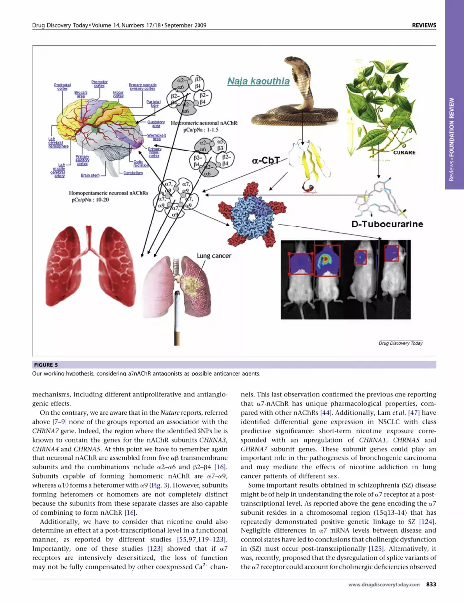
FIGURE 5
Our working hypothesis, considering a7nAChR antagonists as possible anticancer agents.
Reviews�FOUNDATION
REVIEW
mechanisms, including different antiproliferative and antiangio-
genic effects.
On the contrary, we are aware that in the Nature reports, referred
above [7–9] none of the groups reported an association with the
CHRNA7 gene. Indeed, the region where the identified SNPs lie is
known to contain the genes for the nAChR subunits CHRNA3,
CHRNA4 and CHRNA5. At this point we have to remember again
that neuronal nAChR are assembled from five ab transmembrane
subunits and the combinations include a2–a6 and b2–b4 [16].
Subunits capable of forming homomeric nAChR are a7–a9,
whereas a10 forms a heteromer with a9 (Fig. 3). However, subunits
forming heteromers or homomers are not completely distinct
because the subunits from these separate classes are also capable
of combining to form nAChR [16].
Additionally, we have to consider that nicotine could also
determine an effect at a post-transcriptional level in a functional
manner, as reported by different studies [55,97,119–123].
Importantly, one of these studies [123] showed that if a7
receptors are intensively desensitized, the loss of function
may not be fully compensated by other coexpressed Ca2+ chan-
nels. This last observation confirmed the previous one reporting
that a7-nAChR has unique pharmacological properties, com-
pared with other nAChRs [44]. Additionally, Lam et al. [47] have
identified differential gene expression in NSCLC with class
predictive significance: short-term nicotine exposure corre-
sponded with an upregulation of CHRNA1, CHRNA5 and
CHRNA7 subunit genes. These subunit genes could play an
important role in the pathogenesis of bronchogenic carcinoma
and may mediate the effects of nicotine addiction in lung
cancer patients of different sex.
Some important results obtained in schizophrenia (SZ) disease
might be of help in understanding the role of a7 receptor at a post-
transcriptional level. As reported above the gene encoding the a7
subunit resides in a chromosomal region (15q13–14) that has
repeatedly demonstrated positive genetic linkage to SZ [124].
Negligible differences in a7 mRNA levels between disease and
control states have led to conclusions that cholinergic dysfunction
in (SZ) must occur post-transcriptionally [125]. Alternatively, it
was, recently, proposed that the dysregulation of splice variants of
the a7 receptor could account for cholinergic deficiencies observed
www.drugdiscoverytoday.com 833

REVIEWS Drug Discovery Today � Volume 14, Numbers 17/18 � September 2009
Review
s�FOUNDATION
REVIEW
in this disease [126]. These data demonstrated that a7 transcrip-
tion is altered in several ways in SZ, suggesting that transcription-
level mechanisms could account, in part, for the impaired choli-
nergic neurotransmission observed in this disease. These observa-
tions imply that if some a7 isoforms are functional, certain
variants, as well as others associated with both the full-length
and duplicated a7 loci, may translate as novel subunits, whereas
others may also function as antisense regulators of a7 expression.
Additionally, they may be regulatory in nature or may contribute
to altered ligand-gated ion channel activity.
All of these findings supported the ‘special’ role played by a7
nicotinic receptors and reinforced our hypothesis that the inhi-
bition of a7-nAChR by antagonists might represent a strategy to
treat NSCLC or MPM (Fig. 4). Various animal venom toxins,
antagonists of AChR such as a-bungarotoxin from the Bungarus
multicinctus snake and conotoxins purified from the venom of
Conus marine snails have previously been used as molecular
tools for the characterization of neuronal and non-neuronal
nAChR in cells [16]. A direct cytotoxic effect of venom compo-
nents against cancer cells has, however, to the best of our
knowledge, never has been investigated and demonstrated. Sev-
eral very early reports, that did not include any mechanistic
aspect [127,128], or naıve (at best) reports described some antic-
ancer activity associated with the snake venoms. Our group,
following a policy of independent research, has a timely estab-
lished interest in the characterization of the therapeutic (antic-
834 www.drugdiscoverytoday.com
ancer) potential of some natural compounds coming from
libraries in Europe, financed exclusively by National Research
Institutes and Universities, among these plant and snake
venoms. In this setting, we have clearly demonstrated that a-
CbT as a result of multiple mechanisms, including different
antiproliferative and antiangiogenic effects, has a potent anti-
tumor effect against NSCLC or MPM (Fig. 5).
The idea to use venom’s peptides as therapeutic agents in the
treatment of human diseases is not new but, only recently, has
become a reality. In this setting, it has to be borne in mind that
snake venom’s derived compounds (peptides, proteins) have been
used as lead molecules to develop novel strategies in antithrom-
botics, antihemorrhagic, antihypertensive or defibrinogenating
therapy [129]. As a consequence of their high selectivity, venom
peptides have proved particularly useful for in vitro and in vivo
proof-of-concept studies. For therapeutic applications, however,
several issues associated with safety, pharmacokinetics and deliv-
ery need to be addressed.
AcknowledgementsThis review is devoted to the memory of Dr Andre Menez,
President of the French Natural History Museum, a world
renowned expert in the molecular and structural biology of
nicotinic and muscarinic receptors and three-finger fold toxins;
and to the memory of Dr Cassian Bon, President of SFET (Societe
Francaise pour l’Etude des Toxines).
References
1 Wistuba, I.I. (2007) Genetics of preneoplasia: lessons from lung cancer. Curr. Mol.
Med. 7, 3–14
2 Robinson, B.W. and Lake, R.A. (2005) Advances in malignant mesothelioma. N.
Engl. J. Med. 353, 1591–1603
3 Palumbo, C. et al. (2008) Molecular targets and targeted therapies for malignant
mesothelioma. Curr. Med. Chem. 15, 855–867
4 Fennell, D.A. et al. (2008) Advances in the systemic therapy of malignant pleural
mesothelioma. Nat. Clin. Pract. Oncol. 5, 136–147
5 Travis, W.D. et al. eds (2008) World Health Organization Classification of Tumours.
Pathology and Genetics of Tumours of the Lung, Pleura, Thymus and Heart, IARC Press,
Lyon
6 Sun, S. et al. (2007) Lung cancer in never smokers—a different disease. Nat. Rev.
Cancer 7, 778–790
7 Amos, C.I. et al. (2008) Genome-wide association scan of tag SNPs identifies a
susceptibility locus for lung cancer at 15q25.1. Nat. Genet. 40, 616–622
8 Thorgeirsson, T.E. et al. (2008) A variant associated with nicotine dependence,
lung cancer and peripheral arterial disease. Nature 452, 638–642
9 Hung, R.J. et al. (2008) A susceptibility locus for lung cancer maps to nicotinic
acetylcholine receptor subunit genes on 15q25. Nature 452, 633–637
10 Weiss, R.B. et al. (2008) A candidate gene approach identifies the CHRNA5-A3-B4
region as a risk factor for age-dependent nicotine addiction. PLoS Genet. 4,
e1000125
11 Bierut, L.J. et al. (2008) Variants in nicotinic receptors and risk for nicotine
dependence. PLoS Genet. 4 (7), e1000125
12 Spitz, M.R. et al. (2008) The CHRNA5-A3 region on chromosome 15q24–25.1 is a
risk factor both for nicotine dependence and for lung cancer. J. Natl. Cancer Inst.
100, 1552–1556
13 Corringer, P.J. et al. (2000) Nicotinic receptors at the amino acid level. Annu. Rev.
Pharmacol. Toxicol. 40, 431–458
14 Sargent, P.B. (1993) The diversity of neuronal nicotinic acetylcholine receptors.
Annu. Rev. Neurosci. 16, 403–443
15 Lukas, R.J. et al. (1999) Current status of the nomenclature for nicotinic
acetylcholine receptors and their subunits. Pharmacol. Rev. 51, 397–401
16 Dani, J.A. and Bertrand, D. (2007) Nicotinic acetylcholine receptors and nicotinic
cholinergic mechanisms of the central nervous system. Annu. Rev. Pharmacol.
Toxicol. 47, 699–729
17 Changeux, J.P. and Edelstein, S.J. (1998) Allosteric receptors after 30 years. Neuron
21, 959–980
18 Chavez-Noriega, L.E. et al. (1997) Pharmacological characterization of
recombinant human neuronal nicotinic acetylcholine receptors h alpha 2 beta 2, h
alpha 2 beta 4, h alpha 3 beta 2, h alpha 3 beta 4, h alpha 4 beta 2, h alpha 4 beta 4
and h alpha 7 expressed in Xenopus oocytes. J. Pharm. Exp. Ther. 280, 346
19 Wang, F. et al. (1996) Assembly of human neuronal nicotinic receptor alpha5
subunits with alpha3, beta2, and beta4 subunits. J. Biol. Chem. 271, 17656
20 Brejc, K. et al. (2001) Crystal structure of an ACh-binding protein reveals the
ligand-binding domain of nicotinic receptors. Nature 411, 269–276
21 Celie, P.H. et al. (2005) Crystal structure of nicotinic acetylcholine receptor
homolog AChBP in complex with an alpha-conotoxin PnIA variant. Nat. Struct.
Mol. Biol. 12, 582
22 Celie, P.H. et al. (2005) Crystal structure of acetylcholine-binding protein from
Bulinus truncatus reveals the conserved structural scaffold and sites of variation in
nicotinic acetylcholine receptors. J. Biol. Chem. 280, 26457
23 Lummis, S.C. et al. (2005) Cis–trans isomerization at a proline opens the pore of a
neurotransmitter-gated ion channel. Nature 438, 248
24 Lee, W.Y. and Sine, S.M. (2005) Principal pathway coupling agonist binding to
channel gating in nicotinic receptors. Nature 438, 243
25 Celie, P.H. et al. (2004) Nicotine and carbamylcholine binding to nicotinic
acetylcholine receptors as studied in AChBP crystal structures. Neuron 41, 907–914
26 Cashin, A.L. et al. (2005) Using physical chemistry to differentiate nicotinic from
cholinergic agonists at the nicotinic acetylcholine receptor. J. Am. Chem. Soc. 127,
350–356
27 Benowitz, N.L. (2003) Basic cardiovascular research and its implications for the
medicinal use of nicotine. J. Am. Coll. Cardiol. 41, 497–498
28 Czernin, J. and Waldherr, C. (2003) Cigarette smoking and coronary blood flow.
Prog. Cardiovasc. Dis. 45, 395–404
29 Pickworth, W.B. and Fant, R.V. (1998) Endocrine effects of nicotine
administration, tobacco and other drug withdrawal in humans.
Psychoneuroendocrinology 23, 131–141
30 Dani, J.A. and de Biasi, M. (2001) Cellular mechanisms of nicotine addiction.
Pharmacol. Biochem. Behav. 70, 439–446
31 Benowitz, N.L. (1992) Cigarette smoking and nicotine addiction. Med. Clin. North
Am. 76, 415–437

Drug Discovery Today � Volume 14, Numbers 17/18 � September 2009 REVIEWS
Reviews�FOUNDATION
REVIEW
32 Buisson, B. and Bertrand, D. (2002) Nicotine addiction: the possible role of
functional upregulation. Trends Pharmacol. Sci. 23, 130–136
33 Monod, J. et al. (1965) On the nature of allosteric transitions: a plausible model.
J. Mol. Biol. 12, 88–118
34 Bertrand, D. and Gopalakrishnan, M. (2007) Allosteric modulation of nicotinic
acetylcholine receptors. Biochem. Pharmacol. 74, 1155–1163
35 Fucile, S. et al. (2003) Fractional Ca2+ current through human neuronal a7
nicotinic acetylcholine receptors. Cell Calcium 34, 205–209
36 Lax, P. et al. (2002) Ca2+ permeability of human heteromeric nAChRs expressed by
transfection in human cells. Cell Calcium 32, 53–58
37 Gilbert, D. et al. (2009) Local and global calcium signals associated with the
opening of neuronal alpha7 nicotinic acetylcholine receptors. Cell Calcium 45 (2),
198–207
38 Fucile, S. (2004) Ca2+ permeability of nicotinic acetylcholine receptors. Cell
Calcium 35, 1–8
39 Wang, H. et al. (2003) Nicotinic acetylcholine receptor alpha7 subunit is an
essential regulator of inflammation. Nature 421, 384–388
40 de Jonge, W.J. and Ulloa, L. (2007) The alpha7 nicotinic acetylcholine receptor as a
pharmacological target for inflammation. Br. J. Pharmacol. 151, 915–929
41 Ulloa, L. (2005) The vagus nerve and the nicotinic anti-inflammatory pathway.
Nat. Rev. Drug Discov. 4, 673–684
42 Maneckjee, R. and Minna, J.D. (1990) Opioid and nicotine receptors affect growth
regulation of human lung cancer cell lines. Proc. Natl. Acad. Sci. U. S. A. 87, 3294–
3298
43 Schuller, H.M. (1994) Carbon dioxide potentiates the mitogenic effects of nicotine
and its carcinogenic derivative, NNK, in normal and neoplastic neuroendocrine
lung cells via stimulation of autocrine and protein kinase C-dependent mitogenic
pathways. Neurotoxicology 15, 877–886
44 Gotti, C. and Clementi, F. (2004) Neuronal nicotinic receptors: from structure to
pathology. Prog. Neurobiol. 74, 363–396
45 Lindstrom, J. (1996) Neuronal nicotinic acetylcholine receptors. Ion Channels 4,
377–450
46 Sharma, G. and Vijayaraghavan, S. (2002) Nicotinic receptor signaling in
nonexcitable cells. J. Neurobiol. 53, 524–534
47 Lam, D.C. et al. (2007) Expression of nicotinic acetylcholine receptor subunit
genes in non-small-cell lung cancer reveals differences between smokers and
nonsmokers. Cancer Res. 67, 4638–4647
48 Paleari, L. et al. (2008) Role of a7-nicotinic acetylcholine receptor in human non
small cell lung cancer proliferation. Cell. Prol. 41, 936–959
49 Racke, K. and Matthiesen, S. (2004) The airway cholinergic system: physiology and
pharmacology. Pulm. Pharmacol. Ther. 17, 181–198
50 Russo, P. et al. (2006) Development of novel therapeutic strategies for lung cancer:
targeting the cholinergic system. Curr. Med. Chem. 13, 3493–3512
51 Olale, F. et al. (1997) Chronic nicotine exposure differentially affects the function
of human alpha3, alpha4, and alpha7 neuronal nicotinic receptor subtypes.
J. Pharmacol. Exp. Ther 283, 675–683
52 Buisson, B. and Bertrand, D. (2001) Chronic exposure to nicotine upregulates the
human (alpha)4((beta)2 nicotinic acetylcholine receptor function. J. Neurosci. 21,
1819–1829
53 Kawai, H. and Berg, D.K. (2001) Nicotinic acetylcholine receptors containing
alpha 7 subunits on rat cortical neurons do not undergo long-lasting inactivation
even when up-regulated by chronic nicotine exposure. J. Neurochem. 78, 1367–
1378
54 Roman, J. et al. (2004) Nicotine and fibronectin expression in lung fibroblasts:
implications for tobacco-related lung tissue remodeling. FASEB J. 18, 1436–1438
55 Wang, Y. et al. (2001) Human bronchial epithelial and endothelial cells express
alpha7 nicotinic acetylcholine receptors. Mol. Pharmacol. 60, 1201–1209
56 Tsai, J.R. et al. (2006) Mitogen-activated protein kinase pathway was significantly
activated in human bronchial epithelial cells by nicotine DNA and cell biology. 25,
312–322
57 Maus, A.D. et al. (1998) Human and rodent bronchial epithelial cells express
functional nicotinic acetylcholine receptors. Mol. Pharmacol. 54, 779–788
58 Benowitz, N.L. (1986) Clinical pharmacology of nicotine. Annu. Rev Med. 37, 21–32
59 Guo, J. et al. (2005) Persistent nicotine treatment potentiates amplification of the
dihydrofolate reductase gene in rat lung epithelial cells as a consequence of Ras
activation. J. Biol. Chem. 280, 30422–30431
60 West, K.A. et al. (2003) Rapid Akt activation by nicotine and a tobacco carcinogen
modulates the phenotype of normal human airway epithelial cells. J. Clin. Invest.
111, 81–90
61 Dey, R.D. et al. (1981) Morphology, histochemistry, and distribution of serotonin-
containing cells in tracheal epithelium of adult rabbit. Anat. Rec. 199, 23–31
62 Scheuermann, D.W. (1987) Morphology and cytochemistry of the endocrine
epithelial system in the lung. Int. Rev. Cytol. 106, 35–88
63 Freitag, A. et al. (1996) Adrenoceptor- and cholinoceptor-mediated mechanisms in
the regulation of 5-hydroxytryptamine release from isolated tracheae of newborn
rabbits. Br. J. Pharmacol. 119, 91–98
64 Jull, B.A. et al. (2001) Nicotinic receptor-mediated activation by the tobacco-
specific nitrosamine NNK of a Raf-1/MAP kinase pathway, resulting in
phosphorylation of c-myc in human small cell lung carcinoma cells and
pulmonary neuroendocrine cells. J. Cancer Res. Clin. Oncol. 127, 707–717
65 Chen, M.F et al. (1987) Human fetal lung changes associated with maternal
smoking during pregnancy. Pediatr. Pulmonol. 3, 51–58
66 Rehan, V.K. et al. (2005) Mechanism of nicotine-induced pulmonary fibroblast
transdifferentiation. Am. J. Physiol. Lung Cell Mol. Physiol. 289, 667–676
67 Torday, J.S. et al. (2001) Biologic role of fetal lung fibroblast triglycerides as
antioxidants. Pediatr. Res. 49, 843–849
68 Torday, J.S. et al. (1995) Metabolism and fate of neutral lipids of fetal lung
fibroblast origin. Biochim. Biophys. Acta 1254, 198–206
69 Torday, J.S. et al. (2003) The role of fibroblast transdifferentiation in lung epithelial
cell proliferation, differentiation, and repair in vitro. Pediatr. Pathol. Mol. Med. 22,
189–207
70 Leslie, K.O. et al. (1990) Alpha smooth muscle actin expression in developing and
adult human lung. Differentiation 44, 143–149
71 Toti, P. et al. (1997) Bronchopulmonary dysplasia of the premature baby: an
immunohistochemical study. Pediatr. Pulmonol. 24, 22–28
72 Sekhon, H.S. et al. (1999) Prenatal nicotine increases pulmonary alpha7 nicotinic
receptor expression and alters fetal lung development in monkeys. Clin. Invest.
103, 637–647
73 Sekhon, H.S. et al. (2001) Prenatal nicotine exposure alters pulmonary function in
newborn rhesus monkeys. Am. J. Respir. Crit. Care Med. 164, 989–994
74 Sekhon, H.S. et al. (2002) Maternal nicotine exposure upregulates collagen gene
expression in fetal monkey lung. Association with alpha7 nicotinic acetylcholine
receptors. Am. J. Respir. Cell Mol. Biol. 26, 31–41
75 Sekhon, H.S. et al. (2005) Expression of lynx1 in developing lung and its
modulation by prenatal nicotine exposure. Cell Tissue Res. 320, 287–297
76 Ding, L. et al. (2008) Somatic mutations affect key pathways in lung
adenocarcinoma. Nature 455, 1069–1075
77 Song, P. et al. (2003) Acetylcholine is synthesized by and acts as an autocrine
growth factor for small cell lung carcinoma. Cancer Res. 63, 214–221
78 Paleari, L. et al. (2009) Inhibition of non-neuronal a7-nicotinic receptor reduces
tumorigenicity in A549 NSCLC xenografts. Int. J. Cancer 125 (1), 199–211
79 Dasgupta, P. et al. (2006) Nicotine inhibits apoptosis induced by
chemotherapeutic drugs by up-regulating XIAP and survivin. Proc. Natl. Acad. Sci.
U. S. A. 103, 6332–6337
80 Dasgupta, P. et al. (2006) Nicotine induces cell proliferation by beta-arrestin-
mediated activation of Src and Rb-Raf-1 pathways. J. Clin. Invest. 116,
2208–2217
81 Xin, M. et al. (2005) Nicotine inactivation of the proapoptotic function of Bax
through phosphorilation. J. Biol. Chem. 280, 10781–10784
82 Jin, Z. et al. (2004) Nicotine induces multi-site phosphorylation of Bad in
association with suppression of apoptosis. J. Biol. Chem. 279, 23837–
23844
83 Heeschen, C. et al. (2006) Endothelial progenitor cells participate in nicotine-
mediated angiogenesis. J. Am. Coll. Cardiol. 48, 2553–2560
84 Zhu, B.Q. et al. (2003) Second hand smoke stimulates tumor angiogenesis and
growth. Cancer Cell 4, 191–196
85 Gallowitsch-Puerta, M. and Pavlov, V.A. (2007) Neuro-immune interactions via
the cholinergic anti-inflammatory pathway. Life Sci. 80, 2325–2329
86 Rosas-Ballina, M. et al. (2008) Splenic nerve is required for cholinergic
antiinflammatory pathway control of TNF in endotoxemia. Proc. Natl. Acad. Sci.
U. S. A. 105, 11008–11013
87 Su, X. et al. (2007) Activation of the alpha7 nAChR reduces acid-induced acute
lung injury in mice and rats. Am. J. Respir. Cell Mol. Biol. 37, 186–192
88 Lena, D. and Changeux, J.P. (1998) Allosteric nicotinic receptors, human
pathologies. J. Physiol. 92, 63–74
89 Tsetlin, V.I. and Hucho, F. (2004) Snake and snail toxins acting on nicotinic
acetylcholine receptors: fundamental aspects and medical applications. FEBS Lett.
557, 9–13
90 Tsetlin, V.I. (1999) Snake venom alpha-neurotoxins and other ‘three-finger’
proteins. Eur. J. Biochem. 264, 281–286
91 Servent, D. and Menez, A. (2001) In Handbook of Neurotoxicology, (Vol. 1) (Massaro,
E.J., ed.), pp. 385–425, Humana Press Inc., Totowa, NJ
92 Nirthanan, S. and Gwee, M.C. (2004) Three-finger alpha-neurotoxins and the
nicotinic acetylcholine receptor, forty years on. J. Pharmacol. Sci. 94, 1–17
93 Kini, R.M. (2002) Molecular moulds with multiple missions: functional sites in
three-finger toxins. Clin. Exp. Pharmacol. Physiol. 29, 815–822
www.drugdiscoverytoday.com 835
REVIEWS Drug Discovery Today � Volume 14, Numbers 17/18 � September 2009
Review
s�FOUNDATION
REVIEW
94 Fruchart-Gaillard, C. et al. (2002) Experimentally based model of a complex
between a snake toxin and the alpha 7 nicotinic receptor. Proc. Natl. Acad. Sci.
U. S. A. 99, 3216–3221
95 Antil-Delbeke, S. et al. (2000) Molecular determinants by which a long chain toxin
from snake venom interacts with the neuronal alpha 7-nicotinic acetylcholine
receptor. J. Biol. Chem. 275, 29594–29601
96 Michalet, S. et al. (2000) Relative spatial position of a snake neurotoxin
and the reduced disulfide bond alpha (Cys192–Cys193) at the alpha gamma
interface of the nicotinic acetylcholine receptor. J. Biol. Chem. 275, 25608–
25615
97 Carlisle, D.L. et al. (2007) Nicotine activates cell-signaling pathways through
muscle-type and neuronal nicotinic acetylcholine receptors in non-small cell lung
cancer cells. Pulm. Pharmacol. Ther. 20, 629–641
98 Takacs, Z. et al. (2004) Cobra (Naja spp.) nicotinic acetylcholine receptor exhibits
resistance to Erabu sea snake (Laticauda semifasciata) short-chain alpha-
neurotoxin. J. Mol. Evol. 58, 516–526
99 Sher, E. et al. (2000) Peptide neurotoxins, small cell-lung carcinoma and
neurological paraneoplastic syndromes. Biochimie 82, 927–936
100 Pillet, L. et al. (1993) Genetic engineering of snake toxins. Role of invariant
residues in the structural and functional properties of a curaremimetic toxin, as
probed by site-directed mutagenesis. J. Biol. Chem. 268, 909–916
101 Endo, T. and Tamiya, N. (1991) Structure–function relationships of postsynaptic
neurotoxins from snake venoms. In Snake Toxins (Harvey, A.L., ed.), pp. 165–222,
Pergamon Press Inc., New York
102 Taylor, P. et al. (2002) Toxins as probes for structure and specificity of synaptic
target proteins. In Perspectives in Molecular Toxicology (Menez, A., ed.), pp. 271–280,
John Wiley & Sons, Chichester, UK
103 Menez, A. et al. (2002) In Perspectives in Molecular Toxicology, (Vol. 10) (Menez, A.,
ed.), pp. 175–202, John Wiley & Sons, Ltd., Chichester
104 Bourne, Y. et al. (2005) Crystal structure of a Cbtx–AChBP complex reveals
essential interactions between snake a-neurotoxins and nicotinic receptors. EMBO
J. 24, 1512–1522
105 Yi, M. et al. (2008) Spontaneous conformational change and toxin binding in
alpha7 acetylcholine receptor: insight into channel activation and inhibition.
Proc. Natl. Acad. Sci. U. S. A. 105, 8280–8285
106 Fruchart-Gaillard, C. et al. (2006) How three-finger-fold toxins interact with
various cholinergic receptors. J. Mol. Neurosci. 30, 7–8
107 Hogg, R.C. et al. (2003) a-Conotoxins PnIA and [A10L] PnIA stabilize different
states of the a7-L247T nicotinic acetylcholine receptor. J. Biol. Chem. 278, 26908–
26914
108 Dutertre, S. and Lewis, R.J. (2006) Toxin insights into nicotinic acetylcholine
receptors. Biochem. Pharmacol. 72, 661–670
109 Trombino, S. et al. (2004) Alpha7-nicotinic acetylcholine receptors affect growth
regulation of human mesothelioma cells: role of mitogen-activated protein kinase
pathway. Cancer Res. 64, 135–145 [Erratum: Cancer Res. 64, 1559]
110 Trombino, S. et al. (2004) Role of the non-neuronal human cholinergic system in
lung cancer and mesothelioma: possibility of new therapeutic strategies. Curr. Med.
Chem. – Anticancer Agents 4, 535–542
111 Catassi, A. et al. (2008) Multiple roles of nicotine on cell proliferation and
inhibition of apoptosis: implications on lung carcinogenesis. Mutat. Res. Rev. 659
(3), 221–231
112 Grozio, A. et al. (2007) Nicotine, lung and cancer. Anticancer Agents Med. Chem. 7,
461–466
113 Grozio, A. et al. (2008) Natural agents targeting the alpha7-nicotinic-receptor in
NSCLC: a promising prospective in anti-cancer drug development. Int. J. Cancer
122, 1911–1915
836 www.drugdiscoverytoday.com
114 Catassi, A. et al. (2006) a-Cobratoxin, a natural (7-nicotinic receptor antagonist, as
putative anticancer drug in non small cell lung cancer and mesothelioma. In
Toxines and Cancer (Goudey-Perriere et al. eds), pp. 99–106, Lavoisier
115 Catassi, A. et al. (2008) a7-Cobratoxin as a nicotinic receptor-targeted drug for
pleural mesothelioma. Eur. J. Cancer 44, 2296–2311
116 Paleari, L. etal. (2009) Inhibitionofnon-neuronal a7-nicotinic receptoras anticancer
treatment for human NSCLC. Am. J. Resp. Crit. Care Med. 179, 1141–1150
117 Russo, P. et al. (in press) Neuronal Cholinergic Toxines: possible drugs for lung
cancer treatment. In Toxines et fonctions cholinergiques neuronales et non
neuronales (Goudey-Perriere et al., eds). Lavoisier, pp. 63–67
118 Stevens, K.E. et al. (1996) Genetic correlation of inhibitory gating of hippocampal
auditory evoked response and alpha-bungarotoxin-binding nicotinic cholinergic
receptors in inbred mouse strains. Neuropsychopharmacology 15, 152–162
119 Sheppard, B.J. et al. (2000) Activation of voltage-operated Ca2+-channels in human
small cell lung carcinoma by the tobacco-specific nitrosamine 4-
(methylnitrosamino)-1-(3-pyridyl)-1-butanone. Int. J. Oncol. 16, 513–518
120 Cattaneo, M.G. et al. (1993) Nicotine stimulates a serotonergic autocrine loop in
human small-cell lung carcinoma. Cancer. Res. 53, 5566–5568
121 Nelson, M.E. et al. (2003) Alternate stoichiometries of alpha4beta2 nicotinic
acetylcholine receptors. Mol. Pharmacol. 63, 332–341
122 Quik, M. et al. (1994) a-Bungarotoxin blocks the nicotinic receptor mediated
increase in cell number in a neuroendocrine cell line. Brain Res. 655, 161–167
123 Tsuneki, H. et al. (2007) Novel G423S mutation of human alpha7 nicotinic
receptor promotes agonist-induced desensitization by a protein kinase C-
dependent mechanism. Mol. Pharmacol. 71, 777–786
124 De Luca, V. et al. (2006) Regulation of alpha7-nicotinic receptor subunit and
alpha7-like gene expression in the prefrontal cortex of patients with bipolar
disorder and schizophrenia. Acta Psychiatr. Scand. 114, 211–215
125 Leonard, S. and Freedman, R. (2006) Genetics of chromosome 15q13–q14 in
schizophrenia. Biol. Psychiatry 60, 115–122
126 Severance, E.G. et al. (2004) The alpha7 nicotinic acetylcholine receptor subunit
exists in two isoforms that contribute to functional ligand-gated ion channels.
Mol. Pharmacol. 66, 420–429
127 Selinger, H. et al. (1951) New results with cobratoxin in caner therapy. Med. Welt.
20, 638
128 Calmette, A. et al. (1933) Effetts du venim de cobra sur les greffles cancereuses et sur
le cancer spontane (adeno-carcinome) de la souris. C. R. Acad. Sci. 197–205
129 Lewis, R.J. and Garcia, M.L. (2003) Therapeutic potential of venom peptides. Nat.
Drug Discov. 2, 791–802
130 Proskocil, B.J. et al. (2004) Acetylcholine is an autocrine or paracrine hormone
synthesized and secreted by airway bronchial epithelial cells. Endocrinology 145,
2498–2506
131 Arredondo, J. et al. (2006) The nicotinic receptor antagonists abolish pathobiologic
effects of tobacco-derived nitrosamines on BEP2D cells. J. Cancer Res. Clin. Oncol.
132, 653–663
132 Tsurutani, J. et al. (2005) Tobacco components stimulate Akt-dependent
proliferation and NFkappaB-dependent survival in lung cancer cells. Carcinogen 26,
1182–1195
133 Plummer, H.K., III et al. (2005) Expression of the alpha-7 nicotinic acetylcholine
receptor in human lung cells. Respir. Res. 6, 29
134 Chini, B. et al. (1992) Neuronal-type alpha-bungarotoxin receptors and the alpha
5-nicotinic subunit gene are expressed in neuronal and nonneuronal human cell
lines. Proc. Natl. Acad. Sci. USA 89 (5), 1572–1576
135 Zhang, Q. et al. (2007) Nicotine induces hypoxia-inducible factor-1alpha
expression in human lung cancer cells via nicotinic acetylcholine receptor
mediated signaling pathways. Clin. Cancer Res. 13, 4686–4694


![[Malignant mesothelioma in garment sewing-machine workers]](https://img.pdfslide.net/doc/110x75/634ef3e02630aed500083a34/malignant-mesothelioma-in-garment-sewing-machine-workers.jpg)


