Embed Size (px)
Citation preview
A Capillary Electrophoresis Sequencing Methodfor the Identification of Mutations in the Inverted Terminal
Repeats of Adeno-Associated Virus
Cameron Mroske,1 Hector Rivera,2 Taihra Ul-Hasan,3 Saswati Chatterjee,3 and K.K. Wong1
Abstract
Inverted terminal repeat (ITR) integrity is critical for the replication, packaging, and transduction of recombinantadeno-associated virus (rAAV), a promising gene therapy vector. Because AAV ITRs possess 70% GC contentand are palindromic, they are notoriously difficult to sequence. The purpose of this work was to develop areliable ITR sequencing method. The ITRs of two molecular clones of AAV2, pTZAAV and pAV2, were (1)sequenced directly from plasmid DNA in the presence of denaturant (direct sequencing method, DSM) or (2)first amplified in a reaction in which 7-deaza-dGTP was substituted for dGTP and the resultant amplificationproduct sequenced (amplification sequencing method, ASM). The DSM and ASM techniques yielded clearchromatograms, read through the ITR hairpin, and revealed hitherto unreported mutations in each ITR.pTZAAV and pAV2 possess identical mutations at the upstream MscI site of the 5¢ ITR (T > G, nt 2) and thedownstream MscI site of the 3¢ ITR (del. nt 4672–4679). The chromatograms for pAV2 also revealed that the ITRsof this construct were arranged in a FLOP/FLOP orientation. In addition, the DSM was successfully employed torecover ITR–chromosomal junction sequences from a variety of rAAV-transduced tissue types. Both the DSMand ASM can be employed to sequence through the AAV ITR hairpin, and both techniques reliably detectmutations in the ITR. Because the DSM and ASM offer a way to verify ITR integrity, they constitute powerfultools for the process development of rAAV gene therapy.
Introduction
The clinical success of adeno-associated virus (AAV)-mediated gene therapy for the treatment of hemophilia B
demonstrates that AAV-based vectors have the potential to bea transformative medicine (Nathwani et al., 2011). AAV is asmall, nonenveloped single-stranded DNA virus that is con-sidered attractive for therapeutic gene transfer because it isnonpathogenic and occurs naturally in a large number of se-rotypes that may confer selective tropism to specific tissues(Michelfelder and Trepel, 2009). In addition to hemophilia B,AAV gene therapy vectors have been used successfully totreat Leber’s congenital amaurosis (LCA) (Bainbridge et al.,2008; Cideciyan et al., 2008; Hauswirth et al., 2008; Maguireet al., 2008; Simonelli et al., 2010) and Parkinson’s disease(Feigin et al., 2007; Kaplitt et al., 2007; LeWitt et al., 2011). In all,there are currently 38 AAV protocols approved by the Re-combinant DNA Advisory Committee (National Institutes ofHealth, Bethesda, MD) and U.S. Food and Drug Adminis-
tration (Silver Spring, MD) for the treatment of diseasesranging from cystic fibrosis to rheumatoid arthritis (Daya andBerns, 2008; Mingozzi and High, 2011).
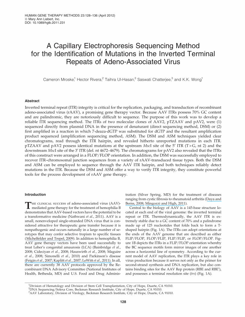
Central to the biology of AAV is a 145-base structure lo-cated at each end of the viral genome: the inverted terminalrepeat or ITR. Thermodynamically, the AAV ITR is ex-tremely stable due to a GC content of 70% and a palindromemade up of 125 nucleotides that folds back to form a T-shaped hairpin (Fig. 1A). The ITRs can adopt orientations atthe ends of the AAV genome that are described as eitherFLIP/FLOP, FLOP/FLIP, FLIP/FLIP, or FLOP/FLOP. Fig-ure 1B depicts the ITRs in a FLIP/FLOP orientation wherebythe BC sequence motifs form mirror images of one anotheracross a horizontal line of symmetry. According to the cur-rent model of AAV replication, the ITR plays a key role invirus production because it serves not only as the primer forsecond-strand synthesis and DNA replication, but also con-tains binding sites for the AAV Rep protein (RBE and RBE¢),and possesses a terminal resolution site (trs) (Fig. 1A).
1Division of Hematology and Division of Stem Cell Transplantation, City of Hope, Duarte, CA 91010.2DNA Sequencing/Solexa Core, Beckman Research Institute, City of Hope, Duarte, CA 91010.3AAV Laboratory, Division of Virology, Beckman Research Institute, City of Hope, Duarte, CA 91010.
HUMAN GENE THERAPY METHODS 23:128–136 (April 2012)ª Mary Ann Liebert, Inc.DOI: 10.1089/hgtb.2011.231
128
Importantly, rAAV vectors used for gene therapy do notcontain the endogenous rep and cap genes but instead pos-sess a therapeutic transgene expression cassette, located be-tween the ITRs, which act as cis signals for packaging andtransduction (Fig. 1C). Because the presence of intact ITRs iscritical for the production of high-titer, fully functionalrAAV, and given the fact that it is not uncommon for eachITR to mutate when present in plasmid form, it is criticallyimportant to develop a rapid, low-cost, high-resolutionprotocol to verify ITR sequence content.
Traditionally, ITR integrity and orientation have beenverified through restriction analysis; however, this techniquehas several drawbacks. Restriction mapping provides onlylimited information regarding the nature of the mutation,and interpretation of the restriction pattern, which is oftenequivocal, can be time-consuming. Thus, without accurateknowledge of the nature and extent of sequence change(point mutation vs. microdeletion), it may be difficult tomake an informed decision as to whether a particular clonewill be suitable for vector production. For these reasons, oneor more methods to sequence through the AAV ITR wouldbe advantageous. It has been asserted that the only way tosequence through the hairpin structure of the AAV ITR is to
perform a two-step protocol whereby the hairpin is ampli-fied in the presence of 7-deaza-dGTP before sequencing(Kieleczawa, 2006). Not only did we want to substantiate thisclaim, because of the paucity of ITR sequencing chromato-grams in the literature, we also wished to have a more directmethod for sequencing the AAV ITR. Moreover, our objectivehas been to develop a rapid, consistent, and accurate methodto sequence each AAV ITR, using contemporary fluorescencecapillary electrophoresis sequencing technology.
By taking advantage of developments in amplification andsequencing chemistry for high GC content DNA (Henkeet al., 1997; Kieleczawa, 2006; Musso et al., 2006), we havebeen able to develop/adapt two independent methods tosequence the AAV ITR. Herein we show that direct se-quencing of each ITR on AAV plasmids (direct sequencingmethod, or DSM), and sequencing of an amplificationproduct containing a complete 7-deaza-dGTP substitutionfor dGTP (amplification sequencing method, or ASM), pro-duce noise-free chromatograms that reveal mutations inconcordance with the respective restriction patterns. In con-trast to a previous report (Kieleczawa, 2006), we find that theDSM reads through the ITR hairpin region as well as theASM. Using both the DSM and ASM, we demonstrate that
FIG. 1. Inverted terminalrepeat (ITR) structure. (A)Secondary structure of theAAV-2 3¢ ITR (GenBank ac-cession number NC_001401).Shown are the Rep-bindingelements RBE (GAGCGAGCGAGCGCGC) and RBE¢(CTTTG), as well as the ter-minal resolution site (trs,GTTGG). (B) Representationof the AAV-2 genome (dia-gram not according to scale).In the case of wild-type AAV-2 (GenBank accession numberNC_001401), the 5¢ ITR shownat right adopts a FLIP config-uration whereas the 3¢ ITR atleft adopts a FLOP orienta-tion, so that the BC sequencemotifs, as well as the SmaIrestriction sites, mirror eachother across a horizontal lineof symmetry (red line). (C)Schematic of a typical rAAVcassette containing the ele-ments necessary to drivetransgene expression.
DSM/ASM SEQUENCING OF AAV ITR 129
the infectious clone pTZAAV (Chatterjee, 1992), as well asclone pAV2 from the American Type Culture Collection(ATCC, Manassas, VA) (Laughlin et al., 1983), possess iden-tical mutations in the upstream MscI site of the 5¢ ITR (T > G,nt 2) and the downstream MscI site of the 3¢ ITR (Del.GTGGC/A, nt 4672–4679). In contrast to pTZAAV, wedemonstrate that the ITRs of pAV2 are arranged in a FLOP/FLOP orientation. As well, we have successfully employedthe DSM to retrieve chromosomal junction sequences frommouse liver transduced with a novel rAAV vector, AAV-HSC15 (Aravind et al., 2012). We provide an example inwhich the DSM clearly shows that rAAV integrated intomurine chromosome 2 at position 134373802 in the minusorientation. Future experiments will be necessary to deter-mine how the particular ITR mutations identified in thisstudy affect AAV packaging efficiency. Nevertheless, thedirect sequencing and amplification sequencing methods areaccurate and consistent in verifying ITR integrity and shouldprove useful in the process of large-scale rAAV vector pro-duction for gene therapy.
Materials and Methods
Plasmid DNA
Plasmid DNA for construct pTZAAV was isolated from a200-ml overnight Escherichia coli DH5a culture, using a Qiagenplasmid maxi kit (cat. no. 12163; Qiagen, Valencia, CA). Con-struct pAV2 was purchased from the ATCC (no. 37216) as alyophilized E. coli HB101 pellet and reconstituted according tothe manufacturer’s instructions. In brief, the bacterial pellet wasdissolved in 5 ml of Luria broth containing ampicillin (50lg/ml) and grown overnight at 37�C. The next day, 250ll ofovernight culture was seeded into 200 ml of fresh Luria broth–ampicillin and grown for 20 hr at 37�C in a shaker incubator.Plasmid DNA was then isolated according to the Qiagen maxi-prep protocol. For both pTZAAV and pAV2, isolated plasmidDNA was dissolved in sterile Tris–EDTA buffer.
Primers
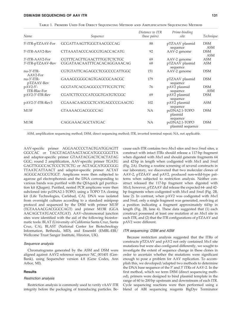
Primers used for PCR amplification and cycle sequencingare listed in Table 1. DNA primers were chemically synthe-sized by Integrated DNA Technologies (San Diego, CA) andpurified by standard desalting.
Restriction analysis
Restriction enzymes were purchased from New EnglandBioLabs (Ipswich, MA). One microgram of plasmid DNAwas digested with MscI at 37�C, or with MscI at 37�C fol-lowed by SmaI at 25�C. After digestion, about 400 ng ofenzyme-treated DNA was resolved on a 4% agarose–0.5 · TBE gel (TBE: 45 mM Tris, 45 mM borate, 1 mM EDTA)and imaged by staining in 3 · GelRed solution (cat. no. 41002;Biotium, Hayward, CA) for 1 hr.
PCR amplification: ASM
ASM templates were generated by amplifying the 5¢ and 3¢ITRs of the AAV2 genome carried on plasmids pTZAAV andpAV2 in a manner such that 7-deaza-dGTP was completelysubstituted for dGTP. 7-Deaza-dGTP solution was pur-chased (cat. no. 10988537001; Roche Applied Science, Man-
nheim, Germany) and mixed at an equimolar concentrationwith dATP, dTTP, and dCTP in order to constitute a 7-deaza-dGTP–dNTP stock. Each amplification reaction consisted of20 ng of plasmid template, 1 · cloned Pfu (Pyrococcus furiosus)reaction buffer, forward and reverse primers at a final con-centration 0.4 lM, 7-deaza-dGTP–dNTP at a final concen-tration of 250 lM, and 2.5 U of PfuTurbo DNA polymerase(cat. no. 600250; Stratagene/Agilent Technologies, La Jolla,CA). Reactions were performed in a total volume of 25 ll ona Bio-Rad MyCycler thermocycler (cat. no. 170-9701; Bio-Rad, Hercules, CA) according to the following program:96�C for 2 min (1 cycle)/96�C for 45 sec, 50�C for 45 sec, 72�Cfor 1 min (30 cycles)/72�C for 10 min (1 cycle)/4�C (hold).Resultant amplicons were purified with a Qiagen QIAquickPCR purification kit (cat. no. 28104). DNA was eluted in10 mM Tris, pH 8.5, and quantified on a NanoDrop 1000spectrophotometer (Thermo Scientific, Wilmington, DE).
Cycle sequencing: ASM and DSM
Before cycle sequencing, 2 ng of purified amplificationproduct (ASM) or 200 ng of plasmid DNA (DSM) wasdiluted to a volume of 7 ll in 10 mM Tris, pH 8.0, and de-natured at 98�C for 10 min before being immediately snap-cooled on ice. The following reagents were then added toeach denatured sample for a total reaction volume of 14.5 llper sample: 1 ll of 5 lM sequencing primer, 2 ll of BigDyeTerminator version 3.1, 1 ll of BigDye Terminator dGTPversion 3.0, 1.5 ll of 5 · DNA sequencing buffer (AppliedBiosystems/Life Technologies, Foster City, CA), and 2 ll of 5M betaine solution (Sigma-Aldrich, St. Louis, MO). Sampleswere then subjected to cycle sequencing in a Veriti thermo-cycler (Applied Biosystems/Life Technologies) according tothe following program: 96�C for 1 min (1 cycle)/96�C for10 sec, 50�C for 5 sec, 60�C for 75 sec (25 cycles)/4�C (hold).After thermocycling, samples were purified with a BigDyeXterminator purification kit and electrophoresed on a 3730DNA analyzer (Applied Biosystems/Life Technologies)equipped with a 50-cm capillary array filled with POP-7polymer and 1 · capillary electrophoresis buffer. Purifiedsamples were injected electrokinetically at 1.2 kV and elec-trophoresed at 8.5 kV for 1.56 hr with a current stability of3.0 lA and an oven temperature of 60�C.
Determination of AAV–chromosomaljunction sequences
Genomic DNA was isolated from the livers of mice thathad been injected with the novel rAAV vector, AAV-HSC15.Liver tissue was suspended in digestion buffer (10 mM NaCl,10 mM Tris-HCl [pH 8.0], 25 mM EDTA [pH 8.0]) along withRNase (Roche, Indianapolis, IN) and incubated on a rotatingrack for 2 hr at 37�C. Sodium dodecyl sulfate (SDS) andproteinase K were then added to final concentrations of 0.5%and 0.1 mg/ml, respectively, whereupon samples were fur-ther incubated for 16 hr at 56�C. Genomic DNA was isolatedfrom the reactions through phenol–chloroform/ethanol pu-rification and digested with either RsaI or DraI before un-dergoing an additional round of purification. Using T4ligase, GenomeWalker adaptors (Clontech, Mountain View,CA) were ligated to the digested product for 16 hr at 16�Cand integration junctions were amplified through nestedPCR using the following primer pairs: round 1 amplification,
130 MROSKE ET AL.
AAV-specific primer AGGAACCCCTAGTGATGGAGTTGGCCAC or TACGTAGATAAGTAGCATGGCGGGTTAand adaptor-specific primer GTAATACGACTCACTATAGGGC; round 2 amplification, AAV-specific primer TGATGGAGTTGGCCACTCCCTCTCTG or AGTAGCATGGCGGGTTAATCATTAACT and adaptor-specific primer ACTATAGGGCACGCGTGGT. Amplicons were then subjected toagarose gel electrophoresis and the DNA corresponding tovarious bands was purified with the QIAquick gel purifica-tion kit (Qiagen). Purified, nested PCR amplicons were thensubcloned into pcDNA2.1-TOPO, using a TOPO TA cloningkit (Life Technologies, Carlsbad, CA). DNA was isolatedfrom overnight cultures according to a standard miniprepprotocol and sequenced by the DSM with primer M13F(TGTAAAACGACGGCCAGT) and primer M13R (GGAAACAGCTATGACCATGAT). AAV–chromosomal junctionsites were identified with the aid of the following bioinfor-matic tools: BLAT (University of California Santa Cruz, SantaCruz, CA), BLAST (National Center for BiotechnologyInformation, Bethesda, MD), and Ensembl (EMBL-EBI/Wellcome Trust Sanger Institute, Hinxton, UK).
Sequence analysis
Chromatograms generated by the ASM and DSM werealigned against AAV2 reference sequence NC_001401 (Gen-Bank), using Sequencher version 4.8 (Gene Codes, AnnArbor, MI).
Results
Restriction analysis
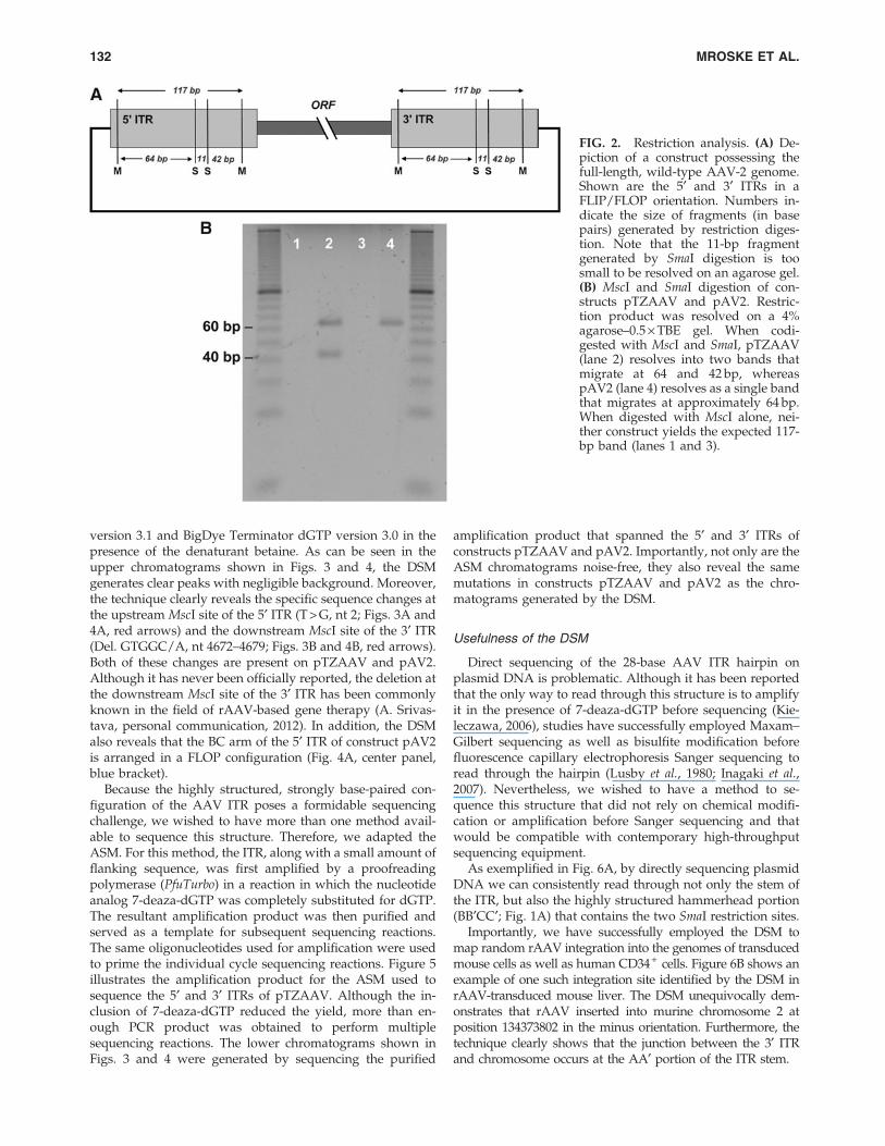
Restriction analysis is commonly used to verify rAAV ITRintegrity before the packaging of transducing particles. Be-
cause each ITR contains two MscI sites and two SmaI sites, aconstruct with intact ITRs should release a 117-bp fragmentwhen digested with MscI and should generate fragments 64and 42 bp in length when codigested with MscI and SmaI(Fig. 2A). During a routine screening of several constructs inour laboratory, we discovered that two molecular clones ofAAV-2, pTZAAV and pAV2, produced non-wild-type pat-terns when subjected to restriction analysis. Neither con-struct released the 117-bp fragment when digested withMscI; however, pTZAAV did release the expected 64- and 42-bp fragments when codigested with MscI and SmaI (Fig. 2B,lane 2). In contrast, when pAV2 was codigested with MscIand SmaI, only a single fragment was generated, resolving ata position indicating a fragment approximately 64 bp inlength (Fig. 2B, lane 4). These data suggested that (1) eachconstruct possessed at least one mutation at an MscI site ineach ITR, and (2) that the ITR configurations of pTZAAV andpAV2 were different.
ITR sequencing: DSM and ASM
Because restriction analysis suggested that the ITRs ofconstructs pTZAAV and pAV2 not only contained MscI sitemutations but were also configured differently, we sought toinvestigate the extent of sequence change in these clones inorder to ascertain whether the mutations were significantenough to pose a problem for AAV replication. To accom-plish this, we developed/adapted two methods to determinethe DNA base sequence of the 5¢ and 3¢ ITRs of AAV-2. In thefirst method, which we term DSM (direct sequencing meth-od), primers were designed to bind plasmid template in therange of 60 to 200 bp upstream and downstream of each ITR.Cycle sequencing reactions were then performed using ablend of ABI sequencing reagents BigDye Terminator
Table 1. Primers Used For Direct Sequencing Method and Amplification Sequencing Method
Name SequenceDistance to ITR
(base pairs)Prime-binding
site Technique
5¢-ITR-pTZAAV-For GCGATTAAGTTGGGTAACGCCAG 88 pTZAAV plasmidsequence
DSMASM
5¢-ITR-AAV2-Rev CTTAAATACCCAGCGTGACCACATG 92 AAV-2 genome DSMASM
3¢-ITR-AAV2-For CGTTTCAGTTGAACTTTGGTCTCTGC 69 AAV-2 genome ASM3¢-ITR-pTZAAV-Rev CGGATAACAATTTCACACAGGAAACAG 68 pTZAAV plasmid
sequenceASM
nu-3¢-ITR-AAV2-For
CGTGTATTCAGAGCCTCGCCCCATTGGC 151 AAV-2 genome DSM
nu-3¢-ITR-pTZAAV-Rev
GAAAGCGGGCAGTGAGCGCAACGC 179 pTZAAV plasmidsequence
DSM
pAV2-5¢-ITR-Blac-For
GCGTATCACGAGGCCCTTTCGTCTTC 76 pAV2 plasmidsequence
DSMASM
pAV2-3¢-ITR-Rev CGATCTTCCCCATCGGTGATGTCGGC 69 pAV2 plasmidsequence
DSM
pAV2-3¢-ITR-Rev3 CGAAACAAGCGCTCATGAGCCCGAAGTG 102 pAV2 plasmidsequence
ASM
M13F GTAAAACGACGGCCAG NA pcDNA2.1-TOPOplasmidsequence
DSM
M13R CAGGAAACAGCTATGAC NA pcDNA2.1-TOPOplasmid sequence
DSM
ASM, amplification sequencing method; DSM, direct sequencing method; ITR, inverted terminal repeat; NA, not applicable.
DSM/ASM SEQUENCING OF AAV ITR 131
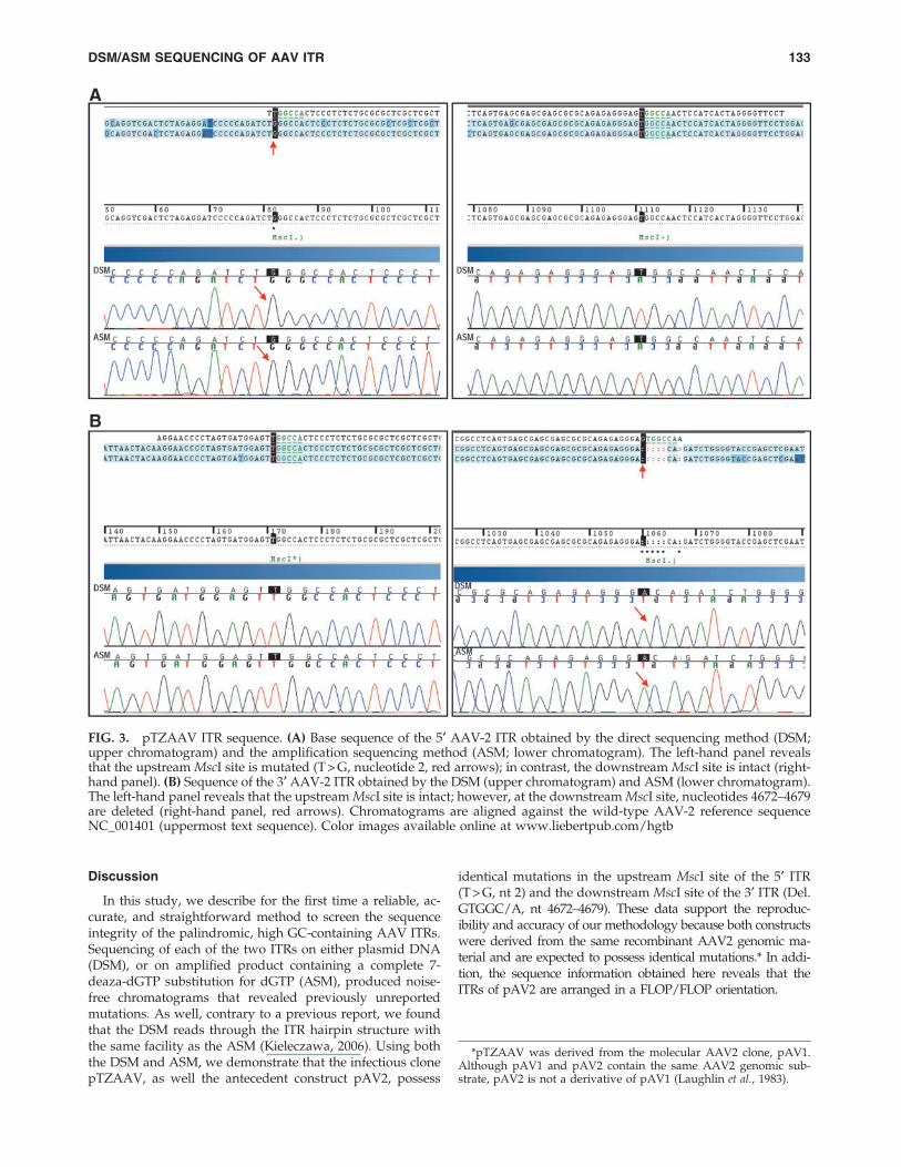
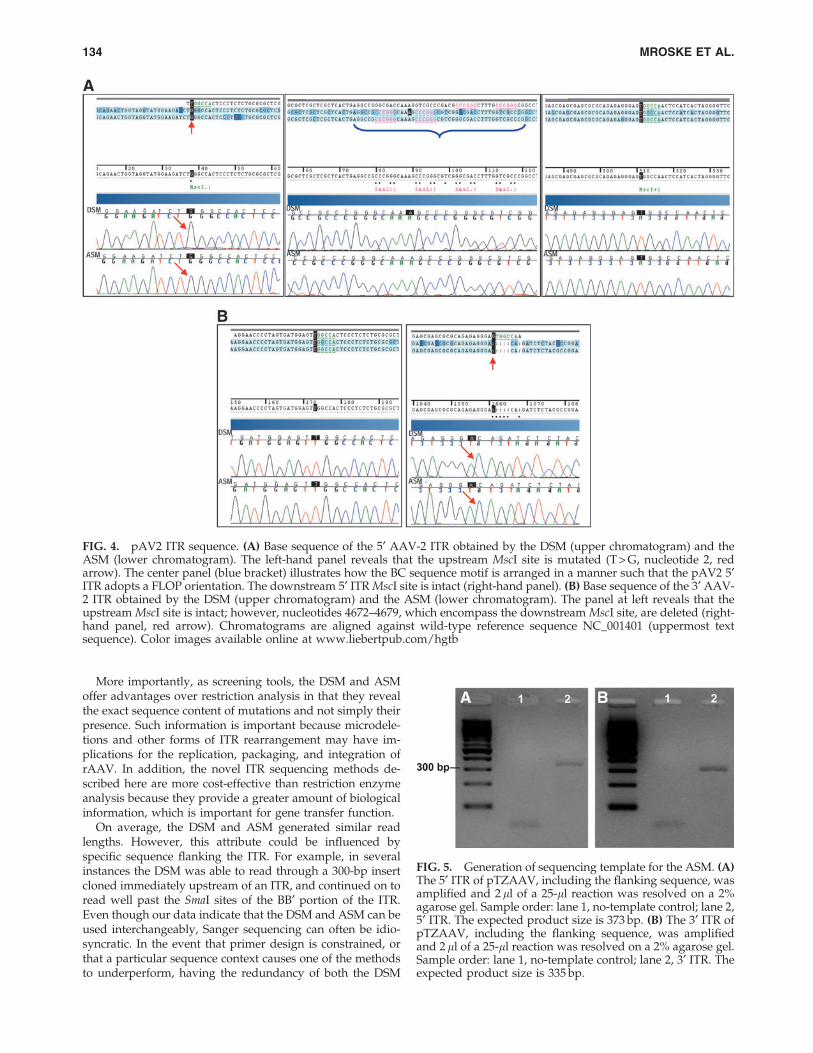
version 3.1 and BigDye Terminator dGTP version 3.0 in thepresence of the denaturant betaine. As can be seen in theupper chromatograms shown in Figs. 3 and 4, the DSMgenerates clear peaks with negligible background. Moreover,the technique clearly reveals the specific sequence changes atthe upstream MscI site of the 5¢ ITR (T > G, nt 2; Figs. 3A and4A, red arrows) and the downstream MscI site of the 3¢ ITR(Del. GTGGC/A, nt 4672–4679; Figs. 3B and 4B, red arrows).Both of these changes are present on pTZAAV and pAV2.Although it has never been officially reported, the deletion atthe downstream MscI site of the 3¢ ITR has been commonlyknown in the field of rAAV-based gene therapy (A. Srivas-tava, personal communication, 2012). In addition, the DSMalso reveals that the BC arm of the 5¢ ITR of construct pAV2is arranged in a FLOP configuration (Fig. 4A, center panel,blue bracket).
Because the highly structured, strongly base-paired con-figuration of the AAV ITR poses a formidable sequencingchallenge, we wished to have more than one method avail-able to sequence this structure. Therefore, we adapted theASM. For this method, the ITR, along with a small amount offlanking sequence, was first amplified by a proofreadingpolymerase (PfuTurbo) in a reaction in which the nucleotideanalog 7-deaza-dGTP was completely substituted for dGTP.The resultant amplification product was then purified andserved as a template for subsequent sequencing reactions.The same oligonucleotides used for amplification were usedto prime the individual cycle sequencing reactions. Figure 5illustrates the amplification product for the ASM used tosequence the 5¢ and 3¢ ITRs of pTZAAV. Although the in-clusion of 7-deaza-dGTP reduced the yield, more than en-ough PCR product was obtained to perform multiplesequencing reactions. The lower chromatograms shown inFigs. 3 and 4 were generated by sequencing the purified
amplification product that spanned the 5¢ and 3¢ ITRs ofconstructs pTZAAV and pAV2. Importantly, not only are theASM chromatograms noise-free, they also reveal the samemutations in constructs pTZAAV and pAV2 as the chro-matograms generated by the DSM.
Usefulness of the DSM
Direct sequencing of the 28-base AAV ITR hairpin onplasmid DNA is problematic. Although it has been reportedthat the only way to read through this structure is to amplifyit in the presence of 7-deaza-dGTP before sequencing (Kie-leczawa, 2006), studies have successfully employed Maxam–Gilbert sequencing as well as bisulfite modification beforefluorescence capillary electrophoresis Sanger sequencing toread through the hairpin (Lusby et al., 1980; Inagaki et al.,2007). Nevertheless, we wished to have a method to se-quence this structure that did not rely on chemical modifi-cation or amplification before Sanger sequencing and thatwould be compatible with contemporary high-throughputsequencing equipment.
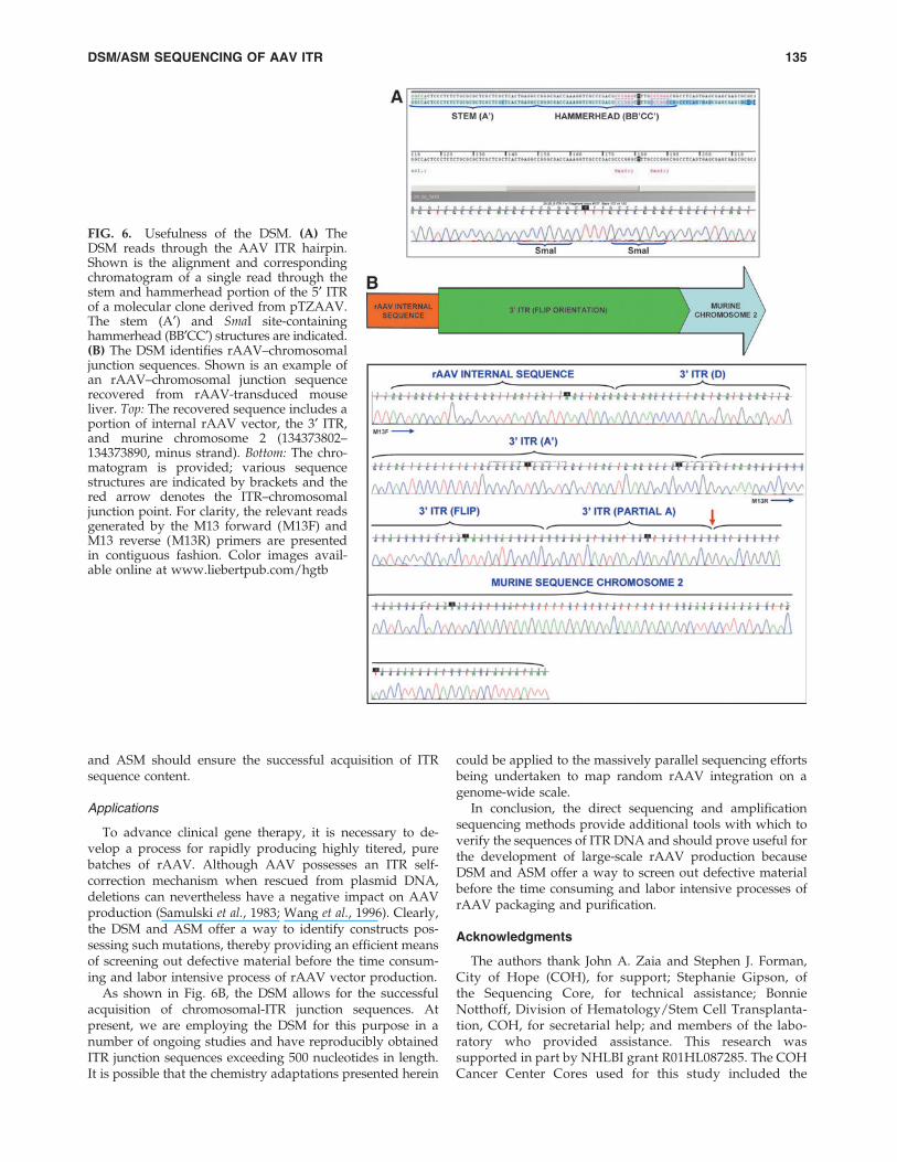
As exemplified in Fig. 6A, by directly sequencing plasmidDNA we can consistently read through not only the stem ofthe ITR, but also the highly structured hammerhead portion(BB¢CC¢; Fig. 1A) that contains the two SmaI restriction sites.
Importantly, we have successfully employed the DSM tomap random rAAV integration into the genomes of transducedmouse cells as well as human CD34 + cells. Figure 6B shows anexample of one such integration site identified by the DSM inrAAV-transduced mouse liver. The DSM unequivocally dem-onstrates that rAAV inserted into murine chromosome 2 atposition 134373802 in the minus orientation. Furthermore, thetechnique clearly shows that the junction between the 3¢ ITRand chromosome occurs at the AA¢ portion of the ITR stem.
FIG. 2. Restriction analysis. (A) De-piction of a construct possessing thefull-length, wild-type AAV-2 genome.Shown are the 5¢ and 3¢ ITRs in aFLIP/FLOP orientation. Numbers in-dicate the size of fragments (in basepairs) generated by restriction diges-tion. Note that the 11-bp fragmentgenerated by SmaI digestion is toosmall to be resolved on an agarose gel.(B) MscI and SmaI digestion of con-structs pTZAAV and pAV2. Restric-tion product was resolved on a 4%agarose–0.5 · TBE gel. When codi-gested with MscI and SmaI, pTZAAV(lane 2) resolves into two bands thatmigrate at 64 and 42 bp, whereaspAV2 (lane 4) resolves as a single bandthat migrates at approximately 64 bp.When digested with MscI alone, nei-ther construct yields the expected 117-bp band (lanes 1 and 3).
132 MROSKE ET AL.
Discussion
In this study, we describe for the first time a reliable, ac-curate, and straightforward method to screen the sequenceintegrity of the palindromic, high GC-containing AAV ITRs.Sequencing of each of the two ITRs on either plasmid DNA(DSM), or on amplified product containing a complete 7-deaza-dGTP substitution for dGTP (ASM), produced noise-free chromatograms that revealed previously unreportedmutations. As well, contrary to a previous report, we foundthat the DSM reads through the ITR hairpin structure withthe same facility as the ASM (Kieleczawa, 2006). Using boththe DSM and ASM, we demonstrate that the infectious clonepTZAAV, as well the antecedent construct pAV2, possess
identical mutations in the upstream MscI site of the 5¢ ITR(T > G, nt 2) and the downstream MscI site of the 3¢ ITR (Del.GTGGC/A, nt 4672–4679). These data support the reproduc-ibility and accuracy of our methodology because both constructswere derived from the same recombinant AAV2 genomic ma-terial and are expected to possess identical mutations.* In addi-tion, the sequence information obtained here reveals that theITRs of pAV2 are arranged in a FLOP/FLOP orientation.
FIG. 3. pTZAAV ITR sequence. (A) Base sequence of the 5¢ AAV-2 ITR obtained by the direct sequencing method (DSM;upper chromatogram) and the amplification sequencing method (ASM; lower chromatogram). The left-hand panel revealsthat the upstream MscI site is mutated (T > G, nucleotide 2, red arrows); in contrast, the downstream MscI site is intact (right-hand panel). (B) Sequence of the 3¢ AAV-2 ITR obtained by the DSM (upper chromatogram) and ASM (lower chromatogram).The left-hand panel reveals that the upstream MscI site is intact; however, at the downstream MscI site, nucleotides 4672–4679are deleted (right-hand panel, red arrows). Chromatograms are aligned against the wild-type AAV-2 reference sequenceNC_001401 (uppermost text sequence). Color images available online at www.liebertpub.com/hgtb
*pTZAAV was derived from the molecular AAV2 clone, pAV1.Although pAV1 and pAV2 contain the same AAV2 genomic sub-strate, pAV2 is not a derivative of pAV1 (Laughlin et al., 1983).
DSM/ASM SEQUENCING OF AAV ITR 133
More importantly, as screening tools, the DSM and ASMoffer advantages over restriction analysis in that they revealthe exact sequence content of mutations and not simply theirpresence. Such information is important because microdele-tions and other forms of ITR rearrangement may have im-plications for the replication, packaging, and integration ofrAAV. In addition, the novel ITR sequencing methods de-scribed here are more cost-effective than restriction enzymeanalysis because they provide a greater amount of biologicalinformation, which is important for gene transfer function.
On average, the DSM and ASM generated similar readlengths. However, this attribute could be influenced byspecific sequence flanking the ITR. For example, in severalinstances the DSM was able to read through a 300-bp insertcloned immediately upstream of an ITR, and continued on toread well past the SmaI sites of the BB¢ portion of the ITR.Even though our data indicate that the DSM and ASM can beused interchangeably, Sanger sequencing can often be idio-syncratic. In the event that primer design is constrained, orthat a particular sequence context causes one of the methodsto underperform, having the redundancy of both the DSM
FIG. 4. pAV2 ITR sequence. (A) Base sequence of the 5¢ AAV-2 ITR obtained by the DSM (upper chromatogram) and theASM (lower chromatogram). The left-hand panel reveals that the upstream MscI site is mutated (T > G, nucleotide 2, redarrow). The center panel (blue bracket) illustrates how the BC sequence motif is arranged in a manner such that the pAV2 5¢ITR adopts a FLOP orientation. The downstream 5¢ ITR MscI site is intact (right-hand panel). (B) Base sequence of the 3¢ AAV-2 ITR obtained by the DSM (upper chromatogram) and the ASM (lower chromatogram). The panel at left reveals that theupstream MscI site is intact; however, nucleotides 4672–4679, which encompass the downstream MscI site, are deleted (right-hand panel, red arrow). Chromatograms are aligned against wild-type reference sequence NC_001401 (uppermost textsequence). Color images available online at www.liebertpub.com/hgtb
FIG. 5. Generation of sequencing template for the ASM. (A)The 5¢ ITR of pTZAAV, including the flanking sequence, wasamplified and 2 ll of a 25-ll reaction was resolved on a 2%agarose gel. Sample order: lane 1, no-template control; lane 2,5¢ ITR. The expected product size is 373 bp. (B) The 3¢ ITR ofpTZAAV, including the flanking sequence, was amplifiedand 2 ll of a 25-ll reaction was resolved on a 2% agarose gel.Sample order: lane 1, no-template control; lane 2, 3¢ ITR. Theexpected product size is 335 bp.
134 MROSKE ET AL.
and ASM should ensure the successful acquisition of ITRsequence content.
Applications
To advance clinical gene therapy, it is necessary to de-velop a process for rapidly producing highly titered, purebatches of rAAV. Although AAV possesses an ITR self-correction mechanism when rescued from plasmid DNA,deletions can nevertheless have a negative impact on AAVproduction (Samulski et al., 1983; Wang et al., 1996). Clearly,the DSM and ASM offer a way to identify constructs pos-sessing such mutations, thereby providing an efficient meansof screening out defective material before the time consum-ing and labor intensive process of rAAV vector production.
As shown in Fig. 6B, the DSM allows for the successfulacquisition of chromosomal-ITR junction sequences. Atpresent, we are employing the DSM for this purpose in anumber of ongoing studies and have reproducibly obtainedITR junction sequences exceeding 500 nucleotides in length.It is possible that the chemistry adaptations presented herein
could be applied to the massively parallel sequencing effortsbeing undertaken to map random rAAV integration on agenome-wide scale.
In conclusion, the direct sequencing and amplificationsequencing methods provide additional tools with which toverify the sequences of ITR DNA and should prove useful forthe development of large-scale rAAV production becauseDSM and ASM offer a way to screen out defective materialbefore the time consuming and labor intensive processes ofrAAV packaging and purification.
Acknowledgments
The authors thank John A. Zaia and Stephen J. Forman,City of Hope (COH), for support; Stephanie Gipson, ofthe Sequencing Core, for technical assistance; BonnieNotthoff, Division of Hematology/Stem Cell Transplanta-tion, COH, for secretarial help; and members of the labo-ratory who provided assistance. This research wassupported in part by NHLBI grant R01HL087285. The COHCancer Center Cores used for this study included the
FIG. 6. Usefulness of the DSM. (A) TheDSM reads through the AAV ITR hairpin.Shown is the alignment and correspondingchromatogram of a single read through thestem and hammerhead portion of the 5¢ ITRof a molecular clone derived from pTZAAV.The stem (A¢) and SmaI site-containinghammerhead (BB¢CC¢) structures are indicated.(B) The DSM identifies rAAV–chromosomaljunction sequences. Shown is an example ofan rAAV–chromosomal junction sequencerecovered from rAAV-transduced mouseliver. Top: The recovered sequence includes aportion of internal rAAV vector, the 3¢ ITR,and murine chromosome 2 (134373802–134373890, minus strand). Bottom: The chro-matogram is provided; various sequencestructures are indicated by brackets and thered arrow denotes the ITR–chromosomaljunction point. For clarity, the relevant readsgenerated by the M13 forward (M13F) andM13 reverse (M13R) primers are presentedin contiguous fashion. Color images avail-able online at www.liebertpub.com/hgtb
DSM/ASM SEQUENCING OF AAV ITR 135
Sequencing Core, Oligonucleotide Synthesis Core, and theCloning Laboratory. Core facilities were supported by NCIgrant P30CA33572.
Author Disclosure Statement
The authors have no competing financial interests.
References
Aravind, A., Schaffer, D.V., and Samulski, R.J. (2012). The AAVvector toolkit: Poised at the clinical crossroads. Mol. Ther. 20,699–708.
Bainbridge, J.W., Smith, A.J., Barker, S.S., et al. (2008). Effect ofgene therapy on visual function in Leber’s congenital amau-rosis. N. Engl. J. Med. 358, 2231–2239.
Chatterjee, S., Johnson, P.R., and Wong, K.K., Jr. (1992). Dual-target inhibition of HIV-1 in vitro by means of an adenoasso-ciated virus antisense vector. Science 258, 1485–1488.
Cideciyan, A.V., Aleman, T.S., Boye, S.L., et al. (2008). Humangene therapy for RPE65 isomerase deficiency activates theretinoid cycle of vision but with slow rod kinetics. Proc. Natl.Acad. Sci. U.S.A. 105, 15112–15117.
Daya, S., and Berns, K.I. (2008). Gene therapy using adeno-associated virus vectors. Clin. Microbiol. Rev. 21, 583–593.
Feigin, A., Kaplitt, M.G., Tang, C., et al. (2007). Modulation ofmetabolic brain networks after subthalamic gene therapy forParkinson’s disease. Proc. Natl. Acad. Sci. U.S.A. 104, 19559–19564.
Hauswirth, W.W., Aleman, T.S., Kaushal, S., et al. (2008).Treatment of Leber congenital amaurosis due to RPE65 mu-tations by ocular subretinal injection of adeno-associated virusgene vector: Short-term results of a phase I trial. Hum. GeneTher. 19, 979–990.
Henke, W., Herdel, K., Jung, K., et al. (1997). Betaine improvesthe PCR amplification of GC-rich DNA sequences. NucleicAcids Res. 25, 3957–3958.
Inagaki, K., Ma, C., Storm, T.A., et al. (2007). The role of DNA-PKcs and Artemis in opening viral DNA hairpin termini invarious tissues in mice. J. Virol. 81, 11304–11321.
Kaplitt, M.G., Feigin, A., Tang, C., et al. (2007). Safety and tol-erability of gene therapy with an adeno-associated virus(AAV) borne GAD gene for Parkinson’s disease: An open la-bel, phase I trial. Lancet 369, 2097–2105.
Kieleczawa, J. (2006). Fundamentals of sequencing of difficulttemplates—an overview. J. Biomol. Tech. 17, 207–217.
Laughlin, C.A., Tratschin, J.D., Coon, H., and Carter, B.J. (1983).Cloning of infectious adeno-associated virus genomes in bac-terial plasmids. Gene 23, 65–73.
LeWitt, P., Rezai, A., Leehey, M., et al. (2011). AAV2-GAD genetherapy for advanced Parkinson’s disease: A double-blind,
sham-surgery controlled, randomised trial. Lancet Neurol. 10,309–319.
Lusby, E., Fife, K.H., and Berns, K.I. (1980). Nucleotide sequenceof the inverted terminal repetition in adeno-associated virusDNA. J. Virol. 34, 402–409.
Maguire, A.M., Simonelli, F., Pierce, E.A., et al. (2008). Safety andefficacy of gene transfer for Leber’s congenital amaurosis. N.Engl. J. Med. 358, 2240–2248.
Michelfelder, S., and Trepel, M. (2009). Adeno-associated viralvectors and their redirection to cell-type specific receptors.Adv. Genet. 67, 29–60.
Mingozzi, F., and High, K. (2011). Therapeutic in vivo genetransfer for genetic disease using AAV: Progress and chal-lenges. Nat. Rev. Genet. 12, 341–355.
Musso, M., Bocciardi, R., Parodi, S., et al. (2006). Betaine, di-methyl sulfoxide, and 7-deaza-dGTP, a powerful mixture foramplification of GC-rich DNA sequences. J. Mol. Diagn. 8,544–550.
Nathwani, A.C., Tuddenham, E.G., Rangarajan, S., et al. (2011).Adenovirus-associated virus vector-mediated gene transfer inhemophilia B. N. Engl. J. Med. 365, 2357–2365.
Samulski, R.J., Srivastava, A., Berns, K.I., and Muzyczka, N.(1983). Rescue of adeno-associated virus from recombinantplasmids: Gene correction within the terminal repeats of AAV.Cell 33, 135–143.
Simonelli, F., Maguire, A.M., Testa, F., et al. (2010). Gene therapyfor Leber’s congenital amaurosis is safe and effectivethrough 1.5 years after vector administration. Mol. Ther. 18,643–650.
Wang, X.S., Ponnazhagan, S., and Srivastava, A. (1996). Rescueand replication of adeno-associated virus type 2 as well asvector DNA sequences from recombinant plasmids containingdeletions in the viral inverted terminal repeats: Selective en-capsidation of viral genomes in progeny virions. J. Virol. 70,1668–1677.
Address correspondence to:Dr. K.K. Wong, Jr.
Divisions of Hematology/Stem Cell Transplantation1500 E. Duarte Road
Beckman Research Institute, City of HopeDuarte, CA 91010
E-mail: [email protected]
Received for publication January 5, 2012;accepted after revision April 10, 2012.
Published online: April 19, 2012.
136 MROSKE ET AL.


