Embed Size (px)
Citation preview

A Consensus-Binding Structure for Adenine at the AtomicLevel Permits Searching for the Ligand Site in a WideSpectrum of Adenine-Containing ComplexesYosef Y. Kuttner, Vladimir Sobolev,* Alexander Raskind, and Marvin EdelmanDepartment of Plant Sciences, Weizmann Institute of Science, Rehovot, Israel
ABSTRACT Attempts to derive structural fea-tures of ligand-binding sites have traditionally in-volved seeking commonalities at the residue level.Recently, structural studies have turned to atomicinteractions of small molecular fragments to extractcommon binding-site properties. Here, we explorethe use of larger ligand elements to derive a consen-sus binding structure for the ligand as a whole. Wesuperimposed multiple molecular structures from anonredundant set of adenosine-5�-triphosphate(ATP) protein complexes, using the adenine moietyas template. Clustered binding-site atoms of compat-ible atomic classes forming attractive contacts withthe adenine probe were extracted. A set of atomicclusters characterizing the adenine binding pocketwas then derived. Among the clusters are threevertices representing the interactions of adenineatom N6 with its protein-binding niche. These verti-ces, together with atom C6 of the purine ring sys-tem, complete the set of four vertices for the pyra-mid-like structure of the N6 anchor atom. Also, thesequence relationship for the adenine-binding loopinteracting with the C2–N6 end of the conjugatedring system is expanded to include a third hydro-philic cluster interacting with atom N1. A searchprocedure involving interatomic distances betweencluster centers was formulated and applied to seekputative binding sites in test cases. The results showthat a consensus network of clusters, based on anadenine probe and an ATP-complexed training setof proteins, is sufficient to recognize the experimen-tal cavity for adenine in a wide spectrum of ligand–protein complexes. Proteins 2003;52:400–411.© 2003 Wiley-Liss, Inc.
Key words: ATP; ligand binding; atom–atom con-tacts; ligand design; structural similar-ity
INTRODUCTION
The search for conserved aspects of protein structurehas often generated fundamental insights in biochemistry.Many attempts to derive structural features of bindingsites were performed on proteins complexing adenosine-5�-triphosphate (ATP), an important and frequently appear-ing ligand in the structural Protein Data Bank (PDB).1,2
Various sequence motifs have been proposed for the ATP-binding proteins: The A motif,3 the P- loop,4 the GXGXXG
motif in the protein kinase family5; and the HSP70 proteinfamily signature.6 Comparative structural studies on mono-nucleotide-binding proteins have also been carried out.These identified various sequence motifs7 and a variety ofprotein folds.8 However, the information was not sufficientto define the ligand-binding schemes. Therefore, struc-tural studies were initiated by several groups to extractcommon binding-site properties. Moodie et al.9 describedthe recognition of adenylate by proteins in terms of a fuzzyrecognition template, with hydrophobic residues forming asandwich-like structure as its basis. They concluded thatalthough certain properties of the protein–ligand interfaceare common for all complexes, there is no specific recogni-tion motif in terms of particular residue–ligand interac-tions. To find commonalities at the residue level in hydro-phobic regions of adenine-binding sites, it is necessary toconsider families of proteins separately.10
Structural analysis of atomic interactions between thenucleotide base of ATP or guanosine-5�-triphosphate andproteins revealed the structural variety existing for purinebase recognition11 and showed that proteins having totallydifferent folds may adopt similar recognition schemes.12 Amethod was developed to search for similar local proteinstructures at ligand-binding sites with the use of pairwisesuperimposition of proteins.13 Analyzing hydrogen-bondinteractions, Denessiouk and Johnson14 showed that 12different fold types share a specific recognition pattern forthe adenine moiety, and 8 of these have a commonstructural framework at the residue level for recognition ofthe adenosine monophosphate molecule. A computerizedlisting of crystallographic and theoretical data on non-bonded intermolecular interactions was developed,15 andthe generated library was used to identify interactionsites. This was achieved by superimposition of molecularfragments from several files, with placement of all contact-ing groups in the same system of coordinates. Catalogingthe contacting groups provides a valuable resource thatmight be applicable for deriving a consensus structure.
Grant sponsor: V.S. received partial support from the Israel Minis-try of Absorption, the Center for Absorption of Scientists.
*Correspondence to: Vladimir Sobolev, Department of Plant Sci-ences, Weizmann Institute of Science, Rehovot 76100, Israel. E-mail:[email protected]
Received 22 July 2002; Accepted 10 January 2003
PROTEINS: Structure, Function, and Genetics 52:400–411 (2003)
© 2003 WILEY-LISS, INC.

Recently, with the use of multiple-ligand superimposi-tion, the three-dimensional (3D)-distribution of bindingpocket atoms around the adenine ring was illustrated, andclusters of hydrophilic and hydrophobic protein atomswere recognized in both nonredundant16 and redundant17
data sets. Using molecular fragment superimposition,18,19
Rantanen et al.20 went on to derive clusters of contactingatoms for fragments consisting of three to four atoms andproduced a protein–atom/ligand-fragment interaction li-brary. Comparable libraries have also been generated5,21,22
from analysis of the Cambridge Structural Database.These libraries have been used to predict interactionswithin a binding site and to identify the ligand atom typethat most probably fits at different locations within thebinding site.23
We seek specific modes of recognition at the atomic levelfor larger, self-contained ligand moieties to derive a consen-sus-binding structure for the whole ligand. The relativepositions of certain protein atoms in contact with the probeelement and common to a nonredundant group of struc-tures are calculated, and interatomic distances betweenkey protein atoms are then applied to search for putativebinding sites. There are several procedures to find cavitieswithin protein structures (see Chakravarty et al.24 andreferences therein). However, approaches to search forspecific binding sites have, so far, been restricted to findingmetal binding sites,25–27 determining enzyme active sitesat the residue level,28 and predicting surface patchesresponsible for carbohydrate binding.29 In this study, weemploy the adenine moiety as a probe molecule and LPCsoftware30 as a tool for deriving the ligand–protein inter-face. A search procedure is formulated that successfullylocates the receptor cavity for adenine-containing ligandsin a wide spectrum of proteins.
MATERIALS AND METHODSLPC Software
LPC software definitions are used throughout this ar-ticle to describe ligand–protein interfaces.30 Data providedby this tool are based on analyses of interatomic con-tacts.31 The software lists the residues in contact with theligand, thus delineating the binding site. It also defines thebinding-site atoms in direct contact with specific ligandatoms and their atomic hydrophobic–hydrophilic proper-ties. Eight classes of atoms, defined by atom type andbonding arrangement, were used in this study.32
Defining the Data SetsTraining set of ATP-containing entries
A list of proteins complexed with ATP and resolved to 2.2A or better was prepared (PDB1,2 release, October 1999).ClustalW software33 was used to estimate the sequencesimilarity among proteins in the list. If a pair of proteinshad a score �30, one protein was omitted. The ligand–solvent accessibility ratio of complexed versus uncom-plexed forms was determined for all structures left in thelist. Entry 1b0u proved to be anomalous, with the solventaccessibility differing by more than 14 standard deviationsfrom the average of the other entries (the adenine ring of
entry 1b0u is surface located and has stacking interactionwith one aromatic residue34); therefore, this entry wasexcluded. The 14 final entries in the training set are listedin Table I.
Test set of ATP-containing entries
All recent protein structures from 2000 and 2001 appear-ing in the PDB (release, June 2002) complexed with ATPand resolved to 2.15 A or better were taken. When twoentries for the same protein were encountered, the onewith the highest resolution was taken (or the first re-leased, when resolutions were equal). Entries with anadditional heterogroup contacting adenine (1ee1, 1dv3,1esv), or with adenine contacting two separate chains(1f2u), were discarded. The 9 entries of this test set arelisted in Table II.
Test set of adenine-containing ligands other thanATP
An independent data set resolved to 2.0 A or better wasgenerated from PDB entries having an adenine-containingligand other than ATP. In order to identify a maximalnumber of appropriate entries, an approach based onbiological function was chosen. The PROSITE database(http://www.expasy.ch/prosite/) that relates primary struc-ture of proteins to their function was analyzed, and
TABLE I. Training Set of Protein Complexes ContainingATP
PDBentry Protein
Resolution(Å)
1a49 Pyruvate kinase 2.101a6o Protein kinase ck2 alpha-subunit 2.101a82 Dethiobiotin synthetase 1.801atp C-Amp-dependent protein kinase 2.201ayl Phosphoenolpyruvate carboxykinase 1.801b8a Aspartyl-trna synthetase 1.901csn Casein kinase-1 2.001hck Cyclin-dependent kinase 2 1.901kay 70 kd Heat shock cognate protein 1.701mjh Universal stress protein family 1.701nsf N-Ethylmaleimide sensitive factor 1.901phk Phosphorylase kinase 2.201qmm Phosphatidylinositol 3-kinase 2.201yag Gelsolin 1.90
TABLE II. Test Set of Protein Complexes Containing ATP
PDBentry Protein
Resolution(Å)
1e2q Thymidylate kinase 1.701f9a Nmn adenylyltransferase 2.001fmw Myosin motor domain 2.151g5t Corrinoid adenosyltransferase 1.801gn8 Phosphopantetheine adenylyltransferase 1.831hp1 5�-Nucleotidase 1.701j7k DNA helicase RUVB 1.801jjv Dephosphocoenzyme-A kinase 2.001kp2 Argininosuccinate synthetase 2.00
CONSENSUS SEARCHING FOR ADENINE BINDING SITES 401

families of proteins known to interact with nucleotideswere selected. The corresponding PDB entries were ex-tracted from PROSITE records. Some additional struc-tures were identified by scanning PDB with PROSITEconsensus patterns (http://www.npsa-pbil.ibcp.fr/cgi-bin/pattern_pattinprot.pl). A small, web-driven database (http://sgedg.weizmann.ac.il/lpraskind/atpbinfam.html) was thencreated and used to compose an independent data set ofentries. The highest resolution structure for each ligandwas selected. When two entries for the same protein wereencountered, the one with the highest resolution wastaken (or the first released, when resolutions were equal).The final “non-ATP” test set of 11 PDB entries is listed inTable III.
Superimposition of Binding Pockets and Clusteringof Atoms
Multiple structural alignment of binding sites and clus-tering of atoms in 3D space was carried out in the followingsteps:
1. For each entry in the input list of PDB entries, proteinatoms in contact with the ligand were found using LPCsoftware,30 and their coordinates assigned to a file.
2. Molecular structures from these files were superim-posed using the adenine moiety as template. The ad-enine rings from each structure were placed in acommon coordinate system, such that C1 atoms were atthe origin, N2 atoms at the X ordinate, and C7 atoms onthe XY plane.
3. A list of all the protein atoms in contact with the ligandwas created, and for each atom in this list, the numberof atomic neighbors within a designated thresholddistance, and contacting the same ligand atom, wasrecorded.
4. Protein atoms were then sorted by decreasing numberof neighbors.
5. The atom with the most neighbors, and all its neighborswithin a given threshold distance, were deleted fromthe table and outputted to a file containing the coordi-nates of atoms for a given cluster.
6. Step 5 was repeated to obtain additional clusters, untilno further atoms remained in the table.
Finally, in each cluster, only atoms forming attractivecontact with the ligand were accepted, and only oneatom (if any), that closest to the cluster center, was keptfor each PDB entry. The clustering procedure defines a“cluster type” based on the identity of the most frequent,non-neutral atom class contributing to the cluster.Thus, a cluster is composed of accepted atoms from oneor more classes,32 where all atoms within one clusterform attractive contacts with the ligand. After prelimi-nary trials, we settled on a maximum cluster radius of1.5 A, and accepted clusters as representative if theyconsisted of 50% or more of the PDB entries underanalysis.
RESULTS AND DISCUSSIONDefining a Cluster
Using procedures described in the Materials and Meth-ods section, 12 clusters for the adenine moiety wereextracted from our nonredundant data set of 14 ATP-containing files. These clusters and the atom types withineach cluster are listed in Table IV. Note that we accepteddonor atoms (class III) from PDB entries 1mjh and 1ayl asbona fide members of cluster 1 (cluster type, Hydrophobic).This is because the aromatic rings of adenine can act ashydrogen-bond acceptors35–39; therefore, in cluster 1, bothhydrophobic and donor atoms can form attractive contactswith the conjugated system of the adenine rings, and bothclasses may participate. Similarly, we accepted hydro-philic atoms (classes I and III) in Hydrophobic clusters 3and 4, and an aromatic atom (class V) in Hydrogen BondAcceptor cluster 10 (Table IV). However, backbone oxygen(a hydrogen-bond acceptor) of Asp176 from entry 1a82,although within the radial range of cluster 2, would form arepulsive contact with the electron cloud of the aromaticrings of adenine. Thus, this atom was excluded and anempty cell is found for cluster 2 at the 1a82 slot in TableIV. For the same reason, we excluded the oxygen atom ofVal177, entry 1a82 from cluster 7. Furthermore, atom CBof Val882A in entry 1qmm was excluded from cluster 8because of repulsive contact with atom N1 of adenine,whereas atom CB of Phe113 in entry 1a6o was excludedfrom cluster 10 because of repulsive contact with N6. Thus,the sets of potentially compatible atom classes are: I, III,
TABLE III. Test Set of Proteins Complexed with Adenine-ContainingLigands Other Than ATP
PDBEntry Protein Ligand
Resolution(Å)
1ads Aldose reductase NAD phosphate 1.651b4v Cholesterol oxidase Flavin-adenine dinucleotide 1.501bx4 Adenosine kinase Adenosine 1.501byq Heat shock protein 90 Adenosine-5�-diphosphate 1.501csc Citrate synthase Carboxymethyl coenzyme A 1.701kpf Protein kinase C inhibitor Adenosine monophosphate 1.501mmg Myosin Phosphothiophosphorate-adenylate 1.901nhk Nucleoside diphos. kinase Cyclic adenosine monophosphate 1.901zin Adenylate kinase Bis(adenosine)-5�-pentaphosphate 1.602src Tyrosine-protein kinase 5�-Adenyly-imido-triphosphate 1.509ldt Neuraminidase Nicotinamide adenine dinucleotide 2.00
402 Y.Y. KUTTNER ET AL.

IV, V, VI, and VII for Hydrophobic clusters; I and III forHydrogen Bond Donor clusters; and I, II, V, VI, and VIIIfor Hydrogen Bond Acceptor clusters. However, for analy-sis of the clusters in Table IV, only the compatible atomclasses actually found were included (for cluster 1, onlyclasses III, IV, and VI were accepted, etc.).
There is a total of 352 nonhydrogen atoms in contactwith adenine within our data set. Of these, 138 are inthe defined cluster space, and 105 were accepted in the12 clusters shown in Table IV. The 33 that were notincluded represent either repulsive contacts or multipleatoms from the same entry. The average density in thecluster space is 2.4 atoms per A3 (range 1.4 – 4.5 atomsper A3) and 0.7 atoms per A3 in the nonclustered space.The total average density for the 352 contacting atoms isapproximately one atom per A3. Our clustering proce-dure captured 40% of the atoms in contact with theadenine moiety. Concerning the remaining 60%, theexclusion of a portion can undoubtedly be ascribed toimperfect definition of clusters, incompleteness of thecluster set, insufficient resolution of crystal structures,or the natural diversity of the adenine-binding pocket inour wide-spectrum training set (Table I). However, asignificant portion of the nonclustered, contacting atomsis likely to have an intrinsically distinct role—that ofproviding a matrix for the clusters. We speculate thatwhereas clustered atoms impart specificity, nonclus-tered ones impart stability: in this case, an energeticallynot-insignificant complementary shell to the planar,conjugated ring system of adenine.
Cluster Description
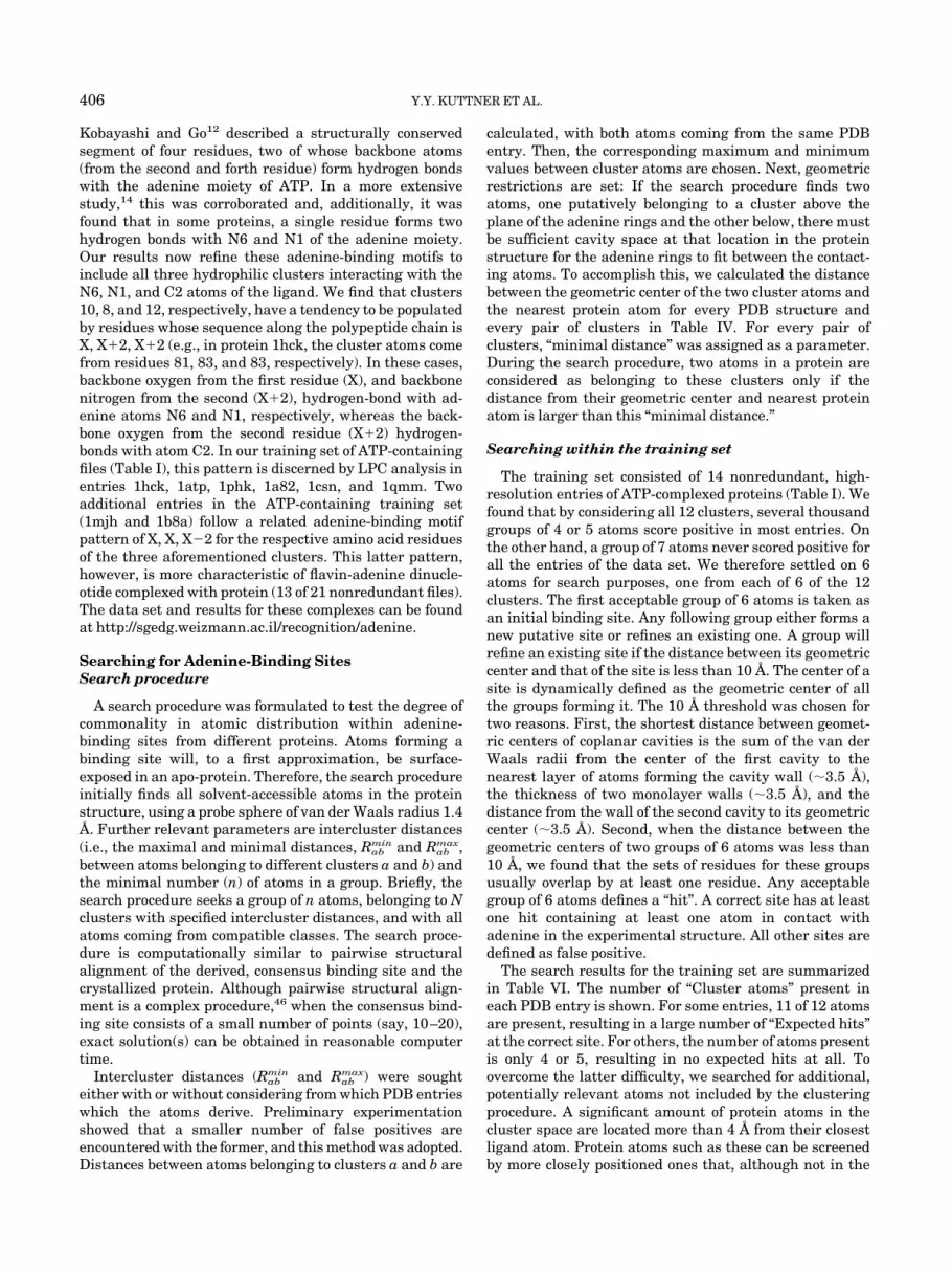
The 3D location of the clusters around the adenine ringis depicted in a perspective projection from two vantagepoints in Figure 1. In a frontal view, perpendicular to theplane of the rings (Fig. 1, upper panel), all clusters appearwell delineated except clusters 1 and 3, whose closestmember atoms are 0.7 A apart. There are eight Hydropho-bic clusters (clusters 1–7 and 11) distributed above andbeneath the plane of the conjugated ring system, and fourclusters, two (clusters 8 and 10) strongly and two (clusters9 and 12) weakly hydrogen-bonded to the distal end of theadenine moiety (at atoms N6, N1, and C2). In Figure 1,lower panel, the plane of the ring system is rotated 90°,such that the four clusters (clusters 1, 3, 4, and 11) beneaththe plane in Figure 1, upper panel, are now more spreadout.
Multiple contacts
Table V shows the dominant adenine atom(s) contactedby each cluster, indicating the general position of a clusterrelative to the adenine moiety. The full set of such contactscan be found at http://sgedg.weizmann.ac.il/recognition/adenine. The hydrophobic clusters have a total of 82nonhydrogen protein atoms in contact with adenine: 90%are derived from amino acid sidechains, and more than90% of these are carbon atoms. Conversely, a preponder-ance (19 of 23) of hydrophilic cluster atoms are from theprotein backbone, in agreement with the description byDenessiouk et al.17 Thus, although the adenine pocket isbasically hydrophobic, hydrophilic interactions can be
TABLE IV. Cluster Properties
PDBentry
Cluster number
1 2 3 4 5 6 7 8 9 10 11 12
Atom classa
1phk IV IV IV — IV IV IV III IV II IV II1hck IV IV IV — V IV IV III IV II IV II1atp — IV IV IV V IV IV III IV II — II1csn IV IV — IV IV IV IV III — II IV —b
1mjh III IV IV I — IV IV III — II — II1a82 IV —b IV IV V — —b III IV II IV II1a6o IV V IV III IV — IV — IV —b — VI1ayl III VI — — IV — — — IV I IV II1a49 VI VI IV — — — IV — VI V IV —1kay — IV IV VI — VI — I — — — —1qmm IV — — IV — IV — —b V — IV —1b8a VI — III — — IV IV III — — — —b
1yag — IV IV I VI VI — — — — — —1nsf IV IV — IV IV — — — — — — —
A filled cell indicates that the entry contributes a member to the cluster, an empty cell (—) indicates that it does not. Shading indicates clustertype, which is assigned based on the most frequent class in the cluster: white, Hydrophobic (IV); light gray, Hydrogen Bond Donor (III); dark gray,Hydrogen Bond Acceptor (II).aAtom classes32: I, Hydrophilic: N and O that can donate and accept hydrogen bonds; II, Acceptor: N or O that can only accept a hydrogen bond; III,Donor: N that can only donate a hydrogen bond; IV, Hydrophobic: CI, Br, I, and all C atoms that are not in aromatic rings and do not have acovalent bond to an N or O atom; V, Aromatic: C in aromatic rings irrespective of any other bonds formed by the atom; VI, Neutral: S, F, P, andmetal atoms in all cases, and nonaromatic C atoms that have a covalent bond to at least one atom from class I, or two or more atoms from class IIor III; VII, Neutral donor: Nonaromatic C atoms that have a covalent bond with only one atom of class III; VIII, Neutral acceptor: Nonaromatic Catoms that have a covalent bond with only one atom of class II.bAlthough an atom is within the cluster radius, it is not accepted as a cluster member due to unfavorable interaction (see text).
CONSENSUS SEARCHING FOR ADENINE BINDING SITES 403

Figure 1.
Figure 2.
404 Y.Y. KUTTNER ET AL.

formed with polar backbone atoms. Hence, atomic (ratherthan residue) recognition imparts specificity to adeninebinding. Hydrophobic cluster atoms often form multiple(up to six) atomic contacts when positioned above orbeneath the plane of the adenine ring system, whereasatoms from all four hydrogen-bonded clusters form basi-cally a single contact with adenine. The average number ofring contacts per atom per cluster is given at the bottom ofTable V.
Positioning
Seven hydrophobic clusters (clusters 1–4, 6, 7, and 11)are located 3.4–3.8 A from the plane of the adenine rings(Zc, Table V). These values are close to the sum of van derWaals radii for COC and CON interactions, indicatingthat these clusters are situated almost directly above andbeneath the rings (cf. Fig. 1, upper panel). On the otherhand, hydrophobic cluster 5, which interacts with atomsC2 and N1, is located 2.4 A above the ring plane, indicatingan oblique positioning. Interestingly, cluster 12 geometri-
cally counterbalances cluster 5 (cf. Fig. 1, upper panel),being is positioned 2.2 A beneath the ring plane. Cluster 12is composed mainly of oxygen atoms in contact withadenine atom C2. Such CH…O contact is generally ac-cepted as a weak hydrogen bond.10,40–44
Hydrophobic cluster 9 is also obliquely positioned (dis-tance, �2.2 A) compared to the ring plane. The atoms inthis cluster interact solely with hydrophilic atom N6 ofadenine (Table V). At first glance, this would appear toindicate a repulsive contact. However, Luisi et al.45 showedthat the amino group of adenine has the highest propen-sity among nucleobases to accept a hydrogen bond from aCH group, and they consider this contact to be attractive.The positions of clusters 9 and 10 relative to the C6ON6covalent bond of adenine show characteristic pyramidaliza-tion of the amino group, supporting the assumption thatthe N6 atom of the adenine moiety contacting cluster 9 isindeed acting as a hydrogen-bond acceptor. Furtherbacking can be gleaned from a recent report10 of asecond, strong hydrogen-bond location interacting withadenine atom N6 in addition to cluster 10. Essentially,all four vertices of a pyramid-like structure representingthe interactions of atom N6 with the adenine ringsystem and with its protein binding niche can now bedescribed (namely, atom N6 at the center, and atom C6,cluster 9, cluster 10, and the strong hydrogen-bond loca-tion of Cappello et al.10 at the vertices). Thus, N6 appearsto be an anchor atom, interacting with its proteinaceoussurrounding by up to three hydrogen bonds: two strongand one weak.
The atomic positions forming hydrogen bonds with N6and N1 of adenine have been well described.9–11,14,20
Clusters 10 and 8 in our study fit into those positions,forming strong hydrogen bonds with atoms N6 and N1 ofadenine, respectively. These two clusters are situatedalmost on the plane of the ring (Fig. 1, upper panel).
Fig. 1. Positions of clusters around the adenine moiety. The adeninemoieties of the 14 structures in the ATP-containing training set weresuperimposed and clusters of atoms extracted as described in Materialsand Methods section. Cluster numbers follow those in Table IV. Carbonatoms are colored gray; oxygen atoms, red; nitrogen atoms, blue; andsulphur atoms, yellow. The pictures were prepared with WebLab Viewersoftware (Molecular Simulations, Inc.), using a perspective projectionview. Upper panel, frontal view perpendicular to the plane of the adeninerings. Lower panel: plane of ring system rotated 90° such that clusters 1,3, 4, and 11 are now extended.
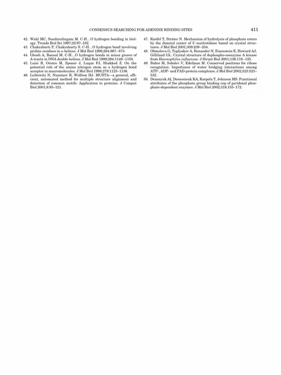
Fig. 2. Positions of the five putative binding sites of entry 1mjh.Training set entry 1mjh is predicted to have five putative binding sites (cf.Table VI). Four of the sites are located at the convex surface, whereas theremaining one (colored green) is immersed in the protein core. This site isthe correct prediction. The ATP ligand at the experimental position isshown in purple. Analysis was performed by extracting the coordinates forthe putative sites from the PDB file and highlighting their positions inWebLab Viewer (MSI).
TABLE V. Atoms of Adenine in Contact with Clustered Atoms of Proteinsa
Adenine Cluster No.
Atomic designation Atom 1 2 3 4 5 6 7 8 9 1 1 10 1 2
N1 � � � �C2 � � �N3 � �C4 � �C5 � �C6 � �N6 � � � �N7 � � �C8 � �N9 �
No. of contactsb 3.3 3.3 2.9 1.6 1.3 3.8 2.9 1.3 1.0 1.0 1.1 1.3Zc(A)c �3.8 �3.5 �3.7 �3.4 �2.4 �3.8 �3.5 �0.6 �2.2 �0.3 �3.6 �2.5aAdenine atoms marked as (�) collectively contact �70% of the protein atoms in that cluster.bAverage number of contacts of a cluster atom with atoms of the adenine moiety.cDistance of the cluster center from the plane of the adenine rings. Minus (�) or plus (�) indicates the position of the cluster relative to theconjugated ring system, as pictured in Figure 1, upper panel.
CONSENSUS SEARCHING FOR ADENINE BINDING SITES 405
Kobayashi and Go12 described a structurally conservedsegment of four residues, two of whose backbone atoms(from the second and forth residue) form hydrogen bondswith the adenine moiety of ATP. In a more extensivestudy,14 this was corroborated and, additionally, it wasfound that in some proteins, a single residue forms twohydrogen bonds with N6 and N1 of the adenine moiety.Our results now refine these adenine-binding motifs toinclude all three hydrophilic clusters interacting with theN6, N1, and C2 atoms of the ligand. We find that clusters10, 8, and 12, respectively, have a tendency to be populatedby residues whose sequence along the polypeptide chain isX, X�2, X�2 (e.g., in protein 1hck, the cluster atoms comefrom residues 81, 83, and 83, respectively). In these cases,backbone oxygen from the first residue (X), and backbonenitrogen from the second (X�2), hydrogen-bond with ad-enine atoms N6 and N1, respectively, whereas the back-bone oxygen from the second residue (X�2) hydrogen-bonds with atom C2. In our training set of ATP-containingfiles (Table I), this pattern is discerned by LPC analysis inentries 1hck, 1atp, 1phk, 1a82, 1csn, and 1qmm. Twoadditional entries in the ATP-containing training set(1mjh and 1b8a) follow a related adenine-binding motifpattern of X, X, X�2 for the respective amino acid residuesof the three aforementioned clusters. This latter pattern,however, is more characteristic of flavin-adenine dinucle-otide complexed with protein (13 of 21 nonredundant files).The data set and results for these complexes can be foundat http://sgedg.weizmann.ac.il/recognition/adenine.
Searching for Adenine-Binding SitesSearch procedure
A search procedure was formulated to test the degree ofcommonality in atomic distribution within adenine-binding sites from different proteins. Atoms forming abinding site will, to a first approximation, be surface-exposed in an apo-protein. Therefore, the search procedureinitially finds all solvent-accessible atoms in the proteinstructure, using a probe sphere of van der Waals radius 1.4A. Further relevant parameters are intercluster distances(i.e., the maximal and minimal distances, Rab
min and Rabmax,
between atoms belonging to different clusters a and b) andthe minimal number (n) of atoms in a group. Briefly, thesearch procedure seeks a group of n atoms, belonging to Nclusters with specified intercluster distances, and with allatoms coming from compatible classes. The search proce-dure is computationally similar to pairwise structuralalignment of the derived, consensus binding site and thecrystallized protein. Although pairwise structural align-ment is a complex procedure,46 when the consensus bind-ing site consists of a small number of points (say, 10–20),exact solution(s) can be obtained in reasonable computertime.
Intercluster distances (Rabmin and Rab
max) were soughteither with or without considering from which PDB entrieswhich the atoms derive. Preliminary experimentationshowed that a smaller number of false positives areencountered with the former, and this method was adopted.Distances between atoms belonging to clusters a and b are
calculated, with both atoms coming from the same PDBentry. Then, the corresponding maximum and minimumvalues between cluster atoms are chosen. Next, geometricrestrictions are set: If the search procedure finds twoatoms, one putatively belonging to a cluster above theplane of the adenine rings and the other below, there mustbe sufficient cavity space at that location in the proteinstructure for the adenine rings to fit between the contact-ing atoms. To accomplish this, we calculated the distancebetween the geometric center of the two cluster atoms andthe nearest protein atom for every PDB structure andevery pair of clusters in Table IV. For every pair ofclusters, “minimal distance” was assigned as a parameter.During the search procedure, two atoms in a protein areconsidered as belonging to these clusters only if thedistance from their geometric center and nearest proteinatom is larger than this “minimal distance.”
Searching within the training set
The training set consisted of 14 nonredundant, high-resolution entries of ATP-complexed proteins (Table I). Wefound that by considering all 12 clusters, several thousandgroups of 4 or 5 atoms score positive in most entries. Onthe other hand, a group of 7 atoms never scored positive forall the entries of the data set. We therefore settled on 6atoms for search purposes, one from each of 6 of the 12clusters. The first acceptable group of 6 atoms is taken asan initial binding site. Any following group either forms anew putative site or refines an existing one. A group willrefine an existing site if the distance between its geometriccenter and that of the site is less than 10 A. The center of asite is dynamically defined as the geometric center of allthe groups forming it. The 10 A threshold was chosen fortwo reasons. First, the shortest distance between geomet-ric centers of coplanar cavities is the sum of the van derWaals radii from the center of the first cavity to thenearest layer of atoms forming the cavity wall (�3.5 A),the thickness of two monolayer walls (�3.5 A), and thedistance from the wall of the second cavity to its geometriccenter (�3.5 A). Second, when the distance between thegeometric centers of two groups of 6 atoms was less than10 A, we found that the sets of residues for these groupsusually overlap by at least one residue. Any acceptablegroup of 6 atoms defines a “hit”. A correct site has at leastone hit containing at least one atom in contact withadenine in the experimental structure. All other sites aredefined as false positive.
The search results for the training set are summarizedin Table VI. The number of “Cluster atoms” present ineach PDB entry is shown. For some entries, 11 of 12 atomsare present, resulting in a large number of “Expected hits”at the correct site. For others, the number of atoms presentis only 4 or 5, resulting in no expected hits at all. Toovercome the latter difficulty, we searched for additional,potentially relevant atoms not included by the clusteringprocedure. A significant amount of protein atoms in thecluster space are located more than 4 A from their closestligand atom. Protein atoms such as these can be screenedby more closely positioned ones that, although not in the
406 Y.Y. KUTTNER ET AL.

cluster, are in contact with the ligand target atom (cf. Fig.2 in Sobolev and Edelman31). In such instances, thedistant atom does not appear in the clustering data ofTable IV (because the clustering procedure requires con-tact with the ligand atom and this, by definition, isprovided by the closer atom), but is recognized by thesearch procedure (because cluster space and atom type arethe parameters scored in the search). Such additionalatoms allowed the search for groups of 6 atoms to proceedin entries with less than 6 clusters.
The total number of hits (“Total hits” column) exceededthe expected hits for each entry in the training set (TableVI). This is readily explained, because only sets of atoms inwhich all 6 members are in contact with adenine arerepresented in “Expected hits.” On the other hand, in“Total hits,” additional sets populate false-positive cavi-ties, or even the correct cavity, but have less than 6 atomsin contact with adenine. Critically, the data in Table VIshow that the correct experimental cavity for adenine wasfound in all 14 cases. This is indicated by the smalldistance between the center of the experimental site foreach entry and the closest hit from the search results(“Best-hit distance” column). The only seeming exceptionis entry 1qmm. However, in spite of the relatively largedistance (8.7 A) of the best hit from the center of theexperimentally correct cavity, 3 of 6 search atoms provedto be in contact with adenine.
Searching with the test sets
The search procedure was applied to a nonredundant,high-resolution test set composed of all the suitably re-solved structures complexed with ATP that appeared inthe PDB following completion of our training set (Table II).The results are summarized in Table VII. With the excep-tion of one case, the experimentally correct cavity foradenine was found for all entries (cf. “Best-hit distances”column). In the exception (entry 1hp1), the adenine ringproved to be stacked between two residues of phenylala-nine.47 A review of the atom class data in Table IV revealsthat an aromatic-ring carbon atom (class V) occurred inonly 3 of the 9 Hydrophobic clusters of the training set(namely, clusters 2, 5, and 9). Thus, the search procedureaccepted aromatic input only for those three Hydrophobicclusters. Relaxation of the procedure to include aromatic-ity for all Hydrophobic clusters readily revealed the experi-mental binding site for entry 1hp1, with a best-hit distanceof 3.6 A, whereas the total number of putative sitesincreased only moderately from 18 to 21 (cf. Table VII).
We note that inclusion of hydrophilic atoms in Hydropho-bic clusters 1, 3, and 4, and aromatic atoms in HydrogenBond Accepter cluster 10 (as described for the training set;Table IV), proved to be significant for the search procedurein the ATP test set as well (Table VII). Without suchinclusion, the correct site was not found in 2 of 9 ATP teststructures: 1jjv and 1e2q. Indeed, Obmolova et al.48 con-
TABLE VI. Search Results for Training Set of ATP-Complexed Proteins
PDBentry
Clusteratomsa
Expectedhitsb
Totalhitsc
Best-hitdistanced
(Å)
Putative sites
Totalno.e
Narrowing of false positives by:
Chainsizef Visualg
Mosthitsh
Hits �visuali
1phk 11 462 534 0.1 9 4 1 0 01hck 11 462 518 0.1 12 9 5 0 01atp 10 210 471 0.1 11 9 2 0 01csn 9 84 230 0.2 8 4 0 0 01mjh 9 84 179 0.2 5 4 0 0 01a82 9 84 143 0.1 5 4 0 0 01a6o 8 28 103 0.2 12 7 0 0 01ayl 7 7 92 0.5 18 14 5 0 01a49 7 7 51 0.4 13 9 5 0 01kay 5 0 71 1.0 12 8 5 0 01qmm 5 0 239 8.7j 25 22 6 0 01b8a 5 0 60 2.4 17 12 2 7–12k 11yag 5 0 51 2.4 16 9 1 1 01nsf 4 0 56 1.2 10 9 2 0 0aNumber of clustered atoms at the authentic site (derived from Table IV).bNumber of hits (K) theoretically expected for 6 atoms contacting adenine at the correct site given the number of cluster atoms (k) in the specifiedPDB entry: K � k!/(6! (k � 6)!).cTotal number of hits found for all putative sites.dDistance between the center of the experimental binding site and the center of atoms forming the closest hit.eTotal number of putative sites found.fNumber of false positives remaining following a chain-length restriction of at least 15 residues between distal amino acids of a putative site.gNumber of false positives remaining following visual elimination of convex surface sites using WebLab Viewer (MSI).hNumber of false positives remaining after applying “highest frequency of hits at a binding site” as a limiting parameter.iNumber of false positives remaining after applying “Visual” and “Most hits” techniques in combination.jIn spite of the large distance, three atoms of the best hit are in contact with adenine.kIdentical frequency of hits was found for multiple putative sites.
CONSENSUS SEARCHING FOR ADENINE BINDING SITES 407

cluded that Arg140A in entry 1jjv does not determine basespecificity but is important for formation of the ATPpocket, and the same holds for Arg143A of entry 1e2q.
The search procedure derived for the adenine moiety ofATP-binding proteins was, in addition, applied to anindependent high-resolution set of PDB entries, eachmember of which is complexed with an adenine-containingligand other than ATP (Table III). These search results aresummarized in Table VIII. Two of the 11 test structures ofthis data set contain more than one unique adenine moietycomplexed to proteins: in entry 1bx4, two molecules ofadenosine, and in entry 1zin, a single molecule of bis(ade-nosine)-5�-pentaphosphate. Thus, this test set provides 13adenine-binding sites for the search. The experimentalbinding site was found for 12 of the 13 adenines using thestandard search procedure, and for one of the ligands in1bx4 following inclusion of aromaticity for all Hydrophobicclusters (“Best-hit distance” column). Thus, consensusclusters and a search procedure based on an ATP-complexed training set are sufficient to recognize theexperimental cavity for adenine in a wide spectrum ofligand–protein complexes containing that purine.
For both test sets, the best hit at each correct bindingsite was checked with LPC software30 and found to have atleast one atom in contact with adenine. Hence, the testresults show that the correct experimental cavity foradenine was discovered in all cases.
Narrowing the Number of Putative Binding Sites
Whereas the correct binding cavity for adenine wasindeed discovered in all cases examined, the data alsoshow that, all in all, about 400 putative binding sites werescored. In fact, in every case, more than one putative sitewas counted (Tables VI–VIII, “Total No.” column) and, onaverage, in addition to the correct one, 10 false positives
were registered. For a search procedure to be effective, itought to find the actual binding cavity with a minimalnumber of false-positive sites (preferably, none). Severalmethods were thus surveyed in an attempt to narrow thenumber of false positives. The results are summarized be-low and in Tables VI–VIII. The data sets for these tablescan be found at http://sgedg.weizmann.ac.il/recognition/adenine.
First, we experimented with reducing the number ofclusters in the training set. Searching with 6 of 8 ratherthan 6 of 12 clusters, a grouping was found (namely,clusters 1–5, 8, 11, 12) that, on average, reduced thenumber of false-positive sites by 59% and the total numberof hits by 89%, while retaining the experimental bindingsite in each training set entry. However, our search with 6of 8 (or 6 of 9) clusters failed to find the experimentalbinding site for several entries of the test sets, and weabandoned this approach.
Next, we sought a common chain-length minimum thatsatisfied all correct sites in the training and test sets.Restrictions were set for the distal amino acids of thebinding domain. Inspection of the amino acid chain encom-passing the correct binding site for all members of thethree data sets revealed that it is at least 15 residues long.When this empiricism is introduced as a constraint (see“Chain size” column), the number of false positives ismoderately reduced, on average by 22% in the ATP-containing training set (Table VI), 27% in the ATP-containing test set (Table VII), and 23% in the non-ATPadenine-containing test set (Table VIII). However, in nocase was the number of false positives reduced to nil.
In a different approach, preliminary observations showeda high percentage of bogus binding sites on the convexsurface of the protein. We attempted to discriminatevisually between these and putative binding sites partly
TABLE VII. Search Results for Test Set of ATP-Complexed Proteins
PDBentry Total hitsa Best-hit distanceb (A)
Putative sites
Total no.cNarrowing of false positives by:
Chain sized Visuale Most hitsf Hits � visualg
1e2q 23 0.8 7 3 0 0 01f9a 42 2.6 5 2 1 0 01fmw 144 1.6 27 16 6 3 21g5t 7 3.3 3 1 0 0 01gn8 23 5.0 7 4 1 6 11hp1 65 10.0h (3.6)i 18 (21)i 15 (16)i 2 (2)i n.f.h (1)i n.f.h (0)i
1j7k 45 2.2 10 5 2 0–1 01jjv 39 1.5 9 5 2 5–6 21kp2 49 6.9j 15 8 3 4–6 0–2aSee Table VIc.bSee Table VId.cSee Table VIe.dSee Table VIf.eSee Table VIg.fSee Table VIh.gSee Table VIi.hCorrect site not found (n.f.) using the standard search procedure (no atoms in contact with adenine in best hit).iExpansion of the search function to include aromaticity in all Hydrophobic clusters (see legend, Table IV) revealed the experimental binding site.jIn spite of the relatively large distance, two atoms of the best hit are in contact with adenine.
408 Y.Y. KUTTNER ET AL.

buried in the protein core that might accommodate anadenine moiety. Figure 2 reveals the potential of thismethod: In the case of training set entry 1mjh, the numberof false positives was reduced from four to nil. Each entryin each data set was analyzed. The procedure retained thecorrect binding site in all cases except one (entry 1csc,Table VIII). The atomic coordinates for all putative sitesfor an entry were extracted, highlighted, and overlaid on a3D representation of the protein using WebLab Viewer(MSI). Rotation about all axes was used to discriminateconvex surface sites from partly buried ones. When convexsurface sites are eliminated in this fashion, the number offalse positives (“Visual” column) is reduced, on average, by79% in the ATP-containing training set (Table VI), 82% inthe ATP-containing test set (Table VII), and 88% in thenon-ATP adenine-containing test set (Table VIII). In overone fourth of the cases, the number of false positives wasreduced to nil.
In yet another approach, we noticed that in a majority ofcases, the search procedure scored a larger number of hitsat the correct binding-site cavity than at any of theincorrect ones. Parenthetically, we note that if sufficienthits exist in the correct cavity, their positional densitymight be useful to home in on the precise location of thecorrect binding position. With use of the highest frequencyof hits at a binding site as a limiting parameter (see “Mosthits” column), the number of false positives is reduced, onaverage, by 92–95% in the ATP-containing training set(Table VI), 76–80% in the ATP-containing test set (TableVII), and 80–81% in the non-ATP adenine-containing test
set (Table VIII). In almost two thirds of the cases, thenumber of false positives was reduced to nil.
Finally, by combining the “Visual” and “Most hits”approaches (“Hits � visual” column), the number of falsepositives is reduced, on average, by �99% in the ATP-containing training set (Table VI), 93–95% in the ATP-containing test set (Table VII), and 97–98% in the non-ATP adenine-containing test set (Table VIII). In over threefourths of the cases, the number of false positives wasreduced to nil, and in no case was it greater than two, whenthese two approaches were used in concert.
Currently, the consensus-binding structure approachhas been applied only to rigid (adenine; this work) oralmost-rigid (ribose)49 ligand elements. In principle, thisapproach may be applied to rigid ligands, or ligandshaving a few well-defined conformations, for which suffi-cient cases are documented in the structural databases.With regard to flexible ligands, a two-step procedure wouldapply: Consensus-binding sites for individual rigid ele-ments (e.g., the adenine, ribose, and phosphate moieties ofATP) can be derived separately, after which a searchprocedure accounts for the relative positioning of theelements. Such a search procedure might exploit a consen-sus-binding site obtained by different methods.16,50 Exten-sion of our work to flexible ligands is currently underdevelopment.
CONCLUSIONS
Our objective in this study was to determine whether aconsensus network of atomic coordinates, derived from the
TABLE VIII. Search Results for Test Set of Proteins Complexed with Adenine-Containing Ligands Other Than ATP
PDBentry Total hitsa Best-hit distanceb (Å)
Putative sites
Total no.cNarrowing of false positives by:
Chain sized Visuale Most hitsf Hits � visualg
1ads 47 5.4i 8 6 0 0 01b4v 65 2.5 18 12 1 3 01bx4h 72 4.8 11 8 1 0–1 0–1
72 13.0j (8.2i)k 11 (12)k 8 1 n.f.j (10)k n.f.j (1)k
1byq 40 2.7 9 7 0 1 01csc 68 8.1i 12 8 3l 0 01kpf 49 0.8 3 2 1 0 01mmg 103 2.7 26 22 3 2 01nhk 31 6.3i 6 2 0 0 01zinh 28 3.2 5 2 0 2 0
28 1.3 5 2 0 0 02src 94 0.3 16 12 4 0 09ldt 61 3.0 11 8 1 3–4 1aSee Table VIc.bSee Table VId.cSee Table VIe.dSee Table VIf.eSee Table VIg.fSee Table VIh.gSee Table VIi.hThese entries have two unique adenine moieties complexed with their proteins; in 1bx4, two molecules of adenosine; in 1zin, a single molecule ofbis(adenosine)-5�-pentaphosphate.iIn spite of the relatively large distances, at least one atom of the best hit is in contact with adenine.jCorrect site not found (n.f.) using the standard search procedure (no atoms in contact with adenine in best hit).kExpansion of the search function to include aromaticity in all Hydrophobic clusters revealed the experimental binding site.lThe correct binding site was not retained in this one case.
CONSENSUS SEARCHING FOR ADENINE BINDING SITES 409

structural database, could be extracted and applied to-ward recognizing the binding-site cavity for a given hetero-group in any of its diverse protein complexes. The resultsshow that for a frequently appearing PDB ligand elementsuch as adenine, this is indeed feasible. A consensus-binding structure for adenine can be scored to predict thecorrect binding cavity within the protein volume withacceptable fidelity (�90%). As a consequence, it is likelythat if a protein is known, or suspected, to be a target for aheterogroup containing adenine, our search procedure willallow resolution of the binding site. Nevertheless, ourcurrent procedure probably would not allow us to excludenontarget proteins with much fidelity. This remains achallenge.
ACKNOWLEDGMENTS
We thank Dr. B. McConkey, R. Najmanovich, E.Eyal,and M. Babor for helpful discussions.
REFERENCES
1. Bernstein FC, Koetzle TF, Williams GJ, Meyer EF Jr, Brice MD,Rodgers JR, Kennard O, Shimanouchi T, Tasumi M. The ProteinData Bank: A computer-based archival file for macromolecularstructures. J Mol Biol 1977;112:535–542.
2. Berman HM, Westbrook J, Feng Z, Gilliland G, Bhat TN, WeissigH, Shindyalov IN, Bourne PE. The Protein Data Bank. NucleicAcids Res 2000;28:235–242.
3. Walker J, Saraste M, Runswick M, Gay N. Distantly relatedsequences in a- and b- subunits of ATP synthase, myosin, kinasesand other ATP-requiring enzymes and common nucleotide bindingfold. EMBO J 1982;1:945–951.
4. Saraste M, Sibbald PR, Wittinghofer A. The P-loop—a commonmotif in ATP-binding and GTP-binding proteins. Trends BiochemSci 1990;15:430–434.
5. Hanks SK, Quinn AM, Hunter T. The protein-kinase family—conserved feartures and deduced phylogeny of the catalytic do-mains. Science 1988;241:42–52.
6. Bairoch A, Bucher P, Hofmann K. The PROSITE database, itsstatus in 1997. Nucleic Acids Res 1997;25:217–221.
7. Traut TW. The functional and consensus motifs of 9 types ofpeptide segments that form different types of nucleotide-bindingsites. Eur J Biochem 1994;222:9–19.
8. Schulz G. Binding of nucleotides by proteins. Curr Opin StructBiol 1992;2:61–67.
9. Moodie SL, Mitchell JBO, Thornton JM. Protein recognition ofadenylate: An example of a fuzzy recognition template. J Mol Biol1996;263:486–500.
10. Cappello V, Tramontano A, Koch U. Classification of proteinsbased on the properties of the ligand-binding site: The case ofadenine-binding proteins. Proteins 2002;47:106–115.
11. Nobeli I, Laskowski RA, Waldar WSJ, Thornton JM. On moleculardiscrimination between adenine and guanine by proteins. NucleicAcids Res 2001;29:4294–4309.
12. Kobayashi N, Go N. ATP binding proteins with different foldsshare a common ATP-binding structural motif. Nat Struct Biol1997a;4:6–7.
13. Kobayashi N, Go N. A method to search for similar protein localstructures at ligand-binding sites and its application to adeninerecognition. Eur Biophys J 1997b;26:135–144.
14. Denessiouk KA, Johnson MS. When fold is not important: Acommon structural framework for adenine and AMP binding in 12unrelated protein families. Proteins 2000;38:310–326.
15. Bruno IJ, Cole JC, Lommerse JP, Rowland RS, Taylor R, VerdonkML. IsoStar: A library of information about nonbonded interac-tions. J Comput Aided Mol Des 1997;11:525–537.
16. Kuttner YY, Babor M, Edelman M, Sobolev V. Structural common-ality in protein binding sites for ATP. In: El-Mabrouk, N, Len-gauer T, Sankoff D, editors. Currents in computational molecularbiology. Montreal: CRM; 2001. p 91–92.
17. Denessiouk LA, Rantanen VV, Johnson MS. Adenine recognition:
A motif present in ATP-, CoA-, NAD-, NADP-, and FAD-dependent proteins. Proteins 2001;44:282–291.
18. Klebe G. The use of composite crystal-field environments inmolecular recognition and the de novo design of protein ligands. JMol Biol 1994;237:212–235.
19. Laskowski RA, Thornton JM, Humblet C, Singh J. X-site: Use ofempirically derived atomic packing preferences to identify favour-able interaction regions in the binding sites of proteins. J Mol Biol1996;259:175–201.
20. Rantanen VV, Denessiouk KA, Gyllenberg M, Koski T, JohnsonMS. A fragment library based on Gaussian mixtures predictingfavorable molecular interactions. J Mol Biol 2001;313:197–214.
21. Verdonk ML, Cole JC, Taylor R. SuperStar: A knowledge-basedapproach for identifying interaction sites in proteins. J Mol Biol1999;289:1093–1108.
22. Verdonk ML, Cole JC, Watson P, Gillet V, Willett P. SuperStar:Improved knowledge-based interaction field for protein bindingsites. J Mol Biol 2001;307:841–859.
23. Nissink JWM, Verdonk ML, Klebe G. Simple knowledge-baseddescriptors to predict protein–ligand interactions: Methodologyand validation. J Comput Aided Mol Des 2000;14:787–803.
24. Chakravarty S, Bhinge A, Varadarajan R. A procedure for detec-tion and quantitation of cavity volumes in proteins. J Biol Chem2002;30:31345–31353.
25. Yamashita MM, Wesson L, Eisenman G, Eisenberg D. Wheremetal ions bind in proteins. Proc Natl Acad Sci U S A 1990;87:5648–5652.
26. Hellinga HW, Richards FM. Construction of new ligand bindingsites in proteins of known structure: I. Computer-aided model-ling of sites with pre-defined geometry. J Mol Biol 1991;222:763–785.
27. Yang W, Lee H-W, Hellinga H, Yang JJ. Structural analysis,identification, and design of calcium-binding sites in proteins.Proteins 2002;47:344–356.
28. Walles AC, Borkakoti N, Thornton JM. TESS: A geometrichashing algorithm for deriving 3D coordinate templates for search-ing databases: Application to enzyme active sites. Protein Sci1997;6:2308–2323.
29. Taroni C, Jones S, Thornton JM. Analysis and prediction ofcarbohydrate binding sites. Protein Eng 2000;13:89–98.
30. Sobolev V, Sorokine A, Prilusky J, Abola EE, Edelman M.Automated analysis of interatomic contacts in proteins. Bioinfor-matics 1999;15:327–332.
31. Sobolev V, Edelman M. Modeling the quinone-B binding site ofthe photosystem-II reaction center using notions of complemen-tarity and contact surface between atoms. Proteins 1995;21:214–225.
32. Sobolev V, Vriend G, Wade RC, Edelman M. Molecular dockingusing surface complementarity. Proteins 1996;25:120–129.
33. Thompson JD, Higgins DG, Gibson TJ. Clustal-W—improving thesensitivity of progressive multiple sequence alignment throughsequence weighting, position-specific gap penalties and weightmatrix choice. Nucleic Acids Res 1994;22:4673–4680.
34. Hung LW, Wang IX, Nikaido K, Liu PQ, Ames GF, Kim SH.Crystal structure of the ATP-binding subunit of an ABC trans-porter. Nature 1998;396:703–707.
35. Burley SK, Petsko GA. Amino-aromatic interactions in proteins.FEBS Lett 1986;203:139–143.
36. Levitt M, Perutz MF. Aromatic rings act as hydrogen-bondacceptors. J Mol Biol 1988;201:751–754.
37. Basharov MA, Volkenstein MV, Golovanov IB, Nauchitel VV,Sobolev VM. The fragment–fragment interaction method: Part I.Estimating molecule–molecule interactions. J Gen Chem USSR1989;59:435–447.
38. Mitchell JBO, Nandi CL, McDonald IK, Thornton JM, Price SL.Amino/aromatic interactions in proteins—is the evidencestacked against against hydrogen-bonding? J Mol Biol 1994;239:315–331.
39. Felder C., Jiang HL., Zhu WL, Chen KX, Silman I, Botti SA,Sussman JL. Quantum/classical mechanical comparison of cat-ion–pi interactions between tetramethylammonium and benzene.J Phys Chem A 2002;105:1326–1333.
40. Derewenda ZS, Lee L, Derewenda U. The occurrence C-H…Ohydrogen-bonds in proteins. J Mol Biol 1995;252:248–262.
41. Bella J, Berman HM. Crystallographic evidence for C�H…OAChydrogen bonds in a collagen triple helix. J Mol Biol 1996;264:734–742.
410 Y.Y. KUTTNER ET AL.
42. Wahl MC, Sundaralingam M. C-H…O hydrogen bonding in biol-ogy. Trends Biol Sci 1997;22:97–102.
43. Chakrabarti P, Chakrabarty S. C-H…O hydrogen bond involvingproline residues in �-helices. J Mol Biol 1998;284:867–873.
44. Ghosh A, Bansal M. C-H…O hydrogen bonds in minor groove ofA-tracts in DNA double helices. J Mol Biol 1999;294:1149–1158.
45. Luisi B, Orozco M, Sponer J, Luque FJ, Shakked Z. On thepotential role of the amino nitrogen atom as a hydrogen bondacceptor in macromolecules. J Mol Biol 1998;279:1123–1136.
46. Leibowitz N, Nussinov R, Wolfson HJ. MUSTA—a general, effi-cient, automated method for multiple structure alignment anddetection of common motifs: Application to proteins. J ComputBiol 2001;8:93–121.
47. Knofel T, Strater N. Mechanism of hydrolysis of phosphate estersby the dimetal center of 5�-nucleotidase based on crystal struc-tures. J Mol Biol 2001;309:239–254.
48. Obmolova G, Teplyakov A, Bonander N, Eisenstein E, Howard AJ,Gilliland GL. Crystal structure of dephospho-coenzyme A kinasefrom Haemophilus influenzae. J Struct Biol 2001;136:119–125.
49. Babor M, Sobolev V, Edelman M. Conserved positions for riboserecognition: Importance of water bridging interactions amongATP-, ADP- and FAD-protein complexes. J Mol Biol 2002;323:523–532.
50. Denesyuk AI, Denessiouk KA, Korpela T, Johnson MS. Functionalattributes of the phosphate group binding cup of pyridoxal phos-phate-dependent enzymes. J Mol Biol 2002;316:155–172.
CONSENSUS SEARCHING FOR ADENINE BINDING SITES 411


