Embed Size (px)
Citation preview
452 IEEE TRANSACTIONS ON MEDICAL IMAGING, VOL. 26, NO. 4, APRIL 2007
A Deformable Registration Method forAutomated Morphometry of MRI Brain Images in
Neuropsychiatric ResearchDaniel Schwarz*, Tomas Kasparek, Ivo Provaznik, Member, IEEE, and Jiri Jarkovsky
Abstract—Image registration methods play a crucial role in com-putational neuroanatomy. This paper mainly contributes to thefield of image registration with the use of nonlinear spatial trans-formations. Particularly, problems connected to matching mag-netic resonance imaging (MRI) brain image data obtained fromvarious subjects and with various imaging conditions are solvedhere. Registration is driven by local forces derived from multi-modal point similarity measures which are estimated with the useof joint intensity histogram and tissue probability maps. A spatialdeformation model imitating principles of continuum mechanicsis used. Five similarity measures are tested in an experiment withimage data obtained from the Simulated Brain Database and aquantitative evaluation of the algorithm is presented. Results of ap-plication of the method in automated spatial detection of anatom-ical abnormalities in first-episode schizophrenia are presented.
Index Terms—Computational neuroanatomy, deformable regis-tration, first-episode schizophrenia, magnetic resonance imaging.
I. INTRODUCTION
COMPUTATIONAL neuroanatomy is a new growing fieldof powerful applications in neuroscience. It promises
an automated methodology to characterize neuroanatomicalconfiguration of structural magnetic resonance imaging (MRI)brain scans. One of the crucial techniques in this methodologyis image registration. Its task is to find a spatial transformationwhich maps each point of an image onto its correspondingpoint of another image [1]. In computational neuroanatomy,atlas-based registration performs the task of spatial normal-ization of images according to a common reference anatomytermed as a brain atlas. It allows interpreting results of an imageanalysis in a standard anatomical coordinate system. Further,an atlas of brain makes it possible to find out how different isa subject brain compared with the common reference anatomywhich represents certain population [2].
Manuscript received September 15, 2006; revised December 29, 2006. Thiswork was supported in part by the Research Programme of Masaryk UniversityMSM0021622404 and in part by the Research Programme of Brno Universityof Technology MSM 0021630513.Asterisk indicates corresponding.
*D. Schwarz is with the Masaryk University, Institute of Biostatistics andAnalyses, 625 00 Brno, Czech Republic (e-mail: [email protected]).
T. Kasparek is with The Faculty Hospital Brno, Clinic of Psychiatry, 625 00Brno, Czech Republic.
I. Provaznik is with the Brno University of Technology, Department ofBiomedical Engineering, 612 00 Brno, Czech Republic.
J. Jarkovsky is with the Masaryk University, Institute of Biostatistics andAnalyses,625 00 Brno, Czech Republic.
Digital Object Identifier 10.1109/TMI.2007.892512
Image registration methods with the use of various typesof geometric transformations ranging from simple transla-tions with rotations, over affine transformations and smoothnonlinear parametric transformations, to high-dimensionalnonlinear nonparametric transformations are used in the fieldof computational neuroanatomy. As the registration methodsemploying global linear transformations have been satisfactorysettled already [3]–[5], this paper mainly contributes to thefield of registration with the use of nonlinear locally adaptivetransformations. Many approaches to deformable registration,e.g., [6]–[8] assume same or functionally dependent intensitiesin the images across subjects. Thus, they allow the registrationprocess to be driven by differences in intensities. They, however,do not count on intensity variations caused by various imagingconditions or tissue atrophy and degradation induced by neu-rological diseases. On the other hand, the image registrationmethods which are robust to such intensity variations, e.g., [9],[10], enable only low-dimensional parametric transformations,which make it impossible to detect localized image differenceswithout additional efforts. However, several registration algo-rithms focused on high-dimensional matching of multimodaldata have recently emerged. In [11], an interesting two-stagescheme is proposed. It combines the popular monomodal“demons” algorithm [12] with an intensity transformation toaccount for the contrast differences between modalities. In [13],a variational formulation for nonparametric multimodal imagematching is developed, which allows coupling of multimodalsimilarity measures to physically motivated deformations. Thisapproach is extended in [14], where the mutual informationbased registration functional is used together with a viscousfluid deformation model simplified to Gaussian kernel con-volution. Similar methods presented in [15] and [16] differ inexpressions for local forces which are all, however, derivedfrom the variation of the global mutual information between theimages to be aligned. Recently, Rogelj et al. proposed severalpoint multimodal similarity measures [17] and a combinedelastic-incremental spatial deformation model [18]. Thanksto extreme locality of the point similarity measures, the de-formable registration was solved in two independent stages:estimation of local forces and spatial deformation model.
In this paper, some of Rogelj’s ideas are adapted to the spe-cific problem of MRI brain images located in the stereotaxicspace. The next section introduces the deformable registrationframework and involved methods. Section III shows results ofquantitative evaluation of the registration algorithm and its ap-plication in deformation-based morphometry (DBM) on real
0278-0062/$25.00 © 2007 IEEE

SCHWARZ et al.: A DEFORMABLE REGISTRATION METHOD FOR AUTOMATED MORPHOMETRY OF MRI BRAIN IMAGES 453
Fig. 1. The scheme of the deformable registration algorithm. The displacementfield u which maximizes global mutual information between a reference imageM and a floating image N is searched in an iterative process which involvescomputation of local forces f in each individual voxel and their regularizationby Rogelj’s elastic-incremental spatial deformation model. The resolution ofM ,N , f and u gradually increases according to a multiresolution schedule.
3-D patient images. The results and the advantages of the pro-posed method are discussed in Section IV and the paper is thenconcluded in Section V.
II. DEFORMABLE REGISTRATION IN STEREOTAXIC SPACE
The algorithm proposed here is inspired by the approachof [17], [18]. The main contribution compared to the originalmethod lies in incorporating tissue probability maps into com-putation of point similarity measures and thus improving theregistration accuracy. Thanks to other enhancements, which aredescribed further, a clearly specified convergence criterion isused instead of a preset number of iterations applied by Rogelj.The scheme of the algorithm is in Fig. 1. There are two mainparts repeated in an iterative process in which a floating image
is deformed to match a reference image : extraction oflocal forces by measurements of similarity and a spatial de-formation model producing the displacement field . These twoparts are completely independent: the similarity of one pointdoes not suppose any spatial relation with neighboring imagepoints. A multiresolution strategy propagating solutions from
coarser to finer levels is used here to speed up the convergenceof the algorithm and to avoid local optima.
A. Symmetric Voxel Matching
In [17], point similarity measures are derived from globalsimilarity measures. Supposing image intensities to be discreterandom variables, their mutual information (MI) can be definedin terms of entropy known from information theory [19]:
(1)
where is the mutual information of random variablesand , , and are entropies of and re-
spectively, is the joint entropy of and ,and are marginal probability density functions (PDF) and
is the joint PDF of the random variables andrespectively. The (1) can be rewritten to [17]:
(2)
The global mutual information of the images and is thuscomputed as an average of point similarities defined foreach voxel . The final summation is taken over spatial coordi-nates instead of intensities thanks to the fact that is thenumber of occurrences of the intensity pair and is thetotal number of intensity pairs, which equals to the number ofoverlapping voxels. The point similarity measure de-rived from the global mutual information is thus defined as [17]:
(3)
In [20], conditional probability densities are used for a regionsimilarity measure, which is here rewritten as another point sim-ilarity measure
(4)
In [17], other measures are proposed
(5)
(6)
454 IEEE TRANSACTIONS ON MEDICAL IMAGING, VOL. 26, NO. 4, APRIL 2007
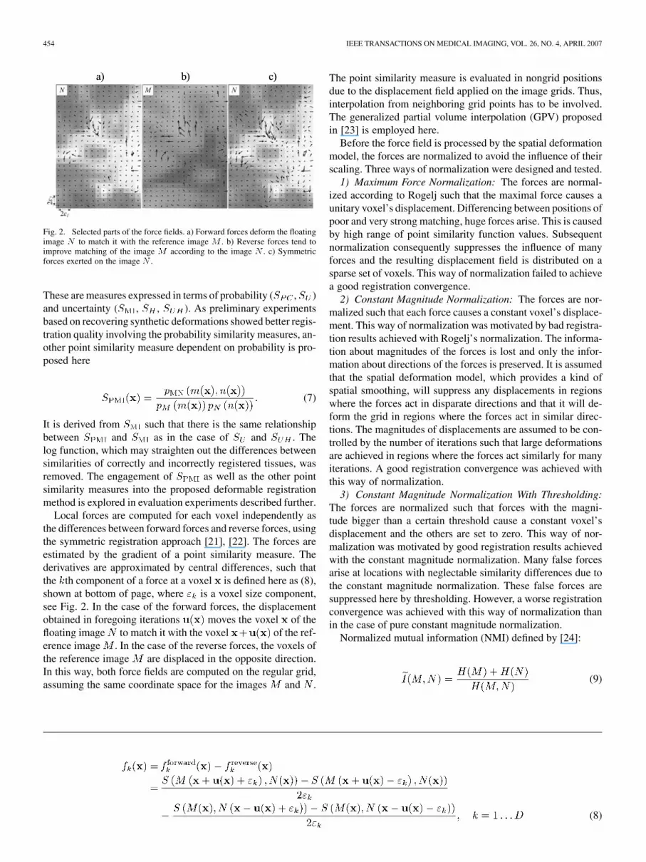
Fig. 2. Selected parts of the force fields. a) Forward forces deform the floatingimage N to match it with the reference image M . b) Reverse forces tend toimprove matching of the image M according to the image N . c) Symmetricforces exerted on the image N .
These are measures expressed in terms of probability ( , )and uncertainty ( , , ). As preliminary experimentsbased on recovering synthetic deformations showed better regis-tration quality involving the probability similarity measures, an-other point similarity measure dependent on probability is pro-posed here
(7)
It is derived from such that there is the same relationshipbetween and as in the case of and . Thelog function, which may straighten out the differences betweensimilarities of correctly and incorrectly registered tissues, wasremoved. The engagement of as well as the other pointsimilarity measures into the proposed deformable registrationmethod is explored in evaluation experiments described further.
Local forces are computed for each voxel independently asthe differences between forward forces and reverse forces, usingthe symmetric registration approach [21], [22]. The forces areestimated by the gradient of a point similarity measure. Thederivatives are approximated by central differences, such thatthe th component of a force at a voxel is defined here as (8),shown at bottom of page, where is a voxel size component,see Fig. 2. In the case of the forward forces, the displacementobtained in foregoing iterations moves the voxel of thefloating image to match it with the voxel of the ref-erence image . In the case of the reverse forces, the voxels ofthe reference image are displaced in the opposite direction.In this way, both force fields are computed on the regular grid,assuming the same coordinate space for the images and .
The point similarity measure is evaluated in nongrid positionsdue to the displacement field applied on the image grids. Thus,interpolation from neighboring grid points has to be involved.The generalized partial volume interpolation (GPV) proposedin [23] is employed here.
Before the force field is processed by the spatial deformationmodel, the forces are normalized to avoid the influence of theirscaling. Three ways of normalization were designed and tested.
1) Maximum Force Normalization: The forces are normal-ized according to Rogelj such that the maximal force causes aunitary voxel’s displacement. Differencing between positions ofpoor and very strong matching, huge forces arise. This is causedby high range of point similarity function values. Subsequentnormalization consequently suppresses the influence of manyforces and the resulting displacement field is distributed on asparse set of voxels. This way of normalization failed to achievea good registration convergence.
2) Constant Magnitude Normalization: The forces are nor-malized such that each force causes a constant voxel’s displace-ment. This way of normalization was motivated by bad registra-tion results achieved with Rogelj’s normalization. The informa-tion about magnitudes of the forces is lost and only the infor-mation about directions of the forces is preserved. It is assumedthat the spatial deformation model, which provides a kind ofspatial smoothing, will suppress any displacements in regionswhere the forces act in disparate directions and that it will de-form the grid in regions where the forces act in similar direc-tions. The magnitudes of displacements are assumed to be con-trolled by the number of iterations such that large deformationsare achieved in regions where the forces act similarly for manyiterations. A good registration convergence was achieved withthis way of normalization.
3) Constant Magnitude Normalization With Thresholding:The forces are normalized such that forces with the magni-tude bigger than a certain threshold cause a constant voxel’sdisplacement and the others are set to zero. This way of nor-malization was motivated by good registration results achievedwith the constant magnitude normalization. Many false forcesarise at locations with neglectable similarity differences due tothe constant magnitude normalization. These false forces aresuppressed here by thresholding. However, a worse registrationconvergence was achieved with this way of normalization thanin the case of pure constant magnitude normalization.
Normalized mutual information (NMI) defined by [24]:
(9)
(8)

SCHWARZ et al.: A DEFORMABLE REGISTRATION METHOD FOR AUTOMATED MORPHOMETRY OF MRI BRAIN IMAGES 455
is used here as the global similarity measure which serves forobservation of the registration convergence. It is defined in termsof marginal and joint entropies as well as MI. The convergencecriterion is defined here as the relative change of NMI from apreceding iteration to a current iteration
(10)
B. Multiresolution Deformation
Estimation of local forces by symmetric voxel matching doesnot consider any spatial relations among neighboring voxels.Voxel interdependencies are introduced by spatial deformationmodel which converts the force field into a displacement fieldpreventing physically or anatomically unlikely deformations.Here, the combined elastic-incremental spatial deformationmodel proposed in [18] is used. It combines the elastic and theincremental elastic approach to deformable registration basedon continuum mechanics. Its design follows the concept ofsolving partial differential equation associated with linearizedelasticity or viscosity by convolution filtering, where the filterkernel equals the impulse response of the deformable media.The displacement is computed as a reaction of local forcesexerted in the floating image by [18]:
(11)
The first part of (11) follows Hook’s law to compute unregu-larized displacements. It says that the points move proportion-ally to the applied forces with a constant . The second partof (11) regularizes the displacements by convolution filtersand which define spatial deformation properties of the mod-eled material. Gaussian kernel as a separable approximation tothe elastic kernel is used here. Besides its lower computationalcost, the registration results obtained with the Gaussian kernelare reported in [18] to be more precise than the results obtainedwith the elastic kernel in the case when the forces driving theregistration differ from the forces which in reality deformed theanatomy. This is the case of intersubject registration which is thegoal of the proposed algorithm. Separable Gaussian kernel doesnot provide control over compressibility, due to independenceof spatial dimensions. While this property is disadvantageousin particular registration tasks, it is required in the case of inter-subject registration.
The force field normalization is designed in the proposed al-gorithm with the assumption that smoothing provided by thespatial deformation model suppresses false forces arisen dueto the normalization. If it is true, the convergence of registra-tion will be achieved in a number of iterations corresponding tothe degree of initial misregistration of the images. Fig. 3 showsthat the assumption is satisfied. Four processes of registration ofbrain images misaligned by synthetic deformations with variousmagnitudes are observed. The number of iterations isthe highest in the case of registration of images misaligned bythe largest deformation and it is lowest in the case of registration
Fig. 3. Convergence of the registration for various degrees of initial image mis-alignment. The initial misalignment is expressed by the maximum displacementu and the root mean squared displacementu . The number of iterationsi is the highest when the images are misaligned by the largest deformation andit is lowest in the case of registration of images which are closely aligned.
of images which are closely aligned. Fig. 3 also shows that reg-istration is less precise for large initial misalignments accordingto lower values of NMI at the end of the registration process. Itis caused by two types of error: 1) incorrect estimation of pointsimilarity function from misaligned images leads to computa-tion of forces which do not drive the registration properly; 2) thecomplexity of brain images together with gradient-based com-putation of forces lead to suboptimal registration solutions.
The influence of the second type of the error is reduced herewith the use of the multiresolution strategy. Registration is per-formed in multiple resolutions on a prepared sequence of down-sampled images. To generate these images, Gaussian smoothingand subsequent subsampling are performed. Registration startsat the coarsest resolution level and the resulting deformation ini-tializes registration on the finer resolution level. Each changeof resolution requires resampling of the displacement field ob-tained in the preceding level. The standard deviations of theGaussians and in the spatial deformation model, aswell as the parameter c remain constant regarding them in voxelcoordinates, but their actual values in world coordinates are dou-bled for each coarser resolution level. The registration conver-gence with and without the multiresolution strategy is comparedin Fig. 4. The first type of the error was suppressed in this ex-ample, as the point similarity function was estimated from per-fectly aligned images which were available. The discontinuitiesin and are caused by various intensity distributions ofthe images used in various resolution levels. The intensity dis-tribution varies due to Gaussian smoothing. It can be observedthat besides improving the correctness of registration accordingto higher value of NMI the multiresolution strategy acceleratesthe registration process, because the number of iterations per-formed on the finest resolution level is lowered substantially.
C. Joint PDF Estimation in Stereotaxic Space
All the described point similarity depend on the joint PDF, which is however unknown. Therefore, its estimate based
on the global joint histogram is frequently used. The real inten-sity relationship can be described only with the joint histogramof the correctly registered images. Hence, the joint histogramof the images aligned by preceding linear registration is the

456 IEEE TRANSACTIONS ON MEDICAL IMAGING, VOL. 26, NO. 4, APRIL 2007
Fig. 4. The convergence of the registration with the use of the three-level mul-tiresolution scheme (solid line) and without the multiresolution scheme (dashedline).
choice. In the proposed algorithm, the joint histogram and a cor-responding point similarity function are recomputedafter each change of the displacement field. The interpolationrequired for the computation of the joint histogram involves theGPV algorithm.
The first type of the error described above is caused by theuse of the joint histogram of misaligned images for estimationof . To reduce this type of error, tissue probability maps(TPMs) are used here. For each spatial location in the stereo-taxic space, the TPM value expresses the probability of the oc-currence of the given tissue in that location. The spatial prob-ability distribution represents certain subject population whichthe particular atlas is made from. Another estimate of the jointPDF is computed with the use of this kind of prior information.The way of the computation is illustrated in Fig. 5. It is in facta Parzen windowing technique which generally uses randomsamples drawn from an unknown joint intensity distribution tomodel its joint PDF by
(12)
where is an intensity pair for which the joint PDFvalue is computed by summing over values of kernel or windowfunctions , which are centered around each sample inten-sity pair from containing sample intensity pairs. In theproposed algorithms, the set of sample intensity pairs con-tains intensity pairs sampled at the most probable locations ofmain brain tissue occurrences. The same number of samples istaken for each of the tissues: white matter (WM), gray matter(GM), and cerebrospinal fluid (CSF). The kernel functions areweighted by the probabilities obtained from TPMs
(13)
where is the number of TPMs and is the number of inten-sity pairs sampled in each TPM at points together with the
Fig. 5. Estimation of the joint PDF p with the use of tissue probabilitymaps P . The joint PDF is constructed from intensity pairs [m;n] sampledat a predefined number of locations of the main brain tissues in the stereotaxicspace. The locations are sorted in descending order by probabilities P .
probabilities . In the proposed algorithm, Gaussian isused as the kernel function . The tissue probability maps arecomputed here by applying 3-D Gaussian filter with standarddeviation 4 mm on fuzzy tissue membership volumes providedin Simulated Brain Database (SBD) [25].
In the estimate of the joint PDF , only the main brain tis-sues WM, GM, and CSF are represented. Extracerebral voxelsas well as partial volume voxels are at locations which are char-acterized by low probabilities in the TPMs of the main braintissues. Thus, the influence of intensities representing the ex-tracerebral voxels and the partial volume voxels is reduced bychoosing a low number of samples. However, these intensitiesare represented in the estimate based on the joint histogram
. The resulting estimate of the joint PDF is computedas a combination of and
(14)
where is a weighting parameter. Setting , the fractionof false tissue pairs brought by computed from mis-aligned images is reduced. Consequently, the error caused byincorrect local forces estimation is reduced too. This fact is seenin Fig. 6. A synthetic deformation is recovered during the reg-istration process and values of the residual root mean squarederror and residual maximum error between the ini-tial displacement field and a current recovering displacementfield are plotted. The residual error and the rate of the conver-gence of registration with the use of combined estimate of thejoint PDF are confronted to the trial registration with the use ofthe joint PDF estimated from the joint histogram of correctlyregistered images.
D. Synthetic Deformations
In real registration problems, the correct deformations are un-known. To assess the quality of registration, ability to recoversynthetic deformations is measured. This evaluation technique

SCHWARZ et al.: A DEFORMABLE REGISTRATION METHOD FOR AUTOMATED MORPHOMETRY OF MRI BRAIN IMAGES 457
Fig. 6. The convergence of the registration with the use of the combinedestimate of the joint PDF. The registration process is observed by the root meansquared residual displacement e and maximum residual displacemente . Solid line: � = 0:5. Dashed line: � = 0. Dotted line: the joint PDFwas estimated from the joint histogram of correctly registered images.
requires an availability of correctly registered images togetherwith their segmentations in advance. Appropriate measures arethe root mean squared residual displacement , the max-imum absolute residual displacement and the Jaccardoverlap measure defined as the voxel ratio of intersections andunions between corresponding labeled regions in the registeredimages. Realistic T1-, T2-, and proton density (PD)-weighteddata with 3% noise and 20% intensity nonuniformity (INU)from SBD are used here for evaluation purposes. The values ofnoise and INU are reported as typical artifact severity in [26].Instead of images with segmented anatomical features, imageswith voxels classified according to the tissue they represent areavailable.
The disadvantage of the evaluation technique may residein the synthetic character of the initial deformations whichmay be different from the real intersubject anatomical vari-ability. Thus, the synthetic deformations are produced hereby two completely different anatomical variability simulators,which are described further. Additionally, the performanceof the proposed methods is evaluated in complex studies in-volving registration experiments on many images deformedby various synthetic deformations. Due to the computationalcomplexity of the registration methods, the evaluation is doneon 2-D data. Both anatomical variability simulators are usedto produce 20 random, image dependent deformations for 20transversal slices extracted from each SBD volume (T1-, T2-,and PD-weighted). The deformations are normalized, in orderto use them for simulating various degrees of anatomical differ-ences, expressed by the maximum absolute initial displacement
. The evaluation measuresare computed for each registered 2-D image. The sets of valuesof evaluation measures are used to find the best experimentalsetups of the proposed registration method and average valuesare used to present its performance.
1) RGsim—Simulator Based on Rogelj’s Gaussians: Ro-gelj’s combined elastic-incremental spatial deformation modelis used to regularize random force fields in an iterative process.The standard deviations of the Gaussian convolution filters areset randomly to andin each iteration. Random forces are generated at 10% ran-domly selected points from the points which form edges in the
TABLE ISETUP OF THE DEFORMABLE REGISTRATION METHOD
image. The maximum force magnitude is gradually decreasing, where is a randomly preset total
number of iterations and is the current iteration. The diffeo-morphicity of the obtained deformation withis checked with the minimum determinant of its Jacobian ineach iteration and and are linearly increased, if thecondition of topology preservation is not met.
2) TPSsim—Simulator Based on Thin-Plate Splines: Multi-level deformations with thin-plate splines as basis functions aregenerated. Forces (translations of control points) are set in cen-ters of blocks. The number of blocks is increasing in the subdi-vision scheme from 16 in the second level to 4096 in the sixthlevel (the first level is left out). The translations are set equallyto the displacements obtained in previous levels. Only in ran-domly selected blocks, the translations are set differently to re-fine the deformation. The probability of the block selection isdecreasing as well as the maximum force mag-nitude . The total number of changingtranslations is 95 on the average. The diffeomorphicity of theobtained deformation with is checked withthe minimum determinant of its Jacobian in each iteration andand are linearly decreased, if the condition of topologypreservation is not met.
III. RESULTS
The proposed deformable registration method is quantita-tively evaluated with the use of 2-D simulated images andsynthetic deformations. In addition, the registration consistencyis analyzed on real 3-D patient images and finally results ofDBM in first-episode schizophrenia with the use of the pro-posed method are presented.
Detailed experimental exploration of the combined elastic-in-cremental spatial deformation model in [18] shows the most pre-cise recover of synthetic deformations for ,
, and (in voxel coordinates). In preliminary eval-uation studies, other settings of the model were tested with thegoal of precise registration with a rapid convergence. The bothkinds of deformations RGsim as well as TPSsim are more com-plex than the synthetic deformations used in [18]. Increasing thegain , the overall regularization given by standard deviations
and has to be increased too, in order to meet the con-dition of topology preservation. Registration with a more rapidconvergence turned out to be less precise according tothe evaluation measures. The setup of the high-dimensional reg-istration method is summarized in Table I.
1) Quantitative Evaluation: The results of quantitative eval-uation of the registration method with the use of various point

458 IEEE TRANSACTIONS ON MEDICAL IMAGING, VOL. 26, NO. 4, APRIL 2007
TABLE IIROOT MEAN SQUARED RESIDUAL DISPLACEMENTS AND THE MAXIMUM
DISPLACEMENTS ACHIEVED BY THE DEFORMABLE REGISTRATION METHOD
WITH THE USE OF VARIOUS POINT SIMILARITY MEASURES; HIGHLIGHTED
VALUES SHOW THE BEST RESULTS ACHIEVED IN PARTICULAR EXPERIMENTS
similarity measures are presented in Table II and Table III. Themethod is able to recover up to 87% of smooth TPSsim defor-mations and 80% of complex RGsim deformations. The pro-posed point similarity measure gives the lowest residualerror and the highest average overlaps of corresponding braintissues. It is worth to note that the shapes of the labeled regions(the brain tissues) are very complex and hence an increase in thenumber of overlapping voxels with the same label as a result ofan applied deformation does not imply correctness of the de-formation. Together with the error introduced by the necessarynearest neighbor interpolation preserving the integer labels, thisfact rejects as a measure suitable for demonstration of the reg-istration quality. It is, however, still suitable to use it for a com-parison among various similarity measures.
The influence of the weighting parameter in the computa-tion of the joint PDF estimate was experimentally judged in pre-liminary evaluation studies with the use of the similarity mea-sures and . The root mean squared residual error waslowered by 6% on the average and the maximum absolute errorwas lowered by 11% on the average compared to the case of thejoint PDF estimate based on the joint histogram only. Roughlythe same accuracy was however in the case of the combined esti-mate achieved in a lower number of iterations; the decrease was19% on the average. The drop in the maximum absolute errorshows that more local suboptimal solutions are avoided with theuse of the combined joint PDF estimate.
2) Patient Data: High-resolution T1-weighted MRI brainscans of 48 subjects were obtained with a Siemens 1.5 T systemin Faculty Hospital Brno. The group contained 28 male subjectswith first-episode schizophrenia (mean age 24.3 years, stan-dard deviation 5.2 years) and 20 male control volunteers (mean
TABLE IIIVALUES OF THE JACCARD OVERLAP MEASURE ACHIEVED BY THE
DEFORMABLE REGISTRATION METHOD WITH THE USE OF VARIOUS POINT
SIMILARITY MEASURES. HIGHLIGHTED VALUES SHOW THE BEST RESULTS
ACHIEVED IN PARTICULAR EXPERIMENTS
age 23.3 years, standard deviation 2.0 years) without any man-ifested psychopathology or neuropathology. Images were 1.17mm thick in the sagittal plane, the in-plane resolution was 0.48mm 0.48 mm (the 3-D array dimension was
) and the intensity was represented by unsigned 12bit integers.
Original DICOM data were corrected for INU artifact [26]and transformed into the stereotaxic space with the use of a stan-dard brain from the Montreal Neurological Institute (MNI) andthe MNI software toolbox [27]. A template with subtle anatomicstructures should be used to allow accurate deformation-basedmorphometry (DBM). Hence, a template based on a single sub-ject seems to be more suitable than the average brain templates.The T1-weighted dataset from SBD, which is based on 27 scansof one subject, was used. In contrast to the collected images, theSBD reference image is available without noise and INU artifact.
The registration method’s parameters were set to the samevalues as in the case of evaluation studies on 2-D image data, seeTable I. The only difference was in the numbers of TPM samples,which were set to 500 in the third level, 1000 in the second leveland 2000 in the first level of the multiresolution scheme. The reg-istrations were completed in 43.5 iterations on the average (thethird level: 14.2, the second level: 13.4, the first level: 15.9) andthe computation time for one subject was about 35 min on a PCAthlon 3-GHz, 1-GB RAM. The registrations were performedwith the use of and in both directions with the viewof subsequent measurements of the registration consistency. Theerrors and computed by adding up obtained transfor-mations from subject-to-template and template-to-subject regis-trations of images with the voxel size were lowerthan 1 mm over the brain area in all 48 subjects. The experiments

SCHWARZ et al.: A DEFORMABLE REGISTRATION METHOD FOR AUTOMATED MORPHOMETRY OF MRI BRAIN IMAGES 459
Fig.7. Consistencyof thedeformable registration measuredbye andeof 48 residual deformations computed with the use of a) S and b) S .The boxes have lines at the lower quartile, median, and upper quartile values.Outliers are data with values beyond the dashed lines which extend to 1.5 timesthe interquartile range.
which involved showed lower error of residual deforma-tions than the experiments with , see Fig. 7. The consistencymeasurements are another way of quantifying registration accu-racy besides the ways described above.
The deformation fields obtained were put into the statisticalanalysis which included assessing normality and parametric sig-nificance testing. Regarding the registration consistency results,the maps obtained from the deformation fields in the experi-ment with the similarity measure and the template-to-sub-ject direction were used to interpret the findings. The F statisticwas computed from the statistic in each voxel. The obtainedspatial map of values was thresholded at the significancelevel and was overlaid on the image of templateanatomy, see Fig. 8. In this way, clusters of voxels locating re-gions with significant differences in displacements between thegroups arised. In order to detect local volume differences be-tween the groups, morphometry on Jacobian determinants wasfurther performed. The Jacobian determinant can be viewed alsoas a parameter which characterizes local volume changes, i.e.,local shrinkage or enlargement caused by a deformation. Theanalysis of the scalar fields produced spatial map of statisticwhich allowed to localize regions with significant differences involumes of anatomical structures between the groups. The ob-tained spatial map of values was thresholded at the significancelevel as well, see Fig. 8.
Three clusters of local brain shrinkage were identified in thefirst-episode schizophrenia patients. They are all located in theprefrontal cortex: left inferior frontal gyrus (extending to thesuperior orbital gyrus), right inferior frontal gyrus (its posteriorpart adjacent to insula), and cingulate gyrus (cluster comprisingboth sides and adjacent supplemental motor area). Prefrontalcortex is considered to play a crucial role in the pathogenesisof schizophrenia [28], [29], it is thought to be associated withthe abnormal coordination with temporal cortex and cerebellum[30]. The presence of the prefrontal dysfunction is linked withcognitive and negative symptoms of the illness.
IV. DISCUSSION
An image registration method which produces high-dimen-sional nonparametric deformations involving gross shape dif-
Fig. 8. Representative slices of a) F statistic and b) t statistic overlaid over thetemplate from simulated brain database in the MNI coordinate system. The F
values and the t values were thresholded at the level of significance a = 0:1%
and the resulting clusters of significant voxels are displayed as white.
ferences as well as local subtle differences between a subjectand a template anatomy is presented in this paper. This kind ofregistration is suitable for DBM, where the resulting deforma-tions are the objects of the statistical analysis. This is in con-trast to widely used voxel-based morphometry (VBM), whereaffine or low-dimensional parametric nonlinear transformationsare used to spatially normalize images and the following anal-ysis of residual anatomical variability is performed on so-calledtissue density for which additional segmentation and blurringhave to be involved.
Another way of obtaining parametric transformations isto choose an algorithm from the family of block-matchingmethods. The floating image is divided into blocks which areregistered to the reference image independently. The resultingtransformation is composed from local simple transformations,frequently only translations. To achieve high-dimensionaldeformations, block size has to be reduced, what increases thenumber of local optimization tasks and the computational time.However, this drawback was solved in [31] by using parallelversion of block matching on a cluster of computers.
Other high-dimensional registration methods which producenonparametric deformations differ from the method presentedhere in the computation of local forces and in the employeddeformation model. The first issue is addressed in [13], [16] bycomputing derivatives of a global similarity measure whereas ahigher sensitivity in detection of localized image discrepanciesis achieved here by measuring similarity of individual voxels.The viscous fluid deformations are represented in [14]–[16]by concatenation of incremental displacement fields associatedwith regridded images. Rogelj’s elastic-incremental model,which was employed here, prevented the registration processfrom regridding even for large deformations (up to 15 mm in

460 IEEE TRANSACTIONS ON MEDICAL IMAGING, VOL. 26, NO. 4, APRIL 2007
the case of real patient data). This fact brings a straightforwarddefinition of local volume changes by Jacobian determinants aswell as a reduction in the computational load.
The constant magnitude normalization introduces anotherchange in the computation of local forces comparing to theoriginal method of Rogelj et al. [17], which substantially in-spired the method presented here. It was experimentally provedthat although the local forces are equal in their size, magnitudesof resulting displacements vary across the image thanks to theemployed deformation model. On the other hand, preliminaryexperiments did not show good results for the promising ideaof thresholding false forces introduced by the normalization.The reason for that might lie in the fact that a certain portion ofcorrect forces is always neglected together with the false ones.
Another considerable modification of original Rogelj’smethod is the use of the combined estimate of the joint PDF onwhich the point similarity functions are dependent. During thefirst several iterations of the registration process, the level ofthe image misalignment is very high and so is the error of localforces caused by false tissue pairs in . Assuming thatboth images are in the stereotaxic space, the most probablelocations of correctly aligned tissues are assessed with the useof TPMs and is constructed from the intensity pairs atthose locations. The number of samples is kept low to preventintroducing other false intensity pairs. On the other hand, thelow number of samples brings limited coverage of the brainarea and thus incomplete intensity ranges of particular tissues.Hence, both available estimates are combined with an exper-imentally assessed weighting parameter . All the mentionedpreliminary experiments were similar to those involved in thequantitative evaluation of the whole method.
Studying the first-episode populations in the schizophreniaresearch brings the advantage of controlling many potential con-founding factors such as long term medication, possible pro-gression of morphological changes or unfavorable course of theillness. Thus, the changes present at the beginning of the illnessmay reflect its primary pathology. Unfortunately, relevant firstepisode studies that used whole-brain voxel-based morphom-etry have brought inconsistent findings. Reductions in frontotemporal areas are the most often replicated result [32]–[36].Here, we are able to replicate only changes in the prefrontallobe. This is consistent with the results obtained using opti-mized voxel based morphometry on a similar group of subjects[37]. Such results may be obtained due to the clinical hetero-genity of the condition, differences in the study samples in age,medication status, gender structure, and duration of the illness.It is also possible that around the time of the illness onset, anongoing neuroprogressive process is affecting fronto-temporalareas [35], [38] and thus the full manifestation of the changesmay be apparent later in the course of the condition.
V. CONCLUSION
In this paper, a deformable registration method based onRogelj’s spatial deformation model was described and fivevarious multimodal point similarity measures were studied. Allcomputations of the point similarity measures were based onjoint PDF. Images were registered in a standardized stereo-taxic space, what allowed introducing prior information from
available TPMs into the computation of the joint PDF estimate.Having combined the estimates based on TPMs and on the jointhistogram, the process of the registration became similar to thecase when the joint histogram of correctly registered imageswas known in advance—the registration converged faster andthe error of residual deformations was reduced.
Intersubject deformable registration methods are difficult tovalidate as the correct transformations are unknown. Thus, eval-uation of the proposed method was based here on measuringits ability to recover synthetic deformations in 2-D images ob-tained from Simulated Brain Database. Two spatial deformationmodels were designed for that purpose. The synthetic deforma-tions might have not been biologically plausible. However, themodels produced smooth as well as more complex deformationsand the registration algorithms were tested for various degreesof misregistration. The performance of the proposed methodwas quantified and the best experimental setup was found. Incontrast to Rogelj’s results, the similarity measures andwere found not to give better results than . The proposedsimilarity measure gave the best results in cases of largeinitial misregistration. As all interpolation tasks were performedin the feature space, it was unnecessary to resample image in-tensities during the registration process, what lowered the com-putational load.
In the clinical application, brain regions with anatomicaldifferences between a group of 28 patients with first-episodeschizophrenia and a group of 20 healthy subjects were revealedby an automated morphometry with the use of the proposeddeformable registration algorithm. Multivariate significancetests on the deformation fields and univariate significance testson their Jacobian determinants were performed in each voxel.Resulting statistical maps localized regions with significantstructural differences and regions with significant local volumedifferences between the groups. The most significant findingsshowed volume reductions in the prefrontal cortex in firstepisode schizophrenia patients.
REFERENCES
[1] K. Rohr, “Elastic registration of multimodal medical images: Asurvey,” Künstliche Intelligenz, vol. 3, pp. 11–17, 2001.
[2] A. W. Toga and P. Thompson, “The role of image registration in brainmapping,” Image Vision Comput. J., vol. 19, pp. 3–24, 2001.
[3] P. Thévenaz and M. Unser, “Optimization of mutual information formultiresolution image registration,” IEEE Trans. Image Process., vol.9, no. 12, pp. 2083–2099, Dec. 2000.
[4] B. Zitova and J. Flusser, “Image registration methods: A survey,” ImageVision Comput., vol. 21, pp. 977–1000, 2003.
[5] J. Modersitzki, Numerical Methods for Image Registration. NewYork: Oxford Univ. Press, 2004.
[6] J. Ashburner and K. J. Friston, “Nonlinear spatial normalization usingbasis functions,” Human Brain Map., vol. 7, pp. 254–266, 1999.
[7] J. P. Thirion, “Image matching as a diffusion process: An analogy withMaxwell’s demons,” Med. Image Anal., vol. 2, pp. 243–260, 1998.
[8] M. Bro-Nielsen and C. Gramkow, “Fast fluid registration of medicalimages,” in Proc. 4th Int. Conf. Visualization Biomed. Comput., 1996,pp. 267–276.
[9] T. Rohlfing, C. R. Maurer, D. A. Bluemke, and M. A. Jacobs, “Volume-preserving nonrigid registration of MR breast images using free-formdeformation with an incompresibility constraint,” IEEE Trans. Med.Imag., vol. 12, no. 6, pp. 730–741, Jun. 2003.
[10] Y. Pauchard, M. R. Smith, and M. P. Mintchev, “Modeling suscepti-bility difference artifacts produced by metallic implants in magneticresonance imaging with point-based thin-plate spline image registra-tion,” in Proc. 26th Annu. Int. Conf. IEEE Eng. Med. Biol. Soc., 2004,pp. 1766–1769.
SCHWARZ et al.: A DEFORMABLE REGISTRATION METHOD FOR AUTOMATED MORPHOMETRY OF MRI BRAIN IMAGES 461
[11] A. Guimond, A. Roche, N. Ayache, and J. Meunier, “Three-dimen-sional multimodal brain warping using the demons algorithm and adap-tive intensity corrections,” IEEE Trans. Med. Imag., vol. 20, no. 1, pp.58–69, Jan. 2001.
[12] J. P. Thirion, “Image matching as a diffusion process: Analogy withMaxwell’s demons,” Med. Image Anal., vol. 2, pp. 243–260, 1998.
[13] G. Hermosillo, C. Chefd’Hotel, and O. Faugeras, A variational ap-proach to multi-modal image matching INRIA, Sophia Antipolis,France, Res. Rep. 4117, 2001.
[14] E. D’Agostino, F. Maes, D. Vandermuelen, and P. Suetens, “A viscousfluid model for multimodal non-rigid image registration using mutualinformation,” Med. Image Anal., vol. 7, pp. 541–548, 2003.
[15] W. R. Crum, D. L. G. Hill, and D. J. Hawkes, “Information theoreticsimilarity measures in non-rigid registration,” in Pro. 18th Int. Conf.IPMI, 2003, pp. 378–387.
[16] R. Bansal, L. H. Staib, and B. S. Peterson, “Mutual information basednon-rigid image registration: A stochastic formulation,” in Proc.ISMRM, 2005.
[17] P. Rogelj, S. Kovacic, and J. C. Gee, “Point similarity measures fornon-rigid registration of multi-modal data,” Computer Vision ImageUnderstand., vol. 92, pp. 112–140, 2003.
[18] P. Rogelj and S. Kovacic, “Spatial deformation models for non-rigidimage registration,” in Proc. 9th Computer Vision Winter WorkshopCVWW’04, 2004, pp. 79–88.
[19] L. Ibanez, W. Schroeder, L. Ng, and J. Cates, The ITK Software Guide.Clifton Park, PA: Kitware, Inc., 2003.
[20] J. B. A. Maintz, E. H. W. Meijering, and M. A. Viergever, “Generalmultimodal elastic registration based on mutual information,” in Med.Imag. 1998—Image Process., 1998, pp. 144–154.
[21] G. E. Christensen and H. J. Johnson, “Consistent image registration,”IEEE Trans. Med. Imag., vol. 20, no. 7, pp. 568–582, Jul. 2001.
[22] P. Rogelj and S. Kovacic, “Symmetric image registration,” in Med.Imag. 2003: Image Process., 2003, pp. 334–343.
[23] H. M. Chen and P. K. Varshney, “Mutual information-based CT-MRbrain image registration using generalized partial volume point his-togram estimation,” IEEE Trans. Med. Imag., vol. 22, pp. 1111–1119,2003.
[24] C. Studholme, D. L. G. Hill, and D. J. Hawkes, “An overlap invariantentropy measure of 3-D medical image alignment,” Pattern Recognit.,vol. 32, pp. 71–86, 1999.
[25] D. L. Collins, A. P. Zijdenbos, V. Kollokian, J. G. Sled, N. J. Kabani,C. J. Holmes, and A. C. Evans, “Design and construction of a realisticdigital brain phantom,” IEEE Trans. Med. Imag., vol. 17, no. 3, pp.463–468, Jun. 1998.
[26] J. G. Sled, A. P. Zijdenbos, and A. C. Evans, “A nonparametric methodfor automatic correction of intensity nonuniformity in MRI data,” IEEETrans. Med. Imag., vol. 17, pp. 87–97, 1998.
[27] D. L. Collins, P. Neelin, T. M. Peters, and A. C. Evans, “Automatic3-D inter-subject registration of MR volumetric data in standardizedTalairach space,” J. Comput. Assist. Tomogr., vol. 18, pp. 192–205,1994.
[28] D. R. Weinberger, “Dopamine, the prefrontal cortex, and geneticmechanism of schizophrenia,” in Dopamine in the Pathophysiologyand Treatment of Schizophrenia, S. Kapur and S. Y. Lecrubier,Eds. London, U.K.: Martin Dunitz, 2003, pp. 129–154.
[29] P. S. Goldman-Rakic and L. D. Selemon, “Functional and anatomicalaspects of prefrontal pathology in schizophrenia,” Schizophrenia Bull.,vol. 23, pp. 437–458, 1997.
[30] N. C. Andreasen, S. Paradiso, and D. S. O’Leary, “Cognitive dys-metria as an integrative theory of schizophrenia a dysfunction in cor-tical-subcortical-cerebellar circuitry?,” Schizophrenia Bull, vol. 24, pp.203–218, 1998.
[31] O. Clatz, H. Delingette, I. F. Talos, A. J. Golby, R. Kikinis, F. A. Jolesz,N. Ayache, and S. K. Warfield, “Robust nonrigid registration to capturebrain shift from intraoperative MRI,” IEEE Trans. Med. Imag., vol. 24,no. 11, pp. 1417–1427, Nov. 2005.
[32] D. E. Job, H. C. Whalley, S. McConnell, M. Glabus, E. C. Johnstone,and S. M. Lawrie, “Structural gray matter differences between first-episode schizophrenics and normal controls using voxel-based mor-phometry,” NeuroImage, vol. 17, pp. 880–889, 2002.
[33] M. Kubicki, M. E. Shenton, D. F. Salisbury, Y. Hirayasu, K. Kasai, R.Kikinis, F. A. Jolesz, and R. W. McCarley, “Voxel-based morphometricanalysis of gray matter in first episode schizophrenia,” NeuroImage,vol. 17, pp. 1711–1719, 2002.
[34] P. Salgado-Pineda, I. Baeza, M. Perez-Gomez, P. Vendrell, C. Junque,N. Bargallo, and M. Bernardo, “Sustained attention impairment corre-lates to gray matter decreases in first episode neuroleptic-naive schizo-phrenic patients,” NeuroImage, vol. 19, pp. 365–375, 2003.
[35] T. F. Farrow, T. J. Whitford, L. M. Williams, L. Gomes, and A. W.Harris, “Diagnosis-related regional gray matter loss over two years infirst episode schizophrenia and bipolar disorder,” Biol. Psychiatry, vol.58, pp. 713–723, 2005.
[36] P. N. Jayakumar, G. Venkatasubramanian, B. N. Gangadhar, N.Janakiramaiah, and M. S. Keshavan, “Optimized voxel-based mor-phometry of gray matter volume in first episode, antipsychotic-naiveschizophrenia,” Prog. Neuropsychopharmacol Biol. Psychiatry, vol.29, pp. 587–591, 2005.
[37] T. Kasparek, R. Prikryl, D. Schwarz, M. Mikl, E. Ceskova, and P.Krupa, “Prefrontal but not temporal gray matter changes in first-epi-zode schizophrenia men,” Prog. Neuropsychopharmacol. Biol. Psychi-atry, vol. 31, pp. 151–157, 2007.
[38] D. Velakoulis, S. J. Wood, M. T. Wong, P. D. McGorry, A. Yung, L.Phillips, D. Smith, W. Brewer, T. Proffitt, P. Desmond, and C. Pantelis,“Hippocampal and amygdala volumes according to psychosis stage anddiagnosis,” Arch. Gen. Psychiatry, vol. 63, pp. 139–149, 2006.


