Embed Size (px)
Citation preview
Antibody induction of lupus-like neuropsychiatric manifestations
David A. Lawrence*, Valerie J. Bolivar, Chad A. Hudson, Tapan K. Mondal, and Nina G. PabelloWadsworth Center, Albany, NY 12201-0509
AbstractAlthough systemic lupus erythematosis (SLE) is usually evaluated with regard to autoimmunereactivity toward the kidney, there are multiple psychiatric abnormalities associated with thisautoimmune disease. Lupus-prone male NZM88 mice, derived from NZB/NZW F1 mice, developearly neuropsychiatric manifestations without any signs of nephritis. In addition to the usualrepertoire of antibody specificities, including autoantibodies to dsDNA and renal antigens, mice ofthis inbred strain express autoantibodies to numerous brain antigens. Here, we show thatautoantibodies to brain antigens, assessed by Western analysis, are as individually varied as are thediverse neuropsychiatric manifestations observed in SLE patients. Additionally, a monoclonalantibody derived from the spleen of an untreated NZM88 male when injected into healthy BALB/cByJ, but not C57BL/6J, mice induced behaviors similar to those of lupus-prone NZM88 mice. Thismonoclonal antibody, which is specific to dynamin-1, binds preferentially in BALB/cByJ cortex andinduces substantial expression of cytokines mainly in the hypothalamus. Thus, an antibody to justone brain antigen can induce multiple behavioral changes, and multiple autoantibodies to differentbrain antigens exist in lupus-prone mice; however, susceptibility to the induction of neurobehavioraldeficits is dependent on host genetics.
Keywordslupus; neuropsychiatric; dynamin-1; autoantibody
IntroductionAs many as 70 % of the individuals inflicted with SLE develop clinically noticeable psychiatricdisorders, including cognitive impairment (Van Dam, 1991; Sibley et al., 1992; Hietaharju etal., 1993; Hay, 1994; West, 1994). Due to the diverse array of aberrant manifestations (Hanly,1998; Nadeau, 2002), these nervous system disorders have been collectively referred to asneuropsychiatric SLE (NPSLE). It has been suggested that NPSLE is more common thanglomerulonephritis (Sibbitt et al., 2002). Some NPSLE symptoms have even been observed inchildren (Schor, 2000). Although the proportion of patients with NPSLE is debatable (Ainialaet al., 2001; Monastero et al., 2001), there is agreement that diagnosis and therapeuticintervention can be difficult and complex, owing to the variety of nervous systemmanifestations (Navarrete and Brey, 2000; Brey et al., 2002). Assessment of NPSLE isdependent upon clinical evaluation, in addition to information from studies of autoantibodies(Bleiberg and Bunning, 1998), brain structure (Sibbitt et al., 1999), and neural function (Sibbittet al., 1997).
*Address correspondence to: David A. Lawrence, PhD, Biggs Laboratory, Wadsworth Center, Empire State Plaza, Albany, NY12201-0509 phone: (518)402-5684; fax: (518) 474-1412; e-mail: [email protected]'s Disclaimer: This is a PDF file of an unedited manuscript that has been accepted for publication. As a service to our customerswe are providing this early version of the manuscript. The manuscript will undergo copyediting, typesetting, and review of the resultingproof before it is published in its final citable form. Please note that during the production process errors may be discovered which couldaffect the content, and all legal disclaimers that apply to the journal pertain.
NIH Public AccessAuthor ManuscriptJ Neuroimmunol. Author manuscript; available in PMC 2008 January 1.
Published in final edited form as:J Neuroimmunol. 2007 January ; 182(1-2): 185–194.
NIH
-PA Author Manuscript
NIH
-PA Author Manuscript
NIH
-PA Author Manuscript
The mechanisms associated with the variant NPSLE manifestations need to be differentiallyinvestigated, and a mouse model can be especially useful for this purpose. The NZB/NZW F1mouse has long been the classical model with which to assess human SLE, in that theautoimmune disease pathologies of human and mouse lupus are very similar, including thefacts that female mice are significantly more lupus-prone than are male mice and that neprhritisis the major cause of mortality. Unfortunately, nephritis can lead to potential complications forthe investigation of NPSLE, because renal pathology can affect nervous system functions, asa result of metabolic disturbances. With the development of the NZM mouse strains derivedfrom NZB/NZWF1 mice (Rudofsky et al., 1993; Rudofsky and Lawrence, 1999), genetic andenvironmental influences on different SLE pathologies may now be dissected with fewerconfounding factors. The NZM strains derive their MHC loci from the NZW mouse; their otherloci are a mixture of NZB and NZW loci. Behavioral aberrance can readily be discerned bythe postural reflex tests (PRT) as described by Kier (1990); the NZM strains appear to haveinherited their high PRT responses from the NZB strain, which has high PRT values and hasbeen shown to have plasma cells in the central nervous system (Kier, 1990). Importantly, malemice of one of the NZM strains (the NZM88 strain) develop behavioral changes long beforeexhibiting any nephritic problems (Rudofsky and Lawrence, 1999), which makes male NZM88mice a useful model to further evaluate NPSLE and the immune-associated parametersinvolved with the behavioral changes.
Material and MethodsMice
We tested male mice from four NZM strains (88, 391, 2328 and 2758), bred and maintainedin our colony in the AAALAC-approved animal facility of Wadsworth Center. In addition,BALB/cByJ (BALB/c) males and females were bred at Wadsworth Center, and C57BL/6J (B6)mice were purchased from Jackson Laboratory (Bar Harbor, ME). Mice were housed in clearPlexiglas cages (29 × 18 × 12.5 cm) with stainless steel wire lids and filter tops, in a temperature(21-23°C) controlled room, and maintained on a 12:12 hr light:dark cycle (lights on at 7:00AM). All behavioral testing was done during the light phase of the light:dark cycle.
Morris Water MazeThe maze was a circular swimming pool (150 cm in diameter and 40 cm high), filled to a depthof 21 cm with water (20-22°C) made opaque by the addition of nontoxic white tempera paint.A circular escape platform (10 cm in diameter) was placed in the center of one of the quadrants(SW), 0.5 cm below the surface of the water. A camera was mounted over the center of thepool and connected to a video tracking system (VideoScan 2000, Accuscan Instruments, Inc.,Columbus, OH). A combination of posters and large high-contrast geometric symbols on thewalls served as cues. The 4-day training phase was completed in blocks of trials (four trialsper block, 2 blocks per day, with an inter-session interval of 1 hr). On each trial the mouse wasplaced in the water facing the wall of the maze (the quadrant of entry was randomly determined)and allowed to swim freely until successfully reaching the platform (a maximum of 60 sec ifunsuccessful). When the mouse reached the platform it was allowed to remain there for 5 sec,but if the platform was not found during the trial, the mouse was guided to the platform andallowed to stay there for a additional 30 sec. Latency to reach the platform was measured foreach trial. One hour after the end of the last training trial on day 4, the platform was removedand mice were given a 60 sec probe trial. The amount of time spent in each quadrant andcrossings in the area where the platform was previously were recorded.
Open-field behaviorA large circular open-field chamber (70 cm diameter) was divided into 10 cm squares. Thechamber was further divided into an inner circle and perimeter. Mice were individually placed
Lawrence et al. Page 2
J Neuroimmunol. Author manuscript; available in PMC 2008 January 1.
NIH
-PA Author Manuscript
NIH
-PA Author Manuscript
NIH
-PA Author Manuscript
in the chamber and the number of squares crossed, number of wall rears, and time spent in thecenter and the margin of the open field during a 5 min period were recorded.
T-mazeThe T-maze was constructed of wood, with a stem section measuring 24 cm long and 7 cmwide, two arms measuring 17 cm long and 7 cm wide, and walls 15 cm high. To measuregeneral exploratory activity, the mouse was placed in the start arm and the time until it enteredone of the two “goal” arms was recorded.
Light dark boxMice were tested in a light-dark box (54 cm long × 42 cm wide) for a 5-minute period. Thebox was divided equally into light and darkened chambers. At the beginning of each trial, themouse was placed in the darkened chamber and the latency to enter the light chamber wasmeasured, as well as the total time spent in each half of the apparatus.
Elevated Plus MazeThe maze consisted of four arms 30 cm long and 5 cm wide connected to a center zone (5 cm× 5 cm). The two open arms had a slightly raised edge (0.5 cm high) and the two closed armshad clear plastic walls (15 cm high). The floor of the maze was constructed of black plasticand mounted on a clear pedestal 50 cm above the floor. The only light source in the room wasa 15 W bulb 2 m away from the maze. Mice were placed in the center and allowed to freelyexplore the maze for 5 minutes. The apparatus was cleaned with 20% isopropyl alcohol betweenmice. The number of entries and the time spent in each arm, as well as the center zone, werecoded from videotapes.
Exploratory ActivityTest chambers were Digiscan 16-beam automated activity monitors (42 cm × 42 cm × 30 cm;AccuScan, Columbus, OH) enclosed in melamine sound-attenuating chambers (65 cm × 55cm × 55 cm). Approximately 1 hr before the start of testing, mice were placed in the testingroom to acclimate to the room. Each mouse was weighed, placed in a holding cage for 15 min,and then placed in the center of a darkened activity monitor for 15 min. The total distancetraveled during the session, as well as the percentage of time spent, and the percentage ofdistance traveled, in the center of the monitor were measured. The activity monitor was cleanedwith a solution of 20% isopropyl alcohol between mice.
Monoclonal antibody productionThe spleen of an untreated NZM88 male mouse with a PRT of 4+ was used to generatehybridomas by fusion with the X63Ag8.653 cell line. Fused cells were plated into a methylcellulose-based selection medium (ClonaCel-HY medium D, StemCell Technologies,Vancouver BC); after each cell reproduced to generate a visible colony, the colony was pickedup with a 10 microliter pipet and transferred to a growth medium (medium D) for expansion.Cloned hybridomas were first screened for IgG production by surface plasmon resonance(Pharmacia Biacore 3000) followed by assessment of specificity for a brain antigen by ELISAwith wells coated with brain or kidney homogenates or dsDNA, as previously described(Hudson et al., 2003). Clone SB31 was determined to be brain-specific, and SB31 was isotypedas IgG2a and purified by protein G affinity chromatography. The affinity purified IgG2aimmunoglobulins were quantified by spectroscopy (E280 of 1 mg/ml = 1.5), and confirmed tobe low in endotoxin (< ) with use of Limulus Amebocyte Lysate QCL-1000 (Biowhittaker,Walersville, MD).
Lawrence et al. Page 3
J Neuroimmunol. Author manuscript; available in PMC 2008 January 1.
NIH
-PA Author Manuscript
NIH
-PA Author Manuscript
NIH
-PA Author Manuscript
Immunoprecipitation analysisSB31 (2-4 μg), a commercial monoclonal antibody to mouse dynamin-1 (NB28) fromOncogeneResearch Products (Boston, MA), or a anti-neurofilament-L (DA2) from Chemicon,Inc. (Temecula, CA) was added to 1 mg of brain homogenate (pre-cleared with protein G-agarose, Sigma) and incubated overnight at 4°C. After incubation, the mixture was incubatedwith protein G-agarose (30 ml) at 4°C for 3hr, washed 3 times with NP-40 washing buffer(50mM Tris-Cl, 150 mM NaCL, 2mM EDTA, 1mM Sodium orthovanadate, 5 mM NaF and1% NP-40) containing 0.5 M NaCl, three times with NP-40 washing buffer, two times withsterile phosphate buffered saline (cold), pH 7.2. Finally, the washed immune complexes on theagarose were suspended in SDS-PAGE sample buffer, which contains 6 % (w/v) sodiumdodecylsulphate (SDS), 8% (v/v) glycerol, 4% (v/v) 2-ME, 62.5 mM Tris-HCL buffer (pH 6.8)and 0.1% (w/v) bromophenol blue and heated in boiling water for 5 min, centrifuged (1000 g),and the supernatant loaded onto the gel. After running, the gel was fixed (5% acetic acid and7% methanol) for 30 min or gels were used for Western analysis. The fixed gel was stainedwith SYPRO-RUBY (Molecular Probes (Eugene, OR) room temp for 3hr and then destainedwith 5% acetic acid and 7% methanol and was assayed with a LAS-1000plus (Fuji).
Western analysisBrain proteins were diluted with one-half volume of sample buffer containing 10% (w/v)sodium dodecyl sulphate (SDS), 30% (v/v) glycerol, 2% (v/v) 2-ME, and 0.25% (w/v)bromophenol blue, 62.5 mM Tris-HCl buffer (pH 6.8) and 10-200 μg were loaded dependenton the well size. SDS-PAGE was performed with 10% separating gels, after which the gelswere electroblotted onto Immobilon-P Transfer Membrane (Millipore Corporation, MA). Thenonspecific sites on the membrane were blocked by incubating with 5% fish gelatin in PBScontaining 0.05% Tween-20 (PBS-T) overnight at 4°C. The membrane was then washed withPBS-T and incubated overnight with SB31 (anti-dynamin-1), NB28, or DA2 (anti-neurofilament-L) (2 μg/ml in blocking buffer) at 4°C. The membrane was further washed withPBS-T and incubated with goat anti mouse IgG with biotin conjugated (1:20,000: PierceBiotechnology Inc.,Rockford, IL) at 4°C for 2 hr. Again the membrane was washed with PBS-T and incubated with streptavidin conjugated with HRP (1:20,000 in blocking buffer; PierceBiotechonology) at at 4°C for 2 hr. The blot was developed by incubating with Super Signal/chemiluminescent substrate (Pierce Biotechnology) for 5 min and was assayed with aLAS-1000plus (Fuji). Other Western analysis used a single well comb and a twenty lane slotblotter (Bio-Rad, Hercules, CA). Each lane was filled with 100 μl of mouse serum diluted1:100 with PBS-T, rocked overnight at 4°C, and further processed as described.
MS/MS protein analysisProtein bands of interest were isolated from SybroRuby stained SDS-PAGE gels, destained,and partially dehydrated with 0.1 M Tris (pH 8)–50% (v/v) acetonitrile for 30 min at 37°C,and this was followed by 5 min in a sonicating water bath to ensure removal of free stain fromthe gel pieces. Gel pieces were then dried in a Speed-Vac at ambient temperature under vacuumfor approximately 30 min and rehydrated in 0.1 M Tris (pH 8)–0.05% (w/v) n-octylglucoside(Sigma) containing modified trypsin (0.1 μg/μl: Boehringer Mannheim) or endoproteinaseGlu-C (protease V8) (Roche Molecular Biochemicals). Proteins were digested for 18–24 h at37°C. After digestion, samples were mixed with an equal volume of 0.1 M Tris (pH 8)–50%(v/v) acetonitrile and dithiothreitol was added to a final concentration of 1 mM beforeincubating at 50°C for 20 min. Free sulfhydryl groups of the digested peptide fragments werealkylated by adding iodoacetamide (Sigma) to a final concentration of 3–4 mM and incubatingat 37°C for 40 min. Peptide fragments were recovered by extracting the gel pieces twice with60% (v/v) acetonitrile–0.1% (v/v) trifluoroacetic acid and shaking for 40 min at roomtemperature. Peptide extracts were combined and concentrated 10-fold in a Speed-Vac vacuum
Lawrence et al. Page 4
J Neuroimmunol. Author manuscript; available in PMC 2008 January 1.
NIH
-PA Author Manuscript
NIH
-PA Author Manuscript
NIH
-PA Author Manuscript
centrifuge prior to mass spectrometry (MS) analysis. Ion-trap mass spectrometry wasperformed with a Finnigan LCQ DECA (Thermo Finnigan, San Jose, CA) equipped with anew objective Pico View nano-ESI source (New Objective, Inc., Cambridge, MA). Peptidedigests were loaded on a Michrom peptide CapTrap (Michrom BioResources, Inc., Auburn,CA) plumbed to a 75-μm ID New Objective Pico Frit liquid chromatography(LC) columnpacked with 5 cm Aquisil C18. Peptide digests were desalted on the CapTrap and then elutedfrom the reverse-phase column with a gradient of 2–70% (v/v) acetonitrile–0.1% (v/v) formicacid vs 0.1% (v/v) formic acid in water over 70 min. The LC gradient was generated by anABI 140B dual-syringe pump (ABI/Kratos, Foster City, CA) and split to a flow of less than500 nl/min with a home-built flow splitter. Data-dependent LC–mass, spectroscopy (MS),zoom scans, and LC–MS–MS of eluting peptides were acquired in repetitive 3-s cycles by thetriple-play software of the LCQ system. MS/MS spectra data sets were used to query the mousegenome (NCBI accession no. NC–000962) with the SEQUEST protein identification software.
Regional distribution of IgG and cytokines in the brainSix brain regions were dissected from mice that were intracardially perfused with 60 ml ofwarmed 0.1 M phosphate-buffered saline (PBS) a wk after the first (cytokine analysis) or thethird (IgG analysis) weekly intravenous injection of 100 μg SB31 (anti-dynamin) or IgG2aisotype control (PC5). After removal of meninges, regions were dissected and each region washomogenized in M-PER extraction reagent (Pierce, Rockford, IL) containing protease inhibitorcocktail (Sigma, St. Louis, MO). The ELISA for IgG used goat anti- mouse (m) γ-chain forcapture and HRP-goat anti-mIgG for detection. The homogenate protein concentration wasassayed by BCA kit (Pierce), and the amount of mIgG in each homogenate was calculated aspg/mg total protein based on a standard curve with mIgG (Sigma).
CNS cytokine analysisMice were euthanized with CO2 and exsanguinated by cardiac puncture. The brain regionswere obtained and homogenized as described for the analysis of IgG binding. Each brain regionhomogenate was prepared for Bio-Plex bead assay with reagents from Bio-Rad Laboratories(Hercules, CA) or Upstate Biotechnology (Lake Placid, NY), as previously described (Hudsonet al., 2004). The assay was performed with 50 μl of homogenate in duplicate with the 18-PlexPanel. The fluorescence was quantified with a Luminex 100 analyzer (Austin, TX) as describedby provided Bio-Rad protocol.
ImmunohistochemistryAnimals were euthanized with CO2 at 1-5 wks following injection of SB31 or at the 5 wktimepoint only for control animals injected with mouse-IgG2a (PC5). They were thenintracardially perfused with warmed PBS followed by cold 4 % paraformaldehyde in 0.1 Mphosphate buffer (pH 7.4). The brains were then removed, postfixed in 4 % paraformaldehydefor 8 hr and then place in a solution of 15 % sucrose in 0.1 M phosphate buffer. Thirty-micronserial sections were cut on a sliding microtome and representative sections at the level of thenucleus accumbens (Nac), medial striatum (Str), hippocampus (H), and cerebellum (Crb) werecollected in PBS. Free floating sections were then quickly washed in PBS, incubated 0.2 %Triton X -100 in 0.1 M PBS (10 min), and incubated overnight at 4°C with Alexa-488conjugated chicken anti-mouse IgG containing (1:10, Molecular Probes, Eugene, OR) in asolution of 5 % normal chicken serum in 0.2 % Triton /PBS. After incubation directly with thefluorescent conjugated antibody the sections were washed extensively with PBS, mounted ongelatin-coated slides, and allowed to air dry before applying coverslips using VectashieldMounting Medium (Vector Laboratories, Burlingame, CA). Negative control staining wasperformed with the primary antibody omitted. For image scans, images were obtained using ahigh-power objective (50X oil-immersion objective). To further evaluate generalized
Lawrence et al. Page 5
J Neuroimmunol. Author manuscript; available in PMC 2008 January 1.
NIH
-PA Author Manuscript
NIH
-PA Author Manuscript
NIH
-PA Author Manuscript
histology, NZM88 brains were placed in Bouin's fixative overnight after perfusion and removalof meninges. The fixed brains were sectioned and stained with hematoxylin and eosin (H & Estaining).
StatisticsThe data were analyzed by one-way analysis of variance (ANOVA) followed by Bonferroni'st-test to compare groups. Student's t-test was employed for comparison of a single treatmentgroup with the control group. Results were considered significant when p < .05.
RESULTSBehavioral testing
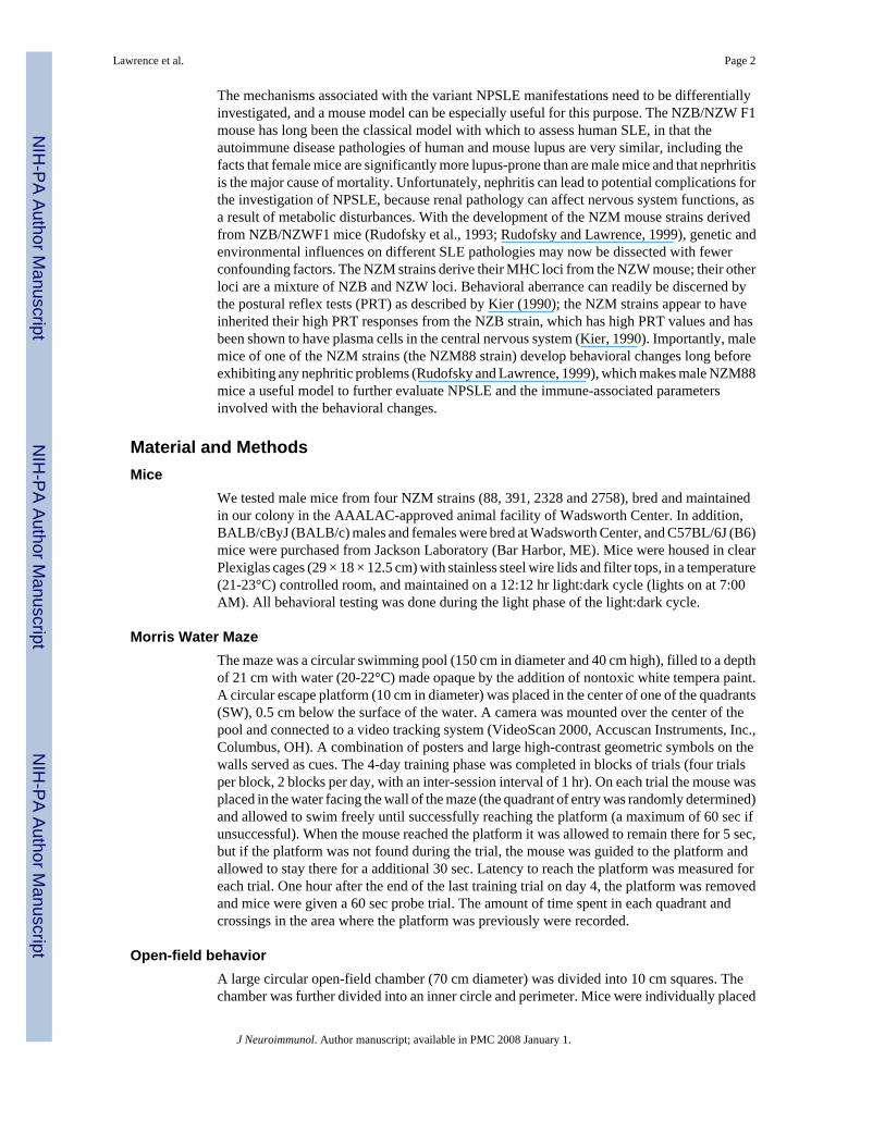
As shown (Fig. 1a), the NZM88 mice had the poorest performance in the Morris water maze,a test designed to evaluate spatial localization and memory (Morris, 1981). NZM88 malesshowed no signs of learning the test, as they did not progressively decrease the time to reachthe platform over trials. In fact, NZM88 mice displayed little swimming behavior; thus, in theprobe trial (Fig. 1b), they did not swim to the platform area, because they rarely found theplatform during training.
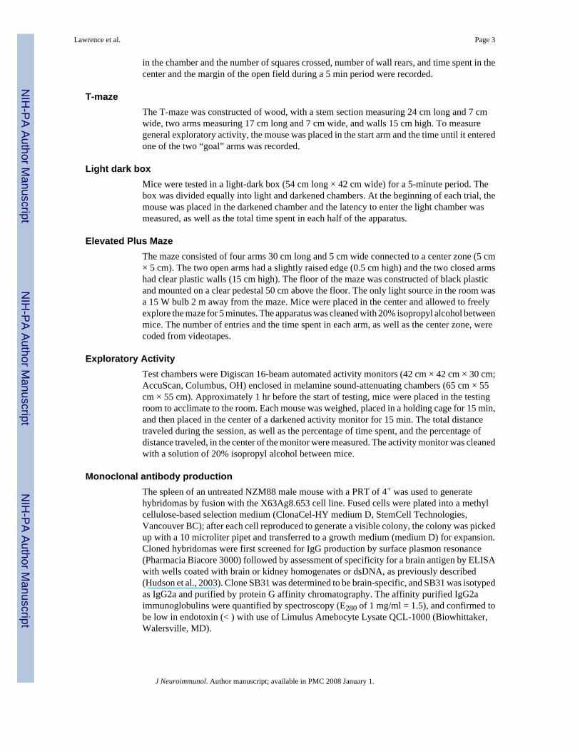
NZM88 mice also displayed other behavioral deficits (Fig. 2). NZM88 mice displayed the leasttime in the inner circle of the open field than did the other strains; however, the time did notsignificantly differ among the NZM88, 391 and 2758 strains (Fig. 2A). Assessment of open-field behavior of the NZM strains showed that NZM2758 mice were more active than werethe other strains in terms of both horizontal (number of squares crossed) and vertical (wallrears) activity (Fig. 2 B & C). The NZM2758 strain has the least lupus pathology of thesestrains (Rudofsky and Lawrence, 1999). The NZM88 males also demonstrated significantlylonger latencies to travel to the end of an arm in the T-maze. Latency was 3-fold greater forNZM88 mice than NZM2758 or NZM2328 mice, but not different from that of the NZM391mice (data not shown).
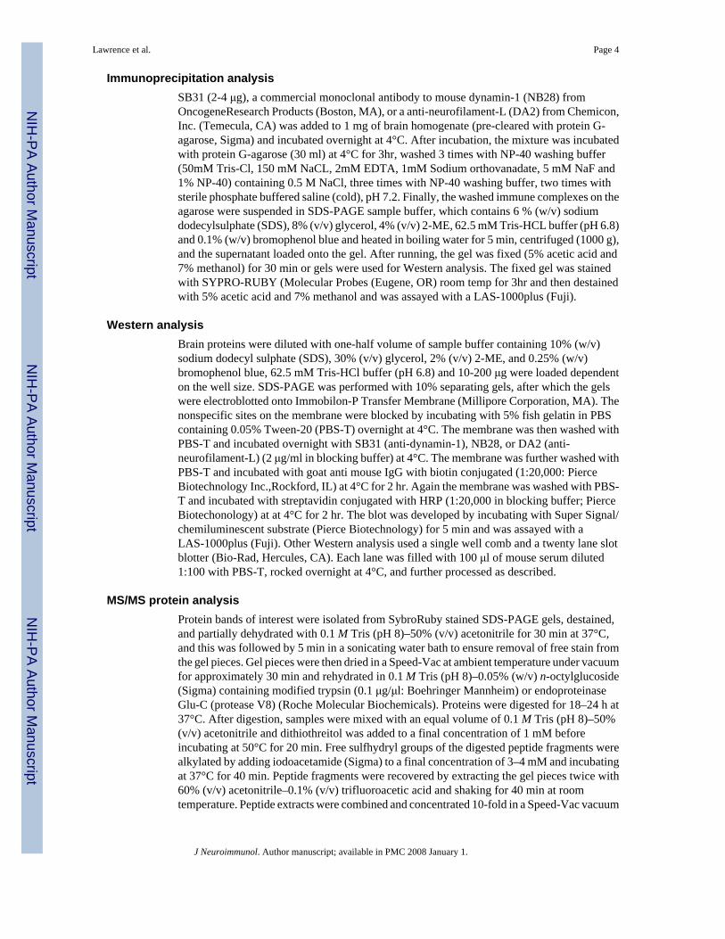
Western analysesSince SLE is principally an autoimmune disease exacerbated by antibodies to self-constituents(autoantibodies), the reactivity of serum antibodies to brain antigens was investigated (Fig. 3).The sera from various NZM88 mice displayed specificity for numerous antigens from the brainof a BALB/c SCID mouse. ELISA analysis indicated that some of these sera also bind to renalantigens and all of the sera bound to dsDNA (data not shown); however, the intensity andnumber of bands to brain antigens of each serum did not correlate with their titers to renalantigens or dsDNA.
Monoclonal antibody productionSince the NZM88 mice spontaneously develop relatively high IgG titers to numerous brainantigens, the spleen from an untreated 3 month-old male NZM88 mouse was used to generatemonoclonal antibodies. Fifty-five IgG-secreting clones were selected for further analysis.Multiple clones possessed anti-dsDNA activity. Four clones had reactivity to kidneyhomogenate antigen(s). Two clones (SB31 & SB302) reacted exclusively with brainhomogenate antigen(s). SB31 was used to immunoprecipitate brain antigens from a BALB/cSCID mouse, and the SDS-treated immunoprecipitate was assayed by SDS-PAGE (Fig. 4a).The proteins were identified by SYPRO Ruby staining. Since additional bands besides thoseof the 50-kDa H chain and the 25-kDa L chain of SB31 (IgG2a) were seen, an identical gelwas blotted, so that it could be probed with SB31 followed by HRP-goat-anti-mouse IgG(Western analysis) for determination of whether the SB31-reactive protein could be identified.Two bands, at 100- and 66-kDa, were observed by Western analysis (Fig. 4b). Both bands were
Lawrence et al. Page 6
J Neuroimmunol. Author manuscript; available in PMC 2008 January 1.
NIH
-PA Author Manuscript
NIH
-PA Author Manuscript
NIH
-PA Author Manuscript
cut from a SYPRO Ruby stained SDS-PAGE gel and processed for MS/MS analysis. The 100-kDa band was identified as dynamin-1, but the 66-kDa band was not initially identified. The66-kDa protein was believed to be a fragment of dynamin, as previously suggested (Tuma etal., 1993). The 100-kDa and 66-kDa bands were also identified with brain homogenates fromC57BL/6J mice (not shown), and NZM88 sera (Fig. 3, lanes 4, 8, and 13) demonstrated bindingto a band at about 100-kDa, which might be dynamin-1. To confirm the specificity of SB31,we immunoprecipitated brain antigens with a commercial mouse anti-dynamin-1 (NB28;OncogeneResearch Products, Boston, MA), and the anti-dynamin-1 specificity of SB31 wasconfirmed by Western analysis (Fig. 5). Further MS/MS analysis of SB31 reactive brainproteins indicated SB31 also captures neurofilament protein L (NF-L). Furtherimmunoprecipitation and Western analysis with SB31 and DA2 (anti-NF-L) has shown thatthe SB31-immunoprecipitated 66 kDa protein is neurofilament- L (68/70 kDa) in that byWestern analysis it is recognized by DA2, and vice versa, an 66 kDa proteinimmunoprecipitated by DA2 is recognized by Western analysis with SB31 (data not shown).
Induction of behavioral changes in BALB/c mice with anti-dynamin-1 (SB31)One hundred μg of SB31 or an isotype control IgG2a (PC5) was injected intravenously once-weekly into healthy BALB/c or C57BL/6J (B6) male mice, and the mice were assessed forbehavioral changes. Only the SB31-injected BALB/c mice demonstrated significant changesin behavior (Fig. 6) after 3-5 wk; SB31 (anti-dynamin-1) induced behaviors similar to thoseseen with NZM88 mice.
Brain region specificity of SB31 anti-dynamin-1To evaluate whether SB31 actually could enter the brain and have preferential binding to aparticular region, an IgG-specific ELISA was performed with proteins isolated from differentbrain regions of the BALB/c and B6 mice injected with SB31 or PC5. We had to utilize SCIDmice for this analysis, because immunocompetent BALB/c and B6 mice had too much IgGbound in the various brain regions to allow identification of the location of the SB31 or PC5.Surprisingly, SB31 bound mainly to antigens in the cortex of the BALB/c mice (Fig. 7).Significantly less SB31 bound to any brain region of B6 mice, and no PC5 binding wasobserved with either BALB/c or B6 brain homogenates. The low binding of SB31 to any B6brain region correlates well with the absence of any SB31-induced behavioral changes withB6 mice. Although negligible amounts of SB31 bound to B6 brain antigen, as stated earlier,Western analysis of BALB/c and B6 brain homogenates confirmed equivalent binding of SB31to a 99 and 66 kDa proteins (data not shown).
SB31 versus PC5 (isotype control antibody) binding to brain tissueThe two brain regions with the most SB31 anti-dynamin-1 binding were cerebellum and cortex.As shown (Fig. 8), binding was relatively restricted to vesicles. PC5 binding was negligible tobrain parenchyma; however, PC5 was still in the vasculature of the cortex (Fig. 8D), whichwas likely due to inadequate perfusion prior to tissue fixation. H & E staining was never ableto detect any gross morphological changes.
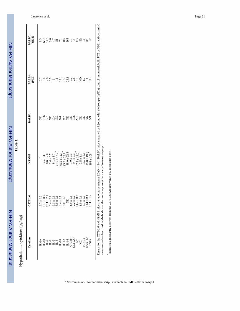
SB31 induces CNS cytokine expression mainly in the hypothalamusTo assess the influence of SB31 on brain function, cytokine expression in various brain regionswas assayed by Luminex technology. Surprisingly, although minimal amounts of SB31 boundto the hypothalamus of the BALB/c mice, hypothalamus expressed the highest increase ofmultiple cytokines. Since the SB31-treated BALB/c mice displayed behaviors similar to theuntreated NZM88 mice, their cytokine expressions were compared. Interestingly, theexpression was similar but the SB31-treated BALB/c mice had levels even higher than theNZM88 mice. Numerous cytokines were almost ten-fold higher than the control levels. For
Lawrence et al. Page 7
J Neuroimmunol. Author manuscript; available in PMC 2008 January 1.
NIH
-PA Author Manuscript
NIH
-PA Author Manuscript
NIH
-PA Author Manuscript
the other brain regions, SB31 enhanced only IL-10 in the substantia nigra and only IL-6 in thestriatum. For the SB31-injected BALB/c and untreated NZM88 mice, the most highlyexpressed cytokines were TNFα>IL-16>IL-12>IFNγ, which suggests Th1-like immunity iselevated in the hypothalamus. Surprisingly, IL-10, which is usually considered as an anti-inflammatory cytokine, also was highly expressed in the NZM88 mice and SB31 injectedBALB/c mice.
DISCUSSIONAlthough the NZM391 and NZM2328 strains have similar overall SLE pathologies as theNZM88 strain, it is interesting that more male NZM88 mice (Rudofsky and Lawrence, 1999)demonstrate 4+ PRT behaviors (Kier, 1990). It is especially important to note that male NZM88mice have behavioral changes before they demonstrate any kidney pathology (proteinuria) orhistological evidence of glomerulonephritis (Rudofsky et al., 1993; Rudofsky and Lawrence,1999).
Instead of actively swimming in the Morris water maze test, the NZM88 mice spent a largeproportion of time floating. As mice will normally try to escape from an aversive stimulus,such as submersion in a pool of water, this behavior is noteworthy. Floating or early cessationof attempts to escape has been used as a rodent model of stress-induced depression (Porsolt etal., 1977). NZM88 mice also showed significant differences in the open-field; spontaneousactivity in the open-field is one of the oldest standardized behavioral assays in rodents and hasbeen used to measure a variety of parameters, including motor function, emotionality, andcognition (Hall, 1934; Henderson, 1967; Crusio and Gerlai,1999; Crawley, 2000). Over thepast decade, it has become one of the most commonly used assays for evaluating the behaviorof knockout and transgenic mice (Bolivar et al., 2000). The tendency for the NZM88 mice tostay at the edges of the monitor (thigmotaxis) has been classified as a fear-related behavior(Treit and Fundytus, 1988). The behavior of the NZM88 mice in the Morris water maze andopen-field tests was confirmed with the T maze and light-dark box assessments, suggestingthat these mice have high anxiety and/or greater depression. As noted in a recent review, thereare no methods currently available “to fully recapitulate human depression or anxiety in themouse and indeed, (we) cannot truly know whether a mouse id depressed or feelinganxious” (Cryan and Holmes, 2005).
Even though the NZM88 mice are genetically identical (>F50), and were of the sameapproximate age (3-4 mos) and were born and raised in the same environment, substantialdifferences existed among these mice with regard to their production of IgG autoantibodies tobrain antigens. However, some of the brain-reactive autoantibodies may have specificity forother self-constituents. It has been reported that lupus autoantibodies to dsDNA have reactivitytoward other self-antigens. One such antigen was shown to be the N-methyl-D-aspartate(NMDA) receptor; these antibodies mediated apoptotic death of neurons (DeGiorgio et al.,2001). The Western analysis of the NZM88 sera clearly indicated that multiple autoantibodyspecificities are present in individual mice, and the specificities are individually different.Previous reports have demonstrated that antibodies can induce behavioral changes, e.g., anti-phospholipid antibodies caused loss of reflex, postural reflex and gripping potential in Rota-Rod test, but induced hyperactive behavior (Ziporen et al., 1997).
Since autoantibodies in the sera from NZM88 mice display binding to numerous brain antigens,the diversity of the NPSLE manifestations is not surprising. It is anticipated that furtheridentification of the brain antigens will provide indicators for the neuronal factors and functionsassociated with the varied aberrant behaviors. Furthermore, the specific regions of the braininvolved in the NPSLE manifestations may be identified through the use of isolated brain-specific NZM88 antibodies or monoclonal antibodies developed from NZM88 plasmacytes.
Lawrence et al. Page 8
J Neuroimmunol. Author manuscript; available in PMC 2008 January 1.
NIH
-PA Author Manuscript
NIH
-PA Author Manuscript
NIH
-PA Author Manuscript
In the case of SB31, this antibody bound preferentially in the cortex; an unexpected findingsince dynamin-1 is involved in synaptic-vesicle endocytosis (Robinson et al., 1994; Daly etal., 2000). There currently is no obvious reason why SB31 was mainly found in the cortex, butit apparently interferes with the endocytotic process creating substantial behavioral disturbancein BALB/c, but not B6, mice. Although low, some IgG binding was detected in a few brainregions of the B6 mice suggesting that SB31 did cross the blood brain barrier. Since BALB/cand B6 mice should express dynamin-1 in all brain regions and the Western analysis with brainhomogenates from BALB/c and B6 mice displayed the same bands, the preferential bindingof SB31 is suggested to be due to the reactive epitope of dynamin-1 being differentiallyaccessible. Although dynamin-1 is expressed more highly in the cortex than some other brainregions, the high binding observed to BALB/c cortex can not be explained by differentialexpression, in that the dynamin-1 content of cortex, amygdala, and striatum is near equal andis significantly greater than that of olfactory bulb, cerebellum, midbrain, and brainstem (Yaoet al., 2003). Thus, the presynaptic terminal membranes in the cortex of BALB/c mice mustmore freely display the epitope for SB31. The loss of dynamin-1 expression in the cortex ofAlzheimer's disease patients has been reported to correlate with loss of synapse function andcognitive decline (Faire et al., 1992).
Although SB31 clearly induces behavioral aberrance and causes a significant alteration of CNScytokine expression, the precise mechanisms involved remain to be delineated. We currentlyhave no explanation for how SB31's preferential binding to particular brain regions inducescytokine expression mainly in the hypothalamus. The neurochemical connections between thevarious brain regions need to be examined.
Identification of additional brain antigens associated with behavioral deficits, through the useof antibodies from lupus-prone mice, may aid in future diagnostic and therapeutic interventionsfor the NPSLE manifestations, as well as in treatment of psychiatric problems associated withother diseases. Although at least 19 distinct neuropsychiatric manifestations have beendelineated in SLE patients (American College of Rheumatology, 1999) and although it hasbeen over two decades since antibodies were first reported to exist in the central nervous systemof SLE patients (Bluestein et al., 1981), this is the first report of lupus-derived antibodiesmediating behavioral aberrance.
Acknowledgements
This work was supported by a grant from NIEHS (ES08983) to DAL. We thank the Biological Mass SpectrometryFacility and the Immunology Core of Wadsworth Center for their assistance.
ReferencesAiniala H, Loukkola J, Peltola J, Korpela M, Hietaharju A. The prevalence of neuropsychiatric syndromes
in systemic lupus erythematosus. Neurology 2001;57:496–500. [PubMed: 11502919]American College of Rheumatology. The American College of Rheumatology nomenclature and case
definitions for neuropsychiatric lupus syndromes. Arthritis Rheum 1999;42:599–608. [PubMed:10211873]
Bankhurst AD, Brooks WM. The incidence and prevalence of neuropsychiatric syndromes in pediatriconset systemic lupus erythematosus. J. Rheumatol 2002;29:1536–1542. [PubMed: 12136916]
Bleiberg J, Bunning R. Cognitive and physical measures in rehabilitation of patients with lupus. CurrOpin Rheumatol 1998;10:442–445. [PubMed: 9746860]
Bluestein HG, Williams GW, Steinberg AD. Cerebrospinal fluid antibodies to neuronal cells: associationwith neuropsychiatric manifestations of systemic lupus erythematosus. Am. J. Med 1981;70:240–246.[PubMed: 7468611]
Bolivar V, Cook M, Flaherty L. List of transgenic and knockout mice: behavioral profiles. MammGenome 2000;11:160–274.
Lawrence et al. Page 9
J Neuroimmunol. Author manuscript; available in PMC 2008 January 1.
NIH
-PA Author Manuscript
NIH
-PA Author Manuscript
NIH
-PA Author Manuscript
Brey RL, Holliday SL, Saklad AR, Navarrete MG, Hermosillo-Romo D, Stallworth CL, Valdez CR,Escalante A, del Rincon I, Gronseth G, Rhine CB, Padilla P, McGlasson D. Neuropsychiatricsyndromes in lupus: prevalence using standardized definitions. Neurology 2002;58:1214–1220.[PubMed: 11971089]
Crawley, JN. What's Wrong With My Mouse?. Wiley-Liss; New York: 2000.Crusio, WE.; Gerlai, RT. Handbook of Molecular-Genetic Techniques for Brain and Behavior Research.
Elsevier; Amsterdam: 1999.Cryan JF, Holmes A. The ascent of mouse: advances in modeling human depression and anxiety. Nature
Rev 2005;4:775–790.Daly C, Sugimori M, Moreira JE, Ziff EB, Llinas R. Synaptophysin regulates clathrin-independent
endocytosis of synaptic vesicles. Proc. Natl. Acad. Sci. U S A 2000;97:6120–6125. [PubMed:10823955]
DeGiorgio LA, Konstantinov KN, Lee SC, Hardin JA, Volpe BT, Diamond B. A subset of lupus anti-DNA antibodies cross-reacts with the NR2 glutamate receptor in systemic lupus erythematosus. Nat.Med 2001;7:1189–1193. [PubMed: 11689882]
Faire K, Trent F, Tepper JM, Bonder EM. Analysis of dynamin isoforms in mammalian brain: dynamin-1expression is spatially and temporally regulated during postnatal development. Proc Natl Acad SciU S A 1992;89:8376–8380. [PubMed: 1387713]
Hall CS. Emotional behavior in the rat. J. Comparative Psychol 1934;18:385–403.Henderson ND. Prior treatment effects on open field behaviour of mice--a genetic analysis. Anim. Behav
1967;15:364–376. [PubMed: 6030962]Hanly JG. Evaluation of patients with CNS involvement in SLE. Baillieres Clin. Rheumatol 1998;12:415–
431. [PubMed: 9890105]Hay EM. Psychiatric disorder and cognitive impairment in SLE. Lupus 1994;3:145–148. [PubMed:
7951298]Hietaharju A, Jantti V, Korpela M, Frey H. Nervous system involvement in systemic lupus erythematosus,
Sjogren syndrome and scleroderma. Acta. Neurol. Scand 1993;88:299–308. [PubMed: 8256578]Hudson CA, Cao L, Kasten-Jolly J, Kirkwood JN, Lawrence DA. Susceptibility of lupus-prone NZM
mouse strains to lead exacerbation of systemic lupus erythematosus symptoms. J. Toxicol Environ.Health 2003;66:895–918.
Hudson CA, Mondal K, Cao L, Kasten-Jolly J, Huber VC, Lawrence DA. The dietary supplementephedrine induces β-adrenergic mediated exacerbation of systemic lupus erythematosus in NZM391mice. Lupus 2004;14:295–307.
Kier AB. Clinical neurology and brain histopathology in NZB/NZW F1 lupus mice. J. Comp. Pathol1990;102:165–177. [PubMed: 2324339]
Morris RGM. Spatial localization does not require the presence of local cues. Learning Motivation1981;12:239–260.
Monastero R, Bettini P, Del Zotto E, Cottini E, Tincani A, Balestrieri G, Cattaneo R, Camarda R, VignoloLA, Padovani A. Prevalence and pattern of cognitive impairment in systemic lupus erythematosuspatients with and without overt neuropsychiatric manifestations. J. Neurol. Sci 2001;184:33–39.[PubMed: 11231030]
Nadeau SE. Neurologic manifestations of connective tissue disease. Neurol. Clin 2002;20:151–178.[PubMed: 11754305]
Navarrete MG, Brey RL. Neuropsychiatric Systemic Lupus Erythematosus. Curr. Treat. Options Neurol2000;2:473–485. [PubMed: 11096772]
Porsolt RD, LePichon M, Jalfre M. Depression: a new animal model sensitive to antidepressanttreatments. Nature 1977;266:730–732. [PubMed: 559941]
Robinson PJ, Liu JP, Powell KA, Fykse EM, Sudhof TC. Phosphorylation of dynamin I and synaptic-vesicle recycling. Trends Neurosci 1994;17:348–353. [PubMed: 7526507]
Rudofsky UH, Evans BD, Balaban SL, Mottiron VD, Gabrielsen AE. Differences in expression of lupusnephritis in New Zealand mixed H-2z homozygous inbred strains of mice derived from New Zealandblack and New Zealand white mice. Origins and initial characterization. Lab. Invest 1993;68:419–426. [PubMed: 8479150]
Lawrence et al. Page 10
J Neuroimmunol. Author manuscript; available in PMC 2008 January 1.
NIH
-PA Author Manuscript
NIH
-PA Author Manuscript
NIH
-PA Author Manuscript
Rudofsky UH, Lawrence DA. New Zealand mixed mice: a genetic systemic lupus erythematosus modelfor assessing environmental effects. Environ. Health Perspect 1999;107(Suppl 5):713–721.[PubMed: 10502536]
Schor NF. Neurology of systemic autoimmune disorders: a pediatric perspective. Semin. Pediatr. Neurol2000;7:108–117. [PubMed: 10914412]
Sibbitt WL Jr, Sibbitt RR, Brooks WM. Neuroimaging in neuropsychiatric systemic lupus erythematosus.Arthritis Rheum 1999;42:2026–2038. [PubMed: 10524673]
Sibbitt WL Jr, Haseler LJ, Griffey RR, Friedman SD, Brooks WM. Neurometabolism of activeneuropsychiatric lupus determined with proton MR spectroscopy. AJNR Am J Neuroradiol1997;18:1271–1277. [PubMed: 9282854]
Sibbitt WL Jr, Brandt JR, Johnson CR, Maldonado ME, Patel SR, Ford CC, Sibley JT, Olszynski WP,Decoteau WE, Sundaram MB. The incidence and prognosis of central nervous system disease insystemic lupus erythematosus. Rheumatol 1992;19:47–52.
Treit D, Fundytus M. Thigmotaxis as a test for anxiolytic activity in rats. Pharmacol. Biochem. Behav1988;31:959–962. [PubMed: 3252289]
Tuma PL, Stachniak MC, Collins CA. Activation of dynamin GTPase by acidic phospholipids andendogenous rat brain vesicles. J Biol Chem 1993;268:17240–17246. [PubMed: 8349610]
Van Dam AP. Diagnosis and pathogenesis of CNS lupus. Rheumatol. Intl 1991;11:1–11.West SG. Neuropsychiatric lupus. Rheum. Dis. Clin. North Am 1994;20:129–158. [PubMed: 8153397]Yao PJ, Zhu M, Pyun EI, Brooks AI, Therianos S, Meyers VE, Coleman PD. Defects in expression of
genes related to synaptic vesicle trafficking in frontal cortex of Alzheimer's disease. Neurobiol Dis2003;12:97–109. [PubMed: 12667465]
Ziporen L, Shoenfeld Y, Levy Y, Korczyn AD. Neurological ysfunction and hyperactive behaviorassociated with antiphospholipid antibodies. J. Clin. Invest 1997;100:613–619. [PubMed: 9239409]
Lawrence et al. Page 11
J Neuroimmunol. Author manuscript; available in PMC 2008 January 1.
NIH
-PA Author Manuscript
NIH
-PA Author Manuscript
NIH
-PA Author Manuscript
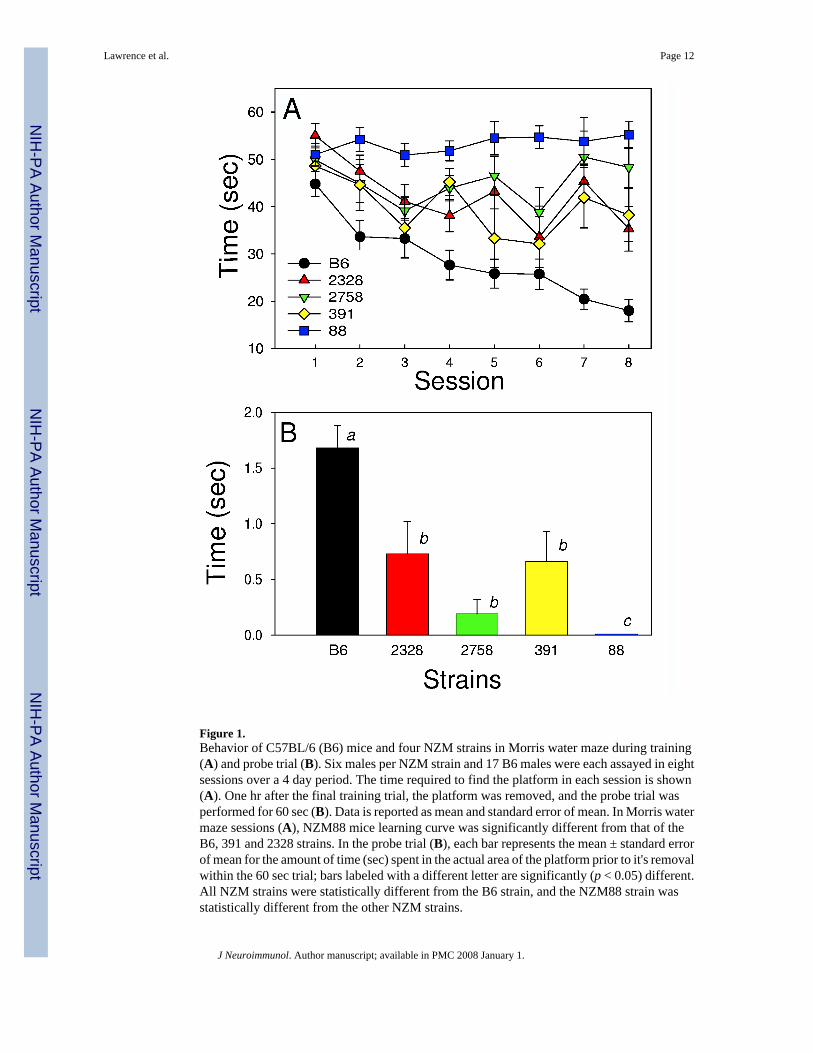
Figure 1.Behavior of C57BL/6 (B6) mice and four NZM strains in Morris water maze during training(A) and probe trial (B). Six males per NZM strain and 17 B6 males were each assayed in eightsessions over a 4 day period. The time required to find the platform in each session is shown(A). One hr after the final training trial, the platform was removed, and the probe trial wasperformed for 60 sec (B). Data is reported as mean and standard error of mean. In Morris watermaze sessions (A), NZM88 mice learning curve was significantly different from that of theB6, 391 and 2328 strains. In the probe trial (B), each bar represents the mean ± standard errorof mean for the amount of time (sec) spent in the actual area of the platform prior to it's removalwithin the 60 sec trial; bars labeled with a different letter are significantly (p < 0.05) different.All NZM strains were statistically different from the B6 strain, and the NZM88 strain wasstatistically different from the other NZM strains.
Lawrence et al. Page 12
J Neuroimmunol. Author manuscript; available in PMC 2008 January 1.
NIH
-PA Author Manuscript
NIH
-PA Author Manuscript
NIH
-PA Author Manuscript
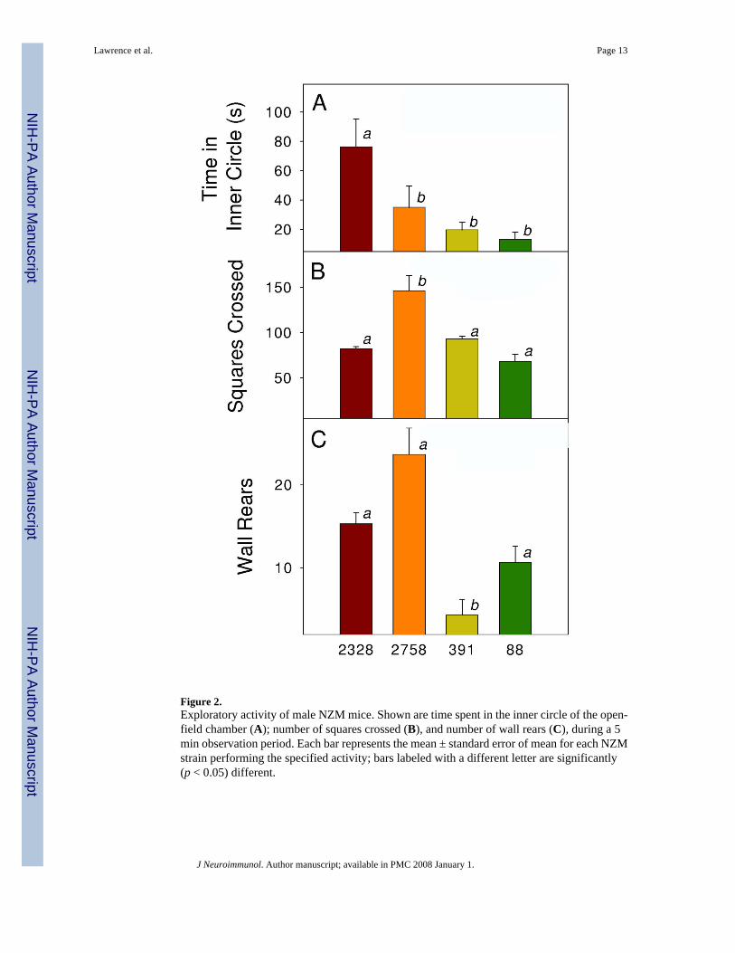
Figure 2.Exploratory activity of male NZM mice. Shown are time spent in the inner circle of the open-field chamber (A); number of squares crossed (B), and number of wall rears (C), during a 5min observation period. Each bar represents the mean ± standard error of mean for each NZMstrain performing the specified activity; bars labeled with a different letter are significantly(p < 0.05) different.
Lawrence et al. Page 13
J Neuroimmunol. Author manuscript; available in PMC 2008 January 1.
NIH
-PA Author Manuscript
NIH
-PA Author Manuscript
NIH
-PA Author Manuscript
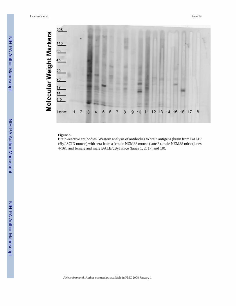
Figure 3.Brain-reactive antibodies. Western analysis of antibodies to brain antigens (brain from BALB/cByJ SCID mouse) with sera from a female NZM88 mouse (lane 3), male NZM88 mice (lanes4-16), and female and male BALB/cByJ mice (lanes 1, 2, 17, and 18).
Lawrence et al. Page 14
J Neuroimmunol. Author manuscript; available in PMC 2008 January 1.
NIH
-PA Author Manuscript
NIH
-PA Author Manuscript
NIH
-PA Author Manuscript
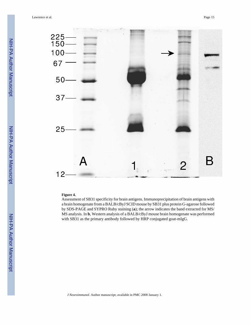
Figure 4.Assessment of SB31 specificity for brain antigens. Immunoprecipitation of brain antigens witha brain homogenate from a BALB/cByJ SCID mouse by SB31 plus protein G-agarose followedby SDS-PAGE and SYPRO Ruby staining (a); the arrow indicates the band extracted for MS/MS analysis. In b, Western analysis of a BALB/cByJ mouse brain homogenate was performedwith SB31 as the primary antibody followed by HRP conjugated goat-mIgG.
Lawrence et al. Page 15
J Neuroimmunol. Author manuscript; available in PMC 2008 January 1.
NIH
-PA Author Manuscript
NIH
-PA Author Manuscript
NIH
-PA Author Manuscript
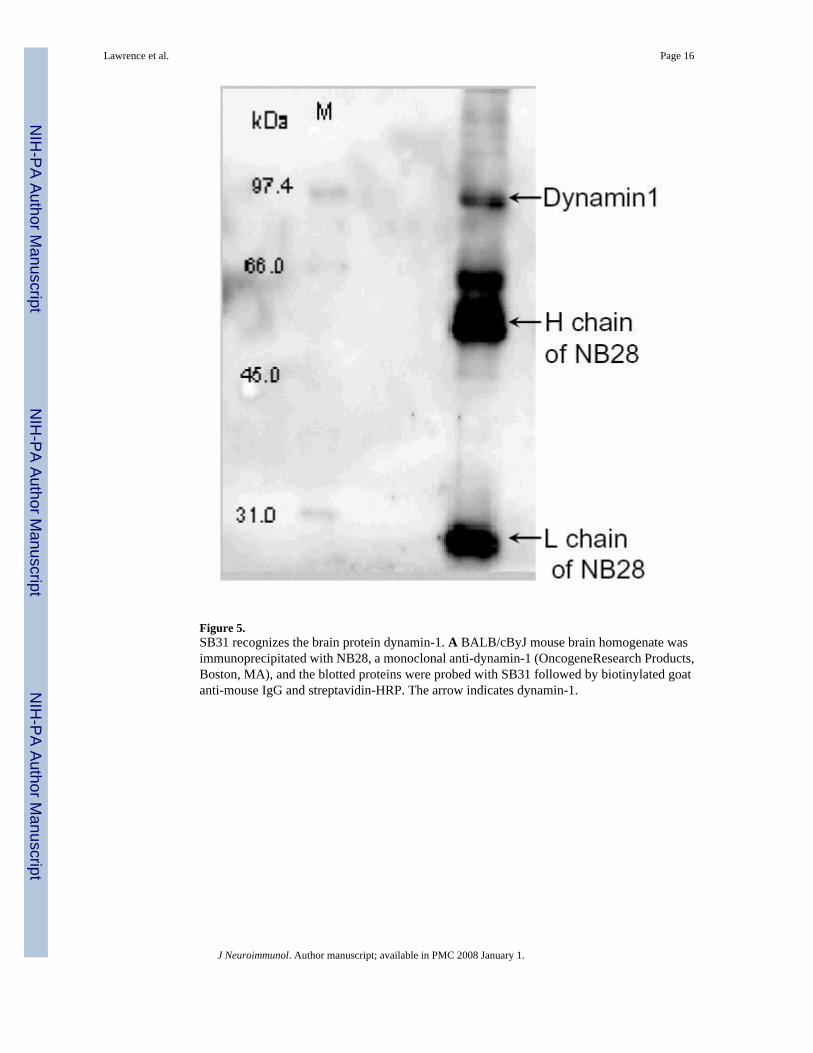
Figure 5.SB31 recognizes the brain protein dynamin-1. A BALB/cByJ mouse brain homogenate wasimmunoprecipitated with NB28, a monoclonal anti-dynamin-1 (OncogeneResearch Products,Boston, MA), and the blotted proteins were probed with SB31 followed by biotinylated goatanti-mouse IgG and streptavidin-HRP. The arrow indicates dynamin-1.
Lawrence et al. Page 16
J Neuroimmunol. Author manuscript; available in PMC 2008 January 1.
NIH
-PA Author Manuscript
NIH
-PA Author Manuscript
NIH
-PA Author Manuscript
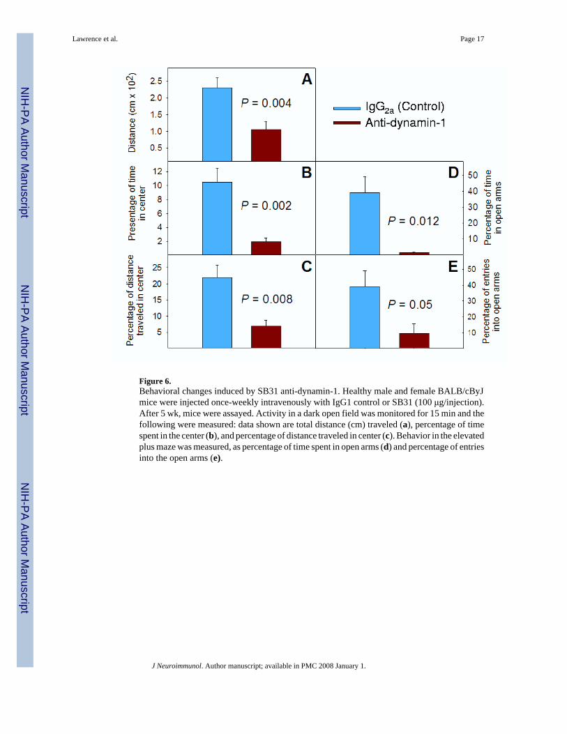
Figure 6.Behavioral changes induced by SB31 anti-dynamin-1. Healthy male and female BALB/cByJmice were injected once-weekly intravenously with IgG1 control or SB31 (100 μg/injection).After 5 wk, mice were assayed. Activity in a dark open field was monitored for 15 min and thefollowing were measured: data shown are total distance (cm) traveled (a), percentage of timespent in the center (b), and percentage of distance traveled in center (c). Behavior in the elevatedplus maze was measured, as percentage of time spent in open arms (d) and percentage of entriesinto the open arms (e).
Lawrence et al. Page 17
J Neuroimmunol. Author manuscript; available in PMC 2008 January 1.
NIH
-PA Author Manuscript
NIH
-PA Author Manuscript
NIH
-PA Author Manuscript
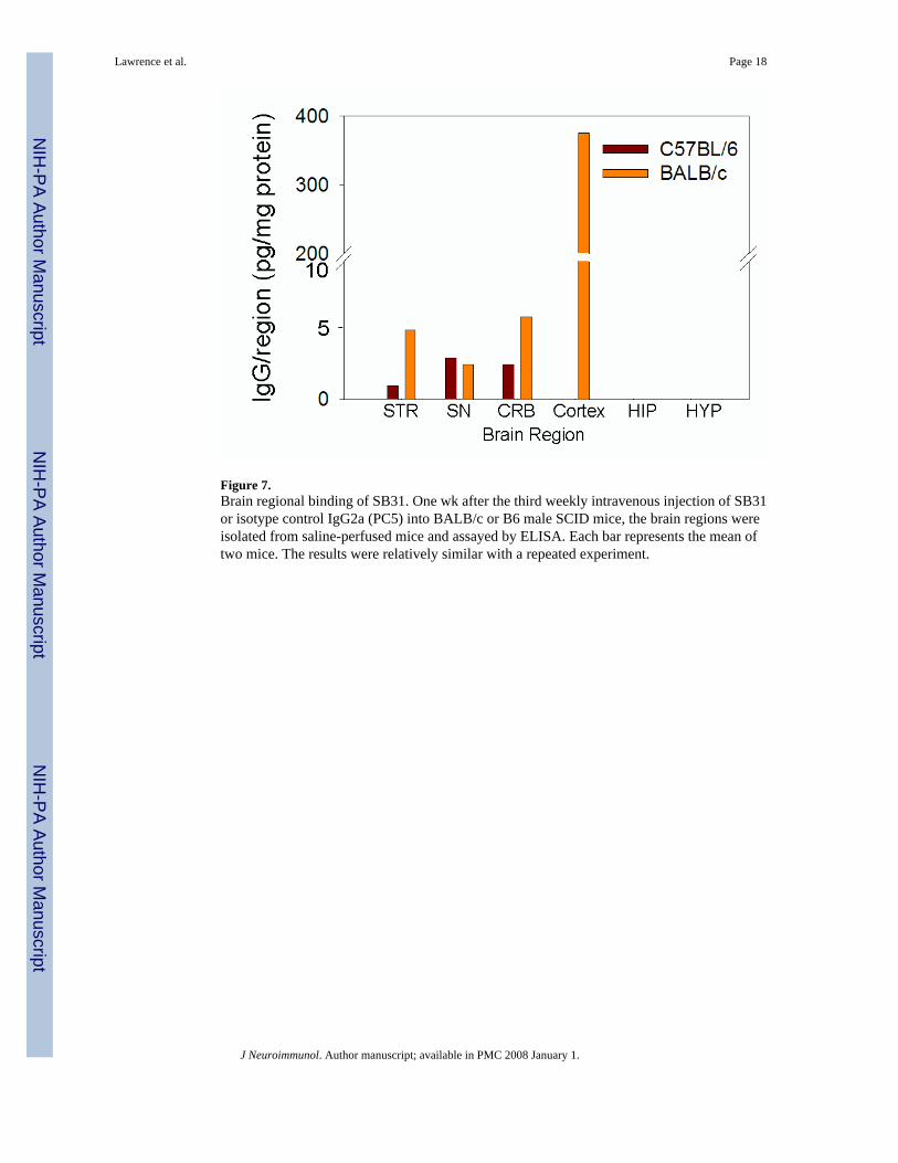
Figure 7.Brain regional binding of SB31. One wk after the third weekly intravenous injection of SB31or isotype control IgG2a (PC5) into BALB/c or B6 male SCID mice, the brain regions wereisolated from saline-perfused mice and assayed by ELISA. Each bar represents the mean oftwo mice. The results were relatively similar with a repeated experiment.
Lawrence et al. Page 18
J Neuroimmunol. Author manuscript; available in PMC 2008 January 1.
NIH
-PA Author Manuscript
NIH
-PA Author Manuscript
NIH
-PA Author Manuscript
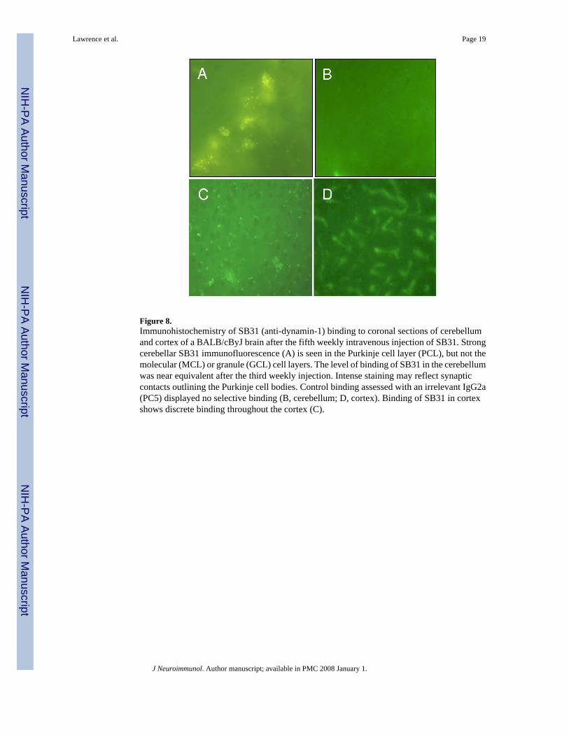
Figure 8.Immunohistochemistry of SB31 (anti-dynamin-1) binding to coronal sections of cerebellumand cortex of a BALB/cByJ brain after the fifth weekly intravenous injection of SB31. Strongcerebellar SB31 immunofluorescence (A) is seen in the Purkinje cell layer (PCL), but not themolecular (MCL) or granule (GCL) cell layers. The level of binding of SB31 in the cerebellumwas near equivalent after the third weekly injection. Intense staining may reflect synapticcontacts outlining the Purkinje cell bodies. Control binding assessed with an irrelevant IgG2a(PC5) displayed no selective binding (B, cerebellum; D, cortex). Binding of SB31 in cortexshows discrete binding throughout the cortex (C).
Lawrence et al. Page 19
J Neuroimmunol. Author manuscript; available in PMC 2008 January 1.
NIH
-PA Author Manuscript
NIH
-PA Author Manuscript
NIH
-PA Author Manuscript
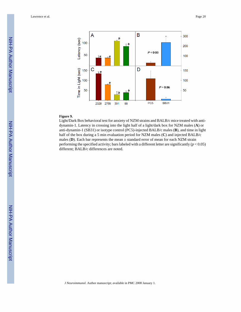
Figure 9.Light/Dark Box behavioral test for anxiety of NZM strains and BALB/c mice treated with anti-dynamin-1. Latency in crossing into the light half of a light/dark box for NZM males (A) oranti-dynamin-1 (SB31) or isotype control (PC5)-injected BALB/c males (B), and time in lighthalf of the box during a 5 min evaluation period for NZM males (C) and injected BALB/cmales (D). Each bar represents the mean ± standard error of mean for each NZM strainperforming the specified activity; bars labeled with a different letter are significantly (p < 0.05)different; BALB/c differences are noted.
Lawrence et al. Page 20
J Neuroimmunol. Author manuscript; available in PMC 2008 January 1.
NIH
-PA Author Manuscript
NIH
-PA Author Manuscript
NIH
-PA Author Manuscript
NIH
-PA Author Manuscript
NIH
-PA Author Manuscript
NIH
-PA Author Manuscript
Lawrence et al. Page 21Ta
ble
1H
ypot
hala
mic
cyt
okin
es (p
g/m
g)
Cyt
okin
eC
57B
L/6
NZ
M88
BA
LB
/cB
AL
B/c
(PC
5)B
AL
B/c
(SB
31)
IL-1α
8.7
± 0.
50*
ND
0.7
8.3
IL-1β
17.8
± 0
.517
.4 ±
4.3
19.6
8.8
60.0
IL-2
28.1
± 2
.18.
6 ±
2.0
12.1
2.6
17.0
IL-3
0.4
± 0.
12.
4 ±
0.7
ND
0.5
3.6
IL-5
2.1
± 0.
18.
1 ±
1.7
10.4
14.
7IL
-63.
0 ±
0.1
41.5
± 1
2.2*
19.3
1151
IL-1
03.
4 ±
0.1
45.5
± 1
2.7*
9.4
13.0
70IL
-12
8.0
± 0.
593
.7 ±
23.
7*9.
717
.018
9IL
-16
ND
88.0
± 2
2.8
ND
28.1
268
G-C
SF1.
0 ±
0.1
1.0
± 0.
3N
D0.
21.
7G
M-C
SF2.
2 ±
0.2
9.1
± 0.
319
.62.
816
IFNγ
14.7
± 0
.747
.5 ±
14.
0*20
.112
119
KC
1.0
± 0.
12.
5 ±
1.1
ND
ND
ND
MIP
-1α
5.7
± 0.
615
.7 ±
6.6
ND
1.7
16R
AN
TES
11.1
± 0
.435
.8 ±
10.
6N
D14
84TN
Fα17
.1 ±
1.5
364
± 10
8*5.
911
163
2
Res
ults
for t
he C
57B
L/6
and
NZM
88 m
ice
are
repo
rted
as m
ean
± SD
(N =
3-4
). B
ALB
/c m
ice
untre
ated
or i
njec
ted
with
the
isot
ype
(IgG
2a) c
ontro
l im
mun
oglo
bulin
PC
5 or
SB
31 a
nti-d
ynam
in-1
wer
e as
saye
d as
des
crib
ed in
Met
hods
, and
the
resu
lts re
pres
ent t
he m
ean
of tw
o m
ice/
grou
p.
* indi
cate
s sig
nific
antly
diff
eren
t fro
m th
e C
57B
L/6
cyto
kine
val
ue. N
D m
eans
not
don
e.
J Neuroimmunol. Author manuscript; available in PMC 2008 January 1.


