Embed Size (px)
Citation preview

A discrete amino terminal domain of Kv1.5 and Kv1.4 potassium
channels interacts with the spectrin repeats of K-actinin-2
D. Cukovica, G.W-K. Lua, B. Wibleb, D.F. Steelea, D. Fedidaa;*aDepartment of Physiology, University of British Columbia, 2146 Health Sciences Mall, Vancouver, BC, Canada V6T 1Z3bRammelkamp Center for Research, Metrohealth Medical Center, Case Western Reserve University, Cleveland, OH, USA
Received 9 March 2001; revised 2 May 2001; accepted 3 May 2001
First published online 18 May 2001
Edited by Gianni Cesareni
Abstract The interaction between the amino terminus of Kv1-type potassium channels and KK-actinin-2 has been investigated.Using a combination of yeast two-hybrid analysis and in vitrobinding assays, KK-actinin-2 was found to bind to the N-termini ofboth Kv1.4 and Kv1.5 but not to the equivalent segments ofKv1.1, Kv1.2 or Kv1.3. Deletion analysis in the in vitro bindingassays delineated the actinin binding region of Kv1.5 to betweenamino acids 73 and 148 of the channel. The Kv1.5 binding sites inKK-actinin-2 were found to lie within actinin's internal spectrinrepeats. Unlike the reported interaction between actinin and theNMDA receptor, calmodulin was found to have no effect onactinin binding to Kv1.5. ß 2001 Published by Elsevier ScienceB.V. on behalf of the Federation of European Biochemical So-cieties.
Key words: Potassium channel; Kv1.5; Actinin-2;Cytoskeleton
1. Introduction
Made up of three fundamental kinds of protein ¢laments,
microtubules, intermediate ¢laments and actin ¢laments, the
cytoskeleton is a complex group of structures involved in vir-
tually every cellular process [1,2]. Actin ¢laments provide me-
chanical strength to the cell, and, like the microtubules, are
important in cytokinesis. Forming a band just beneath the
plasma membrane, the actin cytoskeleton serves also to an-
chor large proteins in the membrane.
Among the proteins likely anchored to the actin cytoskele-
ton are a number of ion channels. The NR2 subunit of the
NMDA receptor, muscle sodium channels, and the voltage-
regulated potassium channel Kv1.5 have all been shown to
associate with molecules linked to the actin cytoskeleton [3^
5]. Calcium-mediated actin depolymerization reduces NMDA
receptor activity [6] and cytochalasin a¡ects the expression of
sodium channels [7,8], and a number of potassium channels
[9^12], including Kv1.5 [5]. Using a yeast two-hybrid system,
we have previously shown that Kv1.5 binds to the actin cy-
toskeleton linking protein, K-actinin-2. We also demonstrated
that the channel and K-actinin-2 are closely associated when
co-expressed in HEK cells, that the two could be co-immuno-
precipitated, and that disruption of their association caused
signi¢cant increases in Kv1.5 current sizes. In this study, we
have used in vitro techniques to demonstrate that K-actinin-2
binds a speci¢c region in the Kv1.4 and Kv1.5 N-termini via
its central spectrin repeats. We further show that calcium and
calmodulin have no e¡ect on the association and that the K-
actinin-2 interaction is speci¢c to Kv1.5 and its close homo-
log, Kv1.4. No such binding of K-actinin-2 occurs with Kv1.1,
Kv1.2 or Kv1.3. These results con¢rm our previous ¢ndings
and provide further insight into the role of the actin cytoskel-
eton in the regulation of Kv1.5.
2. Materials and methods
2.1. Deletion constructsDNA encoding the Kv1.5 N-terminus, and internal deletions, were
cloned as NcoI fragments into either pET42 or pET28 vectors (No-vagen, Madison, WI, USA) as appropriate. Sequence-con¢rmed PCR-derived segments encoding N-terminal fragments of Kv1.1 (aa 1^167),Kv1.2 (aa 1^164), Kv1.3 (aa 1^182) and Kv1.4 (aa 1^305) were clonedas EcoRI^SalI fragments into pGBT9 or pGAD424. The Kv1.1 andKv1.4 N-termini were similarly cloned into pET42. Deletions inKv1.5-encoding segments were made by restriction digests or internaldigestion followed by incubation with nuclease Bal31 for varyingtimes. Digestions were stopped by addition of 20 mM EGTA, andthe DNA was ligated, then recovered after transformation into Esche-richia coli. The presence of in-frame fusions with the glutathione-S-transferase (GST)-tag was con¢rmed by DNA sequencing. K-Actinin-2 deletions were made in appropriate pET21 or pET42 vectors bydigestion with restriction endonucleases followed by religation in theabsence of deleted sequences.
2.2. Preparation of GST- and T7-tagged proteinsRecombinant proteins were expressed in E. coli strain BL21(DE3)
and puri¢ed using BugBuster Extraction Reagent (Novagen). Proteinsdetected in the soluble fraction by Coomassie staining of sodiumdodecyl sulfate^polyacrylamide gel electrophoresis (SDS^PAGE)gels were puri¢ed using T7-tag a¤nity-puri¢ed kit and GST bind resinand bu¡er kit (Novagen). Proteins expressed in the insoluble fraction(inclusion bodies) were washed, and solubilized with 1Usolubilizationbu¡er with 0.3% N-lauroylsarcosine according to Novagen's recom-mendations. They were then dialyzed in 0.02 M Tris^HCl to removeresidual detergent.
2.3. Protein^protein interactions by the yeast two-hybrid systemN-terminal fragments of Kv1.1, Kv1.2, Kv1.3 and Kv1.4 fusions to
the GAL4 DNA binding domain in pGBT9 were tested in yeast strainPJ69-4a [13] against the K-actinin-2 fragment in pactGAD [5]. Meth-ods were as previously described [5].
2.4. In vitro binding assaysApproximately 2 Wg (normalized by comparison to standards on
Coomassie-stained SDS^PAGE gels) of GST, GST N-terminus ofKv1.1, GST N-terminus of Kv1.4, GST N-terminus of Kv1.5 andother GST-fused Kv1.5 fragments (B, C, D, E, F, G and H) werecombined with 2 Wg of T7 fusion K-actinin-2 fragments (clones 1, 2, 3,and 4) in binding bu¡er (2 mM Tris^Cl, pH 7.5, 150 mM NaCl, 1 mMEDTA, 0.5 mM dithiothreitol, 0.1% Triton X-100) [14]. The mixtureswere incubated at room temperature for 1 h with periodic mixing.
0014-5793 / 01 / $20.00 ß 2001 Published by Elsevier Science B.V. on behalf of the Federation of European Biochemical Societies.
PII: S 0 0 1 4 - 5 7 9 3 ( 0 1 ) 0 2 5 0 5 - 4
*Corresponding author. Fax: (1)-604-822 6048.E-mail: [email protected]
FEBS 24928 FEBS Letters 498 (2001) 87^92
Glutathione-Sepharose beads 4B (Amersham Pharmacia) washed inthe binding bu¡er were added to each tube and incubated with mixingfor 30 min at room temperature. The mixtures were spun for 5 min at1000 rpm and pelleted beads were washed and repelleted four times inwash bu¡er (25 mM Tris^HCl, pH 7.5, 1 mM EDTA, 0.5 mM dithio-threitol) [14]. The pelleted bead^protein complexes were boiled in SDSsample bu¡er for 5 min. Aliquots containing 0.2 Wg of the GST fusionwere then resolved by SDS^PAGE. The proteins were transferred toPVDF membranes (Amersham) and probed as Western blots withmonoclonal T7 antibodies (dilution 1:10 000, Novagen). The T7-tagwas deleted in the cloning of fragment 4 of K-actinin-2 for this work,so polyclonal anti-K-actinin antibodies (dilution 1:500, Sigma A3144)were used to detect the protein in Western blots where this fragmentwas used. Antibody binding in all cases was detected using horserad-ish peroxidase-labeled secondary antibodies and a chemiluminescentreagent (ECL, New England Nuclear). To check equivalent loading ofconstructs, identical quantities of GST fusion proteins were subjectedto PAGE on a separate gel and stained with Coomassie blue. Thesame procedure was used in the reverse experiments in which thebinding of GST-fused K-actinin-2 fragments (5, 6, 7, 8, and 9) wastested for their abilities to bind a T7-tagged N-terminus of Kv1.5.
2.5. Calmodulin binding experimentsGST-tagged Kv1.5 N-terminus was incubated with T7-tagged K-
actinin-2 in binding bu¡er containing 2 mM Tris^Cl, pH 7.5, 150mM NaCl, 1 mM EDTA, 0.5 mM dithiothreitol, 0.1% Triton X-100. Calcium was added, where appropriate, to a nominal concentra-tion of 2.5 mM. 1.2 WM calmodulin was included in calmodulin com-petition experiments. Detection of bound protein was as described inSection 2.4, with anti-T7 (1:10 000 dilution). Calmodulin was ob-tained from Calbiochem (La Jolla, CA, USA).
3. Results
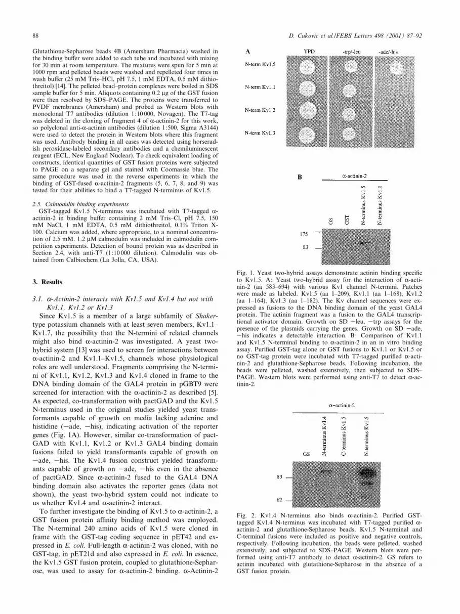
3.1. K-Actinin-2 interacts with Kv1.5 and Kv1.4 but not with
Kv1.1, Kv1.2 or Kv1.3
Since Kv1.5 is a member of a large subfamily of Shaker-
type potassium channels with at least seven members, Kv1.1^
Kv1.7, the possibility that the N-termini of related channels
might also bind K-actinin-2 was investigated. A yeast two-
hybrid system [13] was used to screen for interactions between
K-actinin-2 and Kv1.1^Kv1.5, channels whose physiological
roles are well understood. Fragments comprising the N-termi-
ni of Kv1.1, Kv1.2, Kv1.3 and Kv1.4 cloned in frame to the
DNA binding domain of the GAL4 protein in pGBT9 were
screened for interaction with the K-actinin-2 as described [5].
As expected, co-transformation with pactGAD and the Kv1.5
N-terminus used in the original studies yielded yeast trans-
formants capable of growth on media lacking adenine and
histidine (3ade, 3his), indicating activation of the reporter
genes (Fig. 1A). However, similar co-transformation of pact-
GAD with Kv1.1, Kv1.2 or Kv1.3 GAL4 binding domain
fusions failed to yield transformants capable of growth on
3ade, 3his. The Kv1.4 fusion construct yielded transform-
ants capable of growth on 3ade, 3his even in the absence
of pactGAD. Since K-actinin-2 fused to the GAL4 DNA
binding domain also activates the reporter genes (data not
shown), the yeast two-hybrid system could not indicate to
us whether Kv1.4 and K-actinin-2 interact.
To further investigate the binding of Kv1.5 to K-actinin-2, a
GST fusion protein a¤nity binding method was employed.
The N-terminal 240 amino acids of Kv1.5 were cloned in
frame with the GST-tag coding sequence in pET42 and ex-
pressed in E. coli. Full-length K-actinin-2 was cloned, with no
GST-tag, in pET21d and also expressed in E. coli. In essence,
the Kv1.5 GST fusion protein, coupled to glutathione-Sephar-
ose, was used to assay for K-actinin-2 binding. K-Actinin-2
Fig. 1. Yeast two-hybrid assays demonstrate actinin binding speci¢cto Kv1.5. A: Yeast two-hybrid assay for the interaction of K-acti-nin-2 (aa 583^694) with various Kv1 channel N-termini. Patcheswere made as labeled. Kv1.5 (aa 1^209), Kv1.1 (aa 1^168), Kv1.2(aa 1^164), Kv1.3 (aa 1^182). The Kv channel sequences were ex-pressed as fusions to the DNA binding domain of the yeast GAL4protein. The actinin fragment was a fusion to the GAL4 transcrip-tional activator domain. Growth on SD 3leu, 3trp assays for thepresence of the plasmids carrying the genes. Growth on SD 3ade,3his indicates a detectable interaction. B: Comparison of Kv1.1and Kv1.5 N-terminal binding to K-actinin-2 in an in vitro bindingassay. Puri¢ed GST-tag alone or GST fusions to Kv1.1 or Kv1.5 orno GST-tag protein were incubated with T7-tagged puri¢ed K-acti-nin-2 and glutathione-Sepharose beads. Following incubation, thebeads were pelleted, washed extensively, then subjected to SDS^PAGE. Western blots were performed using anti-T7 to detect K-ac-tinin-2.
Fig. 2. Kv1.4 N-terminus also binds K-actinin-2. Puri¢ed GST-tagged Kv1.4 N-terminus was incubated with T7-tagged puri¢ed K-actinin-2 and glutathione-Sepharose beads. Kv1.5 N-terminal andC-terminal fusions were included as positive and negative controls,respectively. Following incubation, the beads were pelleted, washedextensively, and subjected to SDS^PAGE. Western blots were per-formed using anti-T7 antibody to detect K-actinin-2. GS refers toactinin incubated with glutathione-Sepharose in the absence of aGST fusion protein.
D. Cukovic et al./FEBS Letters 498 (2001) 87^9288

was mixed with the Kv1.5 GST and, after extensive washing,
the complexes were pelleted, boiled in sample bu¡er and used
for Western analysis, probing for K-actinin-2. Controls were
included to ensure that actinin was binding speci¢cally to the
N-terminus, and not binding to GST or glutathione-Sephar-
ose. As shown in Fig. 1B, K-actinin-2 did bind Kv1.5 in this
assay. When a similarly GST-tagged Kv1.1 N-terminus was
used in place of Kv1.5 in otherwise identical in vitro binding
assays, no K-actinin-2 binding could be detected. Thus, it
would appear that K-actinin-2 binding is not a universal prop-
erty of Kv channels and may be speci¢c to only a few. The
same approach was used to determine whether the Kv1.4 N-
terminus also bound K-actinin-2. Most likely due to codon-
usage problems, we were unable to express the full Kv1.4 N-
terminus, but a GST fusion to amino acids 90^305 of this
channel was expressed and was tested in the in vitro system.
The fragment includes all of the Kv1.4 amino acids with ap-
parent homology to the Kv.15 actinin binding domain de¢ned
by deletion analysis (see below). As shown in Fig. 2, this
fusion protein did bind K-actinin-2, albeit more weakly than
Fig. 3. Analysis of K-actinin-2 binding to GST-tagged Kv1.5 N-terminal constructs. A: Schematic diagram of the various Kv1.5 N-terminalconstructs. In the top line, putative transmembrane domains S1^S6 are located in appropriate positions along the sequence, and the stippledupstream region represents the T1 assembly domains. Solid lines in other constructs represent deleted amino acids which are numbered along-side. The actinin-2 binding activity of the constructs is indicated in the column on the right. B: Representative Western blot showing actininbinding by the various constructs. Lanes correspond to the diagrammatic representation in A. All constructs were GST fusions. The fusionswere individually incubated with T7-tagged puri¢ed K-actinin-2 and glutathione-Sepharose beads. Following incubation, the beads were pelleted,washed extensively, then subjected to SDS^PAGE. Western blots were performed using anti-T7 to detect K-actinin-2. GS refers to actinin incu-bated with glutathione-Sepharose in the absence of a GST fusion protein; GST refers to actinin incubated with GST-tag alone. Puri¢ed K-acti-nin-2 was loaded in the lane labeled `K-actinin-2' as a control. C: Multiple sequence alignment. Homologous amino acids are depicted in verti-cal synchrony. The putative K-actinin-2 binding region is boxed. The lowercase letters in the Kv1.4 sequence represent the additional aminoacids present in the Kv1.4 fragment tested for binding that are not homologous to Kv1.5.
D. Cukovic et al./FEBS Letters 498 (2001) 87^92 89

the Kv1.5 N-terminus. Thus, K-actinin-2 binding is a property
apparently shared by both Kv1.5 and Kv1.4.
3.2. K-Actinin-2 binding site in Kv1.5 localizes to a small region
of the channel's N-terminus
In our previous study, using the yeast two-hybrid system,
we showed that the K-actinin-2 binding site in Kv1.5 lies
somewhere within the large, soluble N-terminal domain of
approximately 250 amino acids [5]. Attempts to further de¢ne
the actinin-2 binding site in Kv1.5 using the yeast two-hybrid
system yielded inconclusive results, most likely because of
problems with expression of our constructs in the yeast
(data not shown). Therefore, we decided to employ the
same GST fusion protein a¤nity binding method described
above for this purpose.
A series of internal deletion mutations were constructed by
digestion with restriction endonucleases followed by Bal31
digestion for varying periods. This approach generated a large
number of deletions of varying sizes, of which ¢ve were chos-
en for further analysis. As with the full Kv1.5 N-terminus,
these deletion mutants were cloned in frame with the GST-
tag in pET42, expressed, puri¢ed and assayed for K-actinin-2
Fig. 4. Analysis of Kv1.5 N-terminus binding to K-actinin-2 deletion constructs. A: Schematic diagram of the various K-actinin-2 constructs.The binding activity of the constructs to Kv1.5 is indicated in the column on the right. B: Representative Western blots showing Kv1.5 bindingby the various constructs. GST-tagged Kv1.5 C- or N-termini were incubated separately with full-length K-actinin-2, and constructs 2^4 of Aand glutathione-Sepharose beads were added. Following incubation, the beads were pelleted, washed extensively, then subjected to SDS^PAGE.For constructs 1^3, Western blots were performed using monoclonal anti-T7 (Novagen) to visualize actinin fragments. For construct 4, poly-clonal anti-K-actinin (Sigma, St. Louis, MO, USA) was used to detect the actinin fragment. Constructs 5^9 were expressed as GST fusions.Kv1.5 N-terminus was incubated with the actinin constructs or GST-tag alone. Glutathione-Sepharose beads were added, and the assay wasperformed as for constructs 1^4 to visualize the bound Kv1.5 fragments. C: Deletion constructs retain speci¢city for actinin binding region ofKv1.5. Constructs 2^4 were tested against various Kv1.5 N-terminal constructs by the method described in Fig. 2. Kv1.5 constructs are as pre-sented in Fig. 2A. Western blots were performed using monoclonal anti-T7 (Novagen) to visualize actinin fragments. For construct 4, polyclon-al anti-K-actinin (Sigma) was used to detect the actinin fragment.
D. Cukovic et al./FEBS Letters 498 (2001) 87^9290

binding. A C-terminal fragment of Kv1.5 was similarly ex-
pressed and assayed. Con¢rming yeast two-hybrid studies,
data in Fig. 3 show that K-actinin-2 did not bind to the
Kv1.5 C-terminus GST fusion protein. K-Actinin-2 did bind
separate N-terminal deletion constructs lacking amino acids
149^208 and amino acids 2^19 (Fig. 3, constructs B and D).
However, a large deletion, eliminating amino acids 2^161 of
Kv1.5, completely abolished actinin-2 binding, as did a small-
er deletion of amino acids 86^208 (constructs C and E, re-
spectively). K-Actinin-2 also failed to bind the Kv1.5 N-termi-
nus lacking amino acids 2^91 (construct F), demonstrating
that the K-actinin-2 binding site must be in the area of
Kv1.5 amino acid 90.
To further de¢ne the K-actinin-2 binding region, three addi-
tional N-terminal truncations were constructed using conven-
ient restriction sites. Deletion constructs lacking amino acids
1^49 (construct G) or 1^72 (construct H) bound K-actinin-2,
thus delineating the maximal extent of the K-actinin-2 binding
region in Kv1.5 to between amino acids 73 and 148. An N-
terminal deletion construct ending in amino acid 133 of Kv1.5
(construct I) failed to bind K-actinin-2, indicating that the C-
terminal extent of the binding site lies beyond amino acid 134.
These ¢ndings o¡er a simple explanation for the di¡ering
actinin binding abilities of the various Kv1 channels. Except
the extreme C-terminal of the binding region, known to be
part of the KvL binding site, there are no homologous sequen-
ces to this region in Kv1.1, Kv1.2 or Kv1.3 (Fig. 2C). Kv1.4,
on the other hand, does have limited homology to this region.
Most notably, it shares with Kv1.5 a region rich in glutamate
residues.
3.3. K-Actinin-2 binds to hKv1.5 via its spectrin repeats
A similar approach was taken to identify the Kv1.5 binding
region(s) in K-actinin-2. Numerous fragments, representing
three general regions, were tested for binding to the 240 amino
acid Kv1.5 N-terminal fragment. These were the actin binding
domain/N-terminal region, the central spectrin repeats and the
C-terminal EF-hands. As illustrated in Fig. 4, the actin bind-
ing region failed to bind Kv1.5, as did the EF-hands. In con-
trast, all fragments that included any spectrin repeat bound
Kv1.5 well. While not every individual spectrin repeat was
tested in isolation, fragments containing spectrin repeat 1,
spectrin repeat 2, spectrin repeats 2 and 3, and spectrin repeat
4 along with a part of spectrin repeat 3 all bound Kv1.5. A
long N-terminal fragment outside the spectrin repeats (con-
struct 5, Fig. 4B) bound Kv1.5 weakly.
To con¢rm the speci¢city of these interactions, several of
the K-actinin-2 constructs were tested against Kv1.5 deletion
mutants (Fig. 4C). As expected, spectrin repeat-containing
constructs tested (numbers 2, 3, and 4) bound Kv1.5 construct
D, lacking only amino acids 2^19, but failed to bind any
Kv1.5 construct lacking an intact actinin binding region.
3.4. Neither Ca2+ nor calmodulin modulate Kv1.5 binding to
K-actinin-2The interaction between K-actinin-2 and another ion chan-
nel, the NR1 subunit of the NMDA receptor, is modulated by
calmodulin [3]. This interplay is involved in the calcium-de-
pendent inactivation of the channel. While there has been no
evidence reported that Kv1.5 is a¡ected by calcium, were the
ion to play a role in regulating this channel, it could have
profound implications for the modulation of the cardiac ac-
tion potential. Therefore, the Kv1.5 N-terminus GST fusion
was incubated with full-length K-actinin-2 in the presence of 0
or 500 WM Ca2� plus or minus 1.2 WM calmodulin. As shown
in Fig. 5, neither Ca2� nor calmodulin had any e¡ect on K-
actinin-2 binding to Kv1.5.
4. Discussion
Using in vitro binding techniques, we have delineated a
region of interaction between the actin crosslinking protein
K-actinin-2 and hKv1.5, a human voltage-gated potassium
channel. A similar interaction occurs between K-actinin-2
and the related Kv1.4 channel, but not between actinin-2
and other Kv1-type channels. The Kv1.4 fragment employed
in this study bound actinin much less well than did Kv1.5, but
as we were unable to test the full Kv1.4 N-terminus for bind-
ing, it is unclear whether this weaker binding was due to an
intrinsically lower a¤nity of this channel for K-actinin-2 or to
a disruption of Kv1.4's actinin binding region. The Kv1.4
fragment tested overlaps completely the aligned Kv1.5 actinin
binding region and includes a similar, if more extensive,
stretch of glutamate residues. A stretch of arginines immedi-
ately upstream of the glutamates in Kv1.5, however, is nota-
bly absent in the Kv1.4 sequence. A stretch of arginines does
occur immediately upstream of the Kv1.4 fragment employed
in our study, so we may have inadvertently disrupted the
Kv1.4 K-actinin-2 binding site. Since the spacing of the
stretches di¡ers so radically in the two proteins, it is never-
theless hard to see how the two could act similarly in K-acti-
nin-2 binding (if the arginines are involved in at all). Further
experiments will be necessary to address these possibilities.
The interaction of actinin-2 with Kv1.5 occurs between the
central region in the N-terminus of the channel and the cen-
tral spectrin repeats of K-actinin-2. Because Kv1.5 constructs
lacking amino acids 1^72 and 149^208 both bind K-actinin-2
comparably well as does the full Kv1.5 N-terminus, the max-
imal size of the binding site is 76 amino acids, and the mini-
mum length of the site is 45 amino acids. There is no con-
sensus K-actinin-2 binding site reported in the literature.
Binding sequences vary from 18 residue glycine-rich repeats
interspersed with positively charged residues [15], to 10^20
amino acid proline-rich regions in zyxin [16] to short stretches
rich in glutamate and lysine residues in L1- and L2-integrins
[17,18]. The K-actinin-2 binding region in Kv1.5 has little
Fig. 5. Calcium and calmodulin do not a¡ect Kv1.5 binding to K-actinin-2. GST-tagged Kv1.5 N-terminus was incubated with T7-tagged K-actinin-2 as described in Fig. 3. 1.2 WM calmodulin and2.5 mM CaCl2 were included as indicated. Visualization was withanti-T7 (Novagen). Puri¢ed K-actinin-2 was loaded in the far rightlane as a control.
D. Cukovic et al./FEBS Letters 498 (2001) 87^92 91

direct homology to any of these. There is a proline- and glu-
tamate-rich area embedded within the binding region of
Kv1.5, however. The homologous region of Kv1.4 lacks the
prolines but is even richer in glutamate residues. Substantial
further work will be necessary to identify the precise residues
required for Kv1.5 and Kv1.4 binding to K-actinin-2. That
none of Kv1.1, Kv1.2 or Kv1.3 contains sequences homolo-
gous to the glutamate-rich region very probably underlies the
failure of these latter channels to bind K-actinin-2.
The situation in the K-actinin-2 protein seems comparably
complex. We have found multiple binding sites for Kv1.5,
most likely amounting to one binding site per spectrin-like
repeat. Although only spectrin-like repeat number 2 was
tested in isolation, from experiments with the other K-acti-
nin-2 deletion constructs it is apparent that at least spectrin
repeats 1, and 3 or 4 also bind the Kv1.5 N-terminus (Fig. 3).
Like all spectrin repeats, the repeats in K-actinin-2 are highly
degenerate, overall exhibiting only about 20% amino acid
identity with each other. With this degree of degeneracy, it
will likely be di¤cult to de¢ne the precise residues required for
binding to Kv1.5, especially if residues of like charge su¤ce in
this regard. Replacement of the spectrin repeat-de¢ning resi-
dues, such as the conserved tryptophan in helix A and the
leucine residue near the carboxyl end of helix C [19], may
yield information as to whether it is the spectrin repeat struc-
ture or some other feature of these sequences that underlies
the interaction. That a portion of K-actinin-2 upstream of the
spectrin repeats also bound Kv1.5, albeit weakly, was initially
surprising. A comparison with spectrin repeat 2, however,
showed that the latter 60 amino acid portion of this region
is quite homologous to the spectrin repeat, at least as homol-
ogous, in fact, as the repeat is to those in spectrin. Whether
this homology is responsible for this region's binding to
Kv1.5, or whether some other feature is involved, will require
further investigation.
To gain insight into another possible role for K-actinin-2 in
Kv1.5 regulation, we tested whether calmodulin could com-
pete with actinin for binding to the channel. Calmodulin is
known to compete with K-actinin-2 for binding to another ion
channel, the NR1 subunit of the NMDA receptor [3,20],
thereby mediating both Ca2�-dependent inactivation of the
channels [21,22] and their localization [23]. It would be highly
signi¢cant if calmodulin similarly competed with K-actinin-2
for Kv1.5 binding. Like actinin binding sites, no consensus
exists for those binding calmodulin [24]. Thus, the fact that
Kv1.5 lacks a region homologous to the actinin/calmodulin
binding portion of the NMDA receptor is not informative.
Furthermore, the presence of EF-hands in K-actinin-2 sug-
gests the possibility of a role for calcium independent of cal-
modulin in regulating Kv1.5 binding. However, in our system,
no evidence for a calmodulin or calcium e¡ect on the associ-
ation of Kv1.5 with actinin could be found. Thus, while we
cannot exclude the possibility of a role for another molecule
in mediating a calcium e¡ect, we have found no evidence that
calmodulin can regulate Kv1.5 in the same manner as it does
the NMDA receptor.
In summary, we have found that K-actinin-2, via its central
spectrin-like repeats, binds to a proline- and glutamate-rich
region in the N-terminus of Kv1.5 and to a similarly gluta-
mate-rich Kv1.4 N-terminus. Unlike the interaction of actinin
with the NMDA receptor, the actinin interaction with Kv1.5
is not a¡ected by calcium or calmodulin. Cytochalasin and
antisense experiments suggest that the actinin-2 is involved
in regulating channel expression [5]. In addition, actinin may
be involved in targeting Kv1.5 to speci¢c surfaces on the cell.
This might accomplish in concert with other molecules, such
as PSD-95 [25,26] known to cluster Kv-type channels in neu-
rons. Alternatively, K-actinin-2 might serve merely to anchor
Kv1.5 in the membrane and prevent it from being internalized
as the plasma membrane is cycled through the cell.
Acknowledgements: Supported by grants from the CIHR and theHeart and Stroke Foundations of British Columbia and Yukon toD.F., and by a grant from the National Institutes of Health of theUnited States to B.W. We thank Qin Wang for assistance in preparingprotein.
References
[1] Rogers, S.L. and Gelfand, V.I. (2000) Curr. Opin. Cell Biol. 12,57^62.
[2] Goode, B.L., Drubin, D.G. and Barnes, G. (2000) Curr. Opin.Cell Biol. 12, 63^71.
[3] Wyszynski, M., Lin, J., Rao, A., Nigh, E., Beggs, A.H., Craig,A.M. and Sheng, M. (1997) Nature 385, 439^442.
[4] Abitbol, I., Peretz, A., Lerche, C., Busch, A.E. and Attali, B.(1999) EMBO J. 18, 4137^4148.
[5] Maruoka, N.D., Steele, D.F., Au, B.P.Y., Dan, P., Zhang,X., Moore, E.D.W. and Fedida, D. (2000) FEBS Lett. 473,188^194.
[6] Rosenmund, C. and Westbrook, G. (1993) Neuron 10, 805^814.
[7] Maltsev, V. and Undrovinas, A. (1997) Am. J. Physiol. 273,H1832^H1840.
[8] Undrovinas, A., Shander, G. and Makielski, J.C. (1995) Am. J.Physiol. 269, H203^H214.
[9] Furukawa, T., Yamane, T., Terai, T., Katayama, Y. and Hirao-ka, M. (1996) P£ugers Arch. 431, 504^512.
[10] Yokoshiki, H., Katsube, Y., Sunagawa, M., Seki, T. and Sper-elakis, N. (1997) P£ugers Arch. 434, 203^205.
[11] Brady, P.A., Alekseev, A.E., Aleksandrov, A., Gomez, L.A. andTerzic, A. (1996) Am. J. Physiol. 271, H2710^H2716.
[12] Mazzanti, M., Assandri, R., Ferroni, A. and DiFrancesco, D.(1996) FASEB J. 10, 357^361.
[13] James, P., Halladay, J. and Craig, E.A. (1996) Genetics 144,1425^1436.
[14] Galliano, M.F., Huet, C., Frygelius, J., Polgren, A., Wewer,U.M. and Engvall, E. (2000) J. Biol. Chem. 275, 13933^13939.
[15] Harper, B.D., Beckerle, M.C. and Pomies, P. (2000) Biochem. J.350, 269^274.
[16] Reinhard, M., Zumbrunn, J., Jaquemar, D., Kuhn, M., Walter,U. and Trueb, B. (1999) J. Biol. Chem. 274, 13410^13418.
[17] Otey, C.A., Vasquez, G.B., Burridge, K. and Erickson, B.W.(1993) J. Biol. Chem. 268, 21193^21197.
[18] Sampath, R., Gallagher, P.J. and Pavalko, F.M. (1998) J. Biol.Chem. 273, 33588^33594.
[19] Pascual, J., Castresana, J. and Saraste, M. (1997) Bioessays 19,811^817.
[20] Levitan, I.B. (1999) Neuron 22, 645^648.[21] Hisatsune, C., Umemori, H., Inoue, T., Michikawa, T., Kohda,
K., Mikoshiba, K. and Yamamoto, T. (1997) J. Biol. Chem. 272,20805^20810.
[22] Krupp, J., Vissel, B., Thomas, C., Heinemann, S. and West-brook, G. (1999) J. Neurosci. 19, 1165^1169.
[23] Allison, D., Gelfand, V., Spector, I. and Craig, A.M. (1998)J. Neurosci. 18, 2423^2436.
[24] Gu, C. and Cooper, D.M. (1999) J. Biol. Chem. 274, 8012^8021.[25] Sheng, M. (1996) Neuron 17, 575^578.[26] Kim, E. and Sheng, M. (1996) Neuropharmacology 35, 993^
1000.
D. Cukovic et al./FEBS Letters 498 (2001) 87^9292


