Embed Size (px)
Citation preview

A Fast-Conducting, Stochastic Integrative Mode forNeocortical Neurons In Vivo
Michael Rudolph and Alain DestexheIntegrative and Computational Neuroscience Unit, Centre National de la Recherche Scientifique, 91198 Gif-sur-Yvette, France
During activated states, neocortical neurons receive intense synaptic background activity that induces large-amplitude membranepotential fluctuations and a strong conductance in the membrane. However, little is known about the integrative properties of neuronsduring such high-conductance states. Here we investigated the integrative properties of neocortical pyramidal neurons under in vivoconditions simulated by computational models. We show that the presence of high-conductance fluctuations induces a stochastic state inwhich active dendrites are fast conducting and have a different dynamics of initiation and forward-propagation of Na�-dependentspikes. Synaptic efficacy, quantified as the probability that a synaptic input specifically evokes a somatic spike, was approximatelyindependent of the dendritic location of the synapse. Synaptic inputs evoked precisely timed responses (milliseconds), which also showeda reduced location dependence. This scheme was found to apply to a broad range of kinetics and density distributions of voltage-dependent conductances, as well as to different dendritic morphologies. Synaptic efficacies were, however, modulable by the balance ofexcitation and inhibition in background activity, for all synapses at once. Thus, models predict that the intense synaptic activity in vivocan confer advantageous computational properties to neocortical neurons: they can be set to an integrative mode that is stochastic, fastconducting, and optimized to process synaptic inputs at high temporal resolution independently of their position in the dendrites. Someof these predictions can be tested experimentally.
Key words: computational models; random synaptic inputs; noise; high-conductance state; synaptic integration; dendritic democracy
IntroductionHow the extended dendritic trees of central neurons integratesynaptic inputs is a problem that must be solved to understandhow information is processed or coded in neurons (Yuste andTank, 1996; Magee, 2000; Stuart et al., 2000). The high precisionof patch-clamp and whole-cell recordings, together with the pos-sibility of a fine control of synaptic inputs in vitro, has allowedsignificant advances in this field (Cash and Yuste, 1999; Pouilleand Scanziani, 2001; Williams and Stuart, 2002). Cortical neu-rons possess several types of voltage- and calcium-dependent ionchannels in their dendrites (Llinas, 1975; Johnston et al., 1996;Yuste and Tank, 1996; Stuart et al., 2000), which may significantlyaffect the impact of synaptic inputs at the level of the soma (Crilland Schwindt, 1995; Stuart and Sakmann, 1995; Williams andStuart, 2000a; Berger et al., 2001), and generate calcium- andsodium-dependent spikes in dendrites (Spencer and Kandel,1961; Wong et al., 1979; Benardo et al., 1982; Regehr et al., 1993;Andreasen and Lambert, 1995). Pyramidal neurons can generateaction potentials (APs) that are initiated in the axon and propa-gate backward in the dendritic tree (Stuart and Sakmann, 1994;Stuart et al., 1997b; Hausser et al., 2000) or APs initiated in den-drites that propagate forward to the soma (Schwindt and Crill,
1997, 1998; Stuart et al., 1997a; Golding and Spruston, 1998;Williams and Stuart, 2002).
However, how neocortical neurons integrate synaptic inputsduring activated states in the intact brain is a question yet unan-swered, mainly because of the technical difficulty of controllingidentified subsets of synaptic inputs in vivo. Here we address thisproblem by using computational models of morphologically re-constructed neocortical pyramidal neurons with active dendrites.In vivo conditions were simulated by random excitatory and in-hibitory synaptic inputs in soma and dendrites based on con-straining the model to intracellular recordings in vivo (Destexheand Pare, 1999). In agreement with previous theoretical (Barrett,1975; Holmes and Woody, 1989; Bernander et al., 1991; Rapp etal., 1992) and experimental (Borg-Graham et al., 1998; Pare et al.,1998b) studies, this approach revealed that background activityin vivo is responsible for a major tonic increase of conductancecompared with quiescent states.
Thus, in vitro measurements have demonstrated the impor-tance of active dendritic currents to capture the subthreshold andsuperthreshold dynamics of dendrites. On the other hand, in vivostudies demonstrated that cortical neurons are subject to a high-conductance fluctuating activity during activated states of thebrain. Here we use models based on both in vitro and in vivomeasurements in an attempt to characterize the effect of thesehigh-conductance fluctuations on the integrative properties ofneocortical pyramidal neurons.
Materials and MethodsComputational models of morphologically reconstructed neocorticalpyramidal neurons were simulated using NEURON (Hines andCarnevale, 1997) and were constrained by experimental data obtainedfrom in vitro and in vivo preparations, as detailed below.
Received Oct. 4, 2002; revised Dec. 23, 2002; accepted Dec. 27, 2002.This work was supported by the Centre National de la Recherche Scientifique and the National Institutes of
Health. We thank Y. Fregnac, K. Grant, and L. Borg-Graham for comments on this manuscript. Additional informationabout this paper is available at http://cns.iaf.cnrs-gif.fr.
Correspondence should be addressed to Dr. A. Destexhe, Integrative and Computational Neuroscience Unit,Centre National de la Recherche Scientifique, 1 Avenue de la Terrasse (Bâtiment 33), 91198 Gif-sur-Yvette, France.E-mail: [email protected] © 2003 Society for Neuroscience 0270-6474/03/232466-11$15.00/0
2466 • The Journal of Neuroscience, March 15, 2003 • 23(6):2466 –2476

Dendritic morphologies. Dendritic morphologies were obtained fromthree-dimensional reconstructions of four pyramidal cells (one fromlayer II–III, two from layer V, and one from layer VI) obtained from catcortex (Douglas et al., 1991; Contreras et al., 1997). The cellular geome-tries were corrected for spines assuming that spines represent �45% ofthe dendritic membrane area (DeFelipe and Farinas, 1992).
Passive properties. Passive properties (leak conductance, reversal po-tential, and axial resistance) were obtained by fitting the model to passiveresponses obtained intracellularly after application of tetrodotoxin andsynaptic blockers (Pare et al., 1998b). Two sets of passive properties wereused: (1) a set with uniform leak conductance obtained from sharp-electrode recordings (Destexhe and Pare, 1999) and (2) a non-uniformleak model with low axial resistance, as obtained from dual-patch record-ings (Stuart and Spruston, 1998).
Active properties. Active properties were simulated using Hodgkin andHuxley (Hodgkin and Huxley, 1952) type models for voltage-dependentNa �, K �, and Ca 2� conductances. The densities used were as follows (inmS/cm 2): 3–12 (soma and dendrites) for Na � and 5–10 (soma anddendrites) for K �. Densities in the axon were chosen to be 5–10 timeshigher in the initial segment and nodes of Ranvier. Kinetics of the cur-rents were taken from a model of hippocampal pyramidal cells (Trauband Miles, 1991), in which Na � inactivation was shifted by 10 mV to-ward hyperpolarized values to match voltage-clamp data of cortical py-ramidal cells (Huguenard et al., 1988). Results were checked using dif-ferent kinetic models for Na � and K � currents, as well as for differentpositions of the steady-state inactivation of Na � channels. In some sim-ulations, Ca 2� and Ca 2�-dependent K � currents, as well as A-type K �
currents, were used (for details, see Appendix).Synaptic currents. Synaptic currents were simulated by two-state kinetic
models for glutamate, AMPA, NMDA, and GABA type-A (GABAA) recep-tor types (Destexhe et al., 1994). The densities of synapses were calculated indifferent regions of the cell based on morphological data (White, 1989; Lark-man, 1991; DeFelipe and Farinas, 1992) and were (per 100 �m2 of mem-brane) 10–20 (GABAA, soma), 40–80 (GABAA, axon initial segment), 8–12(GABAA, dendrites), and 55–65 (AMPA–NMDA, dendrites). This led to atotal of 49,699 glutamatergic and 10,669 GABAergic synapses for the layer Vcell shown in Figure 1A (16,563 and 3376, respectively, for the layer VI cellshown in Fig. 1B). Quantal conductances were assumed to be uniform (Wil-liams and Stuart, 2002) and were estimated from fitting the model to record-ings of miniature synaptic events (Destexhe and Pare, 1999). The quantalconductances obtained were of 1.2 and 0.6 nS for glutamatergic andGABAergic synapses, respectively.
Synaptic background activity. Synaptic background activity was simu-lated by random (Poisson-distributed) release events at all synapses. Therelease parameters were estimated by fitting the model to intracellularrecordings in vivo before and after suppression of background activity(Pare et al., 1998b; Destexhe and Pare, 1999). Release rates in the range of0.1–1 and 0.55–5.5 Hz at glutamatergic and GABAergic synapses, respec-tively, gave average membrane potentials, input resistances, and fluctu-ation levels consistent with intracellular recordings. A correlation wasincluded between release events (cross-correlation peak of c � 0.1) (fordetails, see Destexhe and Pare, 1999). In these conditions, we calculatedthat the total membrane conductance attributable to inhibition is ap-proximately four to five times larger than excitation, and, given the dif-ference in driving force, excitatory and inhibitory currents are approxi-mately balanced (with a slight excess for excitation).
Correlations. Correlations were introduced by forcing some of the syn-apses to corelease while keeping the random nature of the release at eachsynapse. This was achieved by generating N0 Poisson-distributed ran-dom presynaptic trains and by redistributing these trains among the Nsynaptic sites in the model. If N0 � N, all synapses still released randomlywith identical statistical properties, but, at any given instant, some of theN synapses released simultaneously and were therefore “correlated.” TheN0 inputs were redistributed randomly among the N synapses at everytime step, such that the average correlation was the same for every pair ofsynapses, regardless of their location in the dendritic tree. Because cor-relations selectively affect the amplitude of voltage fluctuations (Des-texhe and Pare, 1999), this procedure can be used to control voltagefluctuations (by changing N0), with no change in the average conduc-
tance and membrane potential of the cell. Note that the correlation usedcorresponds to a pairwise Pearson correlation of �0.1, which is consis-tent with the values measured experimentally for the “background” cor-relation in cerebral cortex (Zohary et al., 1994; Vaadia et al., 1995).
Static conductance. In some simulations, background activity was re-placed by an equivalent static conductance. This conductance was ob-tained by inserting in each compartment a supplemental leak conduc-tance, which was calculated to equate the average activity of the synapsesconverging to that compartment. The model obtained had a membranepotential, input resistance, and time constant that were equivalent to themodel with background activity but had no membrane potentialfluctuations.
Synaptic stimuli. Synaptic stimuli consisted of a supplementary set ofAMPA-mediated synaptic conductances inserted at different locations inthe dendritic tree. Stimulation intensity was adjusted by varying thenumber of synchronously activated AMPA synapses (quantal conduc-tance of 1.2 nS), colocalized at the same dendritic sites. The stimulationwas repeated every 50 msec. Successive stimuli can be considered asindependent because the period was large compared with the typicalduration of the responses (Fig. 2 B). For each site, a total of 1200 stimu-lations (trials) were used to calculate the poststimulus time histogram(PSTH). For each parameter set, the model was run twice, with andwithout stimulus, and the spikes specifically evoked by the stimulus wereobtained by subtracting spikes attributable to background activity. Thetime integral of the PSTH gives the probability that a somatic spike isspecifically evoked by the stimulus, which is used as a measure of synapticefficacy (for other measures, see London et al., 2002).
ResultsWe start by showing that background activity induces a stochasticdynamics that affects dendritic AP initiation and propagation.We next investigate the impact of individual synapses at the somain this stochastic state, as well as how synaptic efficacy is modu-lated by different factors, such as morphology and the intensity ofbackground activity itself. Finally, we investigate how this sto-chastic state affects the timing of synaptic events as a function oftheir position in the dendrites.
A stochastic state with facilitated action potential initiationWe first characterized how synaptic background activity affectsthe dynamics of AP initiation and propagation in dendrites. Den-dritic AP propagation was simulated in computational models ofmorphologically reconstructed cortical pyramidal neurons,which included voltage-dependent currents in soma, dendrites,and axon (Fig. 1A, top) (see Materials and Methods). In quiescentconditions, backpropagating dendritic APs were reliable up to afew hundred micrometers from the soma (Fig. 1A, bottom, Qui-escent), in agreement with dual soma– dendrite recordings invitro (Stuart and Sakmann, 1994; Stuart et al., 1997b). In thepresence of synaptic background activity, backpropagating APswere still robust but propagated over a more limited distance inthe apical dendrite compared with quiescent states (Fig. 1A, bot-tom, In vivo-like), consistent with the limited backward invasionof apical dendrites observed with two-photon imaging of corticalneurons in vivo (Svoboda et al., 1997).
APs could also be initiated in dendrites after simulated synap-tic stimuli. In quiescent conditions, the threshold for dendriticAP initiation was high (Fig. 1B, left, Quiescent), and the dendritic-initiated APs propagated forward only over limited distances(100 –200 �m) (Fig. 1C, Quiescent), in agreement with previousobservations (Stuart et al., 1997a; Golding and Spruston, 1998;Vetter et al., 2001). Interestingly, background activity tended tofacilitate forward-propagating APs. Dendritic AP initiation washighly stochastic because of the presence of random fluctuations,but computing the probability of AP initiation revealed a signif-
Rudolph and Destexhe • Synaptic Efficacy in Neocortical Neurons In Vivo J. Neurosci., March 15, 2003 • 23(6):2466 –2476 • 2467
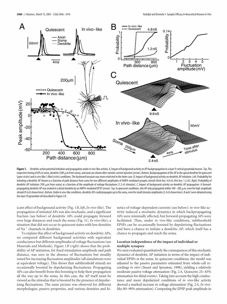
icant effect of background activity (Fig. 1B, left, In vivo-like). Thepropagation of initiated APs was also stochastic, and a significantfraction (see below) of dendritic APs could propagate forwardover large distances and reach the soma (Fig. 1C, In vivo-like), asituation that did not occur in quiescent states with low densitiesof Na� channels in dendrites.
To explain this effect of background activity on dendritic APs,we compared different background activities with equivalentconductance but different amplitudes of voltage fluctuations (seeMaterials and Methods). Figure 1B (right) shows that the prob-ability of AP initiation, for fixed stimulation amplitude and pathdistance, was zero in the absence of fluctuations but steadilyraised for increasing fluctuation amplitudes (all simulations wereat equivalent voltage). This shows that subthreshold stimuli areoccasionally boosted by depolarizing fluctuations. PropagatingAPs can also benefit from this boosting to help their propagationall the way up to the soma. In this case, the AP itself must beviewed as the stimulus that is boosted by the presence of depolar-izing fluctuations. The same picture was observed for differentmorphologies, passive properties, and various densities and ki-
netics of voltage-dependent currents (see below): in vivo-like ac-tivity induced a stochastic dynamics in which backpropagatingAPs were minimally affected, but forward-propagating APs werefacilitated. Thus, under in vivo-like conditions, subthresholdEPSPs can be occasionally boosted by depolarizing fluctuationsand have a chance to initiate a dendritic AP, which itself has achance to propagate and reach the soma.
Location independence of the impact of individual ormultiple synapsesWe next evaluated quantitatively the consequences of this stochasticdynamics of dendritic AP initiation in terms of the impact of indi-vidual EPSPs at the soma. In quiescent conditions, the model wasadjusted to the passive parameters estimated from whole-cell re-cordings in vitro (Stuart and Spruston, 1998), yielding a relativelymoderate passive voltage attenuation (Fig. 2A, Quiescent; 25–45%attenuation for distal events). Taking into account the high conduc-tance and more depolarized conditions of in vivo-like activityshowed a marked increase in voltage attenuation (Fig. 2A, In vivo-like; 80–90% attenuation). Computing the EPSP peak amplitude in
Figure 1. Dendritic action potential initiation and propagation under in vivo-like activity. A, Impact of background activity on AP backpropagation in a layer V cortical pyramidal neuron. Top, Therespective timing of APs in soma, dendrite (300 �m from soma), and axon are shown after somatic current injection (arrow). Bottom, Backpropagation of the AP in the apical dendrite for quiescent(open circles) and in vivo-like ( filled circles) conditions. The backward invasion was more restricted in the latter case. B, Impact of background activity on dendritic AP initiation. Left, Probability forinitiating a dendritic AP shown as a function of path distance from soma for two different amplitudes of AMPA-mediated synaptic stimuli (thick line, 4.8 nS; thin line, 1.2 nS). Right, Probability ofdendritic AP initiation (100 �m from soma) as a function of the amplitude of voltage fluctuations (1.2 nS stimulus). C, Impact of background activity on dendritic AP propagation. A forward-propagating dendritic AP was evoked in a distal dendrite by an AMPA-mediated EPSP (arrow). Top, In quiescent conditions, this AP only propagated within 100 –200 �m, even for high-amplitudestimuli (9.6 nS shown here). Bottom, Under in vivo-like conditions, dendritic APs could propagate up to the soma, even for small stimulus amplitudes (2.4 nS shown here). B and C were obtained usingthe layer VI pyramidal cell described in Figure 2 B.
2468 • J. Neurosci., March 15, 2003 • 23(6):2466 –2476 Rudolph and Destexhe • Synaptic Efficacy in Neocortical Neurons In Vivo

these conditions revealed an attenuation with distance (Fig. 2A,lower panel), which was more pronounced if background activitywas represented by an equivalent static (leak) conductance (see Ma-terials and Methods). Thus, the high-conductance component of
background activity enhances the location-dependent impact ofEPSPs and leads to a stronger individualization of the different den-dritic branches (London and Segev, 2001; Rhodes and Llinas, 2001).
A radically different conclusion was reached if voltage fluctu-
Figure 2. Independence of the somatic response to the location of synaptic stimulation under in vivo-like conditions. A, Impact of background activity on passive voltage attenuation. Top,Somatodendritic membrane potential profile at steady state after current injection at the soma (�0.4nA; layer VI cell shown in B). Two sets of passive properties were used: solid lines, from Destexheand Pare (1999); dashed lines, from Stuart and Spruston (1998). Bottom, Peak EPSP at the soma as a function of path distance for AMPA-mediated 1.2 nS stimuli at different dendritic sites (dendriticbranch shown in B). Peak EPSPs in quiescent conditions are compared with EPSPs obtained with a high static conductance. B, PSTHs of responses to identical AMPA-mediated synaptic stimuli (12 nS)at different dendritic locations (cumulated over 1200 trials after subtraction of spikes attributable to background activity). C, Peak of the PSTH as a function of stimulus amplitude (from 1 to 10coactivated AMPA synapses; conductance range, 1.2–12 nS) and distance to soma. D, Integrated PSTH (probability that a somatic spike was specifically evoked by the stimulus) as a function ofstimulus amplitude and distance to soma. Both C and D show reduced location dependence. E, Top, Comparison of the probability of evoking a dendritic spike (AP initiation) and the probability thatan evoked spike translated into a somatic–axonal spike (AP propagation). Both were represented as a function of the location of the stimulus (AMPA-mediated stimulus amplitudes of 4.8 nS).Bottom, Probability of somatic spike specifically evoked by the stimulus, which was obtained by multiplying the two curves above. This probability was nearly location independent.
Rudolph and Destexhe • Synaptic Efficacy in Neocortical Neurons In Vivo J. Neurosci., March 15, 2003 • 23(6):2466 –2476 • 2469

ations were taken into account. In this case, responses were highlyirregular, and the impact of individual synapses was assessed bycomputing the PSTH over long periods of time with repeatedstimulation of single or groups of colocalized excitatory synapses(see Materials and Methods). The PSTHs obtained for stimulioccurring at different distances from the soma (Fig. 2B) showthat the “efficacy” of these synapses is approximately locationindependent, as calculated from either the peak (Fig. 2C) or theintegral of the PSTH (Fig. 2D). The latter can be interpreted asthe probability that a somatic spike is specifically evoked by asynaptic stimulus. Using this measure of synaptic efficacy, weconclude that, under in vivo-like conditions, the impact of indi-vidual synapses on the soma is nearly independent on their den-dritic location, despite a severe voltage attenuation.
Mechanisms underlying location independenceTo show that this location-independent mode depends onforward-propagating dendritic APs, we selected, for a given syn-aptic location, all trials that evoked a somatic spike. These trialsrepresented a small portion of all trials: from 0.4 to 4.5% depend-ing on the location and the strength of the synaptic stimuli. Forthese “successful” selected trials, the somatic spike was alwayspreceded by a dendritic spike evoked locally by the stimulus. Inthe remaining “unsuccessful” trials, there was a proportion ofstimuli (55–97%) that evoked a dendritic spike but failed to evokesomatic spiking. This picture was the same for different stimula-tion sites: a fraction of stimuli evokes dendritic spikes, and a smallfraction of these dendritic spikes successfully evokes a spike at thesoma–axon.
We analyzed the latter aspect by representing the probabilitiesof initiation and propagation along the distance axis (Fig. 2E).There was an asymmetry between these two measures: the chanceof evoking a dendritic AP was lower for proximal stimuli andincreased with distance (Fig. 2E, AP initiation), because the localinput resistance varies inversely with dendrite diameter and ishigher for thin (distal) dendritic segments. On the other hand,the chance that a dendritic AP propagates down to the soma andleads to soma–axon APs was higher for proximal sites and grad-ually decreased with distance (Fig. 2E, AP propagation). Remark-ably, these two effects compensated such that the probability ofevoking a soma–axon AP (the product of these two probabilities)was approximately independent on the distance to soma (Fig. 2E,Somatic response). This effect was observed only in the presence ofconductance-based background activity and was not present inquiescent conditions or by using current-based models of syn-apses (data not shown). These results show that the location-independent impact of synaptic events under in vivo-like condi-tions is attributable to a compensation between an oppositedistance dependence of the probabilities of AP initiation andpropagation.
The same dynamics were present in four different pyramidalcell morphologies (Fig. 3), suggesting that this principle may ap-ply to a large variety of dendritic morphologies. It was also robustto variations in ion channel densities and kinetics, such as NMDAconductances (Fig. 4A), passive properties (Fig. 4B), and differ-ent types of ion channels (Fig. 4C), including high distal densitiesof leak and hyperpolarization-activated Ih conductances (Fig. 4C,gray line). In the latter case, the presence of Ih affected EPSPs inthe perisomatic region, in which there is a significant contribu-tion of passive signaling, but synaptic efficacy was remarkablylocation independent for the remaining part of the dendrites inwhich the Ih density was highest (see Appendix). Location inde-pendence was also robust to changes in membrane excitability
(Fig. 4D,E) and shifts in the Na� current inactivation (Fig. 4F).Most of these variations changed the absolute probability ofevoking spikes but did not affect the location independence in-duced by background activity. The location-independent synap-tic efficacy was lost when the dendrites had too strong K� con-ductances, with either high IKA in distal dendrites (Fig. 4C, blackdotted line) or a high ratio between K� and Na� conductances(Fig. 4E). In other cases, synaptic efficacy was larger for distaldendrites (Fig. 4D, high excitability, F, inactivation shift of 0).
Activity-dependent modulation of synaptic efficacyTo determine how the efficacy of individual synapses varies as afunction of the intensity of synaptic background activity, we re-peated the same stimulation paradigms as in Figure 2 but byvarying individually the release rates of excitatory (Fig. 5A) orinhibitory (Fig. 5B) inputs of the background, by varying both(Fig. 5C), or by varying the correlation with fixed release rates(Fig. 5D). In all cases, synaptic efficacy (integrated PSTH forstimuli that were subthreshold under quiescent conditions) de-pended on the particular properties of background activity butremained location independent. In the case of “balanced” excita-tory and inhibitory inputs (Fig. 5C), background activity could bechanged continuously from quiescent to in vivo-like conditions.In this case, the probability steadily rose from zero (Fig. 5C, clearregion), showing that subthreshold stimuli can evoke detectableresponses in the presence of background activity, and reached a“plateau” at which synaptic efficacy was independent of both
Figure 3. Location-independent impact of synaptic inputs for different cellular morpholo-gies. The somatic response to AMPA stimulation (12 nS amplitude) is indicated for differentdendritic sites (corresponding branches are indicated by dashed arrows; equivalent electro-physiological parameters and procedures as in Fig. 2 B–D) for four different cells (1 layer II–III,2 layer V, and 1 layer VI) based on cellular reconstructions from cat cortex (Douglas et al., 1991;Contreras et al., 1997). Somatic responses (integrated PSTH) are represented against the pathdistance of the stimulation sites. In all cases, the integrated PSTH shows location independence,but the averaged synaptic efficacy was different for each cell type.
2470 • J. Neurosci., March 15, 2003 • 23(6):2466 –2476 Rudolph and Destexhe • Synaptic Efficacy in Neocortical Neurons In Vivo

synapse location and background intensity (Fig. 5C, dark region).This region corresponds to estimates of background activity onthe basis of intracellular recordings in vivo (Destexhe and Pare,1999). Thus, it seems that synaptic inputs are location indepen-dent for a wide range of background activities and intensities.Modulating the correlation, or the respective weight of excitationand inhibition, allows the network to globally modulate the effi-cacy of all synaptic sites at once.
Location dependence of the timing of synaptic eventsWe next tested whether location independence also applies to thetiming of synaptic events. Figure 6A illustrates the somatic mem-brane potential after synaptic stimuli at different locations. Inquiescent conditions, as predicted by cable theory (Segev et al.,1995; Koch, 1999), proximal synaptic events led to fast rising andfast decaying somatic EPSPs, whereas distal events were attenu-ated in amplitude and slowed in duration (Fig. 6A, Quiescent).The time-to-peak of EPSPs increased monotonically with dis-tance (Fig. 6B, Quiescent). In the presence of background activity,the average amplitude of these voltage deflections was much lessdependent on location (Fig. 6A, In vivo-like), consistent with thePSTHs in Figure 2B, and the time-to-peak of these events wasonly weakly dependent on the location of the synapses in den-drites (Fig. 6B, In vivo-like). Thus, in vivo-like conditions seem toset the dendrites into a fast-conducting mode, in which the tim-
ing of synaptic inputs shows little dependence on their distance tosoma.
To investigate the basis of this fast-conducting mode, we sim-ulated the same paradigm by varying a number of parameters.First, to check whether this effect could be attributable to thedecreased membrane time constant as a result of the high con-ductance imposed by synaptic background activity, we replacedbackground activity by an equivalent static conductance, whichled to an intermediate location-dependent relationship (Fig. 6C,Quiescent, static conductance) between the quiescent and in vivo-like cases depicted above. The reduced time constant thereforecan account for some, but not all, of the diminished locationdependence of the timing. Second, to check for contributions ofdendritic Na� channels, we ran the same stimulation protocolunder in vivo-like conditions but by selectively removing Na�
channels from dendrites. This also led to an intermediate locationdependence (Fig. 6C, In vivo-like, gNa � 0), suggesting that Na�-dependent mechanisms underlie the further reduction of timingbeyond the high-conductance effect. Finally, to show that thisfurther reduction is attributable to dendritic APs, we used a qui-escent state with equivalent static conductance but higher den-dritic excitability (Na� and K� conductances that were twice aslarge), such that strong synaptic stimuli can evoke reliableforward-propagating dendritic APs. In this case only, the reducedlocation dependence of the timing could be fully reconstructed
Figure 4. Location independence for various passive and active properties. A, Synaptic efficacy as a function of path distance and conductance of NMDA receptors. The quantal conductance( gNMDA ) was varied between 0 and 0.7 nS, which corresponds to a fraction of 0 to �60% of the conductance of AMPA channels (Zhang and Trussell, 1994; Spruston et al., 1995). NMDA receptorswere colocalized with AMPA receptors (release frequency of 1 Hz), and stimulation amplitude was 12 nS. B, Synaptic efficacy as a function of path distance and stimulation amplitude for anon-uniform passive model (Stuart and Spruston, 1998) (see Appendix). C, Synaptic efficacy as a function of path distance for different ion channel models or different kinetic models of the same ionchannels (stimulation, 12 nS) (for details of the models, see Appendix). D, Synaptic efficacy as a function of path distance and membrane excitability. Both Na � and K � conductance densities werechanged by a common multiplicative scaling factor. The dotted line indicates a dendritic conductance density of 8.4 mS/cm 2 for the Na � current and 7 mS/cm 2 for the delayed rectifier K � current.The stimulation amplitude was, in all cases, 12 nS. E, Synaptic efficacy obtained by changing the ratio between Na � and K � conductances responsible for action potentials (conductance density of8.4 mS/cm 2 for INa ; the dotted line indicates 7 mS/cm 2 for IKd ). F, Synaptic efficacy as a function of path distance obtained by varying the steady-state inactivation of the fast Na � current. Theinactivation curve was shifted with respect to the original model (Traub and Miles, 1991) toward hyperpolarized values (stimulation amplitude, 12 nS). The dotted line indicates a 10 mV shift, whichapproximately matches the voltage-clamp data of cortical pyramidal cells (Huguenard et al., 1988). All simulations were done using the layer VI cell in which AMPA-mediated synaptic stimuli wereapplied at different sites along the dendritic branch indicated by a dashed arrow in Figure 3.
Rudolph and Destexhe • Synaptic Efficacy in Neocortical Neurons In Vivo J. Neurosci., March 15, 2003 • 23(6):2466 –2476 • 2471

(Fig. 6C, Quiescent, static conductance,high dendritic gNa). The dependence ondendritic APs was also confirmed by theintermediate location dependence ob-tained when EPSPs were constructedfrom trials that did not contain dendriticAPs (Fig. 6B, No dendritic spikes). Thisanalysis shows that the fast-conductingmode is attributable to forward-propagating APs in dendrites of fast timeconstant.
DiscussionIn this paper, we modeled high-conduc-tance states in neocortical pyramidal neu-rons that are associated with a depolarizedand highly fluctuating membrane potential,as shown by intracellular measurements invivo, in anesthetized (Contreras et al., 1996;Pare et al., 1998b) or awake animals (Mat-sumura et al., 1988; Steriade et al., 2001).The simulations suggest that these high-conductance states set pyramidal neuronsinto a stochastic integrative mode that isfast-conducting and in which the impact ofinputs is location independent. The under-lying mechanism depends on forward-propagating APs. The high conductance ofbackground activity lowers the membranetime constant and enhances passive voltageattenuation; this enhanced attenuation lim-its the passive spread to soma and the triggering of backpropagatingAPs by dendritic stimuli and therefore leaves the opportunity forstimuli to influence the soma by forward-propagating APs. At thesame time, the voltage fluctuations associated with synaptic activityinduce a stochastic dynamics that facilitates the initiation and prop-agation of dendritic APs (this effect is consistent with the facilitationof dendritic APs by EPSPs shown by Stuart and Hausser, 2001). Thecombined result of these actions is that colocalized synaptic eventshave a small (but non-zero) chance to trigger a dendritic spike,which itself has a chance to propagate rapidly to the soma in which itcan participate in firing the soma–axon. In contrast with an axon,however, the dynamics is here stochastic in nature, and there is acomplex dependence on distance for the probability of AP initiationand propagation (Fig. 2E). These effects compensate such that theimpact of a given synapse is approximately independent of its posi-tion in the dendrite. Different regions of the neuron, including distaldendrites, can therefore efficiently vote for firing the cell. The samedynamics was observed for different morphologies (Fig. 3), differentpassive and active properties (Fig. 4), and various background activ-ities (Fig. 5).
This stochastic integrative mode was present despite consid-erable variations in the type or kinetics of ions channels in den-drites. It was present with voltage-dependent Ca 2� channels orwith high distal densities of the hyperpolarization-activated cur-rent Ih (Fig. 4C), similar to the densities measured in the apicaldendrite of layer V neocortical pyramidal neurons (Stuart andSpruston, 1998; Larkum et al., 1999a,b; Williams and Stuart,2000a; Berger et al., 2001) (for a model, see Rhodes and Llinas,2001). Our study is therefore applicable to a wide range of den-dritic excitabilities typical of cortical neurons. Because the mech-anism is dependent on forward dendritic spikes, it can fail insituation with suppressed dendritic spike activity, such as strong
dendritic K� conductances (Fig. 4C, INa*, IKd*, IKA; E, large K/Naratio). This suggests that this mechanism would be particularlyrelevant to states in which K� conductances are downregulated.This is the case for aroused states, which are maintained by neu-romodulators such as acetylcholine, noradrenaline, or serotonin,all of which downregulate K� conductances in neocortical neu-rons (McCormick, 1992).
The fact that the same dynamics can be reproduced in differ-ent morphologies (Fig. 3) suggests that the location-independentefficacy of synapses could apply to different types of neurons,regardless of the complexity of their dendritic arborization. If thisprinciple is true, cortical neurons would be free to optimize theirdendritic arbor based on connectivity constraints only (and noton synaptic weight for example). This is valid, however, only forcolocalized synaptic events, as investigated here. There is a largespectrum of possibilities that should be analyzed, including thesynchrony of multiple inputs, their kinetics and receptor type,and the joint impact of paired synaptic events as a function oftheir relative distance and timing. Such an analysis may reveal adependence on the local structure of the dendrites, similar to the“dendritic subunits” postulated in previous models of dendriticintegration (Mel, 1994).
Another interesting property of this stochastic integrativemode is that synaptic efficacy is modulable by network activity.Changing the balance of excitation–inhibition, release rates, orcorrelation in background activity can reconfigure synaptic effi-cacies, but it does so by preserving location independence (Fig.5). The model predicts that the highest synaptic efficacies shouldbe obtained for states of intense “spontaneous” network activity,a somewhat counter-intuitive result. Note that the stochastic as-pect also implies that neocortical neurons would have to usepopulation codes to reliably encode information. In this respect,
Figure 5. Modulation of synaptic efficacy by background activity. A, Integrated PSTH obtained for different intensities ofbackground activity obtained by varying the release rates at glutamatergic synapses (�exc ) while keeping the release rates fixed atGABAergic synapses (�inh � 5.5 Hz). B, Integrated PSTH obtained by varying the release rates at inhibitory synapses (�inh ) withfixed excitatory release rates (�exc � 1 Hz). C, Integrated PSTH obtained by varying both excitatory and inhibitory release rates,using the same scaling factor. The plateau region (dark) shows that the global efficacy of synapses and their location independenceare robust to changes in the intensity of network activity. D, Integrated PSTH obtained for fixed release rates but differentbackground correlations. In all cases, the integrated PSTHs represent the probability that a spike was specifically evoked by synapticstimuli (12 nS, AMPA-mediated), as in Figure 2 D.
2472 • J. Neurosci., March 15, 2003 • 23(6):2466 –2476 Rudolph and Destexhe • Synaptic Efficacy in Neocortical Neurons In Vivo

the modulation of synaptic efficacies by network activity suggeststhat the number of neurons that respond to a given stimulus isdetermined by the spontaneous activity of the network.
Location independence was found and modeled in a numberof previous studies (De Schutter and Bower, 1994; Magee andCook, 2000; Andrasfalvy and Magee, 2001; London and Segev,2001; Rudolph et al., 2001). In hippocampal pyramidal neurons,synaptic conductances are scaled according to their distance to
soma to compensate for dendritic filtering (Magee and Cook,2000; Andrasfalvy and Magee, 2001) (but see London and Segev,2001), a situation that does not seem to apply to neocorticalneurons (Williams and Stuart, 2002). In Purkinje cells, back-ground activity can induce location independence for synchro-nized synaptic inputs by a subthreshold, Ca 2�-mediated mecha-nism (De Schutter and Bower, 1994). In contrast, we show here astochastic mechanism based on Na�-dependent APs that equally
Figure 6. Fast conduction of dendrites under in vivo-like conditions. A, Somatic (black) and dendritic ( gray) voltage deflections after stimuli at different locations (somatic responses are shown with amagnification of 10�). There was a reduction of the location dependence at the soma under in vivo-like conditions (averages over 1200 traces) compared with the quiescent state (all stimuli were 1.2 nS,AMPA-mediated). B, Location dependence of the timing of EPSPs. In the quiescent state, the time-to-peak of EPSPs increased approximately linearly with the distance to soma (Quiescent). This dependence onlocation was markedly reduced under in vivo-like conditions (In vivo-like), defining a fast-conducting state of the dendrites. This location dependence was affected by removing dendritic APs (No dendritic spikes).Inset, Examples of dendritic EPSPs at the site of the synaptic stimulation (50 traces, stimulation with 8.4 nS at 300 �m from soma) are shown under in vivo-like conditions (black) and after dendritic APs wereremoved ( gray). C, Mechanism underlying fast dendritic conduction. Replacing background activity by an equivalent static conductance (Quiescent, static conductance) or suppressing dendritic Na �channels (Invivo-like, gNa � 0) led to an intermediate location dependence of EPSP time-to-peak. On the other hand, using high dendritic excitability together with strong synaptic stimuli (12 nS) evoked reliable dendriticAPs and yielded a reduced location dependence of the time-to-peak in quiescent conditions (Quiescent, static conductance, high dendritic gNa), comparable with in vivo-like conditions. The fast-conducting modeis therefore attributable to forward-propagating dendritic APs in dendrites of fast time constant.
Rudolph and Destexhe • Synaptic Efficacy in Neocortical Neurons In Vivo J. Neurosci., March 15, 2003 • 23(6):2466 –2476 • 2473

applies to single or synchronized synaptic inputs. It would beinteresting to investigate the same behavior in other cell types,such as thalamic neurons, which also contain Na� and Ca 2�
channels in their dendrites (Destexhe et al., 1998; Williams andStuart, 2000b) and are subject to intense synaptic backgroundactivity in vivo (Contreras et al., 1996).
Some of the present observations suggest additional theoreticalwork. First, the fact that dendrites subject to high-conductance fluc-tuations can compensate for the opposite location-dependent effectsof AP initiation and propagation (Fig. 2E) should be investigatedusing the stochastic cable theory to understand the underlyingmechanisms. Second, the observation of an increased traffic of Na�-dependent dendritic APs in these conditions suggests possible con-sequences on spike-timing dependent plasticity (STDP). STDP isthought to occur according to the relative timing between releaseevents and postsynaptic APs (for review, see Bi and Poo, 2001). Thepresent results suggest that, during active states, a significant fractionof postsynaptic APs are local dendritic spikes, which opens the pos-sibility for local dendritic regions to manage their synaptic plasticitymechanisms independently of the soma (Golding et al., 2002). Ad-ditional work is needed to evaluate to what extent such a local plas-ticity rule could duplicate the computational power of cortical neu-rons (Mel, 1994).
Finally, this model formulates a series of experimentally test-able predictions. The predicted stochastic initiation and propa-gation of dendritic APs should be observable by single-cell imag-ing techniques in vivo (Svoboda et al., 1997), if such recordingscan be made in activated states with desynchronized EEG. Theprediction that location-dependent synaptic efficacies should be-come location independent in the presence of intense back-ground activity could be tested by using dual soma– dendriterecordings (for methods, see Stuart and Sakmann, 1994; Larkumet al., 1999a; Berger et al., 2001; Williams and Stuart, 2002). Thiscould be performed in slices either with artificially induced activestates (Buhl et al., 1998; Brumberg et al., 2000) or by recreating invivo-like activity by intracellular injection of fluctuating conduc-tances (Destexhe et al., 2001). In both cases, protocols similar toFigure 2B–D could be used to calculate the efficacy of synapticconductance waveforms injected in dendrites and to compare theefficacy between quiescent and active states.
AppendixThis appendix contains details of the different models used in thepaper. Computational models and scripts for the NEURON sim-ulation environment are available at http://cns.iaf.cnrs-gif.fr/supplement.html.
MorphologySimulations were performed using multicompartment models ofmorphologically reconstructed neocortical pyramidal neuronsfrom layer II–III, V, and VI of cat parietal cortex (Contreras et al.,1997), as well as a layer V cell from cat cortex (Douglas et al.,1991) (Fig. 3) with both simple and detailed axonal geometry(Mainen et al., 1995; Pare et al., 1998a). In all models, passiveproperties and active conductances in dendrites were changed toaccount for the surface correction attributable to dendriticspines, assuming that �45% of the dendritic membrane area arerepresented by spines (DeFelipe and Farinas, 1992).
Uniform passive modelPassive model parameters were adjusted to intracellular (sharp-electrode) recordings obtained after application of TTX and syn-aptic blockers (Destexhe and Pare, 1999), yielding an axial resis-
tance Ra � 250 � cm, a membrane resistance Rm � 22 k� cm 2 insoma and dendrites (Rm � 50 � cm 2 in axon), and membranecapacity Cm � 1 �F/cm 2 in soma and dendrites (Cm � 0.04�F/cm 2 in initial and myelinated segments of axon).
Nonuniform passive modelA non-uniform leak model obtained from dual-patch recordingsin neocortical layer V pyramidal neurons (Stuart and Spruston,1998) was used with Cm � 1.54 �F/cm 2 in soma and dendrites(Cm � 0.04 �F/cm 2 in initial and myelinated segments of axon),and Ra � 68 � cm and Rm � 50 � cm 2 in axonal nodes ofRanvier. The membrane resistance in dendrites scaled in a sig-moidal manner with path distance, with values Rm(end) � 5.36k� cm 2, Rm(soma) � 39.06 k� cm 2, steep � 50 �m, and dhalf �300 �m.
Active membrane conductancesVoltage-dependent conductances were inserted in the soma, den-drites, and the axon. Currents included the fast sodium currentINa, the delayed rectifier potassium current IKd, a slow voltage-dependent potassium current IM, a Ca 2�-dependent potassiumcurrent (C-current) IK[Ca], a high-threshold Ca 2� current (L-current) ICaL, a persistent sodium current INaP, an A-type potas-sium current IKA, and a hyperpolarization-activated conductanceIh. All currents were described by Hodgkin–Huxley-type models(Hodgkin and Huxley, 1952). Various combinations and densi-ties in dendrites and soma were used, as follows.
INa , IKd , IM
The standard kinetic models for these currents were taken from amodel of hippocampal pyramidal cells (see Materials and Meth-ods) (Traub and Miles, 1991), adjusted to match voltage-clampdata of cortical pyramidal cells (Huguenard et al., 1988). In thestandard setup, constant peak conductance densities of 8.4 mS/cm 2 (soma and dendrites; 84 mS/cm 2 in axonal initial segmentand nodes of Ranvier) for INa, 7 mS/cm 2 (soma and dendrites; 70mS/cm 2 in axonal initial segment and nodes of Ranvier) for IKd,and 0.35 mS/cm 2 (soma and dendrites) for IM (no IM in axon)were used. These densities correspond to the values found exper-imentally in adult hippocampal pyramidal neurons (Magee andJohnston, 1995).
INa , IKd , IM , IK[Ca] , ICaL
These currents are as the standard model with additional Ca 2�-dependent potassium current (for kinetics, see Yamada et al.,1989) with conductance density of 1 mS/cm 2 in dendrites andsoma and high-threshold Ca 2� current ICaL (for kinetics, see Mc-Cormick and Huguenard, 1992) with peak conductance density of 3and 1.5 mS/cm2 for proximal and distal dendrites, respectively.
INa , IKd , IM , INaP
These currents are as the standard model with an additional per-sistent sodium current INaP (for kinetics, see French et al., 1990;McCormick and Huguenard, 1992) with conductance density of0.1 mS/cm 2 in soma and dendrites.
INa*, IKd*These currents are described by a different model of action po-tential generation, in which the fast Na� current and the delayedrectifier K� current were taken from a previous model of corticalpyramidal neurons (Mainen et al., 1995). Peak conductance den-sities were 8.4 mS/cm 2 (soma and dendrites; 84 mS/cm 2 in axoninitial segment) for INa* and 7.0 mS/cm 2 (soma and dendrites; 70mS/cm 2 in axon initial segment) for IKd*.
2474 • J. Neurosci., March 15, 2003 • 23(6):2466 –2476 Rudolph and Destexhe • Synaptic Efficacy in Neocortical Neurons In Vivo

INa*, IKd*, IKA
These currents are according to a model of action potential gen-eration for hippocampal pyramidal neurons (Migliore et al.,1999), including an A-type K� current, which conductance den-sity linearly increased with path distance (for details, see Miglioreet al., 1999). Conductance densities were as follows: 7.4 mS/cm 2
(soma and dendrites; 1860 mS/cm 2 in axon initial segment andnodes of Ranvier) for INa*, 10 mS/cm 2 (soma and dendrites; 2500mS/cm 2 in axon initial segment and nodes of Ranvier) for IKd*,and 48 mS/cm 2 for IKA in the soma. Two different kinetics forproximal and distal IKA were used, with densities increasing with48 mS/cm 2 per 100 �m.
INa, IKd, Ih
These currents are as the standard model for INa and IKd with anadditional hyperpolarization-activated conductance Ih (kineticsand density according to non-uniform model by Stuart andSpruston, 1998): sigmoidal scaling with path distance, peak con-ductance density of 0.02 mS/cm 2 at soma and 20 mS/cm 2 in mostdistal dendrites, dhalf � 400 �m, and steep � 50 �m.
ReferencesAndrasfalvy BK, Magee JC (2001) Distance-dependent increase in AMPA
receptor number in the dendrites of adult hippocampal CA1 pyramidalneurons. J Neurosci 21:9151–9159.
Andreasen M, Lambert JD (1995) Regenerative properties of pyramidal celldendrites in area CA1 of the rat hippocampus. J Physiol (Lond)483:421– 441.
Barrett JN (1975) Motoneuron dendrites: role in synaptic integration. FedProc 34:1398 –1407.
Benardo LS, Masukawa LM, Prince DA (1982) Electrophysiology of isolatedhippocampal pyramidal dendrites. J Neurosci 2:1614 –1622.
Berger T, Larkum ME, Luscher HR (2001) High I(h) channel density in thedistal apical dendrite of layer V pyramidal cells increases bidirectionalattenuation of EPSPs. J Neurophysiol 85:855– 868.
Bernander O, Douglas RJ, Martin KA, Koch C (1991) Synaptic backgroundactivity influences spatiotemporal integration in single pyramidal cells.Proc Natl Acad Sci USA 88:11569 –11573.
Bi G, Poo M (2001) Synaptic modification by correlated activity: Hebb’spostulate revisited. Annu Rev Neurosci 24:139 –166.
Borg-Graham LJ, Monier C, Fregnac Y (1998) Visual input evokes transientand strong shunting inhibition in visual cortical neurons. Nature393:369 –373.
Brumberg JC, Sanchez-Vives MV, McCormick DA (2000) Waking thesleeping slice. Soc Neurosci Abstr 26:736.7.
Buhl EH, Tamas G, Fisahn A (1998) Cholinergic activation and tonic exci-tation induce persistent gamma oscillations in mouse somatosensory cor-tex in vitro. J Physiol (Lond) 513:117–126.
Cash S, Yuste R (1999) Linear summation of excitatory inputs by CA1 py-ramidal neurons. Neuron 22:383–394.
Contreras D, Timofeev I, Steriade M (1996) Mechanisms of long-lastinghyperpolarizations underlying slow sleep oscillations in cat corticotha-lamic networks. J Physiol (Lond) 494:251–264.
Contreras D, Destexhe A, Steriade M (1997) Intracellular and computa-tional characterization of the intracortical inhibitory control of synchro-nized thalamic inputs in vivo. J Neurophysiol 77:335–350.
Crill WE, Schwindt PC (1995) Amplification of synaptic current by persis-tent sodium conductance in apical dendrite of neocortical neurons.J Neurophysiol 74:2220 –2224.
DeFelipe J, Farinas I (1992) The pyramidal neuron of the cerebral cortex:morphological and chemical characteristics of the synaptic inputs. ProgNeurobiol 39:563– 607.
De Schutter E, Bower JM (1994) Simulated responses of cerebellar Purkinjecells are independent of the dendritic location of granule cell synapticinputs. Proc Natl Acad Sci USA 91:4736 – 4740.
Destexhe A, Pare D (1999) Impact of network activity on the integrativeproperties of neocortical pyramidal neurons in vivo. J Neurophysiol81:1531–1547.
Destexhe A, Mainen ZF, Sejnowski TJ (1994) An efficient method for com-
puting synaptic conductances based on a kinetic model of receptor bind-ing. Neural Comput 6:14 –18.
Destexhe A, Neubig M, Ulrich D, Huguenard J (1998) Dendritic low-threshold calcium currents in thalamic relay cells. J Neurosci18:3574 –3588.
Destexhe A, Rudolph M, Fellous J-M, Sejnowski TJ (2001) Fluctuating syn-aptic conductances recreate in vivo-like activity in neocortical neurons.Neuroscience 107:13–24.
Douglas RJ, Martin KA, Whitteridge D (1991) An intracellular analysis ofthe visual responses of neurones in cat visual cortex. J Physiol (Lond)440:659 – 696.
French CR, Sah P, Buckett KJ, Gage PW (1990) A voltage-dependent per-sistent sodium current in mammalian hippocampal neurons. J GenPhysiol 95:1139 –1157.
Golding NL, Spruston N (1998) Dendritic sodium spikes are variable trig-gers of axonal action potentials in hippocampal CA1 pyramidal neurons.Neuron 21:1189 –1200.
Golding NL, Staff NP, Spruston N (2002) Dendritic spikes as a mechanismfor cooperative long-term potentiation. Nature 418:326 –331.
Hausser M, Spruston N, Stuart GJ (2000) Diversity and dynamics of den-dritic signaling. Science 290:739 –744.
Hines ML, Carnevale NT (1997) The NEURON simulation environment.Neural Comput 9:1179 –1209.
Hodgkin AL, Huxley AF (1952) A quantitative description of membranecurrent and its application to conduction and excitation in nerve.J Physiol (Lond) 117:500 –544.
Holmes WR, Woody CD (1989) Effects of uniform and non-uniform syn-aptic “activation-distributions” on the cable properties of modeled corti-cal pyramidal neurons. Brain Res 505:12–22.
Huguenard JR, Hamill OP, Prince DA (1988) Developmental changes inNa � conductances in rat neocortical neurons: appearance of a slowlyinactivating component. J Neurophysiol 59:778 –795.
Johnston D, Magee JC, Colbert CM, Christie BR (1996) Active properties ofneuronal dendrites. Annu Rev Neurosci 19:165–186.
Koch C (1999) Biophysics of computation. Oxford: Oxford UP.Larkman AU (1991) Dendritic morphology of pyramidal neurones of the
visual cortex of the rat. III. Spine distributions. J Comp Neurol306:332–343.
Larkum ME, Kaiser KM, Sakmann B (1999a) Calcium electrogenesis in distalapical dendrites of layer 5 pyramidal cells at a critical frequency of back-propagating action potentials. Proc Natl Acad Sci USA 96:14600–14604.
Larkum ME, Zhu JJ, Sakmann B (1999b) A new cellular mechanism forcoupling inputs arriving at different cortical layers. Nature 398:338 –341.
Llinas RR (1975) Electroresponsive properties of dendrites in central neu-rons. Adv Neurol 12:1–13.
London M, Segev I (2001) Synaptic scaling in vitro and in vivo. Nat Neuro-sci 4:853– 855.
London M, Schreibman A, Hausser M, Larkum ME, Segev I (2002) Theinformation efficacy of a synapse. Nat Neurosci 5:332–340.
Magee JC (2000) Dendritic integration of excitatory synaptic input. Nat RevNeurosci 1:181–190.
Magee JC, Cook EP (2000) Somatic EPSP amplitude is independent of syn-apse location in hippocampal pyramidal neurons. Nat Neurosci3:895–903.
Magee JC, Johnston D (1995) Characterization of single voltage-gated so-dium and calcium channels in the apical dendrites of rat CA1 pyramidalneurons. J Physiol (Lond) 487:67–90.
Mainen ZF, Joerges J, Huguenard JR, Sejnowski TJ (1995) A model of spikeinitiation in neocortical pyramidal neurons. Neuron 15:1427–1439.
Matsumura M, Cope T, Fetz EE (1988) Sustained excitatory synaptic inputto motor cortex neurons in awake animals revealed by intracellular re-cording of membrane potentials. Exp Brain Res 70:463– 469.
McCormick DA (1992) Neurotransmitter actions in the thalamus and cere-bral cortex and their role in neuromodulation of thalamocortical activity.Prog Neurobiol 39:337–388.
McCormick DA, Huguenard JR (1992) A model of the electrophysiologicalproperties of thalamocortical relay neurons. J Neurophysiol 68:1384–1400.
Mel BW (1994) Information processing in dendritic trees. Neural Comput6:1031–1085.
Migliore M, Hoffman DA, Magee JC, Johnston D (1999) Role of an A-typeK � conductance in the back-propagation of action potentials in the den-drites of hippocampal pyramidal neurons. J Comp Neurosci 7:5–15.
Rudolph and Destexhe • Synaptic Efficacy in Neocortical Neurons In Vivo J. Neurosci., March 15, 2003 • 23(6):2466 –2476 • 2475

Pare D, Lang EJ, Destexhe A (1998a) Inhibitory control of somatic and den-dritic sodium spikes in neocortical pyramidal neurons in vivo: an intra-cellular and computational study. Neuroscience 84:377– 402.
Pare D, Shink E, Gaudreau H, Destexhe A, Lang EJ (1998b) Impact of spon-taneous synaptic activity on the resting properties of cat neocortical py-ramidal neurons in vivo. J Neurophysiol 79: 1450 –1460.
Pouille F, Scanziani M (2001) Enforcement of temporal fidelity in pyrami-dal cells by somatic feed-forward inhibition. Science 293:1159 –1163.
Rapp M, Yarom Y, Segev I (1992) The impact of parallel fiber backgroundactivity on the cable properties of cerebellar purkinje cells. Neural Com-put 4:518 –533.
Regehr W, Kehoe JS, Ascher P, Armstrong C (1993) Synaptically triggeredaction potentials in dendrites. Neuron 11:145–151.
Rhodes PA, Llinas RR (2001) Apical tuft input efficacy in layer 5 pyramidalcells from rat visual cortex. J Physiol (Lond) 536:167–187.
Rudolph M, Ho N, Destexhe A (2001) Synaptic background activity affectsthe dynamics of dendritic integration in model neocortical pyramidalneurons. In: Computational neuroscience: trends in research 2001(Bower JM, ed), Neurocomputing, Vol 38, pp 327–333. Amsterdam:Elsevier.
Schwindt PC, Crill WE (1997) Local and propagated dendritic action po-tentials evoked by glutamate iontophoresis on rat neocortical pyramidalneurons. J Neurophysiol 77:2466 –2483.
Schwindt PC, Crill WE (1998) Synaptically evoked dendritic action poten-tials in rat neocortical pyramidal neurons. J Neurophysiol 79:2432–2446.
Segev I, Rinzel J, Shepherd GM (1995) The theoretical foundation of den-dritic function: selected papers of Wilfrid Rall with commentaries. Cam-bridge, MA: MIT.
Spencer WA, Kandel ER (1961) Electrophysiology of hippocampal neurons.IV. Fast pre-potentials. J Neurophysiol 24:272–285.
Spruston N, Jonas P, Sakmann B (1995) Dendritic glutamate receptor chan-nels in rat hippocampal CA3 and CA1 pyramidal neurons. J Physiol(Lond) 482:325–352.
Steriade M, Timofeev I, Grenier F (2001) Natural waking and sleep states: aview from inside neocortical neurons. J Neurophysiol 85:1969 –1985.
Stuart GJ, Hausser M (2001) Dendritic coincidence detection of EPSPs andaction potentials. Nat Neurosci 4:63–71.
Stuart GJ, Sakmann B (1994) Active propagation of somatic action poten-tials into neocortical pyramidal cell dendrites. Nature 367:69 –72.
Stuart GJ, Sakmann B (1995) Amplification of EPSPs by axosomatic sodiumchannels in neocortical pyramidal neurons. Neuron 15:1065–1076.
Stuart GJ, Spruston N (1998) Determinants of voltage attenuation in neo-cortical pyramidal neuron dendrites. J Neurosci 18:3501–3510.
Stuart GJ, Schiller J, Sakmann B (1997a) Action potential initiation andpropagation in rat neocortical pyramidal neurons. J Physiol (Lond)505:617– 632.
Stuart GJ, Spruston N, Sakmann B, Hausser M (1997b) Action potentialinitiation and backpropagation in neurons of the mammalian CNS.Trends Neurosci 20:125–131.
Stuart GJ, Spruston N, Hausser M (2000) Dendrites. Cambridge, MA: MIT.Svoboda K, Denk W, Kleinfeld D, Tank DW (1997) In vivo dendritic cal-
cium dynamics in neocortical pyramidal neurons. Nature 385:161–165.Traub RD, Miles R (1991) Neuronal networks of the hippocampus. Cam-
bridge, MA: Cambridge UP.Vaadia E, Haalman I, Abeles M, Bergman H, Prut Y, Slovin H, Aertsen A
(1995) Dynamics of neuronal interactions in monkey cortex in relationto behavioural events. Nature 373:515–518.
Vetter P, Roth A, Hausser M (2001) Propagation of action potentials indendrites depends on dendritic morphology. J Neurophysiol 85:926 –937.
White EL (1989) Cortical circuits. Boston, MA: Birkhauser.Williams SR, Stuart GJ (2000a) Site independence of EPSP time course is
mediated by dendritic I(h) in neocortical pyramidal neurons. J Neuro-physiol 83:3177–3182.
Williams SR, Stuart GJ (2000b) Action potential backpropagation andsomato-dendritic distribution of ion channels in thalamocortical neu-rons. J Neurosci 20:1307–1317.
Williams SR, Stuart GJ (2002) Dependence of EPSP efficacy on synapse lo-cation in neocortical pyramidal neurons. Science 295:1907–1910.
Wong RK, Prince DA, Basbaum AI (1979) Intradendritic recordings fromhippocampal neurons. Proc Natl Acad Sci USA 76:986 –990.
Yamada WM, Koch C, Adams PR (1989) Multiple channels and calciumdynamics. In: Methods in neuronal modeling, Ed 2 (Koch C, Segev I, eds),pp 137–170. Cambridge, MA: MIT.
Yuste R, Tank DW (1996) Dendritic integration in mammalian neurons, acentury after Cajal. Neuron 16:701–716.
Zhang S, Trussell LO (1994) Voltage clamp analysis of excitatory synaptictransmission in the avian nucleus magnocellularis. J Physiol (Lond)480:123–136.
Zohary E, Shadlen MN, Newsome WT (1994) Correlated neuronal dis-charge rate and its implications for psychophysical performance. Nature370:140 –143.
2476 • J. Neurosci., March 15, 2003 • 23(6):2466 –2476 Rudolph and Destexhe • Synaptic Efficacy in Neocortical Neurons In Vivo


