Embed Size (px)
Citation preview

Neocortical Connectivity during EpisodicMemory FormationChristopher Summerfield1*, Matthew Greene1, Tor Wager1, Tobias Egner2, Joy Hirsch2, Jennifer Mangels1
1 Department of Psychology, Columbia University, New York, New York, United States of America, 2 Functional MRI Research Center, Department of Radiology, Columbia
University, New York, New York, United States of America
During the formation of new episodic memories, a rich array of perceptual information is bound together for long-termstorage. However, the brain mechanisms by which sensory representations (such as colors, objects, or individuals) areselected for episodic encoding are currently unknown. We describe a functional magnetic resonance imagingexperiment in which participants encoded the association between two classes of visual stimuli that elicit selectiveresponses in the extrastriate visual cortex (faces and houses). Using connectivity analyses, we show that correlation inthe hemodynamic signal between face- and place-sensitive voxels and the left dorsolateral prefrontal cortex is areliable predictor of successful face–house binding. These data support the view that during episodic encoding, ‘‘top-down’’ control signals originating in the prefrontal cortex help determine which perceptual information is fated to bebound into the new episodic memory trace.
Citation: Summerfield C, Greene M, Wager T, Egner T, Hirsch J, et al. (2006) Neocortical connectivity during episodic memory formation. PLoS Biol 4(5): e128. DOI: 10.1371/journal.pbio.0040128
Introduction
An important goal of memory research is to describe howperceptual experiences are transformed into new memories[1]. Human neuropsychology and neuroimaging have offeredimportant insights into the functional neuroanatomy ofepisodic memory formation, revealing that it involves anetwork of brain regions including the medial temporal lobes(MTLs), the prefrontal cortex (PFC), and neocortical zonesinvolved in perceptual representation of the study materialor task [2,3]. However, consensus has yet to emerge on howthis network of regions works together to make newmemories.
In particular, the contribution of the PFC to episodicmemory formation has been the focus of considerable debate.Among initial proposals was the view that the PFC promotesthe encoding of new associations by mediating the retrievaland elaboration of semantic information relating to the studymaterial [4,5], or that it plays a strategic role, by temporallyorganizing or ‘‘chunking’’ incoming data for more efficientstorage [6–8]. However, more recently, a number of research-ers have argued that during episodic encoding, the functionof the PFC may be to regulate input to the MTL bymodulating activity in the sensory neocortex [1,9–12], whichis in line with evidence from outside the domain of memoryresearch that a key role of the PFC is to select perceptualrepresentations on the basis of their relevance to a currentgoal or task [13,14]. According to this view, differentiationamong neural signals associated with successful and unsuc-cessful encoding begins as perceptual information flowsthrough unimodal and polymodal association cortices, witha prefrontal ‘‘gain control’’ mechanism selecting favoredperceptual codes and rejecting other information at eachsuccessive stage of the processing hierarchy [15]. Selectedperceptual information eventually reaches MTL structuresincluding the hippocampus, whose role is to associate (or‘‘bind’’) the details of an episode (such as objects, colors, orindividuals) into a memory trace for long-term storage [16–
18]. ‘‘Top-down’’ signals from the PFC may thus play a keyrole in controlling which perceptual representations arefated to be included in the new episodic memory trace.Support for this theory comes from neuroimaging studies
that have identified regions of both the PFC and the sensoryneocortex whose evoked hemodynamic response functions(HRFs) vary as a function of encoding success. Activation in‘‘content-specific’’ sensory regions (i.e., those responsive tothe material under study) has often been found to predictencoding success during the learning of new associations. Forexample, successful encoding of object–place associationsyields activation in the lateral occipital complex (LOC) [19–22], a region involved in object representation [23]; learningof face–name pairs leads to robust activation in fusiformgyrus regions involved in face perception [24,25]; and latermemory for word–color associations garners activation invisual regions close to those involved in color processing [26].These studies provide circumstantial evidence for ‘‘top-down’’ selection of perceptual information during encodingby showing that differentiation among neural signals accom-panying successful and unsuccessful associative encoding mayhave already begun en route to the MTL.
Academic Editor: Mick Rugg, University of California Irvine, United States ofAmerica
Received August 26, 2005; Accepted February 21, 2006; Published April 18, 2006
DOI: 10.1371/journal.pbio.0040128
Copyright: ! 2006 Summerfield et al. This is an open-access article distributedunder the terms of the Creative Commons Attribution License, which permitsunrestricted use, distribution, and reproduction in any medium, provided theoriginal author and source are credited.
Abbreviations: BA, Brodmann’s area; DM, difference of memory; FFA, fusiform facearea; FIR, finite impulse response; fMRI, functional magnetic resonance imaging;HRF, hemodynamic response function; LOC, lateral occipital complex; MNI,Montreal Neurological Institute; MTL, medial temporal lobe; PFC, prefrontal cortex;PPA, parahippocampal place area; ROC, receiver operating characteristics; ROI,regions of interest
* To whom correspondence should be addressed. E-mail: [email protected]
PLoS Biology | www.plosbiology.org May 2006 | Volume 4 | Issue 5 | e1280001
PLoS BIOLOGY

However, more substantial evidence in support of this viewwould come from the demonstration that connectivity betweenfrontal and posterior brain regions has predictive power forencoding success. Preliminary evidence for fronto-posteriorinteractions during encoding has been offered by scalpelectroencephalographic studies, in which phase-locking ofneuronal oscillations in the theta-band (4–8 Hz) between theanterior and posterior neocortices has been shown to varywith encoding success [27,28]. However, electroencephalog-raphy lacks the spatial resolution to determine whetherconnectivity is specific to the task-relevant representations,or is occurring in a widespread fashion between the frontaland occipital lobes.
In this report, thus, we turned to functional magneticresonance imaging (fMRI), which can identify, with a highdegree of replicability, discrete regions of extrastriate visualcortex that respond selectively to two classes of stimuli: faces(the ‘‘fusiform face area,’’ or FFA) [29] and houses (the‘‘parahippocampal place area,’’ or PPA) [30,31]. Participantsviewed concurrent images of faces and houses, and wereasked to intentionally encode the association between the twoimages (‘‘try to remember that he lives there’’). We made 2predictions: (1) that evoked responses in the FFA and PPAwould vary with encoding success; and (2) that connectivitybetween the PFC and FFA/PPA would also be a robustpredictor of later memory for the face–house pairings.Additional connectivity analyses were used to explore theextent to which memory-related neocortical regions inter-acted with MTL structures presumably involved in bindingtogether selected perceptual information.
Various techniques have been proposed to meet thechallenges posed by the assessment of functional connectivityin fMRI studies, among the more prominent of which areconfirmatory methods used to assess direction-specificcoupling (‘‘effective connectivity’’) between pre-establishedregions of interest (ROIs) such as structural equationmodeling [32] or dynamic causal modeling [33]. However, inthe absence of a precise hypothesis as to which portion of thePFC would exhibit encoding-sensitive functional connectivitywith the FFA and PPA, we opted for a more exploratoryapproach, in which functional connectivity was calculatedbetween face- and place- sensitive ‘‘seed’’ regions and a maskcomprising the entire frontal lobe. This approach is similar tothat applied in a recent study exploring connectivity betweenthe neocortex and hippocampus during episodic encoding[34]. The use of a mixed block/event-related design with ashort study-test cycle allowed us to assess how bothunivariate, ‘‘event-related’’ hemodynamic responses variedas a function of trial type, and how multivariate, ‘‘state-related’’ patterns of connectivity correlated with memoryperformance on a block-by-block basis, a design which alsodraws upon recent work in memory research [35].
Exploring study-phase hemodynamic responses across thevisual processing hierarchy, we observed that signals associ-ated with subsequent remembering and subsequent forget-ting undergo increasing differentiation as the visual signalfeeds forward from the extrastriate cortex to the hippo-campus. Moreover, connectivity analyses revealed that theextent to which the FFA and PPA coupled their responseswith the left dorsolateral PFC was a robust predictor ofencoding success. One interpretation of these data is that‘‘top-down’’ signals from the PFC modulate the perceptual
signal as it flows through the association neocortex, perhapshelping to select perceptual experience for conversion intoepisodic memory.
Results
Behavioral ResultsParticipants (n ! 16) intentionally encoded 20 blocks of
seven face–house pairings (old pairs; see Figure 1A and 1B)and, following each block, were asked to discriminate thesepairings from another seven recombined pairs drawn fromthe same stimulus pool (new pairs) using a five-point scale.The use of rearranged pairings as distractor stimuli atretrieval ensured that later correct responses could not bemade on the basis of mere familiarity with the face or houseitem, but rather required the successful encoding of the face–house association. Proportions of responses to old pairs (darkgray bars) and new pairs (light gray bars) in each of the fiveresponse categories are shown in Figure 1C. Participantsmade high-confidence ‘‘old’’ responses (key 1) to old pairs on;51% of trials, and rarely made high-confidence false alarms(;5% of trials).The area under the receiver operating characteristics
(ROC) curve (Ag) [36] was calculated for each participant/block (range, 0.35–1.0; mean, 0.82 6 0.15) to give an unbiasedestimate of memory performance, similar to that obtained bycalculating d9 across a Likert confidence scale [37]. Visualinspection suggested that memory performance was reliablypoorer on the first three blocks of the experiment, and thiswas confirmed by statistical analyses (F!5.72, p, 0.01). Theseblocks were excluded from further analyses (Figure 1D) afterwhich no improvement or deterioration in performanceacross the 17 remaining encoding blocks was observed (F, 1).
Event-Related Analyses in the Sensory Neocortex and MTLIn order to identify brain regions whose encoding-related
responses varied with subsequent associative memory for theface–house pairs, encoding trials were sorted according tolater memory performance. A regressor encoding study-phase trials for which a high-confidence ‘‘old’’ response waslater made (key 1; later hits; 50.5% 6 17.4% of trials) wascontrasted with a regressor that modeled study-phase trialsfor which participants later made any other response (keys 2–5; later misses; 49.5% 6 17.4% of trials). Voxels sensitive tothis ‘‘difference of memory’’ (DM) analysis can be seen inFigure 2A, rendered onto a single axial slice. Hemodynamicresponses associated with later hits (blue lines), later misses(green lines), and as a point of reference, unsorted retrievaltrials (red lines) are plotted for the peak voxel from eachcluster. Talairach coordinates for this voxel are given assubheadings.Following correction for false discovery rate [38], statisti-
cally significant responses were observed in regions of theventral inferotemporal cortex sensitive to images of faces(right FFA: F! 35.11, p , 0.001; left FFA: F! 14.73, p , 0.016)and natural scenes (right PPA: F!11.33, p, 0.037; left PPA: F! 12.97, p , 0.024). Bilateral activation in early visual regionsclose to the LOC was also observed to vary with later memory(left LOC: F ! 16.12, p , 0.012; right LOC [not shown;Talairach coordinates 43, "77, "13]: F ! 17.39, p , 0.008).Additional DM effects were observed in the MTL, notably inthe left hippocampus (F!14.97, p , 0.015), the left perirhinal
PLoS Biology | www.plosbiology.org May 2006 | Volume 4 | Issue 5 | e1280002
Connectivity and Memory Formation

cortex (F! 9.72, p , 0.055), and the right entorhinal cortex (F! 10.5, p , 0.045).
Time-To-Peak of the HRFsVisual inspection suggested that HRFs associated with
successful and unsuccessful encoding differed not only inheight but also in latency, with the peak of the HRF occurringprogressively later at each subsequent stage of the processinghierarchy: mean HRFs in the LOC peaked at ;6 s, in face-sensitive regions of the extrastriate visual cortex at ;8 s, andin the MTL at ;10 s. Guided by a previous literature [39], weclassed regions according to their stage in the processinghierarchy (LOC, FFA/PPA, rhinal cortices, and hippocampus),and carried out statistical analysis of the time-to-peak (thelatency of the maximal hemodynamic response after stim-ulus) for later hits and later misses at each region. Nodifferences in time-to-peak were observed for miss trials (allHRFs ;6–8 s), but where an item was successfully encoded,the peak of the HRF fell at ;9–10 s after stimulus in the
rhinal cortices and hippocampus, ;3,000 ms later than inearly visual regions (;6–7 s). Statistical analyses confirmed amain effect of region (F!8.53, p, 0.001), condition (F!9.05,p , 0.01), and a region3 condition interaction (F! 4.0, p ,0.02) for HRF time-to-peak. The time-to-peak for each regionand condition is shown in Figure 2B.
Event-Related Responses in the PFCWithin the PFC, we expected to observe subsequent
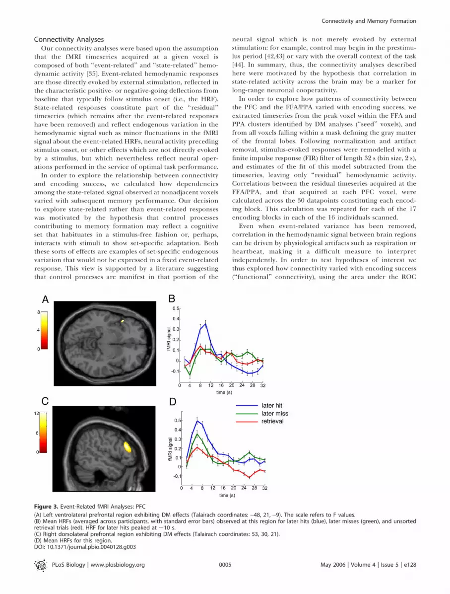
memory effects in the left inferior frontal gyrus, consistentwith a wealth of evidence that this region is involved inepisodic encoding [2] and in particular with the formation ofnew associations [24,40,41]. Indeed, DM effects were observedon the left inferior frontal gyrus in Brodmann’s area (BA) 47(F ! 7.96, p , 0.002), peaking rather late (;10 s afterstimulus). Subsequent memory effects were also observed onthe right middle frontal gyrus in BA46 (F! 12.50, p , 0.001).These two frontal regions, and their accompanying hemody-namic responses, are shown in Figure 3.
Figure 1. Task and Behavioral Data
(A) Selected events (2 trials) comprising part of the encoding phase. At the side of each frame, the amount of time for which it was presented isindicated.(B) Examples of the face and house stimuli employed.(C) Proportion of responses to old pairs (dark gray bars) and new pairs (light gray bars) falling in each of the five reponse categories (shown on the x-axis: 1 [‘‘sure old’’] through 5 [‘‘sure new’’]).(D) Encoding success (the area under the ROC curve, or Ag) averaged across participants for each block. Bars are standard errors. The first three blockswere outliers (in gray). The black line is a fitted linear trend, which was nonsignificant (F , 1).DOI: 10.1371/journal.pbio.0040128.g001
PLoS Biology | www.plosbiology.org May 2006 | Volume 4 | Issue 5 | e1280003
Connectivity and Memory Formation

Figure 2. Event-Related fMRI Analyses: Sensory Neocortex and MTL
(A) DM effects (voxels for which the response for successfully encoded pairs was greater than that for unsuccessfully encoded pairs) suriving a statisticalthreshold of p , 0.001 (uncorrected) are rendered onto a single axial slice of the MNI brain (a single slice was chosen for ease of visualization; voxelsshown are not maxima). The scale refers to F values. Each cluster is linked with a black line to a plot of the HRFs for later hits (blue), later misses (green),and unsorted retrieval trials (red). HRFs are averaged across participants; bars are standard errors. Lineplots are titled with the name of the relevant brainregion. ERC, entorhinal cortex; PRC, perirhinal cortex. Subtitles indicate the Talairach coordinates of the peak voxel for each significant cluster.(B) Time-to-peak estimates of the HRF for LOC (blue), FFA/PPA (green), rhinal cortices (red), and hippocampal (cyan) ROIs in each subsequent memorycondition. Time-to-peak on the y-axis is in seconds.DOI: 10.1371/journal.pbio.0040128.g002
PLoS Biology | www.plosbiology.org May 2006 | Volume 4 | Issue 5 | e1280004
Connectivity and Memory Formation
Connectivity AnalysesOur connectivity analyses were based upon the assumption
that the fMRI timeseries acquired at a given voxel iscomposed of both ‘‘event-related’’ and ‘‘state-related’’ hemo-dynamic activity [35]. Event-related hemodynamic responsesare those directly evoked by external stimulation, reflected inthe characteristic positive- or negative-going deflections frombaseline that typically follow stimulus onset (i.e., the HRF).State-related responses constitute part of the ‘‘residual’’timeseries (which remains after the event-related responseshave been removed) and reflect endogenous variation in thehemodynamic signal such as minor fluctuations in the fMRIsignal about the event-related HRFs, neural activity precedingstimulus onset, or other effects which are not directly evokedby a stimulus, but which nevertheless reflect neural oper-ations performed in the service of optimal task performance.
In order to explore the relationship between connectivityand encoding success, we calculated how dependenciesamong the state-related signal observed at nonadjacent voxelsvaried with subsequent memory performance. Our decisionto explore state-related rather than event-related responseswas motivated by the hypothesis that control processescontributing to memory formation may reflect a cognitiveset that habituates in a stimulus-free fashion or, perhaps,interacts with stimuli to show set-specific adaptation. Boththese sorts of effects are examples of set-specific endogenousvariation that would not be expressed in a fixed event-relatedresponse. This view is supported by a literature suggestingthat control processes are manifest in that portion of the
neural signal which is not merely evoked by externalstimulation: for example, control may begin in the prestimu-lus period [42,43] or vary with the overall context of the task[44]. In summary, thus, the connectivity analyses describedhere were motivated by the hypothesis that correlation instate-related activity across the brain may be a marker forlong-range neuronal cooperativity.In order to explore how patterns of connectivity between
the PFC and the FFA/PPA varied with encoding success, weextracted timeseries from the peak voxel within the FFA andPPA clusters identified by DM analyses (‘‘seed’’ voxels), andfrom all voxels falling within a mask defining the gray matterof the frontal lobes. Following normalization and artifactremoval, stimulus-evoked responses were remodelled with afinite impulse response (FIR) filter of length 32 s (bin size, 2 s),and estimates of the fit of this model subtracted from thetimeseries, leaving only ‘‘residual’’ hemodynamic activity.Correlations between the residual timeseries acquired at theFFA/PPA, and that acquired at each PFC voxel, werecalculated across the 30 datapoints constituting each encod-ing block. This calculation was repeated for each of the 17encoding blocks in each of the 16 individuals scanned.Even when event-related variance has been removed,
correlation in the hemodynamic signal between brain regionscan be driven by physiological artifacts such as respiration orheartbeat, making it a difficult measure to interpretindependently. In order to test hypotheses of interest wethus explored how connectivity varied with encoding success(‘‘functional’’ connectivity), using the area under the ROC
Figure 3. Event-Related fMRI Analyses: PFC
(A) Left ventrolateral prefrontal region exhibiting DM effects (Talairach coordinates: "48, 21,"9). The scale refers to F values.(B) Mean HRFs (averaged across participants, with standard error bars) observed at this region for later hits (blue), later misses (green), and unsortedretrieval trials (red). HRF for later hits peaked at ;10 s.(C) Right dorsolateral prefrontal region exhibiting DM effects (Talairach coordinates: 53, 30, 21).(D) Mean HRFs for this region.DOI: 10.1371/journal.pbio.0040128.g003
PLoS Biology | www.plosbiology.org May 2006 | Volume 4 | Issue 5 | e1280005
Connectivity and Memory Formation

curve (Ag) for each block as a dependent measure. Bycorrelating block-by-block estimates of connectivity (FFA–PFC, PPA–PFC) with encoding success (Ag), we were able todetermine how connectivity between each PFC voxel andFFA/PPA predicted later memory performance for eachparticipant. These correlations were converted to Fisher’s zscores, and t tests were conducted to determine wherefunctional connectivity deviated from zero across theparticipant sample. These t values were rendered ontostatistical maps of the PFC and thresholded in a similarfashion to event-related analyses.
Fronto-Posterior Connectivity ResultsIn Figure 4A, PFC voxels exhibiting statistically significant
functional connectivity with the FFA (red) and PPA (blue) arerendered onto a template brain. Adjacent regions of the leftdorsolateral PFC exhibited reliable connectivity with the FFA(t!3.61, p, 0.003, mean r!0.20) and PPA (t!3.76, p, 0.002,mean r ! 0.17). The FFA cluster was centered on the leftprecentral and superior frontal gyri, at the junction of BA6,BA8, and BA9. The PPA cluster was located immediatelyinferior on the middle frontal gyrus in BA8/9.
Figure 4B shows PFC voxels whose correlation with theaverage of the signal from the FFA and PPA was a reliablepredictor of encoding success. The peak voxel within thiscluster was located at Talairach coordinates "40, 6, 48
(marked with blue crosshairs in Figure 4B, falling on the leftmiddle frontal gyrus) and was statistically significant at ahigher threshold (t ! 4.57, p , 0.0004, mean r ! 0.18). Thevolume of the cluster exceeding a statistical threshold of p ,0.001 was ;375 mm3. Figure 4C shows scatterplots of imageintensity for the peak voxel in the FFA/PFC (left) and PPA/PFC (right), with best-fit lines for individual participantsshown in gray. In each case, positive correlations betweenconnectivity and memory performance can be seen in 15 ofthe 16 participants.In order to demonstrate that connectivity with this region
of the PFC was specific to the FFA and PPA, we performedcontrol analyses exploring how the peak voxel in the frontalcluster ("40, 6, 48) correlated with the other regions of theprocessing hierarchy as defined above (LOC, rhinal cortices,and hippocampus). Connectivity between the PFC and theFFA/PPA was reliably greater than connectivity between thePFC and LOC (t! 2.21, p , 0.05), the PFC and rhinal cortices(t! 2.57, p , 0.03) and the PFC and hippocampus (t! 2.46, p, 0.03), suggesting that this region of the PFC exhibitedfunctional connectivity specifically with the FFA and PPA,and rather than more generally with the posterior brain.
Connectivity with the MTLFurther analyses were conducted to explore patterns of
connectivity linking the sensory neocortex with the MTL
Figure 4. Functional Connectivity
(A) Voxels exhibiting significant (p , 0.01, cluster size . 62 5mm3) functional connectivity with the FFA (red) and PPA (blue).(B) Voxels exhibiting significant (p , 0.01, cluster size . 625 mm3) functional connectivity with the average of the FFA/PPA ROI. The blue crosshairsmark the peak voxel (t ! 4.57, p , 0.001), and the color scale refers to t values.(C) Scatterplots of the correlation between connectivity (timeseries correlation r value) and encoding success (Fisher’s z score) for the peak voxel in thePFC correlating with the FFA (left panel) and PPA (right panel). Each blue cross is one participant/block; gray lines are the fit of the correlation for eachparticipant.DOI: 10.1371/journal.pbio.0040128.g004
PLoS Biology | www.plosbiology.org May 2006 | Volume 4 | Issue 5 | e1280006
Connectivity and Memory Formation

(including the rhinal cortices and hippocampus). The resultsof these analyses were, however, somewhat inconclusive.Although connectivity between the FFA and MTL was arobust predictor of encoding success (t! 2.52, p , 0.03, meanr ! 0.17), no reliable functional connectivity was observedbetween the PPA and MTL (p . 0.5).
Discussion
During performance of a task in which participants learnedpaired face–house associations, encoding success was foundto vary significantly with activation in a network of regionscomprising the PFC, the sensory neocortex, and the medialtemporal lobe. In the occipital and temporal lobes, discretewaystations in the visual processing hierarchy were statisti-cally more active for successfully compared to unsuccessfullyencoded face–house pairs, including the LOC, extrastriatevisual regions responsive to the face and house stimuli understudy (FFA and PPA), and MTL sites including the rhinalcortices and the left hippocampus. This pattern of datareplicates a number of previous studies indicating that inaddition to MTL sites, content-specific activation in thesensory neocortex predicts subsequent memory [19–22,26].Activation in the LOC, often thought to be involved in objectrepresentation [23], may be due to a strategy reportedly usedby some individuals where they attempted to associate theface with object features drawn from the house image (such ascars, or trees), rather than the global scene.
Interestingly, estimates of the hemodynamic response fromeach of these regions revealed an increasing differentiationbetween the HRFs for later hits and later misses, as theperceptual signal processed through successive stages in theunimodal and polymodal association cortices, suggestive ofcontent-specific selection or ‘‘gating’’ of perceptual informa-tion. Moreover, the peak fMRI response to later hits was notonly higher but more delayed than that to later misses, aneffect which increased with subsequent stages of the process-ing hierarchy. This effect was quantified with statisticalanalyses showing that the time-to-peak of the HRF increasedacross regions, with HRFs to hits in the rhinal cortices andhippocampus peaking ;3,000 ms later than those in the earlyvisual cortex. One possibility is that the staggered hemody-namic response across visual regions may reflect thecumulative effects of sustained perceptual processing duringthe stimulus presentation period (which lasted 3,000 ms). Inother words, during relay of information through theassociation cortex, only those perceptual codes which under-go tonic maintenance across the encoding event survive to beprocessed in the subsequent stages of the hierarchy.
The data reported here also offer insight into the source ofthe control signals proposed to regulate activity levels insensory neocortex. Multivariate analyses offered evidencethat long-range interactions spanning the PFC and visualregions occurred during successful encoding, with a strikingpattern of functional connectivity observed between the PFCand the two visual regions sensitive to the stimuli undergoingassociative encoding. The FFA and PPA exhibited functionalconnectivity with adjacent regions of the left dorsolateralPFC stretching across cortical territory located at thejunction of BA6, BA8, and BA9. One interpretation of thisfinding is that control signals originating in the left dorso-lateral PFC selectively target visual regions during associative
encoding, controlling the probability that new information isencoded by regulating activity levels in the sensory neocortex.A plausible neuroanatomical basis for exchange of neuralinformation between the PFC and inferotemporal cortex isoffered by tracing studies, which have shown that they arelinked by cortico-cortical connections [45], although con-nectivity could presumably occur via either monosynaptic orpolysynaptic pathways. The left predominance of this effectsupports a long tradition that encoding-related processesdisplay a left hemisphere advantage in the PFC [46].A current view proposes that a generalized function of the
dorsolateral PFC is to regulate activity levels in the posteriorcortex, selecting for further processing those features orrepresentations which are most relevant to the current goalor task [14]. It is possible that in our study, prefrontal regionswere engaging in ‘‘mnemonic selection,’’ the selective bindingof stimulus attributes chosen for long-term storage [47]. Thisview would be consistent with neuropsychological studiesshowing that patients with damage to the lateral PFCdemonstrate deficits of associative memory that are exacer-bated under conditions of high interference [48], as would beexpected if the PFC were involved in regulating input toepisodic memory [49]. However, our data offer only indirectevidence that the PFC mediates selection during episodicencoding. It could be, for example, that fronto-posteriorconnectivity covaries with another factor which predictsmemory performance—for example, successful elaborationof semantic information linking the face and house images.The model advanced here potentially offers an explanation
for a puzzle in memory research: why does the left dorso-lateral portion of the PFC only rarely exhibit DM effects infMRI experiments [20], when neuropsychological damage tothis region is highly disruptive for memory formation[6,7,50]? Our data suggest that multivariate rather thanunivariate hemodynamic responses associated with thisregion vary with encoding success, a result that may havebeen overlooked by researchers using only conventionalanalyses of imaging data. By contrast, we did observe evokedHRFs in the left ventrolateral regions of the PFC to vary withlater memory, echoing a finding that is routine in neuro-imaging studies of encoding. More ventral portions of thePFC may be supporting semantic elaborative mechanismswhich enrich the binding between face and house represen-tations [4,10,12]. We additionally observed right dorsolateralPFC voxels exhibiting evoked responses that predictedencoding success (but which exhibited no frontoposteriorconnectivity), which may be responsible for generalizedmonitoring or vigilance processes that keep participants’attention oriented towards the encoding task [51].In addition, this theory offers a common framework for
understanding the relationship between working memoryand encoding. A contemporary current in memory researchhas argued against the ‘‘multiple-store’’ view, that non-overlapping neural assemblies subserve dissociable short-and long-term representation of perceptual information.Rather, working memory maintenance may be mediated bytop-down reactivation of task-relevant perceptual codes[11,52,53] via persistent reverberation in neural circuitslinking the PFC with posterior regions [54]. The selectionmechanisms leading to episodic encoding can be seen as aspecial case of working memory maintenance, in whichfavored representations are maintained tonically active for
PLoS Biology | www.plosbiology.org May 2006 | Volume 4 | Issue 5 | e1280007
Connectivity and Memory Formation

sufficient time for relevant neural information to reach theMTL for hippocampus-dependent encoding. This notiongarners further support from the finding that there is acommon prefrontal substrate for working memory rehearsaland episodic encoding [55] with a particular focus in thedorsolateral PFC [20,47].
Little is known about the relationship between the fMRIsignal and the underlying signatures by which neurons shareinformation in wide-scale brain networks. However, a clue tohow functional connectivity may be occurring at the neuro-nal level is offered by electroencephalographic studies, whichhave shown that patterns of neuronal oscillation thought tobe important for long-term potentiation in the hippocampalformation [56] are also instantiated in the neocortex duringsuccessful episodic memory formation [27,57,58]. In partic-ular, neuronal populations in the frontal and posteriorneocortex exhibit theta-band (4–8 Hz) responses withconsistent phase-offset during both successful encoding[27,28] and working memory maintenance [59], a neuralsignature which is also observed when local field potentialsare acquired from within the human MTL during encoding[60]. Additionally, recent evidence suggests that theta-bandactivity may mediate coupling between the frontal lobes andMTL during maze learning [61]. Theta-band activity is thus agood candidate for the carrier signal by which perceptualcodes are selected for encoding. Computational models haveeven suggested that the push–pull dynamics of neuraloscillations may be well suited to mediating target enhance-ment/distractor punishment mechanisms required for effi-c ient arbi trat ion among compet ing perceptualrepresentations [62].
Connectivity between the FFA and MTL was also found topredict later memory, perhaps reflecting the flow ofinformation from the ventral stream to the MTL duringencoding. However, although face-sensitive regions exhibitedfunctional connectivity with the MTL, connectivity betweenthe PPA and MTL did not vary reliably with memoryperformance. One possibility is that this reflects an asymme-try in the nature of face and place representations in thebrain, with place representations serving as ‘‘context’’ to themore central perceptual code for an object or individual(face) present in the scene [63]. During associative encoding acentral ‘‘item’’ (often an object or individual) is oftenassociated with its extrinsic ‘‘context’’ (such as its spatiallocation) [64]. This asymmetry makes intuitive sense when oneconsiders that at one given moment, many objects can beembedded in a single scene, but not vice versa. The morerobust FFA–MTL connectivity may reflect this priority of facestimuli during encoding. It may also be that connectivitybetween ventral visual regions and the MTL is betterexpressed in the event-related hemodynamic signal. Indeed,a recent study which explored correlation in evokedresponses found connectivity between the hippocampus andvisual cortex to be a robust predictor of encoding success [34].
The methods used here attempt to address some of thepotential pitfalls of analyzing connectivity in neuroimagingdatasets. We subjected the timeseries to careful artifactremoval and normalization and removed task-related var-iance using a unconstrained model of the evoked HRF in anattempt to remove reproducibility artifacts in timeseriesconnectivity estimates (this is a rather conservative measure,as it is very likely that connectivity in the event-related
responses also has functional significance). This meant thatalthough posterior ROIs were selected on the basis of theirHRFs, estimates of connectivity were independent of task-evoked responses, making it unlikely that connectivity resultsare a simple restatement of the univariate data. Finally, andmost importantly, we only report connectivity that hadpredictive power for behavioral performance, making it veryunlikely that connectivity results reflect artifacts of physiol-ogy, perfusion, or movement. This approach is related to thatoffered by the ‘‘psychophysiological interaction’’ tool inStatistical Parametric Mapping [65] in that both assess howfunctional connectivity varies with a change in experimentalcontext (in this case, later memory performance).Several aspects of our data suggest that this approach was
successful.Connectivity resultsdidnot simply repeatunivariatedata: for example, functional connectivity results in the PFCwere focused on a left dorsolateral prefrontal region whereevoked responses did not reach threshold for differentiatingtrials on the basis of encoding success. Moreover, observedpatterns of inter-regional connectivity were not ubiquitousacross the brain, but closely matched proposed functionalconnectivity within feedforward visual pathways [39] or dorso-lateral-inferotemporal pathways [45]. Indeed, control analysesindicated that that fronto-posterior connectivity seemed tospecifically target tissue responsive to the face andplace stimulipresented in the study phase of the experiment.In summary, these data offer new insights into the
mechanisms by which perceptual details are selected forinclusion in a new episodic memory trace, and providesupport for a model in which the PFC exerts top-downcontrol over perceptual representations in the posteriorbrain during episodic encoding, contributing to the trans-formation of perceptual experience into memory.
Materials and MethodsParticipants. Participants (n ! 18, 8 females, 10 males) were
neurologically normal individuals ranging in age from 19–34 years.All participants gave informed consent in accordance with ColumbiaUniversity Medical Center Institutional Review Board guidelines.Two participants were excluded from the analyses, one because hismemory performance did not deviate from chance, and the other dueto excessive movement in the scanner (leaving n ! 16).
Stimuli. Stimuli were 300 3 300 pixel grayscale images of malefaces (n! 140) and houses (n! 140). Faces came from various sourcesincluding the AR database [66] and houses were from photographstaken by the authors in Brooklyn, New York, United States. All stimuliwere normalized to a mean scalar luminance of 0.5. Examples of faceand house images can be seen in Figure 1B.
Procedure. The experiment consisted of 20 study-test blocks inwhich participants intentionally encoded seven consecutive face–house pairs, and, following a short pause, were tested on theirmemory for these pairings. In the encoding phase, each trial beganwith a blank interval of variable length (range, 2,000–6,000 ms),followed by a central fixation cross for 1 s. The offset of the crossheralded the presentation of the face and house images, whichappeared on the right and left of center (in a randomized fashion) for3 s. Total trial length thus ranged from 6,000 ms to 10,000 ms, and thetotal length of each encoding block was ;60 s. Participants wereinstructed to memorize the association between the face and thehouse as if they were learning that ‘‘he lived there.’’
During each retrieval block, participants were presented with theseven face–house pairings they had just viewed, intermixed with sevenrecombinations of those same stimuli (14 trials). Each face and eachhouse was thus presented exactly twice at retrieval, once with itspartner from the encoding phase (old pair), and once with a newpartner (new pair). Each retrieval trial began with a crosshair of 1,000ms, followed by presentation of the old or new pair for 3,000 ms.Participants were asked to indicate whether they thought the pair was‘‘old’’ or ‘‘new’’ by pressing one of five buttons, where the leftmost
PLoS Biology | www.plosbiology.org May 2006 | Volume 4 | Issue 5 | e1280008
Connectivity and Memory Formation

indicated that they were highly confident that the pair was old, therightmost indicated that they were highly confident that the pair wasnew, the middle button indicated that they were unsure, and buttons2 and 4 represented low-confidence ‘‘old’’ and ‘‘new’’ responses,respectively. To remind participants of the response mappings, ascale appeared underneath the paired images marked with the words‘‘sure old’’ at the leftmost point, ‘‘sure new’’ at the rightmost point,and ‘‘don’t know’’ in the center.
fMRI data acquisition. Images were acquired with a GeneralElectric Twin-Speed 1.5 Tesla scanner (Milwaukee, Wisconsin, UnitedStates). All images were acquired parallel to the anterior commissure–posterior commissure line with a T2*-weighted echo-planar imagingsequence of 24 contiguous axial slices (repetition time ! 2,000, echotime!40, flip angle!60, field of view! 1903 190 mm, and array size64 3 64) of 4.5-mm thickness and 3 3 3 mm in-plane resolution,providing whole-brain coverage. The task consisted of four runs of345 scans each (five blocks/run). High-resolution anatomical scanswere acquired with a T1*-weighted spoiled gradient-recalled acquis-ition in the steady state sequence (repetition time!19, echo time!5,flip angle ! 20, field of view ! 220), recording 24 slices at a slicethickness of 1.5 mm and in-plane resolution of 0.863 0.86 mm.
fMRI data: preprocessing. Spatial preprocessing and conventionalunivariate statistical mapping were carried out with SPM2 software(Wellcome Department of Imaging Neuroscience, University CollegeLondon, United Kingdom; http://www.fil.ion.ucl.ac.uk/spm/spm2.html). Functional T2* images were slice-timing corrected, spatiallyrealigned to the first volume acquired. The first five functional scansfrom each task were discarded prior to the subsequent analyses. Eachparticipant’s structural T1 image was coregistered to an individualmean echo-planar image. Transformation parameters were derivedfrom normalizing the coregistered structural image to a templatebrain within the stereotactic space of the Montreal NeurologicalInstitute (MNI), and the derived parameters were then applied tonormalize each participant’s echo-planar imaging volumes. Normal-ized images were smoothed with a Gaussian kernel of 939313.5 mmfull width at half maximum. A 256-s temporal high-pass filter wasapplied in order to exclude low-frequency artifacts. Temporalcorrelations were estimated using restricted maximum likelihoodestimates of variance components using a first-order autoregressivemodel. The resulting nonsphericity was used to form maximumlikelihood estimates of the activations.
Trial classification and event-related analyses. Encoding trials werebacksorted on the basis of performance on the subsequent retrievalblock (DM analyses), with trials which later received a high-confidence‘‘old’’ response (key 1) classified as ‘‘later hits’’ and all other trials (keys2–5) classified as ‘‘later misses.’’ Regressors were constructed whichconvolved encoding and retrieval events with the canonical hemody-namic response [67] and its temporal derivative [68], and canonical/derivative regressors associated with later hits and later misses werecompared at the group level using analysis of variance. For displaypurposes, voxels that survived a threshold of p , 0.001 were renderedonto the MNI brain. Although our inferences about significanthemodynamic responses were based upon a parsimonious modelincluding two-basis functions, we used a more comprehensive FIRmodel with 2-s time bins (16 basis functions) to plot these responses.Only voxels with HRFs that were more positive-going for later hitscompared to later misses are described in univariate analyses.
Estimates of performance across each encoding block werecalculated by estimating the area by which the ROC curves for hits
versus misses deviated from the diagonal, using a geometricapproximation procedure [36].
The following procedure was used to estimate the time-to-peak ofthe HRF. First, the height H was estimated by finding where thederivative of the HRF was zero (excluding endpoints). If a dual peakwas identified, the first one was chosen. Hence, our estimate of heightis H ! max fjhj j h9 !0g, where h is the FIR-derived hemodynamicresponse, and h9 denotes the derivative of h. The time-to-peak T isdefined as ft j h (t)!Hg where t is time.
Connectivity analyses. Timeseries were extracted using MarsBaR(http://marsbar.sourceforge.net) from the peak voxel within theaveraged bilateral FFA and PPA regions identified by DM analyses,and from within a mask comprising the gray matter of the frontallobe as defined by the WFU_Pickatlas [69]. Image resolution wasdownsampled by resampling PFC voxels at 53535 mm3 (n!2486) inorder to reduce computational demands. These timeseries weresegmented into 20 blocks of 60 s (repetition time ! 2 s, yielding 30volumes/block), each corresponding to a single cycle of study-phasehemodynamic activity. Each timeseries was mean-centered for eachblock to remove gross variation in mean fMRI signal across blocks,such that univariate block effects could not contribute to variation inconnectivity. Timeseries were windsorized at three standard devia-tions above and below the mean to eliminate spike artifacts.Responses evoked by the task were modeled with an FIR filter oflength 32 s and bin size 2 s, and estimates of the fit of this model weresubtracted from the timeseries. The Pearson’s correlation betweenthe timeseries at the FFA and PPA with every voxel in the PFC maskwas calculated independently for each participant for each of the 17blocks included in the analysis.
In order to calculate how functional connectivity varied withmemory performance for each participant, the Pearson’s correlationbetween FFA–PFC and PPA–PFC connectivity values and encodingsuccess (Ag) was calculated across the 17 encoding blocks. Thesevalues were converted to Fisher’s z scores, and t tests were used todetermine whether they deviated from zero across the participantsample for each region-region pairing. T values were written toAnalyze format images using code adapted from Statistical Para-metric Mapping, and visualized using xjview (http://people.hnl.bcm.tmc.edu/cuixu/xjView). Frontal voxels which were found to function-ally couple with the FFA or PPA at a threshold of p , 0.01 (clusterthreshold n ! 5 or 625 mm3) were rendered onto the MNI brain. Toidentify frontal voxels which exhibited functional connectivity withboth the FFA and PPA, the identical analyses were conducted for theaverage of the FFA and PPA signal.
Acknowledgments
Author contributions. CS and JM conceived and designed theexperiments. CS, MG, and TE performed the experiments. CS, MG,and TE analyzed the data. CS, MG, TW, TE, JH, and JM contributedreagents/materials/analysis tools. CS wrote the paper.
Funding. This work was carried out with support from the WilliamJ. Keck Foundation, the Columbia University Provosts AcademicQuality Fund, and a grant from the National Institutes of Health(R21066129) to JM.
Competing interests. The authors have declared that no competinginterests exist. &
References1. Paller KA, Wagner AD (2002) Observing the transformation of experience
into memory. Trends Cogn Sci 6: 93–102.2. Wagner AD, Schacter DL, Rotte M, Koutstaal W, Maril A, et al. (1998)
Building memories: Remembering and forgetting of verbal experiences aspredicted by brain activity. Science 281: 1188–1191.
3. Scoville WB, Milner B (1957) Loss of recent memory after bilateralhippocampal lesions. J Neurochem 20: 11–21.
4. Gabrieli JD, Poldrack RA, Desmond JE (1998) The role of left prefrontalcortex in language and memory. Proc Natl Acad Sci U S A 95: 906–913.
5. Tulving E, Kapur S, Craik FI, Moscovitch M, Houle S (1994) Hemisphericencoding/retrieval asymmetry in episodic memory: Positron emissiontomography findings. Proc Natl Acad Sci U S A 91: 2016–2020.
6. Milner B, Petrides M, Smith ML (1985) Frontal lobes and the temporalorganization of memory. Hum Neurobiol 4: 137–142.
7. Schacter DL (1987) Memory, amnesia, and frontal lobe dysfunction.Psychobiology 15: 21–36.
8. Shimamura AP (1995) Memory and the prefrontal cortex. Ann N Y AcadSci 769: 151–159.
9. Buckner RL (2003) Functional-anatomic correlates of control processes inmemory. J Neurosci 23: 3999–4004.
10. Fletcher PC, Henson RN (2001) Frontal lobes and human memory: Insightsfrom functional neuroimaging. Brain 124: 849–881.
11. Ranganath C, Blumenfeld RS (2005) Doubts about double dissociationsbetween short- and long-term memory. Trends Cogn Sci 9: 374–380.
12. Simons JS, Spiers HJ (2003) Prefrontal and medial temporal lobeinteractions in long-term memory. Nat Rev Neurosci 4: 637–648.
13. Desimone R, Duncan J (1995) Neural mechanisms of selective visualattention. Annu Rev Neurosci 18: 193–222.
14. Miller EK, Cohen JD (2001) An integrative theory of prefrontal cortexfunction. Annu Rev Neurosci 24: 167–202.
15. Beer JS, Shimamura AP, Knight RT (2004) Frontal lobe contributions toexecutive control of cognitive and social behavior. In: Gazzaniga M, editor.The cognitive neurosciences III. Cambridge (Massachusetts): MIT Press. pp.1091–1104.
16. Marr D (1971) Simple memory: A theory for archicortex. Philos Trans RSoc Lond B Biol Sci 262: 23–81.
17. Squire LR (1992) Memory and the hippocampus: A synthesis from findingswith rats, monkeys, and humans. Psychol Rev 99: 195–231.
PLoS Biology | www.plosbiology.org May 2006 | Volume 4 | Issue 5 | e1280009
Connectivity and Memory Formation

18. Teyler TJ, DiScenna P (1985) The role of hippocampus in memory: Ahypothesis. Neurosci Biobehav Rev 9: 377–389.
19. Cansino S, Maquet P, Dolan RJ, Rugg MD (2002) Brain activity underlyingencoding and retrieval of source memory. Cereb Cortex 12: 1048–1056.
20. Ranganath C, Cohen MX, Brozinsky CJ (2005) Working memory main-tenance contributes to long-term memory formation: Neural and behav-ioral evidence. J Cogn Neurosci 17: 994–1010.
21. Sommer T, Rose M, Glascher J, Wolbers T, Buchel C (2005) Dissociablecontributions within the medial temporal lobe to encoding of object-location associations. Learn Mem 12: 343–351.
22. Sommer T, Rose M, Weiller C, Buchel C (2005) Contributions of occipital,parietal and parahippocampal cortex to encoding of object-locationassociations. Neuropsychologia 43: 732–743.
23. Malach R, Reppas JB, Benson RR, Kwong KK, Jiang H, et al. (1995) Object-related activity revealed by functional magnetic resonance imaging inhuman occipital cortex. Proc Natl Acad Sci U S A 92: 8135–8139.
24. Sperling R, Chua E, Cocchiarella A, Rand-Giovannetti E, Poldrack R, et al.(2003) Putting names to faces: Successful encoding of associative memoriesactivates the anterior hippocampal formation. Neuroimage 20: 1400–1410.
25. Zeineh MM, Engel SA, Thompson PM, Bookheimer SY (2003) Dynamics ofthe hippocampus during encoding and retrieval of face-name pairs.Science 299: 577–580.
26. Ranganath C, Yonelinas AP, Cohen MX, Dy CJ, Tom SM, et al. (2004)Dissociable correlates of recollection and familiarity within the medialtemporal lobes. Neuropsychologia 42: 2–13.
27. Summerfield C, Mangels JA (2005) Coherent theta-band EEG activitypredicts item-context binding during encoding. Neuroimage 24: 692–703.
28. Weiss S, Rappelsberger P (2000) Long-range EEG synchronization duringword encoding correlates with successful memory performance. Brain ResCogn Brain Res 9: 299–312.
29. Kanwisher N, McDermott J, Chun MM (1997) The fusiform face area: Amodule in human extrastriate cortex specialized for face perception. JNeurosci 17: 4302–4311.
30. Epstein R, Kanwisher N (1998) A cortical representation of the local visualenvironment. Nature 392: 598–601.
31. Aguirre GK, Zarahn E, D’Esposito M (1998) An area within human ventralcortex sensitive to ‘‘building’’ stimuli: Evidence and implications. Neuron21: 373–383.
32. Buchel C, Coull JT, Friston KJ (1999) The predictive value of changes ineffective connectivity for human learning. Science 283: 1538–1541.
33. Friston KJ, Harrison L, Penny W (2003) Dynamic causal modelling.Neuroimage 19: 1273–1302.
34. Ranganath C, Heller A, Cohen MX, Brozinsky CJ, Rissman J (2005)Functional connectivity with the hippocampus during successful memoryformation. Hippocampus 15: 997–1005.
35. Otten LJ, Henson RN, Rugg MD (2002) State-related and item-related neuralcorrelates of successful memory encoding. Nat Neurosci 5: 1339–1344.
36. Pollack I, Hsieh R (1969) Sampling variability of the area under the ROC-curve and of d9. Psychol Bull 71: 161–173.
37. Swets J (1964) Indices of signal detectability obtained with variouspsychophysical measures. In: Swets J, Green D, editors. Signal detectionand recognition. New York: Wiley. pp. 164–171.
38. Genovese CR, Lazar NA, Nichols T (2002) Thresholding of statistical mapsin functional neuroimaging using the false discovery rate. Neuroimage 15:870–878.
39. Felleman DJ, Van Essen DC (1991) Distributed hierarchical processing inthe primate cerebral cortex. Cereb Cortex 1: 1–47.
40. Davachi L, Mitchell JP, Wagner AD (2003) Multiple routes to memory:Distinct medial temporal lobe processes build item and source memories.Proc Natl Acad Sci U S A 100: 2157–2162.
41. Jackson O 3rd, Schacter DL (2004) Encoding activity in anterior medialtemporal lobe supports subsequent associative recognition. Neuroimage21: 456–462.
42. Hopfinger JB, Buonocore MH, Mangun GR (2000) The neural mechanismsof top-down attentional control. Nat Neurosci 3: 284–291.
43. Kastner S, Pinsk MA, De Weerd P, Desimone R, Ungerleider LG (1999)Increased activity in human visual cortex during directed attention in theabsence of visual stimulation. Neuron 22: 751–761.
44. Koechlin E, Ody C, Kouneiher F (2003) The architecture of cognitivecontrol in the human prefrontal cortex. Science 302: 1181–1185.
45. Petrides M, Pandya DN (1988) Association fiber pathways to the frontalcortex from the superior temporal region in the rhesus monkey. J CompNeurol 273: 52–66.
46. Cabeza R, Nyberg L (2000) Neural bases of learning and memory:Functional neuroimaging evidence. Curr Opin Neurol 13: 415–421.
47. Ranganath C, DeGutis J, D’Esposito M (2004) Category-specific modulationof inferior temporal activity during working memory encoding andmaintenance. Brain Res Cogn Brain Res 20: 37–45.
48. Mangels JA (1997) Strategic processing and memory for temporal order inpatients with frontal lobe lesions. Neuropsychology 11: 207–221.
49. Moscovitch M, Umilta C (1991) Conscious and nonconscious aspects ofmemory: A neuropsychological framework of modules and central systems.In: Lisker R, Weingartner H, editors. Perspectives in cognitive neuro-science. New York: Oxford University Press. pp 229–266.
50. Janowsky JS, Shimamura AP, Squire LR (1989) Source memory impairmentin patients with frontal lobe lesions. Neuropsychologia 27: 1043–1056.
51. Lawrence NS, Ross TJ, Hoffmann R, Garavan H, Stein EA (2003) Multipleneuronal networks mediate sustained attention. J Cogn Neurosci 15: 1028–1038.
52. Cowan N (1995) Attention and memory: An integrated framework. NewYork: Oxford University Press. 321 p.
53. Ruchkin DS, Grafman J, Cameron K, Berndt RS (2003) Working memoryretention systems: A state of activated long-term memory. Behav Brain Sci26: 709–728.
54. Gazzaley A, Rissman J, Desposito M (2004) Functional connectivity duringworking memory maintenance. Cogn Affect Behav Neurosci 4: 580–599.
55. Davachi L, Maril A, Wagner AD (2001) When keeping in mind supportslater bringing to mind: Neural markers of phonological rehearsal predictsubsequent remembering. J Cogn Neurosci 13: 1059–1070.
56. Huerta PT, Lisman JE (1993) Heightened synaptic plasticity of hippo-campal CA1 neurons during a cholinergically induced rhythmic state.Nature 364: 723–725.
57. Klimesch W, Doppelmayr M, Russegger H, Pachinger T (1996) Theta bandpower in the human scalp EEG and the encoding of new information.Neuroreport 7: 1235–1240.
58. Sederberg PB, Kahana MJ, Howard MW, Donner EJ, Madsen JR (2003)Theta and gamma oscillations during encoding predict subsequent recall. JNeurosci 23: 10809–10814.
59. Sarnthein J, Petsche H, Rappelsberger P, Shaw GL, von Stein A (1998)Synchronization between prefrontal and posterior association cortexduring human working memory. Proc Natl Acad Sci U S A 95: 7092–7096.
60. Fell J, Klaver P, Elfadil H, Schaller C, Elger CE, et al. (2003) Rhinal-hippocampal theta coherence during declarative memory formation:Interaction with gamma synchronization? Eur J Neurosci 17: 1082–1088.
61. Jones MW, Wilson MA (2005) Theta rhythms coordinate hippocampal-prefrontal interactions in a spatial memory task. PLoS Biol 3: e402. DOI:10.1371/journal.pbio.0030402
62. Norman KA, Newman EL, Detre GJ, Polyn SM (2006) How inhibitoryoscillations can train neural networks and punish competitors. NeuralComput: In press.
63. Bar M, Aminoff E (2003) Cortical analysis of visual context. Neuron 38:347–358.
64. Geiselman RE, Bjork RA (1980) Primary versus secondary rehearsal inimagined voices: Differential effects on recognition. Cognit Psychol 12:188–205.
65. Gitelman DR, Penny WD, Ashburner J, Friston KJ (2003) Modeling regionaland psychophysiologic interactions in fMRI: The importance of hemody-namic deconvolution. Neuroimage 19: 200–207.
66. CVC Specialty Chemicals [Martinez M, Benavente R] (1998 June) The ARface database. Moorestown (New Jersey): CVC Specialty Chemicals. ReportNumber 24.
67. Worsley KJ, Poline JB, Friston KJ, Evans AC (1997) Characterizing theresponse of PET and fMRI data using multivariate linear models. Neuro-image 6: 305–319.
68. Friston KJ, Fletcher P, Josephs O, Holmes A, Rugg MD, et al. (1998) Event-related fMRI: Characterizing differential responses. Neuroimage 7: 30–40.
69. Maldjian JA, Laurienti PJ, Kraft RA, Burdette JH (2003) An automatedmethod for neuroanatomic and cytoarchitectonic atlas-based interrogationof fMRI data sets. Neuroimage 19: 1233–1239.
PLoS Biology | www.plosbiology.org May 2006 | Volume 4 | Issue 5 | e1280010
Connectivity and Memory Formation