Embed Size (px)
Citation preview
A genome-wide regulatory network identifies key transcriptionfactors for memory CD8[superscript +] T-cell development
The MIT Faculty has made this article openly available. Please share how this access benefits you. Your story matters.
Citation Hu, Guangan, and Jianzhu Chen. “A Genome-Wide RegulatoryNetwork Identifies Key Transcription Factors for MemoryCD8[superscript +] T-Cell Development.” Nature Communications 4(December 12, 2013).
As Published http://dx.doi.org/10.1038/ncomms3830
Publisher Nature Publishing Group
Version Author's final manuscript
Citable link http://hdl.handle.net/1721.1/88162
Terms of Use Article is made available in accordance with the publisher'spolicy and may be subject to US copyright law. Please refer to thepublisher's site for terms of use.
1
A Genome-wide Regulatory Network Identifies Key Transcription
Factors for Memory CD8+ T Cell Development
Guangan Hu and Jianzhu Chen
David H. Koch Institute for Integrative Cancer Research and Department of Biology,
Massachusetts Institute of Technology, Cambridge, MA 02139, USA
Corresponding to Jianzhu Chen ([email protected])
Author’s contacts:
Guangan Hu ([email protected])
Jianzhu Chen ([email protected])
2
Abstract
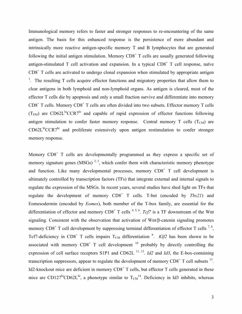
Memory CD8+ T cell development is defined by the expression of a specific set of memory
signature genes (MSGs). Despite recent progress, many components of the transcriptional
control of memory CD8+ T cell development are still unknown. To identify transcription factors
(TFs) and their interactions in memory CD8+ T cell development, we construct a genome-wide
regulatory network and apply it to identify key TFs that regulate MSGs. Most of the known TFs
in memory CD8+ T cell development are rediscovered and about a dozen new TFs are also
identified. Sox4, Bhlhe40, Bach2 and Runx2 are experimentally verified and Bach2 is further
shown to promote both development and recall proliferation of memory CD8+ T cells through
Prdm1 and Id3. Gene perturbation study identifies the mode of interactions among the TFs with
Sox4 as a hub. The identified TFs and insights into their interactions should facilitate further
dissection of molecular mechanisms underlying memory CD8+ T cell development.
3
Immunological memory refers to faster and stronger responses to re-encountering of the same
antigen. The basis for this enhanced response is the persistence of more abundant and
intrinsically more reactive antigen-specific memory T and B lymphocytes that are generated
following the initial antigen stimulation. Memory CD8+ T cells are usually generated following
antigen-stimulated T cell activation and expansion. In a typical CD8+ T cell response, naïve
CD8+ T cells are activated to undergo clonal expansion when stimulated by appropriate antigen 1. The resulting T cells acquire effector functions and migratory properties that allow them to
clear antigens in both lymphoid and non-lymphoid organs. As antigen is cleared, most of the
effector T cells die by apoptosis and only a small fraction survive and differentiate into memory
CD8+ T cells. Memory CD8+ T cells are often divided into two subsets. Effector memory T cells
(TEM) are CD62LloCCR7lo and capable of rapid expression of effector functions following
antigen stimulation to confer faster memory response. Central memory T cells (TCM) are
CD62LhiCCR7hi and proliferate extensively upon antigen restimulation to confer stronger
memory response.
Memory CD8+ T cells are developmentally programmed as they express a specific set of
memory signature genes (MSGs) 2, 3, which confer them with characteristic memory phenotype
and function. Like many developmental processes, memory CD8+ T cell development is
ultimately controlled by transcription factors (TFs) that integrate external and internal signals to
regulate the expression of the MSGs. In recent years, several studies have shed light on TFs that
regulate the development of memory CD8+ T cells. T-bet (encoded by Tbx21) and
Eomesodermin (encoded by Eomes), both member of the T-box family, are essential for the
differentiation of effector and memory CD8+ T cells 4, 5, 6. Tcf7 is a TF downstream of the Wnt
signaling. Consistent with the observation that activation of Wnt/!-catenin signaling promotes
memory CD8+ T cell development by suppressing terminal differentiation of effector T cells 7, 8,
Tcf7-deficiency in CD8+ T cells impairs TCM differentiation 9. Klf2 has been shown to be
associated with memory CD8+ T cell development 10 probably by directly controlling the
expression of cell surface receptors S1P1 and CD62L 11, 12. Id2 and Id3, the E-box-containing
transcription suppressors, appear to regulate the development of memory CD8+ T cell subsets 13.
Id2-knockout mice are deficient in memory CD8+ T cells, but effector T cells generated in these
mice are CD127hiCD62Lhi, a phenotype similar to TCM14. Deficiency in Id3 inhibits, whereas
4
overexpression of Id3 promotes memory CD8+ T cell development 15. The B-cell transcriptional
repressor Blimp-1 (encoded by Prdm1) promotes the terminal differentiation of effector CD8+ T
cells and is required for recall response of memory T cells 16, 17. Despite these progresses, the
current understanding of transcriptional regulation of memory CD8+ T cell development is still
limited, as additional TFs as well as their coordination are likely required to respond to external
and internal signals in order to establish the MSG program for memory CD8+ T cell
development.
In this study, we assemble a genome-wide regulatory network associated with the development
of CD8+ T cells using publicly available gene expression data and a reverse-engineering
algorithm. This regulatory network is applied to identify key TFs that regulate memory CD8+ T
cell development using the master regulator analysis (MRA) of the MSGs of CD8+ T cells. The
inferred TFs include most of the known TFs as well as a dozen new TFs with limited functional
information in CD8+ T cell differentiation. A regulatory module controlling the MSGs is
constructed and the high accuracy of the regulations in the module is verified using ChIP-PCR.
Gene perturbations identify multiple regulatory motifs among the key TFs, suggesting their
complex regulations during the memory CD8+ T cell development. Four of the newly identified
key TFs (Sox4, Bhlhe40, Bach2, and Runx2) are experimentally validated to regulate memory
CD8+ T cell development and function. Bach2 is shown to promote memory CD8+ T cell
development and recall proliferation through Id3 and Prdm1. Our study represents the most
comprehensive analysis of TFs and their interactions in memory CD8+ T cell development to
date. The identified TFs and the insights into their mode of interactions provide a foundation for
further dissecting the molecular mechanisms underlying memory CD8+ T cell development.
Results
Identification of TFs associated with memory CD8+ T cells
We collected 386 gene expression profiles of naïve, effector and memory CD8+ T cells of the
mouse from 35 independent GEO datasets (Supplementary Table S1). 1,445 genes coding
putative TFs 18 were manually mapped to the latest mouse genome to eliminate redundant and
erroneous annotations, resulting in a total of 1,038 putative TFs. Among these putative TFs, 464
were expressed during the naïve to effector to memory CD8+ T cell development (see Methods
5
for detail). Using a reverse-engineering algorithm CLR (context likelihood of relatedness)19, the
386 gene expression profiles and the 1,038 putative TFs, a genome-wide regulatory network was
assembled. The network consisted of 107,157 interactions among 11,032 genes. 62,272
interactions (58%) were between the 276 of the 464 expressed putative TFs and 8,572 target
genes, suggesting that interactions are enriched among the expressed genes (P<0.001, binomial
test). Furthermore, 3,219 of these interactions involve 154 out of 196 (79%) identified MSGs 3
(Supplementary Fig. S1).
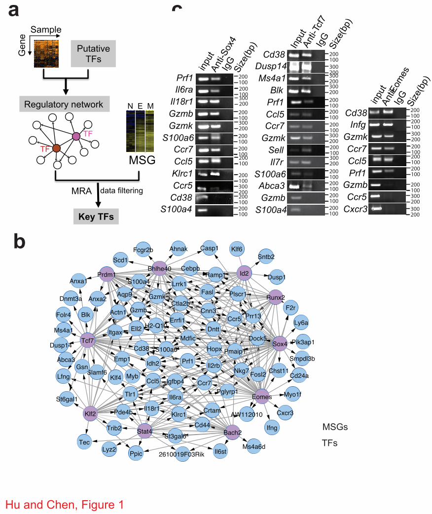
To identify key TFs that regulate MSGs, we applied the MRA to the CLR-inferred
interactions (Fig. 1a, see Methods). The MRA algorithm computes the statistical significance of
overlaps of all interactions of each TF (inferred by CLR) with MSGs or a control gene set by a
binomial test. From the 1,038 putative TFs, MRA identified 60 MSG-specific TFs at P<0.05
(binomial test), all of which are expressed in CD8+ T cells (Supplementary Table S2). These 60
candidates were filtered by removing those whose knockout do not have any immune system
phenotype as defined in MGI (Mouse Genome Informatics) 20. The positive candidates were then
analyzed for enrichment of DNA-binding motifs among MSGs or differential expression among
naïve, effector and memory CD8+ T cells (see Methods). This led to 21 key TFs that were ranked
according to the numbers of MSG they regulate (Table 1). Text-mining of public references on
these 21 TFs revealed that 8 of 12 known TFs, which have been reported to be involved in
memory CD8+ T cell development and function 21, 22, 23, 24, were identified by our analysis. These
results show that our systematic approach is valid for identifying TFs that regulate memory
CD8+ T cell development.
Validation of a regulatory module for memory signature genes
To further explore the relative importance of the 21 identified TFs in regulating MSGs, we
constructed a regulatory module using the top 10-ranked TFs (Fig. 1b). The resulting module
contained 56% (86 out of 154) of MSGs that were present in the entire network (Supplementary
Fig. S1). To verify this regulatory module, chromatin immunoprecipitation (ChIP) was
performed for the top 3 TFs, Sox4, Tcf7 and Eomes, in CD8+ T cells followed by PCR
amplification of promoter regions (within 1 kb upstream of the transcription-starting site) of
randomly selected MSGs that were predicted to be regulated by Sox4 or Tcf7 or Eomes. As
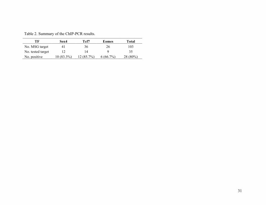
shown in Fig. 1c and 1d, promoter regions of 10 out of the 12 randomly selected Sox4-regulated
6
MSGs were amplified. Similarly, 12 out of 14 randomly selected Tcf7-regulated MSGs and 6 out
of 9 randomly selected Eomes-regulated MSGs were amplified. On average, 80% of the tested
promoter regions were immunoprecipitated with antibodies specific for each of the three TFs
(Table 2), confirming the high accuracy of the constructed regulatory module. Furthermore,
when the cumulative coverage of MSGs was plotted as a function of each of the 21 TFs, the top
2 TFs, Sox4 and Tcf7, were shown to regulate 42% of MSGs (Supplementary Fig. S2).
Perturbation network of key TFs
Although the constructed regulatory module predicts interactions between TFs (Fig. 1b), the
directions of regulation are not known. To find out these, the top 10 TFs and another two known
memory-regulating TFs (Id3 and Tbx21, #12 and #19 in the list, Table 1) 6, 15, 25 were perturbed
in CD8+ T cells in vitro by overexpression through retroviral transduction. The transcript level of
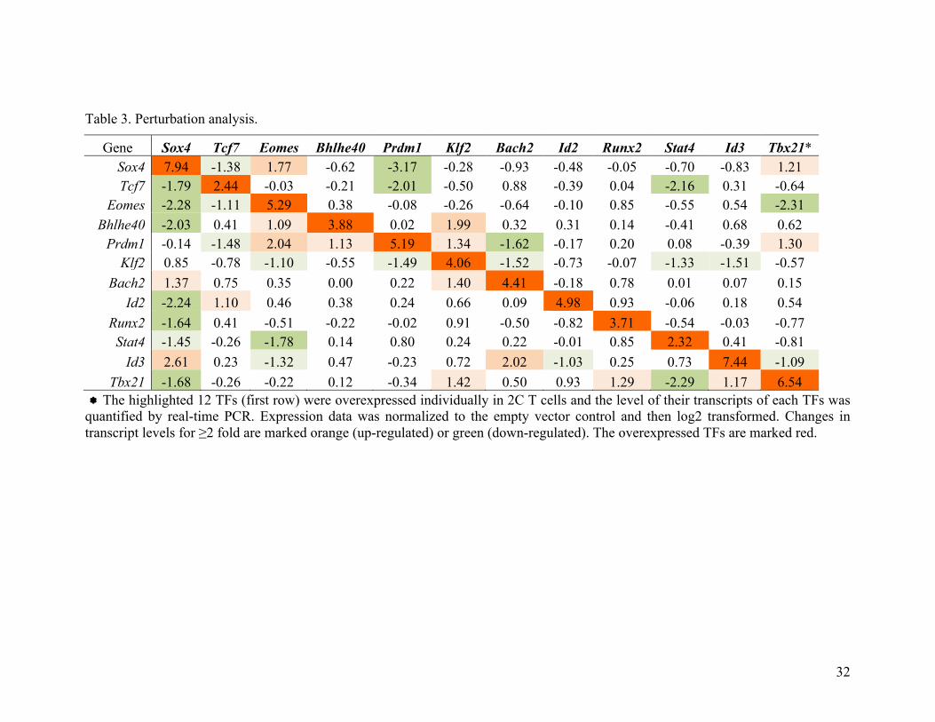
each of the 12-selected TFs was measured by quantitative real-time PCR (Table 3). If changes in
transcript level of "2 fold were taken as directional regulations, the perturbation results identified
41 regulations among the 12x12 matrix (31%). Notably, the top 3 TFs (Sox4, Tcf7 and Eomes)
directed 19 of the 41 regulations. To verify these regulations, ChIP-PCR was performed using
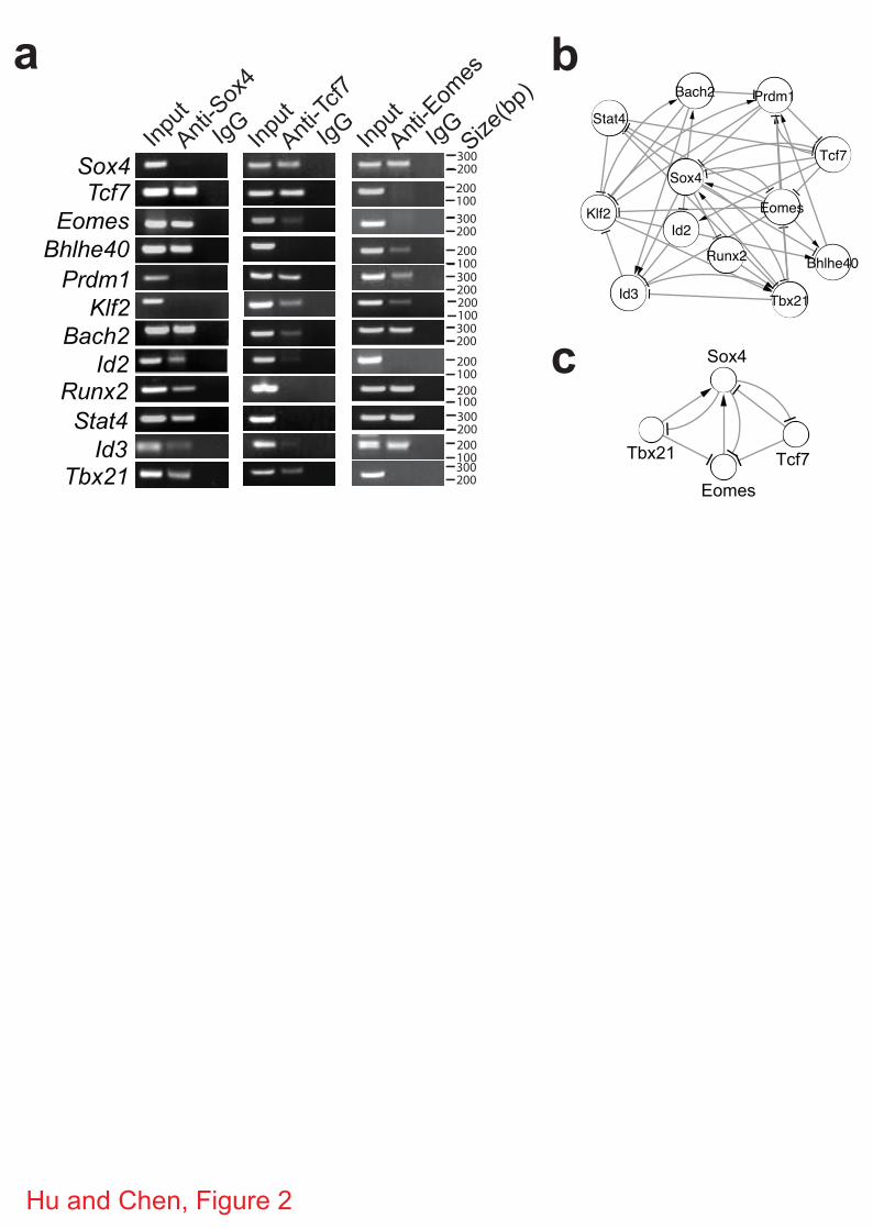
antibodies specific for Sox4, Tcf7 and Eomes. As shown in Fig. 2a, 18 of the 19 regulations were
confirmed. ChIP-PCR also identified 4 more regulations that were not observed in the
perturbation study. Thus, compared to ChIP-PCR, perturbation studies is able to identify the
directional regulations with 82% sensitivity and 91% specificity.
We then constructed a perturbation network of the 12 TFs with directional regulations
(Fig. 2b). The top 3 TFs (Sox4, Tcf7 and Eomes) and Bach2 had more downstream targets than
the number of TFs that regulate them (Supplementary Fig. S3), suggesting that they are at the
upstream of a regulatory structure. TFs in the perturbation network formed multiple motifs, such
as feedback and feed-forward loops (Supplementary Fig. S4). For example, in a feedback motif
of Sox4-Tcf7-Eomes-Tbx21 (Fig. 2c), Sox4 and Tcf7 regulate each other and they also regulate
expression of Eomes and/or Tbx21. The latter regulations were further confirmed at the protein
level as indicated by suppression of Eomes and Tbx21 by overexpression of Sox4 or Tcf7
(Supplementary Fig. S5). These results suggest that complex regulations involving multiple
regulatory motifs among these TFs are involved in memory CD8+ T cell development.
7
Validation of Sox4 and Bach2 in memory CD8+ T cells
Among the top 10 TFs (Table 1), 6 are known to play important roles in memory CD8+ T cell
development and/or function. We then investigated whether the other 4 TFs (Sox4, Bhlhe40,
Bach2 and Runx2) are also involved in memory CD8+ T cell development/function by examining
the effect of overexpression and knockdown of these TFs on the recall proliferation of memory
CD8+ T cells in vitro and in vivo. CD8+ T cells expressing the 2C TCR were activated with
cognate peptide SIYRYYGL (SIY) and then transduced with retroviruses expressing GFP plus
Sox4, Bhlhe40, Bach2 or Runx2 or expressing GFP plus shRNA specific for one of the four TFs
(Supplementary Table S3 and S4). The 2C T cells were then cultured in the presence of cytokine
IL-7 to induce the development of memory CD8+ T cells (Supplementary Fig. S6). To assay
recall proliferation, the in vitro memory 2C T cells were restimulated with SIY and the number
of transduced (GFP+) and non-transduced (GFP-) 2C T cells were quantified on day 4 and 6.
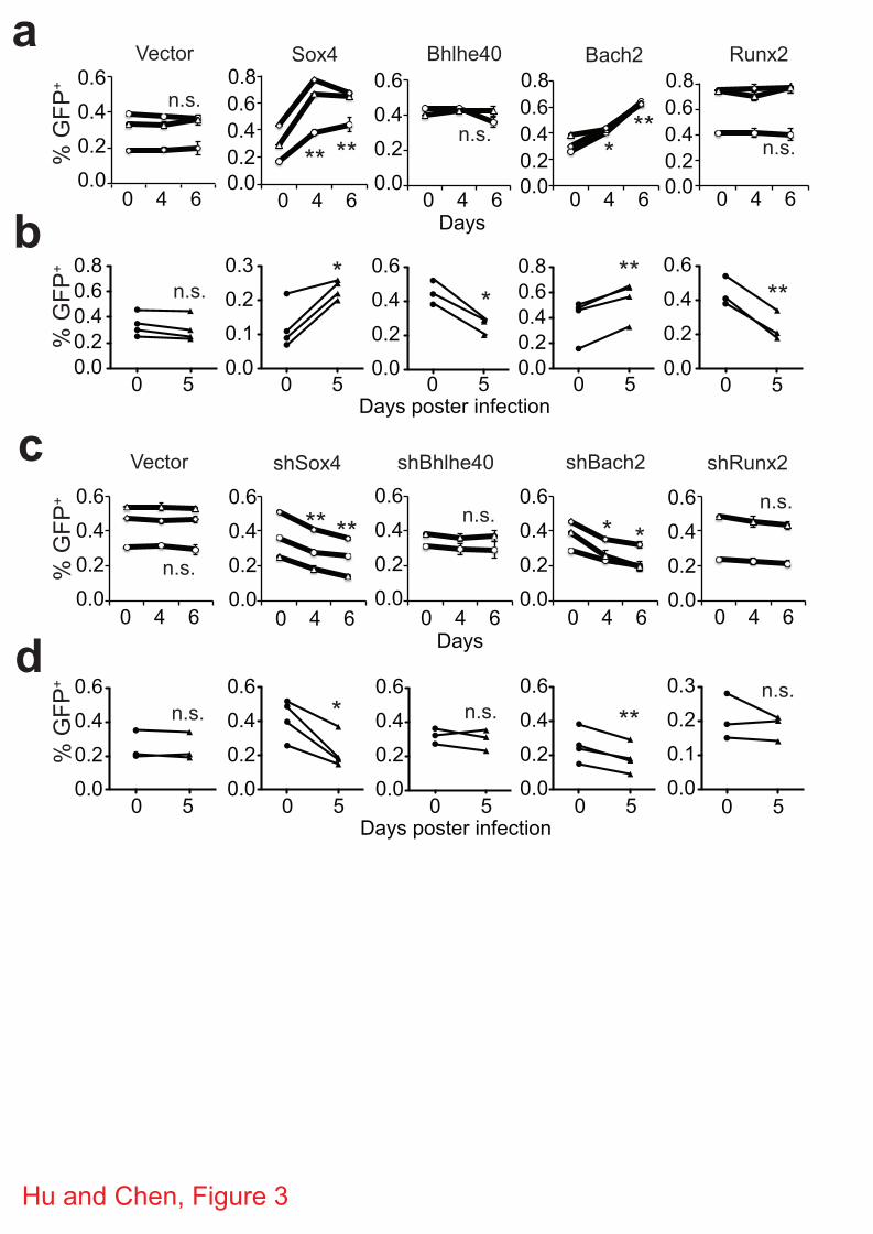
Compared to the vector control, overexpression of Sox4 or Bach2 led to a significant increase in
the proportions of GFP+ cells (Fig. 3a), suggesting a higher recall proliferation. When the in vitro
generated memory 2C T cells were adoptively transferred into C57BL/6 (B6) mice followed by
activation through infection with influenza virus that express SIY (WSN-SIY virus) 26, a
significant increase in the proportion of GFP+ 2C T cells was also observed in the draining
lymph nodes (DLN) (Fig. 3b), the blood, lung and spleen (Supplementary Fig. S7) 5 days post
infection (dpi) if the transduced memory T cells expressed Sox4 or Bach2. Conversely,
knockdown of Sox4 or Bach2 (Supplementary Fig. S8) resulted in a significant inhibition of the
recall proliferation of memory 2C T cells both in vitro and in vivo (Fig. 3c,d). Although
overexpression of Bhlhe40 and Runx2 inhibited the in vivo recall proliferation of the transduced
memory 2C T cells (Fig. 3b), no significant change was observed in in vitro recall response (Fig.
3a) and in knockdown assay (Fig. 3c,d). As positive controls, we tested in parallel known TFs:
overexpression of Eomes promoted the recall proliferation whereas overexpression of Klf2
inhibited the recall proliferation (Supplementary Fig. S9), consistent with previous reports 9, 11.
These results show that Sox4 and Bach2 likely promote the recall proliferation of memory CD8+
T cells.
Enhanced memory T cell development by Bach2 overexpression

8
To confirm the effect of overexpression of Bach2 on recall proliferation of memory CD8+ T cells,
we activated 2C T cells in vitro for two days, transduced the activated T cells with retroviruses
expressing GFP alone or GFP plus Bach2. The cells were cultured in the presence of IL-2 for
two more days and then adoptively transferred into antigen-free B6 mice to induce in vivo
memory 2C T cells (Fig. 4a). Twenty-three days after transfer, the frequency, phenotype and
function of persisting 2C T cells were analyzed. Both transduced (GFP+) and non-transduced
(GFP-) 2C cells persisted in the recipient mice (Fig. 4b), GFP+ 2C cells exhibited a typical
memory phenotype as indicated by expression of CD62L and IL-7 receptor (IL-7R), similar to
the GFP- 2C cells in the same recipient (Fig. 4c). The persisting memory 2C cells, both
transduced and non-transduced, were rapidly induced to express IFN! and TNF" following
antigen stimulation (Fig. 4d). Furthermore, some recipient mice were infected with WSN-SIY
virus and the recall proliferation of persisting 2C cells in the spleen and DLN were analyzed 7
days later. As shown in Fig. 4e, if the 2C cells were originally transduced with GFP-expressing
retrovirus, the proportion of GFP+ versus GFP- 2C cells did not changed following WSN-SIY
challenge. However, if the 2C cells were originally transduced with GFP and Bach2-expressing
retrovirus, the proportion of GFP+ cells was significantly higher in both DLN and spleen,
suggesting a stronger recall proliferation by Bach2-expressing memory 2C cells.
The observed stronger recall response by Bach2-expressing memory 2C cells could be due to the
generation of more memory T cells and/or that the Bach2-expressing memory T cells are more
responsive to restimulation. To investigate these possibilities, we activated 2C T cells in vitro
for two days and transduced them with retroviruses expressing GFP alone (vector) or GFP plus
Bach2 (Fig. 5a). Twenty-four hours later, the T cells were adoptively transferred into B6 mice
followed by WSN-SIY virus infection. 2C T cell responses were analyzed by flow cytometry 7
dpi. Compared to the non-transduced 2C T cells, vector-transduced 2C T cells had the same
expression profiles for CD62L, IL-7R, Klrg1 and CD27 in the same organs of the same mice
(Fig. 5b, upper panel). In contrast, a significantly higher proportion of Bach2-transduced 2C T
cells expressed CD62L and CD27, but a significantly lower fraction expressed Klrg1, in the
DLN and spleen compared to non-transduced 2C cells in the same organs of the same recipients.
When the persisting 2C T cells were analyzed 30 dpi, no significant differences in IFN! and IL-2
expression were observed among non-transduced, vector-transduced and Bach2-transduced 2C T
9
cells in response to restimulation in vitro (Fig. 5c and Supplementary Fig. S10). Although no
difference in CD62L and IL-7R expression was detected, CD27 was higher in Bach2-transduced
than vector-transduced 2C T cells in the lung and DLN. (Fig. 5c and Supplementary Fig. S10).
Importantly, significantly more Bach2-transduced 2C T cells persisted in the DLN, spleen and
bone marrow (Fig. 5d,e). These result suggest that overexpression of Bach2 likely promotes the
generation of memory CD8+ T cells in vivo.
We further investigated the effect of Bach2 overexpression in naïve 2C T cells on memory CD8+
T cell development in vivo. Bone marrow progenitor cells isolated from 2C TCR transgenic mice
were transduced with retroviruses expressing GFP alone or GFP plus Bach2 and adoptively
transferred into sublethally irradiated Rag2-/- mice to generate naïve 2C T cells that express GFP
or GFP plus Bach2 (Fig. 6a). The resulting transduced and non-transduced naïve 2C cells were
then adoptively transferred into B6 mice followed by infection with WSN-SIY virus. The
frequency, phenotype and function of 2C T cells were analyzed 7 dpi. Vector-transduced and
non-transduced 2C T cells from the same recipient mice had the same expression profile of
CD62L, IL-7R, Klrg1, IFN! and TNF" (Fig. 6b). While Bach2-transduced 2C T cells expressed
similar levels of IFN! and TNF" as non-transduced and vector-transduced 2C T cells, more cells
expressed CD62L but fewer cells expressed Klrg1, resembling to CD62LhiKlrg1low memory
precursors (Fig. 6b and Supplementary Fig. S11). By 30 dpi, no significant differences were
observed among non-transduced, vector-transduced and Bach2-transduced 2C T cells in
expression of CD62L, IL-7R, Klrg1, IFN! and TNF" (Fig. 6c). However, more Bach2-
transduced 2C T cells were found in the blood, lung, DLN and spleen 30 dpi (Fig. 6d,e).
Together, these data suggest that overexpression of Bach2 promotes the development of memory
CD8+ T cells.
Diminished memory T cell development due to Bach2 deficiency
We also examined the effect of Bach2 knockdown on memory T cell development. The approach
was the same as outlined as in Figure 5a, except the retrovirus expressed shRNA specific for
Bach2. Briefly, activated 2C T cells were transduced with shRNA-expressing retrovirus and
adoptively transferred into B6 mice followed by infection with WSN-SIY influenza virus. The
number and frequency of GFP+ and GFP- 2C T cells in various organs were analyzed 7 and 30

10
dpi. No significant difference was observed among non-transduced, vector-transduced and
shRNA-transduced 2C T cells in terms of CD62L, IL-7R, Klrg1, CD27 and IFN! expression
(Supplemental Fig. S12). However, the proportion of shRNA-transduced 2C T cells was reduced
significantly in the blood, lung, DLN, spleen and bone marrow 30 but not 7 dpi (Fig. 5f).
Consistently, the number of shRNA-transduced 2C T cells in the spleen was lower as compared
to the numbers of non-transduced and vector-transduced 2C T cells 30 dpi (Supplemental Fig.
S12c).
To investigate the effect of Bach2 knockout on memory T cell development, we constructed
chimeric mice where T and B cells were deficient in Bach2 by adoptively transferring bone
marrow cells from Bach2 knockout mice into sublethally irradiated Rag2-/- mice. Three months
after reconstitution, mice were infected with WSN-SIY influenza virus and analyzed for the
presence of SIY-specific memory CD8+ T cells 30 days later. The percentage of SIY-specific
CD8+ T cells was lower in the DLN and spleen of chimeric mice that were reconstituted with
Bach2-/- than Bach2+/+ bone marrow cells (Fig. 5g). Consistently, the number of SIY-specific
CD8+ T cells in the spleen was lower in mice reconstituted with Bach2-/- than Bach2+/+ bone
marrow cells. Considering that Bach2 expression was down-regulated in effector and then up-
regulated in memory T cells during naïve to effector to memory cell transition (Table 1), together
these results show that Bach2 promotes memory CD8+ T cell development.
Enhanced proliferation of T cells by Bach2 overexpression
To further explore the mechanism underlying the observed effect of Bach2 on memory T cell
development and response, we examined whether Bach2 affects T cell proliferation. 2C T cells
were activated in vitro and transduced with either vector or Bach2-expressing retroviruses. The
cells were cultured in the presence of either IL-2 or IL-7 and the proportion of transduced (GFP+)
versus non-transduced (GFP-) cells in the same cultures was quantified over time. In the IL-7
culture, the proportion of transduced versus non-transduced 2C cells remained stable regardless
whether the 2C T cells were transduced with vector or Bach2 (Fig. 7a). Similarly, the proportion
of vector-transduced versus non-transduced 2C cells remained stable in the IL-2 cultures.
However, the proportion of Bach2-transduced 2C T cells increased significantly over time in the
IL-2 cultures (Fig. 7a). When the cells were lableled with eFluor and followed over time, Bach2-
11
transduced 2C T cells diluted the flourescent dye more extensively than non-transduced 2C T
cells (Fig. 7b). These data suggest that Bach2 promotes proliferation of activated CD8+ T cells.
We noticed a feed-forward regulatory motif of Sox4-Bach2-Prdm1-Id3 in the perturbation
network (Fig. 7c and Supplementary Fig. S4). We verified this regulatory motif by showing that
overexpression of Bach2 suppressed expression of Prdm1 but stimulated expression of Id3 at
transcriptional (Table 3) and translational levels (Fig. 7d). ChIP-PCR analysis with anti-Bach2
antibody also confirmed that Bach2 binds directly to the promoter regions of Prdm1 and Id3 (Fig.
7e, f). Further supporting the regulatory motif, overexpression of Prdm1 significantly reduced
the Bach2-mediated proliferation of activated T cells (Supplementary Fig. S13). As Prdm1 is
known to inhibit T cell proliferation whereas Id3 stimulates survival of effector T cells 15, 16, we
determined the effect of Bach2 overexpression on molecules that regulate cell cycle and survival.
Overexpression of Bach2 stimulated CDK4, CDK6 and Bcl6 expression and Rb phosphorylation
at amino acid residues 780 and 795 but inhibited expression of p27kip (Fig. 7g). These results
suggest that Bach2 regulates memory T cell development and recall proliferation by regulating
cell cycle control possibly through Prdm1 and Id3.
Discussion
At the molecular level, development of memory CD8+ T cells is the establishment of MSG
expression program, which ultimately is controlled by TFs. Although several TFs have been
described to regulate memory CD8+ T cell development, for a comprehensive understanding of
transcriptional regulation of memory CD8+ T cell development, it is necessary to identify most,
if not all, key TFs that regulate MSGs and construct a genome-wide transcriptional network that
supports memory CD8+ T cell development. Using systems biology approaches and publically
available gene expression data, here we have assembled a genome-wide regulatory network
associated with CD8+ T cells of the mouse. Applying MRA to this network, we have identified
twenty-one key TFs, down-narrowed from 1038 putative TFs, which regulate the expression of
70% of MSGs. Our approach is valid based on the following considerations. First, our method
identified eight of the twelve TFs known to be involved in memory CD8+ T cell development,
including Tcf7, Eomes, Prdm1, Klf2, Id2, Stat4, Id3 and Tbx21 (Table 1). Although Bcl6, Stat3
and Myc are known to regulate memory CD8+ T cell development, they were not within the top
12
21 TFs identified using our methodology because CLR-inferred targets do not overlap with
MSGs possibly due to limited data on MSGs or the network. NF-!b plays important roles in both
effector and memory T cell development 27, 28. We found that NF-!b was a key TF that regulates
effector signature genes (ESG) when MRA was applied to the network and ESGs. Second, our
method identified several TFs that are not known to function in the memory CD8+ T cell
development, including Sox4, Bhlhe40, Bach2 and Runx2 in the top 10 TFs (Table 1). Follow-up
experimentation showed that these newly identified TFs indeed play important roles in memory
CD8+ T cell development and function. Overexpression of Sox4 and Bach2 promoted a
significantly higher recall proliferation of memory CD8+ T cells both in vitro and in vivo.
Conversely knockdown of Sox4 and Bach2 inhibited the recall proliferation of the transduced
memory T cells (Fig. 3). Overexpression of Bhlhe40 and Runx2 inhibited the in vivo recall
proliferation, although no significant change was observed in in vitro recall response and in
knockdown assay (Fig. 3). Further analysis showed that overexpression of Bach2 also promotes
memory CD8+ T cell development (Fig. 4-6). Third, compared to the traditional method of
differential gene expression analysis 2, 3, which generates a long list of candidates using fold-
change-based approaches, our network methods identify and rank order TFs according to their
statistical importance. Reduction of hundreds of TFs to two-dozen key TFs makes direct
experimental validation more manageable. The network approach and methodologies developed
here can be applied to any phenotypic transition, such as effector T cells and exhausted T cells,
to identify novel transcriptional modules and TFs.
Studies have suggested that memory CD8+ T cell development is coordinately regulated by
several TFs (reviewed in Refs. 22, 24), including Eomes and Tbx21 4. Our network and
perturbation studies have now greatly expanded the understanding of the mode of interactions to
the top 21 TFs. Our analysis reveals a dense overlapping regulation (DOR) among the key TFs
(Fig. 2b). This mode of regulation is essential for sensing multiple external signals and integrate
them into distinct cell fate outcomes 29. As two classes of TFs have been proposed to control the
developmental potentials of effector and memory fates in a quantitative manner 22, 24, 30, the DOR
might contribute to the quantitative regulations during effector to memory CD8+ T cell
development. Our analysis also shows complex regulations with both feedback and feed-forward
motifs among the key TFs (Table 3 and Fig. 2b). In the regulatory motif of Sox4-Tcf7-Eomes-
13
Tbx21, Tcf7, Eomes and Tbx21 are known to be critical for memory CD8+ T cell development.
The association of Sox4 with these three TFs and especially its “hub” position in this motif are
intriguing. Sox4 is not known to regulate memory CD8+ T cell development. However, it
stabilizes !-catenin to modulate Wnt-Tcf7 signaling 31, 32, which promotes memory CD8+ T cell
development. Sox4 also regulates ‘stemness’ of cancer cells 33, a property shared by memory T
cells. Furthermore, evidence suggests that Sox4 might be a direct regulatory target of TGF!
signaling 34, which is essential for the differentiation of CD8+ T cells 35. These previous
observations, together with our finding of the “hub” position of Sox4 in the regulatory motif of
Sox4-Tcf7-Eomes-Tbx21, suggest that Sox4 is a critical TFs regulating memory CD8+ T cell
development. While this hypothesis has yet to be validated, our finding that overexpression of
Sox4 promotes recall proliferation of memory T cells suggests that Sox4 is involved in memory
CD8+ T cell development.
In the regulatory motif of Sox4-Tcf7-Eomes-Tbx21, we found that Tcf7 binds to Eomes promoter
(Fig. 2a) and retroviral expression of Tcf7 leads to a downregulation of Eomes transcript (Table
3) and protein (Supplementary Fig. S5). The latter results contradict with the previous report
showing that the level of Eomes transcript and protein are decreased in memory CD8+ T cells
from Tcf7-deficient mice 9. Although we do not know the precise causes underlying the observed
opposite effects, the following differences between the two studies may provide part of the
explanation. First, our study was carried out in in vitro, using activated CD8+ T cells that are in
transition to memory T cells, whereas the previous study used memory CD8+ T cells directly
from mice. Second, in our study, we overexpressed Tcf7 for a short period (72 hrs) before
assaying the effect on Eomes expression, whereas the previous study examined the accumulated
effect of germline Tcf7 knockout on Eomes expression. The differences in the stage of T cells, in
vitro vs. in vivo, overexpression vs. deficiency, and the length of Tcf7 overexpression or
deficiency could all contribute to the observed differences in the two studies. Although the
discrepancy raises concern of our approach, results from our perturbation study on the effect of
Bach2 on Prdm1, Prdm1 on Tcf7, and Id2 on Id3 are all consistent with previous reports 17, 36, 37,
suggesting the validity of our in vitro assay in most cases.
14
Our detailed analysis of the feed-forward motif of Sox4-Bach2-Prdm1-Id3 (Fig. 7c) reveals new
insight into memory CD8+ T cell development. This regulatory motif includes two known TFs
(Id3 and Prdm1) and two unknown TFs (Sox4 and Bach2) in memory CD8+ T cell development.
Through both overexpression and knockdown/knockout in CD8+ T cells both in vitro and in mice,
we provide extensive evidence showing that Bach2 promotes memory CD8+ T cell development
(Fig. 4-6). One mechanism appears to be by stimulating the induction of memory T cell
precursors as Bach2-expression in effector T cells leads to a phenotype of CD62LhiKlrgloCD27+
(Fig. 5b-c and Supplementary Fig. S10), which is considered as central memory T cell precursors
with high proliferative potential 38, 39. Recently, two studies report that Bach2 regulates CD4+ T
cell development and function by suppressing effector gene expression 40, 41. Our observation
that Klrg1 is suppressed by Bach2 suggests that suppression of effector function may also be
important for the development of memory CD8+ T cells. Another mechanism is by stimulating T
cell proliferation. We showed that overexpression of Bach2 enhances IL-2 driven T cell
proliferation in vitro and recall proliferation in vitro and in vivo. In addition, when in vitro
memory 2C T cells were labeled with eFluro dye and adoptively transferred into Rag-/- mice,
Bach2-transduced T cells diluted the flourescent dye more extensively than the non-transduced
and vector-transduced T cells (Supplemental Fig. S14). The enhanced proliferation could lead to
development and/or survival of memory T cells.
Our study further sheds light on the mechanisms by which Bach2 promotes T cell proliferation.
In the regulatory motif, Bach2 promotes Id3 expression but suppresses Prdm1 expression
through direct binding to their promoter regions (Fig. 7c-f), the latter is consistent with Bach2
suppression of Prdm1 expression in B cells 37. Id3 is known to promote cell cycle and recall
proliferation of memory CD8+ T cells by binding to and inhibiting E proteins 15, 42. Thus, by
promoting Id3 expression, Bach2 stimulates T cell proliferation. Prdm1 is known to antagonize
Bcl6, which promotes cell cycle by suppressing the expression of cell cycle inhibitor p27kip 43.
Bcl6-/- mice exhibit a profound deficiency of memory T cells 44, 45, whereas in the absence of
p27kip, memory CD8+ T cells exhibit enhanced homeostatic and recall proliferation 46.
Consistently, we show that overexpression of Bach2 promotes Bcl6 expression but inhibits
p27kip expression (Fig. 7g). Thus, Bach2 also stimulates T cell proliferation by suppressing
15
Prdm1 expression. Together, these findings suggest that Bach2 promotes memory CD8+ T cell
development and recall proliferation through Id3- and Prdm1-mediated cell cycle control.
Development of memory CD8+ T cells requires integration of multiple external and internal
signals to establish a new transcriptional program of MSGs that endows memory CD8+ T cells
with characteristic features in phenotype, tissue distribution, homeostasis and recall potentials. In
this study, we have shown that integrated systems biology approaches can be effectively used to
identify key TFs and their mode of interactions that underlies memory CD8+ T cell
differentiation and function. Further analysis of motifs in the regulatory network should help to
elucidate in detail the molecular mechanisms underlying memory CD8+ T cell development and
function.
Methods Regulatory network and master regulator analysis
386 public microarrays related to CD8+ T cells from 35 independent GEO datasets (till
September 2009) were downloaded from the NCBI database of Gene Expression Omnibus
(GEO) (Supplementary Table S1). All raw image files were reprocessed to normalize the data
using R program with a gcRMA method. Gene expression data was used to construct the
regulatory network with the putative TFs using a reverse engineering algorithm CLR 19. Among
the 1,445 putative TFs identified according to the domain predictions of protein sequences 18,
1,038 were manually mapped to the latest mouse genome. To compare gene expression in
different CD8+ T cells, samples were grouped into naïve, effector and memory based the cell
types from which the microarray analysis were done (Supplementary Table S1). Gene was
considered as expressed in CD8+ T cells if the average gene expression level in one of the three
groups was more than 8 (gcRMA values).
To identify TFs associated with memory CD8+ T cell development, we used Master
Regulator Analysis (MRA) to compute the statistical significance of overlaps of all interactions
of each TF (inferred by CLR) with MSGs or a control gene set by a binomial test. The MSGs
were differentially expressed genes between memory CD8+ T cells and naïve/effector CD8+ T
cells identified previously 3. 332 background genes were identified from the 386 gene expression
profiles based on high levels of gene expression (gcRMA value >10) but minimal variation
16
among 386 samples (variation from mean <0.5). This criterion minimizes the potential of the
selected genes not being regulated by TFs in CD8+ T cells. From the 1,038 putative TFs, MRA
identified 60 MSG-specific TFs at P<0.05 (binomial test), all of which are expressed in CD8+ T
cells (Supplementary Table S2). These 60 candidates were filtered by removing those whose
knockout does not have any immune system phenotype as defined in MGI 20. The positive
candidates were analyzed for enrichment of DNA-binding motifs in the promoter regions (-2000
to -1) of the MSGs using the program MatInspector 47 or differential expression among naïve,
effector and memory CD8+ T cells. This led to 21 key TFs that exhibit immune system
phenotype with either an enrichment of DNA-binding motifs among MSGs or differential
expression.
Mice and virus
The 2C TCR transgenic mice on Rag2-/- and C57BL/6 (B6 Thy1.1+) background (2C+Rag-/-)
were maintained in the animal facility at the Massachusetts Institute of Technology (MIT). These
mice express the 2C TCR on CD8+ T cells specific for SIYRYYGL peptide (SIY) in association
with MHC class I Kb molecule 48. B6 and Rag2-/- mice were from the Jackson Laboratory. Mice
were used at 8-16 weeks of age. All animal studies and procedures were approved by the
Massachusetts Institute of Technology’s Committee for Animal Care. Recombinant WSN-SIY
virus encoding the SIY epitope in the neuroaminidase stalk was constructed by plasmid-based
reverse genetics and grown in Madin-Darby canine kidney cells 26. For infection, mice were
anesthetized and given 100 pfu (sublethal dose) intranasally.
Flow cytometry and cell sorting
Antibodies specific for CD8", Thy1.1, Klrg1, CD62L, CD127 (IL-7R), CD27, IFN!, TNF", IL-
2, Eomes and T-bet (Tbx21) were purchased from BioLegend or eBiosciences and used at the
recommended concentration. Single cell suspensions were prepared from spleens and
mediastinal (draining) lymph nodes (DLN), peripheral blood, and lung. Splenocytes and
lymphocytes were collected in 8 ml HBSS by crushing the spleen and lymph node with frosted
glass slides and filtering the cell suspension through 80 #m nylon filters, respectively. Lungs
were harvested and ground through a cell strainer, followed by incubation with 2 ml of digestion
buffer (RPMI 1640 medium containing 3mg/ml of collagenase A (Roche), 5% FBS and 10mM
17
HEPES) at 37°C for 1 hour. Red blood cells (RBCs) in the spleen, blood and lung were lysed
with RBC lysis buffer (Gibco) and the cells were washed with complete RPMI. The cells were
counted and 1-3 x10⁶ cells were used for surface staining. Cells were washed twice with PBS
plus 2% FBS before cytometry analsysis. For intracellular staining, splenocytes were stimulated
with SIY peptide for 5 hours in the presence of GolgiPlug (BD Biosciences). Cells were washed
twice with PBS with 2% FBS and stained with indicated antibodies. The cells were then fixed
and stained with labeled antibodies using an intracellular staining kit (Cytofix/Cytoperm kit; BD
Biosciences) according to the manufacture’s instructions. Stained cells were analyzed on either a
FACSCalibur or AccuriTM C6 flow cytometer (BD Biosciences). 0.5-2 x 10⁶ events were
collected and analyzed with FlowJo software. Cell sorting was carried out with a MoFlo cell
sorter or FACSAria (BD Biosciences).
Retrovirus production and infection
Retroviral pMIGw-GW gateway vector was constructed by inserting a gateway cassette at EcoRI
site of the pMIGw vector (Addgene #9044) using a gateway construction kit (Invitrogen). All
ORFs encoding 12 TFs were amplified with primers (Supplementary Table S3) and cDNA from
mouse splenocytes and cloned into pMIGw-GW using the gateway cloning technology. shRNAs
for specific TFs (Supplementary Table S4) were chosen from the predicted TRC library and
cloned into pMKO.1 GFP retroviral vector (Addgene #10676). Briefly, synthesized single-strand
sense and antisense oligonucleotides were annealed into double-strand oligonucleotides for short
hairpin RNA in the annealing buffer (10mM TrisCl (pH7.5), 50mM NaCl and 1mM EDTA). The
double-strand oligonucleotides were directly treated with T4 polynucleotide kinase (NEB) and
ligated into pMKO.1 GFP vector) between AgeI and EcoRI sites.
293FT cells were cultured to 60% confluency in 6-well plates. Cells were co-transfected
with retroviral vector plasmid (4#g) and packing plasmids pCL-Eco (1#g) with 150#l DMEM
and 15#l TransIT®-LT1 (Mirus) according to the manufacture’s instructions. On the second day,
the culture was replaced with fresh medium. On the third day, supernatant was collected and
filtered through a 0.45 #m low-protein binding membrane (Pall Life Science). Fresh viral
supernatants were used for spin infection of CD8+ T cells in all experiments.
For infection, cells from spleens and lymph nodes were harvested from 2C+RAG-/- mice,
pooled and cultured in 6-well plates in the presence of SIY peptide (1#g/ml) in the complete
18
RPMI medium (RPMI 1640 supplemented with 10% FBS, 5mM HEPES, 2 mM glutamine,
100U/ml penicillin, 100#g/ml streptomycin and 50#M #-mercaptoethanol (Invitrogen)). Two
days later, activated 2C T cells were collected, washed and resuspended at 2 x 10⁶ cells per ml in
the complete RPMI medium. 1ml fresh retrovirus supernatants and 0.25ml 2C cells with a final
concentration of 5µg/ml polybrene (American Bioanalytical) were added to one well of a 24-well
plate and spun for 90min at 2500rpm at 32°C to infect T cells. 24 hours later, cells were
collected for direct adoptive transfer, or resuspended and cultured in 3ml fresh RPMI medium
with 100U/ml IL-2 (eBioscience). After culture for 24 hours, 2C T cells were analyzed for GFP
expression by flow cytometry and prepared for injection into mice to generate in vivo memory T
cells or further culture to generate in vitro memory T cells.
Generation and recall proliferation of memory T cells
To generate in vitro memory T cells, activated (and transduced) 2C T cells were cultured in
complete RPMI medium supplemented with 5ng/ml IL-7 (Peprotech) for 7 days with change of
fresh IL-7-supplemented medium every two days. Cells were analyzed for memory phenotype by
flow cytometry on day 7. To test the recall proliferation, in vitro memory 2C T cells (1x10⁵)
were cultured with B6 splenocytes (5x10⁵) in a 12 well plate in complete RPMI medium
supplemented with 1 µg/ml SIY peptide and 100 units/ml IL-2. The numbers and phenotype of
2C T cells were analyzed by flow cytometry 4 and 6 days later. Alternatively, in vitro memory
2C T cells (2x10⁵) were transferred to B6 recipients and challenged with WSN-SIY virus. 2C
cells were analyzed by flow cytometry 5 dpi.
To generate in vivo memory T cells, activated and transduced 2C T cells were adoptively
transferred into B6 recipients. Twenty-third days later, the frequency, phenotype and function of
persisting 2C T cells were analyzed by flow cytometry. To assay for recall response, mice were
infected with 100pfu WSN-SIY virus and the number and phenotype of 2C T cells in different
organs were analyzed by flow cytometry 7 dpi.
Bone marrow chimera mice
Bone marrow cells were collected from the tiba and femur of 2C+Rag-/- mice. Stem and
progenitor cells were enriched using a progenitor enrichment kit (Stemcell Technologies)
according to the manufacture instructions. The enriched cells were cultured for 48 hours in
19
complete RPMI medium supplemented with IL-3 (30ng/ml), IL-6 (10ng/ml) and SCF (15ng/ml).
The cells were resuspended at 2 x 10⁶ cells per ml in complete RPMI. 600µl fresh retrovirus
supernatants and 400µl cells plus a final concentration of 6 µg/ml polybrene were added to one
well of a 24-well plate and spun for 90min at 2500rpm at 32°C. One the second day, cells were
collected and washed and injected into Rag-/- mice that had been irradiated for 500rads 4 hours
earlier. Eight weeks later, mice were bleeded to determine the reconstitution of CD8+ T cells and
GFP proportion by flow cytometry. Twelve weeks later, cells were collected from spleen and
analyzed for 2C T cell percentage and phenotype. Splenocytes containing 5x10⁴ 2C T cells were
adoptively transferred into B6 mice followed by WSN-SIY infection. The number, phenotype
and function of 2C T cells in the recipient mice were analyzed 7 and 30 dpi. To generate Bach2-/-
chimeric mice, bone marrow cells from Bach2-/- mice 49 (kindly gift of Dr. Kazuhiko Igarash of
Tohoko University, Japan) were directly injected into sublethally irradiated Rag2-/- mice. Eight
weeks later, reconstitution of CD8+ T cells were verified by flow cytometry of peripheral blood
mononuclear cells. Twelve weeks later, mice were infected with 50 pfu WSN-SIY virus and 30
dpi SIY-specific CD8+ T cells in various tissues were identified by H-2Kb DimerX (BD
Biosciences) loaded with SIY peptide plus anti-CD8 by flow cytometry.
Gene perturbations and quantitative PCR
To perturb the network, selected TFs were overexpressed in CD8+ 2C T cells by retrovirus
transduction as described above. Transduced 2C T cells were cultured in the presence of IL-7
for 24 hours and GFP+ 2C T cells were purified by sorting (>95% viable by PI staining). Total
RNA was extracted from the purified 2C T cells using RNeasy micro kit (Qiagen) according to
the manufacture’s instructions. First strand cDNA was synthesized from 1 µg total RNA using
the TaqMan® Reverse Transcription Reagents (ABI). 2 µl of diluted cDNA (total 200 µl) were
used as template for the quantitative PCR with LightCycler®480 SYBR Green and
LightCycler®480 machine (Roche). For each TF transduced CD8+ T cells, the transcript levels
of 12 TFs were measured by qPCR using gene specific primers (Supplementary Table S5). To
measure the transcript level of TFs in naïve, effector and memory CD8+ T cells, naïve 2C T cells
were adoptively transferred into B6 mice followed with WSN-SIY virus infection, effector and
memory 2C T cells were sorted from spleen 7 dpi and 30 dpi, respectively. Total RNA was
20
isolated from naïve, effector and memory 2C T cells and used for quantification of the transcript
level of each TF by PCR.
ChIP and ChIP-PCR
A Millipore ChIP kit was used for chromatin immunoprecipitation. DNA-protein complexes
were cross-linked with formaldehyde at a final concentration of 1%, sheared by sonication to
800~1000bp, followed by precipitation with nonspecific goat anti-IgG (Sigma) or rabbit anti-IgG
(Cell Signaling Technology) or chromatin ChIP-grade anti-Sox4 (C-20, Santa Cruz
Biotechnology), anti-Tcf7 (H-118, Santa Cruz Biotechnology), and anti-Eomes (ab23345,
Abcam). DNA-protein complex was eluted, and ChIP DNA was purified by PCR purification kit
(Qiagen). The promoter regions of the indicated TFs or MSGs were amplified using specific
primers (Supplementary Table S6). Primers used to amplify the promoter regions were all within
this 1 kb upstream of the transcription-starting site. For ChIP of Bach2 with anti-Bach2 (E-16,
Santa Cruz Biotechnology), cross-linked DNA-protein complexes were digested to 400~600bp
by micrococcal nuclease (Cell Signaling Technology).
Protein extraction and western blotting
Proteins were extracted from transduced 2C T cells with the CelLyticTM Lysis Reagent (Sigma).
Samples containing 20µg total protein (BCA™ Protein Assay Kit, Pierce Biotechnology) were
resolved on a 10% SDS-PAGE gel and electro-transferred onto a PVDF membrane (Millipore
Corporation). The membrane was blocked in 5% (w/v) fat-free milk in PBST (PBS containing
0.1% Tween-20). The blot was hybridized overnight with primary antibodies: anti-GAPDH
(HRP-conjugated, Cell Signaling Technology, 1:2000), anti-Sox4 (C-20, Santa Cruz
Biotechnology, 1:500), anti-Bach2 (AP10133b, Abgent, 1:500), anti-Id3 (6-1, CalBioreagents,
1:2500), anti-Blimp-1 (6D3, eBioscience, 1:1000), anti-Bcl2 (BioLegend, 1:500), anti-Bcl6
(BioLegend, 1:2000), anti-pFoxO1 (Cell Signaling Technology, 1:1000), anti-CDK4 (Cell
Signaling Technology, 1:1000), anti-CDK6 (Cell Signaling Technology, 1:1000), anti-Rb-p780
(Cell Signaling Technology, 1:1000), anti-Rb-p795 (Cell Signaling Technology, 1:1000) and
anti-p27kip (Cell Signaling Technology, 1:1000) according to the recommended dilution in 5%
fat free milk. The blot was washed twice in PBST and then incubated with HRP-conjugated
secondary antibody (Cell Signaling Technology: anti-Rabbit, 1:2000; anti-mouse,1:3000. Santa
21
Cruz Biotechnology: anti-Rat, 1:2000; anti-Goat 1:3000) in 5% fat-free milk. The membrane was
washed twice in PBST and subjected to protein detection by ECL Plus Western Blotting
Detection System (GE Healthcare) before being exposed to a Koda BioMax XAR film. The
membrane was stripped and re-blotted with the rabbit anti-mouse HRP-conjugated anti-Gapdh
antibody (Cell Signaling Technology) for protein loading control.
Statistical analysis
Statistical significance was determined with the two-tailed unpaired or paired Student’s t-test. P-
values for MRA and promoter enrichment results were calculated with a binomial test. The
FDRs were computed with q = p*n/i, (p = P value, n = total number of tests, i = sorted rank of
P$value).
References
1. Goldrath AW, Bevan MJ. Selecting and maintaining a diverse T-cell repertoire. Nature 402, 255-261 (1999).
2. Kaech SM, Hemby S, Kersh E, Ahmed R. Molecular and functional profiling of memory
CD8 T cell differentiation. Cell 111, 837-851 (2002). 3. Wherry EJ, et al. Molecular signature of CD8+ T cell exhaustion during chronic viral
infection. Immunity 27, 670-684 (2007). 4. Intlekofer AM, et al. Effector and memory CD8+ T cell fate coupled by T-bet and
eomesodermin. Nat Immunol 6, 1236-1244 (2005). 5. Pearce EL, et al. Control of effector CD8+ T cell function by the transcription factor
Eomesodermin. Science 302, 1041-1043 (2003). 6. Sullivan BM, Juedes A, Szabo SJ, von Herrath M, Glimcher LH. Antigen-driven effector
CD8 T cell function regulated by T-bet. Proc Natl Acad Sci U S A 100, 15818-15823 (2003).
7. Gattinoni L, et al. Wnt signaling arrests effector T cell differentiation and generates
CD8+ memory stem cells. Nat Med 15, 808-813 (2009). 8. Zhao DM, et al. Constitutive activation of Wnt signaling favors generation of memory
CD8 T cells. J Immunol 184, 1191-1199 (2010).
22
9. Zhou X, Yu S, Zhao DM, Harty JT, Badovinac VP, Xue HH. Differentiation and persistence of memory CD8(+) T cells depend on T cell factor 1. Immunity 33, 229-240 (2010).
10. Schober SL, Kuo CT, Schluns KS, Lefrancois L, Leiden JM, Jameson SC. Expression of
the transcription factor lung Kruppel-like factor is regulated by cytokines and correlates with survival of memory T cells in vitro and in vivo. J Immunol 163, 3662-3667 (1999).
11. Bai A, Hu H, Yeung M, Chen J. Kruppel-like factor 2 controls T cell trafficking by
activating L-selectin (CD62L) and sphingosine-1-phosphate receptor 1 transcription. J Immunol 178, 7632-7639 (2007).
12. Carlson CM, et al. Kruppel-like factor 2 regulates thymocyte and T-cell migration.
Nature 442, 299-302 (2006). 13. Yang CY, et al. The transcriptional regulators Id2 and Id3 control the formation of
distinct memory CD8+ T cell subsets. Nat Immunol 12, 1221-1229 (2011). 14. Cannarile MA, et al. Transcriptional regulator Id2 mediates CD8+ T cell immunity. Nat
Immunol 7, 1317-1325 (2006). 15. Ji Y, et al. Repression of the DNA-binding inhibitor Id3 by Blimp-1 limits the formation
of memory CD8+ T cells. Nat Immunol 12, 1230-1237 (2011). 16. Kallies A, Xin A, Belz GT, Nutt SL. Blimp-1 transcription factor is required for the
differentiation of effector CD8(+) T cells and memory responses. Immunity 31, 283-295 (2009).
17. Rutishauser RL, et al. Transcriptional repressor Blimp-1 promotes CD8(+) T cell
terminal differentiation and represses the acquisition of central memory T cell properties. Immunity 31, 296-308 (2009).
18. Gray PA, et al. Mouse brain organization revealed through direct genome-scale TF
expression analysis. Science 306, 2255-2257 (2004). 19. Faith JJ, et al. Large-scale mapping and validation of Escherichia coli transcriptional
regulation from a compendium of expression profiles. PLoS Biol 5, e8 (2007). 20. Eppig JT, Blake JA, Bult CJ, Kadin JA, Richardson JE, Group MGD. The Mouse
Genome Database (MGD): comprehensive resource for genetics and genomics of the laboratory mouse. Nucleic acids research 40, D881-886 (2012).
21. Angelosanto JM, Wherry EJ. Transcription factor regulation of CD8+ T-cell memory and
exhaustion. Immunol Rev 236, 167-175 (2010).
23
22. Rutishauser RL, Kaech SM. Generating diversity: transcriptional regulation of effector and memory CD8 T-cell differentiation. Immunol Rev 235, 219-233 (2010).
23. D'Cruz LM, Rubinstein MP, Goldrath AW. Surviving the crash: transitioning from
effector to memory CD8+ T cell. Semin Immunol 21, 92-98 (2009). 24. Kaech SM, Cui W. Transcriptional control of effector and memory CD8(+) T cell
differentiation. Nat Rev Immunol 12, 749-761 (2012). 25. Joshi NS, et al. Inflammation directs memory precursor and short-lived effector CD8(+)
T cell fates via the graded expression of T-bet transcription factor. Immunity 27, 281-295 (2007).
26. Shen CH, Ge Q, Talay O, Eisen HN, GarcÌa-Sastre A, Chen J. Loss of IL-7R and IL-15R
expression is associated with disappearance of memory T cells in respiratory tract following influenza infection. J Immunol 180, 171-178 (2008).
27. Hettmann T, Opferman JT, Leiden JM, Ashton-Rickardt PG. A critical role for NF-
kappaB transcription factors in the development of CD8+ memory-phenotype T cells. Immunol Lett 85, 297-300 (2003).
28. Teixeiro E, et al. Different T cell receptor signals determine CD8+ memory versus
effector development. Science 323, 502-505 (2009). 29. Alon U. Network motifs: theory and experimental approaches. Nat Rev Genet 8, 450-461
(2007). 30. Kaech SM, Wherry EJ. Heterogeneity and cell-fate decisions in effector and memory
CD8+ T cell differentiation during viral infection. Immunity 27, 393-405 (2007). 31. Bernard P, Harley VR. Acquisition of SOX transcription factor specificity through
protein-protein interaction, modulation of Wnt signalling and post-translational modification. Int J Biochem Cell Biol 42, 400-410 (2010).
32. Sinner D, et al. Sox17 and Sox4 differentially regulate beta-catenin/T-cell factor activity
and proliferation of colon carcinoma cells. Mol Cell Biol 27, 7802-7815 (2007). 33. Ikushima H, Todo T, Ino Y, Takahashi M, Miyazawa K, Miyazono K. Autocrine TGF-
beta signaling maintains tumorigenicity of glioma-initiating cells through Sry-related HMG-box factors. Cell Stem Cell 5, 504-514 (2009).
34. Kuwahara M, et al. The transcription factor Sox4 is a downstream target of signaling by
the cytokine TGF-! and suppresses T(H)2 differentiation. Nat Immunol 13, 778-786 (2012).
24
35. Sanjabi S, Mosaheb MM, Flavell RA. Opposing effects of TGF-beta and IL-15 cytokines control the number of short-lived effector CD8+ T cells. Immunity 31, 131-144 (2009).
36. Masson F, et al. Id2-mediated inhibition of E2A represses memory CD8+ T cell
differentiation. J Immunol 190, 4585-4594 (2013). 37. Ochiai K, et al. Plasmacytic transcription factor Blimp-1 is repressed by Bach2 in B cells.
J Biol Chem 281, 38226-38234 (2006). 38. Buchholz VR, et al. Disparate individual fates compose robust CD8+ T cell immunity.
Science 340, 630-635 (2013). 39. Gerlach C, et al. Heterogeneous differentiation patterns of individual CD8+ T cells.
Science 340, 635-639 (2013). 40. Roychoudhuri R, et al. BACH2 represses effector programs to stabilize T(reg)-mediated
immune homeostasis. Nature 498, 506-510 (2013). 41. Tsukumo S, et al. Bach2 maintains T cells in a naive state by suppressing effector
memory-related genes. Proc Natl Acad Sci U S A 110, 10735-10740 (2013). 42. Rivera R, Murre C. The regulation and function of the Id proteins in lymphocyte
development. Oncogene 20, 8308-8316 (2001). 43. Shaffer AL, Yu X, He Y, Boldrick J, Chan EP, Staudt LM. BCL-6 represses genes that
function in lymphocyte differentiation, inflammation, and cell cycle control. Immunity 13, 199-212 (2000).
44. Crotty S, Johnston RJ, Schoenberger SP. Effectors and memories: Bcl-6 and Blimp-1 in
T and B lymphocyte differentiation. Nat Immunol 11, 114-120 (2010). 45. Ichii H, Sakamoto A, Kuroda Y, Tokuhisa T. Bcl6 acts as an amplifier for the generation
and proliferative capacity of central memory CD8+ T cells. J Immunol 173, 883-891 (2004).
46. Singh A, Jatzek A, Plisch EH, Srinivasan R, Svaren J, Suresh M. Regulation of memory
CD8 T-cell differentiation by cyclin-dependent kinase inhibitor p27Kip1. Mol Cell Biol 30, 5145-5159 (2010).
47. Cartharius K, et al. MatInspector and beyond: promoter analysis based on transcription
factor binding sites. Bioinformatics 21, 2933-2942 (2005). 48. Chen J, Eisen HN, Kranz DM. A model T-cell receptor system for studying memory T-
cell development. Microbes Infect 5, 233-240 (2003).
25
49. Muto A, et al. The transcriptional programme of antibody class switching involves the repressor Bach2. Nature 429, 566-571 (2004).
50. Kuwahara M, et al. The transcription factor Sox4 is a downstream target of signaling by
the cytokine TGF-beta and suppresses T(H)2 differentiation. Nat Immunol 13, 778-786 (2012).
51. Sun H, Lu B, Li RQ, Flavell RA, Taneja R. Defective T cell activation and autoimmune
disorder in Stra13-deficient mice. Nat Immunol 2, 1040-1047 (2001). 52. Vaillant F, Blyth K, Andrew L, Neil JC, Cameron ER. Enforced expression of Runx2
perturbs T cell development at a stage coincident with beta-selection. J Immunol 169, 2866-2874 (2002).
53. Li Q, Eppolito C, Odunsi K, Shrikant PA. IL-12-programmed long-term CD8+ T cell
responses require STAT4. J Immunol 177, 7618-7625 (2006). 54. Rao RR, Li Q, Odunsi K, Shrikant PA. The mTOR kinase determines effector versus
memory CD8+ T cell fate by regulating the expression of transcription factors T-bet and Eomesodermin. Immunity 32, 67-78 (2010).
55. Cruz-Guilloty F, et al. Runx3 and T-box proteins cooperate to establish the
transcriptional program of effector CTLs. J Exp Med 206, 51-59 (2009). 56. Karwot R, et al. Protective role of nuclear factor of activated T cells 2 in CD8+ long-
lived memory T cells in an allergy model. J Allergy Clin Immunol 121, 992-999.e996 (2008).
57. Zheng W, Flavell RA. The transcription factor GATA-3 is necessary and sufficient for
Th2 cytokine gene expression in CD4 T cells. Cell 89, 587-596 (1997). 58. Cortes M, Wong E, Koipally J, Georgopoulos K. Control of lymphocyte development by
the Ikaros gene family. Curr Opin Immunol 11, 167-171 (1999). 59. Oestreich KJ, Yoon H, Ahmed R, Boss JM. NFATc1 regulates PD-1 expression upon T
cell activation. J Immunol 181, 4832-4839 (2008). 60. Isakov N, Altman A. Protein kinase C(theta) in T cell activation. Annu Rev Immunol 20,
761-794 (2002). 61. Kim HJ, Nel AE. The role of phase II antioxidant enzymes in protecting memory T cells
from spontaneous apoptosis in young and old mice. J Immunol 175, 2948-2959 (2005).
26
End notes Acknowledgements
The authors thank Ching-Hung Shen, Zhuyan Guo, and Camille M. Justino for technique
assistance, Pete S. Bak and Herman Eisen for discussion, and Professor Kazuhiko Igarash for
providing Bach-/- bone marrow cells. This work was supported in part by National Institutes of
Health Grant AI69208, funds from the Singapore-MIT Alliance, and Ivan R. Cottrell
Professorship and Research Fund (to J.C.), and the Koch Institute Support (core) Grant P30-
CA14051 from the National Cancer Institute.
Author contributions: G.H. and C.J. conceived and designed the study. G.H. carried out all
computations and experimentations. G.H. and C.J. performed data analysis and drafted the paper.
All authors read and approved the final manuscript.
Competing financial interests: The authors declare no competing financial interests.
Figure Legends
Figure 1 | Construction of regulatory network of memory CD8+ T cells. (a) Schematic
diagram of regulatory network analysis for identifying key TFs. N, E, and M, naïve, effector and
memory CD8+ T cells, respectively. (b) The regulatory module of the top 10 TFs (orchid circles)
and their MSGs (blue). c, ChIP-PCR analysis of Sox4, Tcf7 and Emoes-regulated MSGs. ChIP
was carried out with CD8+ T cells expressing the 2C TCR using antibodies specific for Sox4,
Tcf7 or Eomes or control IgG antibodies. Promoter regions of the indicted genes were amplified
using the precipitated DNA. Shown are PCR products after electrophoresis.
Figure 2 | Construction of perturbation network of TFs in CD8+ T cells. (a) ChIP-PCR
analysis. ChIP was carried out with 2C T cells using antibodies specific for Sox4, Tcf7 or Eomes
or control IgG antibodies. Promoter regions of the indicted genes were then amplified using the
precipitated DNA. Shown are PCR products after electrophoresis. (b) Perturbation network
based on Table 3. (c) An example of network motifs from the perturbation network.
27
Figure 3 | Effect of overexpression and knockdown of TFs on memory CD8+ T-cell recall
proliferation. Naïve 2C T cells were activated in vitro with SIY peptide and then transduced
with retroviruses expressing Sox4, Bhlhe40, Bach2 or Runx2 or expressing shRNA specific for
one of the TFs. The cells were cultured in the presence of IL-7 to induce memory T cell
development. The resulting memory 2C T cells were either activated in vitro with SIY peptide or
transferred into mice and activated by WSN-SIY virus infection. The proportion of GFP+
(transduced) versus GFP- (non-transduced) 2C T cells was quantified 4 and 6 days post
stimulation in vitro and in draining lymph node (DLN) 5 days post infection (dpi). Shown are
proportion of GFP+ 2C T cells that overexpressed Sox4, Bhlhe40, Bach2 or Runx2 among total
2C T cells in vitro (a) and in vivo (b) or that expressed shRNA specific for Sox4, Bhlhe40, Bach2
or Runx2 among total 2C T cells is in vitro (c) and in vivo (d). Each line was one independent
experiment with one sample per time point for the in vitro experiments and one or two mice per
in vivo experiment. Data shown are mean ± s.e.m. Pairwise two-tailed t-tests were used for
statistical analyses. * P<0.05; ** P<0.01.
Figure 4 | Bach2 promotes recall proliferation of memory CD8+ T cells. (a) Scheme of
experimental protocol. (b-d) Phenotype and function of persisting memory 2C cells. Twenty-two
days post transfer, single cell suspension was prepared from spleen and analyzed for CD8,
Thy1.1, GFP plus CD62L or IL-7R directly or stimulated in vitro with SIY peptide for 5 hours
before staining for CD8, Thy1.1, GFP plus intracellular IFN! or TNF". Comparison of GFP
versus Thy1.1 (b) staining profiles of live cells between vector control and Bach2 overexpression
group. Comparison of CD62L and IL-7R (c) or IFN! and TNF" expression (d) between GFP+
and GFP- 2C T cells. Gray trace, nontransduced (GFP-) 2C T cells; black trace, transduced (GFP+)
2C T cells; dash gray trace, isotype control for intracellular staining; dash black trace, IL-7R
staining of naïve 2C T cells. (e) Recall responses of persisting memory 2C T cells in vivo. Some
recipient mice were infected with WSN-SIY 23-25 days post transfer and the proportions of
GFP+ and GFP- 2C T cells in the DLN and spleen was quantified by flow cytometry 7 dpi.
Comparison of proportions of GFP+ 2C T cells in the DLN and spleen before (d23) and after
antigen restimulation (7dpi). Representative data from three independent experiments with 2-3
mice per group per experiment are shown as mean ± s.e.m. Two-tailed student’s t-tests were used
for statistical analyses. ** P<0.01; n.s., not significant.
28
Figure 5 | Bach2 promotes memory CD8+ T cell development. (a) Scheme of experimental
protocol for b-f. (b-e) Effect of Bach2 overexpression on memory T cell development. b,c,
Persistence and phenotype of transferred 2C T cells over time. Seven and 30 dpi, 2C T cells in
various organs were analyzed for CD62L, IL-7R, Klrg1, CD27 and IFN! as in Fig. 4. Shown are
histograms of CD62L, IL-7R, Klrg1 and CD27 expression of Thy1.1+ CD8+ 2C T cells 7 (b) and
30 dpi (c). Gray trace, nontransduced (GFP-) 2C T cells; black trace, transduced (GFP+) 2C T
cells. Representative data from 6 mice in 3 independent experiments are shown. (d) Proportion
of GFP+ transduced 2C cells in different organs normalized to the average of the blood at 7 dpi.
(e) Total Bach2-transduced 2C T cells (GFP+) in the spleen 7 and 30 dpi. (f) Effect of Bach2
knockdown on memory T cell development. Proportion of Bach2-knockdown 2C T cells (GFP+)
in different organs normalized to the average of the blood at 7 dpi. (g) Effect of Bach2 knockout
on memory T cell development. Chimera mice were constructed by injecting Bach2-/- and
Bach2+/+ bone marrow cells into sublethally irradieated Rag2-/- recipient mice. Following
reconstitution (3 months later), mice were infected with WSN-SIY virus and 30 dpi cells from
DLN and spleen were stained for H-2Kb-SIY and anti-CD8. Shown are staining profiles of H-
2Kb-SIY versus CD8. The numbers in the plots indicate percentage of SIY-specific memory
CD8+ T cells. The numbers in the boxes indicate the number of SIY-specific memory CD8+ T
cells (top) and total CD8+ T cells (bottom). Representative data from three independent
experiments with 2-3 mice per group per experiment (d, e) and from two independent
experiments with 3-4 mice per group per experiment (f) are shown as mean ± s.e.m. Two-tailed
student’s t-tests were used for statistical analyses. * P<0.05; ** P<0.01.
Figure 6 | Bach2 promotes memory CD8+ T cell development. (a) Scheme of experimental
protocol. (b-e) Persistence and phenotype of transferred CD8+ T cells over time. Seven and 30
dpi, 2C T cells in various organs were analyzed as in Fig. 4. Shown are histograms of CD62L,
IL-7R, Klrg1, IFN! and TNF" expression of Thy1.1+ CD8+ 2C T cells 7 (b) and 30 dpi (c). Gray
trace, nontransduced (GFP-) 2C T cells; black trace, transduced (GFP+) 2C T cells; dash gray
trace, isotype control for intracellular staining. Representative data from 9-12 mice in 3
independent experiments are shown. (d) Proportion of GFP+ transduced 2C cells in different
organs normalized to the average of the blood at 7 dpi. (e) Total Bach2-transduced 2C T cells
29
(GFP+) in the spleen 7 and 30 dpi. Data from three independent experiments with 3-4 mice per
group per experiment are shown as mean ± s.e.m. Two-tailed student’s t-tests were used for
statistical analyses. * P<0.05; ** P<0.01.
Figure 7 | Bach2 promotes proliferation of CD8+ T cells. (a) 2C cells were activated in vitro
and transduced with retroviruses expressing GFP alone (vector) or GFP plus Bach2 and cultured
in the presence of either IL-2 or IL-7. The proportion of GFP+ 2C T cells was quantified. Shown
(mean ± s.e.m.) are changes in proportion of GFP+ cells over time from four independent
experiments. Two-tailed student’s t-tests were used for statistical analyses. ** P<0.01. (b) Bach2
transduced 2C T cells (day 4 in Fig. 7a) were labeled with eFluor®670 and cultured in the
presence of IL-2 for four days. Shown are representative histograms of eFluor®670 from one of
the two experiments. Gray trace, nontransduced (GFP-) 2C T cells; black trace, transduced (GFP+)
2C T cells. (c) Sox4-Bach2-Prdm1-Id3 regulatory motif identified in perturbation network. (d)
Bach2 inhibits Prdm1 expression but promotes Id3 expression. Activated 2C T cells were
transduced with retroviruses expressing GFP alone (vector) or GFP plus Bach2, Sox4 or Id3. The
levels of the indicated TFs were assayed by Western blotting. Shown are representative Western
blotting and the average expression level quantified from three independent experiments. (e-f)
Bach2 binds to Prdm1 and Id3 promoter. Activated 2C T cells were cultured with IL-7 for 24
hours and harvested for ChIP using anti-Bach2. DNA was used to amplify different parts of the
Prdm1 (e) and Id3 (f) promoter region indicated by i-v. The location of the predicted Bach2
binding motif was indicted as triangle. Data are from two independent experiments, error bar:
SEM. (g) Bach2 affects Rb phosphorylation and p27kip expression. Activated 2C T cells were
transduced with retroviruses expressing GFP alone (vector) or GFP plus Bach2 and cultured for
8 days (Fig. 7a). The levels of phosphorylated Rb and FoxO1, CDK4, CDK6, Bcl6, Bcl2 and
p27kip in the transduced 2C T cells were assayed by Western blotting.
30
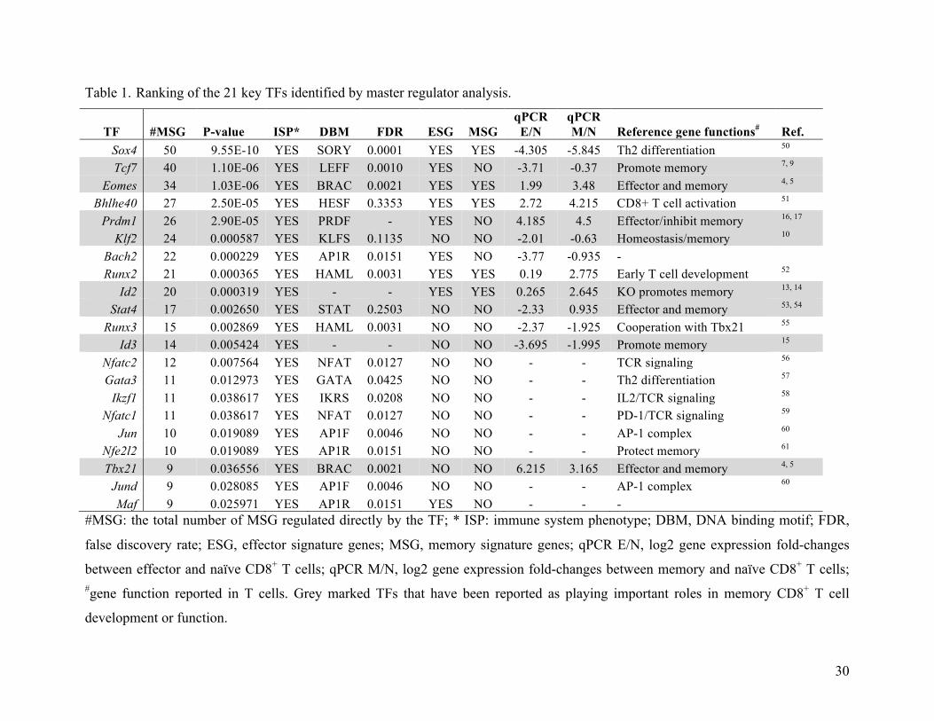
Table 1. Ranking of the 21 key TFs identified by master regulator analysis.
TF #MSG P-value ISP* DBM FDR ESG MSG qPCR E/N
qPCR M/N Reference gene functions# Ref.
Sox4 50 9.55E-10 YES SORY 0.0001 YES YES -4.305 -5.845 Th2 differentiation 50
Tcf7 40 1.10E-06 YES LEFF 0.0010 YES NO -3.71 -0.37 Promote memory 7, 9 Eomes 34 1.03E-06 YES BRAC 0.0021 YES YES 1.99 3.48 Effector and memory 4, 5
Bhlhe40 27 2.50E-05 YES HESF 0.3353 YES YES 2.72 4.215 CD8+ T cell activation 51
Prdm1 26 2.90E-05 YES PRDF - YES NO 4.185 4.5 Effector/inhibit memory 16, 17 Klf2 24 0.000587 YES KLFS 0.1135 NO NO -2.01 -0.63 Homeostasis/memory 10
Bach2 22 0.000229 YES AP1R 0.0151 YES NO -3.77 -0.935 - Runx2 21 0.000365 YES HAML 0.0031 YES YES 0.19 2.775 Early T cell development 52
Id2 20 0.000319 YES - - YES YES 0.265 2.645 KO promotes memory 13, 14 Stat4 17 0.002650 YES STAT 0.2503 NO NO -2.33 0.935 Effector and memory 53, 54
Runx3 15 0.002869 YES HAML 0.0031 NO NO -2.37 -1.925 Cooperation with Tbx21 55
Id3 14 0.005424 YES - - NO NO -3.695 -1.995 Promote memory 15
Nfatc2 12 0.007564 YES NFAT 0.0127 NO NO - - TCR signaling 56
Gata3 11 0.012973 YES GATA 0.0425 NO NO - - Th2 differentiation 57
Ikzf1 11 0.038617 YES IKRS 0.0208 NO NO - - IL2/TCR signaling 58
Nfatc1 11 0.038617 YES NFAT 0.0127 NO NO - - PD-1/TCR signaling 59
Jun 10 0.019089 YES AP1F 0.0046 NO NO - - AP-1 complex 60
Nfe2l2 10 0.019089 YES AP1R 0.0151 NO NO - - Protect memory 61
Tbx21 9 0.036556 YES BRAC 0.0021 NO NO 6.215 3.165 Effector and memory 4, 5 Jund 9 0.028085 YES AP1F 0.0046 NO NO - - AP-1 complex 60
Maf 9 0.025971 YES AP1R 0.0151 YES NO - - - #MSG: the total number of MSG regulated directly by the TF; * ISP: immune system phenotype; DBM, DNA binding motif; FDR,
false discovery rate; ESG, effector signature genes; MSG, memory signature genes; qPCR E/N, log2 gene expression fold-changes
between effector and naïve CD8+ T cells; qPCR M/N, log2 gene expression fold-changes between memory and naïve CD8+ T cells; #gene function reported in T cells. Grey marked TFs that have been reported as playing important roles in memory CD8+ T cell
development or function.
31
Table 2. Summary of the ChIP-PCR results.
TF Sox4 Tcf7 Eomes Total No. MSG target 41 36 26 103 No. tested target 12 14 9 35 No. positive 10 (83.3%) 12 (85.7%) 6 (66.7%) 28 (80%)
32
Table 3. Perturbation analysis.
Gene Sox4 Tcf7 Eomes Bhlhe40 Prdm1 Klf2 Bach2 Id2 Runx2 Stat4 Id3 Tbx21* Sox4 7.94 -1.38 1.77 -0.62 -3.17 -0.28 -0.93 -0.48 -0.05 -0.70 -0.83 1.21 Tcf7 -1.79 2.44 -0.03 -0.21 -2.01 -0.50 0.88 -0.39 0.04 -2.16 0.31 -0.64
Eomes -2.28 -1.11 5.29 0.38 -0.08 -0.26 -0.64 -0.10 0.85 -0.55 0.54 -2.31 Bhlhe40 -2.03 0.41 1.09 3.88 0.02 1.99 0.32 0.31 0.14 -0.41 0.68 0.62
Prdm1 -0.14 -1.48 2.04 1.13 5.19 1.34 -1.62 -0.17 0.20 0.08 -0.39 1.30 Klf2 0.85 -0.78 -1.10 -0.55 -1.49 4.06 -1.52 -0.73 -0.07 -1.33 -1.51 -0.57
Bach2 1.37 0.75 0.35 0.00 0.22 1.40 4.41 -0.18 0.78 0.01 0.07 0.15 Id2 -2.24 1.10 0.46 0.38 0.24 0.66 0.09 4.98 0.93 -0.06 0.18 0.54
Runx2 -1.64 0.41 -0.51 -0.22 -0.02 0.91 -0.50 -0.82 3.71 -0.54 -0.03 -0.77 Stat4 -1.45 -0.26 -1.78 0.14 0.80 0.24 0.22 -0.01 0.85 2.32 0.41 -0.81
Id3 2.61 0.23 -1.32 0.47 -0.23 0.72 2.02 -1.03 0.25 0.73 7.44 -1.09 Tbx21 -1.68 -0.26 -0.22 0.12 -0.34 1.42 0.50 0.93 1.29 -2.29 1.17 6.54
! The highlighted 12 TFs (first row) were overexpressed individually in 2C T cells and the level of their transcripts of each TFs was quantified by real-time PCR. Expression data was normalized to the empty vector control and then log2 transformed. Changes in transcript levels for !2 fold are marked orange (up-regulated) or green (down-regulated). The overexpressed TFs are marked red.
a c
b
Hu and Chen, Figure 1
input
Anti-Sox4
IgG
Prf1 Il6ra Il18r1 Gzmb Gzmk S100a6 Ccr7 Ccl5 Klrc1 Ccr5 Cd38
S100a4
Input
Anti-Tcf7
IgG
Cd38 Dusp14 Ms4a1 Blk Prf1 Ccl5 Ccr7 Gzmk Sell Il7r
S100a6Abca3 Gzmb S100a4
input
Anti-Eomes
IgG
Cd38 Infg
Gzmk Ccr7 Ccl5 Prf1 Gzmb Ccr5 Cxcr3
MRA
Key TFs
TF
TF
Sample Gene
Putative
TFs
Regulatory network N E M
MSG
data filtering
TFs
MSGs
Il6ra
Ccr7
Bach2
Crtam
Pglyrp1
Il6st
Cd44
Klrc1
2610019F03Rik
Igfbp4
St3gal6
Id2
Sntb2
Plscr1Runx2
Klf6
Dusp1
Prf1
Lrrk1
Cebpb
Ctla2b
Errfi1
Mdfic
Ahnak
EomesMyo1f
Cd24aNkg7
AW112010
Chst11
Cxcr3
Ms4a6d
Ifng
Fosl2
Cnn3
Dntt
Ramp1
Hopx
Fasl
Il2rb
Casp1
Ly6a
Sox4Dock5
Pmaip1
Ccr5
Pik3ap1
F2r
Smpdl3b
Prr13
Klf2
Trib2
Lyz2Ppic
Stat4
Tec
Il18r1Pde4b
S100a6Cd38
Ccl5Klf4
Idh2Emp1
Tlr1
MybSlamf6
St6gal1
Folr4
Dnmt3a
Tcf7
Abca3
Gsn
Dusp14
Anxa1
Lfng
Ms4a1
Blk
Ell2H2-Q10
Gzmb
Itgax
Actn1
GzmkAnxa2Aqp9
Bhlhe40
Scd1
Prdm1
S100a4
Fcgr2b
Size(bp)
Size(bp)
Size(bp)
300200
300200
200100
200100
200100
200100
300200
200100200100
200100
200100
200100
200100
300200
200100
300200
300200300200300200
200100
200100
200100
200100
200100
300200
200100
200100
200100
300200
200100
300200
200100
200100
200100
200100
a b
c
Hu and Chen, Figure 2
Tcf7 Eomes
Bhlhe40
Bach2 Id2
Runx2 Stat4
Tbx21
Sox4
Prdm1 Klf2
Id3 Tcf7
Tbx21
Eomes
Sox4
Input
Anti-Sox4
IgGInput
Anti-Tcf7
IgGInput
Anti-Eomes
IgG
Runx2
Klf2
Id3
Id2
Sox4
Bach2
Stat4
Bhlhe40
Eomes
Tbx21
Tcf7
Prdm1
200100
300200
200100
200100
300200
300200
200100
200100
200100
300200
Size(bp)
300200
300200
Vector Sox4 Bhlhe40 Bach2 Runx2a
b
c
d
Vector shSox4 shBhlhe40 shBach2 shRunx2
Hu and Chen, Figure 3
n.s.* **
n.s.
* *****
n.s.
n.s.
** ** *
**
** ** * *
n.s.n.s.
n.s.
n.s.n.s.
n.s.
0.0
0.2
0.4
0.6
0.8
0.0
0.1
0.2
0.3
0.0
0.2
0.4
0.6
0.0
0.2
0.4
0.6
0.0
0.2
0.4
0.6
0.8
0 5 0 5 0 50 5 0 5
Days poster infection
0 5 0 5 0 50 5 0 5
Days poster infection
0.0
0.2
0.4
0.6
0.0
0.2
0.4
0.6
0.0
0.1
0.2
0.3
0.0
0.2
0.4
0.6
0.0
0.2
0.4
0.6
0.0
0.2
0.4
0.6
0.0
0.2
0.4
0.6
0.0
0.2
0.4
0.6
0.0
0.2
0.4
0.6
0.0
0.2
0.4
0.6
0 4 6 0 4 6 0 4 6 0 4 6 0 4 6
Days
0.0
0.2
0.4
0.6
0.0
0.2
0.4
0.6
0.0
0.2
0.4
0.6
0.8
0.0
0.2
0.4
0.6
0.8
0.0
0.2
0.4
0.6
0.8
0 4 6 0 4 6 0 4 6 0 4 6 0 4 6
Days
% GFP+
% GFP+
% GFP+
% GFP+
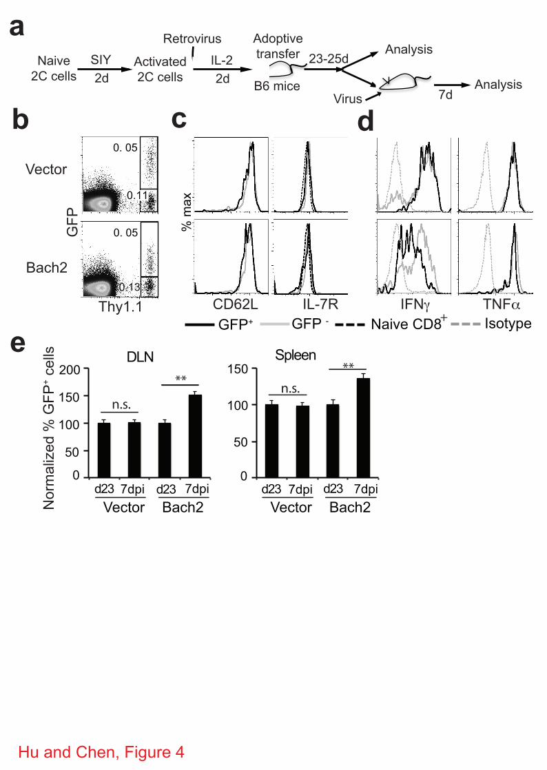
0
50
100
150
200
0
50
100
150
a
b
DLN
IFN TNF
GFP - Naive CD8GFP+ Isotype+
% max
Bach2Vector
7dpid23
**
Spleen**
n.s.n.s.
Hu and Chen, Figure 4
d
CD62L IL-7R
Naive
2C cells
SIY
2d
Activated
2C cells
Retrovirus
IL-2
2d
23-25d
Adoptive
transfer Analysis
Virus 7dAnalysis
GFP
Thy1.1
Vector
Bach2
c
7dpid23 7dpid23 7dpid23
e
Bach2Vector
0.11
0. 05
0. 05
B6 mice
Normalized % GFP+ cells
0.13
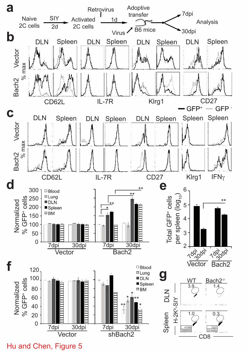
aNaive
2C cells
SIY
2d
Activated
2C cells
Retrovirus
1d
Adoptive
transfer
Analysis
Virus
7dpi
30dpibVector
Bach2
DLN Spleen
CD62L
% max
Klrg1IL-7R
DLNDLN
c
dIL-7RCD62L
SpleenSpleenDLN DLN
IFN
Spleen
Vector
Bach2
% max
Klrg1
Hu and Chen, Figure 5
Spleen
Spleen Spleen
B6 mice
GFP+ GFP -CD27
CD27
SpleenDLN
7dpi 30dpi
Vector Bach2
7dpi 30dpi
***
**300
250
200
150
100
0
50
Normalized
% GFP+ cells
**
DLN Spleen
e5
4
3
2
6
**
7dpi
30dpi
7dpi
30dpi
Vector Bach2
100
80
60
40
0
20
**
**
7dpi 30dpi
Vector shBach2
7dpi 30dpi
f g
*
**
Normalized
% GFP+ cells
120
CD8
H-2Kb-SIYDLN
Spleen
WT Bach2-/-
Total GFP+ cells
per spleen (log10)
3.9 1.4
1.0 0.3
41590
3999000
7227
2210000
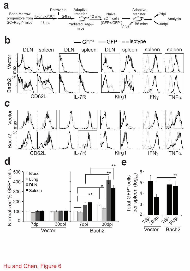
CD62L IFNIL-7R
Vector
Bach2
TNFKlrg1
Hu and Chen, Figure 6
aBone Marrow
progenitors from
2C+Rag-/- mice
IL-3/IL-6/SCF
48hrs
Retrovirus
24hrs
Adoptive
transfer
Analysis
Virus
7dpi
30dpiIrradiated Rag-/-
mice
12 wksNaive
2C T cells
(GFP+/GFP-)
Adoptive
transfer
b
c
Vector
Bach2
d
% max
% max
DLN spleen DLN spleen DLN spleen spleen spleen
CD62L IFNIL-7R TNFKlrg1
Isotype GFP - GFP+
7dpi 30dpi
Vector Bach2
7dpi 30dpi
**
*
**
**500
400
300
200
100
0
B6 mice
Normalized % GFP+ cells
e
5
4
3
2
6
**
7dpi
30dpi
7dpi
30dpi
Vector Bach2
Total GFP+ cells
per spleen (log10)
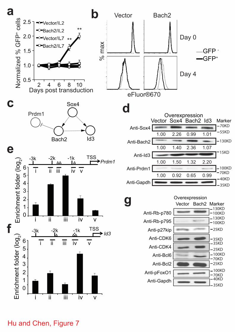
Days post transduction
a
**
**
b Vector Bach2
Day 0
Day 4
eFluor®670Sox4
Bach2 Id3
Prdm1c d
e
Hu and Chen, Figure 7
g
% max
0.5
1.0
1.5
2.0
2.5
2 4 6 8 10 Normalized % GFP
+ cells
GFP - GFP+
Overexpression Vector Bach2
Anti-Rb-p780
Anti-Rb-p795
Anti-p27kip
Anti-CDK6
Anti-CDK4
Anti-Bcl6
Anti-Bcl2
Anti-pFoxO1
Anti-Gapdh
Anti-Sox4
Anti-Bach2
Anti-Id3
Anti-Prdm1
Anti-Gapdh
Overexpression Vector Sox4 Bach2 Id3
1.00 2.26 0.99 1.01
1.00 1.40 2.36 1.07
1.00 1.50 1.32 2.20
1.00 0.92 0.65 0.99
i ii iii iv v Enrichment folder (log 2)
6
4
2
0 1
3
5
TSS Prdm1
-1k -2k -3k
i ii iii iv v
Enrichment folder (log 2)
TSS Id3 -1k -2k -3k
i ii iii iv v 6
4
2
0 1
3
5
i ii iii iv v
f
70KD55KD
130KD
100KD70KD
15KD
40KD35KD
130KD100KD130KD100KD25KD
35KD35KD25KD100KD70KD25KD100KD70KD40KD35KD
Marker
Marker
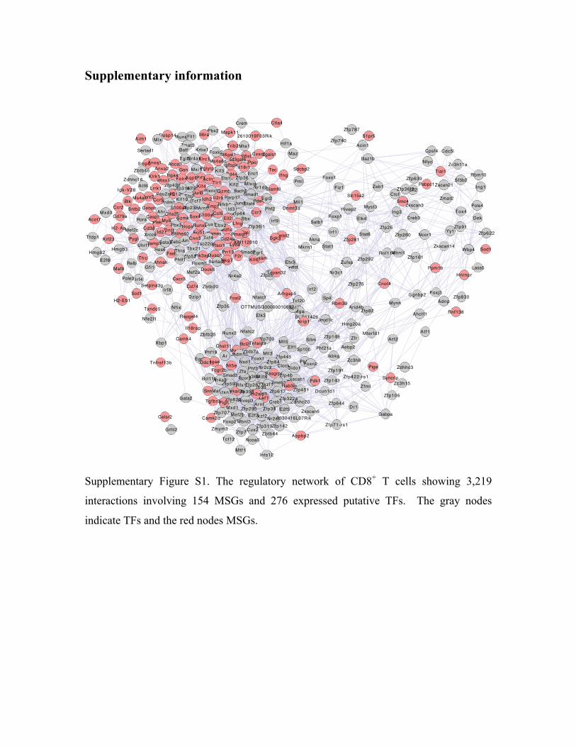
Supplementary information
Supplementary Figure S1. The regulatory network of CD8+ T cells showing 3,219
interactions involving 154 MSGs and 276 expressed putative TFs. The gray nodes
indicate TFs and the red nodes MSGs.
!"##$%&%'()*+,-./0,1
!"#"$
%&'()
*+,-$!"#")%&'(.$
/-&$%&'()0
1234$%&'$)$
53&$$6
*-7+$
83,"6
%&'(9(
%&'(:;
%<&='
>?@
52A$0
%&')((
B,4?6
/3C$
D#==;B'A$2
E2'6
F3-3'-
!,G$%@=+#3$6
%@=+#371H="7
I,J'$/3C7
K4@6
!A+6
8-?27
FLM?'(
%&'.(
!'61H33
NC32'(
52A79
53&$7.
I,JO7
P-LG62
PQ+"&$
PG3'%&'.70
B#2'+$K4&(
%&')79
ILR$
8"+&%&'7)4(
I,J,$
!4+$;#(
%?2$
%&'9$SH$
%A#"(
%=+#3($!&72(
T,J6
TL#4$
8-"#A
%&'(.0+
U4&)
KC-$!A#G:
5?="K"M7
T='#37(
I(-B--$7
%&'7;
I,=4(
*@C:
DH)#
*-6#(
/4$.-$
%&'7)
V#+Q(
8G66
144$
1@-3$
!#"2$
P@3#
>3A"7#
!G+2'(
BA4
/&3C
8+-:
*-$G(!4#A&)
BG?62
BQ&(
KC-(!"#"6
!A#G$
T?+K3+$
14JL'
/-&(
V80$$6()1C#
!'$
*&#"+$T+&(0
P-QC#';
WTT1X!N000000$0);:
K4@7
PE$$(0$0
V2JD&3C
1G&L+I,=BA#L'$
8G(6#
K44(%&')6
!&A2"(!C@7
/-&9
%&'7)4$
*&@2$/G7
NRA2
B#-'$(Y<3G
K"=$
U4&(
FAC2(
T&G'$
P+,":
1#&2
UL&(7
F(ZP#
K(&.
FAC27
1JG7
1?&(+
B4?@
8G7.
8#='$5,-#
!?-'L3#7C
5?42
B,4?7 /-&6
T&-+PQ3#@
N&L$
1H2
>3""B2J7
NRA@
%?2(
VQ4Q?60
Y<3
F,'J
%&';(BQ"&$
I#=4TQ-# T2J($
K,A?=
82J(
T+&6K--&L$
T=+((G$B4=+-$
5<3J(
8337
8<J$
!,J6
I,JA$
1?&(#
BL@7#'$><='$
%2"2(0
>,+@;
!?-"#G(
*&?(4$
TJ3G+;
F(ZK2$
!+G$
F?=)
N#"#75#A'$XQ-&$
BHC4[-++)
/G(
144")
*-(+(
*+,#(NA?2$
%&'.6
5#=C-'$
/@R&$
%2"266
/3"=$(
%&')$:
%&'$6(%&'7$9
*-(+$
8-?2$
%&'79
/@R&(
D?&$FLM?'7
BQ&7
P-3"
%2"277
K'A(#L'$
%&'(.:
%&'7)9
%&'79.B-@#-(#
5+,-7
%&#
BLC3%&'.()
*";?
P"J3$
!A#G7
%&';975&J$
5<3J7
8Q="$$
P-1H,$&
1447YQGA$G
*=G$
%&':09T3&#L'7V+4(
*&#"+(
%2"2:#
I,J@$
K(&7
I,J'(
1"&$
T+&$(
%&'(9;
%&':
12347
*+,#7
8<J(
1?&(2
1JG$
%&':60!$'-;
%&':.:
V#R$2
P+L3$
1#'@$$
T-L2(
8-?A
1"#$
B'L+
!")C#4$
/C&2'6
/4)="!"7C#4)
><='$6
P2+#7
!?-"#G$
!A'G472
14J
P3J#$
PLA$
*&H+
8G+;48'=&6
%+7Q$$#
8"4#6
8JJ+; 1#R
()$00$9I075L@
DC#4=$
FL&$#
1=6#)G
I,J@(
B2J(/4)-#
U4-+$
U-2#$
5<3J$
%A#"7
I4L$
TCL&(
V#"&
*-6#$
P-3"4
!$00#)
B4J3+$
KA'$%&'(7.
/4(-2
8+4;
/GQ(K"=(
T+&:
Y<32
I,4-6P\'9
N=3
*&?(4(%&'7$. U4&6
1JL$
B-&$
%&'6((
/"C#J
8"4#(2
U4&$0
!$00#6
F(Z]$0
B-GA$
B,<(#&$
8G:9#PQ-
DHR(
8+-( 8?2'2
8+-;1=6#$
!3"2(
V4@
1"==$
%2"26)
U4-@$
D--@$
P3J#(
%GQQ+$)
/C@Z^(.P"&)
P+"3$
BC4H-'$
T4-$
U4&7
BQ&.
%@=+#3)
!4"A
54&
!'$00
BG@$
I,J3(
*-L'$
P?2'(
YAOG$+FAC(0#
%+7Q.
%&'$6.
BQ&($#
%&-
/@2@C
%&'$9$%GQQ+7
%+7Q$;
P"&$1"?-&G$
BLC#
P"&(
!H3+-L'
144;
%&'66;
>LG,$
K4&$
84,+@
%&')66
%&'$0)
%&':$Z-=$
N#2'#
>+<3$G$
>-$
%&A4
%&'$67%&'6((Z-=$
P''2'(
)7706$)D0:5L@
K(&;
%&'6;$
%GQQ+(0
5#27L'
%@=+#3$%&'60
%&'7((#
I+C-(2
*-6#7V+4$$2
1#&!33
8#A@(G%&':0:
TC&2-7
N-Q4(
8?4=-(
N#"#(
%AHA7
T3&-=&$72
8#A@6
/"C#?
%2"2(;
KC-7
/4$.-#'
[2'$
BQ&$9
WG+$
*&LJ
>RL'$
8G:6/-&.
5#'C?&6
8J+-7
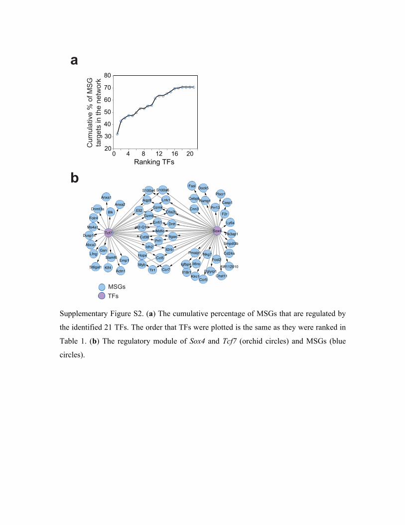
Supplementary Figure S2. (a) The cumulative percentage of MSGs that are regulated by
the identified 21 TFs. The order that TFs were plotted is the same as they were ranked in
Table 1. (b) The regulatory module of Sox4 and Tcf7 (orchid circles) and MSGs (blue
circles).
!"
#"$
%"
&"$
'"
("$
)"$
"$ %$ )$ *!$ *'$ !"$+,-./-0$123$
!
"
Smpdl3b
Pik3ap1
Ly6a
Plscr1Dock5
Casp1
Sox4
Prr13F2r
Ramp1
Cd24a
Ccr5
Pglyrp1Chst11
Nkg7
AW112010
Fosl2
Aqp9
Gzmk
Gzmb
Errfi1
Ell2
S100a4
Lrrk1
S100a6
Prf1
Il2rb
Dntt
Ccl5
ItgaxMdfic
Ccr7
Pmaip1
Il6ra
Il18r1
Igfbp4
Klrc1
St6gal1
Lfng
Abca3
Dusp14
Klf4
Slamf6
Gsn
Tcf7Cd38
Myb
Hopx
Idh2
Actn1
Emp1
H2-Q10
Tlr1
Folr4Blk
Dnmt3a
Ms4a1
Anxa1
Anxa2
Fasl
Cebpb
Cnn3Ctla2b
1234563
7898:,;/<=$>$?@$456
$;,A0=;3$/-$;B=$-=;C?A.$
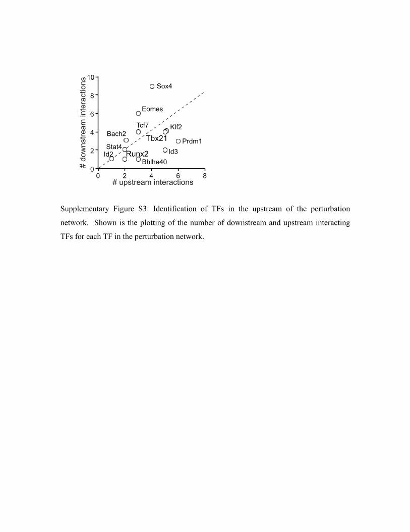
Supplementary Figure S3: Identification of TFs in the upstream of the perturbation
network. Shown is the plotting of the number of downstream and upstream interacting
TFs for each TF in the perturbation network.
!"#$%
&'()%
*"+,-%
./0/,$1%
234+5%
60(7%.8'/7%
947% :;<#7%!=8=$%
94>%
&?#75%
@%4"A<-=3,8+%B<=,38'=B"<-
1%
7%
$%
C%
D%
51%
1% 7% $% C% D%@%;E-=3,8+%B<=,38'=B"<-
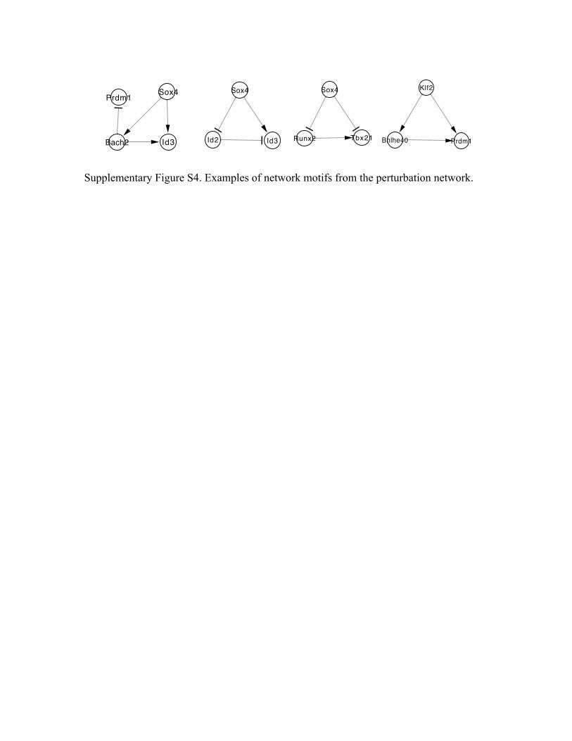
Supplementary Figure S4. Examples of network motifs from the perturbation network.
!"#$
%&' %&(
!"#$
)*+#' ,-#'. /0102$3 45&6.
718'45&6.
!"#$
/9:0' %&(
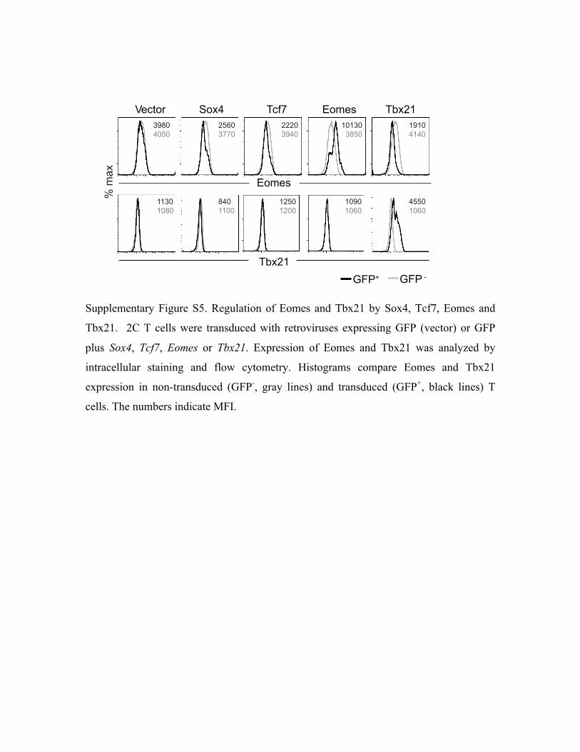
Supplementary Figure S5. Regulation of Eomes and Tbx21 by Sox4, Tcf7, Eomes and
Tbx21. 2C T cells were transduced with retroviruses expressing GFP (vector) or GFP
plus Sox4, Tcf7, Eomes or Tbx21. Expression of Eomes and Tbx21 was analyzed by
intracellular staining and flow cytometry. Histograms compare Eomes and Tbx21
expression in non-transduced (GFP-, gray lines) and transduced (GFP+, black lines) T
cells. The numbers indicate MFI.
!"#$%&'''''''''''(%)*''''''''''''''+#,- .%/"0 +1)23'
.%/"0!
+1)23'
4'/5)
678'9'678:
;<=>*>?>
2?@>;-->
222>;<*>
3>3;>;=?>
3<3>*3*>
33;>3>=>
=*>33>>
32?>32>>
3><>3>@>
*??>3>@>
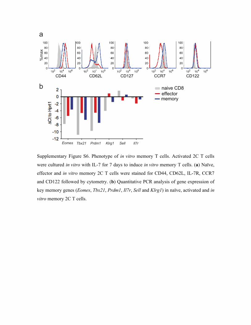
Supplementary Figure S6. Phenotype of in vitro memory T cells. Activated 2C T cells
were cultured in vitro with IL-7 for 7 days to induce in vitro memory T cells. (a) Naïve,
effector and in vitro memory 2C T cells were stained for CD44, CD62L, IL-7R, CCR7
and CD122 followed by cytometry. (b) Quantitative PCR analysis of gene expression of
key memory genes (Eomes, Tbx21, Prdm1, Il7r, Sell and Klrg1) in naïve, activated and in
vitro memory 2C T cells.
!"##$$$$$$$$$$$$$$$$$$$$!"%&'$$$$$$$$$$$$$$$$!"(&)$$$$$$$$$$$$$$$$$$!!*)$$$$$$$$$$$$$$$$$$!"(&&$
+,,+-./01+1/02
3456+$!"7
8149
!"#$% &'()* +,-#* .$//0/,1* 2/3,
4
:
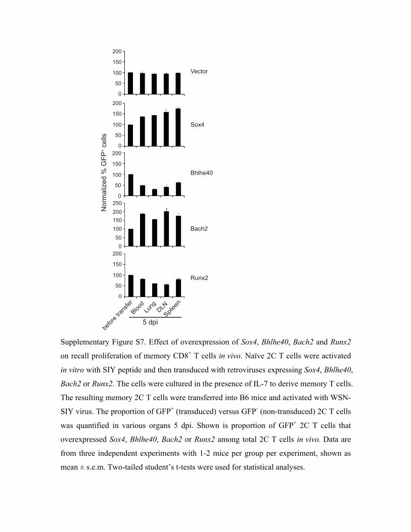
Supplementary Figure S7. Effect of overexpression of Sox4, Bhlhe40, Bach2 and Runx2
on recall proliferation of memory CD8+ T cells in vivo. Naïve 2C T cells were activated
in vitro with SIY peptide and then transduced with retroviruses expressing Sox4, Bhlhe40,
Bach2 or Runx2. The cells were cultured in the presence of IL-7 to derive memory T cells.
The resulting memory 2C T cells were transferred into B6 mice and activated with WSN-
SIY virus. The proportion of GFP+ (transduced) versus GFP- (non-transduced) 2C T cells
was quantified in various organs 5 dpi. Shown is proportion of GFP+ 2C T cells that
overexpressed Sox4, Bhlhe40, Bach2 or Runx2 among total 2C T cells in vivo. Data are
from three independent experiments with 1-2 mice per group per experiment, shown as
mean ± s.e.m. Two-tailed student’s t-tests were used for statistical analyses.
!"#!##$"#$##
#"#
!##
$"#
$##
#
"#
!##
$"#
$##
#
"#
!##
$"#
$##
#
"#
!##
$"#
$##
#
"#
%&'()*+,-./0/123
4 /5-**6
7*&&.89:;<8%=>*--:
?-@&'-/A'):6@-'
B-5A&'
=&CD
7E*E-D#
7)5E!
F9:C!
"/.>+
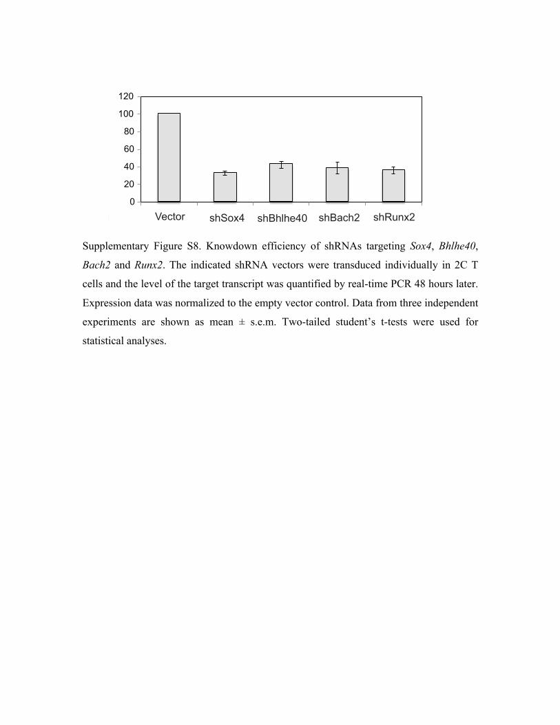
Supplementary Figure S8. Knowdown efficiency of shRNAs targeting Sox4, Bhlhe40,
Bach2 and Runx2. The indicated shRNA vectors were transduced individually in 2C T
cells and the level of the target transcript was quantified by real-time PCR 48 hours later.
Expression data was normalized to the empty vector control. Data from three independent
experiments are shown as mean ± s.e.m. Two-tailed student’s t-tests were used for
statistical analyses.
!"#$%&'"(%)$*+,)&-%(#"'"#
.(
/.(
0.(
1.(
2.(
3..(
3/.(
4",%5) +67580 +696#6"0. +69$,6/ +6!:*8/
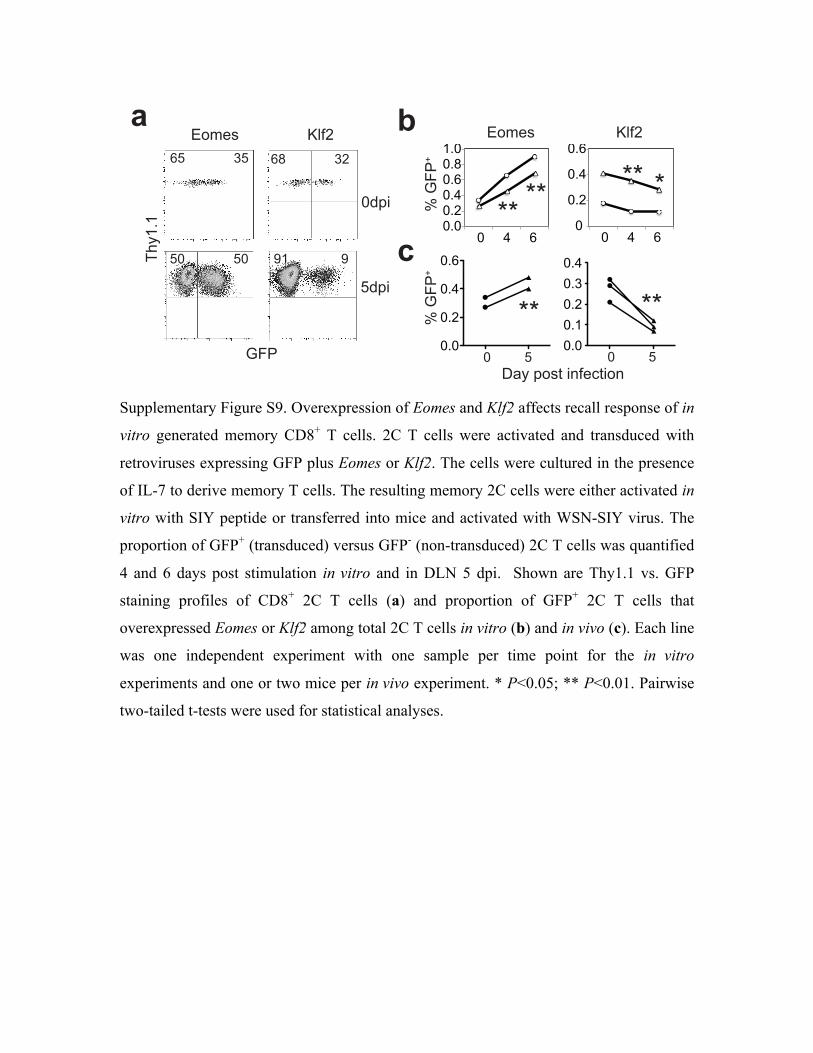
Supplementary Figure S9. Overexpression of Eomes and Klf2 affects recall response of in
vitro generated memory CD8+ T cells. 2C T cells were activated and transduced with
retroviruses expressing GFP plus Eomes or Klf2. The cells were cultured in the presence
of IL-7 to derive memory T cells. The resulting memory 2C cells were either activated in
vitro with SIY peptide or transferred into mice and activated with WSN-SIY virus. The
proportion of GFP+ (transduced) versus GFP- (non-transduced) 2C T cells was quantified
4 and 6 days post stimulation in vitro and in DLN 5 dpi. Shown are Thy1.1 vs. GFP
staining profiles of CD8+ 2C T cells (a) and proportion of GFP+ 2C T cells that
overexpressed Eomes or Klf2 among total 2C T cells in vitro (b) and in vivo (c). Each line
was one independent experiment with one sample per time point for the in vitro
experiments and one or two mice per in vivo experiment. * P<0.05; ** P<0.01. Pairwise
two-tailed t-tests were used for statistical analyses.
!"!#!"$#!"%#!"&#!"'#("!#
!#
!"$#
!"%#
!"&#
))))
))))
)) )
!
"
#
!*+,
-*+,
./012################345$ ./012###################345$
!#########- !#########-#678#+/29#,:51;9,/:
<=8("(
>?@
-!#############-! A(##############A
&-#############B- &'#############B$
!"!
!"$
!"%
!"&
!"!!"(!"$!"B!"%
C#>?@
DC#>?@
D
!#####%#####& !#####%#####&
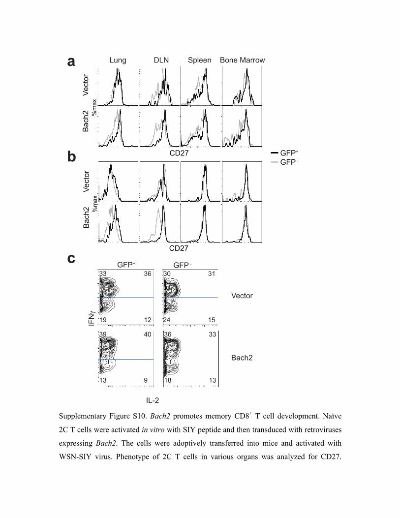
Supplementary Figure S10. Bach2 promotes memory CD8+ T cell development. Naïve
2C T cells were activated in vitro with SIY peptide and then transduced with retroviruses
expressing Bach2. The cells were adoptively transferred into mice and activated with
WSN-SIY virus. Phenotype of 2C T cells in various organs was analyzed for CD27.
!"#$%&'
()#*+'
,-).'
/0+1'
!"#$%&'
()#*+'
,-).'
/0+1'
!
"
#
2345'''''''''''''026'''''''''789""4''''(%4"':)&&%;
<=6
<2>+
!"#$%&
()#*+
?=@A ?=@'>
?=@A''?=@'>
BB BC BD BE
BF GD BC BB
EF E+ +G EH
EB F EI EB
Shown are histograms CD27 expression of Thy1.1+ CD8+ 2C T cells 7 (a) and 30 dpi (b).
Gray trace, nontransduced (GFP-) 2C T cells; black trace, transduced (GFP+) 2C T cells.
IL-2 and IFN! expression 2C T cells 30 dpi were analyzed as Fig.4. Shown is IL-2 vs.
IFN! staining profile of Thy1.1+ CD8+ 2C T cells (c). Representative data from 6 mice in
3 independent experiments are shown.
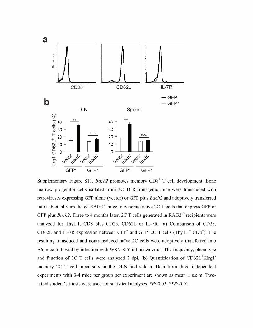
Supplementary Figure S11. Bach2 promotes memory CD8+ T cell development. Bone
marrow progenitor cells isolated from 2C TCR transgenic mice were transduced with
retroviruses expressing GFP alone (vector) or GFP plus Bach2 and adoptively transferred
into sublethally irradiated RAG2-/- mice to generate naïve 2C T cells that express GFP or
GFP plus Bach2. Three to 4 months later, 2C T cells generated in RAG2-/- recipients were
analyzed for Thy1.1, CD8 plus CD25, CD62L or IL-7R. (a) Comparison of CD25,
CD62L and IL-7R expression between GFP+ and GFP- 2C T cells (Thy1.1+ CD8+). The
resulting transduced and nontransduced naïve 2C cells were adoptively transferred into
B6 mice followed by infection with WSN-SIY influenza virus. The frequency, phenotype
and function of 2C T cells were analyzed 7 dpi. (b) Quantification of CD62L+Klrg1-
memory 2C T cell precursors in the DLN and spleen. Data from three independent
experiments with 3-4 mice per group per experiment are shown as mean ± s.e.m. Two-
tailed student’s t-tests were used for statistical analyses. *P<0.05, **P<0.01.
!"#$%
&'() &'*(+ ,+-./
!
**
n.s.
**
n.s.
012334
5$67(
8369:;
5$67(
8369:;
'+<
5$67(
8369:;
5$67(
8369:;
"
=>?@ =>?"-
A2;BC"&'*(+"""D"6322E"F!
G
+
-
=>?@""=>?"-
=>?@ =>?"-
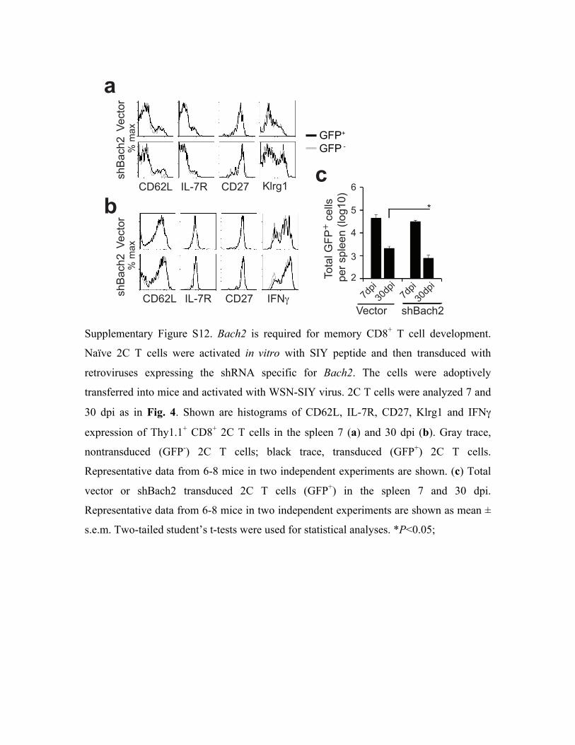
Supplementary Figure S12. Bach2 is required for memory CD8+ T cell development.
Naïve 2C T cells were activated in vitro with SIY peptide and then transduced with
retroviruses expressing the shRNA specific for Bach2. The cells were adoptively
transferred into mice and activated with WSN-SIY virus. 2C T cells were analyzed 7 and
30 dpi as in Fig. 4. Shown are histograms of CD62L, IL-7R, CD27, Klrg1 and IFN!
expression of Thy1.1+ CD8+ 2C T cells in the spleen 7 (a) and 30 dpi (b). Gray trace,
nontransduced (GFP-) 2C T cells; black trace, transduced (GFP+) 2C T cells.
Representative data from 6-8 mice in two independent experiments are shown. (c) Total
vector or shBach2 transduced 2C T cells (GFP+) in the spleen 7 and 30 dpi.
Representative data from 6-8 mice in two independent experiments are shown as mean ±
s.e.m. Two-tailed student’s t-tests were used for statistical analyses. *P<0.05;
!
"
!"#$%&
'()*#(+
,-.+/
012*3
45&678/9:; ,-+:
,-.+/ 8/9:; ,-+: 8<=
!"#$%&
'()*#(+ 012*3
>%$*51?<@111#"55'1
A"&1'A5""B1C5%67DE
+
:FAGHDFAG:FAGHDFAG
!"#$%& '()*#(+
I
J
H
+
.#
K
?<@L11?<@19
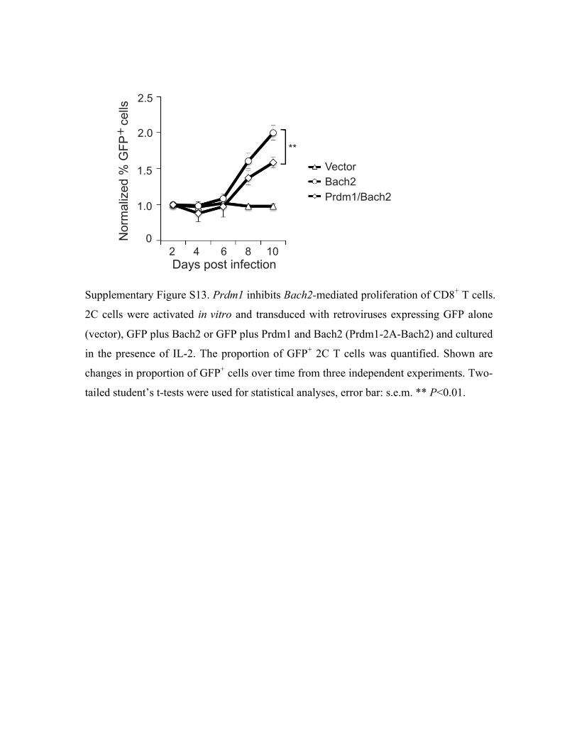
Supplementary Figure S13. Prdm1 inhibits Bach2-mediated proliferation of CD8+ T cells.
2C cells were activated in vitro and transduced with retroviruses expressing GFP alone
(vector), GFP plus Bach2 or GFP plus Prdm1 and Bach2 (Prdm1-2A-Bach2) and cultured
in the presence of IL-2. The proportion of GFP+ 2C T cells was quantified. Shown are
changes in proportion of GFP+ cells over time from three independent experiments. Two-
tailed student’s t-tests were used for statistical analyses, error bar: s.e.m. ** P<0.01.
!"#$%&'$(%)*+,-()'*
..
/'01"2)3,4%5%678%%%-,22$
+
9%%%%%%:%%%%%%%;%%%%%%<%%%%%=>
9?@
9?>
=?@
=?>
A,-('0B"-C98041=DB"-C9
>
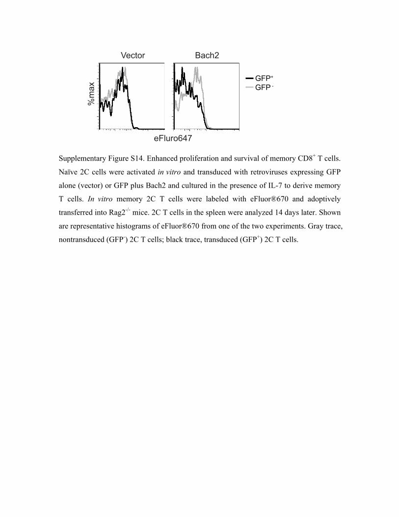
Supplementary Figure S14. Enhanced proliferation and survival of memory CD8+ T cells.
Naïve 2C cells were activated in vitro and transduced with retroviruses expressing GFP
alone (vector) or GFP plus Bach2 and cultured in the presence of IL-7 to derive memory
T cells. In vitro memory 2C T cells were labeled with eFluor®670 and adoptively
transferred into Rag2-/- mice. 2C T cells in the spleen were analyzed 14 days later. Shown
are representative histograms of eFluor®670 from one of the two experiments. Gray trace,
nontransduced (GFP-) 2C T cells; black trace, transduced (GFP+) 2C T cells.
!"#$%&'()
*+,-
.!/0&% 1,/23
4"56774"578
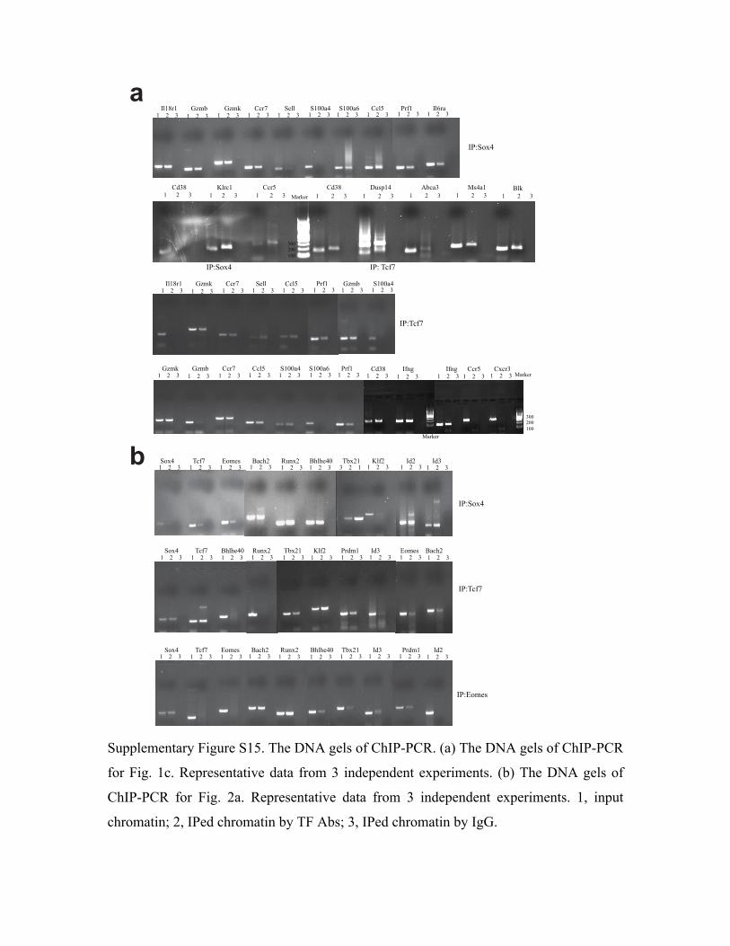
Supplementary Figure S15. The DNA gels of ChIP-PCR. (a) The DNA gels of ChIP-PCR
for Fig. 1c. Representative data from 3 independent experiments. (b) The DNA gels of
ChIP-PCR for Fig. 2a. Representative data from 3 independent experiments. 1, input
chromatin; 2, IPed chromatin by TF Abs; 3, IPed chromatin by IgG.
!
"
!"#$%#&
'((&)((&*((&
*&&&&&&&&)&&&&&&'& *&&&&&&&&)&&&&&&'& *&&&&&&&&)&&&&&&'& *&&&&&&&&)&&&&&&'& *&&&&&&&&)&&&&&&'&*&&&&&&&)&&&&&&'&*&&&&&&)&&&&&&'&+,'-&&&&&&&&&&&&&&&&&&./#0*&&&&&&&&&&&&&&&&&&+0#1&&&&&&&&&&&&&&&&&&&&&&&&&&&&&&+,'-&&&&&&&&&&&&&&&&&2345*6&&&&&&&&&&&&&&&&&&780"'&&&&&&&&&&&&&&&&&!46"*&&&&&&&&&&&&&&& 9/$&
*&&&&&&)&&&&&&'&
:;<=>?6&&&&&&&&&&&&&&&&&&&&&&&&&&&&&&&&&&&&&&&&&&&&&&&&&&&&&&&&&&&&&&&&&&&&&&:;<&@0AB&
:;<=>?6&
*&&&&)&&&&'& *&&&&)&&&&'& *&&&&)&&&&'& *&&&&)&&&&'& *&&&&)&&&&'& *&&&&)&&&&'& *&&&&)&&&&'& *&&&&)&&&&'& *&&&&)&&&&'& *&&&&)&&&&'&:/*-#*&&&&&&&&CDE8&&&&&&&&&&CDE$&&&&&&&&+0#B&&&&&&&&&&=%//&&&&&&&&&=*(("6&&&&&=*(("F&&&&&&&+0/1&&&&&&&&&&;#A*&&&&&&&&&&&&:/F#"&
*&&&&)&&&&'& *&&&&)&&&&'& *&&&&)&&&&'& *&&&&)&&&&'& *&&&&)&&&&'& *&&&&)&&&&'& *&&&&)&&&&'& *&&&&)&&&&'&:/*-#*&&&&&&&&CDE$&&&&&&&&+0#B&&&&&&&&&&=%//&&&&&&&&&&&+0/1&&&&&&&&&&&;#A*&&&&&&&&&CDE8&&&&&&&&=*(("6&
:;<@0AB&
*&&&&)&&&&'& *&&&&)&&&&'& *&&&&)&&&&'& *&&&&)&&&&'& *&&&&)&&&&'& *&&&&)&&&&'& *&&&&)&&&&'&CDE$&&&&&&&&CDE8&&&&&&&&+0#B&&&&&&&&&&+0/1&&&&&&&&&=*(("6&&&&&=*(("F&&&&&&&;#A*&
'((&)((&*((&
!"#$%#&
!"#$%#&*&&&&)&&&&'& *&&&&)&&&&'& *&&&&)&&&&'& *&&&&)&&&&'&+,'-&&&&&&&&&&:AGH&&&&&&&&&&&&&&&&&&&:AGH&&&&&&+0#1&&&&&&&&+?0#'&
*&&&&)&&&&'&
*&&&&)&&&&'& *&&&&)&&&&'& *&&&&)&&&&'& *&&&&)&&&&'& *&&&&)&&&&'& *&&&&)&&&&'&=>?6&&&&&&&&&&@0AB&&&&&&&&9I/I%6(&&&&&J3G?)&&&&&&&&@8?)*&&&&&&&./A)&&&&&&&&&;#,E*&&&&&&&:,'&&&&&&&&&&&&K>E%4&&&&9"0I)&
*&&&&)&&&&'& *&&&&)&&&&'& *&&&&)&&&&'& *&&&&)&&&&'&
:;<@0AB&
*&&&&)&&&&'& *&&&&)&&&&'& *&&&&)&&&&'& *&&&&)&&&&'& *&&&&)&&&&'& *&&&&)&&&&'& '&&&&)&&&&*& *&&&&)&&&&'& *&&&&)&&&&'& *&&&&)&&&&'&=>?6&&&&&&&&&&&@0AB&&&&&&&&&&K>E%4&&&&&&&9"0I)&&&&&&&J3G?)&&&&&&9I/I%6(&&&&&&@8?)*&&&&&&&./A)&&&&&&&&&&&&&:,)&&&&&&&&&&:,'&&&&&&&&&&&&&&&&&&&&&&
:;<=>?6&
*&&&&)&&&&'& *&&&&)&&&&'& *&&&&)&&&&'& *&&&&)&&&&'& *&&&&)&&&&'& *&&&&)&&&&'& *&&&&)&&&&'& *&&&&)&&&&'& *&&&&)&&&&'& *&&&&)&&&&'&=>?6&&&&&&&&&&@0AB&&&&&&&&K>E%4&&&&&&&9"0I)&&&&&&&J3G?)&&&&&&&9I/I%6(&&&&&@8?)*&&&&&&&&:,'&&&&&&&&&&&&;#,E*&&&&&&&&:,)&
:;<K>E%4&
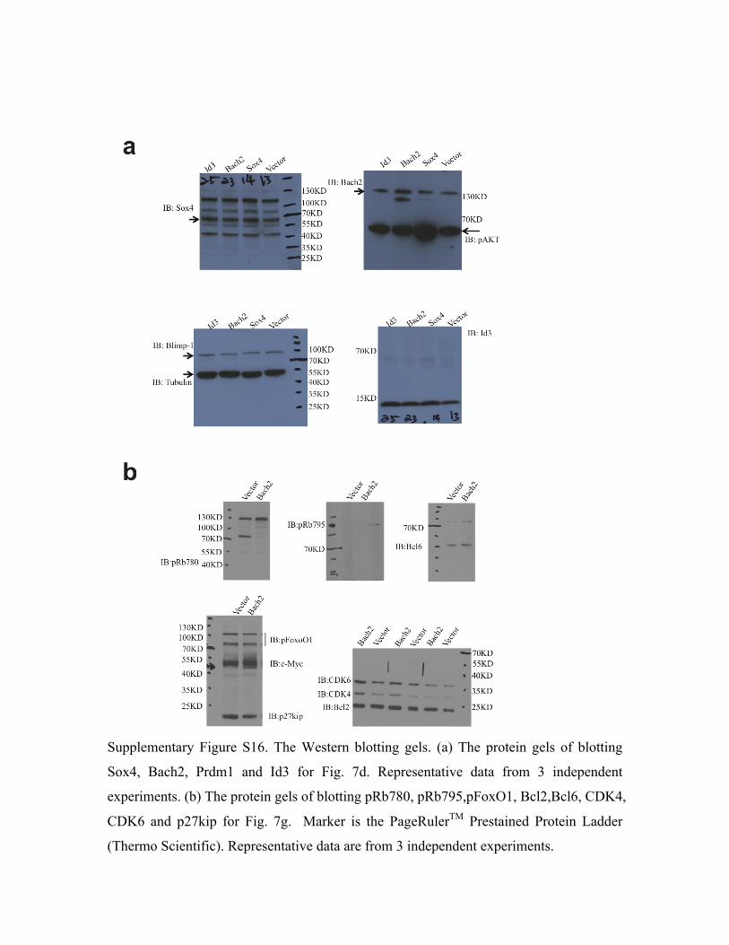
Supplementary Figure S16. The Western blotting gels. (a) The protein gels of blotting
Sox4, Bach2, Prdm1 and Id3 for Fig. 7d. Representative data from 3 independent
experiments. (b) The protein gels of blotting pRb780, pRb795,pFoxO1, Bcl2,Bcl6, CDK4,
CDK6 and p27kip for Fig. 7g. Marker is the PageRulerTM Prestained Protein Ladder
(Thermo Scientific). Representative data are from 3 independent experiments.
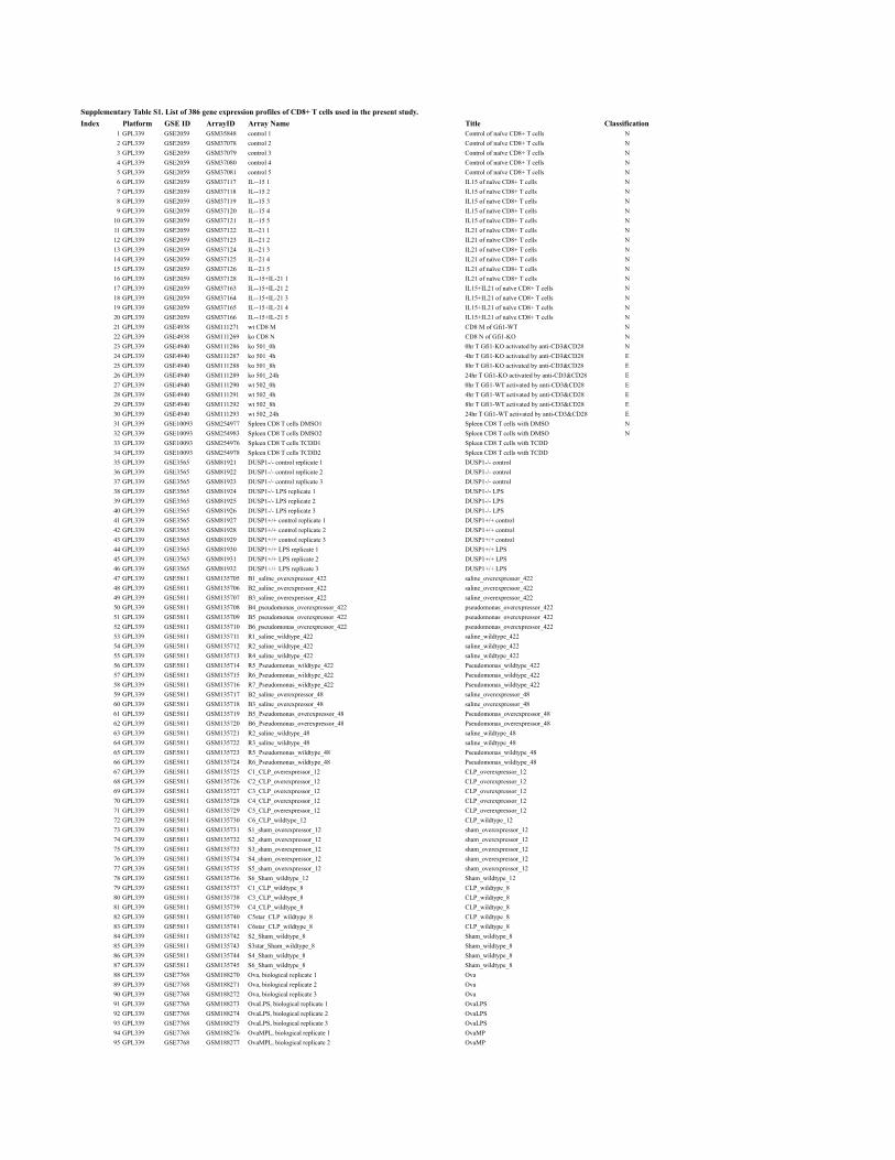
Supplementary Table S1. List of 386 gene expression profiles of CD8+ T cells used in the present study.Index Platform GSE ID ArrayID Array Name Title Classification
1 GPL339 GSE2059 GSM35848 control 1 Control of naïve CD8+ T cells N2 GPL339 GSE2059 GSM37078 control 2 Control of naïve CD8+ T cells N3 GPL339 GSE2059 GSM37079 control 3 Control of naïve CD8+ T cells N4 GPL339 GSE2059 GSM37080 control 4 Control of naïve CD8+ T cells N5 GPL339 GSE2059 GSM37081 control 5 Control of naïve CD8+ T cells N6 GPL339 GSE2059 GSM37117 IL--15 1 IL15 of naïve CD8+ T cells N7 GPL339 GSE2059 GSM37118 IL--15 2 IL15 of naïve CD8+ T cells N8 GPL339 GSE2059 GSM37119 IL--15 3 IL15 of naïve CD8+ T cells N9 GPL339 GSE2059 GSM37120 IL--15 4 IL15 of naïve CD8+ T cells N
10 GPL339 GSE2059 GSM37121 IL--15 5 IL15 of naïve CD8+ T cells N11 GPL339 GSE2059 GSM37122 IL--21 1 IL21 of naïve CD8+ T cells N12 GPL339 GSE2059 GSM37123 IL--21 2 IL21 of naïve CD8+ T cells N13 GPL339 GSE2059 GSM37124 IL--21 3 IL21 of naïve CD8+ T cells N14 GPL339 GSE2059 GSM37125 IL--21 4 IL21 of naïve CD8+ T cells N15 GPL339 GSE2059 GSM37126 IL--21 5 IL21 of naïve CD8+ T cells N16 GPL339 GSE2059 GSM37128 IL--15+IL-21 1 IL21 of naïve CD8+ T cells N17 GPL339 GSE2059 GSM37163 IL--15+IL-21 2 IL15+IL21 of naïve CD8+ T cells N18 GPL339 GSE2059 GSM37164 IL--15+IL-21 3 IL15+IL21 of naïve CD8+ T cells N19 GPL339 GSE2059 GSM37165 IL--15+IL-21 4 IL15+IL21 of naïve CD8+ T cells N20 GPL339 GSE2059 GSM37166 IL--15+IL-21 5 IL15+IL21 of naïve CD8+ T cells N21 GPL339 GSE4938 GSM111271 wt CD8 M CD8 M of Gfi1-WT N22 GPL339 GSE4938 GSM111269 ko CD8 N CD8 N of Gfi1-KO N23 GPL339 GSE4940 GSM111286 ko 501_0h 0hr T Gfi1-KO activated by anti-CD3&CD28 N24 GPL339 GSE4940 GSM111287 ko 501_4h 4hr T Gfi1-KO activated by anti-CD3&CD28 E25 GPL339 GSE4940 GSM111288 ko 501_8h 8hr T Gfi1-KO activated by anti-CD3&CD28 E26 GPL339 GSE4940 GSM111289 ko 501_24h 24hr T Gfi1-KO activated by anti-CD3&CD28 E27 GPL339 GSE4940 GSM111290 wt 502_0h 0hr T Gfi1-WT activated by anti-CD3&CD28 E28 GPL339 GSE4940 GSM111291 wt 502_4h 4hr T Gfi1-WT activated by anti-CD3&CD28 E29 GPL339 GSE4940 GSM111292 wt 502_8h 8hr T Gfi1-WT activated by anti-CD3&CD28 E30 GPL339 GSE4940 GSM111293 wt 502_24h 24hr T Gfi1-WT activated by anti-CD3&CD28 E31 GPL339 GSE10093 GSM254977 Spleen CD8 T cells DMSO1 Spleen CD8 T cells with DMSO N32 GPL339 GSE10093 GSM254983 Spleen CD8 T cells DMSO2 Spleen CD8 T cells with DMSO N33 GPL339 GSE10093 GSM254976 Spleen CD8 T cells TCDD1 Spleen CD8 T cells with TCDD34 GPL339 GSE10093 GSM254978 Spleen CD8 T cells TCDD2 Spleen CD8 T cells with TCDD35 GPL339 GSE3565 GSM81921 DUSP1-/- control replicate 1 DUSP1-/- control36 GPL339 GSE3565 GSM81922 DUSP1-/- control replicate 2 DUSP1-/- control37 GPL339 GSE3565 GSM81923 DUSP1-/- control replicate 3 DUSP1-/- control38 GPL339 GSE3565 GSM81924 DUSP1-/- LPS replicate 1 DUSP1-/- LPS39 GPL339 GSE3565 GSM81925 DUSP1-/- LPS replicate 2 DUSP1-/- LPS40 GPL339 GSE3565 GSM81926 DUSP1-/- LPS replicate 3 DUSP1-/- LPS41 GPL339 GSE3565 GSM81927 DUSP1+/+ control replicate 1 DUSP1+/+ control42 GPL339 GSE3565 GSM81928 DUSP1+/+ control replicate 2 DUSP1+/+ control43 GPL339 GSE3565 GSM81929 DUSP1+/+ control replicate 3 DUSP1+/+ control44 GPL339 GSE3565 GSM81930 DUSP1+/+ LPS replicate 1 DUSP1+/+ LPS45 GPL339 GSE3565 GSM81931 DUSP1+/+ LPS replicate 2 DUSP1+/+ LPS46 GPL339 GSE3565 GSM81932 DUSP1+/+ LPS replicate 3 DUSP1+/+ LPS47 GPL339 GSE5811 GSM135705 B1_saline_overexpressor_422 saline_overexpressor_42248 GPL339 GSE5811 GSM135706 B2_saline_overexpressor_422 saline_overexpressor_42249 GPL339 GSE5811 GSM135707 B3_saline_overexpressor_422 saline_overexpressor_42250 GPL339 GSE5811 GSM135708 B4_pseudomonas_overexpressor_422 pseudomonas_overexpressor_42251 GPL339 GSE5811 GSM135709 B5_pseudomonas_overexpressor_422 pseudomonas_overexpressor_42252 GPL339 GSE5811 GSM135710 B6_pseudomonas_overexpressor_422 pseudomonas_overexpressor_42253 GPL339 GSE5811 GSM135711 R1_saline_wildtype_422 saline_wildtype_42254 GPL339 GSE5811 GSM135712 R2_saline_wildtype_422 saline_wildtype_42255 GPL339 GSE5811 GSM135713 R4_saline_wildtype_422 saline_wildtype_42256 GPL339 GSE5811 GSM135714 R5_Pseudomonas_wildtype_422 Pseudomonas_wildtype_42257 GPL339 GSE5811 GSM135715 R6_Pseudomonas_wildtype_422 Pseudomonas_wildtype_42258 GPL339 GSE5811 GSM135716 R7_Pseudomonas_wildtype_422 Pseudomonas_wildtype_42259 GPL339 GSE5811 GSM135717 B2_saline_overexpressor_48 saline_overexpressor_4860 GPL339 GSE5811 GSM135718 B3_saline_overexpressor_48 saline_overexpressor_4861 GPL339 GSE5811 GSM135719 B5_Pseudomonas_overexpressor_48 Pseudomonas_overexpressor_4862 GPL339 GSE5811 GSM135720 B6_Pseudomonas_overexpressor_48 Pseudomonas_overexpressor_4863 GPL339 GSE5811 GSM135721 R2_saline_wildtype_48 saline_wildtype_4864 GPL339 GSE5811 GSM135722 R3_saline_wildtype_48 saline_wildtype_4865 GPL339 GSE5811 GSM135723 R5_Pseudomonas_wildtype_48 Pseudomonas_wildtype_4866 GPL339 GSE5811 GSM135724 R6_Pseudomonas_wildtype_48 Pseudomonas_wildtype_4867 GPL339 GSE5811 GSM135725 C1_CLP_overexpressor_12 CLP_overexpressor_1268 GPL339 GSE5811 GSM135726 C2_CLP_overexpressor_12 CLP_overexpressor_1269 GPL339 GSE5811 GSM135727 C3_CLP_overexpressor_12 CLP_overexpressor_1270 GPL339 GSE5811 GSM135728 C4_CLP_overexpressor_12 CLP_overexpressor_1271 GPL339 GSE5811 GSM135729 C5_CLP_overexpressor_12 CLP_overexpressor_1272 GPL339 GSE5811 GSM135730 C6_CLP_wildtype_12 CLP_wildtype_1273 GPL339 GSE5811 GSM135731 S1_sham_overexpressor_12 sham_overexpressor_1274 GPL339 GSE5811 GSM135732 S2_sham_overexpressor_12 sham_overexpressor_1275 GPL339 GSE5811 GSM135733 S3_sham_overexpressor_12 sham_overexpressor_1276 GPL339 GSE5811 GSM135734 S4_sham_overexpressor_12 sham_overexpressor_1277 GPL339 GSE5811 GSM135735 S5_sham_overexpressor_12 sham_overexpressor_1278 GPL339 GSE5811 GSM135736 S6_Sham_wildtype_12 Sham_wildtype_1279 GPL339 GSE5811 GSM135737 C1_CLP_wildtype_8 CLP_wildtype_880 GPL339 GSE5811 GSM135738 C3_CLP_wildtype_8 CLP_wildtype_881 GPL339 GSE5811 GSM135739 C4_CLP_wildtype_8 CLP_wildtype_882 GPL339 GSE5811 GSM135740 C5star_CLP_wildtype_8 CLP_wildtype_883 GPL339 GSE5811 GSM135741 C6star_CLP_wildtype_8 CLP_wildtype_884 GPL339 GSE5811 GSM135742 S2_Sham_wildtype_8 Sham_wildtype_885 GPL339 GSE5811 GSM135743 S3star_Sham_wildtype_8 Sham_wildtype_886 GPL339 GSE5811 GSM135744 S4_Sham_wildtype_8 Sham_wildtype_887 GPL339 GSE5811 GSM135745 S6_Sham_wildtype_8 Sham_wildtype_888 GPL339 GSE7768 GSM188270 Ova, biological replicate 1 Ova89 GPL339 GSE7768 GSM188271 Ova, biological replicate 2 Ova90 GPL339 GSE7768 GSM188272 Ova, biological replicate 3 Ova91 GPL339 GSE7768 GSM188273 OvaLPS, biological replicate 1 OvaLPS92 GPL339 GSE7768 GSM188274 OvaLPS, biological replicate 2 OvaLPS93 GPL339 GSE7768 GSM188275 OvaLPS, biological replicate 3 OvaLPS94 GPL339 GSE7768 GSM188276 OvaMPL, biological replicate 1 OvaMP95 GPL339 GSE7768 GSM188277 OvaMPL, biological replicate 2 OvaMP
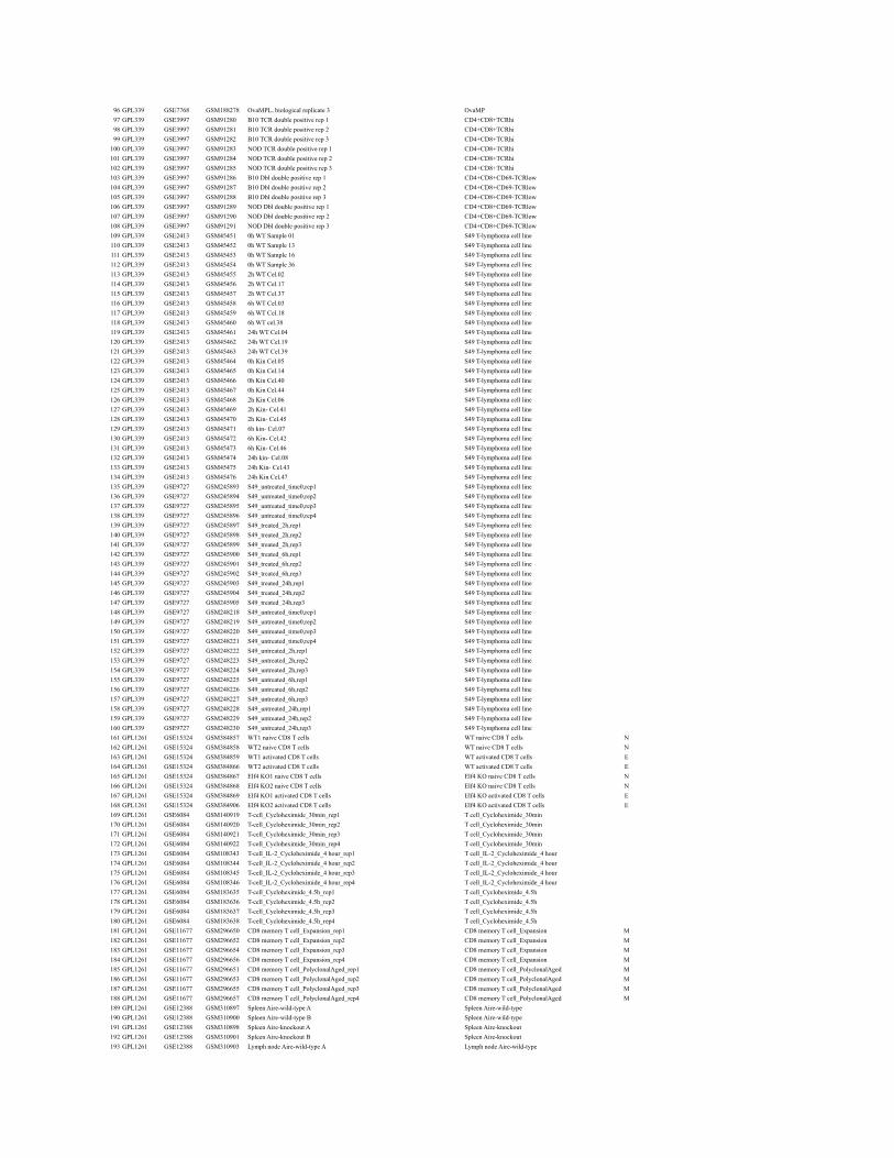
96 GPL339 GSE7768 GSM188278 OvaMPL, biological replicate 3 OvaMP97 GPL339 GSE3997 GSM91280 B10 TCR double positive rep 1 CD4+CD8+TCRhi98 GPL339 GSE3997 GSM91281 B10 TCR double positive rep 2 CD4+CD8+TCRhi99 GPL339 GSE3997 GSM91282 B10 TCR double positive rep 3 CD4+CD8+TCRhi
100 GPL339 GSE3997 GSM91283 NOD TCR double positive rep 1 CD4+CD8+TCRhi101 GPL339 GSE3997 GSM91284 NOD TCR double positive rep 2 CD4+CD8+TCRhi102 GPL339 GSE3997 GSM91285 NOD TCR double positive rep 3 CD4+CD8+TCRhi103 GPL339 GSE3997 GSM91286 B10 Dbl double positive rep 1 CD4+CD8+CD69-TCRlow104 GPL339 GSE3997 GSM91287 B10 Dbl double positive rep 2 CD4+CD8+CD69-TCRlow105 GPL339 GSE3997 GSM91288 B10 Dbl double positive rep 3 CD4+CD8+CD69-TCRlow106 GPL339 GSE3997 GSM91289 NOD Dbl double positive rep 1 CD4+CD8+CD69-TCRlow107 GPL339 GSE3997 GSM91290 NOD Dbl double positive rep 2 CD4+CD8+CD69-TCRlow108 GPL339 GSE3997 GSM91291 NOD Dbl double positive rep 3 CD4+CD8+CD69-TCRlow109 GPL339 GSE2413 GSM45451 0h WT Sample 01 S49 T-lymphoma cell line110 GPL339 GSE2413 GSM45452 0h WT Sample 13 S49 T-lymphoma cell line111 GPL339 GSE2413 GSM45453 0h WT Sample 16 S49 T-lymphoma cell line112 GPL339 GSE2413 GSM45454 0h WT Sample 36 S49 T-lymphoma cell line113 GPL339 GSE2413 GSM45455 2h WT Cel.02 S49 T-lymphoma cell line114 GPL339 GSE2413 GSM45456 2h WT Cel.17 S49 T-lymphoma cell line115 GPL339 GSE2413 GSM45457 2h WT Cel.37 S49 T-lymphoma cell line116 GPL339 GSE2413 GSM45458 6h WT Cel.03 S49 T-lymphoma cell line117 GPL339 GSE2413 GSM45459 6h WT Cel.18 S49 T-lymphoma cell line118 GPL339 GSE2413 GSM45460 6h WT cel.38 S49 T-lymphoma cell line119 GPL339 GSE2413 GSM45461 24h WT Cel.04 S49 T-lymphoma cell line120 GPL339 GSE2413 GSM45462 24h WT Cel.19 S49 T-lymphoma cell line121 GPL339 GSE2413 GSM45463 24h WT Cel.39 S49 T-lymphoma cell line122 GPL339 GSE2413 GSM45464 0h Kin Cel.05 S49 T-lymphoma cell line123 GPL339 GSE2413 GSM45465 0h Kin Cel.14 S49 T-lymphoma cell line124 GPL339 GSE2413 GSM45466 0h Kin Cel.40 S49 T-lymphoma cell line125 GPL339 GSE2413 GSM45467 0h Kin Cel.44 S49 T-lymphoma cell line126 GPL339 GSE2413 GSM45468 2h Kin Cel.06 S49 T-lymphoma cell line127 GPL339 GSE2413 GSM45469 2h Kin- Cel.41 S49 T-lymphoma cell line128 GPL339 GSE2413 GSM45470 2h Kin- Cel.45 S49 T-lymphoma cell line129 GPL339 GSE2413 GSM45471 6h kin- Cel.07 S49 T-lymphoma cell line130 GPL339 GSE2413 GSM45472 6h Kin- Cel.42 S49 T-lymphoma cell line131 GPL339 GSE2413 GSM45473 6h Kin- Cel.46 S49 T-lymphoma cell line132 GPL339 GSE2413 GSM45474 24h kin- Cel.08 S49 T-lymphoma cell line133 GPL339 GSE2413 GSM45475 24h Kin- Cel.43 S49 T-lymphoma cell line134 GPL339 GSE2413 GSM45476 24h Kin Cel.47 S49 T-lymphoma cell line135 GPL339 GSE9727 GSM245893 S49_untreated_time0,rep1 S49 T-lymphoma cell line136 GPL339 GSE9727 GSM245894 S49_untreated_time0,rep2 S49 T-lymphoma cell line137 GPL339 GSE9727 GSM245895 S49_untreated_time0,rep3 S49 T-lymphoma cell line138 GPL339 GSE9727 GSM245896 S49_untreated_time0,rep4 S49 T-lymphoma cell line139 GPL339 GSE9727 GSM245897 S49_treated_2h,rep1 S49 T-lymphoma cell line140 GPL339 GSE9727 GSM245898 S49_treated_2h,rep2 S49 T-lymphoma cell line141 GPL339 GSE9727 GSM245899 S49_treated_2h,rep3 S49 T-lymphoma cell line142 GPL339 GSE9727 GSM245900 S49_treated_6h,rep1 S49 T-lymphoma cell line143 GPL339 GSE9727 GSM245901 S49_treated_6h,rep2 S49 T-lymphoma cell line144 GPL339 GSE9727 GSM245902 S49_treated_6h,rep3 S49 T-lymphoma cell line145 GPL339 GSE9727 GSM245903 S49_treated_24h,rep1 S49 T-lymphoma cell line146 GPL339 GSE9727 GSM245904 S49_treated_24h,rep2 S49 T-lymphoma cell line147 GPL339 GSE9727 GSM245905 S49_treated_24h,rep3 S49 T-lymphoma cell line148 GPL339 GSE9727 GSM248218 S49_untreated_time0,rep1 S49 T-lymphoma cell line149 GPL339 GSE9727 GSM248219 S49_untreated_time0,rep2 S49 T-lymphoma cell line150 GPL339 GSE9727 GSM248220 S49_untreated_time0,rep3 S49 T-lymphoma cell line151 GPL339 GSE9727 GSM248221 S49_untreated_time0,rep4 S49 T-lymphoma cell line152 GPL339 GSE9727 GSM248222 S49_untreated_2h,rep1 S49 T-lymphoma cell line153 GPL339 GSE9727 GSM248223 S49_untreated_2h,rep2 S49 T-lymphoma cell line154 GPL339 GSE9727 GSM248224 S49_untreated_2h,rep3 S49 T-lymphoma cell line155 GPL339 GSE9727 GSM248225 S49_untreated_6h,rep1 S49 T-lymphoma cell line156 GPL339 GSE9727 GSM248226 S49_untreated_6h,rep2 S49 T-lymphoma cell line157 GPL339 GSE9727 GSM248227 S49_untreated_6h,rep3 S49 T-lymphoma cell line158 GPL339 GSE9727 GSM248228 S49_untreated_24h,rep1 S49 T-lymphoma cell line159 GPL339 GSE9727 GSM248229 S49_untreated_24h,rep2 S49 T-lymphoma cell line160 GPL339 GSE9727 GSM248230 S49_untreated_24h,rep3 S49 T-lymphoma cell line161 GPL1261 GSE15324 GSM384857 WT1 naive CD8 T cells WT naive CD8 T cells N162 GPL1261 GSE15324 GSM384858 WT2 naive CD8 T cells WT naive CD8 T cells N163 GPL1261 GSE15324 GSM384859 WT1 activated CD8 T cells WT activated CD8 T cells E164 GPL1261 GSE15324 GSM384866 WT2 activated CD8 T cells WT activated CD8 T cells E165 GPL1261 GSE15324 GSM384867 Elf4 KO1 naive CD8 T cells Elf4 KO naive CD8 T cells N166 GPL1261 GSE15324 GSM384868 Elf4 KO2 naive CD8 T cells Elf4 KO naive CD8 T cells N167 GPL1261 GSE15324 GSM384869 Elf4 KO1 activated CD8 T cells Elf4 KO activated CD8 T cells E168 GPL1261 GSE15324 GSM384906 Elf4 KO2 activated CD8 T cells Elf4 KO activated CD8 T cells E169 GPL1261 GSE6084 GSM140919 T-cell_Cycloheximide_30min_rep1 T cell_Cycloheximide_30min170 GPL1261 GSE6084 GSM140920 T-cell_Cycloheximide_30min_rep2 T cell_Cycloheximide_30min171 GPL1261 GSE6084 GSM140921 T-cell_Cycloheximide_30min_rep3 T cell_Cycloheximide_30min172 GPL1261 GSE6084 GSM140922 T-cell_Cycloheximide_30min_rep4 T cell_Cycloheximide_30min173 GPL1261 GSE6084 GSM108343 T-cell_IL-2_Cycloheximide_4 hour_rep1 T cell_IL-2_Cycloheximide_4 hour174 GPL1261 GSE6084 GSM108344 T-cell_IL-2_Cycloheximide_4 hour_rep2 T cell_IL-2_Cycloheximide_4 hour175 GPL1261 GSE6084 GSM108345 T-cell_IL-2_Cycloheximide_4 hour_rep3 T cell_IL-2_Cycloheximide_4 hour176 GPL1261 GSE6084 GSM108346 T-cell_IL-2_Cycloheximide_4 hour_rep4 T cell_IL-2_Cycloheximide_4 hour177 GPL1261 GSE6084 GSM183635 T-cell_Cycloheximide_4.5h_rep1 T cell_Cycloheximide_4.5h178 GPL1261 GSE6084 GSM183636 T-cell_Cycloheximide_4.5h_rep2 T cell_Cycloheximide_4.5h179 GPL1261 GSE6084 GSM183637 T-cell_Cycloheximide_4.5h_rep3 T cell_Cycloheximide_4.5h180 GPL1261 GSE6084 GSM183638 T-cell_Cycloheximide_4.5h_rep4 T cell_Cycloheximide_4.5h181 GPL1261 GSE11677 GSM296650 CD8 memory T cell_Expansion_rep1 CD8 memory T cell_Expansion M182 GPL1261 GSE11677 GSM296652 CD8 memory T cell_Expansion_rep2 CD8 memory T cell_Expansion M183 GPL1261 GSE11677 GSM296654 CD8 memory T cell_Expansion_rep3 CD8 memory T cell_Expansion M184 GPL1261 GSE11677 GSM296656 CD8 memory T cell_Expansion_rep4 CD8 memory T cell_Expansion M185 GPL1261 GSE11677 GSM296651 CD8 memory T cell_PolyclonalAged_rep1 CD8 memory T cell_PolyclonalAged M186 GPL1261 GSE11677 GSM296653 CD8 memory T cell_PolyclonalAged_rep2 CD8 memory T cell_PolyclonalAged M187 GPL1261 GSE11677 GSM296655 CD8 memory T cell_PolyclonalAged_rep3 CD8 memory T cell_PolyclonalAged M188 GPL1261 GSE11677 GSM296657 CD8 memory T cell_PolyclonalAged_rep4 CD8 memory T cell_PolyclonalAged M189 GPL1261 GSE12388 GSM310897 Spleen Aire-wild-type A Spleen Aire-wild-type190 GPL1261 GSE12388 GSM310900 Spleen Aire-wild-type B Spleen Aire-wild-type191 GPL1261 GSE12388 GSM310898 Spleen Aire-knockout A Spleen Aire-knockout192 GPL1261 GSE12388 GSM310901 Spleen Aire-knockout B Spleen Aire-knockout193 GPL1261 GSE12388 GSM310903 Lymph node Aire-wild-type A Lymph node Aire-wild-type
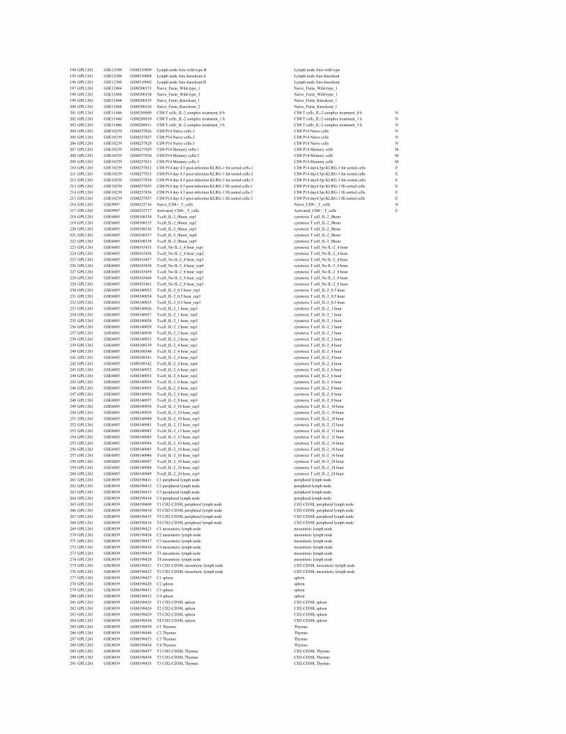
194 GPL1261 GSE12388 GSM310899 Lymph node Aire-wild-type B Lymph node Aire-wild-type195 GPL1261 GSE12388 GSM310904 Lymph node Aire-knockout A Lymph node Aire-knockout196 GPL1261 GSE12388 GSM310902 Lymph node Aire-knockout B Lymph node Aire-knockout197 GPL1261 GSE11884 GSM300153 Naive_Furin_Wild-type_1 Naive_Furin_Wild-type_1198 GPL1261 GSE11884 GSM300154 Naive_Furin_Wild-type_2 Naive_Furin_Wild-type_1199 GPL1261 GSE11884 GSM300155 Naive_Furin_Knockout_1 Naive_Furin_Knockout_1200 GPL1261 GSE11884 GSM300156 Naive_Furin_Knockout_2 Naive_Furin_Knockout_1201 GPL1261 GSE11446 GSM288809 CD8 T cells_IL-2 complex treatment_0 h CD8 T cells_IL-2 complex treatment_0 h N202 GPL1261 GSE11446 GSM288810 CD8 T cells_IL-2 complex treatment_1 h CD8 T cells_IL-2 complex treatment_1 h N203 GPL1261 GSE11446 GSM288811 CD8 T cells_IL-2 complex treatment_3 h CD8 T cells_IL-2 complex treatment_3 h N204 GPL1261 GSE10239 GSM257826 CD8 P14 Naive cells-1 CD8 P14 Naive cells N205 GPL1261 GSE10239 GSM257827 CD8 P14 Naive cells-2 CD8 P14 Naive cells N206 GPL1261 GSE10239 GSM257828 CD8 P14 Naive cells-3 CD8 P14 Naive cells N207 GPL1261 GSE10239 GSM257829 CD8 P14 Memory cells-1 CD8 P14 Memory cells M208 GPL1261 GSE10239 GSM257830 CD8 P14 Memory cells-2 CD8 P14 Memory cells M209 GPL1261 GSE10239 GSM257831 CD8 P14 Memory cells-3 CD8 P14 Memory cells M210 GPL1261 GSE10239 GSM257832 CD8 P14 day 4.5 post-infection KLRG-1 Int sorted cells-1 CD8 P14 day4.5pi KLRG-1 Int sorted cells E211 GPL1261 GSE10239 GSM257833 CD8 P14 day 4.5 post-infection KLRG-1 Int sorted cells-2 CD8 P14 day4.5pi KLRG-1 Int sorted cells E212 GPL1261 GSE10239 GSM257834 CD8 P14 day 4.5 post-infection KLRG-1 Int sorted cells-3 CD8 P14 day4.5pi KLRG-1 Int sorted cells E213 GPL1261 GSE10239 GSM257835 CD8 P14 day 4.5 post-infection KLRG-1 Hi sorted cells-1 CD8 P14 day4.5pi KLRG-1 Hi sorted cells E214 GPL1261 GSE10239 GSM257836 CD8 P14 day 4.5 post-infection KLRG-1 Hi sorted cells-2 CD8 P14 day4.5pi KLRG-1 Hi sorted cells E215 GPL1261 GSE10239 GSM257837 CD8 P14 day 4.5 post-infection KLRG-1 Hi sorted cells-3 CD8 P14 day4.5pi KLRG-1 Hi sorted cells E216 GPL1261 GSE9997 GSM252716 Naive_CD8+_T_cells Naive_CD8+_T_cells N217 GPL1261 GSE9997 GSM252717 Activated_CD8+_T_cells Activated_CD8+_T_cells E218 GPL1261 GSE6085 GSM108334 T-cell_IL-2_0hour_rep1 cytotoxic T cell_IL-2_0hour219 GPL1261 GSE6085 GSM108335 T-cell_IL-2_0hour_rep2 cytotoxic T cell_IL-2_0hour220 GPL1261 GSE6085 GSM108336 T-cell_IL-2_0hour_rep3 cytotoxic T cell_IL-2_0hour221 GPL1261 GSE6085 GSM108337 T-cell_IL-2_0hour_rep4 cytotoxic T cell_IL-2_0hour222 GPL1261 GSE6085 GSM108338 T-cell_IL-2_0hour_rep5 cytotoxic T cell_IL-2_0hour223 GPL1261 GSE6085 GSM183455 T-cell_No IL-2_4 hour_rep1 cytotoxic T cell_No IL-2_4 hour224 GPL1261 GSE6085 GSM183456 T-cell_No IL-2_4 hour_rep2 cytotoxic T cell_No IL-2_4 hour225 GPL1261 GSE6085 GSM183457 T-cell_No IL-2_4 hour_rep3 cytotoxic T cell_No IL-2_4 hour226 GPL1261 GSE6085 GSM183458 T-cell_No IL-2_4 hour_rep4 cytotoxic T cell_No IL-2_4 hour227 GPL1261 GSE6085 GSM183459 T-cell_No IL-2_8 hour_rep1 cytotoxic T cell_No IL-2_8 hour228 GPL1261 GSE6085 GSM183460 T-cell_No IL-2_8 hour_rep2 cytotoxic T cell_No IL-2_8 hour229 GPL1261 GSE6085 GSM183461 T-cell_No IL-2_8 hour_rep3 cytotoxic T cell_No IL-2_8 hour230 GPL1261 GSE6085 GSM140923 T-cell_IL-2_0.5 hour_rep1 cytotoxic T cell_IL-2_0.5 hour231 GPL1261 GSE6085 GSM140924 T-cell_IL-2_0.5 hour_rep2 cytotoxic T cell_IL-2_0.5 hour232 GPL1261 GSE6085 GSM140925 T-cell_IL-2_0.5 hour_rep3 cytotoxic T cell_IL-2_0.5 hour233 GPL1261 GSE6085 GSM140926 T-cell_IL-2_1 hour_rep1 cytotoxic T cell_IL-2_1 hour234 GPL1261 GSE6085 GSM140927 T-cell_IL-2_1 hour_rep2 cytotoxic T cell_IL-2_1 hour235 GPL1261 GSE6085 GSM140928 T-cell_IL-2_1 hour_rep3 cytotoxic T cell_IL-2_1 hour236 GPL1261 GSE6085 GSM140929 T-cell_IL-2_2 hour_rep1 cytotoxic T cell_IL-2_2 hour237 GPL1261 GSE6085 GSM140930 T-cell_IL-2_2 hour_rep2 cytotoxic T cell_IL-2_2 hour238 GPL1261 GSE6085 GSM140931 T-cell_IL-2_2 hour_rep3 cytotoxic T cell_IL-2_2 hour239 GPL1261 GSE6085 GSM108339 T-cell_IL-2_4 hour_rep1 cytotoxic T cell_IL-2_4 hour240 GPL1261 GSE6085 GSM108340 T-cell_IL-2_4 hour_rep2 cytotoxic T cell_IL-2_4 hour241 GPL1261 GSE6085 GSM108341 T-cell_IL-2_4 hour_rep3 cytotoxic T cell_IL-2_4 hour242 GPL1261 GSE6085 GSM108342 T-cell_IL-2_4 hour_rep4 cytotoxic T cell_IL-2_4 hour243 GPL1261 GSE6085 GSM140932 T-cell_IL-2_6 hour_rep1 cytotoxic T cell_IL-2_6 hour244 GPL1261 GSE6085 GSM140933 T-cell_IL-2_6 hour_rep2 cytotoxic T cell_IL-2_6 hour245 GPL1261 GSE6085 GSM140934 T-cell_IL-2_6 hour_rep3 cytotoxic T cell_IL-2_6 hour246 GPL1261 GSE6085 GSM140935 T-cell_IL-2_8 hour_rep1 cytotoxic T cell_IL-2_8 hour247 GPL1261 GSE6085 GSM140936 T-cell_IL-2_8 hour_rep2 cytotoxic T cell_IL-2_8 hour248 GPL1261 GSE6085 GSM140937 T-cell_IL-2_8 hour_rep3 cytotoxic T cell_IL-2_8 hour249 GPL1261 GSE6085 GSM140938 T-cell_IL-2_10 hour_rep1 cytotoxic T cell_IL-2_10 hour250 GPL1261 GSE6085 GSM140939 T-cell_IL-2_10 hour_rep2 cytotoxic T cell_IL-2_10 hour251 GPL1261 GSE6085 GSM140940 T-cell_IL-2_10 hour_rep3 cytotoxic T cell_IL-2_10 hour252 GPL1261 GSE6085 GSM140941 T-cell_IL-2_12 hour_rep1 cytotoxic T cell_IL-2_12 hour253 GPL1261 GSE6085 GSM140942 T-cell_IL-2_12 hour_rep2 cytotoxic T cell_IL-2_12 hour254 GPL1261 GSE6085 GSM140943 T-cell_IL-2_12 hour_rep3 cytotoxic T cell_IL-2_12 hour255 GPL1261 GSE6085 GSM140944 T-cell_IL-2_16 hour_rep1 cytotoxic T cell_IL-2_16 hour256 GPL1261 GSE6085 GSM140945 T-cell_IL-2_16 hour_rep2 cytotoxic T cell_IL-2_16 hour257 GPL1261 GSE6085 GSM140946 T-cell_IL-2_16 hour_rep3 cytotoxic T cell_IL-2_16 hour258 GPL1261 GSE6085 GSM140947 T-cell_IL-2_24 hour_rep1 cytotoxic T cell_IL-2_24 hour259 GPL1261 GSE6085 GSM140948 T-cell_IL-2_24 hour_rep2 cytotoxic T cell_IL-2_24 hour260 GPL1261 GSE6085 GSM140949 T-cell_IL-2_24 hour_rep3 cytotoxic T cell_IL-2_24 hour261 GPL1261 GSE8039 GSM198411 C1 peripheral lymph node peripheral lymph node262 GPL1261 GSE8039 GSM198412 C2 peripheral lymph node peripheral lymph node263 GPL1261 GSE8039 GSM198413 C3 peripheral lymph node peripheral lymph node264 GPL1261 GSE8039 GSM198414 C4 peripheral lymph node peripheral lymph node265 GPL1261 GSE8039 GSM198409 T1 CD2-CD30L peripheral lymph node CD2-CD30L peripheral lymph node266 GPL1261 GSE8039 GSM198410 T2 CD2-CD30L peripheral lymph node CD2-CD30L peripheral lymph node267 GPL1261 GSE8039 GSM198415 T3 CD2-CD30L peripheral lymph node CD2-CD30L peripheral lymph node268 GPL1261 GSE8039 GSM198416 T4 CD2-CD30L peripheral lymph node CD2-CD30L peripheral lymph node269 GPL1261 GSE8039 GSM198423 C1 mesenteric lymph node mesenteric lymph node270 GPL1261 GSE8039 GSM198424 C2 mesenteric lymph node mesenteric lymph node271 GPL1261 GSE8039 GSM198417 C3 mesenteric lymph node mesenteric lymph node272 GPL1261 GSE8039 GSM198418 C4 mesenteric lymph node mesenteric lymph node273 GPL1261 GSE8039 GSM198419 T3 mesenteric lymph node mesenteric lymph node274 GPL1261 GSE8039 GSM198420 T4 mesenteric lymph node mesenteric lymph node275 GPL1261 GSE8039 GSM198421 T1 CD2-CD30L mesenteric lymph node CD2-CD30L mesenteric lymph node276 GPL1261 GSE8039 GSM198422 T2 CD2-CD30L mesenteric lymph node CD2-CD30L mesenteric lymph node277 GPL1261 GSE8039 GSM198427 C1 spleen spleen278 GPL1261 GSE8039 GSM198428 C2 spleen spleen279 GPL1261 GSE8039 GSM198431 C3 spleen spleen280 GPL1261 GSE8039 GSM198432 C4 spleen spleen281 GPL1261 GSE8039 GSM198425 T1 CD2-CD30L spleen CD2-CD30L spleen282 GPL1261 GSE8039 GSM198426 T2 CD2-CD30L spleen CD2-CD30L spleen283 GPL1261 GSE8039 GSM198429 T3 CD2-CD30L spleen CD2-CD30L spleen284 GPL1261 GSE8039 GSM198430 T4 CD2-CD30L spleen CD2-CD30L spleen285 GPL1261 GSE8039 GSM198439 C1 Thymus Thymus286 GPL1261 GSE8039 GSM198440 C2 Thymus Thymus287 GPL1261 GSE8039 GSM198433 C3 Thymus Thymus288 GPL1261 GSE8039 GSM198434 C4 Thymus Thymus289 GPL1261 GSE8039 GSM198437 T1 CD2-CD30L Thymus CD2-CD30L Thymus290 GPL1261 GSE8039 GSM198438 T2 CD2-CD30L Thymus CD2-CD30L Thymus291 GPL1261 GSE8039 GSM198435 T3 CD2-CD30L Thymus CD2-CD30L Thymus
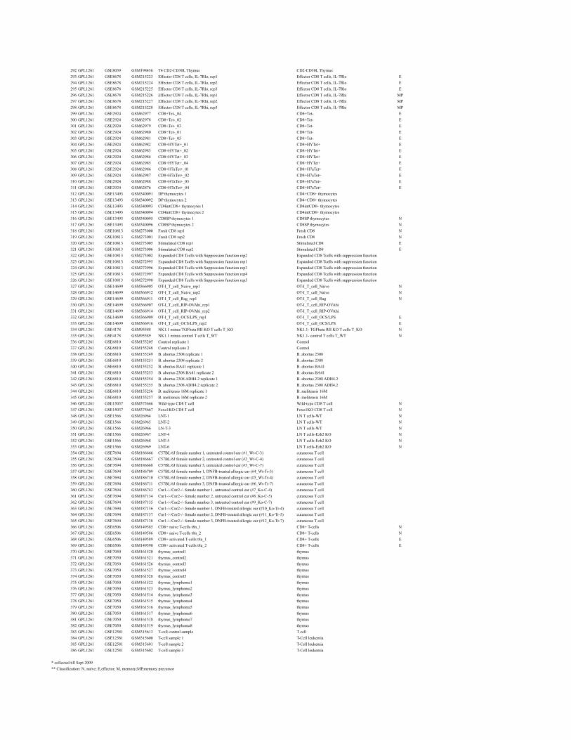
292 GPL1261 GSE8039 GSM198436 T4 CD2-CD30L Thymus CD2-CD30L Thymus293 GPL1261 GSE8678 GSM215223 Effector CD8 T cells, IL-7Rlo, rep1 Effector CD8 T cells, IL-7Rlo E294 GPL1261 GSE8678 GSM215224 Effector CD8 T cells, IL-7Rlo, rep2 Effector CD8 T cells, IL-7Rlo E295 GPL1261 GSE8678 GSM215225 Effector CD8 T cells, IL-7Rlo, rep3 Effector CD8 T cells, IL-7Rlo E296 GPL1261 GSE8678 GSM215226 Effector CD8 T cells, IL-7Rhi, rep1 Effector CD8 T cells, IL-7Rhi MP297 GPL1261 GSE8678 GSM215227 Effector CD8 T cells, IL-7Rhi, rep2 Effector CD8 T cells, IL-7Rhi MP298 GPL1261 GSE8678 GSM215228 Effector CD8 T cells, IL-7Rhi, rep3 Effector CD8 T cells, IL-7Rhi MP299 GPL1261 GSE2924 GSM62977 CD8+Tet-_04 CD8+Tet- E300 GPL1261 GSE2924 GSM62978 CD8+Tet-_02 CD8+Tet- E301 GPL1261 GSE2924 GSM62979 CD8+Tet-_03 CD8+Tet- E302 GPL1261 GSE2924 GSM62980 CD8+Tet-_01 CD8+Tet- E303 GPL1261 GSE2924 GSM62981 CD8+Tet-_05 CD8+Tet- E304 GPL1261 GSE2924 GSM62982 CD8+HYTet+_01 CD8+HYTet+ E305 GPL1261 GSE2924 GSM62983 CD8+HYTet+_02 CD8+HYTet+ E306 GPL1261 GSE2924 GSM62984 CD8+HYTet+_03 CD8+HYTet+ E307 GPL1261 GSE2924 GSM62985 CD8+HYTet+_04 CD8+HYTet+ E308 GPL1261 GSE2924 GSM62986 CD8+H7aTet+_01 CD8+H7aTet+ E309 GPL1261 GSE2924 GSM62987 CD8+H7aTet+_02 CD8+H7aTet+ E310 GPL1261 GSE2924 GSM62988 CD8+H7aTet+_03 CD8+H7aTet+ E311 GPL1261 GSE2924 GSM62876 CD8+H7aTet+_04 CD8+H7aTet+ E312 GPL1261 GSE13493 GSM340091 DP thymocytes 1 CD4+CD8+ thymocytes313 GPL1261 GSE13493 GSM340092 DP thymocytes 2 CD4+CD8+ thymocytes314 GPL1261 GSE13493 GSM340093 CD4intCD8+ thymocytes 1 CD4intCD8+ thymocytes315 GPL1261 GSE13493 GSM340094 CD4intCD8+ thymocytes 2 CD4intCD8+ thymocytes316 GPL1261 GSE13493 GSM340095 CD8SP thymocytes 1 CD8SP thymocytes N317 GPL1261 GSE13493 GSM340096 CD8SP thymocytes 2 CD8SP thymocytes N318 GPL1261 GSE10813 GSM273000 Fresh CD8 rep1 Fresh CD8 N319 GPL1261 GSE10813 GSM273001 Fresh CD8 rep2 Fresh CD8 N320 GPL1261 GSE10813 GSM273005 Stimulated CD8 rep1 Stimulated CD8 E321 GPL1261 GSE10813 GSM273006 Stimulated CD8 rep2 Stimulated CD8 E322 GPL1261 GSE10813 GSM273002 Expanded CD8 Tcells with Suppression function rep2 Expanded CD8 Tcells with suppression function323 GPL1261 GSE10813 GSM272995 Expanded CD8 Tcells with Suppression function rep1 Expanded CD8 Tcells with suppression function324 GPL1261 GSE10813 GSM272996 Expanded CD8 Tcells with Suppression function rep3 Expanded CD8 Tcells with suppression function325 GPL1261 GSE10813 GSM272997 Expanded CD8 Tcells with Suppression function rep4 Expanded CD8 Tcells with suppression function326 GPL1261 GSE10813 GSM272998 Expanded CD8 Tcells with Suppression function rep5 Expanded CD8 Tcells with suppression function327 GPL1261 GSE14699 GSM366905 OT-I_T_cell_Naive_rep1 OT-I_T_cell_Naive N328 GPL1261 GSE14699 GSM366912 OT-I_T_cell_Naive_rep2 OT-I_T_cell_Naive N329 GPL1261 GSE14699 GSM366911 OT-I_T_cell_Rag_rep1 OT-I_T_cell_Rag N330 GPL1261 GSE14699 GSM366907 OT-I_T_cell_RIP-OVAhi_rep1 OT-I_T_cell_RIP-OVAhi331 GPL1261 GSE14699 GSM366914 OT-I_T_cell_RIP-OVAhi_rep2 OT-I_T_cell_RIP-OVAhi332 GPL1261 GSE14699 GSM366909 OT-I_T_cell_OCS/LPS_rep1 OT-I_T_cell_OCS/LPS E333 GPL1261 GSE14699 GSM366916 OT-I_T_cell_OCS/LPS_rep2 OT-I_T_cell_OCS/LPS E334 GPL1261 GSE4178 GSM95588 NK1.1 minus TGFbeta RII KO T cells T_KO NK1.1- TGFbeta RII KO T cells T_KO N335 GPL1261 GSE4178 GSM95589 NK1.1 minus control T cells T_WT NK1.1- control T cells T_WT N336 GPL1261 GSE6810 GSM155205 Control replicate 1 Control337 GPL1261 GSE6810 GSM155248 Control replicate 2 Control338 GPL1261 GSE6810 GSM155249 B. abortus 2308 replicate 1 B. abortus 2308339 GPL1261 GSE6810 GSM155251 B. abortus 2308 replicate 2 B. abortus 2308340 GPL1261 GSE6810 GSM155252 B. abortus BA41 replicate 1 B. abortus BA41341 GPL1261 GSE6810 GSM155253 B. abortus 2308 BA41 replicate 2 B. abortus BA41342 GPL1261 GSE6810 GSM155254 B. abortus 2308 ADH4.2 replicate 1 B. abortus 2308 ADH4.2343 GPL1261 GSE6810 GSM155255 B. abortus 2308 ADH4.2 replicate 2 B. abortus 2308 ADH4.2344 GPL1261 GSE6810 GSM155256 B. melitensis 16M replicate 1 B. melitensis 16M345 GPL1261 GSE6810 GSM155257 B. melitensis 16M replicate 2 B. melitensis 16M346 GPL1261 GSE15037 GSM375666 Wild-type CD8 T cell Wild-type CD8 T cell N347 GPL1261 GSE15037 GSM375667 Foxo1KO CD8 T cell Foxo1KO CD8 T cell N348 GPL1261 GSE1566 GSM26964 LNT-1 LN T cells-WT N349 GPL1261 GSE1566 GSM26965 LNT-2 LN T cells-WT N350 GPL1261 GSE1566 GSM26966 LN-T-3 LN T cells-WT N351 GPL1261 GSE1566 GSM26967 LNT-4 LN T cells-Ezh2 KO N352 GPL1261 GSE1566 GSM26968 LNT-5 LN T cells-Ezh2 KO N353 GPL1261 GSE1566 GSM26969 LNT-6 LN T cells-Ezh2 KO N354 GPL1261 GSE7694 GSM186666 C57BL/6J female number 1, untreated control ear (#1_Wt-C-3) cutaneous T cell355 GPL1261 GSE7694 GSM186667 C57BL/6J female number 2, untreated control ear (#2_Wt-C-4) cutaneous T cell356 GPL1261 GSE7694 GSM186668 C57BL/6J female number 3, untreated control ear (#3_Wt-C-7) cutaneous T cell357 GPL1261 GSE7694 GSM186709 C57BL/6J female number 1, DNFB-treated allergic ear (#4_Wt-Tr-3) cutaneous T cell358 GPL1261 GSE7694 GSM186710 C57BL/6J female number 2, DNFB-treated allergic ear (#5_Wt-Tr-4) cutaneous T cell359 GPL1261 GSE7694 GSM186711 C57BL/6J female number 3, DNFB-treated allergic ear (#6_Wt-Tr-7) cutaneous T cell360 GPL1261 GSE7694 GSM186743 Cnr1-/-/Cnr2-/- female number 1, untreated control ear (#7_Ko-C-4) cutaneous T cell361 GPL1261 GSE7694 GSM187134 Cnr1-/-/Cnr2-/- female number 2, untreated control ear (#8_Ko-C-5) cutaneous T cell362 GPL1261 GSE7694 GSM187135 Cnr1-/-/Cnr2-/- female number 3, untreated control ear (#9_Ko-C-7) cutaneous T cell363 GPL1261 GSE7694 GSM187136 Cnr1-/-/Cnr2-/- female number 1, DNFB-treated allergic ear (#10_Ko-Tr-4) cutaneous T cell364 GPL1261 GSE7694 GSM187137 Cnr1-/-/Cnr2-/- female number 2, DNFB-treated allergic ear (#11_Ko-Tr-5) cutaneous T cell365 GPL1261 GSE7694 GSM187138 Cnr1-/-/Cnr2-/- female number 3, DNFB-treated allergic ear (#12_Ko-Tr-7) cutaneous T cell366 GPL1261 GSE6506 GSM149585 CD8+ naive T-cells t8n_1 CD8+ T-cells N367 GPL1261 GSE6506 GSM149586 CD8+ naive T-cells t8n_2 CD8+ T-cells N368 GPL1261 GSE6506 GSM149589 CD8+ activated T-cells t8a_1 CD8+ T-cells E369 GPL1261 GSE6506 GSM149590 CD8+ activated T-cells t8a_2 CD8+ T-cells E370 GPL1261 GSE7050 GSM161520 thymus_control1 thymus371 GPL1261 GSE7050 GSM161521 thymus_control2 thymus372 GPL1261 GSE7050 GSM161526 thymus_control3 thymus373 GPL1261 GSE7050 GSM161527 thymus_control4 thymus374 GPL1261 GSE7050 GSM161528 thymus_control5 thymus375 GPL1261 GSE7050 GSM161522 thymus_lymphoma1 thymus376 GPL1261 GSE7050 GSM161523 thymus_lymphoma2 thymus377 GPL1261 GSE7050 GSM161514 thymus_lymphoma3 thymus378 GPL1261 GSE7050 GSM161515 thymus_lymphoma4 thymus379 GPL1261 GSE7050 GSM161516 thymus_lymphoma5 thymus380 GPL1261 GSE7050 GSM161517 thymus_lymphoma6 thymus381 GPL1261 GSE7050 GSM161518 thymus_lymphoma7 thymus382 GPL1261 GSE7050 GSM161519 thymus_lymphoma8 thymus383 GPL1261 GSE12581 GSM315613 T-cell control sample T cell384 GPL1261 GSE12581 GSM315600 T-cell sample 1 T-Cell leukemia385 GPL1261 GSE12581 GSM315601 T-cell sample 2 T-Cell leukemia386 GPL1261 GSE12581 GSM315602 T-cell sample 3 T-Cell leukemia
* collected till Sept 2009** Classification: N, naïve; E,effector; M, memory;MP,memory precursor
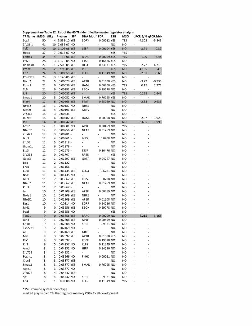
!"##$%&%'()*+,-).$%,!/0,,123(,45,(6%,78,-93,2:%'(252%:,.+,&)3(%*,*%;"$)(4*,)')$+3230,-9,<)&% =>!? =@A; BCD)$"% E!BF G<H,>4(25 9GI J!? >!? KBLI0JM< KBLI0>M<!"#$ %& $ '(%%)*+& ,)! !-., &(&&&+/ ,)! ,)! *$(0&% *%(1$%234056+ $+ +& 7(&%)*&7 8- * * 8- 8- * *9:37 $& +& +(+&)*&5 ,)! ;)<< &(&&+&$ ,)! 8- *0(7+ *&(07="4# 07 7 '(&+)*&7 8- * * ,)! ,)! * *)">?@ 0$ $ +)*&5 ,)! A.BC &(&&/&' ,)! ,)! +('' 0($1)D@/ /1 0 +(+7)*&% 8- )9!< &(+5$75 ,)! 8- * *AE6E?$& /7 + /(%&)*&% ,)! =)!< &(00%0+ ,)! ,)! /(7/ $(/+%FGH>+ /5 / /(')*&% ,)! F.I< * ,)! 8- $(+1% $(%J63/ /$ ' &(&&&%' ,)! J;<! &(++0$' 8- 8- */(&+ *&(50F"K/L3+ /0 0 '(+$)*&% ,)! * * 8- 8- * *AL:E/ // % &(&&&/0 ,)! BF+. &(&+%&1 ,)! 8- *0(77 *&('0%.KM#/ /+ & &(&&&05 ,)! =BN; &(&&0&1 ,)! ,)! &(+' /(77%9:3$ /+ ' &(&&+'+ ,)! )A-O &(/'771 8- 8- * *PH/ /& / &(&&&0/ ,)! * * ,)! ,)! &(/5% /(5$%!>LH+ /& % &(&&&%/ 8- !NBI &(75/'% ,)! 8- * *!DLD$ +7 5 &(&&/5% ,)! !9B9 &(/%&/' 8- 8- */(00 &('0%8G$L/ +5 + &(&&+17 8- 8A.) * 8- 8- * *N?3/: +5 $ &(&&+'+ ,)! N)</ * 8- 8- * *2340+1 +% 0 &(&&/0$ * * * ,)! 8- * *.KM#0 +% $ &(&&/17 ,)! =BN; &(&&0&1 8- 8- */(07 *+('/%PH0 +$ & &(&&%$/ ,)! * * 8- 8- *0(5'% *+(''%<"@6/ +/ + &(&&11+ 8- BF+< &(&&$%' 8- ,)! * *83LD:/ +/ / &(&&7%5 ,)! 8<B9 &(&+/5' 8- 8- * *234$// +/ 0 &(&&71+ * * * 8- 8- * *PQR3/ +/ $ &(&&'5+ * PJ.! &(&/&1 8- 8- * *234%/ +/ % &(&+0+1 * * * 8- 8- * *SEH>+H +/ 5 &(&+171 * * * 8- 8- * *)DT0 +/ 7 &(&/57% * )9!< &(+5$75 8- 8- * *234/01 ++ & &(&+7&7 * .F%1 * ,)! 8- * *ULDL0 ++ + &(&+/'7 ,)! UB9B &(&$/$7 8- 8- * *AV# ++ / &(&++// * * * 8- 8- * *)M:+ ++ 0 &(&++55 * * * 8- 8- * *CK#+ ++ $ &(&+$0% ,)! C;-O &(5/1+ 8- 8- * *8@H+ ++ $ &(&+$0% 8- * * 8- 8- * *PQR3+ ++ 7 &(&015/ ,)! PJ.! &(&/&1 8- 8- * *83LD:+ ++ 7 &(&015/ ,)! 8<B9 &(&+/5' 8- 8- * *FE30 ++ 7 &(&015/ * * * 8- 8- * *SKM +& + &(&+'&' ,)! BF+< &(&&$%' 8- 8- * *8G$L+ +& + &(&+'&' ,)! 8A.) * 8- 8- * *83?/6/ +& + &(&+'&' ,)! BF+. &(&+%&1 8- 8- * *)WG+ +& $ &(&/+$ 8- )U.< &(/$/+5 8- 8- * *N#X+ ' & &(&05%5 ,)! )A-O &(/'771 8- 8- * *FV#0 ' & &(&05%5 8- * * ,)! 8- * *9V#/+ ' & &(&05%5 ,)! A.BC &(&&/&' 8- 8- 5(/+% 0(+5%SKMH ' + &(&/1&1 ,)! BF+< &(&&$%' 8- 8- * *J63+& ' + &(&/1&1 8- !F+< &('0/+ 8- 8- * *9@://H+ ' / &(&/$5' 8- * * 8- 8- * *BG ' / &(&/$5' ,)! U.)< * 8- 8- * *NL3 ' 0 &(&/%'7 ,)! BF+. &(&+%&1 ,)! 8- * *.3#+ ' 0 &(&/%'7 * OAA< &(+'&'1 8- 8- * *J630 ' % &(&$/%7 8- J;<! &(++0$' 8- 8- * *BGMD6 1 + &(&$+0/ 8- =P<< &(0$%'5 8- 8- * *2347&' 1 + &(&$+0/ * * * 8- 8- * *<"#>+ 1 / &(&0555 8- <J=I &(&&&/+ 8- 8- * *OG::5 1 0 &(&0177 ,)! * * 8- 8- * *!>LH0 1 0 &(&0177 ,)! !NBI &(75/'% 8- 8- * *BD#M+ 1 0 &(&0177 8- * * 8- 8- * *2341/5 1 $ &(&$7$/ ,)! * * 8- 8- * *!4+ 1 $ &(&$7$/ 8- !F+< &('0/+ 8- 8- * *J63$ 7 + &(&5&1 8- J;<! &(++0$' 8- ,)! * *
YZP!F[ZX>>KM?Z@\@D?>Z4E?M"D\4?>LGQ?HZWGL\[QM"]MZ9<@ZDELDZG?WK6LD?Z>?>"G\ZCI1^Z9Z:?66ZH?T?6"4>?MD
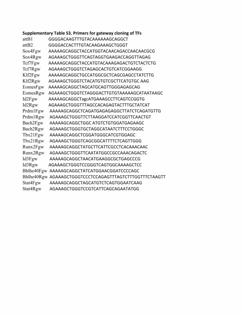
!"##$%&%'()*+,-).$%,!/0,1*2&%*3,45*,6)(%7)+,8$5'2'6,54,-93attB1 !!!!"#""!$$$!$"#""""""!#"!!#$attB2 !!!!"##"#$$$!$"#""!"""!#$!!!$Sox4Fgw """""!#"!!#$"##"$!!$"#""#"!"##""#""#!#!Sox4Rgw "!"""!#$!!!$$#"!$"!!$!""!"##"!!$$"!"!Tcf7Fgw """""!#"!!#$"##"$!$"#"""!"!"#$!$#$"#$#$!Tcf7Rgw "!"""!#$!!!$#$"!"!#"#$!$#"$#!!""!!Klf2Fgw """""!#"!!#$!##"$!!#!#$#"!#!"!##$"$#$$!Klf2Rgw "!"""!#$!!!$#$"#"$!$!$#!#$$#"$!$!#%""!EomesFgw """""!#"!!#$"!#"$!#"!$$!!!"!"!#"!EomesRgw "!"""!#$!!!$#$"!!!"#$$!$!$"""""!#"$""$""!#Id2Fgw """""!#"!!#$&'("$!"""!##$$#"!$##!!$!Id2Rgw "!"""!#$!!!$$$"!##"#"!"!$"#$$$!#$"$#"$%Prdm1Fgw %"""""!#"!!#$#"!"$!"!"!"!!#$$"$#$#"!"$!$$!Prdm1Rgw %"!"""!#$!!!$$#$$""!!"$##"$#!!$$#""#$!$Bach2Fgw %"""""!#"!!#$!!#%"$!$#$!$!!"$!"!""!#Bach2Rgw %"!"""!#$!!!$!#$"!!#"$""$#$$$##$!!!#Tbx21Fgw %"""""!#"!!#$#!!"$!!!#"$#!$!!"!#Tbx21Rgw %"!"""!#$!!!$#"!#!!#"$$$$#$#"!$$!!!Runx2Fgw %"""""!#"!!#$"$!#$$#"$$#!##$#"#"""#""#Runx2Rgw %"!"""!#$!!!$$#""$"$!!##!##"""#"!"#$#Id3Fgw %"""""!#"!!#$""#"$!""!!#!#$!"!###!Id3Rgw "!"""!#$!!!$##!!!$#"!$!!#""""!#$##Bhlhe40Fgw %"""""!#"!!#$"$#"$!!""#!!"$####"!#Bhlhe40Rgw %"!"""!#$!!!$###$##"!"!$$$"!$#$$$!!$$$#$""!$$Stat4Fgw %"""""!#"!!#$"!#"$!$#$#"!$!!""$#""!Stat4Rgw %"!"""!#$!!!$##!$#"$$#"!#"!""$"$!!
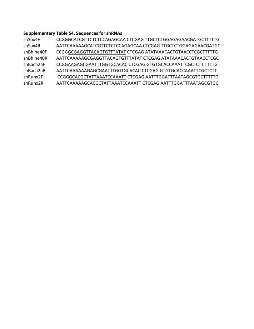
!"##$%&%'()*+,-).$%,!/0,!%1"%'2%3,45*,367893!"#$%&' (()))(*+()++(+(+((*)*)(**,(+()*),++)(+(+))*)*)**()*+)(+++++)!"#$%&- **++(*****)(*+()++(+(+((*)*)(**,(+()*),++)(+(+))*)*)**()*+)(!"."/"0&1' (()))()*))++*(*)+)+++*+*+,(+()*),*+*+***(*(+)+**((+()(+++++)!"."/"0&1- **++(*****)()*))++*(*)+)+++*+*+,(+()*),*+*+***(*(+)+**((+()(!".23"42' (())**)*)()**+++))+)(*(*(,(+()*),)+)+)(*((***++()(+(++,++++)!".23"42- **++(******)*)()**+++))+)(*(*(,(+()*),)+)+)(*((***++()(+(++!"-56%4' ,(()))(*()(+*++***+((***++,(+()*),**+++))*+++**+*)()+)(+++++)!"-56%4- **++(*****)(*()(+*++***+((***++,(+()*),**+++))*+++**+*)()+)(
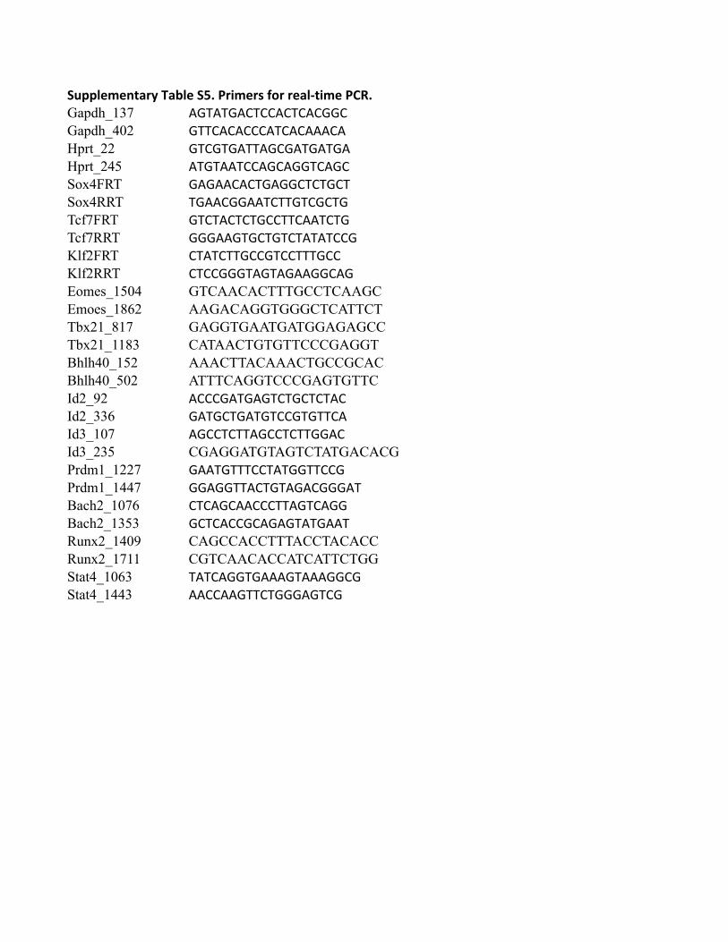
!"##$%&%'()*+,-).$%,!/0,1*2&%*3,45*,*%)$6(2&%,1780Gapdh_137 !"#!#"!$#$$!$#$!$""$Gapdh_402 "##$!$!$$$!#$!$!!!$!Hprt_22 "#$"#"!##!"$"!#"!#"!Hprt_245 !#"#!!#$$!"$!""#$!"$Sox4FRT "!"!!$!$#"!""$#$#"$#Sox4RRT #"!!$""!!#$##"#$"$#"Tcf7FRT "#$#!$#$#"$$##$!!#$#"Tcf7RRT """!!"#"$#"#$#!#!#$$"Klf2FRT $#!#$##"$$"#$$###"$$Klf2RRT $#$$"""#!"#!"!!""$!"Eomes_1504 GTCAACACTTTGCCTCAAGCEmoes_1862 AAGACAGGTGGGCTCATTCTTbx21_817 GAGGTGAATGATGGAGAGCCTbx21_1183 CATAACTGTGTTCCCGAGGTBhlh40_152 AAACTTACAAACTGCCGCACBhlh40_502 ATTTCAGGTCCCGAGTGTTCId2_92 !$$$"!#"!"#$#"$#$#!$Id2_336 "!#"$#"!#"#$$"#"##$!Id3_107 !"$$#$##!"$$#$##""!$Id3_235 CGAGGATGTAGTCTATGACACGPrdm1_1227 "!!#"###$$#!#""##$$"Prdm1_1447 ""!""##!$#"#!"!$"""!#Bach2_1076 $#$!"$!!$$$##!"#$!""Bach2_1353 "$#$!$$"$!"!"#!#"!!#Runx2_1409 CAGCCACCTTTACCTACACCRunx2_1711 CGTCAACACCATCATTCTGGStat4_1063 #!#$!""#"!!!"#!!!""$"Stat4_1443 !!$$!!"##$#"""!"#$"
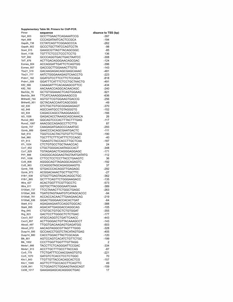
Supplementary Table S6. Primers for ChIP-PCR.Pimer sequence disance to TSS (bp)Hprt_603 GCCTTGAACTCAGAAATCCG -397Hprt_806 CCCAGATAATCACTCCGCA -194Gapdh_738 CCTATCAGTTCGGAGCCCA -262Gapdh_902 GCCCTGCTTATCCAGTCCTA -98Sox4_915 GAAGCGTTAGTTACAGCGGC -85Sox4_1136 TGTTTCTCCCTCCCTCCTG 136Tcf7_692 GCCCAGGTGACTGACTAATCC -308Tcf7_876 ACTTGACAGGGAACAGCGAC -124Eomes_604 ACCAGGATTGATTCTCAGTGG -396Eomes_857 GACCGCTTGGAAACTTGTG -143Tbx21_519 GACAAGAGACAGCGAGCAAAC -481Tbx21_777 AATCTGGGAAAGAGTCAACCTG -223Prdm1_182 GGATGTCCTTCCTTCTCCAGA -818Prdm1_509 GGATTTCATTTCTCCTGCTAACTG -491Klf2_566 CAAAGATTTCACAGAGCGTTCC -434Klf2_760 AACAAACCAGGCACAACAGC -240Bach2a_79 GCTGTGGAAACTCAGTGGAAG -921Bach2a_364 TTCATCAAAGGGAAAGCCG -636Bhlhe40_744 AGTGTTCGTGGAAGTGACCG -256Bhlhe40_951 GCTACAACCAATCAGCGGG -49Id2_630 GTCTGCTGTGCGGAGAGAGT -370Id2_848 AGCCAATGCCTGTAGGGTG -152Id3_834 CAGACCAGCCTAAGGAAGCC -166Id3_1026 GAGACACCTAAAGCAGCAAACA 26Runx2_883 GGCAGTCCCACTTTACTTTGAG -117Runx2_1087 AAACGCCAGAGCCTTCTTG 87Gzmb_707 CAAGAGATGAGCCCAAATGC -293Gzmb_889 GAACCCACAGCGAATGACTC -111Sell_810 TGGTCCACTACTGTGTTCTTGG -190Sell_960 TGCTTTCTTTCATTTCTCCAGC -40Il7r_813 TGAAGTCTACCACCTTGCTCAA -187Il7r_1024 CTCTGTGCCTGCTAAACCAC 24Ccr7_652 CTGCTGGGACAATAGCCACT -348Ccr7_829 TGTAGAGACTCAGGGAGGAGC -171Prf1_888 CAGGGCAGGAAGTAGTAATGATATG -112Prf1_1036 CTTCCTCCTCCTTACCTGAAGTC 36Ccl5_808 AGGGCAGTTAGAGGCAGAGTC -192Ccl5_963 CCAGGGTAGCAGAGGAAGTG 37Gzmk_708 GTGACCCACAGGTTGAGAGC -292Gzmk_973 ACGGACAAACTGCTTGCTTC -27Il18r1_638 GTGGTTGAGTAGACAGGCTGC -362Il18r1_865 GCTTTCAGTTCTGGGAAGACC -135Il6ra_427 ACACTGGTTTCGTTGCCTC -573Il6ra_611 GGTGCTTACGGGAATCAAA -389S100a4_737 TTCCTAAACTTCTGGCTGAGC -263S100a4_906 TGATGTAGTAAATGTCATAGCACCC -94S100a6_781 ACCACCACAACTTGAAGAACAG -219S100a6_936 GGACTGGGAACCACACTGAT -64Stat4_612 AGAGAAGAATCCAGGTGGCAC -388Stat4_895 AGACATTGAGGACCAGGCAG -105Ifng_645 CTGTGCTGTGCTCTGTGGAT -355Ifng_823 GACTCCTTGGGCTCTCTGAC -177Cxcr3_557 ATGCCAGGTCTGATTCAACC -443Cxcr3_857 ACTTGGGACTGTTACAAAGCCT -143Abca3_497 TTGGTGACAAGAGTGAGATGG -503Abca3_672 AACAGTAGGCGTTAGTTTGGG -328Dusp14_595 GCCAACCTGGTCTACATAGTGAG -405Dusp14_880 CACCTGGACTTACTCGCAGA -120Blk_801 AGTCCAGTCACATCTGTTCTGC -199Blk_1002 CCCTTGGTTGGTTTGTTAGG 2Ms4a1_666 TACCTTCTCAGGGATTCCGAC -334Ms4a1_913 ACCTTGCTTTGCCTTACCAG -87Ccr5_779 TTCTGATTTCCAACGAAGTGTG -221Ccr5_1070 GATGTCTCACCTCCTCTGGC 70Klrc1_843 TTGTTGTTACCACAGCACTCG -157Klrc1_1049 AGTTCTTTGCCACCTTCAGTTC 49Cd38_841 TCTGGAGTCTGGAAGTAAGCAGT -159Cd38_1017 GAAGAGAGCACAGGGCTGAC 17


