Embed Size (px)
Citation preview
IntroductionHeart failure is a major cause of morbidity and mor-tality worldwide (1). Regardless of the initiating dis-ease process, pathogenesis inevitably proceedsthrough a final common state termed dilated car-diomyopathy, in which heart chambers becomemarkedly enlarged and contractile function deterio-rates. Although abnormalities in cell membrane andcytoskeletal organization (2), adrenergic signaling (3),intracellular calcium handling (4, 5), and myocardialenergetics (6) have been observed, the molecular andcellular mechanisms that mediate the pathogenesis ofheart failure are poorly understood.
Recently, myocyte apoptosis has been noted in fail-ing human hearts. While initial studies reportedunrealistically high levels of cell death, probably due to methodological problems (7), later work has
consistently shown that approximately 80–250 heartmuscle cells per 105 cardiac nuclei commit suicide atany given time in patients with late-stage dilated car-diomyopathy (8–10). In contrast, the base-line rate ofapoptosis in healthy human hearts is only one to tencardiac myocytes per 105 nuclei. Whether the chroni-cally elevated but extremely low levels of myocyteapoptosis observed in failing hearts play a causal rolein the disease process remains a controversial issuewith major therapeutic implications.
Apoptosis in all metazoan cells is mediated by cas-pases, a multigene family of cysteine proteases thathydrolyzes peptide bonds carboxyl to aspartic acidresidues (11). Synthesized as zymogens, procaspaseactivation involves cleavage and the noncovalentreassociation of two p20 and two p10 fragments intoan active holoenzyme. Once activated, caspases cutkey cellular proteins, leading to the apoptotic demiseof the cell. Caspases can be activated by at least twobiochemical mechanisms. First, already activatedupstream caspases cleave downstream procaspases.Second, the most upstream procaspases, such as pro-caspase-8, possess low but significant basal caspaseactivity and are thought to autoactivate in trans whenforced into close proximity through interactions withother proteins (12).
We exploited this forced-proximity mechanism tocreate transgenic mice with cardiac-specific expressionof a ligand-activated caspase. These mice provided ameans to specifically manipulate the central deathmachinery and quantitatively modulate the induction
The Journal of Clinical Investigation | May 2003 | Volume 111 | Number 10 1497
A mechanistic role for cardiacmyocyte apoptosis in heart failure
Detlef Wencker,1,2 Madhulika Chandra,1 Khanh Nguyen,1,2 Wenfeng Miao,1
Stavros Garantziotis,1 Stephen M. Factor,1,3 Jamshid Shirani,1,3 Robert C. Armstrong,4
and Richard N. Kitsis1,2
1Department of Medicine (Molecular Cardiology),2Department of Cell Biology, and3Department of Pathology, Albert Einstein College of Medicine, Bronx, New York, USA4Idun Pharmaceuticals, San Diego, California, USA
Heart failure is a common, lethal condition whose pathogenesis is poorly understood. Recent stud-ies have identified low levels of myocyte apoptosis (80–250 myocytes per 105 nuclei) in failing humanhearts. It remains unclear, however, whether this cell death is a coincidental finding, a protectiveprocess, or a causal component in pathogenesis. Using transgenic mice that express a conditionallyactive caspase exclusively in the myocardium, we demonstrate that very low levels of myocyte apop-tosis (23 myocytes per 105 nuclei, compared with 1.5 myocytes per 105 nuclei in controls) are suffi-cient to cause a lethal, dilated cardiomyopathy. Interestingly, these levels are four- to tenfold lowerthan those observed in failing human hearts. Conversely, inhibition of cardiac myocyte death in thismurine model largely prevents the development of cardiac dilation and contractile dysfunction, thehallmarks of heart failure. To our knowledge, these data provide the first direct evidence that myocyteapoptosis may be a causal mechanism of heart failure, and they suggest that inhibition of this celldeath process may constitute the basis for novel therapies.
J. Clin. Invest. 111:1497–1504 (2003). doi:10.1172/JCI200317664.
Received for publication December 19, 2002, and accepted in revised formFebruary 25, 2003.
Address correspondence to: Richard N. Kitsis, Departments ofMedicine (Molecular Cardiology) and Cell Biology, AlbertEinstein College of Medicine, 1300 Morris Park Avenue, Bronx, New York 10461, USA. Phone: (718) 430-2609; Fax: (718) 430-8989; E-mail: [email protected] of interest: Robert C. Armstrong is an employee of IdunPharmaceuticals. Richard N. Kitsis is a consultant to IdunPharmaceuticals.Nonstandard abbreviations used: FK-binding protein (FKBP);maximal rate of increase of left ventricular systolic pressure(+dP/dt); maximal rate of decrease of left ventricular systolicpressure (–dP/dt); N-[(1,3-dimethylindole-2-carbonyl)valinyl]-3-amino-4-oxo-5-fluoropentanoic acid (IDN 1965).
See the related Commentary beginning on page 1457.
of apoptosis in cardiac myocytes in vivo. Using thissystem, we discovered that very low levels of myocyteapoptosis, levels that are four- to tenfold lower thanthose seen in human heart failure, are, in fact, suffi-cient to cause a lethal, dilated cardiomyopathy. More-over, inhibition of this cell death markedly inhibits thedevelopment of this syndrome. These experimentsprovide direct evidence that chronically elevated butlow levels of apoptosis may play a causal role in thepathogenesis of heart failure.
MethodsMaterials. Chemicals were purchased from Sigma-Aldrich (St. Louis, Missouri, USA) unless otherwisenoted. C57BL/6 mice were supplied by Taconic (Ger-mantown, New York, USA) and Charles River Labo-ratories (Wilmington, Massachusetts, USA). All ani-mal experimental protocols were approved by theInstitute for Animal Studies of the Albert EinsteinCollege of Medicine.
Generation of FK-binding-protein–caspase-8 transgenicmice. To construct the transgene, we first subclonedthe NotI-EcoRI fragment encoding the FK-binding-protein–caspase-8 (FKBP–caspase-8) fusion protein(Figure 1a; gift of V.M. Dixit, Genentech Inc., SouthSan Francisco, California, USA) (13) into pBluescriptII KS+ (Stratagene, La Jolla, California, USA). Wethen subcloned the NotI blunted–HindIII fragmentof this construct into the SalI blunted–HindIII sitesof clone 26, which contains 4.4 kb of mouse α-car-diac myosin heavy chain 5′ flanking sequence (gift ofJ. Robbins, Children’s Hospital, Cincinnati, Ohio,USA). The transgene construct was liberated fromprokaryotic sequences by BamHI digestion andinjected into the pronuclei of fertilized mouse eggsderived from an FVB/N–C57BL/6 mating. Trans-genic progeny were subsequently backcrossed onto aC57BL/6 background. Animals derived from four toeight backcrosses were used for experiments. WT lit-termates were always used for controls. The C360Amutant transgene construct was made by PCR site-directed mutagenesis (PfuTurbo DNA Polymerase;Stratagene) of codon TGT to GCT in the FKBP–cas-pase-8 transgene construct described above. Then theentire construct was resequenced to exclude addi-tional mutations.
Founders were identified by Southern analysis ofEcoRI-digested tail DNA with a probe consisting of anEcoRI-XbaI genomic fragment of the mouse α-cardiacmyosin heavy chain gene spanning from the 5′ flank-ing region to intron 1. This probe identified a 3.6-kbtransgene fragment and a 2.5-kb fragment of theendogenous α-cardiac myosin heavy chain. Subsequentgenerations were screened by PCR of toe DNA (14)using a three-primer reaction that identified a 324-bpproduct from the transgene and a 506-bp product fromthe endogenous α-cardiac myosin heavy chain gene.The forward primer was GACAGCAGATCACGATTCTC(corresponding to the α-cardiac myosin heavy chain
promoter). The reverse primers were TCCTTAG-GCTTGCTCTTGC (corresponding to the myristoylationsite of the transgene protein) and TTGCCACCATTG-CACGTAC (corresponding to endogenous α-cardiacmyosin heavy chain sequences distal to those in thetransgene construct).
Immunoblots. Mouse hearts were homogenized in 10volumes of 50 mM KCl, 50 mM PIPES (pH 7.4), 10mM ethylene glycol-bis(2-aminoethylether)-N,N,N′,N′-tetraacetic acid, 2 mM MgCl2, and 1 mM DTT at 4°Cwith five 5-second pulses at 9,600 rpm, using an Ultra-Turrax T25 homogenizer (Jahnke and Kunkel,Staufen, Germany). Homogenates were then cen-trifuged at 2,200 g for 15 minutes, the supernatant wasrecovered, and centrifugation was repeated. The pro-tein concentrations of the final supernatant weredetermined using the BCA Protein Assay Reagent(Pierce Chemical Co., Rockford, Illinois, USA). Twen-ty-microgram aliquots were resolved on 10% SDS-PAGE gels and electroblotted onto 0.45-µm nitrocel-lulose membranes (Bio-Rad Laboratories Inc.,Hercules, California, USA). The blots were reacted witha 1:2,500 dilution of a rabbit polyclonal antibodyagainst human caspase-8 (Idun Pharmaceuticals, SanDiego, California, USA).
DNA ladder assay. A DNA ladder assay was performedas previously described (15).
TUNEL/actin costaining. A TUNEL/actin costainingassay was performed as previously described (15) withthe following modifications pertaining to costainingfor α-sarcomeric actin. The TUNEL reaction was per-formed using a TACS 2 TdT DAB apoptosis detectionkit (Trevigen Inc., Gaithersburg, Maryland, USA)according to the manufacturer’s directions, exceptthat the streptavidin used to detect the terminaldeoxynucleotidyl transferase–ligated (TdT-ligated)biotinylated dNTPs was temporarily omitted. Stain-ing for α-sarcomeric actin was performed with a 1:30dilution of rabbit polyclonal antibody (clone 5C5;Sigma-Aldrich). Following incubation with TRITC-conjugated anti-rabbit secondary antibody (1:30 dilu-tion) to detect the α-sarcomeric actin antibody, Strep-tavidin-Orange-Green 488 (1:50 dilution; JacksonImmunoResearch Laboratories Inc., West Grove,Pennsylvania, USA) was applied to detect ligatedbiotinylated dNTPs in apoptotic nuclei. Total nucleion each section were visualized by staining with 500ng/ml bisbenzimide (Hoechst 33258). Sections weremounted on gridded coverslips (Bellco Glass Inc.,Vineland, New Jersey, USA) and visualized by confo-cal light microscopy (Zeiss Axiophot; Carl Zeiss Inc.,Thornwood, New York, USA).
For each section, the number of TUNEL-positivemyocyte nuclei in the left ventricular free wall wasmanually counted. Only nuclei that were clearlylocated in cardiac myocytes were scored. The totalnumber of nuclei in the left ventricular free wall wasdetermined by automated counting of the bisbenz-imide-positive signals using IPLab (Scanalytics Inc.,
1498 The Journal of Clinical Investigation | May 2003 | Volume 111 | Number 10
Vienna, Virginia, USA). Five transverse sectionsspaced through the heart were analyzed for each ani-mal. For controls and each of the three transgeniclines, three animals were studied per group.
Echocardiographic measurements. Echocardiographicstudies (16) were performed with an HDI 5000cv ultra-sonograph system (Advanced Technologies Laborato-ries, Andover, Massachusetts, USA). Mice were lightlyanesthetized with methoxyflurane and placed in thesupine position on a heating pad. A 10-MHz transduc-er was applied to the left hemithorax, and two-dimen-sionally directed M-mode images of the heart wererecorded on videotape. Left ventricular dimensions andwall thicknesses were assessed at end diastole and endsystole according to the guidelines of the AmericanSociety of Echocardiography. Data from three to sixconsecutive cardiac cycles were analyzed and averaged.Heart rate was determined from simultaneous electro-cardiographic recordings. Left ventricular fractionalshortening (LVFS) was calculated by the following for-mula: LVFS (%) = [(LVEDD – LVESD) / LVEDD] × 100,where LVEDD indicates left ventricular end-diastolicdimension and LVESD indicates left ventricular end-systolic dimension. The data were analyzed by anobserver blinded to genotype and/or treatment group.
In vivo hemodynamic measurements. Left ventricularcatheterization was performed under light methoxyflu-rane anesthesia with mice supine on a heating pad. A1.4 F Millar catheter (Millar Instruments Inc., Houston,Texas, USA) was introduced into the right carotidartery and advanced to the left ventricle. Heart rate andleft ventricular pressures were recorded under basalconditions and in response to 500 pg isoproterenol i.v.Maximal rate of rise (+dP/dt) and fall (–dP/dt) of leftventricular systolic pressure was derived from the pri-mary measurements. The data were analyzed by anobserver blinded to genotype.
Statistical analysis. Results are presented as means ±SEM. Statistical comparisons were performed usingANOVA and the Tukey multiple-comparison test,
with differences considered significant at P < 0.05.Kaplan-Meier survival analysis was used to assess allcauses of mortality in the different groups.
ResultsGeneration of transgenic mice with inducible cardiacmyocyte apoptosis. To explore whether cardiac myocyteapoptosis may be a mechanism of heart failure, wecreated transgenic mice in which heart muscle celldeath could be activated at will. This was accom-plished by the cardiac-specific expression of a fusionprotein (Figure 1a) consisting of three modules ofhuman FKBP-12 (pk mutant) attached to the p20and p10 catalytic domains of human procaspase-8(13). This FKBP–caspase-8 fusion protein would bepredicted to be catalytically inactive unless forcedinto close proximity. Forced approximation can bestimulated by systemic administration to mice ofFK1012H2, a small molecule that can simultaneous-ly bind two FKBP modules and, thereby, induce theoligomerization of the transgene protein (17).
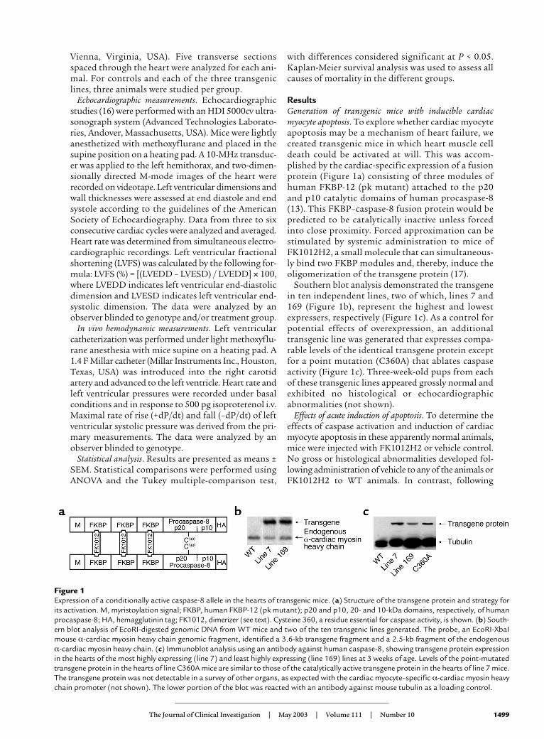
Southern blot analysis demonstrated the transgenein ten independent lines, two of which, lines 7 and169 (Figure 1b), represent the highest and lowestexpressers, respectively (Figure 1c). As a control forpotential effects of overexpression, an additionaltransgenic line was generated that expresses compa-rable levels of the identical transgene protein exceptfor a point mutation (C360A) that ablates caspaseactivity (Figure 1c). Three-week-old pups from eachof these transgenic lines appeared grossly normal andexhibited no histological or echocardiographicabnormalities (not shown).
Effects of acute induction of apoptosis. To determine theeffects of caspase activation and induction of cardiacmyocyte apoptosis in these apparently normal animals,mice were injected with FK1012H2 or vehicle control.No gross or histological abnormalities developed fol-lowing administration of vehicle to any of the animals orFK1012H2 to WT animals. In contrast, following
The Journal of Clinical Investigation | May 2003 | Volume 111 | Number 10 1499
Figure 1Expression of a conditionally active caspase-8 allele in the hearts of transgenic mice. (a) Structure of the transgene protein and strategy forits activation. M, myristoylation signal; FKBP, human FKBP-12 (pk mutant); p20 and p10, 20- and 10-kDa domains, respectively, of humanprocaspase-8; HA, hemagglutinin tag; FK1012, dimerizer (see text). Cysteine 360, a residue essential for caspase activity, is shown. (b) South-ern blot analysis of EcoRI-digested genomic DNA from WT mice and two of the ten transgenic lines generated. The probe, an EcoRI-XbaImouse α-cardiac myosin heavy chain genomic fragment, identified a 3.6-kb transgene fragment and a 2.5-kb fragment of the endogenousα-cardiac myosin heavy chain. (c) Immunoblot analysis using an antibody against human caspase-8, showing transgene protein expressionin the hearts of the most highly expressing (line 7) and least highly expressing (line 169) lines at 3 weeks of age. Levels of the point-mutatedtransgene protein in the hearts of line C360A mice are similar to those of the catalytically active transgene protein in the hearts of line 7 mice.The transgene protein was not detectable in a survey of other organs, as expected with the cardiac myocyte–specific α-cardiac myosin heavychain promoter (not shown). The lower portion of the blot was reacted with an antibody against mouse tubulin as a loading control.
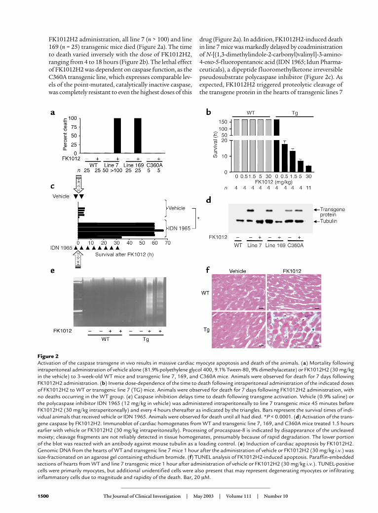
FK1012H2 administration, all line 7 (n > 100) and line169 (n = 25) transgenic mice died (Figure 2a). The timeto death varied inversely with the dose of FK1012H2,ranging from 4 to 18 hours (Figure 2b). The lethal effectof FK1012H2 was dependent on caspase function, as theC360A transgenic line, which expresses comparable lev-els of the point-mutated, catalytically inactive caspase,was completely resistant to even the highest doses of this
drug (Figure 2a). In addition, FK1012H2-induced deathin line 7 mice was markedly delayed by coadministrationof N-[(1,3-dimethylindole-2-carbonyl)valinyl]-3-amino-4-oxo-5-fluoropentanoic acid (IDN 1965; Idun Pharma-ceuticals), a dipeptide fluoromethylketone irreversiblepseudosubstrate polycaspase inhibitor (Figure 2c). Asexpected, FK1012H2 triggered proteolytic cleavage ofthe transgene protein in the hearts of transgenic lines 7
1500 The Journal of Clinical Investigation | May 2003 | Volume 111 | Number 10
Figure 2Activation of the caspase transgene in vivo results in massive cardiac myocyte apoptosis and death of the animals. (a) Mortality followingintraperitoneal administration of vehicle alone (81.9% polyethylene glycol 400, 9.1% Tween-80, 9% dimethylacetate) or FK1012H2 (30 mg/kgin the vehicle) to 3-week-old WT mice and transgenic line 7, 169, and C360A mice. Animals were observed for death for 7 days followingFK1012H2 administration. (b) Inverse dose-dependence of the time to death following intraperitoneal administration of the indicated dosesof FK1012H2 to WT or transgenic line 7 (TG) mice. Animals were observed for death for 7 days following FK1012H2 administration, withno deaths occurring in the WT group. (c) Caspase inhibition delays time to death following transgene activation. Vehicle (0.9% saline) orthe polycaspase inhibitor IDN 1965 (12 mg/kg in vehicle) was administered intraperitoneally to line 7 transgenic mice 45 minutes beforeFK1012H2 (30 mg/kg intraperitoneally) and every 4 hours thereafter as indicated by the triangles. Bars represent the survival times of indi-vidual animals that received vehicle or IDN 1965. Animals were observed for death until all had died. *P < 0.0001. (d) Activation of the trans-gene caspase by FK1012H2. Immunoblot of cardiac homogenates from WT and transgenic line 7, 169, and C360A mice treated 1.5 hoursearlier with vehicle or FK1012H2 (30 mg/kg intraperitoneally). Processing of procaspase-8 is indicated by disappearance of the uncleavedmoiety; cleavage fragments are not reliably detected in tissue homogenates, presumably because of rapid degradation. The lower portionof the blot was reacted with an antibody against mouse tubulin as a loading control. (e) Induction of cardiac apoptosis by FK1012H2.Genomic DNA from the hearts of WT and transgenic line 7 mice 1 hour after the administration of vehicle or FK1012H2 (30 mg/kg i.v.) wassize-fractionated on an agarose gel containing ethidium bromide. (f) TUNEL analysis of FK1012H2-induced apoptosis. Paraffin-embeddedsections of hearts from WT and line 7 transgenic mice 1 hour after administration of vehicle or FK1012H2 (30 mg/kg i.v.). TUNEL-positivecells were primarily myocytes, but additional unidentified cells were also present that may represent degenerating myocytes or infiltratinginflammatory cells due to magnitude and rapidity of the death. Bar, 20 µM.
and 169 mice, consistent with caspase activation; in con-trast, the catalytically inactive mutant protein in C360Ahearts remained intact (Figure 2d). Analysis of cardiacDNA from line 7 transgenic mice treated withFK1012H2 revealed strong internucleosomal ladderingconsistent with apoptosis (Figure 2e). Similarly, TUNELof heart sections demonstrated abundant DNA-strandbreaks in line 7 transgenic mice that received FK1012H2(Figure 2f). Echocardiography of these mice revealedmarked increases in wall thickness, which, upon histo-logical examination, were found to represent edema (notshown). Thus, activation of exogenous caspase-8 in theheart results in massive cardiac myocyte apoptosis anddeath of the mouse.
Effects of chronically low levels of cardiac myocyte apoptosis.Our intention in generating the FKBP–caspase-8 micewas to create a model in which the effects of low levels of
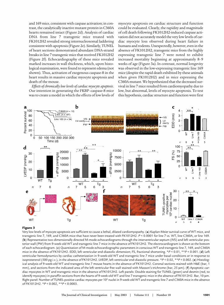
myocyte apoptosis on cardiac structure and functioncould be evaluated. Clearly, the rapidity and magnitudeof cell death following FK1012H2-induced caspase acti-vation did not accurately model the very low levels of car-diac myocyte loss observed during heart failure inhumans and rodents. Unexpectedly, however, even in theabsence of FK1012H2, transgenic mice from the highlyexpressing transgenic line 7 were noted to exhibitincreased mortality beginning at approximately 8–9weeks of age (Figure 3a). In contrast, normal longevitywas observed in the low-expressing transgenic line 169mice (despite the rapid death exhibited by these animalswhen given FK1012H2) and in mice expressing theC360A mutant. We hypothesized that the decreased sur-vival in line 7 mice resulted from cardiomyopathy due tolow, but abnormal, levels of myocyte apoptosis. To testthis hypothesis, cardiac structure and function were first
The Journal of Clinical Investigation | May 2003 | Volume 111 | Number 10 1501
Figure 3Very low levels of myocyte apoptosis are sufficient to cause a lethal, dilated cardiomyopathy. (a) Kaplan-Meier survival curve of WT mice, andtransgenic line 7, 169, and C360A mice that have never been treated with FK1012H2. P < 0.0001 for line 7 vs. WT, line C360A, or line 169.(b) Representative two-dimensionally directed M-mode echocardiograms through the interventricular septum (IVS) and left ventricular pos-terior wall (PW) from 9-week-old WT and transgenic line 7 mice in the absence of FK1012H2. The electrocardiogram is shown at the bottomof each echocardiogram. (c) Quantitation of M-mode echocardiographic parameters in conscious WT and transgenic line 7, 169, and C360Amice in the absence of FK1012H2. EDD, left ventricular end-diastolic dimension; FS, fractional shortening. *P < 0.01, **P < 0.001. (d) Leftventricular hemodynamics by cardiac catheterization in 9-week-old WT and transgenic line 7 mice under basal conditions or in response toisoproterenol (500 pg i.v.), in the absence of FK1012H2. LVEDP, left ventricular end-diastolic pressure. *P < 0.02, **P < 0.002. (e) Histolog-ical analysis of 9-week-old WT and transgenic line 7 mouse hearts in the absence of FK1012H2. Coronal sections stained with H&E (bar, 1mm), and sections from the indicated area of the left ventricular free wall stained with Masson’s trichrome (bar, 25 µm). (f) Apoptotic car-diac myocytes in WT and transgenic mice in the absence of FK1012H2. Left panels: Double staining for TUNEL (green) and desmin (red, toidentify myocytes) in paraffin sections from the hearts of 9-week-old WT and line 7 transgenic mice in the absence of FK1012H2. Bar, 10 µm.Right panel: Number of TUNEL-positive cardiac myocytes per 105 nuclei in 9-week-old WT and transgenic line 7 and C360A mice in the absenceof FK1012H2. *P < 0.002, **P < 0.0003.
evaluated at several time points. At 3 weeks of age,echocardiography and cardiac histology were normal(not shown). In contrast, by 9 weeks of age, line 7 trans-genic mice showed left ventricular dilation and markeddepression of fractional shortening (Figure 3, b and c).This was accompanied by histological myocyte dropout,interstitial fibrosis, thinning of the myocardium, anddilation of all four cardiac chambers (Figure 3e), indica-tive of dilated cardiomyopathy. Cardiac catheterizationmeasurements showed elevated left ventricular end-dias-tolic pressures and depressed basal and isoproterenol-stimulated +dP/dt and –dP/dt (Figure 3d), consistentwith combined systolic and diastolic dysfunction. Thelower-expressing line 169, which had normal longevity,exhibited an intermediate level of left ventricular dilationand contractile dysfunction (Figure 3c). In contrast, miceexpressing the C360A mutant transgene had normal car-diac dimensions, function, and histology (Figure 3c anddata not shown). These data demonstrate that theFKBP–caspase-8 transgenic mice spontaneously developa dilated cardiomyopathy between 3 and 9 weeks of age.This phenotype requires a catalytically active caspase,and the severity and mortality of this syndrome are relat-ed to the dose of the transgene protein.
To investigate the potential role of myocyte apopto-sis in the dilated cardiomyopathy of the FKBP–caspase-8 mice, TUNEL staining was performed on heart sec-tions from 7.5- to 8.0-week-old animals that had neverreceived FK1012H2 (Figure 3f). The frequency of spon-taneous myocyte apoptosis in WT murine hearts was1.59 ± 0.7 myocytes per 105 cardiac nuclei, similar tothat noted previously in healthy human hearts (8–10).Mice expressing the C360A inactive caspase exhibitedsimilar basal rates. In contrast, the frequency ofmyocyte apoptosis in line 7 mice was 23.2 ± 2.8 myo-cytes per 105 cardiac nuclei, 15 times higher than thatin WT (P < 0.001). Despite being abnormally elevated,however, this frequency is still quite low. In fact, it isfour to ten times lower than the most conservative esti-mates of myocyte death in failing human hearts (Table1). These data demonstrate that induction of a very lowlevel of myocyte apoptosis, lower than that observed inhuman heart failure, is sufficient to induce a lethal,dilated cardiomyopathy.
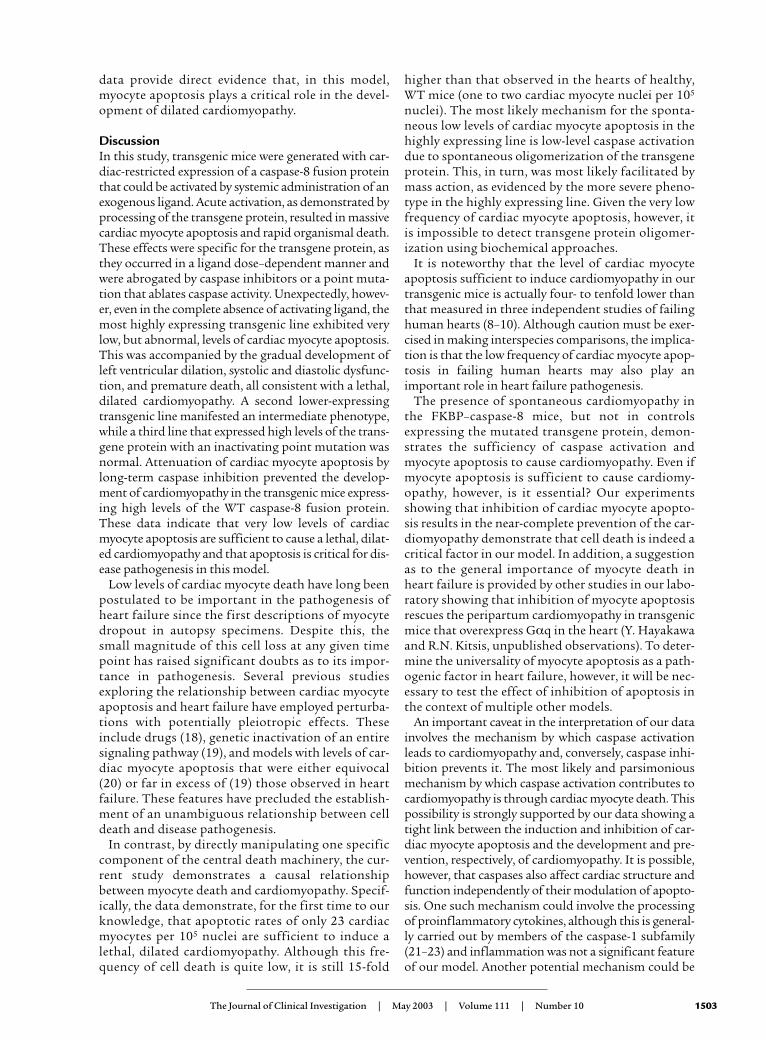
Prevention of cardiomyopathy by caspase inhibition. Ifthe low levels of myocyte apoptosis in the FKBP–cas-pase-8 transgenic mice really play a causal role in theresulting heart failure phenotype, then inhibition ofthis cell death should ameliorate the pathology. Totest this, the polycaspase inhibitor IDN 1965 wasadministered to line 7 animals by continuous subcu-taneous infusion via osmotic minipump. The infu-sion was started at 3.5–4.0 weeks of age, when thehearts were still normal, and continued to 7.5–8.0weeks of age, when the cardiomyopathy is florid inuntreated animals. As expected, caspase inhibitionmarkedly reduced the rates of myocyte apoptosis(Figure 4a). Strikingly, inhibition of myocyte deathwas accompanied by complete abrogation of left ven-tricular dilation (Figure 4, b and c), dramatic im-provement in left ventricular systolic function (Fig-ure 4b), and lessening of histological features ofcardiomyopathy such as fibrosis (Figure 4c). These
1502 The Journal of Clinical Investigation | May 2003 | Volume 111 | Number 10
Table 1Frequency of cardiac myocyte apoptosis in human heart failure vs.FKBP–caspase-8 mice
Species Apoptotic frequency(myocytes per 105 nuclei)
Controls Dilated cardiomyopathy
Human heart failure
Olivetti et al. (8) 1 237Saraste et al. (9) 11 119Guerra et al. (10) 2 80–180FKBP–caspase-8 mice 1.6 23
Apoptosis was assessed using the TUNEL assay.
Figure 4Abrogation of dilated cardiomyopathy by caspase inhibition. Vehicle or the polycaspase inhibitor IDN 1965 (12.5 µg/h) was administeredto line 7 transgenic mice by continuous subcutaneous infusion using osmotic minipumps (model 1002; ALZET Corp., Cupertino, Califor-nia, USA), beginning at 3.5–4.0 weeks of age, when cardiac dimensions, function, and histology are normal, and continuing until sacrificeat 7.5–8.0 weeks of age, when these transgenic mice uniformly exhibit a severe dilated cardiomyopathy. At 7.5–8.0 weeks of age, echocar-diography, TUNEL, and histological examination of cardiac tissue were performed. (a) Number of TUNEL-positive cardiac myocytes per 105
nuclei in vehicle- and IDN 1965–treated line 7 mice. *P < 0.03. (b) M-mode echocardiographic parameters from vehicle- and IDN 1965–treat-ed line 7 mice. *P < 0.0003. (c) Coronal sections from vehicle- and IDN 1965–treated line 7 mice stained with H&E (bar, 1 mm), and sec-tions from the indicated area of the left ventricular free wall stained with Masson’s trichrome (bar, 20 µm).
data provide direct evidence that, in this model,myocyte apoptosis plays a critical role in the devel-opment of dilated cardiomyopathy.
DiscussionIn this study, transgenic mice were generated with car-diac-restricted expression of a caspase-8 fusion proteinthat could be activated by systemic administration of anexogenous ligand. Acute activation, as demonstrated byprocessing of the transgene protein, resulted in massivecardiac myocyte apoptosis and rapid organismal death.These effects were specific for the transgene protein, asthey occurred in a ligand dose–dependent manner andwere abrogated by caspase inhibitors or a point muta-tion that ablates caspase activity. Unexpectedly, howev-er, even in the complete absence of activating ligand, themost highly expressing transgenic line exhibited verylow, but abnormal, levels of cardiac myocyte apoptosis.This was accompanied by the gradual development ofleft ventricular dilation, systolic and diastolic dysfunc-tion, and premature death, all consistent with a lethal,dilated cardiomyopathy. A second lower-expressingtransgenic line manifested an intermediate phenotype,while a third line that expressed high levels of the trans-gene protein with an inactivating point mutation wasnormal. Attenuation of cardiac myocyte apoptosis bylong-term caspase inhibition prevented the develop-ment of cardiomyopathy in the transgenic mice express-ing high levels of the WT caspase-8 fusion protein.These data indicate that very low levels of cardiacmyocyte apoptosis are sufficient to cause a lethal, dilat-ed cardiomyopathy and that apoptosis is critical for dis-ease pathogenesis in this model.
Low levels of cardiac myocyte death have long beenpostulated to be important in the pathogenesis ofheart failure since the first descriptions of myocytedropout in autopsy specimens. Despite this, thesmall magnitude of this cell loss at any given timepoint has raised significant doubts as to its impor-tance in pathogenesis. Several previous studiesexploring the relationship between cardiac myocyteapoptosis and heart failure have employed perturba-tions with potentially pleiotropic effects. Theseinclude drugs (18), genetic inactivation of an entiresignaling pathway (19), and models with levels of car-diac myocyte apoptosis that were either equivocal(20) or far in excess of (19) those observed in heartfailure. These features have precluded the establish-ment of an unambiguous relationship between celldeath and disease pathogenesis.
In contrast, by directly manipulating one specificcomponent of the central death machinery, the cur-rent study demonstrates a causal relationshipbetween myocyte death and cardiomyopathy. Specif-ically, the data demonstrate, for the first time to ourknowledge, that apoptotic rates of only 23 cardiacmyocytes per 105 nuclei are sufficient to induce alethal, dilated cardiomyopathy. Although this fre-quency of cell death is quite low, it is still 15-fold
higher than that observed in the hearts of healthy,WT mice (one to two cardiac myocyte nuclei per 105
nuclei). The most likely mechanism for the sponta-neous low levels of cardiac myocyte apoptosis in thehighly expressing line is low-level caspase activationdue to spontaneous oligomerization of the transgeneprotein. This, in turn, was most likely facilitated bymass action, as evidenced by the more severe pheno-type in the highly expressing line. Given the very lowfrequency of cardiac myocyte apoptosis, however, itis impossible to detect transgene protein oligomer-ization using biochemical approaches.
It is noteworthy that the level of cardiac myocyteapoptosis sufficient to induce cardiomyopathy in ourtransgenic mice is actually four- to tenfold lower thanthat measured in three independent studies of failinghuman hearts (8–10). Although caution must be exer-cised in making interspecies comparisons, the implica-tion is that the low frequency of cardiac myocyte apop-tosis in failing human hearts may also play animportant role in heart failure pathogenesis.
The presence of spontaneous cardiomyopathy inthe FKBP–caspase-8 mice, but not in controlsexpressing the mutated transgene protein, demon-strates the sufficiency of caspase activation andmyocyte apoptosis to cause cardiomyopathy. Even ifmyocyte apoptosis is sufficient to cause cardiomy-opathy, however, is it essential? Our experimentsshowing that inhibition of cardiac myocyte apopto-sis results in the near-complete prevention of the car-diomyopathy demonstrate that cell death is indeed acritical factor in our model. In addition, a suggestionas to the general importance of myocyte death inheart failure is provided by other studies in our labo-ratory showing that inhibition of myocyte apoptosisrescues the peripartum cardiomyopathy in transgenicmice that overexpress Gαq in the heart (Y. Hayakawaand R.N. Kitsis, unpublished observations). To deter-mine the universality of myocyte apoptosis as a path-ogenic factor in heart failure, however, it will be nec-essary to test the effect of inhibition of apoptosis inthe context of multiple other models.
An important caveat in the interpretation of our datainvolves the mechanism by which caspase activationleads to cardiomyopathy and, conversely, caspase inhi-bition prevents it. The most likely and parsimoniousmechanism by which caspase activation contributes tocardiomyopathy is through cardiac myocyte death. Thispossibility is strongly supported by our data showing atight link between the induction and inhibition of car-diac myocyte apoptosis and the development and pre-vention, respectively, of cardiomyopathy. It is possible,however, that caspases also affect cardiac structure andfunction independently of their modulation of apopto-sis. One such mechanism could involve the processingof proinflammatory cytokines, although this is general-ly carried out by members of the caspase-1 subfamily(21–23) and inflammation was not a significant featureof our model. Another potential mechanism could be
The Journal of Clinical Investigation | May 2003 | Volume 111 | Number 10 1503
the cleavage of sarcomeric proteins, as has been suggest-ed for caspase-3 (24–26). The effects of caspase-mediat-ed cleavage of contractile proteins on muscle functionand long-term cellular viability remain to be determined.
In summary, we believe that this study provides thefirst direct evidence that chronic, low levels of cardiacmyocyte apoptosis are a causal component in thepathogenesis of heart failure, and it raises the possi-bility that inhibition of this cell death may provide anovel target for treatments directed at this commonand lethal disorder.
AcknowledgmentsR.N. Kitsis was supported by grants from the NIH (R01HL60665 and R01 HL61550) and by the Monique Weill-Caulier Scholar Award. R.N. Kitsis is the Charles andTamara Krasne Faculty Scholar in CardiovascularResearch of the Albert Einstein College of Medicine. D.Wencker was supported by the Glorney-Raisbeck Fellow-ship in Cardiovascular Diseases of the New York Acade-my of Medicine and an Individual National Research Ser-vice Award from the NIH. We are indebted to ARIADPharmaceuticals Inc. (Cambridge, Massachusetts, USA)for providing FK1012H2 (http://www.ariad.com/regulationkits) and to Idun Pharmaceuticals for IDN1965 and the caspase-8 antibody.
1. Braunwald, E., and Bristow, M.R. 2000. Congestive heart failure: fiftyyears of progress. Circulation. 102:IV14–IV23.
2. Chien, K.R. 1999. Stress pathways and heart failure. Cell. 98:555–558.3. Lefkowitz, R.J., Rockman, H.A., and Koch, W.J. 2000. Catecholamines, car-
diac beta-adrenergic receptors, and heart failure. Circulation. 101:1634–1637.4. Marks, A.R. 2002. Ryanodine receptors, FKBP12, and heart failure. Front.
Biosci. 7:d970–d977.5. Luo, W., et al. 1994. Targeted ablation of the phospholamban gene is
associated with markedly enhanced myocardial contractility and loss ofbeta-agonist stimulation. Circ. Res. 75:401–409.
6. Taegtmeyer, H. 2002. Switching metabolic genes to build a better heart.Circulation. 106:2043–2045.
7. Narula, J., et al. 1996. Apoptosis in myocytes in end-stage heart failure.N. Engl. J. Med. 335:1182–1189.
8. Olivetti, G., et al. 1997. Apoptosis in the failing human heart. N. Engl. J.Med. 336:1131–1141.
9. Saraste, A., et al. 1999. Cardiomyocyte apoptosis and progression ofheart failure to transplantation. Eur. J. Clin. Invest. 29:380–386.
10. Guerra, S., et al. 1999. Myocyte death in the failing human heart is gen-der dependent. Circ. Res. 85:856–866.
11. Thornberry, N.A., and Lazebnik, Y. 1998. Caspases: enemies within.Science. 281:1312–1316.
12. Salvesen, G.S., and Dixit, V.M. 1999. Caspase activation: the induced-proximity model. Proc. Natl. Acad. Sci. U. S. A. 96:10964–10967.
13. Muzio, M., Stockwell, B.R., Stennicke, H.R., Salvesen, G.S., and Dixit,V.M. 1998. An induced proximity model for caspase-8 activation. J. Biol.Chem. 273:2926–2930.
14. Walter, C.A., Nasr-Schirf, D., and Luna, V.J. 1989. Identification of trans-genic mice carrying the CAT gene with PCR amplification. Biotechniques.7:1065–1070.
15. Bialik, S., et al. 1997. Myocyte apoptosis during acute myocardial infarc-tion in the mouse localizes to hypoxic regions but occurs independent-ly of p53. J. Clin. Invest. 100:1363–1372.
16. Chandra, M., et al. 2002. Cardioprotective effects of verapamil onmyocardial structure and function in a murine model of chronic Try-panosoma cruzi infection (Brazil Strain): an echocardiographic study.Int. J. Parasitol. 32:207–215.
17. Spencer, D.M., Wandless, T.J., Schreiber, S.L., and Crabtree, G.R. 1993.Controlling signal transduction with synthetic ligands. Science.262:1019–1024.
18. Li, Z., Bing, O.H., Long, X., Robinson, K.G., and Lakatta, E.G. 1997.Increased cardiomyocyte apoptosis during the transition to heartfailure in the spontaneously hypertensive rat. Am. J. Physiol.272:H2313–H2319.
19. Hirota, H., et al. 1999. Loss of a gp130 cardiac muscle cell survival path-way is a critical event in the onset of heart failure during biomechanicalstress. Cell. 97:189–198.
20. Condorelli, G., et al. 2001. Heart-targeted overexpression of caspase3 inmice increases infarct size and depresses cardiac function. Proc. Natl.Acad. Sci. U. S. A. 98:9977–9982.
21. Li, P., et al. 1995. Mice deficient in IL-1 beta-converting enzyme are defec-tive in production of mature IL-1 beta and resistant to endotoxic shock.Cell. 80:401–411.
22. Kuida, K., et al. 1995. Altered cytokine export and apoptosis in mice defi-cient in interleukin-1 beta converting enzyme. Science. 267:2000–2003.
23. Gu, Y., et al. 1997. Activation of interferon-gamma inducing factor medi-ated by interleukin-1beta converting enzyme. Science. 275:206–209.
24. Laugwitz, K.L., et al. 2001. Blocking caspase-activated apoptosisimproves contractility in failing myocardium. Hum. Gene Ther.12:2051–2063.
25. Moretti, A., et al. 2002. Essential myosin light chain as a target forcaspase-3 in failing myocardium. Proc. Natl. Acad. Sci. U. S. A.99:11860–11865.
26. Communal, C., et al. 2002. Functional consequences of caspase activa-tion in cardiac myocytes. Proc. Natl. Acad. Sci. U. S. A. 99:6252–6256.
1504 The Journal of Clinical Investigation | May 2003 | Volume 111 | Number 10