Embed Size (px)
Citation preview

A Mechanistic View of the Role of E3 in SumoylationMelda Tozluoglu1, Ezgi Karaca1, Ruth Nussinov2,3*, Turkan Haliloglu1*
1 Polymer Research Center & Chemical Engineering Department, Bogazici University, Istanbul, Turkey, 2 Basic Science Program, SAIC-Frederick, Inc., Center for Cancer
Research Nanobiology Program, NCI-Frederick, Frederick, Maryland, United States of America, 3 Sackler Institute of Molecular Medicine, Department of Human Genetics
and Molecular Medicine, Sackler School of Medicine, Tel Aviv University, Tel Aviv, Israel
Abstract
Sumoylation, the covalent attachment of SUMO (Small Ubiquitin-Like Modifier) to proteins, differs from other Ubl (Ubiquitin-like) pathways. In sumoylation, E2 ligase Ubc9 can function without E3 enzymes, albeit with lower reaction efficiency. Here,we study the mechanism through which E3 ligase RanBP2 triggers target recognition and catalysis by E2 Ubc9. Twomechanisms were proposed for sumoylation. While in both the first step involves Ubc9 conjugation to SUMO, thesubsequent sequence of events differs: in the first E2-SUMO forms a complex with the target and E3, followed by SUMOtransfer to the target. In the second, Ubc9-SUMO binds to the target and facilitates SUMO transfer without E3. Usingdynamic correlations obtained from explicit solvent molecular dynamic simulations we illustrate the key roles played byallostery in both mechanisms. Pre-existence of conformational states explains the experimental observations thatsumoylation can occur without E3, even though at a reduced rate. Furthermore, we propose a mechanism for enhancementof sumoylation by E3. Analysis of the conformational ensembles of the complex of E2 conjugated to SUMO illustrates thatthe E2 enzyme is already largely pre-organized for target binding and catalysis; E3 binding shifts the equilibrium andenhances these pre-existing populations. We further observe that E3 binding regulates allosterically the key residues in E2,Ubc9 Asp100/Lys101 E2, for the target recognition.
Citation: Tozluoglu M, Karaca E, Nussinov R, Haliloglu T (2010) A Mechanistic View of the Role of E3 in Sumoylation. PLoS Comput Biol 6(8): e1000913.doi:10.1371/journal.pcbi.1000913
Editor: Burkhard Rost, Columbia University, United States of America
Received October 13, 2009; Accepted July 29, 2010; Published August 26, 2010
This is an open-access article distributed under the terms of the Creative Commons Public Domain declaration which stipulates that, once placed in the publicdomain, this work may be freely reproduced, distributed, transmitted, modified, built upon, or otherwise used by anyone for any lawful purpose.
Funding: We would like to acknowledge financial support from the following sources: EU FP6-2004-ACC-SSA-2 (517991); Turkish Academy of Sciences (TUBA);The Scientific and Technological Research Council of Turkey (TUBITAK, 107T382); the Betil Fund. This project has been funded in whole or in part with Federalfunds from the National Cancer Institute, National Institutes of Health, under contract number HHSN261200800001E. The content of this publication does notnecessarily reflect the views or policies of the Department of Health and Human Services, nor does mention of trade names, commercial products, ororganizations imply endorsement by the U.S. Government. This research was supported (in part) by the Intramural Research Program of the NIH, NCI, Center forCancer Research. The funders had no role in study design, data collection and analysis, decision to publish, or preparation of the manuscript.
Competing Interests: The authors have declared that no competing interests exist.
* E-mail: [email protected] (TH); [email protected] (RN)
Introduction
Protein function is regulated by numerous mechanisms, one of
which is post-translational modification. Covalent binding of
ubiquitin (Ub) and ubiquitin-like (Ubl) modifiers to target proteins
constitute a key step in cellular processes including differentiation,
apoptosis, cell cycle, and stress response [1–4]. Here, we focus on
one member of the Ubl super-family, SUMO, with the aim of
figuring out the mechanism through which SUMO is conjugated
to its target proteins.
SUMO-1 (Small ubiquitin-like modifier, also known as PIC1,
UBL1, GMP1, Sentrin), -2, -3 and -4 exist in mammals [5–10].
Sumoylation can change the proteins’ intracellular localization,
interaction patterns with other proteins and modifications by other
post-translational events. It is important in development [11] and
is related to cancer drug resistance [12,13]. For simplicity, below,
SUMO refers to SUMO-1. At least 100 different proteins have
been reported as targets for sumoylation [14–17]. Analogous to
conjugation mechanisms of Ub/Ubls, SUMO is attached to target
proteins following sequential activation by E1, E2 and in most
cases, E3 enzymes [18]. Following activation of the SUMO
precursor [4], the E1 enzyme Aos1/Uba2 and SUMO form a
thioester bond. The SUMO thioester is next transferred to the
active cysteine of Ubc9, the single known E2 enzyme of the
sumoylation pathway [1,4,18]. Then SUMO is transferred from
E2 to a target protein lysine residue. E3 enzymes that ensure target
specificity and increase reaction efficiency usually mediate this step
(Figure 1). Among the sumoylation targets, RanGAP1, p53 and
IkBa are modified without an E3 ligase in vitro, although the
reaction rates are slower compared to E3-mediated conjugation
[1]. E2 ligase Ubc9 is essential [1,19] and conserved [1]. It
recognizes a consensus sumoylation motif, ‘‘Y-K-x-D/E’’, where
Y represents a hydrophobic residue, K is the SUMO acceptor
lysine, x is any amino acid and D/E is an acidic residue [4]. The
E2 ligase also interacts with E3 enzymes during the transfer of
SUMO to targets [20]. In addition to the consensus sumoylation
motif, sumoylation target RanGAP1 has a second contact surface
with the E2 ligase Ubc9, which is thought to be responsible for the
higher efficiency of modification compared to other substrates [4].
A fragment of the E3 enzyme RanBP2, consisting of the IR1-M-
IR2 domains is sufficient for E3 activity in vivo and in vitro [18].
Moreover, IR1-M and M-IR2 constructs are also functional with
IR1-M being the catalytic core domain [20–22]. The activity of
the E3 fragment indicates that E3 exerts its catalytic effect by
altering the structural properties of the E2-SUMO complex,
increasing the affinity of the complex for specific protein targets,
rather than by forming direct target interactions [21]. The crystal
structure of the SUMO-RanGAP1-Ubc9-RanBP2 complex sup-
ports this idea [20]. Recent work also shows that E3 ligase
RanBP2 prevents dissociation of SUMO from its target Ran-
PLoS Computational Biology | www.ploscompbiol.org 1 August 2010 | Volume 6 | Issue 8 | e1000913

GAP1, leading to an increase in the sumoylated RanGAP1 levels
[23]. Due to the strong interactions between RanGAP1 and E2, it
has been a debated question whether RanBP2 exerts its E3 activity
for RanGAP1 or whether it only maintains the complex at the
nuclear pore complex (NPC) [4,20].
Our aim is to understand the mechanism through which the E3
ligase RanBP2 triggers target recognition and catalysis by E2 in
sumoylation. We carried out explicit solvent molecular dynamic
simulations for the E2 ligase Ubc9, SUMO, and the E2-SUMO
complex with and without the E3 enzyme RanBP2. We modeled
the conjugated E2-SUMO complex, in RanBP2 bound and
unbound forms, based on the SUMO-RanGAP1-Ubc9-RanBP2
crystal structure (Figure 2). Our results indicate that E3 binding
induces a higher population of target binding and catalysis-ready
E2, restricting the conformational space of the E2-SUMO
complex. We observe that RanBP2 binding enhances the
correlations between the fluctuations of E2 residues involved in
catalytic activity and target recognition, which implies that
RanBP2 is indeed an E3 ligase for the sumoylation of the target
protein RanGAP1. Our results further lead us to propose that the
mechanism through which E3 ligase RanBP2 triggers E2 target
recognition and catalysis in sumoylation is allostery: RanBP2 is an
allosteric effector of E2 ligase Ubc9. Below, we refer to the specific
proteins simulated (Ubc9, RanGAP1, RanBP2) rather than the
protein functional class (E2, target protein, E3, respectively) to
which they belong. These were the ones crystallized by Reverter
and Lima [20].
Results
RanBP2 binding reduces the conformational space of theUbc9-SUMO complex
When simulated without RanBP2, the Ubc9-SUMO complex
structure displays a significant deviation from its crystal structure.
The two representative conformations from the clustering analysis
of the sampled conformational space of the complexes display the
change in the quaternary structure of the Ubc9-SUMO complex
(Figure 3). The Ubc9-SUMO rotates and moves away from its
position in the crystal structure (Figure 3). Accompanying the
orientation change of SUMO, there is a minor re-organization of
the hydrogen bond network in the catalytic area (Figure S1). The
rmsd (root mean square deviation) of the Ubc9-SUMO complex
shows that the deviation is more dramatic between 5–12 ns and
stabilizes at the end of this period (Figure S2A). Yet, the monomers
do not show increased deviations from their initial structures
(Figure S2B, Figure S2C). This indicates that the rmsd increase of
the complex structure originates from a change in the relative
positions of the chains with respect to each other. On the other
hand, with RanBP2, SUMO does not move or rotate but
fluctuates around its original crystal conformation.
For a more quantitative measure of this orientation change, we
utilize the representative structures from the clustering analysis.
We carried out rmsd calculations for Ubc9 and SUMO, with
alignments of Ubc9-SUMO and Ubc9-SUMO-RanBP2 complex-
es on the individual proteins. The results are listed in Table 1. As
expected, when the proteins are aligned on the corresponding
chains in the complex structure, the rmsd values are low; however,
they show some increase when the complex is aligned on the other
chain. The rmsd values for SUMO and Ubc9 are not significantly
different for Ubc9-SUMO and Ubc9-SUMO-RanBP2 simulations
when each is aligned on the corresponding chain in the complex.
When the structures are aligned with Ubc9 as the pivot, the
SUMO rmsd in the Ubc9-SUMO-RanBP2 complex increases up
to less than 2 fold (representative structure 3), while the SUMO
rmsd in the Ubc9-SUMO complex increases more than 4 fold
(representative structure 4). This implies an overall quaternary
structure change; that is, an orientation change of SUMO in the
Ubc9-SUMO complex, which is not observed in the Ubc9-
SUMO-RanBP2 complex. An additional set of simulations further
validate this major orientation change (Table S1).
Along with the limitation of SUMO orientation, E3 binding
restricts the conformational space of Ubc9. We combined the
Ubc9 conformations from the simulations of the Ubc9-SUMO
and Ubc9-SUMO-RanBP2 complexes, eliminating the initial
10 ns from each simulation, and clustering the remaining
conformations. In the three resulting clusters, nearly all the
Ubc9 conformations from the Ubc9-SUMO-RanBP2 complex are
in one cluster, and the Ubc9 conformations from the Ubc9-
SUMO complex are distributed among all three clusters. The
distributions of the Ubc9 conformations are given in Table 2. The
time distribution of cluster members displayed in Figure 4D shows
Figure 1. Sumoylation mechanism by E1-E2 and E1-E2-E3enzymes. (A) Produced as an inactive precursor, the SUMO protein iscleaved, exposing its Gly-Gly motif, and gets activated. (B) Active SUMOis transferred by E1 enzyme Aous1/Uba2 heterodimer to the E2 enzymeUbc9. (C) Two alternative pathways can follow as the third step. Ubc9can directly transfer SUMO to specific targets (top). AlternativelyRanBP2, an E3 enzyme, can also join the complex, increasing the Ubc9catalytic activity and transferring SUMO to its targets (bottom).doi:10.1371/journal.pcbi.1000913.g001
Author Summary
Post-translational modifications constitute key regulatorymechanisms in the cell. One of these modifications is thetagging of the target protein with a smaller molecule.SUMO is such a ubiquitin-like tag protein, and sumoylationis the process of tagging proteins with SUMO. Themalfunctioning of sumoylation is linked with diseasessuch as Alzheimer’s, Parkinson’s, and cancer. Based onexperimental observations, two paths were suggested forsumoylation, the first and more efficient involves the E1, E2and E3 enzymes; the second only the E1 and E2. Here weinvestigate these alternative paths of sumoylation. Ourresults offer an explanation for how sumoylation can takeplace with only the E1 and E2 enzymes, and for themechanistic role of E3. They emphasize that E2 bound toSUMO is already pre-organized for the transfer of SUMO toa target protein and E3 binding further stabilizes theconformations, shifting the ensemble and thus increasingthe efficiency of the sumoylation.
Mechanistics of E3 in Sumoylation
PLoS Computational Biology | www.ploscompbiol.org 2 August 2010 | Volume 6 | Issue 8 | e1000913

that Ubc9 from the Ubc9-SUMO complex samples conformations
from the Ubc9-SUMO-RanBP2 complex throughout the whole
simulation time. The distribution of the conformations among the
clusters shows that RanBP2 binding restricts the conformational
space of Ubc9. To further validate this restriction, we projected
the Ubc9 conformational space from the Ubc9-SUMO and Ubc9-
SUMO-RanBP2 complexes on the principal components
(Figure 4A–C, Figure S4). The projections and the clustering
analysis demonstrate the restriction of Ubc9 conformational space
upon RanBP2 binding.
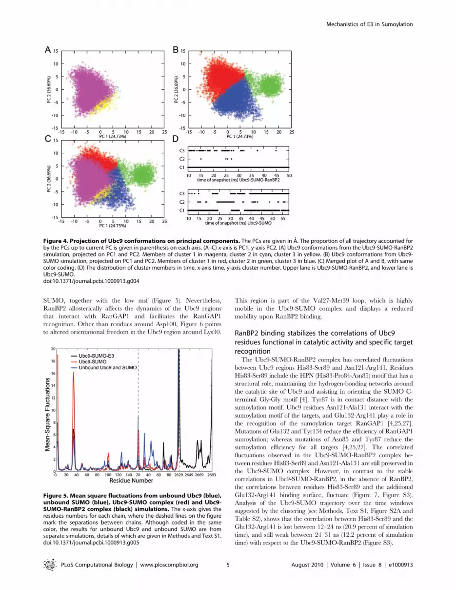
The mean-square fluctuations (msf) of the proteins in their
unbound states (Ubc9 and SUMO only), and in the complexes are
given in Figure 5. Ubc9 Cys93 is the active cysteine, and residues
from Asn124-Pro128 are part of the loop region that is in contact
with the tetrapeptide motif of the sumoylation targets [4]. The
fluctuations of Cys93 and Asn124-Pro128 are restricted in both
Ubc9-SUMO and Ubc9-SUMO-RanBP2 complexes, as compared
to the unbound state of Ubc9 (Figure 5). Thus, the catalytic residue
and the residues maintaining the structural integrity around the
catalytic residue of Ubc9 already display a reduced mobility in the
Ubc9-SUMO complex. In terms of reduced conformational states
of the catalytic region, Cys93 and Asn124-Pro128, the RanBP2
binding does not stabilize further these regions. This reduction in
Ubc9 mobility is a direct result of SUMO binding. Ubc9 residues
Val27-Met39 comprise the loop between b-sheets that serve as
RanBP2 binding sites [20]. These residues already display high
fluctuations in the isolated state, but their mobilities are allosterically
further enhanced by SUMO binding. RanBP2 binding reduces the
Figure 2. The structure of the Ubc9-SUMO-RanBP2-RanGAP1 complex [20]. The chains are colored as indicated in the legend. The insetshighlight the residue groups that are of interest. Top: The Ubc9 loop with the Asp100 and Lys101, active Cys93 is also represented in sticks. Bottom:The residues interacting with the sumoylation motif of target proteins. Detailed version is given in Figure S5.doi:10.1371/journal.pcbi.1000913.g002
Figure 3. SUMO orientation change. The orientations in the crystalstructure [20] (Ubc9 in yellow and SUMO in dark green) and arepresentative structure from the simulation trajectory (Ubc9 in brown,SUMO in light green) are aligned via the Ubc9 molecule. The orientationchange in SUMO can clearly be seen. Additionally, the motion of themobile loop of Ubc9 (Val27-Met39) can also be observed.doi:10.1371/journal.pcbi.1000913.g003
Mechanistics of E3 in Sumoylation
PLoS Computational Biology | www.ploscompbiol.org 3 August 2010 | Volume 6 | Issue 8 | e1000913

fluctuations to the values in the unbound state of Ubc9 (Figure 5).
These residues also show cooperative fluctuations with RanBP2
binding sites on SUMO (discussed below).
The mobility of residues Glu98-Asp102 of Ubc9 increase upon
SUMO binding, and again decrease upon RanBP2 binding
(Figure 5). Residues Asp100 and Lys101 take part in target
recognition, interacting with the approaching RanGAP1 [24,25].
RanBP2 does not have direct contacts with this loop. The
fluctuations of Asp100 and Lys101 in the Ubc9-SUMO-RanBP2
complex are lower than in the unbound Ubc9, whereas their
fluctuations in the Ubc9-SUMO complex are higher than in the
unbound Ubc9. This suggests that the restricted fluctuations of
Asp100 and Lys101 are the outcome of E3 binding. On the whole,
the reduced mobility of this region upon RanBP2 binding is
consistent with the proposed allosteric effect of RanBP2 on the
Ubc9 target recognition [20,21]. Although the fluctuations of some
SUMO residues demonstrate dramatic changes amongst all three
(the two complex and one isolated) states, most of these do not
coincide with functional residues. The high mobility of the
unbound SUMO structure (see Text S1) necessitates further
experimental evidence.
The allosteric effect of RanBP2 on Ubc9 in sumoylation:Restriction of Asp100-Lys101 orientational motion
The time delayed auto-correlations of the backbone bond
vectors is a measure of their orientational freedom. They provide
information on the time dependent changes in the orientations of
the backbone bonds. Here, the backbone bond vector refers to the
virtual bond vector between two successive Ca atoms (see
Methods, Text S1). A backbone bond vector will be closely
correlated with the same vector calculated after a short time
interval. As the time delay increases, the correlation between the
vectors will decrease. High auto-correlation of a backbone bond
vector indicates a restricted orientational freedom for the
backbone bond. In the Ubc9-SUMO-RanBP2 complex, the
correlations of backbone bonds are not lost with time delays as
high as 30 ns, with almost all virtual bond vectors having auto-
correlations above 0.9, unlike the Ubc9-SUMO complex
(Figure 6). This indicates that RanBP2 binding restricts the
orientational freedom of Ubc9-SUMO residues. Upon RanBP2
binding, the backbone vectors between Glu99-Asp100 and
Asp100-Lys101 of Ubc9 display the most significant restraint in
their orientational behavior (Figure 6), particularly compared to
the other loops of Ubc9. Residues Asp100-Lys101 are not in close
vicinity to the RanBP2 binding regions or the catalytic region of
Ubc9. The average distances of Asp100 to Cys93 throughout the
Ubc9-SUMO and Ubc9-SUMO-RanBP2 simulations are 13.25 A
and 12.82 A, respectively. Similarly, the average distances
between Lys101 and Cys93 throughout the Ubc9-SUMO and
Ubc9-SUMO-RanBP2 simulations are 11.54 A and 9.83 A,
respectively. Asp100 and Lys101 of Ubc9 are known to be
important for target recognition and functional defects have been
observed for Ubc9 upon mutations of these residues [24,25].
Restriction of the mobility and orientational freedom of these
residues upon RanBP2 binding can hinder a pre-organization of
the target binding site on Ubc9-SUMO leading to a shift of the
conformational ensemble [26] of Ubc9. Since these residues are
far from the RanBP2 binding site on Ubc9, the rigidification of the
99–102 loop on Ubc9 is allosterically induced by RanBP2 binding.
The Ubc9 residues Asn121-Ala131 interact with the consensus
sumoylation motif and Glu132-Arg141 are important for specific
RanGAP1 target binding [4,25,27]. The orientational freedom of
the bond vectors Pro128-Ala129, Gln130-Ala131, Glu132-Ala133
and Ala133-Tyr134 are significantly reduced when bound to
RanBP2. Yet, the bond vectors Asp127-Pro128 and Ala129-
Gln130 already have a restricted orientational freedom in Ubc9-
Table 1. RMSD with chain based alignments.
Ubc9-SUMO-E3 complex Ubc9 rmsd (A) SUMO rmsd (A)
aligned on Ubc9 aligned on SUMO aligned on Ubc9
Representative Structure 1 1.28 0.95 1.80
Representative Structure 2 1.31 1.42 2.25
Representative Structure 3 1.19 1.30 2.47
Representative Structure 4 1.23 1.60 2.75
Ubc9-SUMO complex Ubc9 rmsd (A) SUMO rmsd (A)
aligned on Ubc9 aligned on SUMO aligned on Ubc9
Representative Structure 1 1.34 1.45 1.94
Representative Structure 2 1.00 1.16 2.97
Representative Structure 3 1.23 1.46 2.63
Representative Structure 4 1.00 1.27 5.30
Representative Structure 5 2.15 1.41 5.49
Representative Structure 6 1.32 1.37 4.93
doi:10.1371/journal.pcbi.1000913.t001
Table 2. Clustering analysis of Ubc9 conformations from thejoined ensemble.
Percentage of ensemble belonging tocluster
Cluster 1 Cluster 2 Cluster 3 Total
Ubc9 from Ubc9-SUMO-E3complex
97.97 0.06 1.97 100.00
Ubc9 from Ubc9-SUMO complex 48.19 15.07 36.74 100.0
doi:10.1371/journal.pcbi.1000913.t002
Mechanistics of E3 in Sumoylation
PLoS Computational Biology | www.ploscompbiol.org 4 August 2010 | Volume 6 | Issue 8 | e1000913
SUMO, together with the low msf (Figure 5). Nevertheless,
RanBP2 allosterically affects the dynamics of the Ubc9 regions
that interact with RanGAP1 and facilitates the RanGAP1
recognition. Other than residues around Asp100, Figure 6 points
to altered orientational freedom in the Ubc9 region around Lys30.
This region is part of the Val27-Met39 loop, which is highly
mobile in the Ubc9-SUMO complex and displays a reduced
mobility upon RanBP2 binding.
RanBP2 binding stabilizes the correlations of Ubc9residues functional in catalytic activity and specific targetrecognition
The Ubc9-SUMO-RanBP2 complex has correlated fluctuations
between Ubc9 regions His83-Ser89 and Asn121-Arg141. Residues
His83-Ser89 include the HPN (His83-Pro84-Asn85) motif that has a
structural role, maintaining the hydrogen-bonding networks around
the catalytic site of Ubc9 and assisting in orienting the SUMO C-
terminal Gly-Gly motif [4]. Tyr87 is in contact distance with the
sumoylation motif. Ubc9 residues Asn121-Ala131 interact with the
sumoylation motif of the targets, and Glu132-Arg141 play a role in
the recognition of the sumoylation target RanGAP1 [4,25,27].
Mutations of Glu132 and Tyr134 reduce the efficiency of RanGAP1
sumoylation; whereas mutations of Asn85 and Tyr87 reduce the
sumoylation efficiency for all targets [4,25,27]. The correlated
fluctuations observed in the Ubc9-SUMO-RanBP2 complex be-
tween residues His83-Ser89 and Asn121-Ala131 are still preserved in
the Ubc9-SUMO complex. However, in contrast to the stable
correlations in Ubc9-SUMO-RanBP2, in the absence of RanBP2,
the correlations between residues His83-Ser89 and the additional
Glu132-Arg141 binding surface, fluctuate (Figure 7, Figure S3).
Analysis of the Ubc9-SUMO trajectory over the time windows
suggested by the clustering (see Methods, Text S1, Figure S2A and
Table S2), shows that the correlation between His83-Ser89 and the
Glu132-Arg141 is lost between 12–24 ns (20.9 percent of simulation
time), and still weak between 24–31 ns (12.2 percent of simulation
time) with respect to the Ubc9-SUMO-RanBP2 (Figure S3).
Figure 5. Mean square fluctuations from unbound Ubc9 (blue),unbound SUMO (blue), Ubc9-SUMO complex (red) and Ubc9-SUMO-RanBP2 complex (black) simulations. The x-axis gives theresidues numbers for each chain, where the dashed lines on the figuremark the separations between chains. Although coded in the samecolor, the results for unbound Ubc9 and unbound SUMO are fromseparate simulations, details of which are given in Methods and Text S1.doi:10.1371/journal.pcbi.1000913.g005
Figure 4. Projection of Ubc9 conformations on principal components. The PCs are given in A. The proportion of all trajectory accounted forby the PCs up to current PC is given in parenthesis on each axis. (A–C) x-axis is PC1, y-axis PC2. (A) Ubc9 conformations from the Ubc9-SUMO-RanBP2simulation, projected on PC1 and PC2. Members of cluster 1 in magenta, cluster 2 in cyan, cluster 3 in yellow. (B) Ubc9 conformations from Ubc9-SUMO simulation, projected on PC1 and PC2. Members of cluster 1 in red, cluster 2 in green, cluster 3 in blue. (C) Merged plot of A and B, with samecolor coding. (D) The distribution of cluster members in time, x-axis time, y-axis cluster number. Upper lane is Ubc9-SUMO-RanBP2, and lower lane isUbc9-SUMO.doi:10.1371/journal.pcbi.1000913.g004
Mechanistics of E3 in Sumoylation
PLoS Computational Biology | www.ploscompbiol.org 5 August 2010 | Volume 6 | Issue 8 | e1000913

Coinciding with the orientation change of SUMO, the
correlations between the fluctuations of the Ubc9 region including
the mobile loop, Val27-Glu42, and the rest of the Ubc9 residues,
become more negative. Furthermore, the same region, Val27-
Glu42, displays cooperative fluctuations with SUMO residues
Phe36-Leu47 and Asp73-Ile88. The two regions of SUMO are
either on the b-sheets packed against RanBP2 or in the vicinity of
these b-sheets in RanBP2 binding site in SUMO. Upon RanBP2
binding these correlations become more prevalent. The loop in
Ubc9 and those SUMO regions are spatially far away. When using
the centers of mass, the average distance between Val27-Glu42
loop of Ubc9 and Phe36-Leu47 of SUMO is ,45.0 A and 47.6 A
for Ubc9-SUMO and Ubc9-SUMO-RanBP2 simulations. Simi-
larly, the average distance between the Val27-Glu42 loop and the
Asp73-Ile88 of SUMO is 44.8 A for Ubc9-SUMO and 45.6 A for
the Ubc9-SUMO-RanBP2 complex simulations.
Alterations in the correlated fluctuations of regions that are not
listed above are observed from the correlation maps. In many of
these cases, although an overall change is observed in a region in
the vicinity of a key residue, the correlation of the residue itself is
conserved. One such example is the reduction in the correlated
fluctuations of Ubc9 residues around position 70, with both
residues around 100 and the first 10 residues of Ubc9. Lys74, the
key residue in this region which contacts the consensus sumoyla-
tion motif in targets [4], conserves its correlations.
To summarize, the correlated fluctuations among the residues
responsible for the structural integrity of the complex catalytic
region (His83-Ser89) and the residues that interact with the
sumoylation motif of the targets (Asn121-Ala131) are stable for
both Ubc9-SUMO and Ubc9-SUMO-RanBP2 complexes. On
the other hand, the correlated fluctuations between His83-Ser89
and residues playing a role in selective target recognition (Glu132-
Arg141) are less stable in the Ubc9-SUMO complex. These
correlations are allosterically stabilized upon RanBP2 binding.
Further, the correlations between RanBP2 binding sites on Ubc9
and on SUMO pre-exist RanBP2 binding; yet, as expected, they
are enhanced upon binding. Our results are summarized in
Table 3.
Discussion
Two mechanisms have been proposed [1,4] for RanGAP1
target sumoylation (Figure 1). In both, the first step involves Ubc9
conjugation to SUMO. In the first mechanism Ubc9-SUMO
binds to the target and an E3 ligase, whereas in the second Ubc9-
SUMO can bind and sumoylate the target without an E3 ligase. In
order to understand the role of E3 enzymes in the pre-
organization of the Ubc9-SUMO complex in the mechanism of
sumoylation, we simulated the Ubc9-SUMO complex with and
without the E3 ligase RanBP2. Based on the two conformational
ensembles with and without E3, Ubc9 is already largely pre-
organized for target binding and catalysis [28,29], yet the
orientation of SUMO differs in Ubc9-SUMO and Ubc9-
SUMO-RanBP2 complexes. Analysis of the conformational
ensembles of Ubc9, SUMO, Ubc9-SUMO, and Ubc9-SUMO-
RanBP2 revealed that RanBP2 binding allosterically shifts the
equilibrium of Ubc9 conformations, restricts SUMO orientation,
Figure 6. Auto-correlations of the backbone bond vectors(virtual bond vectors between successive alpha carbons). (A)The auto-correlations for Ubc9-SUMO-RanBP2 complex with time delayof 0.4 ns (black), 1 ns (red) and 30 ns (blue). (B) The auto-correlationsfor Ubc9-SUMO complex with time delay of 0.4 ns (black), 1ns (red),5 ns (green) and 30 ns (blue). At time delay of 30 ns, there is overall lossof correlations in Ubc9-SUMO complex in comparison to the highcorrelations observed for Ubc9-SUMO-RanBP2. The correlations with5 ns delay present first regions to loose correlations as around vector 30and vector 101 of Ubc9.doi:10.1371/journal.pcbi.1000913.g006
Figure 7. Correlations between fluctuations of residues. (A) Correlations of Ubc9-SUMO from Ubc9-SUMO-RanBP2 overall trajectory. (B)Correlations of Ubc9-SUMO from Ubc9-SUMO trajectory between 12–24 ns of simulation time. In both A and B, the dashed rectangle surrounds thecorrelations between His83-Ser89 Asn121-Ala131 of Ubc9, the solid rectangle surrounds the correlations between His83-Ser89 and Ala131-Arg141 ofUbc9. (C) The color bar indicating the correlations for both A and B.doi:10.1371/journal.pcbi.1000913.g007
Mechanistics of E3 in Sumoylation
PLoS Computational Biology | www.ploscompbiol.org 6 August 2010 | Volume 6 | Issue 8 | e1000913

and enhances the populations of the pre-organized conformational
states [26,28,29,30]. RanBP2 binding reduces the conformational
space sampled by Ubc9 (Figure 4, Figure S4). At the same time,
the RanBP2 binding allosterically reduces the mobility and the
orientational freedom of the Ubc9 residues, with the effect being
particularly dramatic around Asp100 and Lys101 target recogni-
tion residues [24,25]. RanBP2 binding allosterically enhances the
correlated fluctuations of Ubc9, mainly for residues around the
catalytic and specific target recognition sites. The RanBP2 binding
sites on Ubc9 and on SUMO which are spatially far apart display
correlated fluctuations even in the absence of E3; however upon
E3 binding these correlations get stronger.
RanBP2 was proposed to limit the available conformations of
the Ubc9-SUMO complex and prevent non-productive confor-
mations [20,21]. Our simulations demonstrate that upon RanBP2
removal there is a change in the relative position of SUMO with
respect to Ubc9, yet the removal of RanGAP1 does not affect
SUMO’s position. Furthermore, in the absence of RanBP2,
RanGAP1 is not sufficient to prevent SUMO’s position change
(unpublished data). This leads us to propose a mechanism where
RanBP2 binding to the Ubc9-SUMO complex triggers SUMO’s
stabilization in a catalytically efficient orientation, with a
subsequent target binding. This is consistent with RanBP2
enhanced allosteric effects on Ubc9’s Asp100-Lys101, the specific
target recognition regions, and the correlated fluctuations of
RanBP2 binding sites in Ubc9 and SUMO in the absence of
RanBP2.
A correlation between the fluctuations implicates a network of
interacting residues. It is highly plausible to expect an overlap of
such a network with functional residues. We observe coupled
fluctuations displaying changes between RanBP2 bound and
unbound states. Ubc9 residues Lys74, Tyr87, Ser89, Thr91,
Cys93, Asp127, Pro128, Ala129, Gln130 and Ala131, interact
with the consensus sumoylation tetrapeptide motif in most
sumoylation targets [4]. The correlations between Ubc9 residues
responsible for the structural integrity of the complex catalytic
region (His83-Ser89) and residues Asn121-Ala131 are conserved
in both Ubc9-SUMO and Ubc9-SUMO-RanBP2. Additionally,
there is a second contact surface on Ubc9 (Glu132-Asn140),
specific for the sumoylation target RanGAP1. Because of the
strong interactions between Ubc9 and RanGAP1 [4] through this
additional binding region, the need for E3 activity for sumoylation
of this target has not been apparent [4,20]. The enhanced
correlations between Ubc9 residues His83-Ser89 and Glu132-
Arg141 upon RanPB2 binding indicate that this additional
binding surface is linked to the catalytic activity. The stronger
correlations (Figure 7) suggest that RanBP2 increases the efficiency
of this additional target binding surface on Ubc9.
The mobilities of the catalytic Cys93, Asp127 and Pro128 of
Ubc9, which contact the consensus sumoylation tetrapeptide motif
in sumoylation targets [4], are reduced with the SUMO binding,
and RanBP2 binding does not lead to a further stability of these
residues. Additionally, we observe the conservation of the
correlations between His83-Ser89 and Asn121-Ala131 of Ubc9,
and the restrictions of the orientational freedom for Asp127-
Pro128 and Ala129-Gln130 in the Ubc9-SUMO complex without
RanBP2. The pre-existing tendencies of these residues which have
roles in catalysis and target recognition in the absence RanBP2
may indicate why Ubc9 can function without the aid of an E3
enzyme. Nevertheless, the correlations between other functional
regions, such as between His83-Ser89 and Glu132-Arg141, are
enhanced with a restriction in the conformational freedom of
Ubc9-SUMO with RanBP2. Indeed, the restriction in the
conformational space of Ubc9-SUMO with RanBP2 was suggest-
ed as a means of increasing sumoylation efficiency [20,21].
The mobility and orientational freedom of the Ubc9 Val27-
Met39 loop is affected by RanBP2 binding. This loop displays
correlated fluctuations with SUMO residues Phe36-Leu47 and
Asp73-Ile88, which are in close vicinity to the RanBP2 binding
sites. Together, these point to a sequence of events in the
formation of the complex which translate to SUMO binding to
Ubc9, followed by RanBP2 binding. From a mechanistic point of
view, SUMO binds to Ubc9, allosterically enhancing the mobility
of the Val27-Met39 loop and residues Asp100-Lys101, with the
Table 3. Summary of key residues and related observations.
Ubc9 Residue(s) Function Observations from Dynamics
Cys93 Catalytic cysteine [1,4,18]. Restricted in both Ubc9-SUMO and Ubc9-SUMO-RanBP2 complexes relative tounbound Ubc9 structure.
Val27 – Glu42 Includes loop between b-sheets serving asRanBP2 binding sites [20].
The loop has high mobility in unbound state. The mobility is further increased inUbc9-SUMO complex. RanBP2 binding reduces mobility to unbound Ubc9 level.
In Ubc9-SUMO complex, displays correlated fluctuations with RanBP2 bindingregions of SUMO, which are spatially distant.
His83 – Ser89 Maintains hydrogen bonding networks aroundcatalytic site, assists orientation of SUMO C-terminal Gly-Gly motif [4,25,27].
Correlated fluctuation with Asn121-Ala131 protected in Ubc9-SUMO and Ubc9-SUMO-RanBP2 complexes.
Correlated fluctuation with Glu132-Arg141 are mostly lost in Ubc9-SUMO complex,but enhanced by RanBP2 binding.
Asp100 – Lys101 Target recognition [24,25]. Increased msf in Ubc9-SUMO complex relative to unbound state. Fluctuationsreduced below unbound state in Ubc9-SUMO-RanBP2 complex.
Increased orientational freedom in Ubc9-SUMO complex, rigidification in Ubc9-SUMO-RanBP2 complex.
Asn124 – Pro128 Loop in contact with consensus sumoylationmotif in targets [4].
Restricted in both Ubc9-SUMO and Ubc9-SUMO-RanBP2 complexes relative tounbound Ubc9 structure.
Glu132 – Arg 141 RanGAP1 specific target binding [4,25,27]. Orientational freedom of region reduced in Ubc9-SUMO-RanBP2 complex.
Correlations with His83 – Ser89 are mostly lost in Ubc9-SUMO complex, butenhanced by RanBP2 binding.
doi:10.1371/journal.pcbi.1000913.t003
Mechanistics of E3 in Sumoylation
PLoS Computational Biology | www.ploscompbiol.org 7 August 2010 | Volume 6 | Issue 8 | e1000913

Val27-Met39 loop coordinated with the RanBP2 binding sites on
SUMO. Next RanBP2 binds to the Ubc9-SUMO complex,
moving SUMO closer to the target binding orientation, restricting
the Ubc9 conformations, and at the same time RanBP2 induces
allosteric changes in Ubc9 target recognition and catalytic sites.
The changes in the network of hydrogen-bonds between Ubc9 and
SUMO between the RanBP2 bound and unbound states appears
to be the driving force behind the orientation limitation of SUMO
induced by RanBP2 (Figure S1). The limitations imposed on the
orientational freedom of Asp100-Lys101 of Ubc9, and increased
correlations between Ubc9 catalytic site residues (His83-Ser89)
and specific target recognition residues (Glu132-Asn140) [4,20]
induced by RanBP2 binding, support the proposed mechanism.
Here we propose that the role of E3 ligase RanBP2 in
sumoylation is to restrict the conformational freedom of the E2-
SUMO complex and to increase the reaction efficiency via
allosteric effects [31] on the E2 Ubc9. RanBP2 binding to the E2-
SUMO complex limits the accessible conformations of Ubc9 and
the orientational space of the Ubc9 and SUMO monomers. In
particular, the positional and orientational freedom of Ubc9
residues Asp100-Lys101, important for target recognition [24,25]
is restricted upon RanBP2 binding. RanBP2 binding stabilizes the
correlations among Ubc9 residues that are functional in specific
target recognition (Glu132-Asn140) and catalytic activity (His83-
Ser89) [4,20]. Mechanistically, the correlations we observe in the
dynamics of the E2-SUMO complex argue for such sequence of
events in sumoylation and provide an explanation to the question
of why sumoylation can also take place in the absence of an E3,
although with lower efficiency.
Methods
Molecular dynamics protocolMolecular dynamics (MD) simulations are run on Ubc9,
SUMO, and the Ubc9-SUMO and Ubc9-SUMO-RanBP2
complexes, using Amber 8 [32,33]. Software and simulation
parameter details are provided in the Text S1.
The structures of human Ubc9 (PDB code: 1A3S) and human
SUMO-1 (PDB code: 1A5R) provide a base to analyze the effects
of RanBP2 binding to the Ubc9-SUMO complex. The modeled
structures for the intermediate complexes Ubc9-SUMO and
Ubc9-SUMO-RanBP2 are extracted from the crystal structure
of Ubc9-SUMO-RanGAP1-RanBP2 complex (PDB code: 1Z5S).
In the latter complex, as SUMO is bound to the acceptor lysine of
target protein RanGAP1, the thioester bond between Ubc9 active
cysteine and SUMO C-terminal glycine is modeled in the co-
crystal complexes of Ubc9-SUMO and Ubc9-SUMO-RanBP2.
The modeling of the thioester bond is detailed in Text S1. Two
sets of simulations are carried out. In the set where detailed time
window based analysis is carried out, the simulation lengths for the
structures are: unbound Ubc9, 32.5 ns; unbound SUMO, 35 ns;
Ubc9-SUMO complex, 58 ns; Ubc9-SUMO-RanBP2 complex,
50 ns. The second set is carried out to validate the major
orientation change of SUMO observed in the Ubc9-SUMO
structure in the first set of simulations. The simulation lengths for
each complex or protein consider the specific characteristics of the
structure, details of which are given in the Text S1.
Clustering analysis and principal component analysisThe simulations generate a large number of different confor-
mations of the structures, many of which may be similar. To
obtain distinct representative conformations, we clustered the
conformations with k-means clustering (MMTSB Toolset’s kclust
utility [34]), taking the rmsd of residue positions from the cluster
centroid as the similarity measure. Rmsd values of 2 A, 1.7 A and
1.5 A are tested; a smaller number of clusters appear as the rmsd
increases. The rmsd is set to 1.7 A for Ubc9-SUMO complex and
2 A for Ubc9-SUMORanBP2 complex. For the joined confor-
mational space analysis on Ubc9, the alignment is made on the
Ubc9 conformation from the Ubc9-SUMO complex at 10 ns, and
1.7 A cut-off is used.
The principal component analysis is carried out using the ptraj
module of AMBER 8.0. The alignment of the joined ensemble,
Ubc9 conformations from the Ubc9-SUMO and the Ubc9-
SUMO-RanBP2 complexes, followed the same procedure as in
the clustering analysis. The results are generated by projecting the
Ubc9 conformations on the PC of the joined ensemble. The
proportion of the eigenvalue of each PC to the sum of all
eigenvalues represents the contribution of the PC to the all
conformations in the trajectory. The cumulative contribution of all
PCs up the PC of interest is given in the axes of Figure 4 and
Figure S4.
Residues’ mean-square fluctuations and cross-correlations
The equilibration periods of the simulation (2.5 ns for Ubc9,
5 ns for SUMO, 0.5 ns for Ubc9-SUMO and 0.4 ns for Ubc9-
SUMO-RanBP2) are excluded in the calculations. For the msf
calculations of each chain in the complexes, the alignment is
carried out on the corresponding chain only, to eliminate the poor
alignment that may possibly result from the structural changes in
the quaternary structure. In the case of SUMO, the flexible N-
terminal tail (residues 21 to 19 in PDB 1A5R) of SUMO is
eliminated for the same reason.
The normalized correlations between the fluctuations of
residues, the cross-correlations, are defined by Eq. 1 as:
COi,j~SDRiDRjT
SDR2i T
1=2SDR2j T
1=2ð1Þ
Where DRi and DRj are the fluctuation in the position vectors, Ri
and Rj of residues i and j, respectively. The cross-correlations vary
in the range [21, 1] with the lower and upper limit indicating fully
anti-correlated and correlated fluctuations, respectively. The
correlations are calculated for the total length of the trajectory
and for the time windows defined by the clustering analysis.
The time delayed auto-correlations of the backbonebond vectors
The backbone bond vector is defined as the normalized vector
Mi from the a carbon Cai21 of residue i21 to the a carbon Ca
i of
residue i. The normalized time-delayed auto-correlations of these
virtual bond vectors are defined as in Eq. 2:
CMi tð Þ~SMi tð ÞMi tztð ÞT ð2Þ
Where Mi (t) and Mi (t+t) are the virtual bond vectors of i at time t
and t+t, respectively. The brackets represent averages over
recorded snapshots. The auto-correlations are in the range [21,
1] with the lower and upper limit indicating fully anti-correlated
and correlated virtual bonds, respectively. t = 0 gives the equal-
time auto-correlations, which is 1 for all virtual bond vectors. The
correlations are calculated for several time delays t, from 0 to
30 ns. The highest value of the time delay (30 ns) is selected to be
slightly longer than half simulation times, for both Ubc9-SUMO-
RanBP2 and Ubc9-SUMO complexes.
Mechanistics of E3 in Sumoylation
PLoS Computational Biology | www.ploscompbiol.org 8 August 2010 | Volume 6 | Issue 8 | e1000913

Supporting Information
Text S1 Detailed methodology of the study.
Found at: doi:10.1371/journal.pcbi.1000913.s001 (0.05 MB
DOC)
Figure S1 Distances between potential hydrogen bonds. (A)
Distance between alpha carbon atoms of residues Arg63 of
SUMO and Glu122 of Ubc9 throughout the trajectories. Upper
lane is the distance for Ubc9-SUMO-RanBP2 complex, and lower
lane is the distance for Ubc9-SUMO complex. (B) Distance
between a carbon atoms of residues Gln29 of SUMO and Gln111
of Ubc9 throughout the trajectories. Upper lane is the distance for
Ubc9-SUMO-RanBP2 complex, and lower lane is the distance for
Ubc9-SUMO complex.
Found at: doi:10.1371/journal.pcbi.1000913.s002 (4.23 MB TIF)
Figure S2 Rmsd values for both complexes throughout the
simulation. (A) The rmsd values with the alignment of the whole
complex structure, throughout the simulation for Ubc9-SUMO
complex. The jump in rmsd can be observed around 10 ns. The
rmsd values for the same simulation, calculated by alignment of
individual chains are illustrated for (B) Ubc9 and for (C) SUMO.
The rmsd jump is not observed in B and C. (D) The rmsd values
with the alignment of the whole complex structure, throughout the
simulation for Ubc9-SUMO-RanBP2 complex.
Found at: doi:10.1371/journal.pcbi.1000913.s003 (4.49 MB TIF)
Figure S3 Correlations of mean-square fluctuations. (A) Corre-
lations of Ubc9-SUMO overall trajectory. (B) Correlations of
Ubc9-SUMO from Ubc9-SUMO trajectory between 24–31 ns of
simulation time. In both A and B, the rectangles surround the
correlations between His83-Ser89 and Asn121-Ala131 of Ubc9,
and correlations between His83-Ser89 and Ala131-Arg141 of
Ubc9. (C) The color bar indicating the correlations for both A and
B.
Found at: doi:10.1371/journal.pcbi.1000913.s004 (2.14 MB TIF)
Figure S4 The projections of Ubc9 conformations on principal
components. The projections of Ubc9 conformations from Ubc9-
SUMO and Ubc9-SUMO-RanBP3 simulations are given in blue
and red, respectively. The principal components are given in A.
All plots are in the range [210:10] in x- and y-axes. The
proportion of all trajectory accounted for accounted for by the PCs
up to current PC is given in parenthesis on each axis.
Found at: doi:10.1371/journal.pcbi.1000913.s005 (1.80 MB TIF)
Figure S5 The structure of the Ubc9-SUMO-RanBP2-Ran-
GAP1 complex [20]. This figure is a detailed version of Figure 2 of
manuscript. The chains are colored as indicated in the legend. The
insets highlight the residue groups that are of interest. Top left:
Ubc9 mobile loop Val27 to Glu42. Bottom left: Ubc9 residues
Glu132-Arg141, responsible for specific target recognition. Top
right: SUMO residues Phe36 to Leu47 and Asp73 to Ile88. These
regions mark the proximity of SUMO residues that pack with E3
and the also show correlated fluctuations with Ubc9 residues
Val27 to Glu42. Middle right: Ubc9 catalytic Cys93, residues
functional in target recognition Asp100, Lys101. Bottom right:
Ubc9 HPN (His83-Pro84-Asn85) motif, has a structural role,
maintains the hydrogen-bonding networks around the catalytic site
of Ubc9. Ubc9 residues which interact with the consensus
sumoylation motif, see text for functional details of individual
residues.
Found at: doi:10.1371/journal.pcbi.1000913.s006 (1.61 MB TIF)
Figure S6 Rmsd values for unbound Ubc9 and SUMO
throughout the simulation. (A) The rmsd values of Ubc9. (B)
The rmsd values for SUMO. Values for full length protein are
displayed in red, values for N-terminal truncated protein are in
blue. The effect of the first 21 residues of protein on calculations
can be seen from the difference between two plots. The truncated
values are used for comparison through text.
Found at: doi:10.1371/journal.pcbi.1000913.s007 (0.31 MB TIF)
Table S1 RMSD with chain based alignments, second set of
simulations.
Found at: doi:10.1371/journal.pcbi.1000913.s008 (0.04 MB
DOC)
Table S2 Time windows defined by the clustering analysis.
Found at: doi:10.1371/journal.pcbi.1000913.s009 (0.03 MB
DOC)
Acknowledgments
MT and EK would like to thank Bulent Balta for valuable discussions.
Author Contributions
Conceived and designed the experiments: RN TH. Performed the
experiments: MT EK. Analyzed the data: MT. Contributed reagents/
materials/analysis tools: MT EK. Wrote the paper: MT RN TH.
References
1. Melchior F (2000) SUMO-Nonclassical Ubiquitin. Annu Rev Cell Dev Biol 16:
591–626.
2. Capili AD, Lima CD (2007) Taking it Step by Step: Mechanistic insights from
structural studies of Ubiquitin/Ubiquitin-like protein modification pathways.
Curr Op Struct Bio 17: 726–735.
3. Muller S, Cartsen H, Pyrowolakis G, Jentsch S (2001) SUMO, Ubiquitin’s
Mysterious Cousin. Nat Rev Mol Cell Biol 2: 202–210.
4. Bernier-Villamor V, Sampson DA, Matunis MJ, Lima CD (2001) Structural
basis for E2 mediated SUMO conjugation revealed by a complex between
ubiquitin-conjugating enzyme Ubc9 and RanGAP1. Cell 108: 345–356.
5. Boddy MN, Howe K, Etkin LD, Solomon E, Freemont PS (1996) PIC, novel
ubiquitin-like protein which interacts with the PML component of a multi-
protein complex that is disrupted in acute promyelocytic leukaemia. Oncogene
13: 971–982.
6. Mahajan R, Delphin C, Guan T, Gerace L, Melchinor F (1997) A small
ubiquitin-related polypeptide involved in targeting RanGAP1 to nuclear pore
complex protein RanBP2. Cell 88: 97–107.
7. Matunis MJ, Coutavas E, Blobel G (1996) A novel ubiquitin-like modification
modulates the partitioning of the Ran GTPase-activating protein RanGAP1
between the cytosol and the nuclear pore complex. J Cell Biol 135: 1457–1470.
8. Okura T, et al. (1996) Protection against Fas/APO-1- and tumor necrosis factor-
mediated cell death by a novel protein, sentrin. J Immunol 157: 4277–4281.
9. Shen Z, Pardington-Purtymun PE, Comeaux JC, Moyzis RK, Chen DJ (1996)
UBL1, a human ubiquitin-like protein associating with human RAD51/RAD52
proteins. Genomics 36: 271–279.
10. Capili AD, Lima CD (2007) Structure and Analysis of a Complex between
SUMO and Ubc9 Illustrates Features of a Conserved E2-Ubl Interaction. J Mol
Biol 369: 608–618.
11. Ulrich HD (2009) Ubiquitin, SUMO and the maintenance of genome stability.
DNA Repair (Amst) 8: 429.
12. Mo Y, Yu Y, Theodosiou E, Ee PLR, Beck WT (2005) A Role for Ubc9 in
Tumorigenesis. Oncogene 24: 2677–2683.
13. Vigodner M, Weisburg JH, Shrivastava V, Marmor RA, Fathy J, et al. (2009)
Differential expression patterns of SUMO proteins in HL-60 cancer cell lines
support a role for sumoylation in the development of drug resistance. Cell Tissue
Res 336: 277–286.
14. Panse V-G, Hardeland U, Werner T, Kuster B, Hurt E (2004) A proteome wide
approach identifies sumoylated substrate proteins in yeast. J Biol Chem 279:
41346–41351.
15. Wohlschlegel JA, Johnson ES, Reed SI, 3rd, Yates JR (2004) Global analysis of
protein sumoylation in Saccharomyces cerevisiae. J Biol Chem 279: 45662–45668.
16. Zhao Y, Kwon SW, Anselmo A, Kaur K, White MA (2004) Broad spectrum
identification of cellular small ubiquitin-related modifier (SUMO) substrate
proteins. J Biol Chem 279: 20999–21002.
Mechanistics of E3 in Sumoylation
PLoS Computational Biology | www.ploscompbiol.org 9 August 2010 | Volume 6 | Issue 8 | e1000913

17. Zhou W, Ryan JJ, Zhou H (2004) Global analyses of sumoylated proteins in
Saccharomyces cerevisiae induction of protein sumoylation by cellular stresses. J BiolChem 279: 32262–32268.
18. Tang Z, Hecker CM, Scheschonka A, Betz H (2009) Protein interactions in the
sumoylation cascade–lessons from X – ray structures. FEBS Journal 275:3003–3015.
19. van Waardenburg RCAM, Duda DM, Lancaster CS, Schulman B-A,Bjornsti M-A (2006) Distinct Functional Domains of Ubc9 Dictate Cell Survival
and Resistance to Genotoxic Stress. Mol Cell Biol 26: 4958–4969.
20. Reverter D, Lima CD (2005) Insights into E3 ligase activity revealed by aSUMO-RanGAP1-Ubc9-Nup358 complex. Nature 435: 687–692.
21. Pichler A, Gast A, Seeler JS, Dejean A, Melchior F (2002) The NucleoproteinRanBP2 Has SUMO1 E3 Ligase Activity. Cell 108: 109–120.
22. Pichler A, Knipscheer P, Saitoh H, Sixma TK, Melchior F (2004) The RanBP2SUMO E3 ligase is neither HECT- nor RING-type. Nat Struct Mol Biol 11:
984–991.
23. Zhu S, Goeres J, Sixt KM, Bekes M, Zhang X-D, et al. (2009) Protection fromisopeptidase-mediated deconjugation regulates paralog-selective sumoylation of
RanGAP1. Mol Cell 33: 570–580.24. Tatham MH, Chen Y, Hay RT (2003) Role of two residues proximal to the
active site of Ubc9 in substrate recognition by the Ubc9.SUMO-1 thiolester
complex. Biochemistry 42: 3168–3179.
25. Yunus AA, Lima CD (2006) Lysine activation and functional analysis of E2-
mediated conjugation in the SUMO pathway. Nat Struct Mol Biol 13: 491–499.26. Gunasekaran K, Ma B, Nussinov R (2004) Is allostery an intrinsic property of all
dynamic proteins? Proteins 15: 433–443.
27. Wu PY, Hanlon M, Eddins M, Tsui C, Rogers RS, et al. (2003) A conservedcatalytic residue in the ubiquitin-conjugating enzyme family. EMBO J 22:
5241–5250.28. Ma B, Kumar S, Tsai CJ, Nussinov R (1999) Folding funnels and binding
mechanisms. Protein Eng 12: 713–720.
29. Kumar S, Ma B, Tsai CJ, Sinha N, Nussinov R (2000) Folding and bindingcascades: dynamic landscapes and population shifts. Protein Sci 9: 10–19.
30. Tsai CJ, del Sol A, Nussinov R (2009) Protein allostery, signal transmission anddynamics: a classification scheme. Mol BioSyst 5: 207–216.
31. Tsai CJ, del Sol A, Nussinov R (2008) Absence of a change in shape does notimply that allostery is not at play. J Mol Bio 18: 1–11.
32. Case DA, Darden TA, Cheatham TE, III Simmerling CL, Wang J, et al. (2004)
AMBER 8, University of California, San Francisco. .33. Case DA, Cheatham 3rd TE, Darden T, Gohlke H, Luo R, et al. (2005) The
Amber biomolecular simulation programs. J Computat Chem 26: 1668–1688.34. Feig MJ, Karanicolas J, Brooks CL (2004) MMTSB Tool Set: enhanced
sampling and multiscale modeling methods for applications in structural biology.
J Mol Graph Model 22: 377–395.
Mechanistics of E3 in Sumoylation
PLoS Computational Biology | www.ploscompbiol.org 10 August 2010 | Volume 6 | Issue 8 | e1000913