Embed Size (px)
Citation preview
Cell, Vol. 121, 37–47, April 8, 2005, Copyright ©2005 by Elsevier Inc. DOI 10.1016/j.cell.2005.01.019
Sumoylation Silences the Plasma MembraneLeak K+ Channel K2P1
Sindhu Rajan,1 Leigh D. Plant,1 Michael L. Rabin,Margaret H. Butler, and Steve A.N. Goldstein*Department of Pediatrics and Institute for Molecular
Pediatric SciencesPritzker School of MedicineUniversity of Chicago5721 South Maryland AvenueChicago, Illinois 60637
Summary
Reversible, covalent modification with small ubiqui-tin-related modifier proteins (SUMOs) is known to me-diate nuclear import/export and activity of transcrip-tion factors. Here, the SUMO pathway is shown tooperate at the plasma membrane to control ion chan-nel function. SUMO-conjugating enzyme is seen to beresident in plasma membrane, to assemble withK2P1, and to modify K2P1 lysine 274. K2P1 had notpreviously shown function despite mRNA expressionin heart, brain, and kidney and sequence features likeother two-P loop K+ leak (K2P) pores that control ac-tivity of excitable cells. Removal of the peptide adductby SUMO protease reveals K2P1 to be a K+-selective,pH-sensitive, openly rectifying channel regulated byreversible peptide linkage.
Introduction
Background potassium conductances were recognizedto influence resting membrane potential and activity ofexcitable cells (Goldman, 1943; Hodgkin and Katz,1949; Hodgkin and Huxley, 1952) long before they wererevealed to pass via dedicated portals rather than ill-defined seepage pathways (Goldstein et al., 2001). Mo-lecular identification was presaged by cloning of potas-sium channels in S. cerevisiae and C. elegans with twopore-forming P loops in each subunit (Ketchum et al.,1995) and realized in characterization of K2P0 fromD. melanogaster, which manifests the canonical attri-butes: a K+-selective pore open across the physiologi-cal voltage range that manifests open (Goldman-Hodg-kin-Katz type) rectification (Goldstein et al., 1996; Ilanand Goldstein, 2001) and regulated activity (Zilberberget al., 2000; Zilberberg et al., 2001).
Fifteen animal genes have since been discovered toencode subunits with two P loops and four transmem-brane spans, and these are designated K2P channels(Goldstein et al., 2002). As expected for backgroundconductances, K2P channels that show function revealdynamic regulation by a panoply of chemical and phys-ical stimuli (i.e., kinases, phosphatases, lipids, G pro-teins, and mechanical stretch) that change open prob-ability (Zilberberg et al., 2000; Zilberberg et al., 2001;
*Correspondence: [email protected]
1These authors contributed equally to this work.Bockenhauer et al., 2001) or forward transport to theplasma membrane (O’Kelly et al., 2002; Rajan et al.,2002). The process of matching K2P subtypes to nativecurrents is now well underway (Kim et al., 1999; Lopeset al., 2000; Sirois et al., 2000; Bockenhauer et al., 2001;Washburn et al., 2002). It appears that each channelpore is formed by a pair of subunits (Lopes et al., 2001)and thus assembly of nonidentical subunits is ex-pected, although it has not yet been seen. Some K2Pgenes do not produce currents in experimental cells(K2P1, K2P7, K2P12 and K2P15). As synthesis of mRNAin native cells indicates that these are not pseudogenes(Washburn et al., 2002), their silence has been sus-pected to result from absolute localization to internalmembranes or strict downregulation (Orias et al., 1997;Goldstein et al., 1998; Bockenhauer et al., 2000; Rajanet al., 2001).
K2P1, the first mammalian isolate of the K2P super-family (Lesage et al., 1996), is expressed widely withabundant transcript in heart (ventricle greater than at-rium [Wang et al., 1998]), brain (cerebellum and thala-mus [Talley et al., 2001]), and kidney (thick ascendingloop of Henle and distal nephron [Levy et al., 2004]).While K2P1 was first reported to be a weak, inwardlyrectifying potassium channel, and so named TWIK (Le-sage et al., 1996), others observed no channel activity(Orias et al., 1997; Goldstein et al., 1998). In this report,K2P1 is shown to be inactive despite residence in theplasma membrane so long as the protein is covalentlymodified by small ubiquitin-related modifier protein,SUMO (also called sentrin). Sumoylation, the covalentmodification of substrate proteins with SUMO, is a re-versible, posttranslational modification primarily ob-served with nuclear proteins and implicated in nuclearimport/export, target stability, and transcriptional activ-ity (Muller et al., 2001; Li and Hochstrasser, 2003;Melchior et al., 2003). SUMO subtypes, SUMO-conju-gating enzyme, and SUMO proteases are conservedfrom yeast to humans.
Here, SUMO-conjugating enzyme (Ubc-9) is discov-ered to be uniformly distributed at the plasma mem-brane of Xenopus laevis oocytes until assembly withK2P1 channels induces polarized expression over theanimal pole. Endogenous Ubc-9 sumoylates K2P1 onan intracellular lysine at position 274. Desumoylation bya SUMO protease (SENP-1) activates the mute pore,allowing its characterization. K2P1 is now seen to be apotassium-selective leak channel with a 32 pS unitaryconductance that passes larger outward than inwardcurrents under physiological conditions (that is, high in-ternal and low external potassium) due to open rectifi-cation. Active at all physiological voltages, K2P1 is sen-sitive to external pH via protonation of a histidine in thefirst P loop that is conserved in the TASK (acid-sensi-tive) K2P channels K2P3 and K2P9 (Lopes et al., 2000,2001; Rajan et al., 2000). SUMO regulation of K2P1 isalso demonstrated in the mammalian renal fibroblastcell line COS-7. K2P1 is thus a canonical leak channelshowing tightly regulated activity at the plasma mem-brane via SUMO modification and removal.
Cell38
R
KaEc(cwbonbtiptc
rf(fapf(tSwtn
MSKapotwwcvtuf
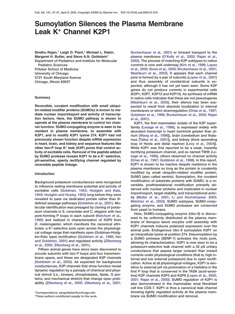
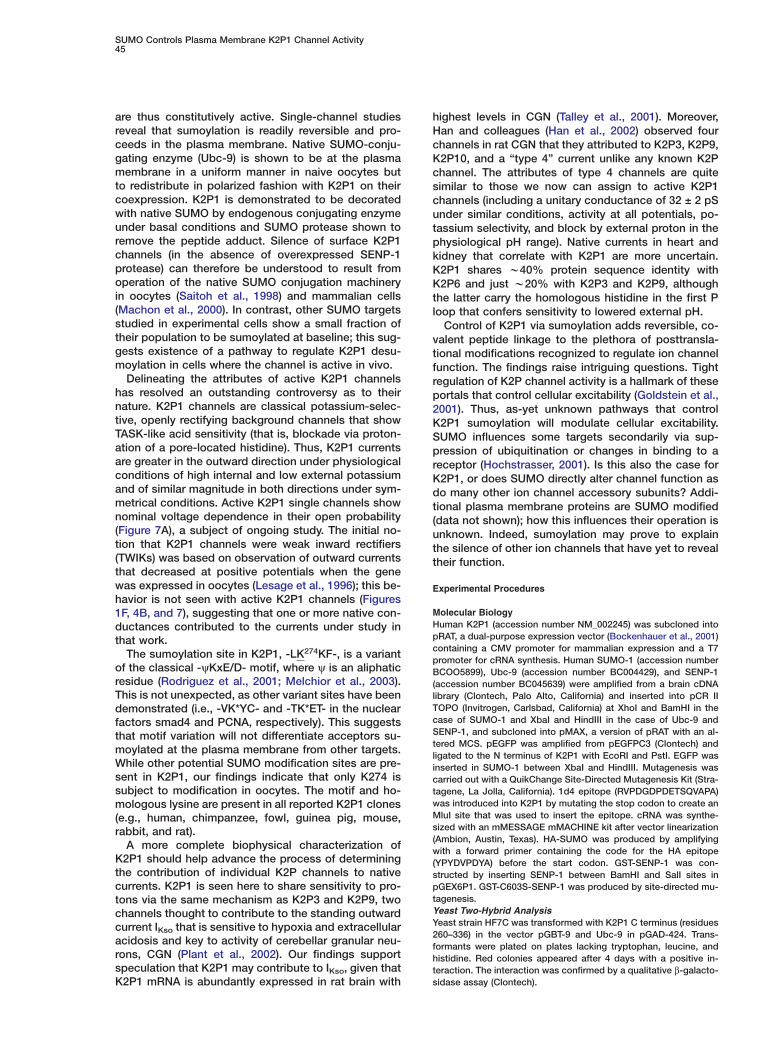
Figure 1. K2P1 Reaches the Plasma Membrane but Remains nElectrically Silent (The indicated subunits were expressed in Xenopus oocytes for 48 Phr before surface currents were recorded by two-electrode voltage wclamp and surface protein visualized by confocal microscopy. k(A) GFP-K2P1 shows polarized surface expression in oocytes. Light 1microscopy reveals K2P1 to be at the animal (dark) pole of the cells ((data not shown). Inset indicates oocyte position for microscopy; adark side represents animal pole. ((B) Expression of uncoupled GFP in oocytes shows no plasma imembrane fluorescence. p(C) K2P1-1d4 expressed in oocytes and visualized by immuno- Istaining with anti-1d4 antibodies also shows polarized expression. P(D) Cells expressing wild-type K2P1 without an epitope tag show i
As before (Orias et al., 1997; Goldstein et al., 1998),
o plasma membrane staining with anti-1d4 antibodies.E) Wild-type K2P1. Current trace from a representative oocyte.rotocol: holding −80 mV, 1 s test pulses from −120 mV to +80 mVith 2.5 s interpulse interval, sampled and filtered at 5 kHz and 1Hz, respectively. Bath solution with 4 mM potassium. Scale bars,�A, 200 ms.
F) K274E-K2P1. Current trace from a representative oocyte studieds in (E).
G) Average current-voltage relationship for groups of six cells asn (E) with bath potassium levels of 4 mM (■) and (F) with bathotassium levels of 4 (B), 20 (7), and 100 mM (,) (mean ± SEM).
nset: Predicted topology of K2P1 indicating lysine 274 (arrow), twoloops (P), and the membrane segments (M); N and C termini are
ntracellular.
esults
2P1 Is Electrically Silent Despite Expressiont the Plasma Membranexpression of K2P1 alone fails to produce potassiumurrents in oocytes or mammalian tissue culture cellsOrias et al., 1997; Goldstein et al., 1998). Lack of ionhannel function can be associated in the first instanceith failure of the protein to reach the plasma mem-rane; thus, K2P7 channels are speculated to operaten intracellular membranes (Salinas et al., 1999). Alter-atively, K2P3 channels reach the plasma membraneut surface levels are determined by regulated forwardransport (O’Kelly et al., 2002; Rajan et al., 2002). Tonvestigate trafficking of human K2P1, the protein wasroduced with an N-terminal green fluorescent proteinag (GFP-K2P1) and studied in oocytes by confocal mi-roscopy, as before (Rajan et al., 2002).Oocytes expressing GFP-K2P1 showed strong fluo-
escence at the plasma membrane (Figure 1A); thisluorescence was absent in cells with untagged K2P1data not shown) or GFP alone (Figure 1B). Of note, sur-ace expression of GFP-K2P1 was polarized to thenimal pole of the cells. Xenopus laevis oocytes arerototypical polarized cells that display structural and
unctional asymmetry of the animal and vegetal polesUbbels, 1997), with targeting of some membrane pro-eins to the animal pole a recognized phenomenon.urface expression in polarized fashion was also seenith K2P1 channels bearing a 16-residue tag at the C
erminus (K2P1-1d4) visualized with anti-1d4 monoclo-al antibodies (Figure 1C).
utation of a Single Residue Yields K2P1 Currentsurface expression without activity suggested that2P1 might require a stimulus to operate. However,gents (including long-chain free fatty acids, lysophos-holipids, volatile anesthetics, and classical regulatorsf kinases and phosphatases, data not shown) knowno upregulate other K2P channels had no effect. Activityas also not seen when the N and/or C termini of K2P1ere replaced by the regions from three functional K2Phannels (K2P2, K2P3, or K2P9, data not shown). Con-ersely, mutating a single intracellular lysine followinghe fourth transmembrane span, a region critical to reg-lation of K2P2 (Bockenhauer et al., 2001), was most in-ormative.
SUMO Controls Plasma Membrane K2P1 Channel Activity39
human K2P1 expression failed to induce currents in oo-cytes (Figure 1E). Like wild-type, GFP-K2P1 and K2P1-1d4 channels were electrically silent (data not shown)despite staining consistent with successful targeting tothe plasma membrane (Figures 1A and 1C). Conversely,changing the lysine at position 274 to glutamate(K274E-K2P1) produced macroscopic currents by two-electrode voltage clamp (Figures 1F–1H). As expectedfor a K2P channel, the portal was constitutively active(that is, open at rest) and potassium selective, showinga shift in reversal potential with altered bath potassiumof 51 ± 3 mV per 10-fold change. It became clear thatlysine had a unique silencing effect when currents werealso observed on alteration of the position to gluta-mine, alanine, cysteine, or arginine, a residue like lysinethat is positively charged at physiological pH. This ledus to speculate that position 274 was in a receptor sitefor a regulator that required lysine to bind; the underly-ing hypothesis was that the regulator suppressed K2P1function and that mutations interfered with regulatorassociation allowing the channels to open.
K2P1 Assembles with Native SUMO-ConjugatingEnzyme at the Plasma MembraneEvaluating the region of K2P1 with lysine 274 for knownmotifs revealed a variant SUMO modification site,-LK274KF-. SUMO-1, -2, and -3 are w100 amino acid,soluble, intracellular proteins, found in all eukaryoticcells, that act via covalent linkage to the � amino groupof lysine on an acceptor protein to be regulated. Su-moylation proceeds by a pathway that is distinct from,but analogous to, ubiquitin conjugation. Thus, SUMOsare first activated and linked to Aos1/Uba2 and thentransferred to the cysteine side chain of the conjugatingenzyme Ubc-9 (Gong et al., 1999; Bernier-Villamor etal., 2002). Ubc-9 then binds to the acceptor protein andtransfers its thioester-linked SUMO to the acceptor-protein lysine. The SUMO conjugation machinery ispresent en toto in extracts from Xenopus laevis oocytes(Saitoh et al., 1998).
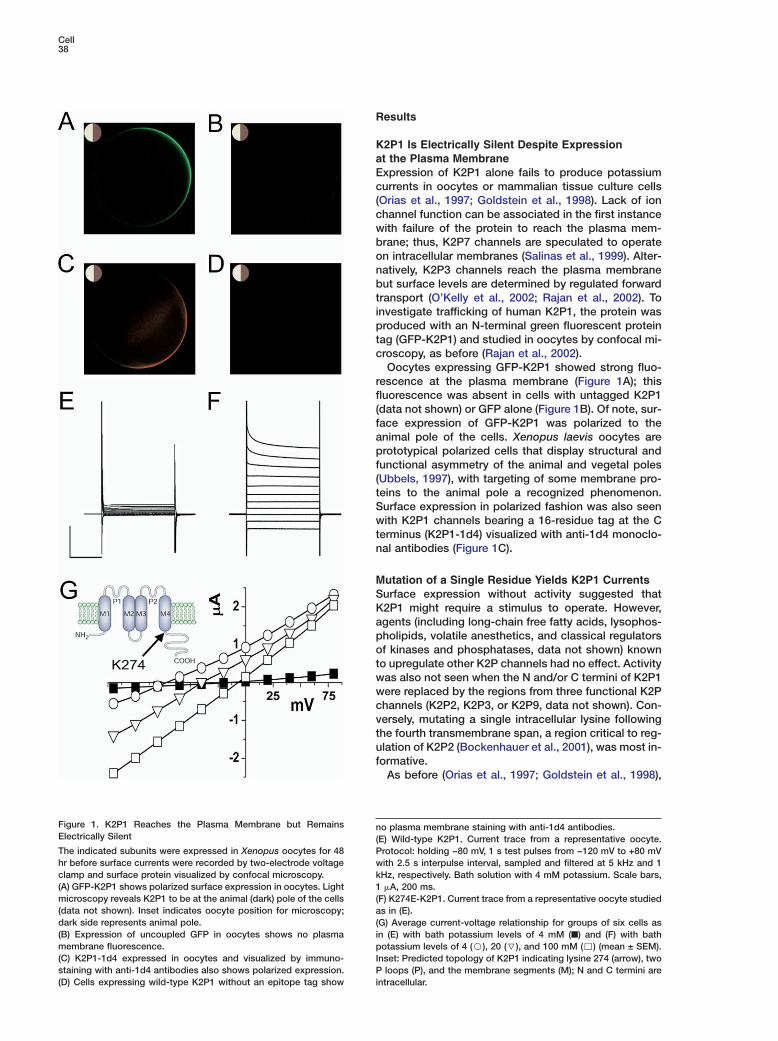
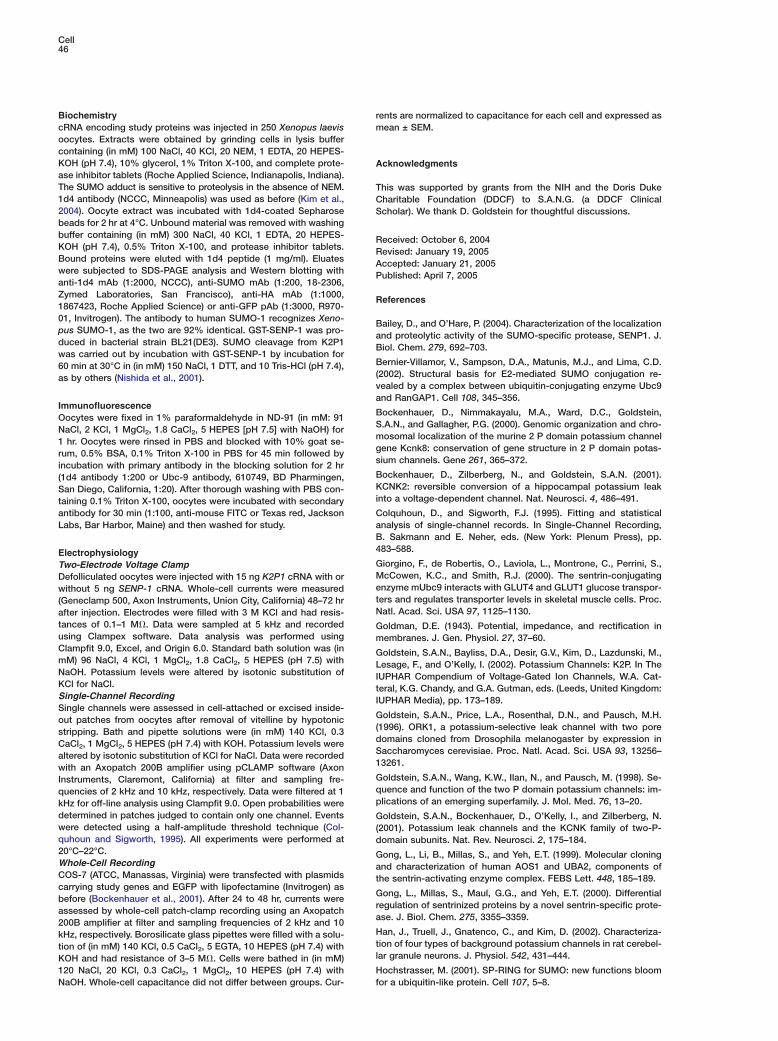
As interaction of Ubc-9 and the acceptor is requiredto accomplish sumoylation, we sought evidence for in-teraction of Ubc-9 and K2P1. First, human Ubc-9 wascloned and confirmed to interact with the C-terminalportion of K2P1 that carries lysine 274 using a yeasttwo-hybrid assay (Figure 2A). To next determine if com-plete K2P1 and Ubc-9 proteins interact in oocytes, aGFP tag was introduced at the N terminus of Ubc-9(GFP-Ubc-9) for studies by confocal microscopy. Ex-pressed on its own, GFP-Ubc-9 was observed at theoocyte plasma membrane in a uniform, nonpolarizedfashion (Figure 2B). In contrast, coexpression of wild-type K2P1 (no tag) and GFP-Ubc-9 led to restricted lo-calization of the conjugating enzyme at the animal pole(Figure 2C), the surface pattern observed for K2P1channels (Figure 1B), arguing for assembly of the twoproteins.
Native Ubc-9 endogenous to oocytes was found to in-teract with K2P1 in the same fashion as overexpressedhuman Ubc-9. The human protein is identical to theXenopus product (accession number BC046273), allow-ing use of a commercial antibody to human Ubc-9. NativeUbc-9 shows a uniform signal at the plasma membrane
Figure 2. K2P1 Assembles with SUMO-Conjugating Enzyme in thePlasma Membrane
(A) Yeast two-hybrid analysis shows strong interaction of the K2P1C terminus and SUMO-conjugating enzyme (Ubc-9). (Left panel)Yeast with the K2P1 C terminus (in pGBT-9) and Ubc-9 (in pGAD-424) show growth on histidine-deficient plates whereas (right panel)yeast with the K2P1 C terminus (in pGBT-9) and empty pGAD 424vector do not.(B) A representative oocyte expressing GFP-Ubc-9 shows uniformsurface staining in confocal images.(C) A representative oocyte expressing GFP-Ubc-9 and K2P1shows that surface staining for the conjugating enzyme becomespolarized to the animal pole.(D) Native Ubc-9 visualized in a naive oocyte stained with anti-Ubc-9 antibody and a Texas red-conjugated secondary antibodyshows uniform surface staining.(E) Native Ubc-9 visualized in an oocyte expressing wild-type K2P1and stained with anti-Ubc-9 antibody as in (D) shows that the na-tive conjugating enzyme also becomes polarized to the animalpole.
in untreated cells (Figure 2D). When K2P1 is expressed,the native Ubc-9 signal is polarized to the animal pole(Figure 2E). This suggested that surface distribution ofnative Ubc-9 was also restricted by assembly with
Cell40
CK2P1. Direct evidence for sumoylation of K2P1 was
uthus sought.
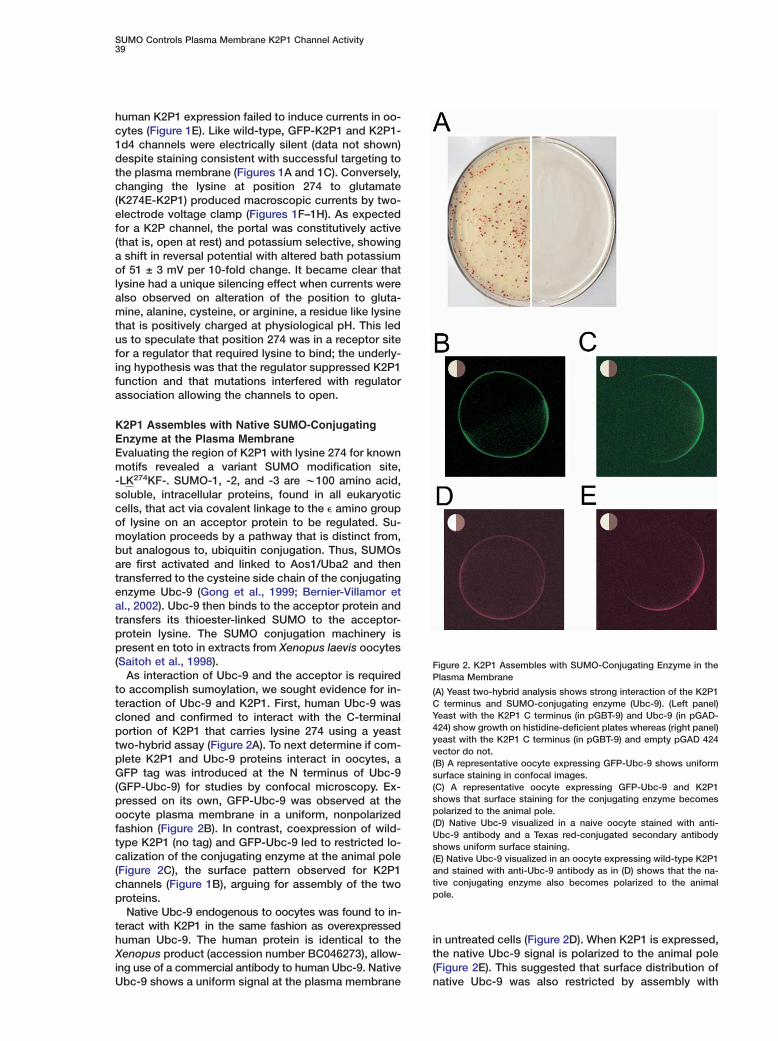
osK2P1 Is Modified by SUMO at Lysine 274
Human K2P1 was shown to be coupled to native SUMO aCby enzymes endogenous to the oocyte as follows.
Wild-type K2P1 and mutant subunits with lysine 274 afchanged to glutamate (K274E-K2P1) were produced
with C-terminal 1d4 tags to allow immunopurification. aeChannels were expressed in oocytes, solubilized in de-
tergent, affinity isolated via the 1d4 tag, subjected to wtseparation by SDS-PAGE, and analyzed by Western
blotting with anti-1d4 (Figure 3A) or anti-SUMO-1 anti- cdbodies (Figure 3B). Consistent with SUMO modification
ommm(gtsKf
daFhSKaha3umfsK
SFigure 3. K2P1 Is Modified by Native or Overexpressed SUMO ontLysine 274IOocytes were injected with cRNA for the indicated subunits, incu-pbated for 48 hr, and proteins purified by immunoprecipitation (IP)
with 1d4 antibodies for separation by SDS-PAGE and Western tblotting. s(A) Cells expressing K2P1-1d4 or K274E-K2P1-1d4 blotted with aanti-1d4 antibodies. Lane 1: K2P1-1d4 total extract. Lane 2: IP of
alane 1 with 1d4 antibody. Lane 3: K274E-K2P1-1d4 total extract.mLane 4: IP of lane 3 with 1d4 antibody; the point mutant migratesswith a lower apparent kDa than wild-type. Lane 5: total extract from
mock injected cells. Lane 6: IP of lane 5 with 1d4 antibody. l(B) Materials as in (A) visualized with an antibody to SUMO-1 show- Wing that many native proteins are sumoylated (lanes 1 and 3), as is oK2P1-1d4 (lane 2), whereas K274E-K2P1-1d4 does not carry SUMO
m(lane 4).t(C) Cells expressing K2P1-1d4 or K274E-K2P1-1d4 and human HA-fSUMO blotted with anti-1d4 antibodies. Lanes as in (A).
(D) Materials as in (C) visualized with an antibody to HA showing tthat many native proteins are modified with HA-SUMO (lanes 1 and s3), as is K2P1-1d4 (lane 2), whereas K274E-K2P1-1d4 does not lbear HA-SUMO (lane 4).
pc
f wild-type K2P1-1d4 on lysine 274 and failure to su-oylate K274E-K2P1-1d4 channels, the latter subunitsigrate at a lower apparent kDa (Figure 3A). Whileany native proteins in oocytes are modified by SUMO
Figure 3B, lane 1), immunoprecipitation isolated a sin-le sumoylated band that migrated at the same posi-ion as K2P1-1d4 (Figure 3B, lane 2). Furthermore, noumoylated band was isolated from cells expressing274E-K2P1-1d4 (Figure 3B, lane 4), despite success-
ul isolation of the subunit (Figure 3A, lane 4).Evidence that lysine 274 was the single essential resi-
ue in K2P1 required for SUMO linkage and activity waslso obtained by cloning and study of human SUMO-1.irst, SUMO was produced with a nine-residue tag fromemagglutinin at the N terminus (HA-SUMO). HA-UMO was overexpressed with K2P1-1d4 or K274E-2P1-1d4, and after purification, anti-1d4 and anti-HAntibodies were used to visualize channel subunits oruman SUMO-1, respectively. Whereas both K2P1-1d4nd K274E-K2P1-1d4 subunits were purified (FigureC), HA-SUMO was observed only with wild-type sub-nits bearing lysine 274 (Figure 3D, lane 2). In addition,utation or deletion of the other potential SUMO modi-
ication sites in K2P1 produced no current (data nothown; examined constructs included K275Q and R,283Q, K286Q and E, and K312Q and E).
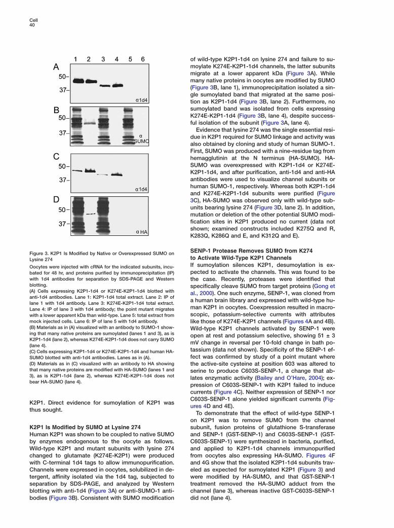
ENP-1 Protease Removes SUMO from K274o Activate Wild-Type K2P1 Channelsf sumoylation silences K2P1, desumoylation is ex-ected to activate the channels. This was found to behe case. Recently, proteases were identified thatpecifically cleave SUMO from target proteins (Gong etl., 2000). One such enzyme, SENP-1, was cloned fromhuman brain library and expressed with wild-type hu-an K2P1 in oocytes. Coexpression resulted in macro-
copic, potassium-selective currents with attributesike those of K274E-K2P1 channels (Figures 4A and 4B).
ild-type K2P1 channels activated by SENP-1 werepen at rest and potassium selective, showing 51 ± 3V change in reversal per 10-fold change in bath po-
assium (data not shown). Specificity of the SENP-1 ef-ect was confirmed by study of a point mutant wherehe active-site cysteine at position 603 was altered toerine to produce C603S-SENP-1, a change that ab-ates enzymatic activity (Bailey and O’Hare, 2004); ex-ression of C603S-SENP-1 with K2P1 failed to induceurrents (Figure 4C). Neither expression of SENP-1 nor603S-SENP-1 alone yielded significant currents (Fig-res 4D and 4E).To demonstrate that the effect of wild-type SENP-1
n K2P1 was to remove SUMO from the channelubunit, fusion proteins of glutathione S-transferasend SENP-1 (GST-SENP-1) and C603S-SENP-1 (GST-603S-SENP-1) were synthesized in bacteria, purified,nd applied to K2P1-1d4 channels immunopurifiedrom oocytes also expressing HA-SUMO. Figures 4Fnd 4G show that the isolated K2P1-1d4 subunits trav-led as expected for sumoylated K2P1 (Figure 3) andere modified by HA-SUMO, and that GST-SENP-1
reatment removed the HA-SUMO adduct from thehannel (lane 3), whereas inactive GST-C603S-SENP-1id not (lane 4).
SUMO Controls Plasma Membrane K2P1 Channel Activity41
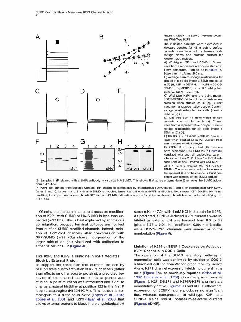
Figure 4. SENP-1, a SUMO Protease, Awak-ens Wild-Type K2P1
The indicated subunits were expressed inXenopus oocytes for 48 hr before surfacecurrents were recorded by two-electrodevoltage clamp and proteins purified forWestern blot analysis.(A) Wild-type K2P1 and SENP-1. Currenttrace from a representative oocyte studied in4 mM potassium. Protocol as in Figure 1A.Scale bars, 1 �A and 200 ms.(B) Average current-voltage relationships forgroups of six cells (mean ± SEM) studied asin (A) (■, K2P1 + SENP-1; $, K2P1 + C603S-SENP-1; B, SENP-1) or in 100 mM potas-sium (:, K2P1 + SENP-1).(C) Wild-type K2P1 and the point mutantC603S-SENP-1 fail to induce currents on ex-pression when studied as in (A). Currenttrace from a representative oocyte. Current-voltage relationship for six cells (mean ±SEM) in (B) ($).(D) Wild-type SENP-1 alone yields no newcurrents when studied as in (A). Currenttrace from a representative oocyte. Current-voltage relationship for six cells (mean ±SEM) in (C) (B).(E) C603S-SENP-1 alone yields no new cur-rents when studied as in (A). Current tracefrom a representative oocyte.(F) K2P1-1d4 immunopurified (IP) from oo-cytes expressing HA-SUMO (as in Figure 3C)visualized with anti-1d4 antibodies. Lane 1:total extract. Lane 2: IP of lane 1 with 1d4 anti-body. Lane 3: lane 2 treated with GST-SENP-1.Lane 4: lane 2 treated with GST-C603S-SENP-1. The active enzyme (lane 3) decreasesthe apparent kDa of the channel subunit con-sistent with removal of the SUMO adduct.
(G) Samples in (F) stained with anti-HA antibody to visualize HA-SUMO. This shows that active enzyme (lane 3) removes the SUMO adductfrom K2P1-1d4.(H) K2P1-1d4 purified from oocytes with anti-1d4 antibodies is modified by endogenous SUMO (lanes 1 and 3) or coexpressed GFP-SUMO(lanes 2 and 4). Lanes 1 and 2 with anti-SUMO antibodies; lanes 3 and 4 with anti-GFP antibodies. Not shown: K274E-K2P1-1d4 is notmodified; the upper band seen with anti-GFP and anti-SUMO antibodies in lanes 2 and 4 also stains with anti-1d4 antibodies identifying it asK2P1-1d4.
Of note, the increase in apparent mass on modifica-tion of K2P1 with SUMO or HA-SUMO is less than ex-pected (w12 kDa). This is best explained by anomalousgel migration, because terminal epitopes are not lostfrom purified SUMO-modified channels. Indeed, isola-tion of K2P1-1d4 channels after coexpression withGFP-SUMO (w35 kDa) shows incorporation of thelarger adduct on gels visualized with antibodies toeither SUMO or GFP (Figure 4H).
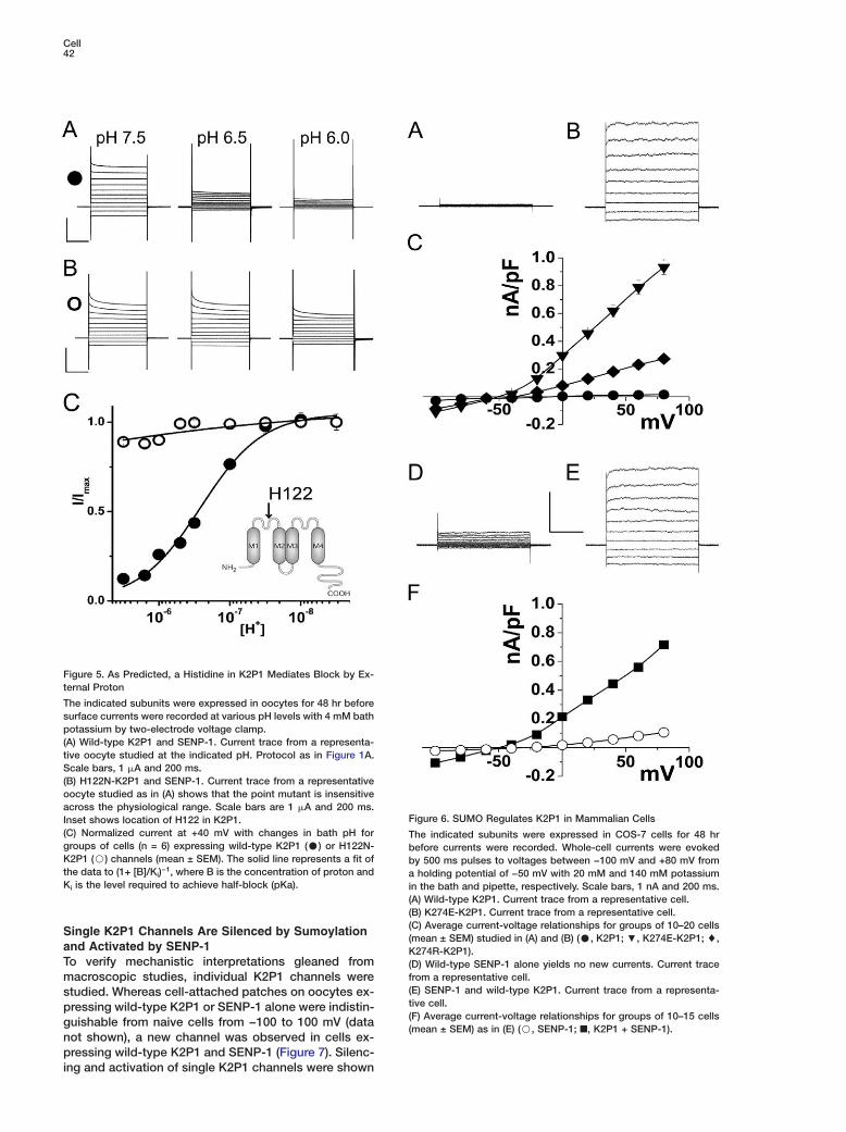
Like K2P3 and K2P9, a Histidine in K2P1 MediatesBlock by External ProtonTo support the conclusion that currents induced bySENP-1 were due to activation of K2P1 channels (ratherthan effects on other oocyte proteins), a predicted be-havior of the channel based on its sequence wasstudied. A point mutation was introduced into K2P1 tochange a natural histidine at position 122 in the first Ploop to asparagine (H122N-K2P1). This residue is ho-mologous to a histidine in K2P3 (Lopes et al., 2000;Lopes et al., 2001) and K2P9 (Rajan et al., 2000) thatallows external protons to block in the physiological pH
range (pKa w 7.24 with 4 mM KCl in the bath for K2P3).As predicted, SENP-1-induced K2P1 currents were in-hibited as external pH was lowered from 8.0 to 6.2(pKa = 6.67 ± 0.04, Hill coefficient 0.99, n = 6 cells),while H122N-K2P1 channels were insensitive to themanipulation (Figure 5).
Mutation of K274 or SENP-1 Coexpression ActivatesK2P1 Channels in COS-7 CellsThe operation of the SUMO regulatory pathway inmammalian cells was confirmed by studies of COS-7,a fibroblast cell line from African green monkey kidney.Alone, K2P1 channel expression yields no current in thecells (Figure 6A), as previously reported (Orias et al.,1997; Goldstein et al., 1998). Conversely, as in oocytes(Figure 1), K274E-K2P1 and K274R-K2P1 channels areconstitutively active (Figures 6B and 6C). Furthermore,expression of SENP-1 alone produces no significantflux, whereas coexpression of wild-type K2P1 andSENP-1 yields robust, potassium-selective currents(Figures 6D–6F).
Cell42
Figure 5. As Predicted, a Histidine in K2P1 Mediates Block by Ex-ternal Proton
The indicated subunits were expressed in oocytes for 48 hr beforesurface currents were recorded at various pH levels with 4 mM bathpotassium by two-electrode voltage clamp.(A) Wild-type K2P1 and SENP-1. Current trace from a representa-tive oocyte studied at the indicated pH. Protocol as in Figure 1A.Scale bars, 1 �A and 200 ms.(B) H122N-K2P1 and SENP-1. Current trace from a representativeoocyte studied as in (A) shows that the point mutant is insensitiveacross the physiological range. Scale bars are 1 �A and 200 ms.
FInset shows location of H122 in K2P1.(C) Normalized current at +40 mV with changes in bath pH for Tgroups of cells (n = 6) expressing wild-type K2P1 (C) or H122N- bK2P1 (B) channels (mean ± SEM). The solid line represents a fit of bthe data to (1+ [B]/Ki)−1, where B is the concentration of proton and aKi is the level required to achieve half-block (pKa). i
((
igure 6. SUMO Regulates K2P1 in Mammalian Cells
he indicated subunits were expressed in COS-7 cells for 48 hrefore currents were recorded. Whole-cell currents were evokedy 500 ms pulses to voltages between −100 mV and +80 mV fromholding potential of −50 mV with 20 mM and 140 mM potassium
n the bath and pipette, respectively. Scale bars, 1 nA and 200 ms.A) Wild-type K2P1. Current trace from a representative cell.B) K274E-K2P1. Current trace from a representative cell.
(C) Average current-voltage relationships for groups of 10–20 cells(mean ± SEM) studied in (A) and (B) (C, K2P1; ;, K274E-K2P1; %,K274R-K2P1).(D) Wild-type SENP-1 alone yields no new currents. Current tracefrom a representative cell.(E) SENP-1 and wild-type K2P1. Current trace from a representa-tive cell.(F) Average current-voltage relationships for groups of 10–15 cells(mean ± SEM) as in (E) (B, SENP-1; ■, K2P1 + SENP-1).
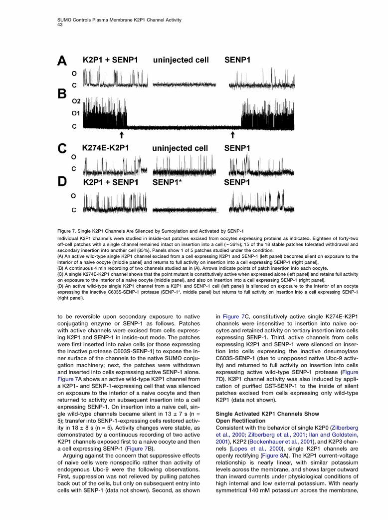
Single K2P1 Channels Are Silenced by Sumoylationand Activated by SENP-1To verify mechanistic interpretations gleaned frommacroscopic studies, individual K2P1 channels werestudied. Whereas cell-attached patches on oocytes ex-pressing wild-type K2P1 or SENP-1 alone were indistin-guishable from naive cells from −100 to 100 mV (datanot shown), a new channel was observed in cells ex-pressing wild-type K2P1 and SENP-1 (Figure 7). Silenc-ing and activation of single K2P1 channels were shown
SUMO Controls Plasma Membrane K2P1 Channel Activity43
Figure 7. Single K2P1 Channels Are Silenced by Sumoylation and Activated by SENP-1
Individual K2P1 channels were studied in inside-out patches excised from oocytes expressing proteins as indicated. Eighteen of forty-twooff-cell patches with a single channel remained intact on insertion into a cell (w36%); 15 of the 18 stable patches tolerated withdrawal andsecondary insertion into another cell (85%). Panels show 1 of 5 patches studied under the condition.(A) An active wild-type single K2P1 channel excised from a cell expressing K2P1 and SENP-1 (left panel) becomes silent on exposure to theinterior of a naive oocyte (middle panel) and returns to full activity on insertion into a cell expressing SENP-1 (right panel).(B) A continuous 4 min recording of two channels studied as in (A). Arrows indicate points of patch insertion into each oocyte.(C) A single K274E-K2P1 channel shows that the point mutant is constitutively active when expressed alone (left panel) and retains full activityon exposure to the interior of a naive oocyte (middle panel), and also on insertion into a cell expressing SENP-1 (right panel).(D) An active wild-type single K2P1 channel from a K2P1 and SENP-1 cell (left panel) is silenced on exposure to the interior of an oocyteexpressing the inactive C603S-SENP-1 protease (SENP-1*, middle panel) but returns to full activity on insertion into a cell expressing SENP-1(right panel).
to be reversible upon secondary exposure to nativeconjugating enzyme or SENP-1 as follows. Patcheswith active channels were excised from cells express-ing K2P1 and SENP-1 in inside-out mode. The patcheswere first inserted into naive cells (or those expressingthe inactive protease C603S-SENP-1) to expose the in-ner surface of the channels to the native SUMO conju-gation machinery; next, the patches were withdrawnand inserted into cells expressing active SENP-1 alone.Figure 7A shows an active wild-type K2P1 channel froma K2P1- and SENP-1-expressing cell that was silencedon exposure to the interior of a naive oocyte and thenreturned to activity on subsequent insertion into a cellexpressing SENP-1. On insertion into a naive cell, sin-gle wild-type channels became silent in 13 ± 7 s (n =5); transfer into SENP-1-expressing cells restored activ-ity in 18 ± 8 s (n = 5). Activity changes were stable, asdemonstrated by a continuous recording of two activeK2P1 channels exposed first to a naive oocyte and thena cell expressing SENP-1 (Figure 7B).
Arguing against the concern that suppressive effectsof naive cells were nonspecific rather than activity ofendogenous Ubc-9 were the following observations.First, suppression was not relieved by pulling patchesback out of the cells, but only on subsequent entry intocells with SENP-1 (data not shown). Second, as shown
in Figure 7C, constitutively active single K274E-K2P1channels were insensitive to insertion into naive oo-cytes and retained activity on tertiary insertion into cellsexpressing SENP-1. Third, active channels from cellsexpressing K2P1 and SENP-1 were silenced on inser-tion into cells expressing the inactive desumoylaseC603S-SENP-1 (due to unopposed native Ubc-9 activ-ity) and returned to full activity on insertion into cellsexpressing active wild-type SENP-1 protease (Figure7D). K2P1 channel activity was also induced by appli-cation of purified GST-SENP-1 to the inside of silentpatches excised from cells expressing only wild-typeK2P1 (data not shown).
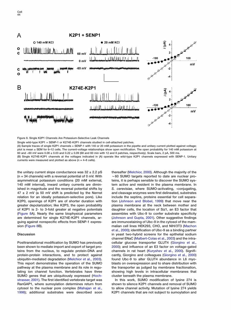
Single Activated K2P1 Channels ShowOpen RectificationConsistent with the behavior of single K2P0 (Zilberberget al., 2000; Zilberberg et al., 2001; Ilan and Goldstein,2001), K2P2 (Bockenhauer et al., 2001), and K2P3 chan-nels (Lopes et al., 2000), single K2P1 channels areopenly rectifying (Figure 8A). The K2P1 current-voltagerelationship is nearly linear, with similar potassiumlevels across the membrane, and shows larger outwardthan inward currents under physiological conditions ofhigh internal and low external potassium. With nearlysymmetrical 140 mM potassium across the membrane,
Cell44
Figure 8. Single K2P1 Channels Are Potassium-Selective Leak Channels
Single wild-type K2P1 + SENP-1 or K274E-K2P1 channels studied in cell-attached patches.(A) Sample traces of single K2P1 channels + SENP-1 with 140 or 20 mM potassium in the pipette and unitary current plotted against voltage;plot is mean ± SEM for 6–12 cells. The current-voltage relationships show open rectification. The open probability for 140 mM potassium at60 and −60 mV were 0.08 ± 0.03 and 0.22 ± 0.09 (68 and 60 min with 12 and 8 patches, respectively). Scale bars, 2 pA, 500 ms.(B) Single K274E-K2P1 channels at the voltages indicated in (A) operate like wild-type K2P1 channels expressed with SENP-1. Unitarycurrents were measured and plotted as above (n = 4–8 cells).
the unitary current slope conductance was 32 ± 2.2 pS tw(n = 34 channels) with a reversal potential of 0 mV. With
asymmetrical potassium conditions (20 mM external, tt140 mM internal), inward unitary currents are dimin-
ished in magnitude and the reversal potential shifts by Sa47 ± 2 mV (a 50 mV shift is predicted by the Nernst
relation for an ideally potassium-selective pore). Like itK2P0, openings of K2P1 are of shorter duration with
greater depolarization; like K2P3, the open probability pdof K2P1 is 2- to 3-fold greater at negative potentials
(Figure 8A). Nearly the same biophysical parameters a(are determined for single K274E-K2P1 channels, ar-
guing against nonspecific effects from SENP-1 expres- amsion (Figure 8B).eiDiscussionccPosttranslational modification by SUMO has previously
been shown to mediate import and export of target pro- 2cteins from the nucleus, to regulate protein-DNA and
protein-protein interactions, and to protect against cfubiquitin-mediated degradation (Melchior et al., 2003).
This report demonstrates the operation of the SUMO btpathway at the plasma membrane and its role in regu-
lating ion channel function. Vertebrates have three scSUMO genes that are ubiquitously expressed (Hoch-
strasser, 2001). The first identified vertebrate target wassRanGAP1, where sumoylation determines return from
cytosol to the nuclear pore complex (Mahajan et al., tK1998); additional substrates were described soon
hereafter (Melchior, 2000). Although the majority of the60 SUMO targets reported to date are nuclear pro-
eins, it is perhaps sensible to discover the SUMO sys-em active and resident in the plasma membrane. In. cerevisiae, where SUMO-activating, -conjugating,nd cleavage enzymes were first delineated, substrates
nclude the septins, proteins essential for cell separa-ion (Johnson and Blobel, 1999) that move near thelasma membrane at the neck between mother andaughter cells, the location of Siz1, an E3 factor thatssembles with Ubc-9 to confer substrate specificity
Johnson and Gupta, 2001). Other suggestive findingsre immunostaining of Ubc-9 in the cytosol of the mam-alian cell lines HEK293, CHO, and NIH/3T3 (Machon
t al., 2000); identification of Ubc-9 as a binding partnern yeast two-hybrid screens for the epithelial sodiumhannel ENaC (Malbert-Colas et al., 2003) and the intra-ellular glucose transporter GLUT4 (Giorgino et al.,000); and influence of an E3 factor on voltage-gatedhannels in rat heart (Kuryshev et al., 2000). Signifi-antly, Giorgino and colleagues (Giorgino et al., 2000)ound Ubc-9 to alter GLUT4 abundance in L6 myo-lasts on overexpression and to share distribution with
he transporter as judged by membrane fractionation,howing high levels in intracellular membranes thatluster beneath the plasma membrane.In this work, SUMO modification of lysine 274 is
hown to silence K2P1 channels and removal of SUMOo allow channel activity. Mutation of lysine 274 yields2P1 channels that are not subject to sumoylation and
SUMO Controls Plasma Membrane K2P1 Channel Activity45
are thus constitutively active. Single-channel studiesreveal that sumoylation is readily reversible and pro-ceeds in the plasma membrane. Native SUMO-conju-gating enzyme (Ubc-9) is shown to be at the plasmamembrane in a uniform manner in naive oocytes butto redistribute in polarized fashion with K2P1 on theircoexpression. K2P1 is demonstrated to be decoratedwith native SUMO by endogenous conjugating enzymeunder basal conditions and SUMO protease shown toremove the peptide adduct. Silence of surface K2P1channels (in the absence of overexpressed SENP-1protease) can therefore be understood to result fromoperation of the native SUMO conjugation machineryin oocytes (Saitoh et al., 1998) and mammalian cells(Machon et al., 2000). In contrast, other SUMO targetsstudied in experimental cells show a small fraction oftheir population to be sumoylated at baseline; this sug-gests existence of a pathway to regulate K2P1 desu-moylation in cells where the channel is active in vivo.
Delineating the attributes of active K2P1 channelshas resolved an outstanding controversy as to theirnature. K2P1 channels are classical potassium-selec-tive, openly rectifying background channels that showTASK-like acid sensitivity (that is, blockade via proton-ation of a pore-located histidine). Thus, K2P1 currentsare greater in the outward direction under physiologicalconditions of high internal and low external potassiumand of similar magnitude in both directions under sym-metrical conditions. Active K2P1 single channels shownominal voltage dependence in their open probability(Figure 7A), a subject of ongoing study. The initial no-tion that K2P1 channels were weak inward rectifiers(TWIKs) was based on observation of outward currentsthat decreased at positive potentials when the genewas expressed in oocytes (Lesage et al., 1996); this be-havior is not seen with active K2P1 channels (Figures1F, 4B, and 7), suggesting that one or more native con-ductances contributed to the currents under study inthat work.
The sumoylation site in K2P1, -LK274KF-, is a variantof the classical -ψKxE/D- motif, where ψ is an aliphaticresidue (Rodriguez et al., 2001; Melchior et al., 2003).This is not unexpected, as other variant sites have beendemonstrated (i.e., -VK*YC- and -TK*ET- in the nuclearfactors smad4 and PCNA, respectively). This suggeststhat motif variation will not differentiate acceptors su-moylated at the plasma membrane from other targets.While other potential SUMO modification sites are pre-sent in K2P1, our findings indicate that only K274 issubject to modification in oocytes. The motif and ho-mologous lysine are present in all reported K2P1 clones(e.g., human, chimpanzee, fowl, guinea pig, mouse,rabbit, and rat).
A more complete biophysical characterization ofK2P1 should help advance the process of determiningthe contribution of individual K2P channels to nativecurrents. K2P1 is seen here to share sensitivity to pro-tons via the same mechanism as K2P3 and K2P9, twochannels thought to contribute to the standing outwardcurrent IKso that is sensitive to hypoxia and extracellularacidosis and key to activity of cerebellar granular neu-rons, CGN (Plant et al., 2002). Our findings supportspeculation that K2P1 may contribute to IKso, given thatK2P1 mRNA is abundantly expressed in rat brain with
highest levels in CGN (Talley et al., 2001). Moreover,Han and colleagues (Han et al., 2002) observed fourchannels in rat CGN that they attributed to K2P3, K2P9,K2P10, and a “type 4” current unlike any known K2Pchannel. The attributes of type 4 channels are quitesimilar to those we now can assign to active K2P1channels (including a unitary conductance of 32 ± 2 pSunder similar conditions, activity at all potentials, po-tassium selectivity, and block by external proton in thephysiological pH range). Native currents in heart andkidney that correlate with K2P1 are more uncertain.K2P1 shares w40% protein sequence identity withK2P6 and just w20% with K2P3 and K2P9, althoughthe latter carry the homologous histidine in the first Ploop that confers sensitivity to lowered external pH.
Control of K2P1 via sumoylation adds reversible, co-valent peptide linkage to the plethora of posttransla-tional modifications recognized to regulate ion channelfunction. The findings raise intriguing questions. Tightregulation of K2P channel activity is a hallmark of theseportals that control cellular excitability (Goldstein et al.,2001). Thus, as-yet unknown pathways that controlK2P1 sumoylation will modulate cellular excitability.SUMO influences some targets secondarily via sup-pression of ubiquitination or changes in binding to areceptor (Hochstrasser, 2001). Is this also the case forK2P1, or does SUMO directly alter channel function asdo many other ion channel accessory subunits? Addi-tional plasma membrane proteins are SUMO modified(data not shown); how this influences their operation isunknown. Indeed, sumoylation may prove to explainthe silence of other ion channels that have yet to revealtheir function.
Experimental Procedures
Molecular BiologyHuman K2P1 (accession number NM_002245) was subcloned intopRAT, a dual-purpose expression vector (Bockenhauer et al., 2001)containing a CMV promoter for mammalian expression and a T7promoter for cRNA synthesis. Human SUMO-1 (accession numberBCOO5899), Ubc-9 (accession number BC004429), and SENP-1(accession number BC045639) were amplified from a brain cDNAlibrary (Clontech, Palo Alto, California) and inserted into pCR IITOPO (Invitrogen, Carlsbad, California) at XhoI and BamHI in thecase of SUMO-1 and XbaI and HindIII in the case of Ubc-9 andSENP-1, and subcloned into pMAX, a version of pRAT with an al-tered MCS. pEGFP was amplified from pEGFPC3 (Clontech) andligated to the N terminus of K2P1 with EcoRI and PstI. EGFP wasinserted in SUMO-1 between XbaI and HindIII. Mutagenesis wascarried out with a QuikChange Site-Directed Mutagenesis Kit (Stra-tagene, La Jolla, California). 1d4 epitope (RVPDGDPDETSQVAPA)was introduced into K2P1 by mutating the stop codon to create anMluI site that was used to insert the epitope. cRNA was synthe-sized with an mMESSAGE mMACHINE kit after vector linearization(Ambion, Austin, Texas). HA-SUMO was produced by amplifyingwith a forward primer containing the code for the HA epitope(YPYDVPDYA) before the start codon. GST-SENP-1 was con-structed by inserting SENP-1 between BamHI and SalI sites inpGEX6P1. GST-C603S-SENP-1 was produced by site-directed mu-tagenesis.Yeast Two-Hybrid AnalysisYeast strain HF7C was transformed with K2P1 C terminus (residues260–336) in the vector pGBT-9 and Ubc-9 in pGAD-424. Trans-formants were plated on plates lacking tryptophan, leucine, andhistidine. Red colonies appeared after 4 days with a positive in-teraction. The interaction was confirmed by a qualitative β-galacto-sidase assay (Clontech).
Cell46
Biochemistry rmcRNA encoding study proteins was injected in 250 Xenopus laevis
oocytes. Extracts were obtained by grinding cells in lysis buffercontaining (in mM) 100 NaCl, 40 KCl, 20 NEM, 1 EDTA, 20 HEPES-KOH (pH 7.4), 10% glycerol, 1% Triton X-100, and complete prote- Aase inhibitor tablets (Roche Applied Science, Indianapolis, Indiana).The SUMO adduct is sensitive to proteolysis in the absence of NEM. T1d4 antibody (NCCC, Minneapolis) was used as before (Kim et al., C2004). Oocyte extract was incubated with 1d4-coated Sepharose Sbeads for 2 hr at 4°C. Unbound material was removed with washingbuffer containing (in mM) 300 NaCl, 40 KCl, 1 EDTA, 20 HEPES- RKOH (pH 7.4), 0.5% Triton X-100, and protease inhibitor tablets. RBound proteins were eluted with 1d4 peptide (1 mg/ml). Eluates Awere subjected to SDS-PAGE analysis and Western blotting with Panti-1d4 mAb (1:2000, NCCC), anti-SUMO mAb (1:200, 18-2306,Zymed Laboratories, San Francisco), anti-HA mAb (1:1000, R1867423, Roche Applied Science) or anti-GFP pAb (1:3000, R970-01, Invitrogen). The antibody to human SUMO-1 recognizes Xeno- Bpus SUMO-1, as the two are 92% identical. GST-SENP-1 was pro- aduced in bacterial strain BL21(DE3). SUMO cleavage from K2P1 Bwas carried out by incubation with GST-SENP-1 by incubation for
B60 min at 30°C in (in mM) 150 NaCl, 1 DTT, and 10 Tris-HCl (pH 7.4),(as by others (Nishida et al., 2001).va
ImmunofluorescenceBOocytes were fixed in 1% paraformaldehyde in ND-91 (in mM: 91SNaCl, 2 KCl, 1 MgCl2, 1.8 CaCl2, 5 HEPES [pH 7.5] with NaOH) form1 hr. Oocytes were rinsed in PBS and blocked with 10% goat se-grum, 0.5% BSA, 0.1% Triton X-100 in PBS for 45 min followed bysincubation with primary antibody in the blocking solution for 2 hrB(1d4 antibody 1:200 or Ubc-9 antibody, 610749, BD Pharmingen,KSan Diego, California, 1:20). After thorough washing with PBS con-itaining 0.1% Triton X-100, oocytes were incubated with secondary
antibody for 30 min (1:100, anti-mouse FITC or Texas red, Jackson CaLabs, Bar Harbor, Maine) and then washed for study.B4ElectrophysiologyGTwo-Electrode Voltage ClampMDefolliculated oocytes were injected with 15 ng K2P1 cRNA with orewithout 5 ng SENP-1 cRNA. Whole-cell currents were measuredt(Geneclamp 500, Axon Instruments, Union City, California) 48–72 hrNafter injection. Electrodes were filled with 3 M KCl and had resis-
tances of 0.1–1 M�. Data were sampled at 5 kHz and recorded Gusing Clampex software. Data analysis was performed using mClampfit 9.0, Excel, and Origin 6.0. Standard bath solution was (in GmM) 96 NaCl, 4 KCl, 1 MgCl2, 1.8 CaCl2, 5 HEPES (pH 7.5) with LNaOH. Potassium levels were altered by isotonic substitution of IKCl for NaCl. tSingle-Channel Recording ISingle channels were assessed in cell-attached or excised inside-
Gout patches from oocytes after removal of vitelline by hypotonic(stripping. Bath and pipette solutions were (in mM) 140 KCl, 0.3dCaCl2, 1 MgCl2, 5 HEPES (pH 7.4) with KOH. Potassium levels wereSaltered by isotonic substitution of KCl for NaCl. Data were recorded1with an Axopatch 200B amplifier using pCLAMP software (AxonGInstruments, Claremont, California) at filter and sampling fre-qquencies of 2 kHz and 10 kHz, respectively. Data were filtered at 1pkHz for off-line analysis using Clampfit 9.0. Open probabilities were
determined in patches judged to contain only one channel. Events Gwere detected using a half-amplitude threshold technique (Col- (quhoun and Sigworth, 1995). All experiments were performed at d20°C–22°C. GWhole-Cell Recording aCOS-7 (ATCC, Manassas, Virginia) were transfected with plasmids tcarrying study genes and EGFP with lipofectamine (Invitrogen) as
Gbefore (Bockenhauer et al., 2001). After 24 to 48 hr, currents were
rassessed by whole-cell patch-clamp recording using an Axopatch
a200B amplifier at filter and sampling frequencies of 2 kHz and 10
HkHz, respectively. Borosilicate glass pipettes were filled with a solu-ttion of (in mM) 140 KCl, 0.5 CaCl2, 5 EGTA, 10 HEPES (pH 7.4) withlKOH and had resistance of 3–5 M�. Cells were bathed in (in mM)
120 NaCl, 20 KCl, 0.3 CaCl2, 1 MgCl2, 10 HEPES (pH 7.4) with HfNaOH. Whole-cell capacitance did not differ between groups. Cur-
ents are normalized to capacitance for each cell and expressed asean ± SEM.
cknowledgments
his was supported by grants from the NIH and the Doris Dukeharitable Foundation (DDCF) to S.A.N.G. (a DDCF Clinicalcholar). We thank D. Goldstein for thoughtful discussions.
eceived: October 6, 2004evised: January 19, 2005ccepted: January 21, 2005ublished: April 7, 2005
eferences
ailey, D., and O’Hare, P. (2004). Characterization of the localizationnd proteolytic activity of the SUMO-specific protease, SENP1. J.iol. Chem. 279, 692–703.
ernier-Villamor, V., Sampson, D.A., Matunis, M.J., and Lima, C.D.2002). Structural basis for E2-mediated SUMO conjugation re-ealed by a complex between ubiquitin-conjugating enzyme Ubc9nd RanGAP1. Cell 108, 345–356.
ockenhauer, D., Nimmakayalu, M.A., Ward, D.C., Goldstein,.A.N., and Gallagher, P.G. (2000). Genomic organization and chro-osomal localization of the murine 2 P domain potassium channelene Kcnk8: conservation of gene structure in 2 P domain potas-ium channels. Gene 261, 365–372.
ockenhauer, D., Zilberberg, N., and Goldstein, S.A.N. (2001).CNK2: reversible conversion of a hippocampal potassium leak
nto a voltage-dependent channel. Nat. Neurosci. 4, 486–491.
olquhoun, D., and Sigworth, F.J. (1995). Fitting and statisticalnalysis of single-channel records. In Single-Channel Recording,. Sakmann and E. Neher, eds. (New York: Plenum Press), pp.83–588.
iorgino, F., de Robertis, O., Laviola, L., Montrone, C., Perrini, S.,cCowen, K.C., and Smith, R.J. (2000). The sentrin-conjugating
nzyme mUbc9 interacts with GLUT4 and GLUT1 glucose transpor-ers and regulates transporter levels in skeletal muscle cells. Proc.atl. Acad. Sci. USA 97, 1125–1130.
oldman, D.E. (1943). Potential, impedance, and rectification inembranes. J. Gen. Physiol. 27, 37–60.
oldstein, S.A.N., Bayliss, D.A., Desir, G.V., Kim, D., Lazdunski, M.,esage, F., and O’Kelly, I. (2002). Potassium Channels: K2P. In The
UPHAR Compendium of Voltage-Gated Ion Channels, W.A. Cat-eral, K.G. Chandy, and G.A. Gutman, eds. (Leeds, United Kingdom:UPHAR Media), pp. 173–189.
oldstein, S.A.N., Price, L.A., Rosenthal, D.N., and Pausch, M.H.1996). ORK1, a potassium-selective leak channel with two poreomains cloned from Drosophila melanogaster by expression inaccharomyces cerevisiae. Proc. Natl. Acad. Sci. USA 93, 13256–3261.
oldstein, S.A.N., Wang, K.W., Ilan, N., and Pausch, M. (1998). Se-uence and function of the two P domain potassium channels: im-lications of an emerging superfamily. J. Mol. Med. 76, 13–20.
oldstein, S.A.N., Bockenhauer, D., O’Kelly, I., and Zilberberg, N.2001). Potassium leak channels and the KCNK family of two-P-omain subunits. Nat. Rev. Neurosci. 2, 175–184.
ong, L., Li, B., Millas, S., and Yeh, E.T. (1999). Molecular cloningnd characterization of human AOS1 and UBA2, components ofhe sentrin-activating enzyme complex. FEBS Lett. 448, 185–189.
ong, L., Millas, S., Maul, G.G., and Yeh, E.T. (2000). Differentialegulation of sentrinized proteins by a novel sentrin-specific prote-se. J. Biol. Chem. 275, 3355–3359.
an, J., Truell, J., Gnatenco, C., and Kim, D. (2002). Characteriza-ion of four types of background potassium channels in rat cerebel-ar granule neurons. J. Physiol. 542, 431–444.
ochstrasser, M. (2001). SP-RING for SUMO: new functions bloomor a ubiquitin-like protein. Cell 107, 5–8.
SUMO Controls Plasma Membrane K2P1 Channel Activity47
Hodgkin, A.L., and Huxley, A.F. (1952). A quantitative description ofmembrane current and its application to conduction and excitationin nerve. J. Physiol. 117, 500–544.
Hodgkin, A.L., and Katz, B. (1949). The effect of sodium ions on theelectrical activity of the giant axon of the squid. J. Physiol. 108,37–77.
Ilan, N., and Goldstein, S.A.N. (2001). KCNK0: single, cloned potas-sium leak channels are multi-ion pores. Biophys. J. 80, 241–254.
Johnson, E.S., and Blobel, G. (1999). Cell cycle-regulated attach-ment of the ubiquitin-related protein SUMO to the yeast septins. J.Cell Biol. 147, 981–994.
Johnson, E.S., and Gupta, A.A. (2001). An E3-like factor that pro-motes SUMO conjugation to the yeast septins. Cell 106, 735–744.
Ketchum, K.A., Joiner, W.J., Sellers, A.J., Kaczmarek, L.K., andGoldstein, S.A.N. (1995). A new family of outwardly-rectifying po-tassium channel proteins with two pore domains in tandem. Nature376, 690–695.
Kim, Y., Bang, H., and Kim, D. (1999). TBAK-1 and TASK-1, two-pore K(+) channel subunits: kinetic properties and expression in ratheart. Am. J. Physiol. 277, H1669–H1678.
Kim, L.A., Furst, J., Gutierrez, D., Butler, M.H., Xu, S., Goldstein,S.A., and Grigorieff, N. (2004). Three-dimensional structure of I(to);Kv4.2-KChIP2 ion channels by electron microscopy at 21 Angstromresolution. Neuron 41, 513–519.
Kuryshev, Y.A., Gudz, T.I., Brown, A.M., and Wible, B.A. (2000).KChAP as a chaperone for specific K+ channels. Am. J. Physiol.Cell Physiol. 278, C931–C941.
Lesage, F., Guillemare, E., Fink, M., Duprat, F., Lazdunski, M., Ro-mey, G., and Barhanin, J. (1996). TWIK-1, a ubiquitous humanweakly inward rectifying K+ channel with a novel structure. EMBOJ. 15, 1004–1011.
Levy, D.I., Velazquez, H., Goldstein, S.A., and Bockenhauer, D.(2004). Segment-specific expression of 2P domain potassiumchannel genes in human nephron. Kidney Int. 65, 918–926.
Li, S.J., and Hochstrasser, M. (2003). The Ulp1 SUMO isopeptidase:distinct domains required for viability, nuclear envelope localiza-tion, and substrate specificity. J. Cell Biol. 160, 1069–1081.
Lopes, C.M.B., Gallagher, P.G., Buck, M.E., Butler, M.H., andGoldstein, S.A.N. (2000). Proton block and voltage-gating are po-tassium-dependent in the cardiac leak channel Kcnk3. J. Biol.Chem. 275, 16969–16978.
Lopes, C.M.B., Zilberberg, N., and Goldstein, S.A.N. (2001). Blockof Kcnk3 by protons: evidence that 2-P-domain potassium channelsubunits function as homodimers. J. Biol. Chem. 276, 24449–24452.
Machon, O., Backman, M., Julin, K., and Krauss, S. (2000). Yeasttwo-hybrid system identifies the ubiquitin-conjugating enzymemUbc9 as a potential partner of mouse Dac. Mech. Dev. 97, 3–12.
Mahajan, R., Gerace, L., and Melchior, F. (1998). Molecular charac-terization of the SUMO-1 modification of RanGAP1 and its role innuclear envelope association. J. Cell Biol. 140, 259–270.
Malbert-Colas, L., Nicolas, G., Galand, C., Lecomte, M.C., andDhermy, D. (2003). Identification of new partners of the epithelialsodium channel alpha subunit. C. R. Biol. 326, 615–624.
Melchior, F. (2000). SUMO–nonclassical ubiquitin. Annu. Rev. CellDev. Biol. 16, 591–626.
Melchior, F., Schergaut, M., and Pichler, A. (2003). SUMO: ligases,isopeptidases and nuclear pores. Trends Biochem. Sci. 28, 612–618.
Muller, S., Hoege, C., Pyrowolakis, G., and Jentsch, S. (2001).SUMO, ubiquitin’s mysterious cousin. Nat. Rev. Mol. Cell Biol. 2,202–210.
Nishida, T., Kaneko, F., Kitagawa, M., and Yasuda, H. (2001). Char-acterization of a novel mammalian SUMO-1/Smt3-specific isopep-tidase, a homologue of rat axam, which is an axin-binding proteinpromoting beta-catenin degradation. J. Biol. Chem. 276, 39060–39066.
O’Kelly, I., Butler, M.H., Zilberberg, N., and Goldstein, S.A. (2002).
Forward transport. 14-3-3 binding overcomes retention in endo-plasmic reticulum by dibasic signals. Cell 111, 577–588.
Orias, M., Velazquez, H., Tung, F., Lee, G., and Desir, G.V. (1997).Cloning and localization of a double-pore K channel, KCNK1: ex-clusive expression in distal nephron segments. Am. J. Physiol. 273,F663–F666.
Plant, L.D., Kemp, P.J., Peers, C., Henderson, Z., and Pearson, H.A.(2002). Hypoxic depolarization of cerebellar granule neurons byspecific inhibition of TASK-1. Stroke 33, 2324–2328.
Rajan, S., Wischmeyer, E., Liu, G.X., Muller, R.P., Daut, J., Karschin,A., and Derst, C. (2000). TASK-3, a novel tandem pore domain acid-sensitive K+ channel. An extracellular histidine as pH sensor. J.Biol. Chem. 275, 16650–16657.
Rajan, S., Wischmeyer, E., Karschin, C., Preisig-Muller, R.,Grzeschik, K.H., Daut, J., Karschin, A., and Derst, C. (2001). THIK-1and THIK-2, a novel subfamily of tandem pore domain K+ channels.J. Biol. Chem. 276, 7302–7311.
Rajan, S., Preisig-Muller, R., Wischmeyer, E., Nehring, R., Hanley,P.J., Renigunta, V., Musset, B., Schlichthorl, G., Derst, C., Karschin,A., and Daut, J. (2002). Interaction with 14-3-3 proteins promotesfunctional expression of the potassium channels TASK-1 andTASK-3. J. Physiol. 545, 13–26.
Rodriguez, M.S., Dargemont, C., and Hay, R.T. (2001). SUMO-1conjugation in vivo requires both a consensus modification motifand nuclear targeting. J. Biol. Chem. 276, 12654–12659.
Saitoh, H., Sparrow, D.B., Shiomi, T., Pu, R.T., Nishimoto, T., Mohun,T.J., and Dasso, M. (1998). Ubc9p and the conjugation of SUMO-1to RanGAP1 and RanBP2. Curr. Biol. 8, 121–124.
Salinas, M., Reyes, R., Lesage, F., Fosset, M., Heurteaux, C., Ro-mey, G., and Lazdunski, M. (1999). Cloning of a new mouse two-Pdomain channel subunit and a human homologue with a uniquepore structure. J. Biol. Chem. 274, 11751–11760.
Sirois, J.E., Lei, Q., Talley, E.M., Lynch, C., 3rd, and Bayliss, D.A.(2000). The TASK-1 two-pore domain K+ channel is a molecularsubstrate for neuronal effects of inhalation anesthetics. J. Neurosci.20, 6347–6354.
Talley, E.M., Solorzano, G., Lei, Q., Kim, D., and Bayliss, D.A. (2001).Cns distribution of members of the two-pore-domain (KCNK) po-tassium channel family. J. Neurosci. 21, 7491–7505.
Ubbels, G.A. (1997). Establishment of polarities in the oocyte ofXenopus laevis: the provisional axial symmetry of the full-grownoocyte of Xenopus laevis. Cell. Mol. Life Sci. 53, 382–409.
Wang, Z., Yue, L., White, M., Pelletier, G., and Nattel, S. (1998).Differential distribution of inward rectifier potassium channel tran-scripts in human atrium versus ventricle. Circulation 98, 2422–2428.
Washburn, C.P., Sirois, J.E., Talley, E.M., Guyenet, P.G., and Bayliss,D.A. (2002). Serotonergic raphe neurons express TASK channeltranscripts and a TASK-like pH- and halothane-sensitive K+ con-ductance. J. Neurosci. 22, 1256–1265.
Zilberberg, N., Ilan, N., Gonzalez-Colaso, R., and Goldstein, S.A.N.(2000). Opening and closing of KCNK0 potassium leak channels istightly regulated. J. Gen. Physiol. 116, 721–734.
Zilberberg, N., Ilan, N., and Goldstein, S.A. (2001). KCNKO: openingand closing the 2-P-domain potassium leak channel entails“C-type” gating of the outer pore. Neuron 32, 635–648.


