Embed Size (px)
Citation preview
Medical Hypotheses (2004) 63, 8–20
http://intl.elsevierhealth.com/journals/mehy
A “mitochondrial cascade hypothesis” for sporadicAlzheimer’s disease
Russell H. Swerdlowa,b,*, Shaharyar M. Khanb,c,d
a Department of Neurology, McKim Hall, University of Virginia Health System, P.O. Box 800394,1 Hospital Drive, Charlottesville, VA 22908, USAb Center for the Study of the Neurodegenerative Diseases, University of Virginia Health System,Charlottesville, VA 22908, USAc Department of Neuroscience, University of Virginia Health System, Charlottesville, VA 22908, USAd Gencia Corporation, 706 D Forrest Street, Charlottesville, VA 22903, USA
Received 14 October 2003; accepted 30 December 2003
Summary Alzheimer’s disease (AD) includes etiologically heterogenous disorders characterized by senile orpresenile dementia, extracellular amyloid protein aggregations containing an insoluble amyloid precursor proteinderivative, and intracytoplasmic tau protein aggregations. Recent studies also show excess neuronal aneuploidy,programmed cell death (PCD), and mitochondrial dysfunction. The leading AD molecular paradigm, the “amyloidcascade hypothesis”, is based on studies of rare autosomal dominant variants and does not specify what initiatesthe common late-onset, sporadic form. We propose for late-onset, sporadic AD a “mitochondrial cascadehypothesis” that comprehensively reconciles seemingly disparate histopathologic and pathophysiologic features. Inour model, the inherited, gene-determined make-up of an individual’s electron transport chain sets basal rates ofreactive oxygen species (ROS) production, which determines the pace at which acquired mitochondrial damageaccumulates. Oxidative mitochondrial DNA, RNA, lipid, and protein damage amplifies ROS production and triggersthree events: (1) a reset response in which cells respond to elevated ROS by generating the b-sheet protein, betaamyloid, which further perturbs mitochondrial function, (2) a removal response in which compromised cells arepurged via PCD mechanisms, and (3) a replace response in which neuronal progenitors unsuccessfully attempt to re-enter the cell cycle, with resultant aneuploidy, tau phosphorylation, and neurofibrillary tangle formation. Inaddition to defining a role for aging in AD pathogenesis, the mitochondrial cascade hypothesis also allows andaccounts for histopathologic overlap between the sporadic, late-onset and autosomal dominant, early onset formsof the disease.
�c 2004 Elsevier Ltd. All rights reserved.
Introduction
As described by Alois Alzheimer in 1906 andnamed by Emil Kraepelin in 1910, Alzheimer’s
* Corresponding author. Tel.: +1-434-924-5785; fax: +1-434-982-1726.
E-mail address: [email protected] (R.H. Swerdlow).
0306-9877/$ - see front matter �c 2004 Elsevier Ltd. All rights reserdoi:10.1016/j.mehy.2003.12.045
disease (AD) applied to a state of presenile de-mentia, extraneuronal protein aggregations (pla-ques), and intraneuronal protein aggregations(tangles) [1,2]. Although it was recognized at thetime that brains of persons with senile dementiacould also manifest plaques and tangles, in theelderly this was not felt to represent an actualdisease state [3–6].
ved.
Alzheimer’s disease 99
In the latter half of the 20th century, the ADspectrum expanded to include all plaque and tan-gle dementias regardless of age [7–10]. It wasfurther proposed that this now common neurode-generative condition was not a consequence ofeither normal or accelerated aging, but ratherdistinctly abnormal pathophysiologic events. Toclarify the nature of this abnormal pathophysiol-ogy, investigators elucidated genetic defects un-derlying multiple (albeit rare) families withautosomal dominant, early onset forms. It wasfound mutation of the amyloid precursor protein(APP) gene and two other genes likely involved withAPP processing, presenilin 1 and presenilin 2, causepresenile dementia with plaque formation [11–13].In particular, the ability of APP mutation to causean AD-consistent clinical and histopathologic phe-notype justified the “amyloid cascade hypothesis”[14,15]. According to this hypothesis, the primaryevent in AD neurodegeneration is production of thebeta amyloid (Ab) derivative of APP [16–18].
Accumulating evidence suggests that althoughthe amyloid cascade hypothesis is potentially (ifnot likely) viable in cases of APP, presenilin 1, orpresenilin 2 derived AD, it may not apply in itscurrent form to the late-onset, sporadic type of thedisease (the vast majority) [19]. First, persons withthe common form of AD generally lack mutations ofthese genes, and so it is unclear what initiatesplaque formation in such cases. Second, plaquesare a relatively common finding in the non-de-mented elderly [20–22]. Third, pathways throughwhich plaques generate tangles and other recentlydescribed AD pathophysiology are unknown. Thisincludes neuronal apoptosis, neuronal aneuploidy,and cerebral/extracerebral mitochondrial dys-function [19,23–25].
AD is now identified as a “disease of aging”,which implies aging itself is not a disease (other-wise the term is an oxymoron). This semantic traprequires one to overlook the fact that boundariesbetween late-onset AD and “normal” aging are notabsolute. Neuropsychologic test performance de-cline, brain atrophy, neuronal loss, and plaque/tangle deposition all occur with aging in the ab-sence of frank dementia [26]. For late-onset AD,therefore, it is reasonable to place the causalmolecular events within an aging spectrum, ratherthan consider them distinct disease phenomena. Bythis logic, some individuals are “set” to developsporadic AD at a relatively young age, others at anintermediate age, and yet others only at a veryadvanced age.
We now propose a hypothesis that places ADwithin the context of developmental and agingtheory. The hypothesis takes into account current
molecular knowledge of cell division, differentia-tion, de-differentiation, and demise. We first re-view relevant scientific principles.
The cell cycle, redox status, andreactive oxygen species
All nucleus-endowed cells contain genetic pro-grams that allow for their division and execution.Recent data suggest a single mediator, the cellredox state (which is reflected by ratios of partic-ular oxidized and reduced substrate variants, suchas NADþ and NADH), and by extension reactiveoxygen species (ROS), regulates the balance be-tween these diametric processes [27–30]. Themain determiner of intracellular ROS and overallcell redox states is the mitochondrial electrontransport chain (ETC) [31–33]. In experimentalsystems, limited ROS (H2O2 and O�
2 ) exposures in-duce multiple cell types to enter the proliferationcycle, while increasing ROS amounts above suchlimited thresholds activates apoptotic cell deathpathways [34]. Redox status and ROS levels outsideranges specifically associated with either cell pro-liferation or cell demise are found in cells that areneither dividing nor dying, but rather existing in astable state of physiologic growth arrest (“G0”).Stem or progenitor cells comprise a unique cate-gory of cells that can undergo growth arrest, yet donot lose their ability to pass through the cell cycle[35].
The avascular status of a developing organismduring embryogenesis limits aerobic metabolism.Thus, the expanding, unperfused cell mass mustflourish under relatively anaerobic conditions [36].It is by necessity over-reliant on glycolytic (anaer-obic) metabolism, which generates NADH. Mito-chondrial ROS production is limited [37].Accordingly, when embryo cells are delivered frommitosis (“M”) into the initial “gap” period (G1) ofinterphase, ROS and NADþ/NADH regulation signalsare not set to prompt the cell’s exit from repro-ductive cycling [38,39]. G0 status is not achieved,G1 proceeds, and proteins ultimately necessary forcell division are produced. Subsequent DNA repli-cation (in the “S” phase) results in tetraploidy.
Cells reaching the post-S phase “second gap”(G2) are not obligated to proceed from interphaseto mitosis (“G2-M arrest”). The bioenergetic statusof the cell, in particular, regulates whether pas-sage from G2 to M occurs. Low ATP levels are as-sociated with G2-M arrest [40,41].
When mitosis does occur, microtubules formspindles that appropriately segregate chromosomes
10 Swerdlow, Khan
into daughter nuclei. Tau protein is likely relevantto cell cycling physiology at this point, because as amicrotubule-associated protein it is designed tobind microtubules [42]. This transpires whethermicrotubules act as cytoskeletal elements in dif-ferentiated cells or mitotic spindles in undifferen-tiated cells [43]. In the rapidly dividing cells ofdeveloping organisms, tau is present in a phos-phorylated state (fetal tau). Tau phosphorylation istherefore seen not only in the neurofibrillary tan-gles of AD and normal aging, but also during earlydevelopment and, in general, mitotic cells[26,42,44–46].
Mitochondria: relation to aging, celldeath, and APP
A “mitochondrial” or “free radical” theory of agingderives from data suggesting (1) ETC activity de-clines with age [47–50], and (2) mitochondrial-based oxidative stress increases with age [51–61].The underlying basis for this age-dependent mito-chondrial decline is controversial. Some emphasizemitochondrial DNA (mtDNA) deletions and pointmutations accumulate with age, perhaps due tooxidative stress [57,58,62–69]. Detractors counterdemonstrable mutational burdens are low, andquestion their phenotypic significance [70]. Someargue within post-mitotic cells malfunctioning mi-tochondria have a replicative advantage, andthereby assume an ever-increasing proportion ofthe total cell mitochondria [71,72]. Others hy-pothesize damaged mitochondria are favored be-cause of reduced degradation rates [73–75].
Mechanistic issues notwithstanding, oxidativestress does appear to influence longevity. Life ex-tension occurs in fruit flies engineered to betterdetoxify the free radical byproducts of oxidativemetabolism [76]. Experimental caloric restrictionin animals also extends life span, perhaps by indi-rectly reducing oxidative metabolism-related oxi-dative stress [77,78].
Recent data now implicate mitochondrial dys-function as an initiating event in apoptotic pro-grammed cell death (PCD) pathways [79,80]. In the“intrinsic” apoptosis pathway, when mitochondrialdepolarization, oxidative stress, or bioenergeticfailure surpasses a threshold, permeability transi-tion is triggered. This allows efflux of moleculestypically sequestered within the mitochondrialcompartment, and subsequent activation of celldeath cascades [81–86].
Proteins that affect ETC function may influencemitochondrial ROS production [87–89]. In this re-
spect APP is relevant, since it is partly targeted tomitochondria and under pathologic conditions mayinduce ETC dysfunction and alter oxidative stresslevels [90]. Oxidative stress, in turn, can inducesoluble proteins to adopt insoluble b-pleated sheetconformations, or else yield b-sheet derivatives.Interestingly, precedent exists for the insertion of b-sheet proteins in mitochondrial membranes, wherethey are predicted to form pores [91]. It is temptingto consider existence of a feedback loop, in whichmitochondria overproducing ROS initiate confor-mational changes in local proteins that then “shutdown” the mitochondria that drive their formation.
The ability of the APP derivative Ab to complexelemental and organic cations may also serve toalter mitochondrial function. Ab is a b-sheet “bio-bioflocculant” that chelates organic and elementaliron and copper, redox-active metal ions abundantin mitochondria [92]. Attomolar concentrations ofiron and copper induce monomeric Ab to oligo-merize, forming insoluble precipitates that in turnsequester the ions that enable their aggregation[93]. As copper and iron are required for electrontransport, chelation of these ions may indirectlyinhibit oxidative phosphorylation. In indirect sup-port of this are two findings: (1) micromolar con-centrations of Ab(25–35) peptide have no effect oncells that do not possess a functional ETC [94], and(2) glycolytic upregulation ameliorates Ab toxicityby decreasing cell reliance on oxidatively derivedATP production [95]. Subsequent extracellular se-cretion of metal-chelated Ab from the cytoplasmvia the Golgi apparatus would predictably give riseto insoluble amyloid plaques, which presumablywould activate local gliosis and microglial invasion.
Pre-translational mRNA oxidation may also con-tribute to a protein aggregation diathesis in bothaging and AD [96,97]. Peptides produced from ox-idized mRNA species are more likely to aggregatethan peptides produced from non-oxidized mRNAspecies [98]. Excessive mRNA oxidation is observedin AD brain, but appears to represent a highly se-lective process that affects only particular tran-scripts [98]. This selectivity may arise from the factthat translation is a cytoanatomically specificevent. Indeed, as is the case with yeast, in humancells certain mRNA species are translated by ribo-somes that reside tethered to the mitochondrialouter membrane [99–101]. In the case of increasedmitochondrial ROS production, peri-mitochondrialtranslation would promote cytoanatomically se-lective mRNA free radical exposure, with sub-sequent aggregation of the translational products.To date, however, it remains to be shown thatAPP and tau mRNA from AD brain exhibit excessiveoxidation [98].
Alzheimer’s disease 1111
One final point about ROS production is in order.ROS are an unavoidable byproduct of cell metabo-lism. Cell metabolism, in turn, is defined by theinterplay between multiple interdependent en-zyme systems that are designed to facilitate sub-strate cycling. Compromise of one biochemicalsystem tends to induce compensatory (althoughnot necessarily advantageous) changes in othersystems. In addition to the mitochondrial ETC,other sites and enzyme systems participate in theredox cycling reactions that maintain appropriatecell NADþ/NADH ratios. These include cytoplasmicglycolysis and lactate production, fatty acid 9-ox-idation and conversion of pyruvate to acetyl CoA inthe mitochondrial matrix, peroxisomal oxidation offatty acids, and activity of the plasma membraneoxidoreductase system. Further, it appears thatdiminished redox cycling by the mitochondrial ETCis associated with increased redox cycling at othercell sites, specifically the plasma membrane oxi-doreductase complex [102–104]. “Shifting” ofcertain redox chemical reactions from one cell lo-cale to another facilitates conservation.
Cytoanatomic redox shifts are seen in variouscell types and under various conditions. Cells thatlack a functional ETC because of mtDNA depletion(q0 cells) show elevated plasma membrane ROSproduction [102,103]. Tumor cells are also char-acterized by low levels of cytoplasmic ROS andelevated levels of plasma membrane ROS[105,106]. Specific ETC enzyme activities andoverall oxidative phosphorylation are reduced intumor cells [107]. Similar mechanisms may alsoapply to non-tumor hepatocytes, in which reducedoxidation phosphorylation capacity is part of aphysiologic “de-differentiation” process that oc-curs when local tissue repair responses are acti-vated [108]. Taken together, these findings areconsistent with the view that relatively anaerobic/glycolytic cells are capable of cell division, andshift redox maintenance from the mitochondrialETC to the plasma membrane oxidoreductasesystem.
Mitochondrial function in developmentand aging
The most distinctive feature of mitochondria istheir ability to perform electron transport. Evolu-tion has facilitated the development of several ETCenzyme complexes for this purpose. Four particularETC complexes (I, II, III, and IV) harness energyfrom mobilized free electrons, and use this energy
to drive proton translocation. An additional com-plex (V) allows protons to re-access the matrix,and couples energy from this proton flux to ADPphosphorylation.
Multimeric ETC complexes contain protein sub-units that derive from two cell genomes, the nu-clear and mitochondrial. For example, 7 of theover 40 proteins that comprise complex I are mi-tochondrial DNA (mtDNA) encoded. One of 11complex III, three of 13 complex IV, and two of 14complex V subunits also arise from mtDNA.
There is substantial polymorphic variability inboth the mtDNA and nuclear DNA (nDNA) ETC sub-unit genes [109,110]. These polymorphisms fre-quently alter amino acids. With so manypolymorphic genes giving rise to participant pep-tides, considerable ETC structural variation existsbetween individuals. Emerging data indicate thisvariability may influence a spectrum of ontologicevents, including development, aging, and neu-rodegeneration [111–114]. Current paradigmsemphasizing mitochondrial contributions to em-bryogenesis, aging, and PCD implicate mitochon-drial ROS as a crucial intermediate in each case.Indeed, a small percentage (1–4%) of electrontransfer normally goes towards production of thesuperoxide radical [53,60]. Although classicallyconsidered detrimental in any form, there is anemerging consensus that ROS in physiologicamounts are required to regulate intracellular sig-naling mechanisms [115,116].
ETC efficiency therefore determines an individ-ual’s basal ROS production rate. ETC efficiency, inturn, is likely influenced by the large number ofpolymorphism combinations generated by the over80 ETC peptide-encoding genes of mtDNA andnDNA. Basal ROS production is potentially relevantto the rate at which mitochondrial oxidative dam-age accumulates in an individual over time. Spe-cifically, over the course of physiologic agingmtDNA progressively acquires deletion and pointmutations [57,58,62,64,65,67]. Precedents existthat show somatic mtDNA mutation influences ETCfunction [117–119].
Unifying hypothesis for ADhistopathology and pathophysiology
We believe low rates of mitochondrial oxidativephosphorylation, increased reliance on anaerobicglycolysis, and high rates of mitochondrial ROSproduction ultimately account for, either directly orindirectly, the histopathologic and pathophysiologic
12 Swerdlow, Khan
features of sporadic, late-onset AD. We thereforepropose a unifying “mitochondrial cascadehypothesis” for this form of the disorder. Informulating the hypothesis, we considered recentdata on cell cycle regulation, programmed celldeath dynamics, ROS-mediated protein modifica-tion, and ROS-mediated DNA modification. Muchof this data post-dates introduction of the mito-chondrial theory of aging and amyloid cascadehypothesis. Accordingly, we attempted to updatethese two constructs within the context of anadvancing body of knowledge. Whenever possible,we modify rather than discard aspects of bothconstructs, and synthesize the most relevantparts into a comprehensive whole. The mito-chondrial cascade hypothesis for sporadic, late-onset AD maintains:
(1) Inherited polymorphic variations in the mtDNAand nDNA genes that encode ETC subunits de-termines ETC efficiency and basal mitochon-drial ROS production;
(2) A correlation exists between basal mitochon-drial ROS production rates and accumulatingmtDNA damage, with higher basal ROS produc-tion rates causing more rapid accumulation ofmtDNA damage;
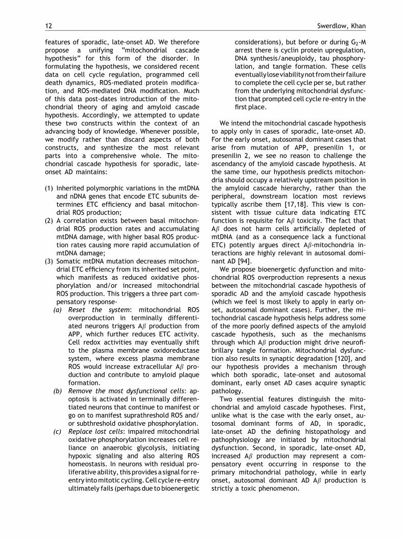
(3) Somatic mtDNA mutation decreases mitochon-drial ETC efficiency from its inherited set point,which manifests as reduced oxidative phos-phorylation and/or increased mitochondrialROS production. This triggers a three part com-pensatory response-(a) Reset the system: mitochondrial ROS
overproduction in terminally differenti-ated neurons triggers Ab production fromAPP, which further reduces ETC activity.Cell redox activities may eventually shiftto the plasma membrane oxidoreductasesystem, where excess plasma membraneROS would increase extracellular Ab pro-duction and contribute to amyloid plaqueformation.
(b) Remove the most dysfunctional cells: ap-optosis is activated in terminally differen-tiated neurons that continue to manifest orgo on to manifest suprathreshold ROS and/or subthreshold oxidative phosphorylation.
(c) Replace lost cells: impaired mitochondrialoxidative phosphorylation increases cell re-liance on anaerobic glycolysis, initiatinghypoxic signaling and also altering ROShomeostasis. In neurons with residual pro-liferativeability, this provides a signal for re-entry intomitotic cycling.Cell cycle re-entryultimately fails (perhaps due to bioenergetic
considerations), but before or during G2-Marrest there is cyclin protein upregulation,DNA synthesis/aneuploidy, tau phosphory-lation, and tangle formation. These cellseventually loseviabilitynotfromtheirfailureto complete the cell cycle per se, but ratherfrom the underlying mitochondrial dysfunc-tion that prompted cell cycle re-entry in thefirst place.
We intend the mitochondrial cascade hypothesisto apply only in cases of sporadic, late-onset AD.For the early onset, autosomal dominant cases thatarise from mutation of APP, presenilin 1, orpresenilin 2, we see no reason to challenge theascendancy of the amyloid cascade hypothesis. Atthe same time, our hypothesis predicts mitochon-dria should occupy a relatively upstream position inthe amyloid cascade hierarchy, rather than theperipheral, downstream location most reviewstypically ascribe them [17,18]. This view is con-sistent with tissue culture data indicating ETCfunction is requisite for Ab toxicity. The fact thatAb does not harm cells artificially depleted ofmtDNA (and as a consequence lack a functionalETC) potently argues direct Ab-mitochondria in-teractions are highly relevant in autosomal domi-nant AD [94].
We propose bioenergetic dysfunction and mito-chondrial ROS overproduction represents a nexusbetween the mitochondrial cascade hypothesis ofsporadic AD and the amyloid cascade hypothesis(which we feel is most likely to apply in early on-set, autosomal dominant cases). Further, the mi-tochondrial cascade hypothesis helps address someof the more poorly defined aspects of the amyloidcascade hypothesis, such as the mechanismsthrough which Ab production might drive neurofi-brillary tangle formation. Mitochondrial dysfunc-tion also results in synaptic degradation [120], andour hypothesis provides a mechanism throughwhich both sporadic, late-onset and autosomaldominant, early onset AD cases acquire synapticpathology.
Two essential features distinguish the mito-chondrial and amyloid cascade hypotheses. First,unlike what is the case with the early onset, au-tosomal dominant forms of AD, in sporadic,late-onset AD the defining histopathology andpathophysiology are initiated by mitochondrialdysfunction. Second, in sporadic, late-onset AD,increased Ab production may represent a com-pensatory event occurring in response to theprimary mitochondrial pathology, while in earlyonset, autosomal dominant AD Ab production isstrictly a toxic phenomenon.
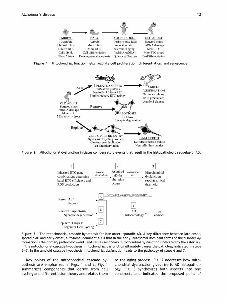
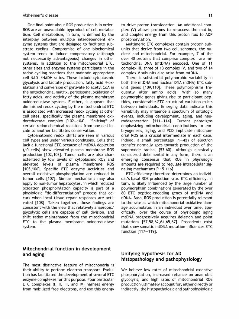
Figure 2 Mitochondrial dysfunction initiates compensatory events that result in the histopathologic sequelae of AD.
EMBRYOAnaerobic
Limited mitosLimited ROSCells divide“Fetal” P-tau
BABYAerobic
More mitosMore ROS
Cell differentiationDevelopmental apoptosis
YOUNG ADULTIntrinsic mito ROSproduction ratedetermines aging(mtDNA+nDNA);Quiescent Neurons
OLD ADULTBattered mitos
mtDNA damageMore ROS
Mito ETC dropsDe-Differentiation
Figure 1 Mitochondrial function helps regulate cell proliferation, differentiation, and senescence.
Inherited ETC genecombinations determinebasal ETC efficiency andROS production
Acquired mtDNAalteration occurs
Mitochondrial dysfunctionreaches criticalthreshold
ADHistopathology
Reset: Aβ/Plaques
Remove: Apoptosis/Synaptic degeneration
Replace: Tangles/Progenitor Cell Cycling
1 2 3
4
5
6
7
Defines rate at which
Determineswhen
Andactivates
Early onset, autosomal dominant AD*
Figure 3 The mitochondrial cascade hypothesis for late-onset, sporadic AD. A key difference between late-onset,sporadic AD and early-onset, autosomal dominant AD is that in the early, autosomal dominant forms of the disorder Abformation is the primary pathologic event, and causes secondary mitochondrial dysfunction (indicated by the asterisk).In the mitochondrial cascade hypothesis, mitochondrial dysfunction ultimately causes the pathology indicated in steps5–7. In the amyloid cascade hypothesis mitochondrial dysfunction leads to the pathology of steps 6 and 7.
Alzheimer’s disease 1313
Key points of the mitochondrial cascade hy-pothesis are emphasized in Figs. 1 and 2. Fig. 1summarizes components that derive from cellcycling and differentiation theory and relates them
to the aging process. Fig. 2 addresses how mito-chondrial dysfunction gives rise to AD histopathol-ogy. Fig. 3 synthesizes both aspects into oneconstruct, and indicates the proposed point of
14 Swerdlow, Khan
overlap between the mitochondrial and amyloidcascade hypotheses.
Support for the hypothesis
Data supporting a role for mitochondria and, inparticular, mtDNA in aging and age-related dis-eases are generally consistent with the first part ofthe AD mitochondrial cascade hypothesis (Fig. 1).Some of these data derive from basic epidemio-logic studies of aging. Longevity analysis of theFramingham cohort, for instance, reveals that al-though the best predictor of an individual’s lon-gevity is biparental longevity, maternal longevitycarries a greater impact [121]. Epidemiologicstudies of both AD and PD further suggest for sub-jects that have a parent with the disease, amongthe affected parents there is maternal overrepre-sentation [122–124]. Taken together, these studiesargue that a maternally inherited genetic factor(mtDNA) helps determine how long one lives, aswell as contributes to AD risk.
Some studies suggest mtDNA haplogroups (whichare defined by mtDNA polymorphism patterns) in-fluence longevity. In northern Italy, haplogroup J ispresent to statistical excess in centenarians [112].Certain mtDNA polymorphisms are more common inthe extremely old than they are in the generalpopulation [113]. Mitochondrial DNA haplogroupvariations may also affect an individual’s odds ofdeveloping a neurodegenerative disease. One re-cent study of mtDNA polymorphisms in Parkinson’sdisease (PD) found that mtDNA haplogroups J and K(which share a common SNP 10398G polymorphism)are associated with a robust PD risk reduction [114].
Cytoplasmic hybrid (“cybrid”) studies of personswith various sporadic neurodegenerative diseasesalso argue mtDNA inheritance at least partly de-termines mitochondrial ETC efficiency, oxidative-phosphorylation capacity, and ROS production.Cybrid cell lines are generated when mtDNA from adesignated subject are expressed within culturedcells depleted of endogenous mtDNA [125,126].This technique allows explorations of mitochon-drial genotype–phenotype relationships whilecontrolling for nDNA variability. If cybrid cell lineswith a common nuclear background but mtDNAfrom different donors have distinct mitochondrialphenotypes, the root cause is likely differencesbetween the donor mtDNAs [127].
Relative to cybrids expressing mtDNA from age-matched control individuals, cybrids expressingmtDNA from AD, PD, amyotrophic lateral sclerosis(ALS), and progressive supranuclear palsy subjectsshow bioenergetic impairment and increased oxi-
dative stress [128–139]. These cybrid lines alsoexhibit altered calcium homeostasis, mitochondrialmembrane potential depolarization, abnormal mi-tochondrial morphology, molecular stress responsepathway activation, increased activation andexpression of apoptotic proteins, and excessiveprotein aggregation (including Ab in AD cybrids anda-synuclein in PD cybrids) [128,132,134, 136,40–149]. Interestingly, mtDNA used to generatecybrid cell lines in these experiments is derived notfrom brain but rather from platelets, a non-de-generating tissue. This suggests the relative func-tional impairment observed in cell lines withdisease subject mtDNA represents a systemic de-fect. Systemic mitochondrial dysfunction is moreconsistent with inherited rather than somaticmtDNA aberration.
To date, only limited published data indicateinherited mtDNA variation influences somaticmtDNA mutation acquisition [61]. There is, how-ever, considerable evidence showing mtDNA doesacquire mutations during the course of an indi-vidual’s lifespan, including the brain, the tissuewith the greatest rate of oxidative metabolismand therefore ROS production [65,67,150,151].Over a decade ago it was shown that mtDNA de-letions (specifically, a particular 5 kb deletioncalled the “common deletion”) accumulate withage [152–156]. Tissues with lower rates of oxida-tive mutation than brain acquire less deletionburden. More recently, investigators have uncov-ered an entire layer of age-dependent mtDNAmutation in the brains of deceased individuals[67,157]. These presumed somatic mutations arenot detected using routine sequencing strategies,but rather require laborious clonal mtDNA analy-sis. It is currently unclear whether these mutationsrepresent low abundance “microheteroplasmy”that is widely distributed between many or mostcells of a brain parenchyma region, or if it arisesfrom limited numbers of individual cells that carryunique homoplasmic mutations. These scenarios,however, may not be mutually exclusive, sincesomatic mutations (that by definition are in lowabundance when they arise) tend to clonallyexpand towards high abundance intracellularheteroplasmy or even homoplasmy [158–160].Ultimately, through either clonal expansion or“compound microheteroplasmy”, mtDNA muta-tional burdens within individual cells may reachthresholds at which the resultant mitochondrialdysfunction becomes critical.
There is some debate as to how aging effectsmitochondrial ETC function. Data supporting anage-related decline are strongest for liver, andalso indicate a similar phenomenon in muscle,
Alzheimer’s disease 1515
fibroblasts, and brain [161–170]. The status ofbrain mitochondria ETC function has also beenextensively evaluated in subjects with neurode-generative disorders. Relevant to this discussion isthe well-replicated finding that complex IV (cyto-chrome oxidase) activity is reduced in AD brain[19]. Some argue this is an epiphenomenal conse-quence of neuronal de-afferentation. Indeed, it hasbeen shown that surgically de-afferented neuronsdownregulate cytochrome oxidase [171,172]. Themechanistic explanation for this is that de-affer-ented neurons have reduced synaptic connections,less synaptic activity, and require less ATP tomaintain ion gradients across their membranes.This cannot entirely explain the AD cytochromeoxidase deficit, which is also present in sporadic ADsubject platelets and fibroblasts [173–178].
One AD chicken-and-egg controversy revolvesaround whether mitochondrial dysfunction causesAb over-production, or whether Ab over-productioncauses mitochondrial dysfunction. When viewedoutside the charged confines of the AD debate, itcertainly seems clear that cytochrome oxidase in-hibition promotes amyloidgenic fragmentation ofAPP, and that Ab inhibits cytochrome oxidase[179–183]. For the sporadic late-onset forms,available data argue amyloidgenesis follows mito-chondrial dysfunction. Specifically, in sporadic ADmitochondrial dysfunction is more anatomicallywidespread than is Ab deposition, and thereforemitochondrial dysfunction cannot entirely be ac-counted for by Ab [19]. Further, cybrid cell linesthat express mtDNA from AD subjects, in addition toshowing reduced cytochrome oxidase activity andincreased ROS production relative to control cybridlines, also produce substantially increased amountsof both intracellular and extracellular Ab [19,146].
AD brain shows evidence of increased apoptoticcell death, cyclin protein expression, and nuclearDNA replication with excess aneuploidy [23–25].The concept that tau phosphorylation promotestangle formation in neurons attempting to re-enterthe cell replication cycle is not novel [24], nor isthe idea that mitochondrial function can determinetau phosphorylation. Indeed, tau phosphorylationis promoted in AD fibroblasts exposed to an ETCuncoupler [184].
Conclusion
The mitochondrial cascade hypothesis provides aunifying framework for AD pathology. In developingthis hypothesis, we approached sporadic AD froman aging theory perspective, since sporadic AD in-
cidence and prevalence progressively rise at leastinto the ninth decade [185]. Indeed, if half thoseover age 85 meet criteria for AD [186], can it trulybe considered a disease?
Our hypothesis posits mitochondrial dysfunctionrepresents primary pathology in sporadic, late-on-set AD, and drives both Ab plaque and neurofibril-lary tangle formation. We further provide arationale for how mitochondrial dysfunction sur-passing certain thresholds triggers compensatoryevents that cause the various histopathologic andpathophysiologic features of AD. Other sporadicneurodegenerative diseases also manifest mito-chondrial dysfunction, oxidative stress, and proteinaggregation [127,187,188]. It is tempting to con-sider whether similar principles may underlie thesedisorders at the molecular level.
References
[1] Alzheimer A. Uber eine eigenartige Erkrankung der Hirnr-inde. Allg Z Psychiat Psych- Gerichtl Med 1907;64:146–8.
[2] Kraepelin E. Psychiatrie. Ein Lehrbuch fur Studierende undArtze. II. Band, Klinische Psychiatrie. Leipzig: VerlagJohann Ambrosius Barth; 1910.
[3] Fischer O. Miliare Nekrosen mit drusigen Wucherungen derNeurofibrillen, eine regelmabige Veranderung der Hirnr-inde bei seniler Demenz. Monatsschr Psychiatr Neurol1907;22:361–72.
[4] Perusini G. Sul valore nosografico di alcuni repertiistopatlogici caratteristici per la senilita. Rivista italianadi Neuropatologia. Psichiatria e Elettroterapia 1911;4:145–51.
[5] Boller F, Forbes MM. History of dementia and dementia inhistory: an overview. J Neurol Sci 1998;158:125–33.
[6] Amaducci LA, Rocca WA, Schoenberg BS. Origin of thedistinction between Alzheimer’s disease and senile de-mentia: how history can clarify nosology. Neurology1986;36:1497–9.
[7] Katzman R. The prevalence and malignancy of Alzheimer’sdisease: a major killer. Arch Neurol 1976;33:217–8.
[8] Katzman R, Terry R, Bick K. Recommendations of thenosology, epidemiology, and etiology and pathophysiologycommissions of the workshop-conference on Alzheimer’sdisease: senile dementia and related disorders. In: Katz-man R, Terry RD, Bick KL, editors. Alzheimer’s disease:senile dementia and related disorders. New York: RavenPress; 1978. p. 579–85.
[9] Katzman R. Alzheimer’s disease. New England J Med1986;314:964–73.
[10] Amaducci L, Rocca W, Schoenberg B. Origin of thedistinction between Alzheimer’s disease and senile de-mentia. How history can clarify nosology. Neurology 1986;36:1497–9.
[11] Goate A, Chartier-Harlin MC, Mullan M, Brown J, CrawfordF, Fidani L, et al. Segregation of a missense mutation inthe amyloid precursor protein gene with familial Alzhei-mer’s disease. Nature 1991;349:704–6.
[12] Sherrington R, Rogaev EI, Liang Y, Rogaeva EA, LevesqueG, Ikeda M, et al. Cloning of a gene bearing missensemutations in early-onset familial Alzheimer’s disease.Nature 1995;375:754–60.
16 Swerdlow, Khan
[13] Levy-Lahad E, Wasco W, Poorkaj P, Romano DM, Oshima J,Pettingell WH, et al. Candidate gene for the chromosome1 familial Alzheimer’s disease locus. Science 1995;269:973–7.
[14] Hardy J, Allsop D. Amyloid deposition as the central eventin the aetiology of Alzheimer’s disease. Trends PharmacolSci 1991;12:383–8.
[15] Hardy JA, Higgins GA. Alzheimer’s disease: the amyloidcascade hypothesis. Science 1992;256:184–5.
[16] Selkoe DJ. Cell biology of the amyloid b-protein precursorand the mechanism of Alzheimer’s disease. Annu Rev CellBiol 1994;10:373–403.
[17] Selkoe DJ. Toward a comprehensive theory for Alzheimer’sdisease. Ann NY Acad Sci 2000;924:17–25.
[18] Cummings JL, Cole G. Alzheimer disease. JAMA 2002;287:2335–8.
[19] Swerdlow RH, Kish SJ. Mitochondria in Alzheimer’s dis-ease. Int Rev Neurobiol 2002;53:341–85.
[20] Davis DG, Schmitt FA, Wekstein DR, Markesbery WR.Alzheimer neuropathologic alterations in aged cogni-tively normal subjects. J Neuropath Exp Neurol 1999;58:376–88.
[21] Neuropathology Group of the Medical Research CouncilCognitive Function and Ageing Study. Pathological corre-lates of late-onset dementia in a multicentre, community-based population in England and Wales. Lancet 2001;357:169–75.
[22] Snowdon DA. Healthy aging and dementia: findings fromthe nun study. Ann Int Med 2003;139:450–4.
[23] Barinaga M. Is apoptosis key in Alzheimer’s disease.Science 1998;281:1303–4.
[24] Arendt T. Alzheimer’s disease as a loss of differentiationcontrol in a subset of neurons that retain immaturefeatures in the adult brain. Neurobiol Aging 2000;21:783–96.
[25] Yang Y, Geldmacher DS, Herrup K. DNA replicationprecedes neuronal cell death in Alzheimer’s disease. JNeurosci 2001;21:2662–8.
[26] Anderton BH. Ageing of the brain. Mech Ageing and Dev2002;123:811–7.
[27] Kwon YW, Masutani H, Nakamura H, Ishii Y, Yodoi J. Redoxregulation of cell growth and cell death. Biol Chem2003;384:991–6.
[28] Fulco M, Schiltz RL, Iezzi S, King MT, Zhao P, Kashiwaya Y,et al. Sir2 regulates skeletal muscle differentiation as apotential sensor of the redox state. Mol Cell 2003;12:51–62.
[29] Chueh PJ. Cell membrane redox systems and transforma-tion. Antioxid Redox Signal 2000;2:177–87.
[30] Klein JA, Ackerman SL. Oxidative stress, cell cycle, andneurodegeneration. J Clin Invest 2002;111:785–93.
[31] Boveris A, Oshino N, Chance B. The cellular production ofhydrogen peroxide. Biochem J 1972;128:617–30.
[32] Chance B, Sies H, Boveris A. Hydroperoxide metabolism inmammalian organs. Physiol Rev 1979;59:527–605.
[33] Shigenaga MK, Hagen TM, Ames BN. Oxidative damage andmitochondrial decay in aging. Proc Natl Acad Sci USA1994;91:10771–8.
[34] Burdon RH. Superoxide and hydrogen peroxide in relationto mammalian cell proliferation. Free Rad Biol Med 1995;18:775–94.
[35] Johansson CB. Mechanism of stem cells in the centralnervous system. J Cell Physiol 2003;196:409–18.
[36] Jauniaux E, Gulbis B, Burton GJ. The human first trimestergestational sac limits rather than facilitates oxygen trans-fer to the foetus – A review. Placenta 2003;24(suppl A):S86–93.
[37] Nasr-Esfahani MH, Aitken JR, Johnson MH. Hydrogenperoxide levels in mouse oocytes and early cleavage stageembryos developed in vitro or in vivo. Development 1990;109:501–7.
[38] Johnson MH, Nasr-Esfahani MH. Radical solutions andcultural problems: could free oxygen radicals be re-sponsible for the impaired development of preimplanta-tion mammalian embryos in vitro. Bioessays 1994;16:31–8.
[39] Trimarchi JR, Liu L, Porterfield DM, Smith PJS, Keefe DL.Oxidative phosphorylation-dependent and – independentoxygen consumption by individual preimplantation mouseembryos. Biol Reprod 2000;62:1866–74.
[40] Sweet S, Singh G. Accumulation of human promyelocyticleukemic (HL-60) cells at two energetic cell cycle check-points. Cancer Res 1995;55:5164–7.
[41] Dorward A, Sweet S, Moorehead R, Singh G. Mitochondrialcontributions to cancer cell physiology: redox balance,cell cycle, and drug resistance. J Bioenerg Biomem 1997;29:385–92.
[42] Lovestone S, Reynolds CH. The phosphorylation of tau: acritical stage in neurodevelopment and neurodegenerativeprocesses. Neuroscience 1997;78:309–24.
[43] Connolly JA, Kalnins VI, Cleveland DW, Kirschner MW.Immunoflourescent staining of cytoplasmic and spindlemicrotubules in mouse fibroblasts with antibody to tauprotien. Proc Natl Acad Sci USA 1977;74:2437–40.
[44] Delobel P, Plament S, Hamdane M, Mailliot C, Sambo AV,Begard S, et al. Abnormal tau phosphorylation of theAlzheimer-type also occurs during mitosis. J Neurochem2002;83:412–20.
[45] Billingsley ML, Kincaid RL. Regulated phosphorylation anddephosphorylation of tau protein: effects on microtubuleinteraction, intracellular trafficking and neurodegenera-tion. Biochem J 1997;323:577–91.
[46] Hartigan JA, Johnson GVW. Tau protein in normal andAlzheimer’s disease brain: an update. Alzheimer’s DiseaseRev 1998;3:125–41.
[47] Trounce I, Byrne E, Marzuki S. Decline in skeletal musclemitochondrial respiratory chain function: possible factorin ageing. Lancet 1989;1:637–9.
[48] Yen TC, Chen YS, King KL, Yeh SH, Wie YH. Livermitochondrial respiratory function declines with age.Biochem Biophys Res Commun 1989;165:994–1003.
[49] Cooper JM, Mann VM, Schapira AHV. Analysis of mitochon-drial respiratory cahin function and mitochondrial DNAdeletion in human skeletal muscle: effect of aging. JNeurol Sci 1992;113:91–8.
[50] Ojaimi J, Masters CL, Opwskin K, McKelvie P, Byrne E.Mitochondrial respiratory chain activity in the humanbrain as a function of age. Mech Ageing Dev 1999;111:39–47.
[51] Harmon D. The biologic clock: the mitochondria. J AmGeriatr Soc 1972;20:145–7.
[52] Miquel J, Economos AD, Fleming J, Johnson JE. Mitochon-drial role in cell aging. Exp Gerontol 1980;15:575–91.
[53] Beckman KB, Ames BN. Mitochondrial aging: open ques-tions. Ann NY Acad Sci 1998;854:118–27.
[54] Melov S. Mitochondrial oxidative stress. Physiologic con-sequences and potential for a role in aging. Ann NY AcadSci 2000;908:219–25.
[55] Lenaz G, Bovina C, D’Aurelio M, Fato R, Formiggini G,Genova ML, et al. Role of mitochondria in oxidative stressand aging. Ann NY Acad Sci 2002;959:199–213.
[56] Cadenas E, Davies KJA. Mitochondrial free radical gener-ation, oxidative stress, and aging. Free Rad Biol Med 2000;29:222–30.
Alzheimer’s disease 1717
[57] Linnane AW, Marzuki S, Ozawa T, Tanaka M. MitochondrialDNA mutations as an important contributor to ageing anddegenerative diseases. Lancet 1989;1:642–5.
[58] Wallace DC. Mitochondrial genetics: a paradigm for agingand degenerative diseases? Science 1992;256:628–32.
[59] Beckman KB, Ames BN. The free radical theory of agingmatures. Physiol Rev 1998;78:548–81.
[60] Gadaleta MN, Cormio A, Pesce V, Lezza AM, Cantatore.Aging and mitochondria. Biochimie 1998;80:863–70.
[61] Ozawa T. Mechanism of somatic mitochondrial DNA muta-tions associated with age and diseases. Biochim BiophysActa 1995;1271:177–89.
[62] Melov S, Shoffner JM, Kaufman A, Wallace DC. Markedincrease in the number and variety of mitochondrial DNArearrangements in aging human skeletal muscle. NuclAcids Res 1995;23:4122–6.
[63] Kovalenko SA, Kopsidas G, Kelso JM, Linnane AW. Deltoidhuman muscle mtDNA is extensively rearranged in old agesubjects. Biochem Biophys Res Commun 1997;232:147–52.
[64] Melov S, Schneider JA, Coskun PE, Bennett DA, WallaceDC. Mitochondrial DNA rearrangements in aging humanbrain in situ PCR of mtDNA. Neurobiol Aging 1999;20:565–71.
[65] Michikawa Y, Mazzucchelli F, Bresolin N, Scarlato G,Attardi G. Aging-dependent large accumulation of pointmutations in the human mtDNA control region for replica-tion. Science 1999;286:774–9.
[66] Wang Y, Michikawa Y, Mallidis C, Bai Y, Woodhouse KE,Yarasheski KE, et al. Muscle-specific mutations accumu-late with aging in critical human mtDNA control sites forreplication. Proc Natl Acad Sci USA 2001;98:4022–7.
[67] Lin MT, Simon DK, Ahn CH, Kim LM, Beal MF. Highaggregate burden of somatic mtDNA point mutations inaging and Alzheimer’s disease brain. Hum Mol Genet2002;11:133–45.
[68] Richter C, Park JW, Ames BN. Normal oxidative damage tomitochondrial and nuclear DNA is extensive. Proc NatlAcad Sci USA 1988;85:6465–7.
[69] Cheng KC, Cahill DS, Kasai H, Nishimura S, Loeb LA. 8-Hydroxyguanine, an abundant form of oxidative DNAdamage, causes G–T and A–C substitutions. J Biol Chem1992;267:166–72.
[70] Brierley EJ, Johnson MA, James OF, Turnbull DM. Mito-chondrial involvement in the ageing process. Facts andcontroversies. Mol Cell Biochem 1997;174:325–8.
[71] Yoneda M, Chomyn A, Martinuzzi A, Hurko O, Attardi G.Marked replicative advantage of human mtDNA carrying apoint mutation that causes the MELAS encephalomyopa-thy. Proc Natl Acad Sci USA 1992;89:11164–8.
[72] Taylor DR, Zeyl C, Cooke E. Conflicting levels of selectionin the accumulation of mitochondrial defects in Saccha-romyces cerevisiae. Proc Natl Acad Sci USA 2002;99:3690–4.
[73] De Grey ADNJ. A mechanism proposed to explain the risein oxidative stress during aging. J Anti Aging Med1998;1:53–66.
[74] Kowald A. The mitochondrial theory of aging: do damagedmitochondria accumulated by delayed degradation? ExpGerontol 1999;34:605–12.
[75] Gershon D. The mitochondrial theory of aging: is theculprit a faulty disposal system rather than indigenousmitochondrial alterations. Exp Gerontol 1999;34:613–9.
[76] Soohal RS, Agarwal A, Agarwal S, Orr WC. Simultaneousoverexpression of copper- and zinc-containing superoxidedismutase and catalase retards age-related oxidativedamage and increases metabolic potential in Drosophilamelanogaster. J Biol Chem 1995;270:15671–4.
[77] Roth GS, Ingram DK, Lane MA. Slowing ageing by caloricrestriction. Nat Med 1995;1:414–5.
[78] Aspnes LE, Lee CM, Weindruch R, Chung SS, Roecker EB,Aiken JM. Caloric restriction reduces fiber loss andmitochondrial abnormalities in aged rat muscle. FASEB J1997;11:573–81.
[79] Zamzami N, Hirsch T, Dallaporta B, Petit PX, Kroemer G.Mitochondrial implication in accidental and programmedcell death: apoptosis and necrosis. J Bioenerg Biomem1997;29:185–93.
[80] Ichas F, Mazat JP. From calcium signaling to cell death:two conformations for the mitochondrial permeabilitytransition pore. Switching from low- to high-conductancestate. Biochim Biophys Acta 1998;1366:33–50.
[81] Zou H, Li Y, Liu X, Wang X. An APAF-1 cytochrome cmultimeric complex is a functional apoptosome thatactivates procaspase-9. J Biol Chem 1999;27:11549–56.
[82] Liu X, Naekyung Kim C, Yang J, Jemmerson R, Wang X.Induction of apoptotic program in cell-free extracts:requirement for dATP and cytochrome c. Cell 1996;86:147–57.
[83] Kluck RM, Bossy-Wetzel E, Green DR, Newmeyer DD. Therelease of cytochrome c from mitochondria: a primarysite for Bcl-2 regulation of apoptosis. Science 1997;275:1132–6.
[84] Petit PX, Zamzami N, Vayssiere JL, Mignotte B, Kroemer G,Castedo M. Implication of mitochondria in apoptosis. MolCell Biochem 1997;174:185–8.
[85] Yang J, Liu X, Bhalla K, Kim CN, Ibrado AM, Cai J, et al.Prevention of apoptosis by Bcl-2: release of cytochrome cfrom mitochondria blocked. Science 1997;275:1129–32.
[86] Li P, Nijhawan D, Budihardjo I, Srinivasula SM, Ahmad M,Alnemri ES, et al. Cytochrome c and dATP-dependentformation of Apaf-1/caspase-9 complex initiates an apop-totic protease cascade. Cell 1997;91:479–89.
[87] Cardoso SM, Swerdlow RH, Oliveira CR. Induction ofcytochrome c-mediated apoptosis by amyloid b25–35requires functional mitochondria. Brain Res 2002;931:117–25.
[88] Negre-Salvayre A, Hirtz C, Carrera G, Cazenave R, Troly M,Salvayre R, et al. A role for uncoupling proteins-2 as aregulator of mitochondrial hydrogen peroxide generation.FASEB J 1997;11:809–15.
[89] Ledesma A, de Lacoba MG, Rial E. The mitochondrialuncoupling proteins. Genome Biol 2003;3:3015, Reviews.
[90] Anandtheerthavarada HK, Biswas G, Robin MA, AvadhaniMG. Mitochondrial targeting and a novel transmembranearrest of Alzheimer’s precursor protein impairs mitochon-drial function in neuronal cells. J Cell Biol 2003;16:41–54.
[91] Dyall SD, Lester DC, Schneider RE, Delgadillo_correa MG,Plumper E, Martinez A, Koehler CM, Johnson PJ. Tricho-monas vaginalis Hmp35, a putative pore-forming hydrog-enosomal membrane protein, can form a complex in yeastmitochondria. J Biol Chem 2003;278:30548–61.
[92] Robinson SR, Bishop GM. Abeta as a bioflocculant: impli-cations for the amyloid hypothesis of Alzheimer’s disease.Neurobiol Aging 2002;23:1051–72.
[93] Atwood CS, Moir RD, Huang X, Scarpa RC, Bacarra NM,Romano DM, et al. Dramatic aggregation of Alzheimerabeta by Cu (II) is induced by conditions representingphysiological acidosis. J Biol Chem 1998;273:12817–26.
[94] Cardoso SM, Santos S, Swerdlow RH, Oliveira CR. Func-tional mitochondria are required for amyloid beta-medi-ated neurotoxicity. FASEB J 2001;15:1439–41.
[95] Arias C, Montiel T, Quiroz-Baez R, Massieu L. b-Amyloidneurotoxicity is exacerbated during glycolysis inhibitionand mitochondrial impairment in the rat hippocampus
[
[
[
[
[
[
[
[
[
[
[
[
[
[
[
[
[
[
[
[
[
[
[
[
[
[
[
[
[
[
[
[
[
[
18 Swerdlow, Khan
in vivo and in isolated nerve terminals: implications forAlzheimer’s disease. Exp Neurol 2002;176:163–74.
[96] Nunomura A, Perry G, Pappolla MA, Wade R, Hirai K, ChibaS, et al. RNA oxidation is a prominent feature of vulnerableneurons in Alzheimer’s disease. J Neurosci 1999;19:1959–64.
[97] Smith MA, Nunomura A, Zhu X, Takeda A, Perry G.Metabolic, metallic, and mitotic sources of oxidativestress in Alzheimer disease. Antioxide Redox Signal 2000;2:413–20.
[98] Shan X, Tashiro H, Lin CL. The identification and charac-terization of oxidized RNAs in Alzheimer’s disease. JNeurosci 2003;23:4913–21.
[99] Sylvestre J, Vialette S, Corral Debrinski M, Jacq C. LongmRNAs coding for yeast mitochondrial proteins of pro-karyotic origin preferentially localize to the vicinity ofmitochondria. Genome Biol 2003;4:R44, Epub 2003 Jun 06.
100] Marc P,Margeot A, Devaux F, BlugeonC, Corral-Debrinski M,Jacq C. Genome-wide analysis of mRNAs targeted to yeastmitochondria. EMBO Rep 2002;3:159–64, Epub 2002 Jan 29.
101] Ginsberg MD, Feliciello A, Jones JK, Avvedimento EV,Gottesman ME. PKA-dependent binding of mRNA to themitochondrial AKAP121 protein. J Mol Biol 2003;327:885–97.
102] Lawen A, Martinus RD, McMullen GL, Nagley P, Vaillant F,Wolvetang EJ, et al. The universality of bioenergeticdisease: the role of mitochondrial mutation and theputative inter-relationship between mitochondria andplasma membrane NADH oxidoreductase. Molec AspectsMed 1994;15(suppl):s13–27.
103] Larm JA, Vaillant F, Linnane AW, Lawen A. Upregulation ofthe plasma membrane oxidoreductase as a prerequisite forthe viability of human Namalwa rho0 cells. J Biol Chem1994;269:30097–100.
104] Morre DM, Lenaz G, Morre J. Surface oxidase and oxidativestress propagation in aging. J Exp Biol 2000;203:1513–21.
105] Capuano F, Guerrieri F, Papa S. Oxidative phosphorylationenzymes in normal and neoplastic cell growth. J BioenergBiomembr 1997;29:379–84.
106] Chueh PJ. Cell membrane redox systems and transforma-tion. Antioxid Redox Signal 2000;2:177–87.
107] Capuano F, Varone D, D’Eri N, Russo E, Tommasi S,Montemurro S, et al. Oxidative phosphorylation and F (O)F(1) ATP synthase activity of human hepatocellular carci-noma. Biochem Mol Biol Int 1996;38:1013–22.
108] Uriel J. Retrodifferentiation and the fetal patterns of geneexpression in cancer. Adv Cancer Res 1979;29:127–74.
109] MITOMAP: A Human Mitochondrial Genome Database.Center for Molecular Medicine, Emory University, Atlanta,GA, USA. Available from: http://www.gen.emory.edu/mitomap.html, 2001.
110] National Center for Biotechnology Information, NationalLibrary of Medicine, National Institutes of Health. Avail-able from: http://www.ncbi.nlm.nih.gov/entrez/, 2002.
111] Roubertoux PL, Sluyter F, Carlier M, Marcet B, Maarouf-Veray F, Cherif C, et al. Mitochondrial DNA modifiescognition in interaction with the nuclear genome and agein mice. Nat Genet 2003;35:65–9.
112] De Benedictis G, Rose G, Carrieri G, De Luca M, Falcone E,Passarino G, et al. Mitochondrial DNA inherited variantsare associated with successful aging and longevity inhumans. FASEB J 1999;13:1532–6.
113] Tanaka M, Gong JS, Zhang J, Yoneda M, Yagi K. Mitochon-drial gentoype associated with longevity. Lancet 1998;351:185–6.
114] van der Walt JM, Nicodemus KK, Martin ER, Scott WK,Nance MA, Watts RL, et al. Mitochondrial polymorphisms
significantly reduce the risk of Parkinson disease. Am JHum Genet 2003;72:804–11.
115] Wolin MS. Reactive oxygen species and vascular signaltransduction mechanisms. Microcirculation 1996;3:1–17.
116] Droge W. Free radicals in the physiologic control of cellfunction. Physiol Rev 2001;82:47–95.
117] Wanagat J, Cao Z, Pathare P, Aiken JM. Mitochondrial DNAdeletion mutations colocalize with segmental electrontransport system abnormalities, muscle fiber atrophy,fiber splitting, and oxidative damage in sarcopenia. FASEBJ 2001;15:322–32.
118] Fayet G, Jansson M, Sternberg D, Moslemi AR, Blondy P,Lombes A, et al. Aging muscle: clonal expansions ofmitochondrial DNA point mutations and deletions causefocal impairment of mitochondrial function. NeuromusculDisord 2002;12:483–93.
119] Chomyn A, Attardi G. MtDNA mutations in aging andapoptosis. BiochemBiophysResCommun2003;304:519–29.
120] Mattson MP, Liu D. Energetics and oxidative stress insynaptic plasticity and neurodegenerative disorders. Neu-romol Med 2002;2:215–31.
121] Brand FN, Kiely DK, Kannel WB, Myers RH. Family patternsof coronary heart disease mortality: the Framinghamlongevity study. J Clin Epidemiol 1992;45:169–74.
122] Duara R, Lopez-Alberola RF, Barker WW, Loewenstein DA,Zatinsky M, Eisdorfer CE, et al. A comparison of familialand sporadic Alzheimer’s disease. Neurology 1993;43:1377–84.
123] Edland SD, Silverman JM, Peskind ER, Tsuang D, WijsmanE, Morris JC. Increased risk of dementia in mothers ofAlzheimer’s disease cases: evidence for maternal inheri-tance. Neurology 1996;47:254–6.
124] Swerdlow RH, Parker WD, Currie LJ, Bennett Jr JP,Harrison MB, Trugman JM, et al. Gender ratio differencesbetween Parkinson’s disease patients and their affectedparents. Parkinsonism Related Disorders 2001;7:47–51.
125] King MP, Attardi G. Human cells lacking mtDNA: repopu-lation with exogenous mitochondria complementation.Science 1989;246:500–3.
126] Chomyn A, Lai ST, Shakeley R, Bresolin N, Scarlato G,Attardi G. Platelet-mediated transformation of mtDNA-less human cells: analysis of phenotypic variability amongclones from normal individuals – and complementationbehavior of the tRNALys mutation causing myoclonicepilepsy and ragged red fibers. Am J Hum Genet 1994;54:966–74.
127] Swerdlow RH. Mitochondrial DNA and dysfunction inneurodegenerative diseases. Arch Path Lab Med2002;126:271–80.
128] Swerdlow RH, Parks JK, Miller SW, Tuttle JB, Trimmer PA,Sheehan JP, et al. Origin and functional consequences ofthe complex I defect in Parkinson’s disease. Ann Neurol1996;40:663–71.
129] Gu M, Cooper JM, Taanman JW, Schapira AHV. Mitochon-drial DNA transmission of the mitochondrial defect inParkinson’s disease. Ann Neurol 1998;44:177–86.
130] Shults CW, Miller SW. Reduced complex I activity inParkinsonian cybrids. Mov Dis 1998;13:217.
131] Davis RE, Miller S, Herrnstadt C, Ghosh SS, Fahy E, ShinobuLA, et al. Mutations in mitochondrial cytochrome c oxidasegenes segregate with late-onset Alzheimer disease. ProcNatl Acad Sci USA 1997;94:4526–31.
132] Swerdlow RH, Parks JK, Cassarino DS, Maguire DJ, MaguireRS, Bennett JP, et al. Cybrids in Alzheimer’s disease: acellular model of the disease. Neurology 1997;49:918–25.
133] Cassarino DS, Fall CP, Swerdlow RH, Smith TS, HalvorsenEM, Miller SW, et al. Elevated reactive oxygen species and
[
[
[
[
[
[
[
[
[
[
[
[
[
[
[
[
[
[
[
[
[
[
[
[
[
[
[
[
[
[
[
[
[
Alzheimer’s disease 1919
antioxidant enzyme activities in animal and cellularmodels of Parkinson’s disease. Biochim Biophys Acta1997;1362:77–86.
134] Swerdlow RH, Parks JK, Davis JN, Cassarino DS, TrimmerPA, Currie LJ, et al. Matrilineal inheritance of complex Idysfunction in a multigenerational Parkinson’s diseasefamily. Ann Neurol 1998;44:873–81.
135] Ghosh SS, Swerdlow RH, Miller SW, Sheeman B, Parker JrWD, Davis RE. Use of cytoplasmic hybrid lines for eluci-dating the role of mitochondrial dysfunction in Alzhei-mer’s disease and Parkinson’s disease. Ann NY Acad Sci2000;893:176–91.
136] Swerdlow RH, Miller SW, Parks JK, Sheehan JP, CassarinoDS, Maguire DJ, et al. Mitochondria in sporadic amyo-trophic lateral sclerosis. Exp Neurol 1998;153:135–42.
137] Swerdlow RH, Golbe LI, Parks JK, Cassarino DS, Binder DR,Grawey AE, et al. Mitochondrial dysfunction in cybrid linesexpressing mitochondrial genes from patients with PSP. JNeurochem 2000;75:1681–4.
138] Trimmer PA, Keeney PM, Borland MK, Simon FA, Almeida J,Swerdlow RH, et al. Mitochondrial abnormalites in cybridcell models of sporadic Alzheimer’s disease worsen withpassage in culture. Neurobiol Dis 2004;15:29–39.
139] Cardoso SM, Santana I, Swerdlow RH, Oliveira CR. Mito-chondria dysfunction of Alzheimer’s disease cybrids en-chances Ab toxicity. J Neurochem, in press.
140] Sheehan JP, Swerdlow RH, Parker WD, Miller SW, Davis RE,Tuttle JB. Altered calcium homeostasis in cells trans-formed by mitochondria from individuals with Parkinson’sdisease. J Neurochem 1997;68:1221–33.
141] Sheehan JP, Swerdlow RH, Miller SW, Davis RE, Tuttle JB.Altered calcium homeostasis and reactive oxygen speciesproduction in cells transformed by Alzheimer’s diseasemitochondrial DNA. J Neurosci 1997;17:4612–22.
142] Cassarino DS, Halvorsen EM, Swerdlow RH, Abramova NN,Parker WD, Sturgill TW, et al. Interaction among mito-chondria, mitogen-activated protein kinases, and nuclearfactor-kappa B in cellular models of Parkinson’s disease. JNeurochem 2000;74:1384–92.
143] Trimmer PA, Swerdlow RH, Parks JK, Miller SW, Davis RE,Parker Jr WD. Abnormal mitochondrial morphology insporadic Parkinson’s and Alzheimer’s disease cybrid lines.Exp Neurol 2000;162:37–50.
144] Veech GA, Dennis J, Keeney PM, Fall CP, Swerdlow RH,Parker Jr WD. Disrupted mitochondrial electron transportfunction increases expression of anti-apoptotic Bcl-2 andBcl-XL proteins in SH-SY5Y neuroblastoma and in Parkin-son’s disease cybrid cell lines. J Neurosci Res 2000;61:693–700.
145] Cassarino DS, Swerdlow RH, Parks JK, Davis Jr WP, BennettJr JP. Cyclosporin A increases resting mitochondrialmembrane potential in SY5Y cells and reverses thedepressed mitochondrial membrane potential of Alzhei-mer’s disease cybrids. Biochem Biophys Res Commun1998;248:168–73.
146] Khan SM, Cassarino DS, Abramova NN, Keeney PM, BorlandK, Trimmer PA, et al. Alzheimer’s disease cybrids replicateb-amyloid abnormalities through cell death pathways. AnnNeurol 2000;48:148–55.
147] Bijur GN, Davis RE, Jope RS. Rapid activation of heat shockfactor-1 DNA binding by H2O2 and modulation by glutathi-one in human neuroblastoma and Alzheimer’s diseasecybrid cells. Brain Res Mol Brain Res 1999;71:69–77.
148] De Sarno P, Bijur GN, Lu R, Davis RE, Jope RS. Alterationsin muscarinic receptor-coupled phosphoinositide hydroly-sis and AP-1 activation in Alzheimer’s disease cybrid cells.Neurobiol Aging 2000;21:31–8.
149] Trimmer PA, Borland K, Keeney PM, Bennett Jr JP, ParkerJr WD. Parkinson’s disease transgenic mitochondrial cy-brids generate Lewy inclusion bodies. J Neurochem 2004;88:800–12.
150] Laderman KA, Penny JR, Mazzucchelli F, Bresolin N,Scarlato G, Attardi G. Aging-dependent functional alter-ations of mitochondrial DNA (mtDNA) from human fibro-blasts transferred into mtDNA-less cells. J Biol Chem 1996;271:15891–7.
151] Diaz F, Bayona-Bafaluy MP, Rana M, Mora M, Hao H, MoraesCT. Human mitochondrial DNA with large deletions repop-ulates organelles faster than full-length genomes underrelaxed copy number control. Nucl Acids Res 2002;30:4626–33.
152] Cortopassi GA, Arnheim N. Detection of a specific mito-chondrial DNA deletion in tissues of older humans. NuclAcids Res 1990;18:6927–33.
153] Cortopassi GA, Shibata D, Soong NW, Arnheim N. A patternof accumulation of a somatic deletion of mitochondrialDNA in aging human tissues. Proc Natl Acad Sci USA 1992;89:7370–4.
154] Soong NW, Hinton DR, Cortopassi G, Arnheim N. Mosaicismfor a specific somatic mitochondrial DNA mutations inadult human brain. Nat Genet 1992;2:318–23.
155] Corral-Debrinski M, Horton MT, Lott JM, Shoffner JM, BealMF, Wallace DC. Mitochondrial DNA deletions in humanbrain: regional variability and increase with advanced age.Nat Genet 1992;2:324–9.
156] Corral-Debrinski M, Horton T, Lott MT, Shoffner JM, McKeeAC, Beal MF, et al. Marked changes in mitochondrial DNAdeletion levels in Alzheimer brains. Genomics 1994;23:471–6.
157] Smigrodzki R, Parks J, Parker WD. High frequency ofmitochondrial complex I mutations in Parkinson’s diseaseand aging. Neurobiol Aging, in press.
158] Elson JL, Samuels DC, Turnbull DM, Chnnery PF. Randomintracellular drift explains the clonal expansion of mito-chondrial DNA mutations with age. Am J Hum Genet2001;68:802–6.
159] Nekhaeva E, Bodyak ND, Kraytsberg Y, McGrath SB, VanOrsouw NJ, Pluzhnikov A, et al. Clonally expanded mtDNApoint mutations are abundant in individual cells in humantissues. Proc Natl Acad Sci USA 2002;99:5521–6.
160] Coller HA, Bodyak ND, Khrapko K. Frequent intracellularclonal expansions of somatic mtDNA mutations. Ann NYAcad Sci 2002;959:434–47.
161] Muller-Hocker J. Cytochrome-c-oxidase deficient cardio-myocytes in the human heart – an age-related phenom-enon. A histochemical ultracytochemical study. Am JPathol 1989;134:1167–73.
162] Muller-Hocker J, Schneiderbanger K, Stefani FH, Kaden-bach B. Progressive loss of cytochrome c oxidase in thehuman extraocular muscles in ageing – a cytochemical–immunohistochemical study. Mutat Res 1992;275:115–24.
163] Trounce I, Byrne E, Marzuki S. Decline in skeletal musclemitochondrial respiratory chain function: possible factorin ageing. Lancet 1989;1:637–9.
164] Yen TC, Chen YS, King KL, Yeh SH, Wei YH. Livermitochondrial respiratory functions decline with age.Biochem Biophys Res Commun 1989;165:944–1003.
165] Bowling AC, Mutisya EM, Walker LC, Price DL, Cork LC,Beal MF. Age-dependent impairment of mitochondrialfunction in primate brain. J Neurochem 1993;60:1964–7.
166] Greco M, Villani G, Mazzucchelli F, Bresolin N, Papa S,Attardi G. Marked aging-related decline in efficiency ofoxidative phosphorylation in human skin fibroblasts. FASEBJ 2003;17:1706–8.
[
[
[
[
[
[
[
[
[
[
[
[
[
[
[
[
[
[
[
[
[
[
20 Swerdlow, Khan
167] Cooper JM, Mann VM, Schapira AHV. Analysis of mitochon-drial respiratory cahin function and mitochondrial DNAdeletion in human skeletal muscle: effect of aging. JNeurol Sci 1992;113:91–8.
168] Ojaimi J, Masters CL, Opwskin K, McKelvie P, Byrne E.Mitochondrial respiratory chain activity in the human brainas a function of age. Mech Ageing Dev 1999;111:39–47.
169] Papa S. Mitochondrial oxidative phosphorylation changesin the life span. Molecular aspects and physiopathologicalimplications. Biochim Biophys Acta 1996;1276:87–105.
170] Chomyn A, Attardi G. MtDNA mutations in aging andapoptosis. BiochemBiophysResCommun2003;304:519–29.
171] Borowsky IW, Collins RC. Histochemical changes in en-zymes of energy metabolism in the dentate gyrus accom-pany deafferentation and synaptic reorganization.Neuroscience 1989;33:253–62.
172] Wong-Riley MT. Cytochrome oxidase: an endogenousmetabolic marker for neuronal activity. Trends Neurosci1989;12:94–101.
173] Parker Jr WD, Filley CM, Parks JK. Cytochrome oxidasedeficiency in Alzheimer’s disease. Neurology 1990;40:1302–3.
174] Parker JrWD, Mahr NJ, Filley CM, Parks JK, Hughes D, YoungDA, et al. Reduced platelet cytochrome c oxidase activity inAlzheimer’s disease. Neurology 1995;44: 1086–90.
175] Bosetti F, Brizzi F, Barogi S, Mancuso M, Siciliano G, TendiEA, et al. Cytochrome c oxidase and mitochondrialF1F0¼ATPase (ATP synthase) activities in platelets andbrain from patients with Alzheimer’s disease. NeurobiolAging 2002;23:371–6.
176] Cardoso SM, Proenca MT, Santos S, Santana I, Oliveira CR.A possible mechanism for the decrease of cytochromeoxidase in Alzheimer’s disease. Soc Neurosci Abstr 1999;25:337.
177] Mancuso M, Filosto M, Bosetti F, Ceravolo R, Rocchi A,Tognoni G, et al. Decreased platelet cytochrome c oxidaseactivity is accompanied by increased blood lactate con-centration during exercise in patients with Alzheimerdisease. Exp Neurol 2003;182:421–6.
178] Curti D, Rognoni F, Gasparini L, Cattaneo A, Paolllo M,Racchi M, et al. Oxidative metabolism in cultured fibro-
blasts derived from sporadic Alzheimer’s disease (AD)patients. Neurosci Lett 1997;236:13–6.
179] Gabuzda D, Busciglio J, Chen LB, Matsudaira P, YanknerBA. Inhibition of energy metabolism alters the processingof amyloid precursor protein and induces a potentiallyamyloidogenic derivative. J Biol Chem 1994;269:13623–8.
180] Webster MT, Pearce BR, Bowen DM, Francis PT. The effecsof perturbed energy metabolism on the processing ofamyloid precursor protein in PC12 cells. J Neural Transm1998;105:839–53.
181] Mark RJ, Keller JN, Kruman I, Mattson MP. Basic FGFattenuates amyloid beta peptide-induced oxidative stress,mitochondrial dysfunction, and impairment of Naþ/Kþ-ATPase activity in hippocampal neurons. Brain Res 1997;756:205–14.
182] Pereira C, Santos MS, Oliveira C. Mitochondrial functionimpairment induced by amyloid beta-peptide on PC12cells. Neuroreport 1998;9:1749–55.
183] Canevari L, Clark JB, Bates TE. b-Amyloid fragment 25–35selectively decreases complex IV activity in isolatedmitochondria. FEBS Lett 1999;457:131–4.
184] Blass JP, Baker AC, Ko L, Black RS. Induction of Alzheimerantigens by an uncoupler of oxidative phosphorylation.Arch Neurol 1990;47:864–9.
185] Zabar Y, Kawas CH. Epidemiology and clinical genetics ofAlzheimer’s disease. In: Clark CM, Trojanowski JQ,editors. Neurodegenerative dementias: clinical featuresand pathological mechanisms. New York: McGraw-Hill;2000. p. 79–94.
186] Evans DA, Funkenstein HH, Albert MS, Scherr PA, Cook NR,Chown MJ, et al. Prevalence of Alzheimer’s disease in acommunity population of older persons. Higher thanpreviously reported. JAMA 1989;262:2551–6.
187] Swerdlow RH. Role of mitochondria in Parkinson’s disease.In: Chesselet MF, editor. Molecular mechanisms of neuro-degenerative diseases. New Jersey: Humana Press Inc;2000. p. 233–70.
188] Swerdlow RH, Parks JK, Pattee G, Parker Jr WD. Roleof mitochondria in amyotrophic lateral sclerosis. Am-yotroph Lateral Sclerosis Motor Neuron Disorders 2000;1:185–90.


