Embed Size (px)
Citation preview

British Journal of Cancer (1998) 77(5), 760-765© 1998 Cancer Research Campaign
Molecular pathogenesis of sporadic duodenal cancer
A Achillel, A Baron', G Zambonil, S Orlandini', G Boginal, C Bassi2, C lacono2 and A Scarpal'Istituto di Anatomia Patologica, Universita di Verona, Strada Le Grazie 8,1-37134 Verona, Italy; 2Dipartimento di Scienze Chirurgiche, Policlinico,1-37134, Verona, Italy
Summary Whether duodenal adenocarcinoma should be considered as a gastrointestinal or as a peripancreatic cancer is a matter ofdebate, as is the opportunity and type of treatment. We investigated 12 such cancers for the genetic anomalies involved in the pathogenesisof gastrointestinal malignancies, including (a) those occurring in common-type cancers - allelic losses at chromosomes 3p, 5q, 1 7p and 1 8q,and Ki-ras and p53 alterations; and (b) those characteristic of mutator-phenotype cancers - microsatellite instability and TGF-PRII genemutations. We found Ki-ras and p53 mutations in five (42%) and eight cancers (67%), respectively; chromosome 3p, 5q, 1 7p and 1 8q alleliclosses in two of nine (22%), six of ten (60%), six of nine (67%) and three of ten (30%) informative cancers, respectively. Finally, three cancers(25%) showed widespread microsatellite instability and two of them had a TGF-pRII gene mutation. Our data suggest that duodenal cancersmay arise from either of the two known pathogenetic molecular pathways of gastric and colorectal cancers. The majority of our cases werehighly aggressive cancers with frequent chromosomal changes and p53 mutations as observed in the common-type gastrointestinalmalignancies, while widespread subtle alterations characteristic of mutator-phenotype cancers occurred in a minority, which also showed afavourable long-term outcome.
Keywords: duodenal cancer; Ki-ras; APC; p53, microsatellite instability; TGF-PRII; loss of heterozygosity
Sporadic cancer of the duodenum not originating from the ampullaof Vater is a very rare disease. Yet it is the most common adeno-carcinoma of the small intestine and kills 70% of affected patients,with a median survival of 20 months from diagnosis (Rose et al,1996; Sexe et al, 1996). The natural history and outcome of thisdisease are poorly defined, because of its rarity and the inclusionof the few cases described in the literature within the category ofperiampullary cancers. The decision as to whether a duodenaladenocarcinoma should be viewed as a gastrointestinal cancer oras a peripancreatic cancer, with which the presentation is usuallyconfused, is unclear (Brennan, 1990; Klempnauer et al, 1995;Rose et al, 1996). This is not a trivial question, as it also involvescontroversy on the opportunity and type of surgical and/orchemotherapeutic treatment (Brennan, 1990; Klempnauer et al,1995; Rose et al, 1996). The study of duodenal cancers may alsohelp to unravel the molecular pathogenesis of cancers arising inthe periampullary area, including cancers of the papilla of Vater, ofthe biliary tract and of the pancreatic head.Two molecular pathways are known to lead to gastric and
colorectal cancer, termed the 'tumour suppressor' and the'mutator' pathways (Perucho, 1996; Shibata, 1996). To the firstbelong the more common sporadic cancers, in which the pace-makers of tumour genesis and progression are the gross chromo-somal changes and discrete mutations leading to the alteration ofoncogenes and tumour suppressor genes, involved in the control ofcell proliferation and death (Vogelstein et al, 1988; Kinzler andVogelstein, 1996; Shibata, 1996). These malignancies are charac-terized by aneuploidy at the cytogenetic level and, at the molecular
Received 23April 1997Revised 23April 1997Accepted 18 July 1997
Correspondence to: A Scarpa
level, by allelic losses in an average of at least 25% of randomlychosen DNA sequences (Aaltonen et al, 1993). The prototypichereditary cancer syndrome of this pathway is familial adenoma-tous polyposis (FAP) (Kinzler and Vogelstein, 1996; Perucho,1996). The 'mutator pathway' has been recognized in a subset ofsporadic gastric and colorectal, but not pancreatic, cancers (Ionovet al, 1993; Thibodeau et al, 1993; Strickler et al, 1994; Hahn et al,1995; Konishi et al, 1996) and is a feature of cancers arising inhereditary non-polyposis colorectal cancer (HNPCC) (Lynch et al,1993; Konishi et al, 1996). These cancers have been defined asUSM+ (ubiquitous somatic mutations positive), RER+ (replicationerror positive) or as being of the mutator phenotype. They havedefective DNA mismatch repair genes (mutator genes) (Umar etal, 1994; Liu et al, 1995a), causing the accumulation of hundredsof thousands of unrepaired mutations during cell replicationcycles. Such a 'DNA phenotype', that is, the existence of wide-spread mutations throughout the genome, is easily detected by theanalysis of simple repeat motifs of 1-4 bp (microsatellites), whichare particularly prone to insertions or deletions (Liu et al, 1995b).In addition, USM+ gastrointestinal cancers are usually euploid ornear-diploid and rarely show chromosomal losses (Strickler et al,1994; Kinzler and Vogelstein, 1996). In these cancers, theimpaired function ofDNA repair genes also causes the mutationalactivation or inactivation of functional genes (Shibata, 1996). Inparticular, the tumour growth factor-p type H receptor gene (TGF-PRII) is inactivated in almost all USM+ gastric and coloniccancers, which escape, in this way, the TGF-p-mediated growthcontrol (Markowitz et al, 1995; Myeroff et al, 1995; Parsons et al,1995; Wang et al, 1995; Shibata, 1996).We analysed paraffin-embedded samples from 12 duodenal
cancers for the genetic anomalies involved in the genesis andprogression of malignancy of gastrointestinal cancers. Theseincluded (a) those characteristic of tumour suppressor pathogenesis- alterations of Ki-ras gene and p53 protein; loss of heterozygosity
760

Duodenal cancer 761
Table 1 Clinicopathological data for the duodenal cancers, listed according to the stage of the disease
Case Sex Age Diagnosis Size Gradea Stageb Ki-ras p53 nuclear Chromosomal lossc RER+ TGF-3RII Follow-up Outcomed(years) (cm) mutation accumulation 3p 5q 17p 18q phenotype mutation (months)
DC 2 M 50 Carcinoma 1.5 M No No No - No No Yes Yes 42 AWDC 6 F 42 Carcinoma 2.7 M Yes No NI No No No No No 79 AWDC 3 M 54 Carcinoma 4 M II Yes Yes No Yes No No No No 33 AWDC 4 M 42 Carcinoma 5.5 Colloid II No No - No - - Yes Yes 25 AWDC 5 F 45 Carcinoma 2 M II No Yes No Yes Yes No No No 66 AWDC 1 M 56 Carcinoma 2.5 P III Yes Yes Yes Yes Yes No No No 12 DODDC 7 M 67 Carcinoma 10 P III No Yes No Yes NI Yes No No 13 ADDC 8 F 62 Carcinoma 7 M IlIl Yes Yes Yes No Yes No No No 15 ADDC 9 F 42 Carcinoma 6.5 M IlIl Yes Yes No Yes Yes Yes No No 16 DODDC 10 M 50 Carcinoma 5 P III No Yes No No Yes Yes No No 5 AWDC 11 M 52 Carcinoma 1 P III No No - - - - Yes No 146 AWDC 12 F 55 Carcinoma 4 M IlIl No Yes No Yes Yes No No No 18 DOD
aM, moderately differentiated; P, poorly differentiated. bi, confined to the duodenal wall; II, transmural involvement; III, lymph node metastases. cNI, notinformative; -, not evaluable for instability of all microsatellites analysed. dAW, alive and well; AD, alive with disease; DOD, died of disease.
(LOH) at chromosomal arms 5q21, 17pl3, 18q21 and 3pl4, whichinclude the loci of APC (adenomatous polyposis coli), p53, DCC(deleted in colon cancer) and FHIT tumour-suppressor genesrespectively (Vogelstein et al, 1988; Kinzler and Vogelstein, 1996;Ohta et al, 1996); and (b) those characteristic of the USM+ cancers- microsatellite instability and truncating mutations in TGF-PRIIreceptor gene. In two cases, for which high-molecular-weight DNAfrom frozen samples was available, we could also characterizeAPCand p53 gene mutations.We report that sporadic cancers of duodenal non-ampullary
origin may arise from either the tumour-suppressor or the mutatorpathway, thus parallelling the molecular pathogenesis of gastricand colorectal cancers.
MATERIALS AND METHODS
Patients and tumours
Twelve cancers of unequivocal duodenal origin were retrievedfrom the files of the Department of Pathology of VeronaUniversity, Italy. They were selected from cancers of patients whounderwent pancreaticoduodenectomy in the period 1985-1997.Ampullary and all periampullary cancers for which unequivocalduodenal origin could not be established were excluded from thestudy. The patients included seven men and five women, whoseclinicopathological characteristics are reported in Table 1. Allcancers but two (DC3 and DC5) presented as ulcerated masses.Staging was performed using the American Joint Committee onCancer (AJCC) criteria for small intestinal malignancies (AJCC,1992) (Table 1). Two cancers were confined to the duodenal wall:DC2 involved the muscularis propria and DC6 the duodenalsubmucosa. Three were transmural cancers (DC3, DC4 and DC5),with infiltration of the periduodenal fat and pancreas. The averagenumber of lymph nodes isolated from the surgical specimens was16 (range 4-33), and nodal metastases were present in sevencancers. All patients had a negative personal and familial historyfor cancer, including FAR
In all cases, cancer cell-rich areas and matched normal mucosawere isolated from formalin-fixed paraffin-embedded blocks, andDNA was extracted as described (Achille et al, 1995). In casesDC1 and DC2, frozen samples were also available and wereenriched to a neoplastic cellularity of more than 70% by cryostat
dissection (Achille et al, 1996a). In these two cases, high-molecular-weight DNA was prepared by standard methods from80 6-jim cryostatic sections, the cell composition of the samplebeing ascertained every 20 sections, and normal DNA was obtainedfrom frozen strips of normal duodenal mucosa of each patient.
Molecular analysis
All cases were studied for mutations of Ki-ras and TGF-PR1Igenes, allelic losses at chromosomes 3p, 5q, 17p and 18q usingpolymerase chain reaction (PCR)-based methods, as well as forp53 protein nuclear accumulation using immunohistochemistry.Mutations of APC and p53 genes were only analysed in the twocases with available high-molecular-weight DNA from frozentissues, because of the inadequacy of degraded DNA fromparaffin-embedded tissues for such analyses.
Mutations of Ki-ras codon 12 were screened by mutant-enriched PCR and characterized by allele-specific oligonucleotidehybridization (ASO), according to Hruban et al (1993). Immuno-histochemistry for p53 protein was performed on paraffin sections,using pAbl801 monoclonal antibody (Scarpa et al, 1993).Truncating mutations of APC gene were searched using the APCprotein truncation test (PTT) analysis of codons 654-1700, aspreviously described in detail (Achille et al, 1996b). Mutations ofthe p53 gene were investigated using single-strand conformationpolymorphism and direct DNA sequencing of PCR-amplifiedDNA fragments (Scarpa et al, 1993).
Microsatellite instability was assessed through PCR amplifica-tion of four loci, including DlS158, DXS538, MAOB and APA3.LOH at specific chromosomal loci was examined by PCR amplifi-cation of ten microsatellites. These included D5S82 and D5S299for chromosome 5q21; D17S513, D17S1176 and D17S559 forchromosome l7pl3; D18S65, D18S61 and D18S55 for chromo-some 18q21; D3S1234 and D3S1300 for chromosome 3pl4. Allappropriate primers for amplification of microsatellites werepurchased from the MapPairs collection (Research Genetics,Huntsville, AL, USA). They were used at the annealing tempera-ture indicated by the manufacturer when using high-quality DNAfrom frozen tissues and at 5°C lower when using DNA fromparaffin-embedded tissues. The PCR reaction (10,l) and productdetection for microsatellite instability and LOH studies wereperformed as previously described in detail for high-quality DNAs
British Journal of Cancer (1998) 77(5), 760-7650 Cancer Research Campaign 1998

762 A Achille et al
Table 2 Detailed results of loss of heterozygosity by microsatellite analysisa
Case Chromosome 3p14 Chromosome 5q21 Chromosome 17p13 Chromosome 18q21
D3S1234 D3S1300 D5S82 D5S299 D17S513 D17S1176 D17S559 D18S65 D18S61 D18S55
DC 2 - No - - b b b _ _ NoDC 6 NI Not ampl. No No No No NI NI No NoDC 3 No NI Loss NI NI No No No No n.i.DC 4 Not ampl. - NI No Not ampl. - - Not ampl. - -DC 5 No No NI Loss NI Loss Loss No NI NIDC 1 NI Loss NI Loss Loss NI Loss No NI NoDC 7 No Not ampl. Loss Loss Not ampl. NI NI NI Loss LossDC 8 Loss Loss NI No Loss Loss Loss No NI Not ampl.DC 9 NI No Loss Loss NI Loss Loss Loss Loss Not ampl.DC 10 No NI No NI Loss Loss NI NI Loss NIDCl1 - - - Not ampl. - Not ampl. - - Not ampl.DC 12 No No Loss NI Loss Not ampl. NI No NI No
aNI, not informative; -, not evaluable for instability of all microsatellites analysed; not ampl. stands for no PCR amplification of either normal or tumour DNA.bThe absence of 1 7p loss was demonstrated by Southern blot (see Results). Note that, of the three USM+ cancers, DC2 had two stable and informative loci,DC4 had two stable but only one informative loci and for DC11 all locus were unstable.
(Achille et al, 1996a and b), with a modification for DNA fromparaffin-embedded tissues, consisting of the addition of five PCRcycles. Microsatellite instability was defined when a shift and/orgain of electrophoretic bands was detected (Kim et al, 1994),whereas LOH was characterized by loss of the bands representingone allele (Achille et al, 1996a).The TGF-ORII gene was analysed by PCR amplification of the
73-bp (nucleotides 665-737) which contains the mutational hotspotrepresented by a 10-bp polyadenine tract (codons 125-128)(Markowitz et al, 1995; Myeroff et al, 1995; Parsons et al, 1995).Genomic DNA (50 and 100 ng) was amplified in duplicate experi-ments using forward [y-32P]ATP 5'-end-labelled primer TAIO-Fl(5'-CTlITATTCTGGAAGATGCTGC-3') and reverse primer TA1O-RI (5'-GAAGAAAGTCTCACCAGG-3') (Myeroff et al, 1995).PCR for both high-quality and partly degraded DNAs consisted of30 cycles of 95°C for 30 s, 55°C for 60 s and 72°C for 60 s. ThePCR products were electrophoresed for 1 h at 60W in 6% polyacry-lamide (5% cross-linker, 0.2 mm thick) gel containing 8 M urea andwere visualized by autoradiography. The standard in each assay wasestablished by amplifying the wild-type TGF-PRII gene fromplasmid H2-3FF (Lin et al, 1992), kindly provided by Professor RAWeinberg (Whithead Institute, MIT, Cambridge, MA, USA).
RESULTS
The results of the molecular studies are summarized in Table 1, anddetails of chromosomal allelic losses are given in Table 2. We firstanalysed the two cancers for which high-quality DNA from frozensamples was available (DC1 and DC2). After testing the suitabilityfor genetic analysis of the partly degraded DNA from formalin-fixed paraffin-embedded samples, as processed at our institution,we extended the study to the additional ten cases of our series.
Molecular analysis of cancers DC1 and DC2
Cancer DCI showed mutations in APC, Ki-ras and p53 genes,together with allelic losses at chromosomal loci 5q21, 17pl3 and3p14 (Figure 1). Chromosome 18q21 alleles were retained, andneither microsatellite instability nor insertions or deletions in theTGF-fRII gene were found.
Cancer DC2 showed instability at all four microsatellites usedfor detection of this anomaly (Figure 2), and also in the majority ofthe microsatellites (eight of ten) used for LOH analyses.Therefore, it was classified as a USM+ neoplasm. The TGF-PRIIgene of this cancer had a 1-bp deletion in both alleles within thepolyadenine tract. Such biallelic mutation predicts an inactivetruncated receptor protein of 161 amino acids (Myeroff et al,1995). This case had neither APC, nor Ki-ras or p53 mutations.The two microsatellites not showing instability, D3S1300 andD18S55, were both informative and showed no allelic loss. Theabsence of allelic losses at the 17pl3 locus was demonstrated bySouthem blot analysis of MspI-digested DNA hybridized to thepYNZ22 probe (American Type Culture Collection, Rockville,MD, USA) (data not shown).
Adequacy of paraffin-embedded samples and summaryof molecular anomalies
We assessed the adequacy of formalin-fixed and paraffin-embeddedtissue from our institution by testing the DNA extracted fromparaffin blocks of cancers DC1 and DC2 for Ki-ras and TGF-,RIImutations, microsatellite instability and LOH at chromosomes 17pand 18q (Figure 3). The results perfectly matched those obtainedwith DNA from frozen tissues. We then extended the study to theremaining ten cases, whose partly degraded DNA could be success-fully tested for all the molecular anomalies analysed in cases DC1and DC2, with the exception of APC and p53 gene mutations.However, the detection of p53 protein accumulation in cell nuclei byimmunohistochemistry serves as a surrogate forp53 gene mutationsin gastrointestinal cancers (Kim et al, 1994; Achille et al, 1996b).
In summary, Ki-ras codon 12 mutations were detected in 5 ofthe 12 cancers (42%). They were GGT to GAT in four instancesand GGT to GCT in one. Abnormal accumulation of p53 proteinwas found in eight cancers (67%), all showing immunostaining inthe vast majority of cancer cell nuclei. Six of these eight cases hada complete loss of p53 function, as suggested by the concomitantloss of the normal p53 allele on chromosome 17p. Allelic losses atchromosomes 3p, Sq, 17p and 18q were found in two of nine(22%), six of ten (60%), six of nine (67%) and three of ten (30%)informative cancers, respectively. Three of the 12 cancers (25%)
British Journal of Cancer (1998) 77(5), 760-765 0 Cancer Research Campaign 1998

Duodenal cancer 763
A
A
i;o
C'. .
B
A B CN T N T N T
TGF-,B reN N T T.....
3p 5q 17pN T N T N T
...
aff:.m,
D
kDa N6 ...~I... ..663w
3926
5
.._IM.
APC
18pN T
O czD A G C TGAT A(-G:-)"p.r
GTT TKi-ras p53
Figure 1 Case DC1. The microphotographs show the moderatelydifferentiated cancer DC1 (A) with p53 nuclear accumulation in the largemajority of cancer cell nuclei, demonstrated by immunohistochemistry (B). In(C) is shown the analysis of PCR-amplified microsatellites D3S1 300,D5S299, D17S513 and D18S55, which are located at chromosomes 3p14,5q21, 17p1 3 and 18q21, respectively. N and T identify normal and tumourDNAs. This cancer has lost the upper allele of the D5S299 locus, and thelower allele of the D3S1 300 and Dl 7S513 loci. Both 18q21 alleles areretained. In (D) are displayed the gene mutations of this cancer. APCmutations were tested by the protein truncation test on codons 1028-1700.Lane N shows the 68.5-kDa product obtained from normal genes (normalduodenal mucosa of the same patient), in which the additional bands visibleunder the major product are due to either degradation or internal initiation oftranslation. Lane DC1 contains a truncated product of about 40 kDa. Ki-rasmutations were investigated by ASO hybridization. Case DC1 shows a GATallelic mutation, whereas case DC2 shows only a germline (GGT) signal; theGTT mutation of a pancreatic cancer was used as control. The p53 sequenceshows a missense point mutation at codon 272 (va/to met)
Figure 2 Case DC2. The upper panel is a whole mount paraffin sectionshowing the duodenal cancer DC2, not involving the papilla of Vater; theasterisk indicates the choledochus. In the lower panel, A, B and C,respectively, correspond to the analysis of microsatellite instability by PCRamplification of simple repeat loci Dl Si 58 (CT ), DXS538 (AT8-AC15), andMAOB (CT4-CA23-CA4) of paired normal (N) and tumour (T) DNAs. In thiscase, the instability is represented by allelic shifts extending the normal allelesize by a variable number of base pairs ranging from 4 to more than 20. Thepolyadenine tract of the TGF-,BRII gene shows a 1-bp deletion of both alleles(T), compared with the wild type pattern (N). This experiment was conductedin duplicate, using 100 ng of genomic DNA on the left N and T lanes, and50 ng on the right N and T lanes
showed widespread microsatellite instability of the type seen inUSM+ cancers. Two of these three cancers (DC2 and DC4) showeda l-bp deletion in the TGF-3RII gene, whereas all the other casesscored negative. The occurrence of chromosomal losses could notbe assessed in every locus of the three USM+ cancers, because ofthe instability at the microsatellite loci tested (Table 2).
Finally, the adenomatous component of case DC1 was micro-dissected and tested for selected genetic anomalies, including Ki-ras mutations and LOH at 5q21, l7pl3 and 3pl4 chromosomalloci. The results confirmed that the Ki-ras mutation was alreadypresent at the adenoma stage, whereas 5q21 and 17pl3 LOH wereconfined to the cancer tissue. In addition, p53 immunohisto-chemistry showed that the p53 mutated protein was accumulatedin more than 90% of cancer cell nuclei but not in adenoma cells.
British Journal of Cancer (1998) 77(5), 760-7650 Cancer Research Campaign 1998
764 A Achille et al
DC3 DC4 DC5KI Tr Ki Tr KI T
DC9 DC1O DC11KiTr K IrT KI T
DC9 DClO DC11
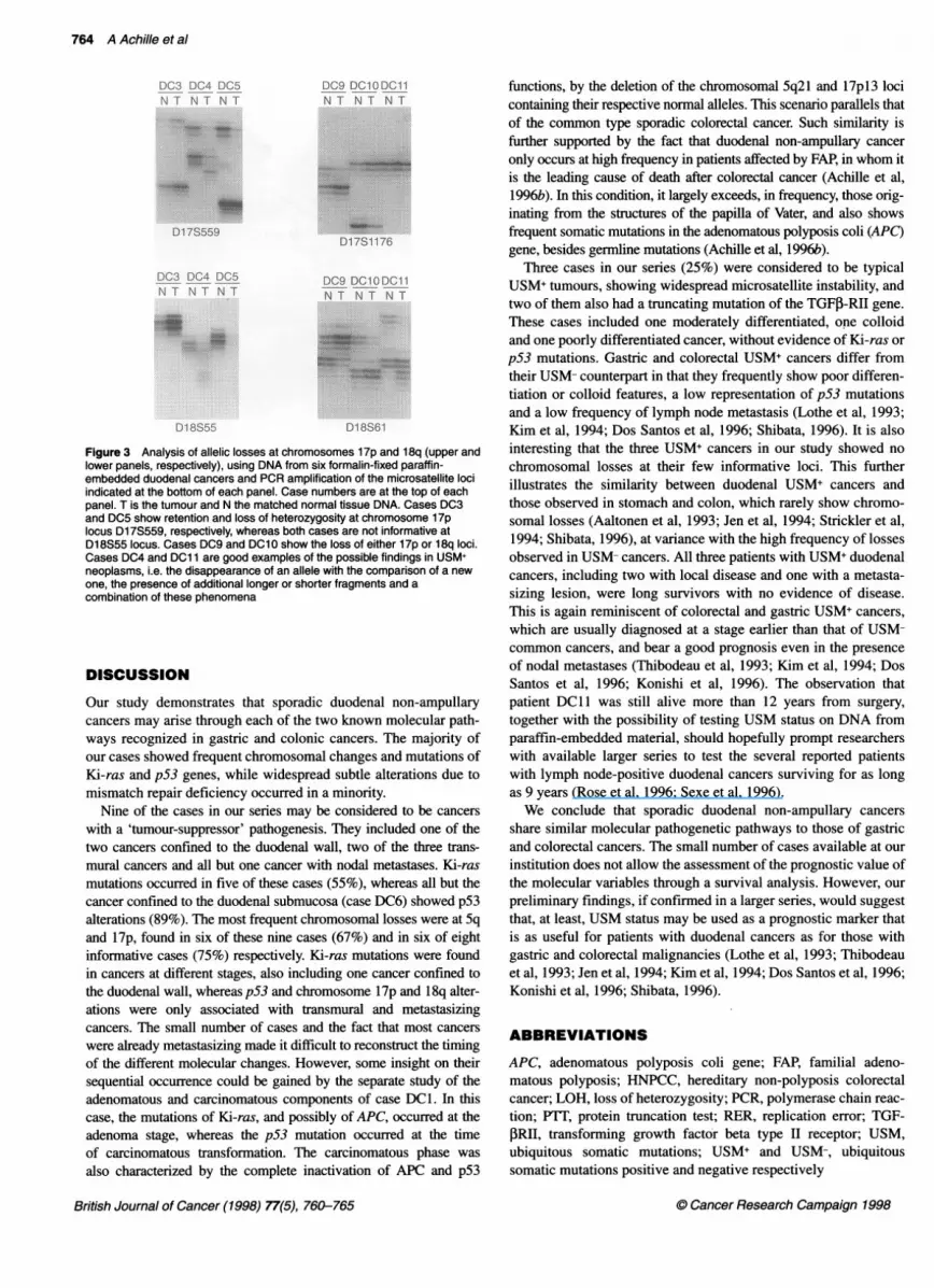
Figure 3 Analysis of allelic losses at chromosomes 1 7p and 1 8q (upper andlower panels, respectively), using DNA from six formalin-fixed paraffin-embedded duodenal cancers and PCR amplification of the microsatellite lociindicated at the bottom of each panel. Case numbers are at the top of eachpanel. T is the tumour and N the matched normal tissue DNA. Cases DC3and DC5 show retention and loss of heterozygosity at chromosome 1 7plocus D17S559, respectively, whereas both cases are not informative atD1 8S55 locus. Cases DC9 and DC10 show the loss of either 17p or 18q loci.Cases DC4 and DC1 1 are good examples of the possible findings in USM+neoplasms, i.e. the disappearance of an allele with the comparison of a newone, the presence of additional longer or shorter fragments and acombination of these phenomena
DISCUSSION
Our study demonstrates that sporadic duodenal non-ampullarycancers may arise through each of the two known molecular path-ways recognized in gastric and colonic cancers. The majority ofour cases showed frequent chromosomal changes and mutations ofKi-ras and p53 genes, while widespread subtle alterations due tomismatch repair deficiency occurred in a minority.
Nine of the cases in our series may be considered to be cancerswith a 'tumour-suppressor' pathogenesis. They included one of thetwo cancers confined to the duodenal wall, two of the three trans-mural cancers and all but one cancer with nodal metastases. Ki-rasmutations occurred in five of these cases (55%), whereas all but thecancer confined to the duodenal submucosa (case DC6) showed p53alterations (89%). The most frequent chromosomal losses were at Sqand 17p, found in six of these nine cases (67%) and in six of eightinformative cases (75%) respectively. Ki-ras mutations were foundin cancers at different stages, also including one cancer confined tothe duodenal wall, whereas p53 and chromosome 17p and 18q alter-ations were only associated with transmural and metastasizingcancers. The small number of cases and the fact that most cancerswere already metastasizing made it difficult to reconstruct the timingof the different molecular changes. However, some insight on theirsequential occurrence could be gained by the separate study of theadenomatous and carcinomatous components of case DC1. In thiscase, the mutations of Ki-ras, and possibly of APC, occurred at theadenoma stage, whereas the p53 mutation occurred at the timeof carcinomatous transformation. The carcinomatous phase wasalso characterized by the complete inactivation of APC and p53
functions, by the deletion of the chromosomal 5q21 and 17pl3 locicontaining their respective normal alleles. This scenario parallels thatof the common type sporadic colorectal cancer. Such similarity isfurther supported by the fact that duodenal non-ampullary canceronly occurs at high frequency in patients affected by FAP, in whom itis the leading cause of death after colorectal cancer (Achille et al,1996b). In this condition, it largely exceeds, in frequency, those orig-inating from the structures of the papilla of Vater, and also showsfrequent somatic mutations in the adenomatous polyposis coli (APC)gene, besides germline mutations (Achille et al, 1996b).
Three cases in our series (25%) were considered to be typicalUSM+ tumours, showing widespread microsatellite instability, andtwo of them also had a truncating mutation of the TGFP-RII gene.These cases included one moderately differentiated, one colloidand one poorly differentiated cancer, without evidence of Ki-ras orp53 mutations. Gastric and colorectal USM+ cancers differ fromtheir USM- counterpart in that they frequently show poor differen-tiation or colloid features, a low representation of p53 mutationsand a low frequency of lymph node metastasis (Lothe et al, 1993;Kim et al, 1994; Dos Santos et al, 1996; Shibata, 1996). It is alsointeresting that the three USM+ cancers in our study showed nochromosomal losses at their few informative loci. This furtherillustrates the similarity between duodenal USM+ cancers andthose observed in stomach and colon, which rarely show chromo-somal losses (Aaltonen et al, 1993; Jen et al, 1994; Strickler et al,1994; Shibata, 1996), at variance with the high frequency of lossesobserved in USM- cancers. All three patients with USM+ duodenalcancers, including two with local disease and one with a metasta-sizing lesion, were long survivors with no evidence of disease.This is again reminiscent of colorectal and gastric USM+ cancers,which are usually diagnosed at a stage earlier than that of USM-common cancers, and bear a good prognosis even in the presenceof nodal metastases (Thibodeau et al, 1993; Kim et al, 1994; DosSantos et al, 1996; Konishi et al, 1996). The observation thatpatient DC11 was still alive more than 12 years from surgery,together with the possibility of testing USM status on DNA fromparaffin-embedded material, should hopefully prompt researcherswith available larger series to test the several reported patientswith lymph node-positive duodenal cancers surviving for as longas 9 years (Rose et al, 1996; Sexe et al, 1996).We conclude that sporadic duodenal non-ampullary cancers
share similar molecular pathogenetic pathways to those of gastricand colorectal cancers. The small number of cases available at ourinstitution does not allow the assessment of the prognostic value ofthe molecular variables through a survival analysis. However, ourpreliminary findings, if confirmed in a larger series, would suggestthat, at least, USM status may be used as a prognostic marker thatis as useful for patients with duodenal cancers as for those withgastric and colorectal malignancies (Lothe et al, 1993; Thibodeauet al, 1993; Jen et al, 1994; Kim et al, 1994; Dos Santos et al, 1996;Konishi et al, 1996; Shibata, 1996).
ABBREVIATIONS
APC, adenomatous polyposis coli gene; FAP, familial adeno-matous polyposis; HNPCC, hereditary non-polyposis colorectalcancer; LOH, loss of heterozygosity; PCR, polymerase chain reac-tion; PTT, protein truncation test; RER, replication error; TGF-PRII, transforming growth factor beta type II receptor; USM,ubiquitous somatic mutations; USM+ and USM-, ubiquitoussomatic mutations positive and negative respectively
British Journal of Cancer (1998) 77(5), 760-765 0 Cancer Research Campaign 1998

Duodenal cancer 765
ACKNOWLEDGEMENTS
This study was supported by grants from Consorzio per gli StudiUniversitari and Banca Popolare di Verona, Verona; MURST 60%and 40%, Roma; and Associazione Italiana Ricerca Cancro(AIRC), Milano.
REFERENCES
Aaltonen L, Peltomaki P, Leach F, Sistonen P, Pylkkanen L, Mecklin J, Jarvinen H,Powell S, Jen J, Hamilton S, Petersen G, Kinzler K, Vogelstein B and de laChapelle A (1993) Clues to the pathogenesis of familial colorectal cancer.Science 260: 812-816
Achille A, Scarpa A, Montresor M, Scardoni M, Zamboni G, Chilosi M, Capelli P,Franzin G and Menestrina F (1995) Routine application of polymerase chainreaction in the diagnosis of monoclonality of B-cell lymphoid proliferations.Diagn Mol Pathol 4: 14-24
Achille A, Biasi MO, Zamboni G, Bogina G, Magalini AR, Pederzoli P, Perucho Mand Scarpa A (1996a) Chromosome 7q allelic losses in pancreatic carcinoma.Cancer Res 56: 3808-3813
Achille A, Scupoli MT, Magalini AR, Zamboni G, Romanelli MG, Orlandini S,Biasi MO, Lemoine NR, Accolla RS and Scarpa A (1996b) APC genemutations and allelic losses in sporadic ampullary tumours. Int J Cancer 68:305-312
AJCC (1992) Small Intestine. In Manualfor Staging of Cancer, Behars 0, HensonD, Hutter R and Kennedy B. (eds), pp. 69-73. JB Lippincott: Philadelphia
Brennan M (1990) Duodenal cancer. Asian J Surg 13: 204-209Dos Santos NR, Seruca R, Constancia M, Seixas M and Sobrinho-Simoes M (1996)
Microsatellite instability at multiple loci in gastric carcinoma:clinicopathologic implications and prognosis. Gastroenterology 110: 38-44
Hahn SA, Seymour AB, Shamsul Hoque ATM, Schutte M, da Costa LT, RedstonMS, Caldas C, Weinstein CL, Fischer A, Yeo CJ, Hruban RH and Kern SE(1995) Allelotype of pancreatic adenocarcinoma using xenograft enrichment.Cancer Res 55: 4670-4675
Hruban R, van-Mansfeld A, Offerhaus G, van-Weering D, Allison D, Goodman S,Kensler T, Bose K, Cameron J and Bos J (1993) K-ras oncogene activation inadenocarcinoma of the human pancreas. A study of 82 carcinomas using acombination of mutant-enriched polymerase chain reaction analysis and allele-specific oligonucleotide hybridization (Review). Am J Pathol 143: 545-554
Ionov Y, Pineado MA, Malkhosyan S, Shibata D and Perucho M (1993) Ubiquitoussomatic mutations in simple repeated sequences reveal a new mechanism forcolonic carcinogenesis. Nature 363: 558-561
Jen J, Kim HG, Piantadosi S, Liu ZF, Levitt RC, Sistonen P, Kinzler KW, VogelsteinB and Hamilton SR (1994) Allelic loss of chromosome 18q and prognosis incolorectal cancer. New Engl J Med 331: 213-221
Kim H, Jen J, Vogelstein B and Hamilton SR (1994) Clinical and pathologicalcharacteristic of sporadic colorectal carcinomas with DNA replication errors inmicrosatellites sequences. Am J Pathol 145: 148-156
Kinzler K and Vogelstein B (1996) Lessons from hereditary colorectal cancer. Cell87: 159-170
Klempnauer J, Ridder GJ and Pichlmayr R (1995) Prognostic factors after resectionof ampullary carcinoma: multivariate survival analysis in comparison withductal cancer of the pancreatic head. Br J Surg 82: 1686-1691
Konishi M, Kikuchi-Yanoshita R, Tanaka K, Muraoka M, Onda A, Okumura Y,Kishi N, Iwama T, Mori T, Koike M, Ushio K, Chiba M, Nomizu S, Konishi F,Utsunomiya J and Miyaki M (1996) Molecular nature of colon tumors inhereditary nonpolyposis colon cancer, familial polyposis, and sporadic coloncancer. Gastroenterology 111: 307-317
Lin HY, Wang XF, Ng-Eaton E, Weinberg RA and Lodish HF (1992) Expressioncloning of the TGF-beta type II receptor, a functional transmembraneserine/threonine kinase. Cell 68: 775-785
Liu B, Nicolaides NC, Markowitz S, Willson JK, Parson RE, Jen J, Papadopolous N,Peltomaki P, de la Chapelle A, Hamilton SR, Kinzler KW and Vogelstein B(1995a). Mismatch repair gene defects in sporadic colorectal cancers withmicrosatellite instability. Nature Genet 9: 48-55
Liu B, Farrington SM, Petersen GM, Hamilton SR, Parsons R, Papadopoulos N,Fujiwara T, Jen J, Kinzler KW, Wyllie AH, Vogelstein B and Dunlop MG(1995b) Genetic instability occurs in the majority of young patients withcolorectal cancer. Nature Med 1: 348-352
Lothe RA, Peltomaki P, Meling GI, Aaltonen LA, Nystrom-Lahti M, Pylkkanen L,Heimdal K, Andersen TI, Moller P, Rognum TO, Fossa SD, Haldorsen T,Langmark F, Brogger A, de la Chapelle A and Borresen AL (1993) Genomicinstability in colorectal cancer: relationship to clinicopathological variables andfamily history. Cancer Res 53: 5849-5852
Lynch HT, Smyrk T, Watson P, Lanspa SJ, Lynch JF, Lynch PM, Cavalieri J andBoland RC (1993) Genetics, natural history, tumor spectrum, and pathology ofhereditary nonpolyposis colorectal cancer: an updated review.Gastroenterology 104: 1535-1549
Markowitz S, Wang J, Myeroff LL, Parsons R, Sun LZ, Lutterbaugh J, Fan RS,Zborowska E, Kinzler KW, Vogelstein B, Brattain M and Willson JKW (1995)Inactivation of the type II TGF-beta receptor in colon cancer cells withmicrosatellite instability. Science 268: 1336-1338
Myeroff LL, Parsons R, Kim SJ, Hedrick L, Cho KR, Orth K, Matis M, Kinzler KW,Lutterbaugh J, Park K, Bang YJ, Lee HY, Park JG, Lynch HT, Roberts AB,Vogelstein B and Markowitz S (1995) A transforming growth factor betareceptor type II gene mutation common in colon and gastric but rare inendometrial cancers with microsatellite instability. Cancer Res 55: 5545-5547
Ohta M, Inoue H, Cotticelli MG, Kastury K, Baffa R, Palazzo J, Siprashvili Z, MoriM, McCue P, Druck T, Croce CM and Huebner K (1996) The FHIT gene,spanning the chromosome 3pl4.2 fragile site and renal carcinoma-associatedt(3;8) breakpoint, is abnormal in digestive tract cancers. Cell 84: 587-597
Parsons R, Myeroff LL, Liu B, Willson JKW, Markowitz SD, Kinzler KW andVogelstein B (1995) Microsatellite instability and mutations of thetransforming growth factor beta type II receptor gene in colorectal cancer.Cancer Res 55: 5548-5550
Perucho M (1996) Microsatellite instability: the mutator that mutates the othermutator. Nature Med 2: 630-631
Rose D, Hochwald S, Klimstra D and Brennan M (1996) Primary duodenaladenocarcinoma: a ten year experience with 79 patients. JAm Coll Surg 183:89-96
Scarpa A, Capelli P, Zamboni G, Oda T, Mukai K, Bonetti F, Martignoni G, IaconoC, Serio G and Hirohashi S (1993) Neoplasia of the ampulla of Vater: Ki-rasand p53 mutations. Am J Pathol 142: 1163-1172
Sexe R, Wade T, Virgo K and Johnson F (1996) Incidence and treatment ofperiampullary duodenal cancer in the U.S. veteran patient population. Cancer77: 251-254
Shibata D (1996) Loss of DNA mismatch repair: life in the fast lane?Gastroenterology 109: 1685-1699
Strickler JG, Zheng J, Shu Q, Burgart LJ, Alberts SR and Shibata D (1994) p53mutations and microsatellite instability in sporadic gastric cancer: whenguardians fail. Cancer Res 54: 4750-4755
Thibodeau SN, Bren G and Schaid D (1993) Microsatellite instability in cancer ofthe proximal colon. Science 260: 816-819
Umar A, Boyer JC, Thomas DC, Nguyen DC, Risinger JI, Boyd J, Ionov Y, PeruchoM and Kunkel TA (1994) Defective mismatch repair in extracts of colorectaland endometrial cancer cell lines exhibiting microsatellite instability. J BiolChem 269:14367-14370
Vogelstein B, Fearon ER, Hamilton SR, Kern SE, Preisinger AC, Leppert M,Nakamura Y, White R, Smits AMM and Bos JL (1988) Genetic alterationsduring colorectal tumor development. New Engl J Med 319: 525-532
Wang J, Sun LZ, Myeroff L, Wang X, Gentry LE, Yang J, Liang J, Zborowska E,Markowitz S, Willson JKV and Brattain MG (1995) Demonstration thatmutation of the type II transforming growth factor beta receptor inactivates itstumor suppressor activity in replication error-positive colon carcinoma cells.J Biol Chem 270: 22044-22049
C Cancer Research Campaign 1998 British Journal of Cancer (1998) 77(5), 760-765


