Embed Size (px)
Citation preview

A New Method for the Characterization of Strain-SpecificConformational Stability of Protease-Sensitive andProtease-Resistant PrPSc
Laura Pirisinu1, Michele Di Bari1, Stefano Marcon1, Gabriele Vaccari1, Claudia D’Agostino1, Paola Fazzi1,
Elena Esposito1, Roberta Galeno2, Jan Langeveld3, Umberto Agrimi1, Romolo Nonno1*
1 Department of Veterinary Public Health and Food Safety, Istituto Superiore di Sanita, Rome, Italy, 2 Department of Cell Biology and Neurosciences, Istituto Superiore di
Sanita, Rome, Italy, 3 Central Veterinary Institute, Wageningen UR, Lelystad, The Netherlands
Abstract
Although proteinacious in nature, prions exist as strains with specific self-perpetuating biological properties. Prion strainsare thought to be associated with different conformers of PrPSc, a disease-associated isoform of the host-encoded cellularprotein (PrPC). Molecular strain typing approaches have been developed which rely on the characterization of protease-resistant PrPSc. However, PrPSc is composed not only of protease-resistant but also of protease-sensitive isoforms. The aim ofthis work was to develop a protocol for the molecular characterization of both, protease-resistant and protease-sensitivePrPSc aggregates. We first set up experimental conditions which allowed the most advantageous separation of PrPC andPrPSc by means of differential centrifugation. The conformational solubility and stability assay (CSSA) was then developed bymeasuring PrPSc solubility as a function of increased exposure to GdnHCl. Brain homogenates from voles infected withhuman and sheep prion isolates were analysed by CSSA and showed strain-specific conformational stabilities, with mean[GdnHCl]1/2 values ranging from 1.6 M for MM2 sCJD to 2.1 for scrapie and to 2.8 M for MM1/MV1 sCJD and E200K gCJD.Interestingly, the rank order of [GdnHCl]1/2 values observed in the human and sheep isolates used as inocula closelymatched those found following transmission in voles, being MM1 sCJD the most resistant (3.3 M), followed by sheep scrapie(2.2 M) and by MM2 sCJD (1.6 M). In order to test the ability of CSSA to characterise protease-sensitive PrPSc, we analysedsheep isolates of Nor98 and compared them to classical scrapie isolates. In Nor98, insoluble PrPSc aggregates were mainlyprotease-sensitive and showed a conformational stability much lower than in classical scrapie. Our results show that CSSA isable to reveal strain-specified PrPSc conformational stabilities of protease-resistant and protease-sensitive PrPSc and that it isa valuable tool for strain typing in natural hosts, such as humans and sheep.
Citation: Pirisinu L, Di Bari M, Marcon S, Vaccari G, D’Agostino C, et al. (2010) A New Method for the Characterization of Strain-Specific Conformational Stability ofProtease-Sensitive and Protease-Resistant PrPSc. PLoS ONE 5(9): e12723. doi:10.1371/journal.pone.0012723
Editor: Jason C. Bartz, Creighton University, United States of America
Received June 15, 2010; Accepted August 2, 2010; Published September 14, 2010
Copyright: � 2010 Pirisinu et al. This is an open-access article distributed under the terms of the Creative Commons Attribution License, which permitsunrestricted use, distribution, and reproduction in any medium, provided the original author and source are credited.
Funding: This work was supported by grants from the European Union (Neuroprion Network of Excellence CT-2004-506579) and the Italian Ministry of Health.The funders had no role in study design, data collection and analysis, decision to publish, or preparation of the manuscript.
Competing Interests: The authors have declared that no competing interests exist.
* E-mail: [email protected]
Introduction
Transmissible spongiform encephalopathies (TSEs), or prion
diseases, are neurodegenerative disorders that afflict humans and
others mammals. TSEs may have genetic, infectious, or sporadic
origins. Creutzfeldt-Jakob disease (CJD) is the most common TSE
in humans and may be sporadic (sCJD), genetic (gCJD), or
acquired (iatrogenic CJD). A novel human acquired prion disease,
variant CJD (vCJD), appeared from 1995 onwards and was
postulated to be caused by consumption of beef from cows infected
with bovine spongiform encephalopathy (BSE). The most common
forms of TSE in animals, scrapie in small ruminants, BSE in cattle
and chronic wasting disease (CWD) in deer, are all acquired. New
atypical forms of BSE in cattle, namely BSE-H [1] and BSE-L or
BASE [2] and atypical scrapie in small ruminants, namely Nor98
[3], are supposed to be sporadic.
All TSEs are characterised by the accumulation of PrPSc, a
misfolded form of the cellular protein PrPC. Besides differing in
conformation, the two forms of the prion protein have divergent
physical properties: while PrPC is soluble in nondenaturating
detergents, rapidly digested by proteases, and rich in a-helical
structure, PrPSc is insoluble in detergents, partially resistant to
proteolysis and contains mostly b-sheet [4,5,6].
A major problem for the protein-only hypothesis, which
postulates that prions are composed mainly or exclusively of
PrPSc [7], was to explain the existence of multiple strains of prions.
Prion strains are infectious isolates that, when propagated in the
same host, exhibit distinct prion disease phenotypes that persist
upon serial transmission [8,9,10]. Thus, prion strains cannot be
encoded by differences in PrP primary structure but anyway carry
information that are independent from the host [11]. The prion
hypothesis equates strains to different self-propagating conforma-
tional variants of PrPSc [12].
Several studies demonstrated that prion strains can be
distinguished based on different biochemical properties of PrPSc,
thus allowing a molecular strain typing approach to TSEs. These
studies are based on the electrophoretic features of the protease
resistant core of PrPSc [13,14,15], the relative proteinase K
PLoS ONE | www.plosone.org 1 September 2010 | Volume 5 | Issue 9 | e12723

resistance of PrPSc [16,17,18], or the physico-chemical behaviour
of PrPSc during denaturation [19,20] and strongly support the
view that distinct strains show different PrPSc conformations.
A conformational stability assay (CSA), combining guanidine
hydrochloride (GdnHCl) denaturation with limited proteolysis
using proteinase K, showed that different prion strains may exhibit
distinct denaturation profiles [20]. A conformation-dependent
immunoassay (CDI) showed the existence of multiple strain-
specified PrPSc conformers by quantifying the immunoreactivity of
native and denatured PrPSc of eight hamster prion isolates [19].
This technique also showed that prion-infected brains contain
both protease-sensitive (sPrPSc) and protease-resistant PrPSc
(rPrPSc). More recently, it was shown by CDI that sPrPSc
represents as much as 90% of total PrPSc in the brain of patients
with sporadic CJD [21].
These findings along with the recent discovery in humans [22]
and animals [3] of previously undetected TSEs characterised by
relatively protease sensitive PrPSc, highlights the need of molecular
strain typing methods able to recognize PrPSc populations based
on their physical properties rather than only based on protease
digestion.
In this study we aimed at developing a new conformational
stability assay based on the differential solubility of PrPC and PrPSc
[23,24], that we called CSSA (conformational stability and
solubility assay). We have previously transmitted CJD and scrapie
isolates to bank voles [25,26], which showed to be a valuable tool
for the biological characterization of the most common forms of
sCJD, gCJD and scrapie. We took advantage of these vole-adapted
strains in order to evaluate the potential of CSSA for strain
discrimination.
We first set up experimental conditions allowing the most
advantageous separation of PrPC and PrPSc, and thus performed
the conformational stability assay by measuring PrPSc solubility in
homogenates treated with increasing concentrations of GdnHCl in
the absence of proteinase K. Indeed, insoluble PrP was inversely
correlated to GdnHCl concentration, and dose-response curves
allowed estimation of the concentration of GdnHCl able to
solubilise 50% of PrPSc.
Additionally, we extended the study to natural isolates of sheep
scrapie and human sCJD cases to investigate the potential of
CSSA for strain discrimination in natural hosts. Our results show
that this method is valuable for the biochemical typing of strains in
voles and it is also a promising tool for molecular analysis of
natural prion isolates.
Results
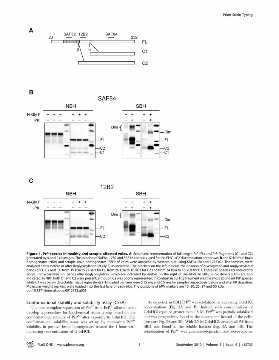
PrP species in healthy and diseased brainIt has been previously reported that normal PrP is composed of
full-length PrP (FL-PrP) as well as of 2 C-terminal fragments
derived from physiological cleavage at the a and b sites: C1, which
is the most represented, derives from a PrP cleavage at position
111/112, while C2 is usually barely detectable and is cleaved
around the octarepeat region [27]. Interestingly, a cleavage
disrupts the conserved neurotoxic and amyloidogenic region
comprising residues 106–126 of PrP, preventing the generation
of PrPSc, while b cleavage occurs upstream this conserved region.
Accordingly, C2 was reported to be enriched in diseased brains
and insoluble in nondenaturing detergents, similarly to PrPres [28].
In order to determine if C1 and C2 were detectable in voles, we
analysed healthy and diseased brain homogenates either before or
after deglycosylation, with SAF84, whose epitope is present in both
fragments, and 12B2 which recognises an epitope that is present
only in C2 (Fig. 1A). In normal brain homogenate (NBH), FL-PrP
was accompanied by substantial amounts of C1 and lower
amounts of C2. In contrast, in voles infected with the Italian
scrapie isolate SS7 (SBH) [26] C2 was strongly enhanced while C1
was not easily detectable (Fig. 1B and C). Furthermore, the PrPres
fragment generated after PK digestion in SBH was similar to C2
(Fig. 1B and C). Finally, in SBH PrP dimers were present which
were completely absent in NBH.
Separation of PrPC and PrPSc
In order to develop a conformational stability assay based on the
differential solubility of PrPC and PrPSc we first set up the
experimental conditions which enabled the most advantageous
separation of PrPC from PrPSc. This was obtained through a
conventional procedure based on centrifugation in the presence of
detergents.
By varying concentrations of different detergents, times of
centrifugation and centrifugal force (see Material and Methods),
we found that treatment with 2% sarcosyl followed by centrifu-
gation at 20.000 g for 1 h enabled an optimal separation of PrPC
from PrPSc.
In these conditions, .95% of the total PrPC in NBH was soluble
and was found in the supernatant fraction (Fig. 2A, left panel)
while in SBH most of PrP was sedimented (Fig. 2A, right panel).
Unfractionated SBH contained high amounts of PrPres which
exclusively sedimented to the pellet fraction (Fig.2A). Insoluble PrP
from SBH was indeed mostly PK-resistant (,90% of insoluble
PrP), while soluble PrP was PK-sensitive. All fractions from NBH
were devoid of PrPres (Fig. 2A).
Furthermore, soluble and insoluble PrP fractions from SBH
displayed slightly different banding patterns which suggested that
C2 was mainly insoluble, while soluble PrP contained FL-PrP and
the C1 fragment (Fig. 2A). Moreover, PrPSc dimers were clearly
segregated in the insoluble fraction (Fig. 2A). The differential
solubility of C1 and C2 in SBH was confirmed by the analysis of
deglycosylated PrP species (Fig. 2B), which showed that C1 was
completely soluble while C2 was mainly sedimented in the pellet
fraction (compare ‘‘S’’ and ‘‘P’’ lanes in Fig. 2B, left panel). With
the aim to mimic a situation comparable to that expected in
animals with pre-clinical disease, we also studied the differential
solubility of C1 and C2 after mixing equal amounts of NBH and
SBH (Fig. 2B). Indeed, under these conditions the PrPC content is
increased compared to SBH, as can be seen by the higher
proportion of C1 in mixed NBH/SBH compared to SBH alone
(compare ‘‘Tot’’ lanes in the two panels of Fig. 2B). Also in this
condition, C1 was completely soluble and the pellet fraction was
enriched in C2 (compare ‘‘S’’ and ‘‘P’’ lanes in Fig. 2B, right
panel).
Finally, we investigated the efficacy of our solubility assay for
separating PrPC and PrPSc in voles infected with other, non
scrapie-derived prion strains (Fig. 2C). For these experiments we
used voles infected with MM1 and MM2 sCJD [25]. In both
strains a substantial amount of PrP was found in the insoluble
fraction after detergent treatment and centrifugation. As already
observed in SBH, the banding patterns of insoluble and soluble
PrP were distinct, suggesting a specific precipitation of PrP species
associated to disease, namely C2 and PrP dimers (compare ‘‘S’’
and ‘‘P’’ lanes in Fig. 2C). Furthermore, after PK digestion, PrPres
was strongly enriched in the pellets and virtually absent in the
supernatants.
Collectively, these findings strongly suggest that under the
experimental conditions described above we were able to
specifically precipitate PrPSc in brain homogenates from voles
infected with different prion strains.
Prion Strain Typing
PLoS ONE | www.plosone.org 2 September 2010 | Volume 5 | Issue 9 | e12723
Conformational stability and solubility assay (CSSA)The near complete separation of PrPC from PrPSc allowed us to
develop a procedure for biochemical strain typing based on the
conformational stability of PrPSc after exposure to GdnHCl. The
conformational solubility assay was set up by measuring PrPSc
solubility in positive brain homogenates treated for 1 hour with
increasing concentrations of GdnHCl.
As expected, in SBH PrPSc was solubilized by increasing GdnHCl
concentrations (Fig. 3A and B). Indeed, with concentrations of
GdnHCl equal or greater than 1.5 M, PrPSc was partially solubilised
and was progressively found in the supernatant instead of the pellet
(compare Fig. 3A and 3B). With 3.5 M GdnHCl, virtually all PrP from
SBH was found in the soluble fraction (Fig. 3A and 3B). The
solubilization of PrPSc was guanidine-dependent and dose-response
Figure 1. PrP species in healthy and scrapie-affected voles. A: Schematic representation of full length PrP (FL) and PrP fragments (C1 and C2)generated by a and b cleavages. The location of SAF84, 12B2 and SAF32 epitopes used for the FL/C1/C2 discrimination are shown. B and C: Normal brainhomogenates (NBH) and scrapie brain homogenates (SBH) of voles were analyzed by western blot using SAF84 (B) and 12B2 (C). The samples, wereanalyzed either before or after deglycosylation (N-Gly F) as indicated. The brackets on the left indicate the position of glycosylated and unglycosylatedbands of FL, C2 and C1: from 35 kDa to 27 kDa for FL, from 26 kDa to 18 kDa for C2 and from 24 kDa to 16 kDa for C1. These PrP species are reduced tosingle unglycosylated PrP bands after deglycosylation, which are indicated by dashes on the right of the blots. In SBH, PrPSc dimers (Dim) are alsoindicated. In NBH both C1 and C2 were present, although C2 was poorly represented; in contrast in SBH C2 fragment was the most abundant PrP specieswhile C1 was barely detectable. Tissue equivalents (TE) loaded per lane were 0,15 mg and 0.5 mg for samples respectively before and after PK digestion.Molecular weight markers were loaded into the last lane of each blot. The positions of MW markers are 15, 20, 25, 37 and 50 kDa.doi:10.1371/journal.pone.0012723.g001
Prion Strain Typing
PLoS ONE | www.plosone.org 3 September 2010 | Volume 5 | Issue 9 | e12723

curves enabled estimation of the concentration of GdnHCl able to
solubilise 50% of PrPSc ([GdnHCl]1/2). The [GdnHCl]1/2 value was
similar when estimated either in the pellet or in the supernatant
fractions (Fig. 3C). In contrast, PrP from NBH remained soluble within
the range of GdnHCl concentrations tested (Fig. 3D). Denaturation of
PrPSc was complete after incubation with GdnHCl for 1 hour, as very
similar denaturation curves were obtained when the treatment was
extended up to 4 hours (Fig. S1). When the denaturation curves where
measured in replica blots with mAbs recognising different PrP species,
namely SAF32 and SAF84 (see scheme in Fig. 1A), we obtained similar
[GdnHCl]1/2 values, which suggest that C2 and FL PrPSc share the
same conformational stability (Fig. S2).
We then studied the relationship between insolubility and PK-
resistance of PrPSc during denaturation. In order to investigate if
denaturation equally affects insolubility and PK-resistance, we
compared denaturation curves derived from the same SBH, either
obtained by insoluble PrPSc (CSSA) or by PK-resistant PrPSc
(CSA). As shown in Fig. 4, the curves of insoluble PrPSc (Fig. 4A)
and PrPres (Fig. 4B) didn’t show differences (Fig. 4C), suggesting
that insolubility and PK-resistance were equally susceptible to
denaturation by GdnHCl. This finding was further confirmed in
experiments aimed at investigating whether, after denaturation,
solubilized PrPSc could partially preserve its resistance to
proteinase K. Indeed, after denaturation with 3 M GdnHCl,
soluble PrP was fully susceptible to protease digestion (Fig. 4D).
Conformational stability of scrapie, sCJD and gCJD PrPSc
from bank volesUnder the experimental conditions described above, we
investigated the potential of CSSA for differentiating prion strains.
As reported in previous studies [25,26] sCJD, gCJD and some
scrapie isolates present distinct and specific patterns of transmis-
sion in voles, based on survival times, lesion profiles, PrPSc
deposition and PrPres biochemical properties. We have shown that
voles infected with MM1/MV1 sCJD and E200K gCJD isolates
were characterised by a PrPres fragment of ,19 kDa, MM2 sCJD
showed a PrPres fragment of ,17 kDa, while natural scrapie
isolates and murine scrapie ME7 were characterised by a PrPres
fragment of ,18 kDa, intermediate between types 1 and 2 CJD.
Since these PrPSc types showed specific molecular characteris-
tics after PK digestion, we investigated the conformational stability
of PrPSc in voles infected with MM1 (n = 4), MV1 (n = 3) and
MM2 (n = 5) sCJD, E200K gCJD (n = 3) and scrapie SS7 (n = 4).
Interestingly, CSSA revealed distinct denaturation profiles
(Fig. 5), with [GdnHCl]1/2 values ranging from 1.5 M to 3 M
(Table 1). Voles infected with MM1 sCJD, MV1 sCJD and E200K
gCJD showed the highest resistance to denaturation, with mean
[GdnHCl]1/2 values of 2.77 M, 2.88 M and 2.88 M, respectively.
In contrast, PrPSc from voles infected with MM2 sCJD was the most
sensitive (mean [GdnHCl]1/2 value of 1.63 M), while scrapie
infected voles showed intermediate [GdnHCl]1/2 values (2.10 M).
We then combined the individual curves within each group in order
to compare the denaturation profiles of the 5 groups (Fig. 5B).
Scrapie SS7 ([GdnHCl]1/2 value of 2.1060.02) and MM2 sCJD
([GdnHCl]1/2 value of 1.6160.03) showed strain specific denatur-
ation profiles, being significantly different from all other groups (SS7
vs MV1, P = 0.0037; SS7 vs MM1, P,0.0001; SS7 vs E200K,
P,0.0001; SS7 vs MM2, P,0.0001; MM2 vs MV1, P = 0.0022;
MM2 vs MM1, P,0.0001; MM2 vs E200K, P,0.0001). In
contrast, MM1 sCJD, MV1 sCJD and E200K gCJD were not
significantly different among them (shared [GdnHCl]1/2 value of
2.7960.06, P = 0.52) possibly representing a distinct strain, in
agreement with previous findings which showed comparable
phenotypes of disease in voles infected with these 3 isolates [25].
Figure 2. Separation of PrPC and PrPSc in vole brain homog-enates. A: Western blot analysis of soluble and insoluble PrP fractionsfrom normal brain homogenate (NBH) and scrapie brain homogenate(SBH) of voles. Samples were centrifuged at 20000 g for 1 h in presenceof 2% sarcosyl, and supernatants (S) and pellets (P) were analysedbefore (+) and after (2) PK treatment. Aliquots of samples beforecentrifugation (Tot) were analysed too. TE per lane were 0,2 mg for‘‘Tot’’ and ‘‘S’’ lanes, and 0.4 mg for ‘‘P’’ lanes. Brackets on the leftindicate the position of the FL, C1 and C2 PrP fragments. Dimers (Dim)of PrPSc are indicated on the right in SBH blot. B: Western blot analysisof soluble and insoluble PrP fractions from scrapie brain homogenate(SBH) and an artificially mixed sample (SBH+NBH). Samples werecentrifuged as described and total (Tot), supernatant (S) and pellet (P)fractions were deglycosylated. Full-length PrP (FL), C1 and C2 PrPfragments are indicated on the left. In each lane, 0.02 mg TE wereloaded. C: Western blot analysis of soluble and insoluble PrP from brainhomogenates of voles infected with MM1 and MM2 sCJD. The sampleswere treated as in panel A and total (Tot), supernatant (S) and pellet (P)fractions were analysed with or without PK treatment. In each lane0.3 mg TE were loaded. Brackets on the left indicate the position of theFL, C1 and C2 fragments. Dimers (Dim) of PrPSc are indicated on the left.A–C: Membranes were probed with SAF84. Molecular size markers areshown in kilodaltons on the right of each panel.doi:10.1371/journal.pone.0012723.g002
Prion Strain Typing
PLoS ONE | www.plosone.org 4 September 2010 | Volume 5 | Issue 9 | e12723

Conformational stability of human and sheep isolatesSince we were interested in exploiting our conformational
solubility assay (CSSA) for strain discrimination in natural prion
diseases, we analysed 3 of the isolates used as inocula for
transmission to voles, namely the human MM1 and MM2 sCJD
isolates and the sheep scrapie SS7.
At first we tested the efficiency of separation of PrPSc from PrPC
in human and sheep brain homogenates, under the same
experimental conditions used for vole brain homogenates. In all
samples, PrPSc was enriched in the pellet, as showed by the
segregation of PrPres in the insoluble fraction and by the distinct
banding patterns shown by soluble and insoluble PrP (Fig. S3). In
negative sheep brain homogenates the fraction of PrP segregating
in the pellet was somewhat higher than in vole brains (.10% in
some experiment). However, this sedimented PrPC was insensitive
to GdnHCl and thus did not interfere with our assay (Fig. S4).
When tested by CSSA, MM1 sCJD, MM2 sCJD and SS7 scrapie
revealed different susceptibilities to denaturation (Fig. 5A and B).
Interestingly, the rank order of conformational stability and the
[GdnHCl]1/2 values observed closely matched those found in the vole
counterpart, being MM1 sCJD the most resistant ([GdnHCl]1/2 =
3.31 M), followed by scrapie SS7 ([GdnHCl]1/2 = 2.23 M) and MM2
sCJD ([GdnHCl]1/2 = 1.63 M).
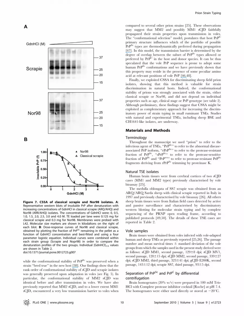
Conformational stability of classical and Nor98 scrapieisolates from sheep
In order to test the ability of CSSA for studying the
conformational stability of protease-sensitive PrPSc, we took
advantage of the recently described atypical scrapie strain,
Nor98 [3], which has been shown to induce the accumulation of
relatively protease-sensitive PrPSc [29]. As previously observed for
all other strains investigated, insoluble and soluble Nor98 PrPSc
showed different segregation of PrP species (Fig. 6). Interestingly
the 12 kDa PrPres fragment, which is a characteristic feature of
Nor98 PrPres [3,30], was observed before PK-digestion and
segregated with insoluble PrP, in a manner similar to what was
observed for C2 in all other strains. However, in Nor98 only a
minimal fraction of insoluble PrP was protease resistant (Fig. 6),
while PrPres represented .90% of insoluble PrP in classical scrapie
(Fig. 6). These findings confirm that in Nor98 samples disease-
Figure 3. Conformational stability and solubility assay in healthy and diseased voles. A and B: Western blot analysis of scrapie brainhomogenate (SBH) after denaturation with various concentrations of GdnHCl and separation of insoluble (A) and soluble (B) fractions bycentrifugation. A: Immunoblot of pellets (P) at different concentrations of GdnHCl. In order to compare the fractions in the same blot, supernatant (S)at 0 M GdnHCl was loaded too. In the pellets, insoluble PrP decreased with increasing concentrations of GdnHCl (M). In each lane 0.4 mg TE wereloaded. B: Immunoblot of supernatants at different concentrations of GdnHCl. In the first lane pellet (P) at 0 M GdnHCl was loaded too. In thesupernatant PrP increased with increasing GdnHCl. In each lane 0.4 mg TE were loaded. Note that the FL and C1 PrP fragments are present in thesupernatant after treatment with 0 M and 1 M GdnHCl, while the PrPSc-specific fragment C2 is visible in the supernatant from 1.5 M GdnHClonwards, in parallel with the decrease of insoluble PrP in panel A. C: The conformational stability of PrPSc in SBH was analysed by denaturation curvesbest-fitted by plotting the fraction of PrPSc in the pellet (P) and in the supernatant (S), depicted in panel A and B respectively, as a function of GdnHClconcentration. [GdnHCl]1/2 values were 1.92 M in the pellet and 1.91 M in the supernatant fraction. D: Western blot analysis of supernatants (S) atdifferent concentrations of GdnHCl from normal brain homogenate (NBH). In the first lane pellet (P) at 0 M GdnHCl was loaded. In NBH, PrPC wasmostly in supernatant fraction and remained soluble at all GdnHCl concentrations tested. Samples were loaded as 0.4 mg tissue equivalent into eachlane. A, B, D: Membranes were probed with SAF84. Molecular size markers are shown in kilodaltons on the left of each panel.doi:10.1371/journal.pone.0012723.g003
Prion Strain Typing
PLoS ONE | www.plosone.org 5 September 2010 | Volume 5 | Issue 9 | e12723

associated PrP is mostly PK-sensitive, although accompanied by
low amounts of genuine PK-resistant PrPSc.
We then studied the conformational stability of Nor98 (n = 5)
and classical (n = 4) Italian field scrapie cases of different PrP
genotypes (Table 2). Classical scrapie samples gave denaturation
curves very similar to that of SS7, with [GdnHCl]1/2 values
ranging from 1.96 to 2.31 (Fig. 7 and Table 2). Nor98 samples
gave remarkably similar denaturation profiles, independently of
PrP genotype, and showed high sensitivity to GdnHCl denatur-
ation (Fig. 7), with [GdnHCl]1/2 values ranging from 1.26 to 1.43
(Table 2). Within group comparison didn’t show significant
differences among classical scrapie (shared [GdnHCl]1/2 value of
2.0960.11, P = 0.52) and Nor98 (shared [GdnHCl]1/2 value of
1.3660.04, P = 0.50). Comparison of the combined classical
scrapie and Nor98 denaturation profiles (Fig. 7B) gave statistically
significant different [GdnHCl]1/2 values (P,0.0001).
Discussion
The main pathogenetic event in TSEs involves the trans-
conformation of PrPC to PrPSc, which leads to the accumulation of
detergent-insoluble and partially protease-resistant aggregates.
Thus, resistance to PK digestion and insolubility are the hallmarks
of PrPSc. However, previous studies on the molecular character-
ization of PrPSc were mainly focused on the PK-resistant core of
PrPSc, due to the difficulties in differentiating PrPC from PrPSc in
infected brain homogenates. Notwithstanding, it is becoming
increasingly clear that insoluble but protease-sensitive isoforms of
PrP are involved in different animal and human prion diseases
[19,22,31,32,33,34,35]. These PrP isoforms have been detected by
several methods, including conformation-dependent immunoassay
[19,21,33], immunological capture [36,37], differential centrifu-
gation [31,32], thermolysin digestion [35] and cold PK digestion
[34].
Among the above-mentioned techniques able to detect sPrPSc,
the CDI was also reported to distinguish eight hamster prion
strains based on PrPSc conformation, by plotting the ratio of
antibody binding to denatured/native PrP as a function of the
concentration of PrPSc [19]. However, CDI depends on the
availability of monoclonal antibodies able to recognise buried
epitopes in PrPSc, such as the 3F4. We developed a protocol for
the molecular characterization of PrPSc aggregates which does not
rely on their protease resistance but is based on a conventional
procedure of differential centrifugation in the presence of
detergents and on the solubilization of PrPSc aggregates upon
denaturation with increasing concentrations of GdnHCl. Our
findings show that CSSA is reliable and straightforward, and that
it is able to discriminate PrPSc conformers associated with different
Figure 4. Relationship between PrPSc insolubility and resistance to PK. The same SBH was analyzed in parallel by CSSA (A) and CSA (B), andthe denaturation curves obtained were compared (C). A and B: Western blot analysis of insoluble PrPSc (A) and PK-resistant PrPSc (B) afterdenaturation of homogenates with increasing concentrations of GdnHCl. In each lane 0.2 mg TE were loaded. The concentrations of GdnHCl were:0.5, 1.0, 1.5, 2.0, 2.5, 3.0 and 4.0 M. C: Graph depicting the denaturation curves obtained by CSSA (panel A) and CSA (panel B). [GdnHCl]1/2 valueswere 2.14 M and 2.23 M for CSSA and CSA, respectively. D: Western blot showing that PrPSc loses PK-resistance upon solubilization. In absence oftreatment with GdnHCl (0), total PrP (Tot) from SBH was resistant to PK digestion (compare PK- and PK+ lanes). After treatment with 3 M GdnHCl (3)and differential centrifugation, most of PrPSc was found in the supernatant (S) and was also made PK-susceptible. In absence of treatment withGdnHCl (0), the supernatant (S) contained normal PrP which was susceptible to PK. Note the different banding patterns in the supernatantscontaining PrPC (0) and solubilised PrPSc (3). A, B, D: Membranes were probed with SAF84. Molecular size markers are shown in kilodaltons on the leftof each panel.doi:10.1371/journal.pone.0012723.g004
Prion Strain Typing
PLoS ONE | www.plosone.org 6 September 2010 | Volume 5 | Issue 9 | e12723

Figure 5. Conformational stability and solubility assay of different strains in voles and natural hosts. A and B: Conformational stabilityof PrPSc from voles inoculated with MM1 sCJD, Scrapie and MM2 sCJD and from the respective human and sheep isolates. A: Representative westernblots of insoluble PrPSc from voles (left column) and natural hosts (on the right column) after treatment of homogenates with increasingconcentrations of GdnHCl. The concentrations of GdnHCl were: 0, 0.5, 1.0, 1.5, 2.0, 2.5, 3.0 and 4.0 M. TE loaded per lane were 0.2 mg for vole samples,0.34 mg for human isolates and 0.125 mg for the scrapie isolate. Membranes were probed with SAF84 (vole samples and sheep scrapie) or with L42(human isolates). B: Dose-response curves in vole strains, obtained by plotting the fraction of PrPSc remaining in the pellet as a function of GdnHClconcentration and best-fitted and using a four parameter logistic equation. Individual curves were combined within each strain group (Scrapie, E200KgCJD, MV1, MM1 and MM2 sCJD) in order to compare the denaturation profiles of the 5 groups. C: Dose-response curves from sheep and humanisolates, obtained by plotting the fraction of PrPSc remaining in the pellet as a function of GdnHCl concentration and best-fitted and using a fourparameter logistic equation. [GdnHCl]1/2 values of MM1, MM2 and scrapie isolates were 3,31 M, 1,63 M and 2,23 M respectively.doi:10.1371/journal.pone.0012723.g005
Prion Strain Typing
PLoS ONE | www.plosone.org 7 September 2010 | Volume 5 | Issue 9 | e12723

TSE strains. Compared to CDI, we believe that CSSA offer some
interesting advantages: i) it does not depend on the antibody used
and can be thus exploited to compare PrPSc conformational
stability in different species, ii) the proof that CDI is able to
discriminate prion strains in natural hosts is still lacking, while here
we show the potential of CSSA to be used in human and sheep
natural isolates.
In previous studies [20,38,39] a conformational stability assay
(CSA) able to discriminate PrPSc conformers was set up by
measuring the extent of loss in protease resistance as a function of
increased exposure to GdnHCl. This method proved to be very
helpful for molecular strain typing in different species [40,41,42]
and was also recently exploited for investigating some basic
mechanisms of prion replication [39]. The protocol that we
developed is conceptually similar to CSA, as it derives information
on the conformational stability of PrPSc aggregates. Indeed, we
showed that CSA and CSSA gave very similar denaturation
profiles in vole-adapted scrapie brain homogenates (see Fig. 4).
However, we believe that CSSA represents a step forward in prion
molecular typing and offers several advantages compared with
previously used protocols. With CSA, in fact, susceptibility to PK
is exploited to distinguish denaturated from native PrPSc.
However, different prion strains may have distinct susceptibilities
to PK, while CSA uses the same PK concentration to derive the
level of PrPSc denaturation in different strains. Furthermore, when
performing CSA it is necessary to dilute the GdnHCl to allow the
activity of proteolytic enzyme. However, it is known that PrPSc
unfolding can be a partially reversible phenomenon and, as
reported, the dilution of the denaturant could restore the original
protease-resistance of PrPSc [43]. With CSSA this problem was
circumvented by avoiding any change in denaturant concentration
during the assay. Indeed, the denaturation step is followed by the
centrifugal separation of soluble and insoluble fractions, which is
performed under conditions identical to the denaturation step.
Most importantly, CSSA allowed the characterization of
protease-sensitive PrPSc, and thus the direct comparison of the
conformational stabilities of TSE strains associated with protease-
sensitive and protease-resistant PrPSc. We investigated the
potential of CSSA for strain typing of ovine field isolates of
Nor98, which are characterised by high amounts of insoluble
sPrPSc [29, present paper]. Our findings seem very encouraging,
since Nor98 samples displayed a distinct denaturation profile,
although the isolates derived from sheep of various PrP genotypes
and presumably at different stage of the disease. Furthermore,
Nor98 samples were easily discriminated from classical scrapie,
which represents a different strain. Thus CSSA enabled
characterization of the conformational stability of a protease
sensitive strain, for which methods based on proteolysis would
have characterised only a minimal amount of PrPSc present in the
brain homogenate. This is promising in view of recent studies
[21,35] showing that in sCJD and vCJD isolates, as much as 90%
of PrPSc in the brains was estimated to be sPrPSc. Furthermore the
recent discovery of protease sensitive prionopathy (PSPr), a
previously unrecognised human prion disease [22], might suggest
that prion diseases characterised by protease-sensitive PrP isoforms
are more frequent than previously thought. Brain homogenates
from PSPr cases were reported to contain mainly a protease
sensitive form of insoluble PrP, accompanied by low amounts of
typical protease-resistant PrP [22], similarly to our findings in
Nor98 samples. It would be interesting to investigate the potential
of CSSA for characterising these human protease-sensitive disease-
related isoforms of PrP.
Another interesting feature of CSSA is that, by avoiding PK
treatment, it allowed the characterization of FL-PrPSc, including
the N-terminus which is cleaved upon PK digestion. Besides FL-
PrP, in vivo trimmed PrPSc fragments were selectively precipitated
and thus CSSA also conveyed information on their conformational
stability. These fragments comprised C2 C-terminal fragments in
scrapie and sCJD isolates, as well as the 12 kDa internal fragment
Table 1. Conformational stability of PrPSc in vole-adaptedsCJD, gCJD and scrapie.
Inoculum Vole ID[GdnHCl]1/2
(M) ± SEM[GdnHCl]1/2 (M)(mean ± SD)
MM1 sCJD 304/2 2.7360.17 2.7760.13
304/4 2.6360.12
304/8 2.7660.11
304/14 2.9460.19
MV1 sCJD 272/1 3.0560.29 2.8860.17
272/4 2.8760.36
272/8 2.7260.11
MM2 sCJD 270/6 1.4560.01 1.6360.11
270/7 1.7160.07
350/3 1.6060.09
350/5 1.6960.04
350/14 1.6960.06
E200K gCJD 271/4 2.7360.15 2.8860.13
271/5 2.9960.25
271/6 2,9160.18
scrapie SS7 388/7 2.2260.04 2.1060.13
388/9 2.1660.03
388/10 1.9260.02
388/11 2.0860.03
doi:10.1371/journal.pone.0012723.t001
Figure 6. Separation of soluble and insoluble PrP fractions inclassical scrapie and Nor98. Western blot analysis of classical scrapieand Nor98 ovine isolates. Supernatant (S) and pellet (P) fractions wereanalysed with (+) or without (2) PK treatment. In each lane 0.8 mg TEwere loaded. Molecular size markers are shown in kilodaltons on the leftof the blot. Membrane was probed with L42.doi:10.1371/journal.pone.0012723.g006
Prion Strain Typing
PLoS ONE | www.plosone.org 8 September 2010 | Volume 5 | Issue 9 | e12723
characteristic of Nor98, and may be easily distinguished from FL-
PrP, also by means of differential antibody binding (data not
shown; see for example Fig. S2). This is interesting because it
enables a comparison of the conformational properties of PrPSc
aggregates made-up of FL-PrP or in vivo trimmed PrPSc fragments.
In situ epitope-mapping of PrPSc, indeed, showed that FL and C2
PrPSc aggregates have different cellular localizations [44].
Furthermore, by analysing thermolysin-resistant PrPSc in sheep
scrapie and BSE isolates, Owen and colleagues have recently
shown the potential of C2 fragments for strain typing [45].
The main potential drawback of CSSA might be the incomplete
separation or PrPC and PrPSc. Herein, we developed a protocol
able to minimise this problem, notwithstanding 1–4% of PrPC in
brain homogenates from healthy voles was found in the pellet
under our working conditions. There are however several lines of
evidence suggesting that this potential drawback did not interfere
with CSSA results. We have shown that brain homogenates from
clinically affected voles contain low levels of soluble PrP (bona fide
PrPC) (see Fig. 2). Based on the results obtained with NBH, less
than 5% of PrPC is expected to be found in the pellet; on the other
hand, in diseased brains bona fide PrPC was 5–30% of total PrP.
From these considerations, it can be argued that the PrPSc/PrPC
ratio in the pellet should be higher than 2 orders of magnitude,
which represents the working range of CSSA. Furthermore, we
obtained clear-cut differential solubility of PrPC and PrPSc even
after increasing the PrPC/PrPSc ratio in the homogenate, by
mixing SBH with NBH (Fig. 2). Most importantly, we found that
the sedimented PrPC fraction was insensitive to denaturation (Fig.
S4) and thus it does not interfere with CSSA.
We investigated the potential of CSSA for strain discrimination
by analysing vole-adapted strains deriving from human sCJD,
gCJD and sheep scrapie which we have previously shown to give
distinct strains in voles [25,26]. In previous studies we have shown
that voles infected with MM1/MV1 sCJD and E200K gCJD
isolates showed identical transmission patterns and were char-
acterised by an unglycosylated PrPres fragment of ,19 kDa, while
MM2 sCJD showed an unglycosylated PrPres fragment of
,17 kDa. In contrast, all Italian scrapie isolates studied so far
[26,46], as well as the murine scrapie strain ME7 [25], were
characterised by the accumulation of an unglycosylated PrPres with
a molecular weight (,18 kDa) intermediate between CJD types 1
and 2, upon transmission to voles. These different PrPres types
derive from different PK-cleavage sites of PrPSc, which in turn are
believed to reflect distinct conformations of PrPSc aggregates. With
CSSA we showed that these three PrPres types are actually
characterised by PrPSc aggregates with distinct susceptibilities to
denaturation by GdnHCl. Indeed, PrPSc from voles infected with
MM1 sCJD, MV1 sCJD and E200K gCJD, characterised by
19 kDa PrPres, showed the highest resistance to denaturation,
while sCJD MM2, characterised by 17 kDa PrPres, was the most
susceptible and scrapie SS7, that was characterised by 18 kDa
PrPres, displayed intermediate susceptibility. Interestingly, the
within group variability of [GdnHCl]1/2 values was very low (see
table 1) and allowed statistical comparisons among the different
groups, which strengthened the view that [GdnHCl]1/2 values
reflect strain-specific rather than individual PrPSc properties.
It has been recently suggested that the conformational stability
of PrPSc is directly proportional to the length of the incubation
time in mice [39]. On this point, it is worth noting that our results
in voles, although based on only 5 isolates, seem to contradict this
conclusion. Indeed, the lowest conformational stability was
associated with prions with the longest survival times, i.e. MM2
sCJD with a survival time of ,330 days post-inoculation (dpi),
while scrapie SS7 (survival time of ,90 dpi) and MM1/MV1
sCJD (survival time of ,130 dpi) showed higher conformational
stabilities and shorter survival times compared to MM2 sCJD.
Further studies with an extended panel of isolates are needed to
investigate if the direct relationship between conformational
stability and incubation time observed in mice holds true also
for vole prions.
We also explored the potential of CSSA for strain typing of
natural prion diseases. To this aim we analysed three of the human
and sheep isolates which were used for bioassay in vole.
Interestingly, we found that the two isolates from MM1 sCJD
and MM2 sCJD patients could be easily discriminated by their
conformational stabilities, with MM1 sCJD displaying lower
susceptibility to denaturation compared to MM2 sCJD
([GdnHCl]1/2 values of 3,31 M for MM1 sCJD and 1,63 M for
MM2 sCJD). These findings compare well with previous studies by
CSA, which showed that PrPSc associated with MM1 sCJD was ,2-fold more stable than that of MM2 sCJD, with [GdnHCl]1/2
values of 2,76 M and 1,63 M for MM1 sCJD and MM2 sCJD
[40]. Of course, this needs confirmation in a larger set of isolates.
Furthermore, this approach allowed us to compare the
conformational stability of prions in their natural host and after
transmission to voles. It has been previously shown that prion
strains can either maintain their biological properties or mutate
upon propagation in a new host species [10]. More recently it was
reported that a change in conformation was accompanied by the
emergence of a new prion strain during interspecies transmission,
Table 2. Conformational stability of PrPSc in sheep field prion isolates.
Strain Sheep ID Age (y) Clinical signs PrP Genotype* [GdnHCl]1/2 (M) [GdnHCl]1/2 (M) (mean±SD)
Classical scrapie ES8/09/3 2 Yes ALRQ/ALRQ 2.27 2.2060.16
ES8/09/1 6 No ALRQ/ALHQ 1.96
241/105 3 No ALRQ/ALRQ 2.27
211/26 3 Yes ALRQ/ALRQ 2.31
Nor98 ES36/08/4 12 No ALHQ/ALRR 1.26 1.3260.07
ES35/07/2 9 No ALRQ/ALHQ 1.26
ES10/07/7 12 No AFRQ/AFRQ 1.35
ES18/07/2 5 No ALRQ/ALHQ 1.31
ES19/07/23 7 No ALRR/ALRR 1.43
*amino acids at codons 136, 141, 154 and 171.doi:10.1371/journal.pone.0012723.t002
Prion Strain Typing
PLoS ONE | www.plosone.org 9 September 2010 | Volume 5 | Issue 9 | e12723
while the conformational stability of PrPSc was preserved when a
strain ‘‘bred true’’ in the new host [38]. Our findings show that the
rank order of conformational stability of sCJD and scrapie isolates
was generally preserved upon adaptation in voles (see Fig. 5). In
particular, the conformational stability of MM2 sCJD was
identical before and after transmission in voles. We have also
previously reported that MM2 sCJD, and to a lower extent MM1
sCJD, encountered a very low transmission barrier to voles when
compared to several other prion strains [25]. These observations
may suggest that MM2 and possibly MM1 sCJD faithfully
propagated their strain properties upon transmission in voles.
The ‘‘conformational selection’’ model, postulates that host PrPC
primary structure influences which of the portfolio of possible
PrPSc types are thermodynamically preferred during propagation
[47]. In this model, the transmission barrier is determined by the
degree of overlap between the subset of PrPSc types allowed or
preferred by PrPC in the host and donor species. It can be thus
speculated that the vole PrP sequence is prone to adopt some
human PrPSc conformations and we have previously shown that
this property may reside in the presence of some peculiar amino
acid at relevant positions of vole PrP [46,48].
Finally, we exploited CSSA for discriminating sheep field prion
isolates, showing that this method is valuable for strain
discrimination in natural hosts. Indeed, the conformational
stability of prions was strongly associated with the strain, either
classical scrapie or Nor98, and did not depend on individual
properties such as age, clinical stage or PrP genotype (see table 2).
Although preliminary, these findings suggest that CSSA might be
exploited as complementary approach for increasing the discrim-
inatory power of strain typing in small ruminant TSEs. Studies
with natural and experimental TSEs, including sheep BSE and
CH1641-like isolates, are underway.
Materials and Methods
TerminologyThroughout the manuscript we used ‘‘prion’’ to refer to the
infectious agent of TSEs, ‘‘PrPSc’’ to refer to the abnormal disease-
associated PrP isoform, ‘‘rPrPSc’’ to refer to the protease-resistant
fraction of PrPSc, ‘‘sPrPSc‘‘ to refer to the protease-sensitive
fraction of PrPSc and ‘‘PrPres’’ to refer to protease-resistant PrPSc
fragments deriving from rPrPSc trimming by proteinase K.
Natural TSE isolatesHuman brain tissues were from cerebral cortices of two sCJD
cases (MM1 and MM2 types) previously characterised by vole
bioassay [25].
The medulla oblongata of SS7 scrapie was obtained from an
ARQ/ARQ Sarda sheep with clinical scrapie reported in Italy in
1997 and previously characterised by vole bioassay [26]. All others
sheep brain tissues were from Italian field cases detected by active
and passive surveillance and characterised by discriminatory
western blotting for molecular strain typing and by complete
sequencing of the PRNP open reading frame, according to
published protocols [49,50]. The details of these TSE cases are
reported in Table 2.
Vole samplesBrain tissues were obtained from voles infected with vole-adapted
human and sheep TSEs as previously reported [25,26]. The passage
number and mean survival times 6 standard deviation of the vole
groups from which the samples used in the present study derived were
as follows: sCJD MM1, second passage, 12968 dpi; sCJD MV1,
second passage, 128615 dpi; sCJD MM2, second passage, 339627
dpi; sCJD MM2, third passage, 323641 dpi; gCJD E200K, second
passage, 143612 dpi; scrapie SS7, third passage, 9365 dpi.
Separation of PrPSc and PrPC by differentialcentrifugation
Brain homogenates (20% w/v) were prepared in 100 mM Tris-
HCl with Complete protease inhibitor cocktail [Roche] at pH 7.4.
The homogenates were either used directly or stored at 220uC.
Figure 7. CSSA of classical scrapie and Nor98 isolates. A:Representative western blots of insoluble PrP after denaturation withincreasing concentrations of GdnHCl in classical scrapie (ARQ/AHQ) andNor98 (ARR/AHQ) isolates. The concentrations of GdnHCl were: 0, 0.5,1.0, 1.5, 2.0, 2.5, 3.0 and 4.0 M. TE loaded per lane were 0.125 mg forclassical scrapie and 0.3 mg for Nor98. Membranes were probed withL42. Molecular size markers are shown in kilodaltons on the right ofeach blot. B: Dose-response curves of Nor98 and classical scrapie,obtained by plotting the fraction of PrPSc remaining in the pellet as afunction of GdnHCl concentration and best-fitted and using a fourparameter logistic equation. Individual curves were combined withineach strain group (Scrapie and Nopr98) in order to compare thedenaturation profiles of the two groups. Individual [GdnHCl]1/2 valuesare shown in Table 2.doi:10.1371/journal.pone.0012723.g007
Prion Strain Typing
PLoS ONE | www.plosone.org 10 September 2010 | Volume 5 | Issue 9 | e12723

The experimental conditions for PrPC/PrPSc separation were
set up in vole brain homogenates by studying the effect of different
detergents, centrifugal force and time of centrifugation.
Brain homogenates (3% to 12% w/v) were added with equal
volumes of different buffers (100 mM Tris-HCl at pH 7.4
containing Sarcosyl 4% or 2%; 100 mM Tris-HCl at pH 7.4
containing NaDoc 1%, NP40 1%; 100 mM Tris-HCl at pH 7.4
containing Triton X-100 2%) and incubated for 1 hour at 37uCwith gentle shaking. Then samples were centrifuged at 10000 to
20000 g for 1 h or 2 h. The obtained pellets were re-suspended
with 100 mM Tris-HCl (pH 7.4) containing the relevant deter-
gent. The experimental conditions then used throughout the paper
in vole, sheep or human brain tissues included solubilisation in
100 mM Tris-HCl at pH 7.4 containing sarcosyl 2% and
centrifugation at 20000 g for 1 h. For each of the different
experimental conditions tested, equivalent aliquots of brain
homogenate before centrifugation, along with supernatant and
pellet fractions, were analysed by western blot either with or
without PK digestion.
Conformational stability and solubility assayAliquots of brain homogenates (3% to 6% w/v) were added
with an equal volume of 100 mM Tris-HCl (pH 7.4) containing
sarcosyl 4% and incubated for 1 h at 37uC with gentle shaking.
Aliquots of 100 ml were treated with 100 ml of guanidine
hydrochloride (GdnHCl) solutions with a final concentration
ranging from 0 to 4.0 M. GdnHCl stock solutions were prepared
from an 8 M solution (Pierce) diluted in water. After treatment
with GdnHCl for 1 h at 37uC with gentle shaking, samples were
centrifuged at 20000 g for 1 h at 22uC. Pellets were re-suspended
in 90 ml NuPage LDS Sample Buffer (Invitrogen) and 10 ml
NuPage Sample Reducing Agent (Invitrogen). Aliquots of
supernatants were precipitated with 4-fold volume excess of pre-
chilled methanol 30 min at 220uC, centrifuged at 15000 g for
30 min at 4uC and then were re-suspended in 90 ml NuPage LDS
Sample Buffer (Invitrogen) and 10 ml NuPage Sample Reducing
Agent (Invitrogen). Supernatant and pellet fractions were analysed
by Western blotting.
Individual denaturation curves were analyzed and best-fitted by
plotting the fraction of PrPSc remaining in the pellet as a function
of GdnHCl concentration, and using a four parameter logistic
equation (GraphPad Prism). In order to fit denaturation curves for
each prion strain, the mean fraction of PrPSc remaining in the
pellet 6 SD were plotted. Statistical comparison of [GdnHCl]1/2
values were made by comparing the best-fit value for each data set
with GraphPad Prism. This was performed by either fitting each
data set independently or doing a global fit with a shared
[GdnHCl]1/2 value, and then the results were compared with an F
test. The simpler model was selected unless the extra sum-of-
squares F test had a P value,0.05.
Digestion with proteinase K after denaturation withGdnHCl
Aliquots of the same brain homogenate were treated in parallel
according to CSSA and conformational stability assay (CSA)
protocols. The CSA was performed as described [20], with minor
modifications. Aliquots of brain homogenates (6% w/v) were
added with an equal volume of 100 mM Tris-HCl (pH 7.4)
containing sarcosyl 4% and incubated for 1 h at 37uC with gentle
shaking. Aliquots of 50 ml were added with 50 ml of GdnHCl to
give a final concentration ranging from 0 to 4.0 M. After 1 h of
incubation at 37uC all samples were diluted to a final
concentration of 0.4 M GdnHCl. Proteinase K (50 mg/ml) was
added and the samples were incubated for 1 h at 37uC with gentle
shaking. The reaction was stopped with 3 mM PMSF (Sigma).
Aliquots of samples were added with an equal volume of
isopropanol/butanol (1:1 v/v) and centrifuged at 20000 g for
5 min. Pellets were re-suspended in NuPage LDS Sample Buffer
(Invitrogen) and were analysed by Western Blotting.
Western blot analysisElectrophoresis and Western blotting were performed as
previously described [25]. Samples were denatured by adding
NuPage LDS Sample Buffer (Invitrogen, Carlsbad, California,
United States) and NuPage Sample Reducing Agent (Invitrogen),
and heating for 10 min at 90uC. After centrifugation at 10000 g
for 5 min each sample was loaded onto 12% bis-Tris polyacryl-
amide gels (Invitrogen). After electrophoresis and Western blotting
on PVDF membranes (Immobilon-P; Millipore, Bedford, MA,
USA), the blots were processed by SNAP i.d.TM Protein Detection
System (Millipore) as described by the manufacturer instructions.
The monoclonal antibodies used, their epitopes on sheep PrP
and the working dilutions were as follow: SAF84, PrP residues
167–173, 1.2 mg/ml; L42, PrP residues 148–153, 0.28 mg/ml;
12B2, PrP residues 93–97, 2.4 mg/ml and SAF32, PrP octarepeat,
2.4 mg/ml. Horseradish peroxidase-conjugated anti-mouse immu-
noglobulin (Pierce Biotechnology, Rockford, Illinois, United
States) was used as secondary antibody (1:13000).
The membranes were developed with an enhanced chemilu-
minescence method (SuperSignal Femto, Pierce). Chemilumines-
cence signal was detected with the VersaDoc imaging system (Bio-
Rad) and was quantified by QuantityOne software (Bio-Rad).
Deglycosylation was performed by adding 18 ml of 0.2 M
sodium phosphate buffer (pH 7.4) containing 0.8% Nonidet P40
(Roche) and 2 ml (80 U/ml) di N-Glycosidase F (Roche) to 5 ml of
denaturated samples and by incubating overnight at 37uC with
gentle shaking. Samples were then analysed by Western blotting as
described above.
Supporting Information
Figure S1 Effect of denaturation time on CSSA. Dose-response
curves of insoluble PrP from brain homogenates of voles infected
with scrapie SS7 (top panel) and MM1 sCJD (bottom panel) after
treatment with increasing concentrations of GdnHCl for 1, 2 or
4 hours. Denaturation curves were best-fitted by plotting the
fraction of PrP remaining in the pellet as a function of GdnHCl
concentration. SS7 and MM1 didn’t reveal differences based on
time of treatment and showed the same [GdnHCl]1/2 values at 1,
2 and 4 hours (2.1 M for SS7 e 3 M for MM1 sCJD).
Found at: doi:10.1371/journal.pone.0012723.s001 (0.13 MB TIF)
Figure S2 CSSA with different mAbs. A: Representative
western blots of insoluble PrP from vole infected with E200K
gCJD after denaturation with increasing concentrations of
GdnHCl. Replica blots were probed with SAF84 (top) and
SAF32 (bottom), as indicated on the left of the blot. The
concentrations of GdnHCl were: 0, 0.5, 1.0, 1.5, 2.0, 2.5, 3.0
and 4.0 M. In each lane 0.2 mg TE were loaded. B: Dose-
response curves derived from the blots in panel A, obtained by
plotting the fraction of PrP remaining in the pellet as a function of
GdnHCl concentration and best-fitted and using a four-parameter
logistic equation.
Found at: doi:10.1371/journal.pone.0012723.s002 (0.57 MB TIF)
Figure S3 Separation of soluble and insoluble PrP fractions from
human and sheep isolates. Western blot analysis of soluble and
insoluble PrP from brain homogenates of sheep with classical
scrapie and human with MM1 sCJD. Samples were centrifuged at
Prion Strain Typing
PLoS ONE | www.plosone.org 11 September 2010 | Volume 5 | Issue 9 | e12723

20000 g for 1 h in presence of 2% Sarcosyl, and supernatants (S)
and pellets (P) were analysed with (+) or without (2) PK treatment.
Aliquots of samples before centrifugation (Tot) were analysed too.
TE loaded per lane were 0.2 mg for classical scrapie and 0.15 mg
for MM1 sCJD. Scrapie membrane was probed with SAF84 and
MM1 sCJD membrane was probed with L42.
Found at: doi:10.1371/journal.pone.0012723.s003 (0.48 MB TIF)
Figure S4 Separation of soluble and insoluble PrP fractions from
sheep normal brain homogenates. A: Western blot analysis of
soluble and insoluble PrP fractions from a representative sheep
negative brain homogenate. Total PrP (Tot) and PrP from
supernatant (S) and pellet (P) fractions were analysed with (+) or
without (2) PK treatment. TE loaded per lane were 0.2 mg. B:
Western blot analysis of insoluble PrP from sheep negative and
scrapie brain homogenates after denaturation with increasing
concentrations of GdnHCl, either after normal (top panel) or long
(bottom panel) exposure times. The concentrations of GdnHCl
were: 0, 0.5, 1.0, 1.5, 2.0, 2.5, 3.0 and 4.0 M. TE loaded per lane
were 0.12 mg. A and B: Membranes were probed with SAF84.
Found at: doi:10.1371/journal.pone.0012723.s004 (0.69 MB TIF)
Acknowledgments
The authors would like to thank Paolo Frassanito and Shimon Simson
(Istituto Superiore di Sanita) for animal care, and Consiglia Parisi (Istituto
Superiore di Sanita) for administrative management of projects and
editorial help.
Author Contributions
Conceived and designed the experiments: LP RN. Performed the
experiments: LP SM GV PF EE. Analyzed the data: LP RN. Contributed
reagents/materials/analysis tools: MADB CD RG JL. Wrote the paper: LP
JL UA RN.
References
1. Biacabe AG, Laplanche JL, Ryder S, Baron T (2004) Distinct molecular
phenotypes in bovine prion diseases. EMBO Rep 5: 110–115.
2. Casalone C, Zanusso G, Acutis P, Ferrari S, Capucci L, et al. (2004)
Identification of a second bovine amyloidotic spongiform encephalopathy:
molecular similarities with sporadic Creutzfeldt-Jakob disease. Proc Natl Acad
Sci U S A 101: 3065–3070.
3. Benestad SL, Sarradin P, Thu B, Schonheit J, Tranulis MA, et al. (2003) Cases
of scrapie with unusual features in Norway and designation of a new type,
Nor98. Vet Rec 153: 202–208.
4. Bolton DC, McKinley MP, Prusiner SB (1982) Identification of a protein that
purifies with the scrapie prion. Science 218: 1309–1311.
5. Meyer RK, McKinley MP, Bowman KA, Braunfeld MB, Barry RA, et al. (1986)
Separation and properties of cellular and scrapie prion proteins. Proc Natl Acad
Sci U S A 83: 2310–2314.
6. Pan KM, Baldwin M, Nguyen J, Gasset M, Serban A, et al. (1993) Conversion of
alpha-helices into beta-sheets features in the formation of the scrapie prion
proteins. Proc Natl Acad Sci U S A 90: 10962–10966.
7. Prusiner SB (1982) Novel proteinaceous infectious particles cause scrapie.
Science 216: 136–144.
8. Fraser H, Dickinson AG (1973) Scrapie in mice. Agent-strain differences in the
distribution and intensity of grey matter vacuolation. J Comp Pathol 83: 29–40.
9. Bruce ME, Fraser H (1991) Scrapie strain variation and its implications. Curr
Top Microbiol Immunol 172: 125–138.
10. Bruce ME (1993) Scrapie strain variation and mutation. Br Med Bull 49:
822–838.
11. Bruce ME, McConnell I, Fraser H, Dickinson AG (1991) The disease
characteristics of different strains of scrapie in Sinc congenic mouse lines:
implications for the nature of the agent and host control of pathogenesis. J Gen
Virol 72(Pt 3): 595–603.
12. Telling GC, Parchi P, DeArmond SJ, Cortelli P, Montagna P, et al. (1996)
Evidence for the conformation of the pathologic isoform of the prion protein
enciphering and propagating prion diversity. Science 274: 2079–2082.
13. Collinge J, Sidle KC, Meads J, Ironside J, Hill AF (1996) Molecular analysis of
prion strain variation and the aetiology of ‘new variant’ CJD. Nature 383:
685–690.
14. Parchi P, Castellani R, Capellari S, Ghetti B, Young K, et al. (1996) Molecular
basis of phenotypic variability in sporadic Creutzfeldt-Jakob disease. Ann Neurol
39: 767–778.
15. Bessen RA, Marsh RF (1994) Distinct PrP properties suggest the molecular basis
of strain variation in transmissible mink encephalopathy. J Virol 68: 7859–7868.
16. Kascsak RJ, Rubenstein R, Merz PA, Carp RI, Wisniewski HM, et al. (1985)
Biochemical differences among scrapie-associated fibrils support the biological
diversity of scrapie agents. J Gen Virol 66(Pt 8): 1715–1722.
17. Kuczius T, Groschup MH (1999) Differences in proteinase K resistance and
neuronal deposition of abnormal prion proteins characterize bovine spongiform
encephalopathy (BSE) and scrapie strains. Mol Med 5: 406–418.
18. Jacobs JG, Langeveld JP, Biacabe AG, Acutis PL, Polak MP, et al. (2007)
Molecular discrimination of atypical bovine spongiform encephalopathy strains
from a geographical region spanning a wide area in Europe. J Clin Microbiol 45:
1821–1829.
19. Safar J, Wille H, Itri V, Groth D, Serban H, et al. (1998) Eight prion strains have
PrP(Sc) molecules with different conformations. Nat Med 4: 1157–1165.
20. Peretz D, Scott MR, Groth D, Williamson RA, Burton DR, et al. (2001) Strain-
specified relative conformational stability of the scrapie prion protein. Protein
Sci 10: 854–863.
21. Safar JG, Geschwind MD, Deering C, Didorenko S, Sattavat M, et al. (2005)
Diagnosis of human prion disease. Proc Natl Acad Sci U S A 102: 3501–3506.
22. Gambetti P, Dong Z, Yuan J, Xiao X, Zheng M, et al. (2008) A novel human
disease with abnormal prion protein sensitive to protease. Ann Neurol 63:
697–708.
23. McKinley MP, Meyer RK, Kenaga L, Rahbar F, Cotter R, et al. (1991) Scrapie
prion rod formation in vitro requires both detergent extraction and limited
proteolysis. J Virol 65: 1340–1351.
24. Caughey B, Raymond GJ, Ernst D, Race RE (1991) N-terminal truncation of
the scrapie-associated form of PrP by lysosomal protease(s): implications
regarding the site of conversion of PrP to the protease-resistant state. J Virol
65: 6597–6603.
25. Nonno R, Di Bari MA, Cardone F, Vaccari G, Fazzi P, et al. (2006) Efficient
transmission and characterization of Creutzfeldt-Jakob disease strains in bank
voles. PLoS Pathog 2: e12.
26. Di Bari MA, Chianini F, Vaccari G, Esposito E, Conte M, et al. (2008) The bank
vole (Myodes glareolus) as a sensitive bioassay for sheep scrapie. J Gen Virol 89:
2975–2985.
27. Mange A, Beranger F, Peoc’h K, Onodera T, Frobert Y, et al. (2004) Alpha- and
beta- cleavages of the amino-terminus of the cellular prion protein. Biol Cell 96:
125–132.
28. Chen SG, Teplow DB, Parchi P, Teller JK, Gambetti P, et al. (1995) Truncated
forms of the human prion protein in normal brain and in prion diseases. J Biol
Chem 270: 19173–19180.
29. Buschmann A, Luhken G, Schultz J, Erhardt G, Groschup MH (2004) Neuronal
accumulation of abnormal prion protein in sheep carrying a scrapie-resistant
genotype (PrPARR/ARR). J Gen Virol 85: 2727–2733.
30. Bruce ME, Nonno R, Foster J, Goldmann W, Di Bari M, et al. (2007) Nor98-like
sheep scrapie in the United Kingdom in 1989. Vet Rec 160: 665–666.
31. Tzaban S, Friedlander G, Schonberger O, Horonchik L, Yedidia Y, et al. (2002)
Protease-sensitive scrapie prion protein in aggregates of heterogeneous sizes.
Biochemistry 41: 12868–12875.
32. Pastrana MA, Sajnani G, Onisko B, Castilla J, Morales R, et al. (2006) Isolation
and characterization of a proteinase K-sensitive PrPSc fraction. Biochemistry 45:
15710–15717.
33. Thackray AM, Hopkins L, Bujdoso R (2007) Proteinase K-sensitive disease-
associated ovine prion protein revealed by conformation-dependent immuno-
assay. Biochem J 401: 475–483.
34. Tremblay P, Ball HL, Kaneko K, Groth D, Hegde RS, et al. (2004) Mutant
PrPSc conformers induced by a synthetic peptide and several prion strains.
J Virol 78: 2088–2099.
35. Cronier S, Gros N, Tattum MH, Jackson GS, Clarke AR, et al. (2008) Detection
and characterization of proteinase K-sensitive disease-related prion protein with
thermolysin. Biochem J 416: 297–305.
36. Zou WQ, Zheng J, Gray DM, Gambetti P, Chen SG (2004) Antibody to DNA
detects scrapie but not normal prion protein. Proc Natl Acad Sci U S A 101:
1380–1385.
37. Nazor KE, Kuhn F, Seward T, Green M, Zwald D, et al. (2005)
Immunodetection of disease-associated mutant PrP, which accelerates disease
in GSS transgenic mice. EMBO J 24: 2472–2480.
38. Peretz D, Williamson RA, Legname G, Matsunaga Y, Vergara J, et al. (2002) A
change in the conformation of prions accompanies the emergence of a new prion
strain. Neuron 34: 921–932.
39. Legname G, Nguyen HO, Peretz D, Cohen FE, DeArmond SJ, et al. (2006)
Continuum of prion protein structures enciphers a multitude of prion isolate-
specified phenotypes. Proc Natl Acad Sci U S A 103: 19105–19110.
40. Cali I, Castellani R, Alshekhlee A, Cohen Y, Blevins J, et al. (2009) Co-existence
of scrapie prion protein types 1 and 2 in sporadic Creutzfeldt-Jakob disease: its
effect on the phenotype and prion-type characteristics. Brain 132: 2643–2658.
Prion Strain Typing
PLoS ONE | www.plosone.org 12 September 2010 | Volume 5 | Issue 9 | e12723

41. Scott MR, Peretz D, Nguyen HO, Dearmond SJ, Prusiner SB (2005)
Transmission barriers for bovine, ovine, and human prions in transgenic mice.J Virol 79: 5259–5271.
42. Green KM, Browning SR, Seward TS, Jewell JE, Ross DL, et al. (2008) The elk
PRNP codon 132 polymorphism controls cervid and scrapie prion propagation.J Gen Virol 89: 598–608.
43. Kocisko DA, Lansbury PT, Jr., Caughey B (1996) Partial unfolding andrefolding of scrapie-associated prion protein: evidence for a critical 16-kDa C-
terminal domain. Biochemistry 35: 13434–13442.
44. Jeffrey M, Martin S, Gonzalez L, Ryder SJ, Bellworthy SJ, et al. (2001)Differential diagnosis of infections with the bovine spongiform encephalopathy
(BSE) and scrapie agents in sheep. J Comp Pathol 125: 271–284.45. Owen JP, Rees HC, Maddison BC, Terry LA, Thorne L, et al. (2007) Molecular
profiling of ovine prion diseases by using thermolysin-resistant PrPSc andendogenous C2 PrP fragments. J Virol 81: 10532–10539.
46. Piening N, Nonno R, Di Bari M, Walter S, Windl O, et al. (2006) Conversion
efficiency of bank vole prion protein in vitro is determined by residues 155 and170, but does not correlate with the high susceptibility of bank voles to sheep
scrapie in vivo. J Biol Chem 281: 9373–9384.
47. Collinge J, Clarke AR (2007) A general model of prion strains and theirpathogenicity. Science 318: 930–936.
48. Agrimi U, Nonno R, Dell’Omo G, Di Bari MA, Conte M, et al. (2008) Prionprotein amino acid determinants of differential susceptibility and molecular
feature of prion strains in mice and voles. PLoS Pathog 4: e1000113.
49. Vaccari G, D’Agostino C, Nonno R, Rosone F, Conte M, et al. (2007) Prionprotein alleles showing a protective effect on the susceptibility of sheep to scrapie
and bovine spongiform encephalopathy. J Virol 81: 7306–7309.50. Mazza M, Iulini B, Vaccari G, Acutis PL, Martucci F, et al. (2010) Co-existence
of classical scrapie and Nor98 in a sheep from an Italian outbreak. Res Vet Sci88: 478–485.
Prion Strain Typing
PLoS ONE | www.plosone.org 13 September 2010 | Volume 5 | Issue 9 | e12723


