Embed Size (px)
Citation preview
Hindawi Publishing CorporationCase Reports in DentistryVolume 2013, Article ID 893791, 5 pageshttp://dx.doi.org/10.1155/2013/893791
Case ReportA Novel Approach for Treatment of an UnusualPresentation of Radicular Cysts Using Autologous Periosteumand Platelet-Rich Fibrin in Combination with DemineralizedFreeze-Dried Bone Allograft
Veena A. Patil,1 Manthan H. Desai,1 Veerendra S. Patil,2 Hanisha Reddy Kaveti,2
Kiran Kumar Ganji,3 and Prasanna M. Danappanavar4
1 Department of Periodontology, HKES’s S.Nijalingappa Institute of Dental Sciences and Research, Sedam Road, Gulbarga,Karnataka 585105, India
2Department of Endodontics, HKES’s S.Nijalingappa Institute of Dental Sciences and Research, Sedam Road, Gulbarga,Karnataka 585105, India
3 Department of Periodontics, Sharad Pawar Dental College & Hospital, Sawangi Meghe Wardha, Maharashtra 442004, India4Department of Oral and Maxillofacial Pathology, MNR Dental College & Hospital, Sangareddy, Medak,Andhra Pradesh 502294, India
Correspondence should be addressed to Veena A. Patil; [email protected]
Received 12 May 2013; Accepted 8 July 2013
Academic Editors: R. A. de Mesquita, J. J. Segura-Egea, and K. Seymour
Copyright © 2013 Veena A. Patil et al. This is an open access article distributed under the Creative Commons Attribution License,which permits unrestricted use, distribution, and reproduction in any medium, provided the original work is properly cited.
Radicular cysts are the most common cystic lesions affecting the jaws.They are most commonly found at the apices of the involvedteeth.This condition is usually asymptomatic but can result in a slow-growth tumefaction in the affected region.The following casereport presents the successful treatment of radicular cysts using autologous periosteum and platelet-rich fibrin with demineralizedfreeze-dried bone allograft.
1. Introduction
Radicular cysts are the most common (52%–68%) cysticlesions affecting the jaw [1]. They are commonly foundat the apices of involved teeth and sometimes lateral toaccessory root canals. They are a direct sequel of chronicperiapical infection [1]. Most of them are asymptomatic andare discovered when periapical radiographs are taken ofteeth with nonvital pulps. Patient often complains of slowlyenlarging swellings. Radiographically, most radicular cystsappear as round or pear shaped unilocular radiolucent lesionsin the periapical region. The cyst may displace adjacent teethor cause mild root resorption [2].
The following case report presents the successful treat-ment of radicular cysts using autologous periosteum andplatelet-rich fibrin (PRF) with demineralized freeze-driedbone allograft (DFDBA).
2. Case Report
A 17-year-old female patient reported to the Departmentof Periodontics, HKES’s S.Nijalingappa Institute of DentalSciences and Research, Gulbarga, India, with a chief com-plaint of pain, swelling ongoing and pus discharge in thelower anterior region since twomonths. Past history revealedtrauma in the lower anterior region 5 years agowith recurrentswelling and pus discharge.
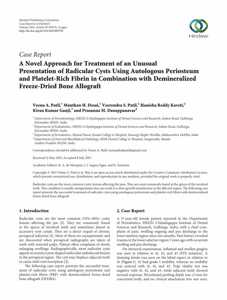
On intraoral examination, inflamed and swollen gingivawas seen in relation to 41, 42, and 43 (FDI notation). Adraining fistula was seen on the labial aspect in relation to41 (Figure 1). 42 had grade I mobility, whereas no mobilitywas noticed with 31, 41, and 43. Pulp vitality test wasnegative with 41, 42, and 43, while adjacent teeth showednormal response. Periodontal probing depth was ≤3mm forconcerned teeth, and no clinical attachment loss was seen.
2 Case Reports in Dentistry
Figure 1: Preoperative view of the lesion.
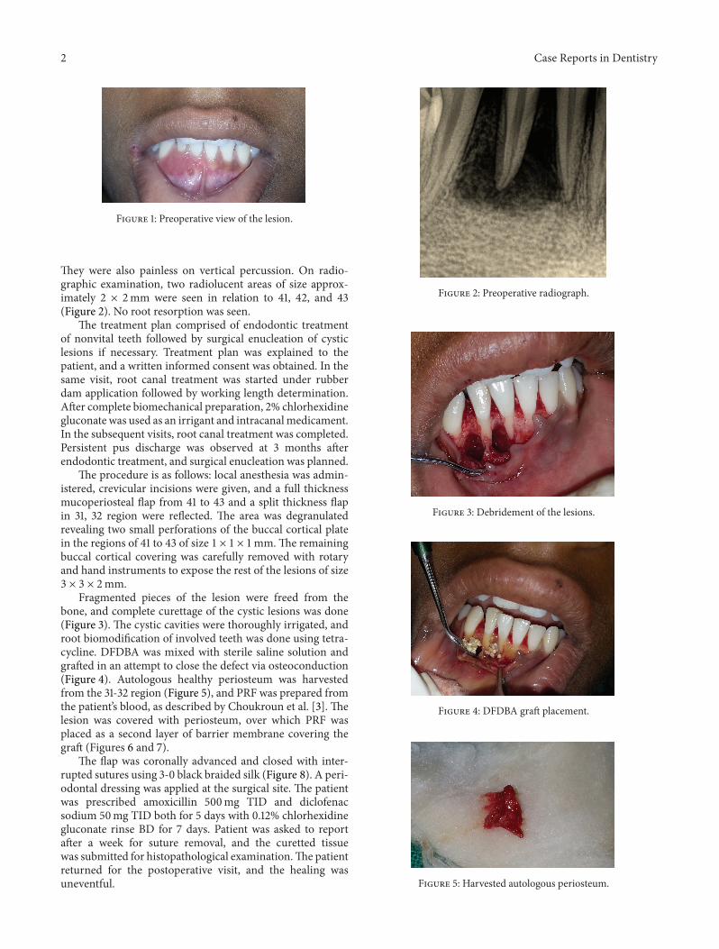
They were also painless on vertical percussion. On radio-graphic examination, two radiolucent areas of size approx-imately 2 × 2mm were seen in relation to 41, 42, and 43(Figure 2). No root resorption was seen.
The treatment plan comprised of endodontic treatmentof nonvital teeth followed by surgical enucleation of cysticlesions if necessary. Treatment plan was explained to thepatient, and a written informed consent was obtained. In thesame visit, root canal treatment was started under rubberdam application followed by working length determination.After complete biomechanical preparation, 2% chlorhexidinegluconate was used as an irrigant and intracanalmedicament.In the subsequent visits, root canal treatment was completed.Persistent pus discharge was observed at 3 months afterendodontic treatment, and surgical enucleation was planned.
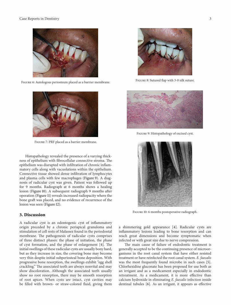
The procedure is as follows: local anesthesia was admin-istered, crevicular incisions were given, and a full thicknessmucoperiosteal flap from 41 to 43 and a split thickness flapin 31, 32 region were reflected. The area was degranulatedrevealing two small perforations of the buccal cortical platein the regions of 41 to 43 of size 1 × 1 × 1mm.The remainingbuccal cortical covering was carefully removed with rotaryand hand instruments to expose the rest of the lesions of size3 × 3 × 2mm.
Fragmented pieces of the lesion were freed from thebone, and complete curettage of the cystic lesions was done(Figure 3). The cystic cavities were thoroughly irrigated, androot biomodification of involved teeth was done using tetra-cycline. DFDBA was mixed with sterile saline solution andgrafted in an attempt to close the defect via osteoconduction(Figure 4). Autologous healthy periosteum was harvestedfrom the 31-32 region (Figure 5), and PRF was prepared fromthe patient’s blood, as described by Choukroun et al. [3]. Thelesion was covered with periosteum, over which PRF wasplaced as a second layer of barrier membrane covering thegraft (Figures 6 and 7).
The flap was coronally advanced and closed with inter-rupted sutures using 3-0 black braided silk (Figure 8). A peri-odontal dressing was applied at the surgical site. The patientwas prescribed amoxicillin 500mg TID and diclofenacsodium 50mg TID both for 5 days with 0.12% chlorhexidinegluconate rinse BD for 7 days. Patient was asked to reportafter a week for suture removal, and the curetted tissuewas submitted for histopathological examination.The patientreturned for the postoperative visit, and the healing wasuneventful.
Figure 2: Preoperative radiograph.
Figure 3: Debridement of the lesions.
Figure 4: DFDBA graft placement.
Figure 5: Harvested autologous periosteum.
Case Reports in Dentistry 3
Figure 6: Autologous periosteum placed as a barrier membrane.
Figure 7: PRF placed as a barrier membrane.
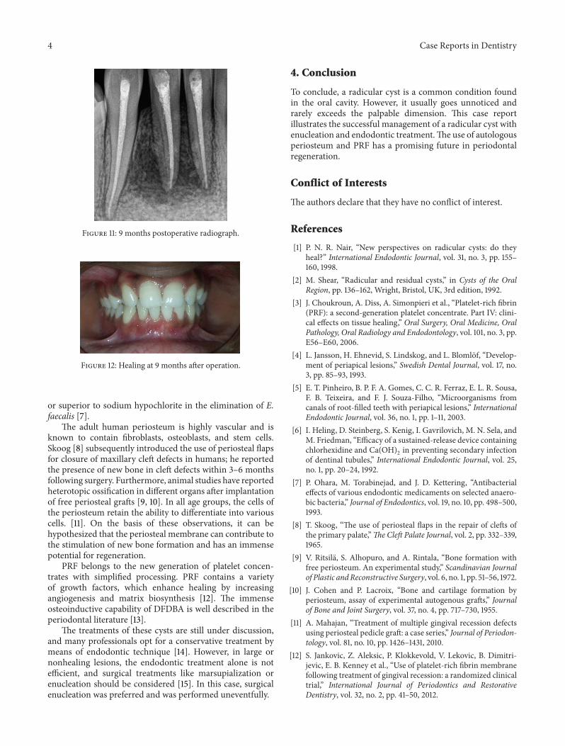
Histopathology revealed the presence of a varying thick-ness of epithelium with fibrocellular connective stroma. Theepithelium was disrupted with infiltration of chronic inflam-matory cells along with vacuolations within the epithelium.Connective tissue showed dense infiltration of lymphocytesand plasma cells with few macrophages (Figure 9). A diag-nosis of radicular cyst was given. Patient was followed upfor 9 months. Radiograph at 6 months shows a healinglesion (Figure 10). A subsequent radiograph 9 months afteroperation (Figure 11) reveals increased radiopacity where thebone graft was placed, and no evidence of recurrence of thelesion was seen (Figure 12).
3. Discussion
A radicular cyst is an odontogenic cyst of inflammatoryorigin preceded by a chronic periapical granuloma andstimulation of cell rests of Malassez found in the periodontalmembrane. The pathogenesis of radicular cysts comprisesof three distinct phases: the phase of initiation, the phaseof cyst formation, and the phase of enlargement [4]. Theinitial swellings of these radicular cysts are usually bony hard,but as they increase in size, the covering bone may becomevery thin despite initial subperiosteal bone deposition. Withprogressive bone resorption, the swellings exhibit “egg shellcrackling.” The associated teeth are always nonvital and mayshow discoloration. Although the associated teeth usuallyshow no root resorption, there may be smooth resorptionof root apices. When cysts are intact, cyst cavities maybe filled with brown- or straw-colored fluid, giving them
Figure 8: Sutured flap with 3-0 silk suture.
Figure 9: Histopathology of excised cyst.
Figure 10: 6 months postoperative radiograph.
a shimmering gold appearance [4]. Radicular cysts areinflammatory lesions leading to bone resorption and canreach great dimensions and become symptomatic wheninfected or with great size due to nerve compression.
The main cause of failure of endodontic treatment isgenerally accepted to be the continuing presence of microor-ganisms in the root canal system that have either resistedtreatment or have reinfected the root canal system. E. faecaliswas the most frequently found microbe in such cases [5].Chlorhexidine gluconate has been proposed for use both asan irrigant and as a medicament especially in endodonticretreatment. As a medicament, it is more effective thancalcium hydroxide in eliminating E. faecalis infection insidedentinal tubules [6]. As an irrigant, it appears as effective
4 Case Reports in Dentistry
Figure 11: 9 months postoperative radiograph.
Figure 12: Healing at 9 months after operation.
or superior to sodium hypochlorite in the elimination of E.faecalis [7].
The adult human periosteum is highly vascular and isknown to contain fibroblasts, osteoblasts, and stem cells.Skoog [8] subsequently introduced the use of periosteal flapsfor closure of maxillary cleft defects in humans; he reportedthe presence of new bone in cleft defects within 3–6 monthsfollowing surgery. Furthermore, animal studies have reportedheterotopic ossification in different organs after implantationof free periosteal grafts [9, 10]. In all age groups, the cells ofthe periosteum retain the ability to differentiate into variouscells. [11]. On the basis of these observations, it can behypothesized that the periosteal membrane can contribute tothe stimulation of new bone formation and has an immensepotential for regeneration.
PRF belongs to the new generation of platelet concen-trates with simplified processing. PRF contains a varietyof growth factors, which enhance healing by increasingangiogenesis and matrix biosynthesis [12]. The immenseosteoinductive capability of DFDBA is well described in theperiodontal literature [13].
The treatments of these cysts are still under discussion,and many professionals opt for a conservative treatment bymeans of endodontic technique [14]. However, in large ornonhealing lesions, the endodontic treatment alone is notefficient, and surgical treatments like marsupialization orenucleation should be considered [15]. In this case, surgicalenucleation was preferred and was performed uneventfully.
4. Conclusion
To conclude, a radicular cyst is a common condition foundin the oral cavity. However, it usually goes unnoticed andrarely exceeds the palpable dimension. This case reportillustrates the successful management of a radicular cyst withenucleation and endodontic treatment.The use of autologousperiosteum and PRF has a promising future in periodontalregeneration.
Conflict of Interests
The authors declare that they have no conflict of interest.
References
[1] P. N. R. Nair, “New perspectives on radicular cysts: do theyheal?” International Endodontic Journal, vol. 31, no. 3, pp. 155–160, 1998.
[2] M. Shear, “Radicular and residual cysts,” in Cysts of the OralRegion, pp. 136–162, Wright, Bristol, UK, 3rd edition, 1992.
[3] J. Choukroun, A. Diss, A. Simonpieri et al., “Platelet-rich fibrin(PRF): a second-generation platelet concentrate. Part IV: clini-cal effects on tissue healing,” Oral Surgery, Oral Medicine, OralPathology, Oral Radiology and Endodontology, vol. 101, no. 3, pp.E56–E60, 2006.
[4] L. Jansson, H. Ehnevid, S. Lindskog, and L. Blomlof, “Develop-ment of periapical lesions,” Swedish Dental Journal, vol. 17, no.3, pp. 85–93, 1993.
[5] E. T. Pinheiro, B. P. F. A. Gomes, C. C. R. Ferraz, E. L. R. Sousa,F. B. Teixeira, and F. J. Souza-Filho, “Microorganisms fromcanals of root-filled teeth with periapical lesions,” InternationalEndodontic Journal, vol. 36, no. 1, pp. 1–11, 2003.
[6] I. Heling, D. Steinberg, S. Kenig, I. Gavrilovich, M. N. Sela, andM. Friedman, “Efficacy of a sustained-release device containingchlorhexidine and Ca(OH)
2in preventing secondary infection
of dentinal tubules,” International Endodontic Journal, vol. 25,no. 1, pp. 20–24, 1992.
[7] P. Ohara, M. Torabinejad, and J. D. Kettering, “Antibacterialeffects of various endodontic medicaments on selected anaero-bic bacteria,” Journal of Endodontics, vol. 19, no. 10, pp. 498–500,1993.
[8] T. Skoog, “The use of periosteal flaps in the repair of clefts ofthe primary palate,”The Cleft Palate Journal, vol. 2, pp. 332–339,1965.
[9] V. Ritsila, S. Alhopuro, and A. Rintala, “Bone formation withfree periosteum. An experimental study,” Scandinavian Journalof Plastic andReconstructive Surgery, vol. 6, no. 1, pp. 51–56, 1972.
[10] J. Cohen and P. Lacroix, “Bone and cartilage formation byperiosteum, assay of experimental autogenous grafts,” Journalof Bone and Joint Surgery, vol. 37, no. 4, pp. 717–730, 1955.
[11] A. Mahajan, “Treatment of multiple gingival recession defectsusing periosteal pedicle graft: a case series,” Journal of Periodon-tology, vol. 81, no. 10, pp. 1426–1431, 2010.
[12] S. Jankovic, Z. Aleksic, P. Klokkevold, V. Lekovic, B. Dimitri-jevic, E. B. Kenney et al., “Use of platelet-rich fibrin membranefollowing treatment of gingival recession: a randomized clinicaltrial,” International Journal of Periodontics and RestorativeDentistry, vol. 32, no. 2, pp. 41–50, 2012.
Case Reports in Dentistry 5
[13] F. Brugnami, P. R.Then, H.Moroi, and C.W. Leone, “Histologicevaluation of human extraction sockets treated with deminer-alized freeze-dried bone allograft (DFDBA) and cell occlusivemembrane,” Journal of Periodontology, vol. 67, no. 8, pp. 821–825, 1996.
[14] M. M. Hoen, G. L. LaBounty, and E. J. Strittmatter, “Con-servative treatment of persistent periradicular lesions usingaspiration and irrigation,” Journal of Endodontics, vol. 16, no. 4,pp. 182–186, 1990.
[15] J. Danin, L. E. Linder, G. Lundqvist, L. Ohlsson, L. O. Ramskold,and T. Stromberg, “Outcomes of periradicular surgery in caseswith apical pathosis and untreated canals,” Oral Surgery, OralMedicine, Oral Pathology, Oral Radiology, and Endodontics, vol.87, no. 2, pp. 227–232, 1999.

Submit your manuscripts athttp://www.hindawi.com
Hindawi Publishing Corporationhttp://www.hindawi.com Volume 2013
Computational and Mathematical Methods in Medicine
International Journal of
BiomaterialsHindawi Publishing Corporationhttp://www.hindawi.com Volume 2013
Hindawi Publishing Corporationhttp://www.hindawi.com Volume 2013
Environmental andPublic Health
Journal of
ScientificaHindawi Publishing Corporationhttp://www.hindawi.com Volume 2013
Drug DeliveryJournal of
Hindawi Publishing Corporationhttp://www.hindawi.com Volume 2013
Preventive MedicineAdvances in
Hindawi Publishing Corporationhttp://www.hindawi.com Volume 2013
Hindawi Publishing Corporationhttp://www.hindawi.com Volume 2013
Radiology Research and Practice
BioMed Research International
Hindawi Publishing Corporationhttp://www.hindawi.com Volume 2013
DentistryInternational Journal of
Hindawi Publishing Corporationhttp://www.hindawi.com Volume 2013
Hindawi Publishing Corporationhttp://www.hindawi.com Volume 2013
Oral OncologyJournal of
Hindawi Publishing Corporationhttp://www.hindawi.com Volume 2013
Case Reports in Dentistry
Hindawi Publishing Corporationhttp://www.hindawi.com Volume 2013
Oral DiseasesJournal of
Hindawi Publishing Corporationhttp://www.hindawi.com Volume 2013
OrthopedicsAdvances in
Hindawi Publishing Corporationhttp://www.hindawi.com Volume 2013
Dental SurgeryJournal of
Hindawi Publishing Corporationhttp://www.hindawi.com Volume 2013
Oral ImplantsJournal of
ISRN Dentistry
Hindawi Publishing Corporationhttp://www.hindawi.com Volume 2013
International Journal of
EndocrinologyHindawi Publishing Corporationhttp://www.hindawi.com
Volume 2013
Hindawi Publishing Corporation http://www.hindawi.com Volume 2013Hindawi Publishing Corporation http://www.hindawi.com Volume 2013
The Scientific World Journal
Hindawi Publishing Corporationhttp://www.hindawi.com Volume 2013
AnesthesiologyResearch and Practice


