Embed Size (px)
Citation preview
RESEARCH ARTICLE Open Access
Immunohistochemical detection of laminin-1 andKi-67 in radicular cysts and keratocysticodontogenic tumorsMohamed S Ayoub1*, Houry M Baghdadi2, Moataz El-Kholy3
Abstract
Background: Odontogenic cysts are those which arise from the epithelium associated with the development ofteeth. Some odontogenic cysts were found to have special biological features that make them distinct from otherlesions. This study was conducted to detect the immunoepxression of laminin-1 and Ki-67 in both radicular cysts(RCs) and keratocystic odontogenic tumors (KCOTs) and to examine the possible predictive value of these markers.
Methods: Thirteen cases of RCs and twelve cases of KCOTs were included in this study. Antibodies against laminin-1 and Ki-67 were used as primary antibodies.
Results: ten cases out of thirteen cases of RCs were immunopositive to laminin-1. The immunonegative cases ofRCs showed high degree of inflammation inside the connective tissue wall. One case out of twelve cases of KCOTswas immunopositive to laminin-1 and the rest were immunonegative. Seven cases out of thirteen cases of RCsshowed immunopositivity for Ki-67 with increased numbers of immunopositive cells when the inflammation wassevere in the connective tissue wall. All KCOTS were immunopositive to Ki-67.
Conclusions: The benign nature of radicular cysts and the aggressive behavior of keratocystic odontogenic tumorscould be explained by the expression of laminin and Ki-67. Laminin-1 and Ki-67 could be valuable markers for theprediction of the biologic behavior of cystic lesions.
BackgroundRadicular cysts are a direct sequel to chronic apical peri-odontitis following the death of dental pulp [1]. Theepithelial rests of Malassez in periapical granuloma maybe stimulated to proliferate by inflammatory stimuli [2].The morphological aspects of the epithelium have beenconsidered to reflect the functional activity of the RCs[3]. RCs depict a thin, regular and atrophic layer of stra-tified squamous epithelium, usually with mild to moder-ate inflammatory reaction [4]. The underlyingsupportive connective tissue might be focally or diffuselyinfiltrated with mixed inflammatory cells population [5].Keratocystic odontogenic tumor (KCOT), previously
known as odontogenic keratocyst (OKC), is a relativelycommon developmental odontogenic cyst that arisesfrom the dental lamina remnants [6]. An important
aspect of the OKC that should be underlined is that itcan represent one component of the nevoid basal cellcarcinoma syndrome (NBCS) [7]. Several studies haveshown that the OKC is well recognized by its invasivepotential [8], thus it tends to grow within the medullarycavity of bone and becomes a large lesion without caus-ing obvious expansion [9].Expression of laminin-1 in normal oral mucosa, odon-
togenic cysts and odontogenic tumors was examined inseveral studies. Sections of normal oral mucosa andodontogenic cysts stained for laminin-1 showed a dis-tinct linear deposit of strong intensity at the basementmembrane junction but not in the cytoplasm of theepithelial cells [10]. Sections of odontogenic tumorsstained for laminin-1 showed strong reactivity at thebasement membrane junction as well as in the cyto-plasm of all tumor cells. The expression of laminin-1 inthe cytoplasm of the tumor cells, but not in the normalmucosa may be a useful marker to distinguish these twotypes of epithelium [11] and it may suggest that
* Correspondence: [email protected], Oral Pathology Department, Faculty of Dentistry, Ain ShamsUniversity, Cairo, EgyptFull list of author information is available at the end of the article
Ayoub et al. BMC Clinical Pathology 2011, 11:4http://www.biomedcentral.com/1472-6890/11/4
© 2011 Ayoub et al; licensee BioMed Central Ltd. This is an Open Access article distributed under the terms of the Creative CommonsAttribution License (http://creativecommons.org/licenses/by/2.0), which permits unrestricted use, distribution, and reproduction inany medium, provided the original work is properly cited.
laminin-1 influences the proliferation activity towardtumor potential [12].Ki-67 antigen is the prototypic cell cycle related
nuclear protein, expressed by proliferating cells in allphases of the active cell cycle (G1, S, G2 and M phase)and reaches a peak in the G2 and M phases. It rapidlydegrades after mitosis with a half life of detectable anti-gen being an hour or less. It is absent in resting (G0)cells. Ki-67 antibodies are useful in establishing the cellgrowing fraction in neoplasms [13].The aims of this study were to detect immunohisto-
chemically the expression of laminin-1 and Ki-67 inradicular cysts and keratocystic odontogenic tumors andalso to examine the possible predictive value of thesemarkers.
MethodSpecimen selectionTwenty-five formalin-fixed, paraffin-embedded tissueblocks of odontogenic cysts were obtained from thearchives of the oral pathology departments, Ain ShamsUniversity, Alexandria University, and National CancerInstitute, Cairo University. Thirteen cases were diag-nosed as radicular cysts (RCs) and twelve cases werediagnosed as keratocystic odontogenic tumors (KCOTs).Haematoxylin and eosin stained sections were used toconfirm the diagnosis.
Immunohistochemical proceduresFor all specimens 4 μm sections were cut and mountedon positively charged glass slides. Sections were deparaf-finized with xylene and rehydrated in graded ethyl alco-hol, sections were immersed in citrate buffer solution ofpH 4.8 and were put in the microwave oven beforestaining procedures.For immunostaining a universal kit (R&D Systems;
USA) was used, peroxidase anti- peroxidase method ofimmunostaining using the streptavidin-biotin systemwas carried out, 3% hydrogen peroxide was applied tothe sections to block the endogenous peroxidase activity.The sections were immunostained with anti-laminin1primary antibody (clone AL-2, R&D Systems, USA) andanti-Ki-67 primary antibody (clone BGX, BiogenixCorp., USA). The tissue sections were incubated over-night at room temperature. Sections were then coveredby the link antibody followed by the streptavidin label-ing antibody. After rinsing with PBS, DAB chromogenwas applied to the sections followed by counter stain,and then sections were dehydrated in graded alcohol,cleared in xylene and mounted.
Image analysisFor each positive section, four microscopic fields showingimmunopositivity were selected and photomicrographs
were captured at a magnification of 20×. Images werethen transferred to the computer system for analysisusing the image analysis software (Image J, 1.43r, NIH,USA), to determine the following:1. Area fraction of immunopositivity for both laminin-
1 and Ki-67. Area fraction was calculated as the ratio ofimmunopositive area to the total area of microscopicfield.2. Number of immunopositive cells for Ki-67
Statistical AnalysisStatistical analysis was carried out on the tabulated datausing (SPSS 16.0) software. The performed statisticaltests included Student’s T-test to compare between theexpression of each marker in the two lesions and Pear-son’s correlation to determine the correlation betweenlaminin-1 and Ki-67.
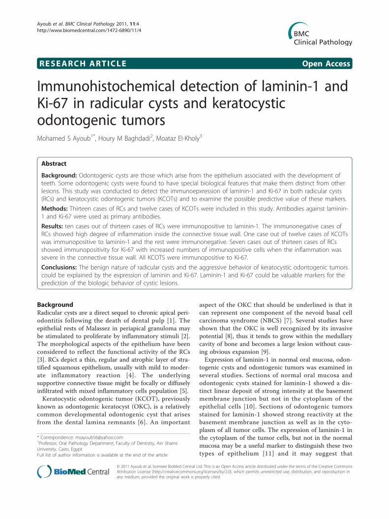
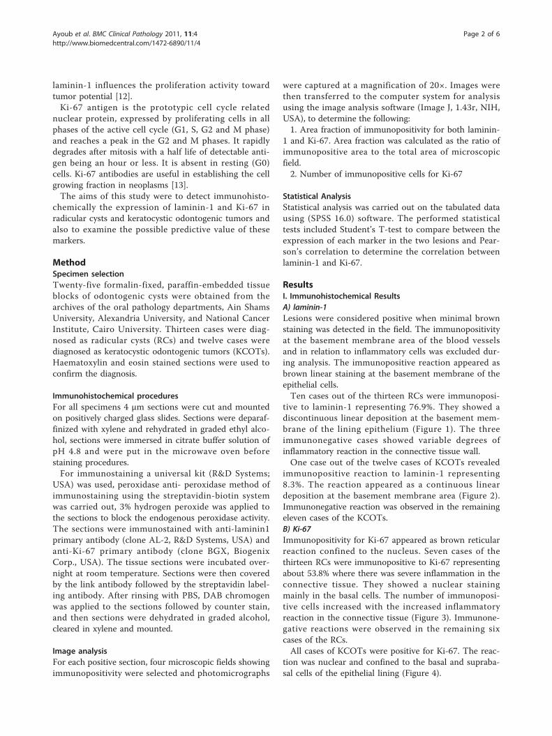
ResultsI. Immunohistochemical ResultsA) laminin-1Lesions were considered positive when minimal brownstaining was detected in the field. The immunopositivityat the basement membrane area of the blood vesselsand in relation to inflammatory cells was excluded dur-ing analysis. The immunopositive reaction appeared asbrown linear staining at the basement membrane of theepithelial cells.Ten cases out of the thirteen RCs were immunoposi-
tive to laminin-1 representing 76.9%. They showed adiscontinuous linear deposition at the basement mem-brane of the lining epithelium (Figure 1). The threeimmunonegative cases showed variable degrees ofinflammatory reaction in the connective tissue wall.One case out of the twelve cases of KCOTs revealed
immunopositive reaction to laminin-1 representing8.3%. The reaction appeared as a continuous lineardeposition at the basement membrane area (Figure 2).Immunonegative reaction was observed in the remainingeleven cases of the KCOTs.B) Ki-67Immunopositivity for Ki-67 appeared as brown reticularreaction confined to the nucleus. Seven cases of thethirteen RCs were immunopositive to Ki-67 representingabout 53.8% where there was severe inflammation in theconnective tissue. They showed a nuclear stainingmainly in the basal cells. The number of immunoposi-tive cells increased with the increased inflammatoryreaction in the connective tissue (Figure 3). Immunone-gative reactions were observed in the remaining sixcases of the RCs.All cases of KCOTs were positive for Ki-67. The reac-
tion was nuclear and confined to the basal and supraba-sal cells of the epithelial lining (Figure 4).
Ayoub et al. BMC Clinical Pathology 2011, 11:4http://www.biomedcentral.com/1472-6890/11/4
Page 2 of 6
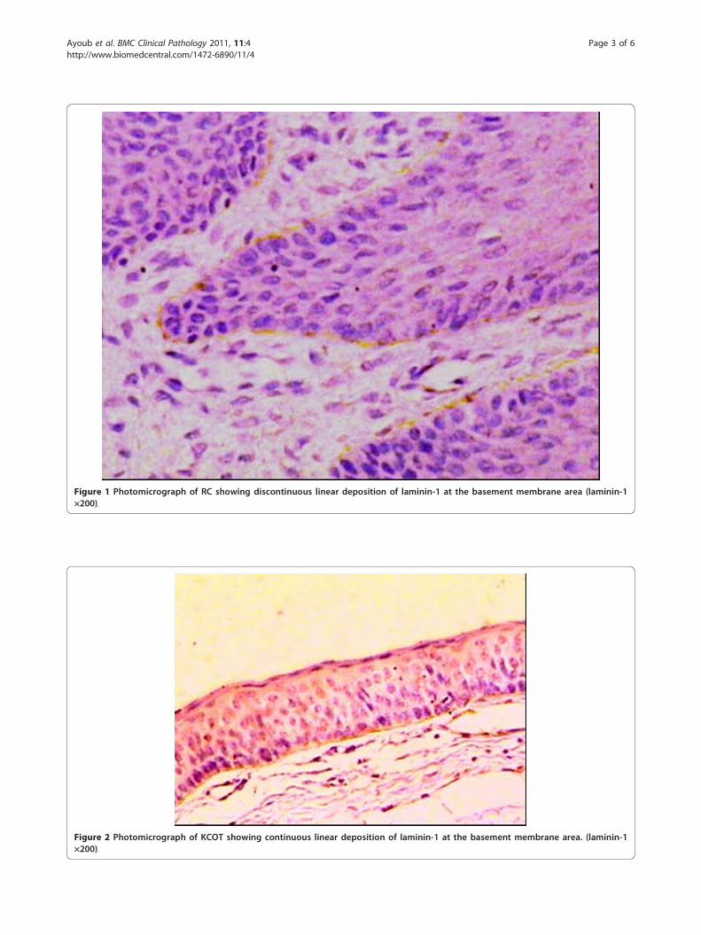
Figure 1 Photomicrograph of RC showing discontinuous linear deposition of laminin-1 at the basement membrane area (laminin-1×200).
Figure 2 Photomicrograph of KCOT showing continuous linear deposition of laminin-1 at the basement membrane area. (laminin-1×200).
Ayoub et al. BMC Clinical Pathology 2011, 11:4http://www.biomedcentral.com/1472-6890/11/4
Page 3 of 6
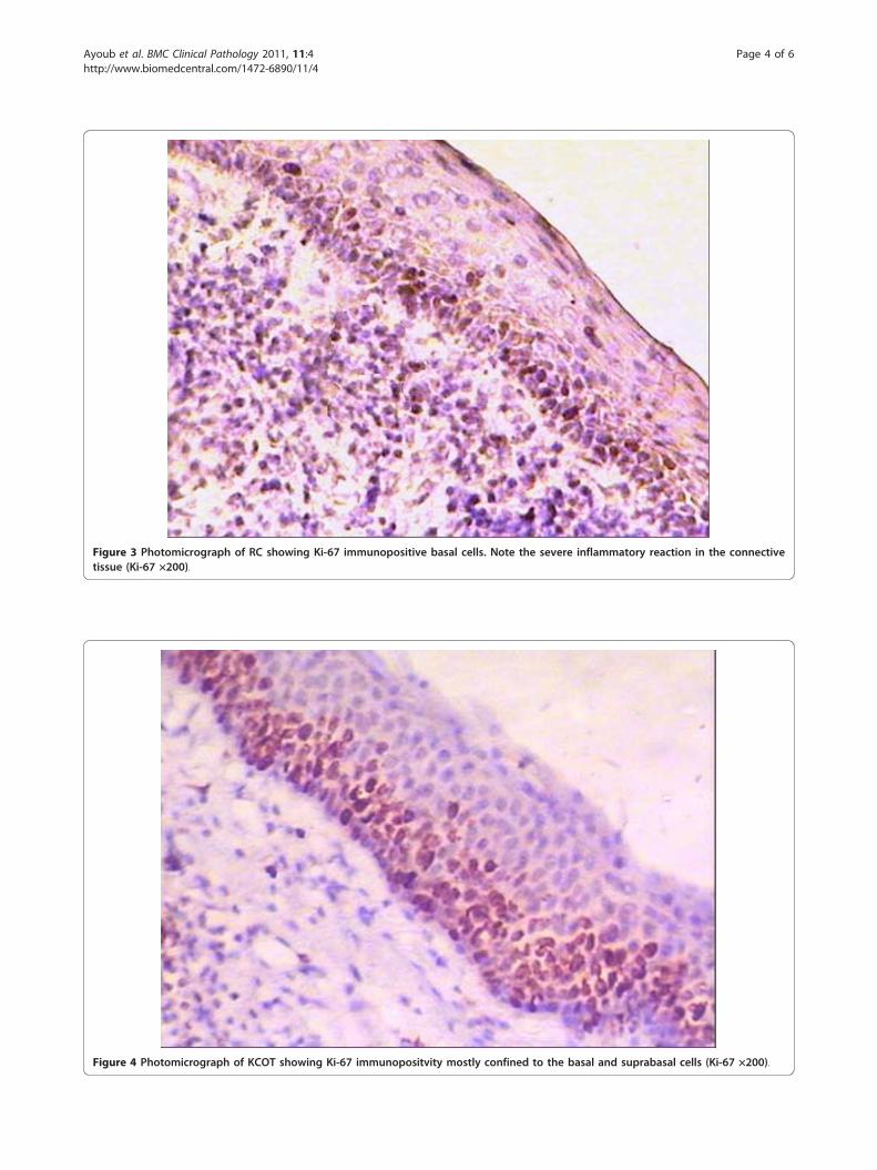
Figure 3 Photomicrograph of RC showing Ki-67 immunopositive basal cells. Note the severe inflammatory reaction in the connectivetissue (Ki-67 ×200).
Figure 4 Photomicrograph of KCOT showing Ki-67 immunopositvity mostly confined to the basal and suprabasal cells (Ki-67 ×200).
Ayoub et al. BMC Clinical Pathology 2011, 11:4http://www.biomedcentral.com/1472-6890/11/4
Page 4 of 6
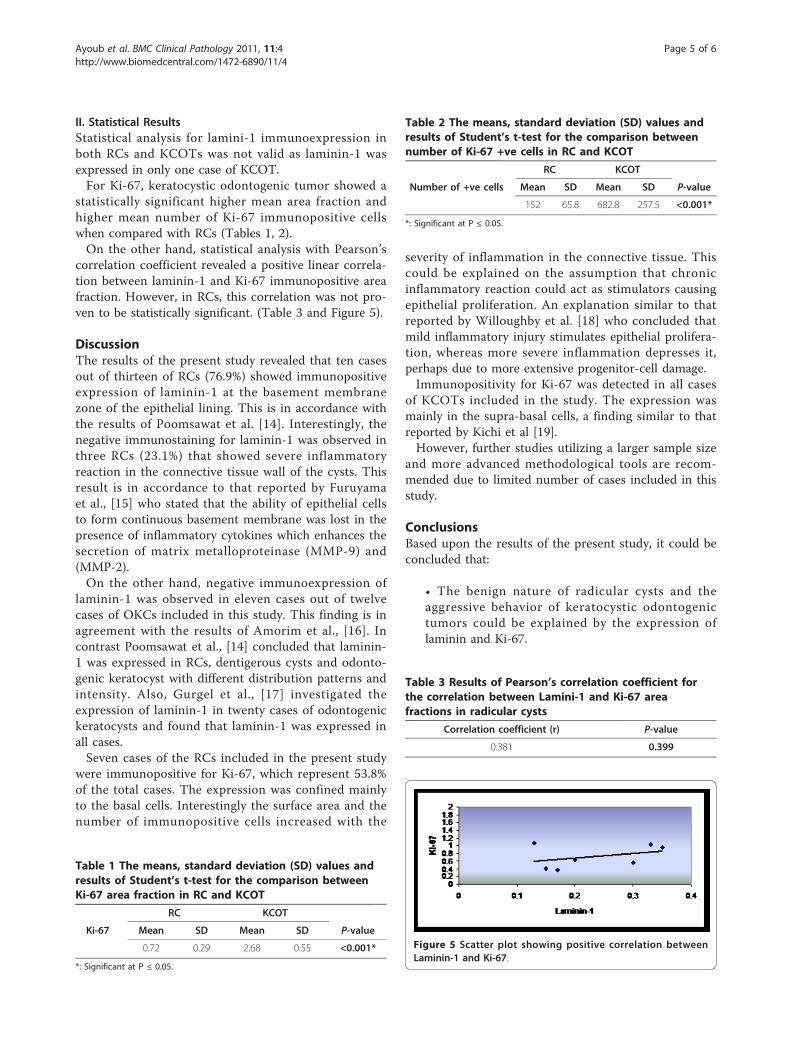
II. Statistical ResultsStatistical analysis for lamini-1 immunoexpression inboth RCs and KCOTs was not valid as laminin-1 wasexpressed in only one case of KCOT.For Ki-67, keratocystic odontogenic tumor showed a
statistically significant higher mean area fraction andhigher mean number of Ki-67 immunopositive cellswhen compared with RCs (Tables 1, 2).On the other hand, statistical analysis with Pearson’s
correlation coefficient revealed a positive linear correla-tion between laminin-1 and Ki-67 immunopositive areafraction. However, in RCs, this correlation was not pro-ven to be statistically significant. (Table 3 and Figure 5).
DiscussionThe results of the present study revealed that ten casesout of thirteen of RCs (76.9%) showed immunopositiveexpression of laminin-1 at the basement membranezone of the epithelial lining. This is in accordance withthe results of Poomsawat et al. [14]. Interestingly, thenegative immunostaining for laminin-1 was observed inthree RCs (23.1%) that showed severe inflammatoryreaction in the connective tissue wall of the cysts. Thisresult is in accordance to that reported by Furuyamaet al., [15] who stated that the ability of epithelial cellsto form continuous basement membrane was lost in thepresence of inflammatory cytokines which enhances thesecretion of matrix metalloproteinase (MMP-9) and(MMP-2).On the other hand, negative immunoexpression of
laminin-1 was observed in eleven cases out of twelvecases of OKCs included in this study. This finding is inagreement with the results of Amorim et al., [16]. Incontrast Poomsawat et al., [14] concluded that laminin-1 was expressed in RCs, dentigerous cysts and odonto-genic keratocyst with different distribution patterns andintensity. Also, Gurgel et al., [17] investigated theexpression of laminin-1 in twenty cases of odontogenickeratocysts and found that laminin-1 was expressed inall cases.Seven cases of the RCs included in the present study
were immunopositive for Ki-67, which represent 53.8%of the total cases. The expression was confined mainlyto the basal cells. Interestingly the surface area and thenumber of immunopositive cells increased with the
severity of inflammation in the connective tissue. Thiscould be explained on the assumption that chronicinflammatory reaction could act as stimulators causingepithelial proliferation. An explanation similar to thatreported by Willoughby et al. [18] who concluded thatmild inflammatory injury stimulates epithelial prolifera-tion, whereas more severe inflammation depresses it,perhaps due to more extensive progenitor-cell damage.Immunopositivity for Ki-67 was detected in all cases
of KCOTs included in the study. The expression wasmainly in the supra-basal cells, a finding similar to thatreported by Kichi et al [19].However, further studies utilizing a larger sample size
and more advanced methodological tools are recom-mended due to limited number of cases included in thisstudy.
ConclusionsBased upon the results of the present study, it could beconcluded that:
• The benign nature of radicular cysts and theaggressive behavior of keratocystic odontogenictumors could be explained by the expression oflaminin and Ki-67.
Table 1 The means, standard deviation (SD) values andresults of Student’s t-test for the comparison betweenKi-67 area fraction in RC and KCOT
RC KCOT
Ki-67 Mean SD Mean SD P-value
0.72 0.29 2.68 0.55 <0.001*
*: Significant at P ≤ 0.05.
Table 2 The means, standard deviation (SD) values andresults of Student’s t-test for the comparison betweennumber of Ki-67 +ve cells in RC and KCOT
RC KCOT
Number of +ve cells Mean SD Mean SD P-value
152 65.8 682.8 257.5 <0.001*
*: Significant at P ≤ 0.05.
Table 3 Results of Pearson’s correlation coefficient forthe correlation between Lamini-1 and Ki-67 areafractions in radicular cysts
Correlation coefficient (r) P-value
0.381 0.399
Figure 5 Scatter plot showing positive correlation betweenLaminin-1 and Ki-67.
Ayoub et al. BMC Clinical Pathology 2011, 11:4http://www.biomedcentral.com/1472-6890/11/4
Page 5 of 6
• Laminin-1 and Ki-67 could be valuable markers forthe prediction of the biologic behavior of cysticlesions.
Author details1Professor, Oral Pathology Department, Faculty of Dentistry, Ain ShamsUniversity, Cairo, Egypt. 2Associate Professor, Oral Pathology Department,Faculty of Dentistry, Ain Shams University, Cairo, Egypt. 3Assistant Lecturer,Oral Pathology Department, Faculty of Dental Surgery, Modern Science andArts University, Cairo, Egypt.
Authors’ contributionsMSA participated in the study design, photomicrography of theimmunohistochemical results, interpreting and displaying the results of thestudy, carried out the sequence alignment and drafted the manuscript. H.M.Bparticipated in displaying the results of the study, writing the discussion ofthe results and alignment of the references. ME carried out theimmunohistochemical technique, collection of the background referencesand participated in writing the discussion of the results.
Competing interestsThe authors declare that they have no competing interests.
Received: 9 December 2010 Accepted: 2 March 2011Published: 2 March 2011
References1. Nair P, Sundqvist G, Sjogren U: Experimental evidence supports the
abscess theory of development of radicular cysts. Oral Surg Oral Med OralPathol Oral Radiol Endod 2008, 106:294-303.
2. Nair P: On the causes of persistent apical periodontitis: a review. IntEndod J 2006, 39:249-81.
3. Loyola A, Cardoso V, Lisa G, Oliveira L, Mesquita R, Carmo M, Aguiar M:Apoptosis in epithelial cells of apical radicular cysts. Int Endod J 2005,38:465-469.
4. Moreria P, Santos D, Martins R, Gomez R: CD57+ cells in radicular cysts. IntEndod J 2000, 33:99-102.
5. Hayashi M, Obsbima T, Obsbima M, Yamaguchi Y, Miyata H, Takeichi O,Ogiso B, Ito K, Ostman A, Otsuka K: Profiling of radicular cyst andodontogenic keratocyst cytokine production suggests common growth.J Endod 2008, 34:14-21.
6. Neville B, Damm D, Allen C, Bouquot J: Oral and Maxillofacial Pathology.Philadelphia: W.B. Saunders, 3 2009, 683.
7. Amorim R, Godoy G, Galvão H, Souza L, Freitas R: Immunohistochemicalassessment of extracellular matrix components in syndrome and non-syndrome odontogenic keratocyst. Oral Disease 2004, 10:265-270.
8. Kimi K, Ohki K, Kumamoto H: Immunohistochemical analysis of cell cycleand apoptosis related factors in lining epithelium of odontogenickeratocyst. J Oral Pathol Med 2001, 30:434-443.
9. Da Silva M, De Sousa S, Correa L, Carvalhosa A, De Araujo V:Immunohistochemical study of the orthokeratinized odontogenic cyst: acomparison with the odontogenic keratocyst. Oral Surg Oral Med OralPathol Oral Radiol Endod 2002, 94:732-7.
10. Poomsawat S, Punyasingh J, Vejchapipat P: Expression of basementmembrane components in odontogenic tumors. Oral Surg Oral Med OralPathol Oral Radiol Endod 2007, 104:666-75.
11. Poomsawat S, Punyasingh J, Weerapradist W: Expression of basementmembrane components in odontogenic cysts. Oral Diseases 2006,12:290-96.
12. De Arcangelis A, Lefebvre O, Mechine-Neuville A, Arnold C, Kline A, Remy L:Overexpression of laminin alpha1 chain in colonic cancer cells inducesan increase in tumor growth. Int J Cancer 2001, 94:44-53.
13. Scholzen T, Gerdens J: The Ki-67 protein from the known and theunknown. J cell Physiol 2000, 182:311-22.
14. Poomsawat S, Punyasingh J, Weerapradist W: Expression of basementmembrane components in odontogenic cysts. Oral Diseases 2006,12:290-96.
15. Furuyama A, Takeshi Hosokawa T, Mochitate K: Interleukin-1β and tumornecrosis factor-α have opposite effects on fibroblasts and epithelial cellsduring basement membrane formation. Matrix Biology 2008, 27:429-440.
16. Amorim R, Godoy G, Galvão H, Souza L, Freitas R: Immunohistochemicalassessment of extracellular matrix component in syndrome & non-syndrome odontogenic keratocyst. Oral Disease 2004, 10:265-270.
17. Gurgel C, Ramos E, Melo L, chlaepfer C, De Souza R, Oliveira M, Santos J:Immunolocalisation of laminin-1 in Keratocystic odontogenic tumor.Acta Histochem 2009, (Journal Article).
18. Willoughbya S, Hoppsa R, Johnson N: Changes in the rate of epithelialproliferation of rat oral mucosa in response to acute inflammationinduced by turpentine. Archives of Oral Biology 1986, 31:193-199.
19. Kichi E, Enokiya Y, Muramatsu T, Hashimoto S, Inoue T, Abiko Y,Shimono M: Cell proliferation, apoptosis and apoptosis-related factors inOKC & DC. J Oral Pathol Med 2005, 34:280-6.
Pre-publication historyThe pre-publication history for this paper can be accessed here:http://www.biomedcentral.com/1472-6890/11/4/prepub
doi:10.1186/1472-6890-11-4Cite this article as: Ayoub et al.: Immunohistochemical detection oflaminin-1 and Ki-67 in radicular cysts and keratocystic odontogenictumors. BMC Clinical Pathology 2011 11:4.
Submit your next manuscript to BioMed Centraland take full advantage of:
• Convenient online submission
• Thorough peer review
• No space constraints or color figure charges
• Immediate publication on acceptance
• Inclusion in PubMed, CAS, Scopus and Google Scholar
• Research which is freely available for redistribution
Submit your manuscript at www.biomedcentral.com/submit
Ayoub et al. BMC Clinical Pathology 2011, 11:4http://www.biomedcentral.com/1472-6890/11/4
Page 6 of 6


