Embed Size (px)
Citation preview

A novel ex vivo skin model to study the susceptibilityof the dermatophyte Trichophyton rubrum to photodynamic
treatment in different growth phases
Threes G. M. Smijs1*, Joke A. Bouwstra2, Hans J. Schuitmaker3, M. Talebi1 and Stan Pavel1
1Leiden University Medical Centre, Skin Research Laboratory, PO Box 9600, 2300 RC Leiden, The Netherlands;2University of Leiden, Leiden/Amsterdam Centre for Drugs Research, Leiden, The Netherlands;
3PhotoBioChem NV, Leiden, The Netherlands
Received 19 July 2006; returned 14 October 2006; revised 20 October 2006; accepted 2 November 2006
Background: Dermatophytes are fungi that can cause infections of skin, hair and nails because of theirability to feed on keratin. Superficial mycoses are among the most prevalent infectious diseases worldwide.Two important restrictions of current therapeutic options are the recurrence of the infection and prolongedtreatment. This is especially true for infections caused by Trichophyton rubrum, a widely distributeddermatophyte. The application of photosensitizers for treatment of fungal infections is, within the field ofphotodynamic treatment (PDT), relatively new. Recently, we demonstrated that the porphyrins 5,10,15-tris(4-methylpyridinium)-20-phenyl-[21H,23H]-porphine trichloride (Sylsens B) and deuteroporphyrin mono-methylester (DP mme) were excellent photosensitizers towards T. rubrum when using red light.
Objectives and methods: To evaluate the photodynamic effectiveness of the porphyrins in a situationthat mimics the clinical situation, we developed an ex vivo model using human stratum corneum. Thismodel offers the possibility of applying PDT at different time points during the germination and sub-sequent development of T. rubrum microconidia. The model was used for two different incubationmedia, Dulbecco’s modified Eagle medium (DMEM) and distilled water.
Results and conclusions: We demonstrated that the PDT susceptibility of T. rubrum depended on thetime of PDT application after spore inoculation. A decrease in susceptibility was observed withincreasing time of PDT application for both photosensitizers in DMEM. Changing the incubationmedium to distilled water resulted in an increased fungicidal effect for Sylsens B and in a decreasedeffect for DP mme. We conclude that T. rubrum is susceptible to PDT in a situation that mimics theclinical situation. The fungicidal effect of PDT on fungal spores is of particular importance.
Keywords: ex vivo model, stratum corneum, porphyrins, tinea, microconidia
Introduction
Dermatophytes are fungi that can cause infections of the skin,hair and nails, in part because of their ability to utilize keratin.The cutaneous infections they cause (also called tinea) areamong the most common infections in humans worldwide.1 Inthe USA, 10% of the population has cutaneous fungal infectionsat any given time, and at least 40% will acquire this skin con-dition at some time in their life.2 The dermatophyteTrichophyton rubrum causes the most common cutaneous infec-tion in humans,3,4 which can be very persistent.5
Two of the most important restrictions limiting the usefulnessof common therapeutic options are the relatively high likelihoodof recurrence of the infection and the need for prolonged
treatment.6 Many current antifungal agents, such as azoles, havea fungistatic effect (a delay in growth) rather than a fungicidaleffect (a complete inactivation of both fungal conidia andhyphae). Fungal conidia appear to be less susceptible to the anti-fungal agents than the hyphae.7,8 In case of widespread, moreinflammatory or persistent dermatophytoses, systemic treatmentis necessary. However, when using systemic therapy one shouldbe aware of the potential risks of drug interactions and adverseeffects, such as hepatotoxicity or general drug reactions. Inaddition, azole resistance appears to be emerging as a seriousproblem in patients treated for yeast infections.9
Photodynamic treatment (PDT) refers to the use of light-activated agents called photosensitizers.10 Upon irradiation withlight of an appropriate wavelength, photosensitizers can initiate
. . . . . . . . . . . . . . . . . . . . . . . . . . . . . . . . . . . . . . . . . . . . . . . . . . . . . . . . . . . . . . . . . . . . . . . . . . . . . . . . . . . . . . . . . . . . . . . . . . . . . . . . . . . . . . . . . . . . . . . . . . . . . . . . . . . . . . . . . . . . . . . . . . . . . . . . . . . . . . . . . . . . . . . . . . . . . . . . . . . . . . . . . . . . . . . . . . . . . . . . . . . . . . . . . . . . . . . . . . . . . . . . . . . . . . . . . . . . . . . . . . . . . . . . . . . . . . . . . . . . . . . . . . . . . . . . . . . . . . . . . . . . . . . . . . . . . . . . . . . . . . . . . . . . . . . . . . . . . . . . . . . . . . . . . . . . . . . . . . . . . . . . . . . . . . . . . . . . . . . . . . . . . . . . . . . . . . . . . . . . . . . . . . . . . . . . . . . . . . . . . . . . . . . . . . . . . . . . . . . . .
*Corresponding author. Tel: þ31-71-5269376 / þ31-71-5269360; Fax: þ31-71-5268286; E-mail: [email protected]
Journal of Antimicrobial Chemotherapy
doi:10.1093/jac/dkl490
. . . . . . . . . . . . . . . . . . . . . . . . . . . . . . . . . . . . . . . . . . . . . . . . . . . . . . . . . . . . . . . . . . . . . . . . . . . . . . . . . . . . . . . . . . . . . . . . . . . . . . . . . . . . . . . . . . . . . . . . . . . . . . . . . . . . . . . . . . . . . . . . . . . . . . . . . . . . . . . . . . . . . . . . . . . . . . . . . . . . . . . . . . . . . . . . . . . . . . . . . . . . . . . . . . . . . . . . . . . . . . . . . . . . . . . . . . . . . . . . . . . . . . . . . . . . . . . . . . . . . . . . . . . . . . . . . . . . . . . . . . . . . . . . . . . . . . . . . . . . . . . . . . . . . . . . . . . . . . . . . . . . . . . . . . . . . . . . . . . . . . . . . . . . . . . . . . . . . . . . . . . . . . . . . . . . . . . . . . . . . . . . . . . . . . . . . . . . . . . . . . . . . . . . . . . . . . . . . . . . .
Page 1 of 8# The Author 2007. Published by Oxford University Press on behalf of the British Society for Antimicrobial Chemotherapy. All rights reserved.
For Permissions, please e-mail: [email protected]
Journal of Antimicrobial Chemotherapy Advance Access published January 9, 2007 by guest on June 3, 2013
http://jac.oxfordjournals.org/D
ownloaded from

a photochemical reaction resulting in the production of reactiveoxygen species, namely singlet oxygen (1O2) and superoxideanion radical (O2
2), which can react with various cellular com-ponents. The sequence of these events is known as the ‘photody-namic effect’ and can result not only in a selective tissue injury,but also in the elimination of different kinds of pathogens.11 Theuse of PDT for fungal infections is a new and promisingapproach.12
Recently, we have demonstrated that the porphyrins5,10,15-tris(4-methylpyridinium)-20-phenyl-[21H,23H]-porphinetrichloride (Sylsens B; Figure 1a) and deuteroporphyrin mono-methylester (DP mme; Figure 1b) are excellent photosensitizersto treat T. rubrum in suspension when red light is used to acti-vate them.13 A single PDT in vitro was sufficient to achieve a100% fungicidal effect, using either Sylsens B or DP mme.
To evaluate the photodynamic activity of these two porphyrinphotosensitizers under conditions that mimic the clinical situ-ation of tinea infections, we developed an ex vivo model usinghuman stratum corneum (SC). This ex vivo model offers thepossibility of applying PDT at different time points during thegermination process of T. rubrum microconidia on human SCand also during the subsequent development of germ tubes andfungal hyphae. We have used the model to investigate the effi-cacy of the porphyrin photosensitizers Sylsens B and DP mmetowards T. rubrum. The susceptibility of T. rubrum to PDT wasinvestigated in different growth phases of the fungus grownunder two different conditions. Because of the importance of asingle treatment for tinea infections, special attention has beenpaid to the fungicidal effect of PDT. Information on the suscep-tibility of different growth phases to PDT can be of greatimportance for the development of a treatment strategy thatwould lead to inactivation of both fungal spores and hyphae.Such a treatment would prevent the recurrence of the infectionand reduce the number of treatments. The growth of T. rubrumon human SC in the ex vivo model was visually investigatedbefore and after PDT using different microscopic techniques.
Materials and methods
Materials
Trichophyton rubrum was purchased from the Centraalbureauvoor Schimmelcultures (CBS; strain no: 304.60), Utrecht, TheNetherlands. For the preparation of a microconidia suspension,T. rubrum cultures were grown on Sabouraud dextrose agar(Sigma-Aldrich Chemie, Germany) at room temperature. Thephotosensitizers Sylsens B (mol. wt: 769.16 g/mol) and DPmme (mol. wt: 524.61 g/mol) were synthesized by theDepartment of Bio-Organic Photochemistry, Leiden University,The Netherlands (purity determined by NMR was more than99.5%) and kindly provided to us. Polyethyleneglycol wasobtained from Genfarma B.V. (Maarssen, The Netherlands) andtrypsin was obtained from Sigma (Zwijndrecht, TheNetherlands), while all other chemicals were purchased fromJ. T. Baker (Deventer, The Netherlands). The following solventswere used: 50 mM sodium phosphate buffer pH 7.4 for SylsensB and polyethyleneglycol/ethanol/water (3 : 2 : 5) for DP mme.Stock solutions of the photosensitizers (4.8 mM) were freshlymade for each new experiment.
Preparation of microconidia suspensions
The protocol to obtain a suspension of microconidia producedby T. rubrum grown on Sabouraud dextrose agar was based onthe method described by Zurita and Hay14 with two modifi-cations. A 0.01% Tween 80 solution in sterile water was usedinstead of phosphate-buffered saline and a 7 mm Millipore filterwas used instead of a 8 mm Nucleopore filter. The protocol wasas follows: To a 14-day-old culture, 8–10 mL of a 0.01% Tween80 solution was added. The surface was brushed with a glass rodand the resulting suspension filtered over a 7 mm diameter filter(Millipore). The filtrate was centrifuged at 3400 g (108C) andthe resulting pellet was washed with sterile water and suspendedagain in sterile water in a total volume of 2–4 mL (10–40 � 106 cfu/mL). The obtained microconidia suspensions were
NH
NH
N
N
OO O OH
NH
NH
N
N
N
N
N CH
H3C
H3C
(a)
(b)
3 Cl–
Figure 1. Chemical structure of the porphyrin photosensitizers (a)
5,10,15-tris(4-methylpyridinium)-20-phenyl-[21H,23H]-porphine trichloride
(Sylsens B) and (b) deuteroporphyrin monomethylester (DP mme).
Smijs et al.
Page 2 of 8
by guest on June 3, 2013http://jac.oxfordjournals.org/
Dow
nloaded from

stored in liquid nitrogen for no longer than 6 months. Countingthe number of colony-forming units on malt extract agar (MEA)dishes was used as a viability check. Identification of the iso-lated spores as microconidia was performed by the CBS.
Preparation of the human stratum corneum (SC)
Abdomen or mammae skin was obtained from a local hospitalafter cosmetic surgery. After removal of the fat tissue, the skinwas cleaned with distilled water and dermatomed to a thicknessof approximately 250 mm using a Padgett Electro DermatomeModel B (Kansas City, USA). The dermatomed skin was incu-bated at the dermal side with a 0.1% trypsin solution in phos-phate buffered saline of pH 7.4 (48C) overnight. After 1 h at378C the SC was removed manually. The obtained SC was air-dried for 24 h and kept under nitrogen over silica gel for nolonger than 3 months.
The ex vivo model
A polycarbonate membrane filter, 25 mm in diameter and with apore size of 2 mm (Omnilabo, Breda, The Netherlands), wasplaced in the central part of a 3 cm culture dish (Greiner,Alphen aan den Rijn, The Netherlands) filled with 5 mL ofMEA. A circular piece of human SC with a diameter of 1 cmwas subsequently placed on the membrane filter and both weregently brushed with a paintbrush dipped in 70% ethanol. Amicroconidia suspension was diluted to 1000 cfu/mL and 15 mLinoculated on the circular piece of human SC in a dish whichwas then placed in an incubator at 288C. At 8, 24, 48 and 72 hafter spore inoculation, PDT was applied using either Sylsens Bor DP mme. The conditions of the ex vivo model are such thatgermination of the microconidia can be detected microscopically(Zeiss Axiovert 25) at 72 h after their inoculation. This impliesthat when PDT is applied at 72 h after spore inoculation fungalhyphae are treated. The test conditions were chosen to resemblethose used in the in vitro experiments,13 except for the durationof the incubation and the concentration of the porphyrins.
A schematic outline of the ex vivo model, including the PDTstadium, is provided in Figure 2.
Light source
Illuminations were performed with a lamp from ‘MASSIVE’(no. 74900/21), 1� max 500 W-230 V-R7s, IP 44. To avoidheating of the samples during illumination, a 5 cm water filterabsorbing infrared light was used. Light intensity was measuredwith an IL1400A photometer equipped with a SEL033/F/Udetector (International Light, Newburyport, MA, USA). A redcutoff filter at 600 nm was used to obtain the red part of thespectrum of the light produced by the lamp. The light emittedby the lamp had a wavelength range of 580–870 nm. The lightintensity at the level of the infected human SC was 30 mW/cm2.
Photodynamic treatment (PDT)
At 8, 24, 48 or 72 h after spore inoculation, the membrane filterwith SC containing spore inoculates was transferred from theMEA dish to a 3 cm culture dish filled with 1035 mL of incu-bation medium. The incubation medium contained either 1 mLof distilled water (pH 5.2) or 1 mL of DMEM (GibcoBRL, UK)
at pH 7.4 supplemented with fetal calf serum (FCS; GibcoBRL,UK) and 35 mL of a photosensitizer solution. Final photosensiti-zer concentrations were 80 and 160 mM. To optimize the contactof the inoculated fungus with the incubation medium, the mem-brane filter with the SC was turned upside down during the incu-bation period of 2 h (see Figure 2c). The incubation wasperformed under continuous shaking conditions (HeidolphShaker, unimax 2010). Shortly before the illumination period,the membrane filter containing the SC was turned back(Figure 2d). In all cases, the illumination time was 1 h using alight flux rate of 30 mW/cm2 (Figure 2e). This corresponds to alight dose of 108 J/cm2. After the illumination the membranefilter with the SC was transferred to a fresh MEA dish(Figure 2f ), placed in an incubator at 288C, and fungal growthwas followed for several weeks. ‘Dark controls’ were included,i.e. the same procedure was carried out except that the inocu-lated microconidia on human skin were treated with solvent orphotosensitizer in the dark. In the ‘light controls’, the microconi-dia were treated with solvent in the presence of light. The effi-cacy of the treatment was expressed as a relative frequency ofcomplete inactivation of fungal growth detected at day 8 afterspore inoculation. To assess this a Zeiss Axiovert 25 microscopewas used. If at day 8 by visual inspection no re-growth could beobserved, a complete inactivation of spores and hyphae as aresult of PDT was established. The treatment effect was fol-lowed for a period of several months. Every experiment con-tained 4 to 6 duplicates of all conditions and the experimentswere repeated at least three times.
Fluorescence microscopy
After the PDT (applied at 72 h after spore inoculation or darktreatment) the SC was placed on an object glass, washed withwater, covered with a glass coverslip and examined with a LeicaDM 5000 microscope equipped with differential interference con-trast (DIC) optics and a digital camera (Leica DFC300FX R2digital colour cam set). A HCX PL FLUOTAR 40 � /0.75Ph2.objective and a HCX PL FLUOTAR 63 � /1.25 oil immersionobjective were used and fluorescence images were taken using afilter cube violetþ blue (H3, excitation filter BP 420–490). Thepictures were taken directly after treatment with 160 mM SylsensB in the dark, after an unsuccessful PDT with 80 mM Sylsens Band after a successful PDT with 160 mM Sylsens B, when a fungi-cidal effect was detected. Pictures of untreated fungus, at 72 hafter spore inoculation, were taken as well.
Results
Changing the incubation medium from DMEM to distilled
water resulted in an increased PDT efficacy for Sylsens B
The results obtained with Sylsens B are provided in Table 1.When incubating in DMEM, we found that 8 h after spore inocu-lation, application of PDT with either 80 or 160 mM Sylsens Bresulted in a complete inactivation of the spores. At 24 h afterspore inoculation under the same conditions, a fungicidal effectwas obtained in 50% of the cases, while in the remaining casesonly a delay in growth was observed. However, the application ofPDT more than 24 h after the spore inoculation did not result in afungicidal effect. In those cases, a growth delay of 1–2 days was
Ex vivo model for photodynamic treatment of Trichophyton rubrum
Page 3 of 8
by guest on June 3, 2013http://jac.oxfordjournals.org/
Dow
nloaded from

obtained, depending on the Sylsens B concentration used.Incubation in distilled water at pH 5.2 resulted in a considerablyhigher number of fungicidal effects compared with incubation inDMEM. When 160 mM Sylsens B was used we obtained a fungi-cidal effect not only after 8 h, but also after 24 and 48 h. At thesetime points the fungicidal effect was almost 100%. In addition,even at 72 h after spore inoculation, when fungal hyphae weretreated, we found a high percentage (65%) with complete fungusinactivation. In the experiments with DMEM the light controlsresulted in 10–15 cfu for every time point at 8 days after sporeinoculation. Very similar results were obtained in the dark controls
with both Sylsens B concentrations. In the case of distilled water,the light controls developed 10–15 cfu for every time point at 8days after spore inoculation and the dark controls 5–12 cfu.
Changing the incubation medium from DMEM to distilled
water caused a decreased PDT efficacy for DP mme
As far as DP mme is concerned (Table 2), the results resembledthose obtained with Sylsens B in DMEM (Table 1): a completeinactivation of the spores 8 h after spore inoculation and a fungi-cidal effect of 50–85% when PDT was applied at 24 h after spore
SC
SC
SC
Membrane
MEA
MEA
MEA
Spore inoculation
Incubationmixture
Incubationmixture
Incubationmixture
Illumination
(a) (b)
(c)
(d)
(e)(f)
After 8–72 hours
After 2 hours
Figure 2. Schematic outline of the ex vivo model. A polycarbonate membrane filter is placed on a MEA dish and a circular piece of human stratum corneum
(SC) on the membrane (a). The SC is inoculated with a microconidia suspension (b). The membrane is turned upside down and transported with the SC to the
incubation medium (c). After 2 h of incubation, the membrane is turned again to allow the surface of the SC to face the illumination source (d). The
illumination takes place (e). After illumination the treated SC is transferred on its membrane to a new MEA dish (f) and kept in an incubator at 288C.
Smijs et al.
Page 4 of 8
by guest on June 3, 2013http://jac.oxfordjournals.org/
Dow
nloaded from

inoculation. When applying PDT at 48 and 72 h after inoculation,the treatment resulted only in a delay of growth, and no fungicidaleffect was observed. However, in contrast to the results obtainedwith Sylsens B, the change of DMEM to water resulted indecreased efficacy. Only a small percentage of fungicidal effectscould be obtained at the 8 h time point and no fungicidal effectwas present at 24 h after spore inoculation or later. Both the lightand dark controls in DMEM developed 10–15 cfu at day 8. Inthe case of the water medium, the light controls contained 10–15cfu at each time point and the dark controls 5–12 cfu.
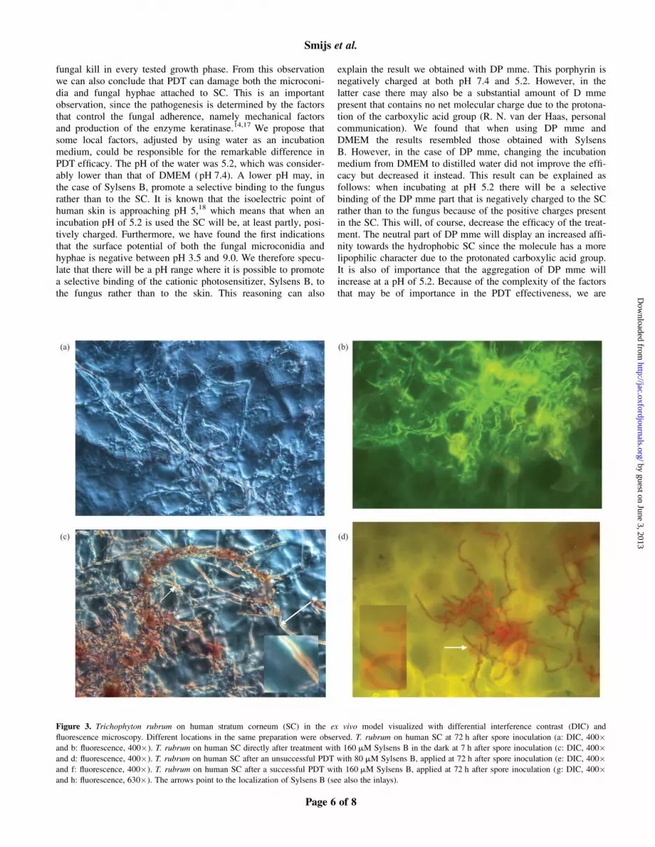
Sylsens B is localized on the fungal wall after dark treatment
and inside the fungal hyphae after successful PDT
The DIC and fluorescence microscopy images shown in Figure 3were made at different locations in the same preparation. In this
way we could select the most representative examples for bothmicroscopic techniques.
Microscopic preparations of T. rubrum in the ex vivo modelwere made for four different conditions. Firstly, T. rubrum wasvisualized at 72 h after spore inoculation (see Figure 3a and b).It can be seen from Figure 3(b) that under the selected fluor-escence microscopic conditions the fungus showed a greencoloured autofluorescence. The images shown in Figure 3 (c andd), were taken from T. rubrum directly after the treatment with160 mM Sylsens B in the dark at 72 h after spore inoculation.Both the DIC (Figure 3c) and the fluorescence images(Figure 3d) show that Sylsens B, represented by the red colourin the DIC images and the red/orange fluorescence in the fluor-escence images, is localized on the fungal wall (see arrow pointsand inlays). Figure 3(e) (DIC) and Figure 3(f) (fluorescence)show images of the fungus after unsuccessful PDT using 80 mMSylsens B, applied 72 h after spore inoculation. Again thearrows point (see inlays as well) to the localization of Sylsens Bon the fungal cell wall.
Figure 3 (g and h) shows images taken from T. rubrum aftera successful PDT with 160 mM Sylsens B, applied at 72 h afterspore inoculation, when a fungicidal effect could be detected.Interestingly, the fluorescence image shown in Figure 3(h), takenat this stage shows that the red fluorescence of Sylsens B ispresent inside the fungal hyphae. The arrows in both the DICand fluorescence images from this stage point to fungal hyphaefilled with Sylsens B.
Discussion
In this study we developed an ex vivo model in order to investi-gate the conditions and evaluate the efficacy of a PDT of thedermatophyte T. rubrum. Since our model offers the possibilityof evaluating the therapeutic effect in different phases of thegermination process, it may contribute to a better understandingof the way in which the therapy affects these individual growthphases. This is important since up to now only very few studieswere concerned with the therapeutic fungicidal effects on differ-ent inoculates.15,16 Recently, Seebacher7 compared the efficiencyof modern antifungal agents on both proliferating fungal strainsand fungal spores.
Using our novel model, it has become clear that microconidiafrom T. rubrum attached to human SC can be completely inacti-vated by the porphyrin photosensitizer Sylsens B when PDT isapplied at an early growth stage (8 h after spore inoculation).This result is in agreement with previous in vitro results wheremicroconidia were used in suspension.13 However, in thesein vitro experiments, using the same light conditions, a muchlower concentration (1 mM) of Sylsens B could be used foreffective treatment. We suggest that the adherence of the micro-conidia to a substrate, like the SC in our ex vivo model, mayinfluence the PDT susceptibility. This situation was different inour in vitro experiments when we worked with the suspensionsand where the attachment to a surface did not play a role.Moreover, from our experiments it has become clear that thephotosensitizer concentration is not the only factor playing apart in achieving a fungicidal effect. The occurrence of the fun-gicidal effect was strongly affected by the medium in whichphotosensitizers were applied. Using distilled water in combi-nation with Sylsens B resulted in a high percentage of complete
Table 1. Occurrence of a fungicidal effect after PDT with Sylsens
B, applied at 8, 24, 48 and 72 h after inoculation of a microconidia
suspension to human SC in the ex vivo modela
PDT application:
time point,
after spore
inoculation (h)
Percentage fungicidal effectb
DMEM distilled water
80 mM
Sylsens B
160 mM
Sylsens B
80 mM
Sylsens B
160 mM
Sylsens B
8 100 100 100 100
24 50 (+0) 50 (+0) 100 100
48 0 0 81 (+6) 96 (+5)
72 0 0 19 (+7) 65 (+8)
The values given are the means of four different experiments (+SEM).aIncubation was either in DMEM (pH 7.4) or in distilled water (pH 5.2) for2 h, followed by an illumination period with red light (108 J/cm2).bA fungicidal effect is defined as the absence of any detectable fungalgrowth at 8 days after spore inoculation.
Table 2. Occurrence of a fungicidal effect after PDT with
DP mme, applied at 8, 24, 48 and 72 h after inoculation of a
microconidia suspension to human SC in the ex vivo modela
PDT
application:
time point,
after spore
inoculation (h)
Percentage fungicidal effectb
DMEM distilled water
80 mM
DP mme
160 mM
DP mme
80 mM
DP mme
160 mM
DP mme
8 100 100 6 (+6) 54 (+6)
24 47 (+3) 85 (+7) 0 0
48 0 0 0 0
72 0 0 0 0
The values given are the means of four different experiments (+SEM).aIncubation was either in DMEM (pH 7.4) or in distilled water (pH 5.2) for2 h, followed by an illumination period with red light (108 J/cm2).bA fungicidal effect is defined as the absence of any detectable fungalgrowth at 8 days after spore inoculation.
Ex vivo model for photodynamic treatment of Trichophyton rubrum
Page 5 of 8
by guest on June 3, 2013http://jac.oxfordjournals.org/
Dow
nloaded from
fungal kill in every tested growth phase. From this observationwe can also conclude that PDT can damage both the microconi-dia and fungal hyphae attached to SC. This is an importantobservation, since the pathogenesis is determined by the factorsthat control the fungal adherence, namely mechanical factorsand production of the enzyme keratinase.14,17 We propose thatsome local factors, adjusted by using water as an incubationmedium, could be responsible for the remarkable difference inPDT efficacy. The pH of the water was 5.2, which was consider-ably lower than that of DMEM (pH 7.4). A lower pH may, inthe case of Sylsens B, promote a selective binding to the fungusrather than to the SC. It is known that the isoelectric point ofhuman skin is approaching pH 5,18 which means that when anincubation pH of 5.2 is used the SC will be, at least partly, posi-tively charged. Furthermore, we have found the first indicationsthat the surface potential of both the fungal microconidia andhyphae is negative between pH 3.5 and 9.0. We therefore specu-late that there will be a pH range where it is possible to promotea selective binding of the cationic photosensitizer, Sylsens B, tothe fungus rather than to the skin. This reasoning can also
explain the result we obtained with DP mme. This porphyrin isnegatively charged at both pH 7.4 and 5.2. However, in thelatter case there may also be a substantial amount of D mmepresent that contains no net molecular charge due to the protona-tion of the carboxylic acid group (R. N. van der Haas, personalcommunication). We found that when using DP mme andDMEM the results resembled those obtained with SylsensB. However, in the case of DP mme, changing the incubationmedium from DMEM to distilled water did not improve the effi-cacy but decreased it instead. This result can be explained asfollows: when incubating at pH 5.2 there will be a selectivebinding of the DP mme part that is negatively charged to the SCrather than to the fungus because of the positive charges presentin the SC. This will, of course, decrease the efficacy of the treat-ment. The neutral part of DP mme will display an increased affi-nity towards the hydrophobic SC since the molecule has a morelipophilic character due to the protonated carboxylic acid group.It is also of importance that the aggregation of DP mme willincrease at a pH of 5.2. Because of the complexity of the factorsthat may be of importance in the PDT effectiveness, we are
(a) (b)
(c) (d)
Figure 3. Trichophyton rubrum on human stratum corneum (SC) in the ex vivo model visualized with differential interference contrast (DIC) and
fluorescence microscopy. Different locations in the same preparation were observed. T. rubrum on human SC at 72 h after spore inoculation (a: DIC, 400�and b: fluorescence, 400�). T. rubrum on human SC directly after treatment with 160 mM Sylsens B in the dark at 7 h after spore inoculation (c: DIC, 400�and d: fluorescence, 400�). T. rubrum on human SC after an unsuccessful PDT with 80 mM Sylsens B, applied at 72 h after spore inoculation (e: DIC, 400�and f: fluorescence, 400�). T. rubrum on human SC after a successful PDT with 160 mM Sylsens B, applied at 72 h after spore inoculation (g: DIC, 400�and h: fluorescence, 630�). The arrows point to the localization of Sylsens B (see also the inlays).
Smijs et al.
Page 6 of 8
by guest on June 3, 2013http://jac.oxfordjournals.org/
Dow
nloaded from

currently focusing on the mechanisms playing a role in the inter-actions between the photosensitizers and the surface of the tar-geted fungi. This investigation concerns research into the factorsthat can be of importance, like the ionic strength and pH of thesurrounding medium, the surface potential of both microconidiaand fungal hyphae, and the photophysical and photochemicalproperties of both photosensitizers.
The fluorescence and DIC images made from the differentstages clearly illustrate that Sylsens B is localized on the fungalwall in the case of the dark controls and unsuccessful light treat-ments (Figure 3c, d, e and f). However, Figure 3 (g and f)shows a localization of the photosensitizer inside the fungalwall. Since the fungus was successfully treated with Sylsens B,a disruption of the fungal wall after PDT could be responsiblefor the penetration of Sylsens B inside the fungus. It can be con-cluded that in a situation that mimics the clinical situation, asrepresented by our ex vivo model, T. rubrum (both fungal sporesand hyphae) can be successfully treated with PDT using160 mM Sylsens B and a light dose of 108 J/cm2 (red light). Thesusceptibility of T. rubrum to PDT is clearly dependent on thetime at which the treatment is performed. Application of thePDT at an increasing time after spore inoculation seems todecrease the susceptibility of the fungus to the treatment. Thisobservation is in contrast with the experiences with regular anti-fungal therapeutics.19 It should be emphasized also that the inac-tivation of the spores by PDT under the given circumstances
seems to be successful. This in contrast to many current thera-peutic treatments where the spores are especially difficult totreat.7,8 These non-responsive spores may remain in the skinwhere they can initiate a relapse of the infection. Our currentresearch focuses on improving the PDT efficacy in the phaseswhere fungal hyphae are treated. One may expect that thefactors that influence the attachment of photosensitizer to fungalwall will be of essential importance in these growth phases.Also, the factors involved in the attachment of the fungus to theSC may be of importance. Controlling these factors is an essen-tial condition for reaching maximal fungicidal effect.
Acknowledgements
This work was supported by the Dutch Technology foundation(STW project LKG 6432). We thank Dr Richard van der Haasfrom the University of Amsterdam (Laboratory of syntheticOrganic Chemistry) and Dr Rob van der Steen from BuchemHolding BV (Lieren, The Netherlands) for the synthesis of thevarious porphyrin photosensitizers and for their valuable advice.
Transparency declarations
None to declare.
(e) (f)
(g)(h)
Figure 3. Continued
Ex vivo model for photodynamic treatment of Trichophyton rubrum
Page 7 of 8
by guest on June 3, 2013http://jac.oxfordjournals.org/
Dow
nloaded from

References
1. Aly R. Ecology and epidemiology of dermatophyte infections.
J Am Acad Dermatol 1994; 31: S21–S25.
2. Rudolf A. Superficial Fungal Infections In: Kalter DC, Sanders
CVS, Nesbitt LT eds. Skin and Infections. Baltimore, MD: Williams &
Wilkins, 1995; 157–161
3. Zaias N, Glick B, Rebell G. Diagnosing and treating onychomy-
cosis. J Fam Pract 1996; 42: 513–18.
4. Rippon JW. The changing epidemiology and emerging patterns
of dermatophyte species. Curr Top Med Mycol 1985; 1: 208–34.
5. Omero C, Dror Y, Freeman A. Trichoderma spp. antagonism to
the dermatophyte Trichophyton rubrum: implications in treatment of
onychomycosis. Mycopathologia 2004; 158: 173–80.
6. Vera JR, Cervera LA. Advantages and disadvantages of topical
antifungal agents. Rev Esp Quimioter 2001; 14: 232–37.
7. Seebacher C. Action mechanisms of modern antifungal agents
and resulting problems in the management of onychomycosis.
Mycoses 2003; 46: 506–10.
8. Fernandez-Torres B, Inza I, Guarro J. Comparison of in vitro
antifungal susceptibilities of conidia and hyphae of dermatophytes with
thick-wall macroconidia. Antimicrob Agents Chemother 2003; 47:
3371–2.
9. Pinjon E, Moran GP, Coleman DC, Sullivan DJ. Azole suscepti-
bility and resistance in Candida dubliniensis. Biochem Soc Trans 2005;
33: 1210–14.
10. Marcus SL, McIntyre WR. Photodynamic therapy systems and
applications. Expert Opin Emerg Drugs 2002; 7: 321–34.
11. Demidova TN, Hamblin MR. Photodynamic therapy targeted to
pathogens. Int J Immunopathol Pharmacol 2004; 17: 245–54.
12. Lambrechts SA, Aalders MC, Van Marle J. Mechanistic study of
the photodynamic inactivation of Candida albicans by a cationic por-
phyrin. Antimicrob Agents Chemother 2005; 49: 2026–34.
13. Smijs TG, van der Haas RN, Lugtenburg J et al. Photodynamic
treatment of the dermatophyte Trichophyton rubrum and its microconi-
dia with porphyrin photosensitizers. Photochem Photobiol 2004; 80:
197–202.
14. Zurita J, Hay RJ. Adherence of dermatophyte microconidia and
arthroconidia to human keratinocytes in vitro. J Invest Dermatol 1987;
89: 529–34.
15. Rashid A. New mechanisms of action with fungicidal antifungals.
Br J Dermatol 1996; 134 Suppl 46: 1–6.
16. Nardoni S, Millants F, Mancianti F. In vitro sensitivity of two
different inocula of microsporum canis versus terbinafine. J Mycol Med
2000; 10: 148–51.
17. Raubitschek F. Mechanical versus chemical keratolysis by der-
matophytes. Sabouraudia 1961; 1: 87–90.
18. Marro D, Guy RH, Delgado-Charro MB. Characterization of the
iontophoretic permselectivity properties of human and pig skin. J
Control Release 2001; 70: 213–17.
19. Dittmar W, Lohaus G. HOE 296, A new antimycotic compound
with a broad antimicrobial spectrum. Arzneimittelforschung 1973; 23:
670–4.
Smijs et al.
Page 8 of 8
by guest on June 3, 2013http://jac.oxfordjournals.org/
Dow
nloaded from


