Embed Size (px)
Citation preview

Journal of Biomedical Optics 5(3), 338–349 (July 2000)
Study of photodynamic reactions in human blood
A. DouplikA. A. StratonnikovV. B. LoshchenovGeneral Physics Institute
of Russian Academy of SciencesLaser Biospectroscopy Laboratory,38 Vavilova St.117942, Moscow, Russia
V. S. LebedevaInstitute Lomonosov State Academy
of Fine Chemical TechnologyPr. Vernadskogo 86117571 Moscow, Russia
V. M. DerkachevaNIOPIK Institute of Organic Intermediates and DyesBolshaya Sadovaya St., 4/1103787 Moscow, Russia
A. VitkinOntario Cancer Institute and University of TorontoDepartment of Medical Biophysics610 University Ave.Toronto, ON M5G 2M9, Canada
V. D. RumyancevaInstitute Lomonosov State Academy
of Fine Chemical TechnologyPr. Vernadskogo 86117571 Moscow, Russia
S. G. KusminNIOPIK Institute of Organic Intermediates and DyesBolshaya Sadovaya St., 4/1103787 Moscow, Russia
A. F. MironovInstitute Lomonosov State Academy
of Fine Chemical TechnologyPr. Vernadskogo 86117571 Moscow, Russia
E. A. Luk’YanetsNIOPIK Institute of Organic Intermediates and DyesBolshaya Sadovaya St., 4/1103787 Moscow, Russia
Abstract. Comparative studies of oxygen consumption, changes ofphotosensitizer fluorescence, and photodestruction of erythrocytes,and photodestruction of oxygen transport protein hemoglobin wereperformed during photodynamic reaction in whole and hemolyzedblood with phthalocyanines, chlorines, porphyrins, and methyleneblue photosensitizers in vitro and in selected cases in vivo. Thepresent work deals with the investigation of blood oxygen saturationSO2 and photosensitizer fluorescence during and immediately afterlight irradiation in the photodynamic therapy process. It has beenobserved that SO2 behavior strongly correlates with the type of pho-tosensitizer. The decrease of photosensitizer fluorescence (pho-tobleaching) during light irradiation can be followed by the recoveryof the photosensitizer fluorescence immediately after interruption ofthe irradiation within 6–8 min. The levels of photodestruction oferythrocytes in whole blood and photodestruction of hemoglobin inhemolyzed blood in combination with the above photosensitizers re-veal the influence of photodynamic reactions upon the ability ofblood to transport oxygen. Maximal photohemolysis activity has beenfound with chlorine p6 photosensitizers. © 2000 Society of Photo-OpticalInstrumentation Engineers. [S1083-3668(00)00303-8]
Keywords: photodynamic therapy; oxygen consumption; photobleaching; photohe-molysis.
Paper ODB-004JBO received Jan. 30, 1998; revised manuscript received Feb. 18,1999; accepted for pulication Jan. 11, 2000.
ti-
f
r
nsi-theenol-
let
eonndtn
1 IntroductionOne of the most important objects of current investigation ishuman blood. This is a wonderful creation of nature and a parof the human body possessing characteristics of both biologcal tissues and fluids. We investigate the interaction of bloodwith photosensitizers and its participation in photodynamicreactions. The current study is part of a broader research efort aimed at developing an accurate and fast clinical systemof photodynamic therapy~PDT! efficacy monitoringin vivo.
The efficiency of many modern photosensitizers used inPDT depends on the presence of dissolved oxygen in the i
Address correspondence to Y. Douplik, current address 1 Greystone Walk Dr.,Penthouse 91, Toronto M1K 5J3 ON, Canada. Electronic mail: [email protected] paper was originally scheduled to appear in the January 1999 special sec-tion on Optical Diagnostics of Biological Fluids, but was not available in time.
338 Journal of Biomedical Optics d July 2000 d Vol. 5 No. 3
-
-
radiated tissues. The interaction of the photons, photosetizer, and oxygen molecules results in the formation ofactive forms of oxygen such as singlet oxygen and oxygradicals. These reactive forms can damage different biomecules, subcellular and cellular structure.1 It is assumed thatthe destroying effect is mainly due to the formation of singoxygen~type II PDT process! resulting from the interaction ofa light excited photosensitizer with molecular oxygen~tripletground state!. Hence, the effect will be proportional to thlevel of singlet oxygen formation, which in turn dependslight intensity, type of photosensitizer, its concentration, amolecular oxygen concentration.2,3 The decrease in the lasquantity is indicative of the level of singlet oxyge
1083-3668/2000/$15.00 © 2000 SPIE

y-
n
,
-e
n
t
-
-
s
,
-
-
l-
loss
hey
e of
mcowof
en-nsen-las
ion
-pec-tionies,
stan-ure-y-
itstheticalourswehin
esti-ipi-cu-
in aododn-
hro-om
Study of Photodynamic Reactions
generation.4 Direct destruction of photosensitizer in the courseof PDT, defined as a photobleaching,2,5 can exhibit itself as adecrease of photosensitizer concentration and or its photodnamic activity in tissue. Hence, one can estimate the biological effects due to photodynamic reactions by means of measuring the photosensitizer photobleaching and the oxygeconsumption within the light-irradiated volume of the biologi-cal tissue during PDT. A complete PDT efficacy monitoringsystem includes explicit evaluation of tissue oxygenationphotosensitizer concentration, and light irradiation dose.2,3 Inthe present comparative study with phthalocyanines, chlorines, porphyrins, and methylene blue photosensitizers, wconcentrated on tissue oxygenation and photosensitizer fluorescence monitoring during PDT. We considered two pro-cesses characterizing tissue oxygenation status: oxygen cosumption during PDT in whole blood and the influence ofphotodynamic reactions upon the ability of blood to transporoxygen. Several aspects of the latter were examined: functional study of the ability of PDT-deoxygenated whole bloodto be reoxygenated and investigations of the levels of photodestruction of blood oxygen transport components includingerythrocytes or red blood cells~RBCs!, and oxygen transportprotein hemoglobin, which is normally located inside RBCs.8
Photohemolysis, the destruction of RBC’s by the photody-namic reaction,9–11was investigated with whole blood. On theother hand, hemoglobin photodestruction is best examined under conditions of significant photohemolysis, as in hemolyzedblood. Concentration of the dissolved oxygen in living tissuewill be proportional to hemoglobin oxygenation@blood oxy-gen saturation, %(SO2)] inside erythrocytes.2 It is defined by
SO25CHbO2
CHbO21CHb
, (1)
whereCHbO2andCHb are the concentrations of oxyhemoglo-
bin and deoxyhemoglobin, respectively. Development of newmethods of monitoring of PDT with oxygen mediated photo-sensitizers is important for scientific and clinical reasons. Specifically, we are interested in thein vivo processes for a par-ticular photosensitizer.In vitro oxygen consumption ispurported to be an irreversible process because of limited content of oxygen in the sample.12,13 In living tissue, the bloodoxygen level is a balance between arterial input and venououtput, which are the parts of blood microcirculation supplysystem of tissue metabolism.14 The photodynamic processcauses damages microvasculature15,16 and causes additionalconsumption of oxygen in the tissue and directly in the bloodthus shifting this balance. The decrease of blood oxygen saturation following PDT depends on irradiation photon density,photosensitizer type, its concentration in blood and surrounding tissue, and extent of blood microcirculation within thevolume of light irradiation.17 Measurements of fluorescencespectra of photosensitizers dissolved in blood reveal the processes of their binding with blood components. For monitor-ing of photosensitizer concentration or its photodynamic activity in the course of PDT we studied the rate and the level ofphotobleaching described by reduction of the photosensitizefluorescence. Actually, the term ‘‘photobleaching’’ is used todesignate chemical destruction of the photosensitizer moecule, but in this current paper the term will relate to the
-
-
-
-
-
-
-
-
-
r
measurable fluorescence decrease of the photosensitizer.2 Weassume that the fluorescence reduction is proportional toof photodynamic activity of the photosensitizer.2 It could beaccompanied by changes of the phototoxicity of tphotosensitizer.5,7 In turn, the photosensitizer phototoxicitdepends on oxygen concentration in surrounding media.18 Onthe other side, photobleaching itself can lead to a decreasoxygen consumption.6,7
2 Materials and Methods
2.1 Materials
2.1.1 Preparation of BloodWe used heparinized, fully oxygenated whole blood frohealthy donors and heart disease patients of the MosMedical Academy within 1–3 h after donation. The valueblood hematocrit was4261. Before preparation of thesample, the blood was incubated with or without a photossitizer for 30 min at37°C. The control sample did not contaia photosensitizer. The concentration of some of the photositizers in the test sampleK ~mg/ml! was close to its maximablood level for a typical PDT treatment. It was calculated
K~mg/l!5KwW/U , (2)
whereKw is the recommended photosensitizer concentratper kilogram of body weight~mg/kg!, W is the body weight~kg!, andU is the total volume of blood~normally about 5000ml!.8
2.1.2 The Methods of Sample PreparationIn VitroThe sample preparation techniquein vitro depends on the duration of the measurement. In these studies, fluorescence stra measurement takes only 1 s, while to measure absorpspectra requires 5–12 min. Therefore for fluorescence studblood contained in a standard cuvette was adequate. Thedard methods of sample preparation for absorption measments did not work for the comparative studies of photodnamic reactions in blood because of the instability ofscattering and absorption properties. The whole blood incuvette undergoes the process of sedimentation and its opproperties cannot be treated as stationary for several hafter sample preparation. To overcome these difficultieshave developed new methods enabling one to work with tlayers of the native whole blood with cells fixed in space.19
2.1.2.1 The Preparation of Blood Sample forFluorescence ResearchFor fluorescence measurement the blood sample was invgated in standard 1 mm cuvette before and after RBC prectation. Before preparation of the sample, the blood was inbated with or without photosensitizer for 30 min at37°C. Thesample was put in the standard 1 mm cuvette and placedspecial light-proof holder. Then for preparation of the blocomponent sample we kept the cuvette with the whole bloin a refrigerator at 4°C for approximately 1.5 h for the spotaneous separation settling of blood into plasma and erytcytes. Before use the sample was held about 30 min at ro
Journal of Biomedical Optics d July 2000 d Vol. 5 No. 3 339

ss
t
f
e
--
-a
ah
e
-
.
s
6-
-ctra-
para-
eter
entof
ure-
al
tondtheing
alom-ion
lof
a intu-
en-
s of
alrdxy-tle
zedthe
eda-inglybe-his
Douplik et al.
temperature in the dark for warming. After that the preparedsample was ready for measurements within 4–6 h.
2.1.2.2 The ‘Glass Slide’ Method0.08 ml of whole blood was deposited on a standard glasslide. Then the blood droplet was covered by another glasslide to achieve homogeneous distribution of the liquid be-tween the slides. Such a ‘‘sandwich’’ is naturally held to-gether by capillary forces. The thickness of the blood layer is2065 mm. Then the prepared sample was left in the dark for;40 min to complete the erythrocyte aggregation process aroom temperature. The resultant sample yielded quite stationary optical properties for at least 2 h. The relative spread othe blood oxygen saturation from spectral measurements wa2%–3%. To estimate the errors and ensure reproducibility, wtypically repeated the measurements on 15–20 samples. Thability to participate in the photodynamic reactions is retainedin these samples for 24–28 h from the time of preparation.
Although simple, this method has a substantial disadvantage of ill-defined blood layer thickness. Hence correct quantitative comparisons, for example of the dynamics of oxygenconsumption of different photosensitizers, is seriously hin-dered. Another method described below was developed fomore accurate comparative studies of photodynamic reactionin blood.
2.1.2.3 The Gel MethodThe washed erythrocytes or whole blood with or without thephotosensitizer were added to 1.8% solution of the ultralowtemperature type of agarose in PBS buffer and poured intocuvette of known thickness.~This method was not successfulwith polyacrilamyde gel because phthalocyanines inhibitedthe gel polymerization process.! The sample was then placedin the refrigerator at 4°C for 5–10 min to solidify the gel viathe polymerization process. The relative scatter in measurements of the blood oxygen saturation by means of spectrmeasurements was less than 1% and was independent of topt-probe position on the sample. For one experiment we usually repeated measurements with 5–10 samples. The gsamples are then capable of photodynamic reactions fo2–3 d.
2.1.3 PhotosensitizersWe studied four groups of the Russian produced watersoluble photosensitizers~Table 1!. They are Phthalocyanines,Chlorines, Porphyrins and Methylene Blue. PBS buffer wasused as a solvent.
2.1.4 In Vivo StudiesThe 75 C57B1/DBA(BDF1) breeding female mice bearingErlich carcinoma were used in the current work. All micewere 10–12 weeks old, with average body weight of 20–22 gThe photosensitizer Photosence~Table 1! has been injectedintravenously at a dose of 4 mg/kg. The photosensitizer habeen dissolved in physiological solution~0.9% NaCl! for in-travenous injection. Two PDT sessions have been applied inand 28 h, respectively, after the photosensitizer administration.
340 Journal of Biomedical Optics d July 2000 d Vol. 5 No. 3
-
s
e
rs
-
e-lr
2.2 Methods
2.2.1 Absorption Measurements
2.2.1.1 Monitoring of the Dynamics of PhotodynamicOxygen ConsumptionBlood oxygen saturation(SO2) was calculated from the relationship between the scatter-corrected transmission spe~range 520–590 nm! of oxy- and deoxy-hemoglobin. The statistical errors in evaluated blood oxygen saturation forin vitromeasurements depended on the experimental sample pretion method described above in Secs.2.1.2.2and2.1.2.3, withreproducibility within 3%–4%. The spectra measurementsinvitro have been conducted in a two-beam spectrophotomwith integrating sphere~HITACHI U-3400, Japan!. Measure-ment of one spectrum took 5–12 min. As an independcheck, we determined the whole blood gas compositionselected PDT irradiated samples with the electrode measments of the gas composition in a blood gas analyzer~CIBA-CORNING 238, England!. The agreement with the spectrmeasurements was 15%.
For the spectral measurementsin vivo during PDT we usedthe fiber optic spectrometer LESA-5~Biospec Lab., Moscow,Russia!.22,23 The light from a halogen lamp was deliveredthe skin tissue through a quartz fiber. The light delivery adetection spectrometer fibers were on the same side oftissue to measure the diffuse reflectance. After passthrough the tissue~blood vessels of derma!, diffuse light wascollected by the receiving fiber. The detector fiber’s proximend served as the input slit of the spectrometer. The spectreter was PC controlled. Custom software allowed acquisitand display of spectra in real time~time resolution of 0.1 s perspectrum, spectral resolution of 7 nm!. Subsequent spectraanalysis yields absorption spectra after scatter correctionthe measured diffuse reflectance.24 The displayed blood oxy-gen saturation was calculated from the absorption spectrreal time. The reproducibility in evaluated blood oxygen saration for thein vivo measurements was about 10%.
2.2.1.2 The Effect of Photodynamic Reactions onOxygen Transport Ability of BloodThe examination of the blood capability to transfer oxygincluded functional study of reoxygenation of PDTdeoxygenated whole blood, and investigations of the levelphotodestruction of erythrocytes~photohemolysis!, and pho-todestruction of hemoglobin. To test the RBC’s functionability for reoxygenation after uniform irradiation in standacapillaries, we withdrew blood that was irradiated and deogenated and tried to restore its full oxygenation by genmechanical shaking in aerobic surroundings.
The photohemolysis level in the irradiated whole bloodinvitro was determined as the relative change~%! of the totaltransmission and diffuse reflectance of the photosensitiblood sample relative to a photosensitizer-free sample, atisosbestic wavelength of 805 nm25,26 during the first 2 h afterirradiation. The sample without photosensitizer was assumto exhibit zero level of photohemolysis under the light irrdiation. Since the erythrocytes are the main light scatterparticles in human blood,26 our measurements are particularsensitive to changes in light transmission and reflectioncause of reduction of scattering due to lysis of RBCs. T

Study of Photodynamic Reactions
Table 1 Photosensitizers examined in this study (synthesized in Russia).
Photosensitizers Composition
Absorption maximum inred range (nm)
Solution in PBS buffer
Extinctioncoefficient
(cm−1/mol/l)
Molecularweight
(Daltons)
Phthalocyanines percentage of groups withvarious degree of sulfonation
(a) Aluminum phthalocyanine(Photosence) (PhSn)
(a) tetra - 30, tri - 50, di - 20 (a) 675 (a) 100 000 (a) 862
(b) Zinc phthalocyanine I(PcZn1)
(b) tetra - 76, tri - 23, di - 1.5 (b) 665 (b) 98 500 (b)–(e) ;870 a
(c) Zinc phthalocyanine II(PcZn2)
(c) tetra - 7, tri - 12, di - 75 (c) 670 (c) 28 300
(d) Zinc phthalocyanine III(PcZn3)
(d) tetra - 28, tri - 50, di - 22 (d) 675 (d) 39 600
(e) Zinc phthalocyanine IV(PcZn4)
(e) tetra - 46, tri - 44, di - 10 (e) 675 (e) 46 000
Chlorines
(a) Chlorine p6 I(Chp6-1)
(a) trisodium salt Clp6 (a) 664 (a) 11 100 (a) 648
(b) Chlorine p6 II(Chp6-2)
(b) trisodium salt 3-devinyl-3 formyl Clp6
(b) 695 (b) 11 780 (b) 650 b
Porphyrins
(a) Hematoporphyrinderivative (photogem)(PhG)
(a) 85% - oligomers,15% monomers
(a) 630 (a) 4400 (a) 598–2920 c
(a) Uroporphyrin III(UPh)
(b) the mixture of isomers Iand III (I/III=4/1)
(b) 625(675)
(b) 4170(400)
(b) 818 d
Methylene Blue 600–665 373,9
(MB) (665) (72 000)
a Reference 15.b Reference 20.c Reference 16.d Reference 21.
ity
irst,tizersn-wasntsr
ce,ced
method allows one to detect rather pronounced~more than5%!, fast developed~1–2 h after irradiation! hemolysis. Thereproducibility in evaluated photohemolysis forin vitro mea-surements was about 6%–25%.
For the photosensitizers exhibiting significant level of pho-tohemolysis, we also examined the hemoglobin photodestruction level as a result of photodynamic reaction in hemolyzedblood. We used fully oxygenated hemolyzed blood. Determi-nation of hemoglobin photodestruction was made from therelative change~%! of the sample absorption~no scattering inthis case! at isosbestic wavelength of 568 and 586 nm27 im-mediately after irradiation, relative to the sample without pho-tosensitizer. Once again, the latter was assumed to displazero level of hemoglobin photodestruction under the light ir-radiation. The intent here was not to identify the hemoglobinderivatives resulting from photodynamic reaction, but ratherto determine the relative changes of hemoglobin concentra
-
y
-
tion in the sample during and after PDT. The reproducibilin evaluated hemoglobin photodestruction forin vitro mea-surements was about 3%–5%.
2.2.2 Fluorescence MeasurementsTwo sets of fluorescence studies have been conducted. Fwe compared fluorescence spectra of selected photosensiin PBS and in blood to determine effect of solvent enviroment. The second study investigated photobleaching, andcarried out for the samples of whole blood, its compone~i.e., separated RBSs and blood plasma!, and photosensitizesolutions in PBS. For all these experimentsin vitro we alsoused the fiber optic spectrometer LESA-5~Biospec Lab.,Moscow, Russia!22,23,28~see Sec.2.2.1.1!. A He–Ne laser wasused instead of a halogen lamp for excitation of fluorescenand as the PDT light source. The sample fluorescence indu
Journal of Biomedical Optics d July 2000 d Vol. 5 No. 3 341

Douplik et al.
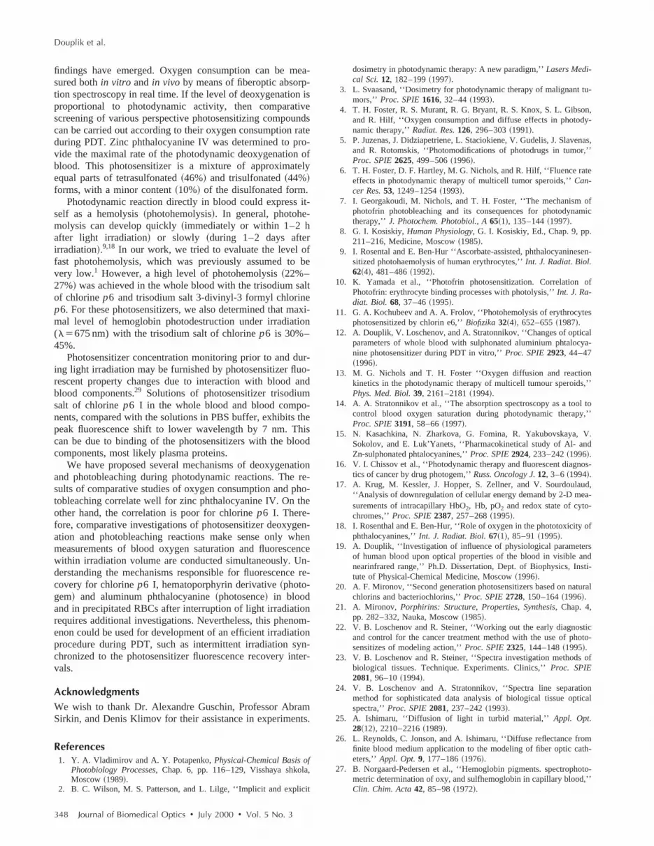
Fig. 1 Monitoring photodynamic deoxygenation in vitro for the whole blood via the glass sandwich method. l5665–675 nm, Pm510 mW/cm2. Concentrations: chlorine p6 II (Chp6-2)—0.01 mg/ml; chlorine p6 I (Chp6-2)—0.01 mg/ml; zinc phthalocyanine II (PcZn2)—0.013mg/ml; zinc phthalocyanine III (PcZn3)—0.012 mg/ml; photosence (PhSn)—0.01 mg/ml; zinc phthalocyanine I (PcZn1)—0.013 mg/ml; zincphthalocyanine IV (PcZn4)—0.013 mg/ml. In this and subsequent graphs, the lines are a guide for the eye.
e
e
nri
-.
e
A
sir-on,d a
areod
Theorp-eac-nsen-nd
thatesbebe-
alo-
entsia-atea-izernce
by the He–Ne irradiation(Pm5100– 250 mW/cm2) was ob-served in the 640–900 nm spectral range. A fluorescencspectrum was acquired in 1 s.
Fluorescence spectra were measured in backscattering gometry and were dependent on both absorption and scatterinproperties of the tested object. To account for the changes iabsorption and scattering of the sample, fluorescence spectdetected during the PDT session were normalized to the maxmum amplitude of the excitation source spectrum.23,24,28 Inother words, fluorescence was calculated as the ratio omaxima of excitation source and fluorescence spectra~i.e.,FL/EX!.28 The monitoring of photobleaching~diminution offluorescence during irradiation! was performed as the ratio offluorescence prior to irradiation~measured after the first sec-ond of switching on the excitation source! and fluorescencemeasured in the course of PDT irradiation. Evaluation of fluo-rescence recovery was carried out after switching off theHe–Ne laser, in the same way as photobleaching monitoringFor excitation of fluorescence during its recovery measurements the He–Ne laser was periodically turned on for 2–3 sThe reproducibility in measured fluorescence forin vitro in-vestigations was about 3%.
2.2.3 PDT Irradiation of Blood and of TissueThe fluence rate was calculated as follows19:
Pm~W/cm2!5Ps~mW/cm2!~12Tt2Rd !, (3)
wherePs is the irradiance rate of the incident beam, andTtandRd are the total transmittance and the diffuse reflectancof the blood layer for the wavelength of interest.
We used a He–Ne laser~633 nm! coupled to a lightguide~diameter 200mm! for both excitation of fluorescence and for
342 Journal of Biomedical Optics d July 2000 d Vol. 5 No. 3
-g
a-
f
.
PDT irradiation during investigations of photobleaching.confocal light emitting diode~LED! matrix ~665–675 nm!~Biospec Lab., Moscow, Russia! was used for experimentwith oxygen consumption evaluation. For more powerfulradiation for comparative studies of oxygen consumptiphotohemolysis, and hemoglobin photodestruction, we usediode laser~670 nm! ~Biospec Lab., Moscow, Russia!. Afrequency-doubled solid state laser~672 nm! was used forinvivo PDT irradiation.
3 Results and DiscussionsThe analysis of the absorption spectra showed that thereno pronounced changes of the optical properties of blosamples without photosensitizers due to laser irradiation.changes of the blood oxygen saturation deduced from abstion spectroscopy measurements due to photodynamic rtions in human bloodin vitro are presented in Figures 1–3. Ithis and other graphs we use abbreviated names of photositizers defined in Table 1. Results with phtalocyanines achlorines photosensitizers are shown in Figure 1. We noteoxygen consumption during light exposure for phtalocyaninis greater than for chlorines by a ratio of 2:20. This maydue to a considerable difference in extinction coefficienttween these two photosensitizer types.15,16,20The fastest pho-todynamic deoxygenation was demonstrated by zinc phthcyanine IV.
Results obtained from absorption spectra measuremfor four other photosensitizers in whole blood under irradtion with l5675 nmare presented in Figure 2. It is clear thuroporphyrin demonstrated no photodynamic activity as msured by oxygen consumption. Moreover, this photosensitdemonstrated the same null result with a much higher flue

Study of Photodynamic Reactions
Fig. 2 Monitoring photodynamic deoxygenation in vitro for the whole blood via the glass sandwich method. l5675 nm, Pm535 mW/cm2.Concentrations: uroporphyrin III (UPh)—0.023 mg/ml; chlorine p6 II (Chp6-2)—0.025 mg/ml; Methylene Blue—0.01 mg/ml; chlorine p6 I (Chp6-1)—0.025 mg/ml.
n
--
nts
forlo-
er,
rate ofPm5300 mW/cm2. Similarly, the photosensitizer pho-togem did not exhibit any measurable blood oxygen saturatiodecrease atl5633 nm and Pm5100 mW/cm2 ~results notshown!. It also appears that the rate of deoxygenation forchlorines and Methylene Blue are similar. For photosensitizers presented in Figure 2, we used a higher incident of irra
diation because of the lower values of extinction coefficiecompared with photosensitizers in Figure 1.
As was shown in Figure 1, we obtained a similar resultphotodynamic oxygen consumption with aluminum phthacyanine and zinc phthalocyanine I~curves 1 and 2! with theslide method. To examine these two phtalocyanines furth
Fig. 3 Monitoring photodynamic deoxygenation in vitro for the erythrocyte suspension via the gel slab method. l5665–675 nm, Pm510 mW/cm2. Concentrations: photosence (PhSn)—0.01 mg/ml; zinc phthalocyanine I (PcZn1)—0.013 mg/ml.
Journal of Biomedical Optics d July 2000 d Vol. 5 No. 3 343

Douplik et al.
Fig. 4 Monitoring of blood oxygen saturation during PDT in vivo. The influence of laser irradiation (Ps585 mW/cm2, l5670 nm) on hemoglobinoxygen saturation in microvasculature of mice during photodynamic therapy with photosence, at the injection dose of 4 mg/kg, is shown. The timecourse of incident laser power is indicated by dotted line. (a) The first PDT session (photosensitizer accumulation time is 6 h); (b) The second PDTsession (photosensitizer accumulation time is 28 h).
te
-s
timeeentto
nt-for
por-
ho-ol-we
oly-tions of
ghtlity-
we used the gel method of the sample preparation. The oucome of this experiment is displayed in Figure 3. As seen, thfaster photodynamic deoxygenation occurs in the bloodsample photosensitized by zinc phtalocyanine I.
Selected independent checks of thein vitro reflectance re-sults in Figures 1–3 were performed through the gas analyzeto determine PDT-induced oxygen consumption in blood withphotosence and Methylene Blue. We observed the decreasepO2 in the samples with photosensitizers after light irradiationfrom 100 to 170 mm Hg down to 0–2 mm Hg. BothpH andpCO2 were virtually stable. The attempts of the reoxygenationof the irradiated deoxygenated blood samples were successfuGas analysis showed that the blood oxygen saturation levewas restored to almost 97%. This implies that the use of photosence and Methylene Blue photosensitizers does not cauoxygen transport dysfunction of blood during PDT.
The results of oxygen consumptionin vivo during PDTmeasured with fiberoptic spectrometer LESA-5 are shown inFigures 4~a! and 4~b!. The irreversible vessel damage wasobserved for the first PDT session, when photosensitizer accumulation time was 6 h~a!. Therefore, only incomplete re-covery of oxygenation is seen. The blood oxygen saturationwas restored reversibly after switching off the laser during the
344 Journal of Biomedical Optics d July 2000 d Vol. 5 No. 3
-
r
of
l.l
e
-
second PDT session, when photosensitizer accumulationwas 28 h~b!. This was enough time for elimination of thphotosensitizer from blood and blood vessel tissue to prevphotodynamic reaction directly in the blood and damagesblood vessels.
Photohemolysis~destruction of RBCs due to developmeof photodynamic reaction9! is presented in Figure 5. This phenomenon was evident for chlorines and was not presentphthalocyanines. Photohemolysis was also detected forphyrins at a rather high fluence rate of500 mW/cm2 (l5633 nm) ~results not shown!.
The differences in the photohemolysis level between ptosensitizers could be in the ability of the photosensitizer mecule to bind with erythrocyte membranes. From Figure 5can suppose that chlorinesp6 are most binding with erythro-cyte membranes, based on their highest determined hemsis. Therefore, we investigated hemoglobin photodestrucspecifically in these photosensitizer systems. The resultthe studies are presented in Figure 6.
We should note that the results of Figures 5 and 6 mihave a similar underlying cause. It is possible that the abiof chlorinep6 I to bind with both erythrocytes and hemoglo

Study of Photodynamic Reactions
Fig. 5 Monitoring of the photohemolysis in vitro in whole blood via the glass slide method. Photohemolysis is measured in percents of the control(nonirradiated) sample, which is assumed to have 0% of hemolysis. l5675 nm, Pm535 mW/cm2. Concentrations: chlorine p6 I (Chp6-1)—0.025mg/ml; chlorine p6 II (Chp6-1)—0.025 mg/ml; zinc phthalocyanine IV (PcZn4)—0.013 mg/ml.
Fig. 6 Monitoring of hemoglobin destruction during irradiation in vitro in hemolyzed blood via the glass slide method. Photodestruction ofhemoglobin is measured in percents of the control (nonirradiated) sample, which is assumed to have 0% of hemoglobin photodestruction. l5675 nm, Pm535 mW/cm2. Concentrations: chlorine p6 I (Chp6-1)—0.025 mg/ml; chlorine p6 II (Chp6-2)—0.025 mg/ml.
Journal of Biomedical Optics d July 2000 d Vol. 5 No. 3 345

d
-e
-
o
ces-o-ure-e
nceIIn-tool-
for
n-e-idethersto-
in-
ncesthe
es-
ured
ofyth-
pli-ap-n ofr.on-nu-er-tompless-di-
m-ce
s of
Douplik et al.
bin is higher than that of chlorinep6 II. Interestingly, we alsoobserved a substantial increase in viscosity of whole bloophotosensitized with chlorinep6 I in the cuvette after laserirradiation (l5675 nm, Pm5150 mW/cm2). This effect didnot occur with any other photosensitizer studied. This phenomenon is perhaps related to protein denaturation in thchlorine p6 I system, due to its expected highest abilityamong investigated photosensitizers for binding with erythrocyte membrane and hemoglobin.
The results of the fluorescence measurements of the phtosensitizers in PBS solution are presented in Figure 7. Concentrations of photosensitizers have been selected to ensusimilar fluorescence intensity for each system, allowing the
Fig. 7 Fluorescence spectra of studied photosensitizers in PBS solu-tion. Excitation l5633 nm. (1) Zinc phthalocyanine I—0.007 mg/ml;(2) zinc phthalocyanine II—0.013 mg/ml; (3) zinc phthalocyanineIII—0.012 mg/ml; (4) zinc phthalocyanine IV—0.013 mg/ml; (5) alu-minum phthalocyanine (photosence)—0.001 mg/ml; (6) MethyleneBlue—0.01 mg/ml; (7) chlorine p6 I—0.01 mg/ml; (8) chlorine p6II—0.01 mg/ml; (9) uroporphyrin III—0.023 mg/ml; (10) hematopor-phyrin derivative (photogem)—0.5 mg/ml.
346 Journal of Biomedical Optics d July 2000 d Vol. 5 No. 3
--re
presentation of all curves in one scale. Essentially, this nesitated concentration adjustment of Uroporphirin III and Phtogem. Fluorescence was very stable during the measments. No photobleaching effect for any of thphotosensitizers in PBS solution has been observed.
The largest fluorescence amplitude is seen for photose~curve 5!. The lowest amplitude is for zinc phthalocyanine~curve 2!, more than two times lower than for other photosesitizers from the Zn phthalocyanine group. It may be duemore pronounced aggregation of such photosensitizer mecules. Despite a very low level of oxygen consumptionUroporphyrin during PDT~probably indicative of its real pho-todynamic activity!, note the high level of fluorescence intesity ~curve 9!. One can assume that uroporphyrin III is a usful dye for diagnostic purposes. From Figure 7, we can divall investigated photosensitizers into two groups based onwavelength range of their fluorescence maxima. In the figroup are zinc phthalocyanine I, II, III, IV, photosence, chlrine p6 I, and uroporphyrin III~curves 1, 2, 3, 4, 5, 7, 9!, withfluorescence maxima at 670–685 nm. The second groupcludes chlorinep6 I, Methylene Blue, and photogem withpeak fluorescence in the range 690–710 nm. These infereobtained from fluorescence spectra of Figure 7 account fordynamic range issues of the fiber optic system for fluorcence measurement in biological liquids and tissues.
Fluorescence spectra presented in Figure 8 were measfor chlorine p6 I in whole blood before~curve 3! and after~curves 2,4! sedimentation. This allows for observationphotosensitizer distribution between blood plasma and errocytes compartments. We supposed that fluorescence amtude indicates photosensitizer concentration. The majoritypears localized in blood plasma; hence, the concentratiochlorine p6 I in whole blood and plasma must be similaHowever the fluorescence amplitude in blood plasma is csiderably higher than in whole blood because of strong atteation of exciting and fluorescent light by erythrocytes scatting and hemoglobin absorption.19 We thus conclude thadetermination of relative photosensitizer concentration frfluorescence measurements can be quantified only in samwith similar types of optical properties. For example, fluorecence spectra from an absorbing only solution cannot berectly correlated with spectra from a turbid tissue. This iposes certain limitations of fiberoptic fluorescenspectroscopy usage in real biological tissues bothin vivo andin vitro. It is seen that the spectral shape and peak position
Fig. 8 Fluorescence spectra of chlorine p6 I in (1) PBS buffer; (2) plasma; (3) whole blood; (4) erythrocytes. Excitation l5633 nm.

Study of Photodynamic Reactions
Fig. 9 Photobleaching of photosensitizers in precipitated erythrocytes. Photobleaching is measured in percent relative to a sample measured in thefirst 1–3 s of excitation, which is assumed to have 100% of its early fluorescence level. Dashed vertical line indicates the time at which theexcitation laser was turned off. Excitation l5633 nm. Photosence (PhSn)—0.001 mg/ml; chlorine p6 I (Chp6-1)—0.01 mg/ml; photogem (PhG)—0.05 mg/ml.
ed
-i
-l
c
-
s
r-
-
on-
tro-
ofngeswere
eplyofho-x-for
htofsth
tohipsach-
cur-entful
photosensitizers in whole blood and erythrocytes are thsame. Photosensitizer spectra in the whole blood and bloocomponents, compared with PBS buffer spectra, exhibit afluorescence peak shift by;7 nm, which can be due to bind-ing of the photosensitizers with the blood components~pro-tein binding–plasma proteins or erythrocytes!.
The main problem of the experimental blood measurements was the change of the fluorescence signal during exctation. There was a diminution of fluorescence of blood withdissolved photosensitizer during irradiation by the He–Ne laser. This so-called photobleaching phenomenon is welknown.7 However, we did not observe this in PBS solutionand blood plasma. Evidently, the presence of membrane strutures was necessary for photosence, chlorinep6 I, and pho-togem photobleaching. We therefore used erythrocytes in ouexperiments to study this phenomenon. The most expressephotobleaching effect took place on precipitated RBCs. Figure 9 summarized the results of photobleaching monitoring inprecipitated erythrocytes.
We hypothesize that the observed photosensitizer fluorescence change during light excitation is caused by a combination of several different processes. In the following discus-sion, the minus sign indicates a process leading to afluorescence decrease, while the plus sign denotefluorescence-increasing process. The first is a formation oreversible complex between photosensitizer and oxygen duing light irradiation. This process causes attenuation of fluorescence without absorption spectra changes~2A process!.The second is oxygen consumption during photodynamic reaction due to singlet oxygen generation~1B process!.2,4 Thedecrease in oxygen concentration can lead to extraction ooxygen from complexes appearing during the2A process.The third is direct bleaching of the photosensitizer because o
-
-
rd
--
f-
f
f
its destruction by light, a ‘‘true photobleaching’’ phenomen~2C process!.5 This leads to diminution of oxygen consumption during PDT for both1B17 and2A reactions. In additionto the above, other factors can also contribute. Let us induce the space factor(S factor! describing the growth ofprobability of 2A, 1B, 2C processes in the presencemembrane suitable for photosensitized substrates. No chaof fluorescence in the absence of membrane structuresobserved. Perhaps theS factor should be critical for the2Aprocess in particular. Forin vivo investigations, we also havto consider the TO factor, indicative of tissue oxygen supand blood microcirculation conditions. The importanceeach process and factor could be very different even for ptosensitizers with similar chemical composition. As an eample, consider oxygen consumption and photobleachingphtalocyanines or chlorinesp6 ~Figures 1 and 9!. The fluo-rescence recovery processes for chlorinep6 I, hematoporphy-rin derivate~photogem! and aluminum phthalocyanine~pho-tosence! ~curves in Figure 9! suggest that the absence of ligirradiation can lead to regression and elimination of all2A, 1B, 2C. The destruction of reversible complexeformed due to the2A process can cause the recovery growof the photosensitizer fluorescence in biological objects.
The presented hypotheses require further investigationfurnish decisive evidence concerning supposed relationsbetween processes of oxygen consumption and photobleing during photodynamic reactions.
4 SummaryGiven the complicity and interdependence of processes ocring in human blood during PDT, the purpose of the presstudy is not to reveal the whole picture. However, some use
Journal of Biomedical Optics d July 2000 d Vol. 5 No. 3 347
e
f
e
i
d
--
-n
e
r
tu-
n,y-
nas,,’’
te
ofmic
sen-
of
es
alya-
n,’’
l to,’’
V.d
os-
ud,a-
of
rsndti-
ural
ticto-
of
ionical
omh-
oto-,’’
Douplik et al.
findings have emerged. Oxygen consumption can be measured bothin vitro and in vivo by means of fiberoptic absorp-tion spectroscopy in real time. If the level of deoxygenation isproportional to photodynamic activity, then comparativescreening of various perspective photosensitizing compoundcan be carried out according to their oxygen consumption ratduring PDT. Zinc phthalocyanine IV was determined to pro-vide the maximal rate of the photodynamic deoxygenation oblood. This photosensitizer is a mixture of approximatelyequal parts of tetrasulfonated~46%! and trisulfonated~44%!forms, with a minor content~10%! of the disulfonated form.
Photodynamic reaction directly in blood could express it-self as a hemolysis~photohemolysis!. In general, photohe-molysis can develop quickly~immediately or within 1–2 hafter light irradiation! or slowly ~during 1–2 days afterirradiation!.9,18 In our work, we tried to evaluate the level offast photohemolysis, which was previously assumed to bvery low.1 However, a high level of photohemolysis~22%–27%! was achieved in the whole blood with the trisodium saltof chlorinep6 and trisodium salt 3-divinyl-3 formyl chlorinep6. For these photosensitizers, we also determined that maxmal level of hemoglobin photodestruction under irradiation(l5675 nm) with the trisodium salt of chlorinep6 is 30%–45%.
Photosensitizer concentration monitoring prior to and dur-ing light irradiation may be furnished by photosensitizer fluo-rescent property changes due to interaction with blood anblood components.29 Solutions of photosensitizer trisodiumsalt of chlorinep6 I in the whole blood and blood compo-nents, compared with the solutions in PBS buffer, exhibits thepeak fluorescence shift to lower wavelength by 7 nm. Thiscan be due to binding of the photosensitizers with the bloodcomponents, most likely plasma proteins.
We have proposed several mechanisms of deoxygenatioand photobleaching during photodynamic reactions. The results of comparative studies of oxygen consumption and photobleaching correlate well for zinc phthalocyanine IV. On theother hand, the correlation is poor for chlorinep6 I. There-fore, comparative investigations of photosensitizer deoxygenation and photobleaching reactions make sense only whemeasurements of blood oxygen saturation and fluorescencwithin irradiation volume are conducted simultaneously. Un-derstanding the mechanisms responsible for fluorescence rcovery for chlorinep6 I, hematoporphyrin derivative~photo-gem! and aluminum phthalocyanine~photosence! in bloodand in precipitated RBCs after interruption of light irradiationrequires additional investigations. Nevertheless, this phenomenon could be used for development of an efficient irradiationprocedure during PDT, such as intermittent irradiation syn-chronized to the photosensitizer fluorescence recovery intevals.
AcknowledgmentsWe wish to thank Dr. Alexandre Guschin, Professor AbramSirkin, and Denis Klimov for their assistance in experiments.
References1. Y. A. Vladimirov and A. Y. Potapenko,Physical-Chemical Basis of
Photobiology Processes, Chap. 6, pp. 116–129, Visshaya shkola,Moscow ~1989!.
2. B. C. Wilson, M. S. Patterson, and L. Lilge, ‘‘Implicit and explicit
348 Journal of Biomedical Optics d July 2000 d Vol. 5 No. 3
-
s
-
n
e
-
-
-
dosimetry in photodynamic therapy: A new paradigm,’’Lasers Medi-cal Sci.12, 182–199~1997!.
3. L. Svaasand, ‘‘Dosimetry for photodynamic therapy of malignantmors,’’ Proc. SPIE1616, 32–44~1993!.
4. T. H. Foster, R. S. Murant, R. G. Bryant, R. S. Knox, S. L. Gibsoand R. Hilf, ‘‘Oxygen consumption and diffuse effects in photodnamic therapy,’’Radiat. Res.126, 296–303~1991!.
5. P. Juzenas, J. Didziapetriene, L. Staciokiene, V. Gudelis, J. Slaveand R. Rotomskis, ‘‘Photomodifications of photodrugs in tumorProc. SPIE2625, 499–506~1996!.
6. T. H. Foster, D. F. Hartley, M. G. Nichols, and R. Hilf, ‘‘Fluence raeffects in photodynamic therapy of multicell tumor speroids,’’Can-cer Res.53, 1249–1254~1993!.
7. I. Georgakoudi, M. Nichols, and T. H. Foster, ‘‘The mechanismphotofrin photobleaching and its consequences for photodynatherapy,’’ J. Photochem. Photobiol., A65~1!, 135–144~1997!.
8. G. I. Kosiskiy,Human Physiology, G. I. Kosiskiy, Ed., Chap. 9, pp.211–216, Medicine, Moscow~1985!.
9. I. Rosental and E. Ben-Hur ‘‘Ascorbate-assisted, phthalocyaninesitized photohaemolysis of human erythrocytes,’’Int. J. Radiat. Biol.62~4!, 481–486~1992!.
10. K. Yamada et al., ‘‘Photofrin photosensitization. CorrelationPhotofrin: erythrocyte binding processes with photolysis,’’Int. J. Ra-diat. Biol. 68, 37–46~1995!.
11. G. A. Kochubeev and A. A. Frolov, ‘‘Photohemolysis of erythrocytphotosensitized by chlorin e6,’’Biofizika32~4!, 652–655~1987!.
12. A. Douplik, V. Loschenov, and A. Stratonnikov, ‘‘Changes of opticparameters of whole blood with sulphonated aluminium phtalocnine photosensitizer during PDT in vitro,’’Proc. SPIE2923, 44–47~1996!.
13. M. G. Nichols and T. H. Foster ‘‘Oxygen diffusion and reactiokinetics in the photodynamic therapy of multicell tumour speroidsPhys. Med. Biol.39, 2161–2181~1994!.
14. A. A. Stratonnikov et al., ‘‘The absorption spectroscopy as a toocontrol blood oxygen saturation during photodynamic therapyProc. SPIE3191, 58–66~1997!.
15. N. Kasachkina, N. Zharkova, G. Fomina, R. Yakubovskaya,Sokolov, and E. Luk’Yanets, ‘‘Pharmacokinetical study of Al- anZn-sulphonated phtalocyanines,’’Proc. SPIE2924, 233–242~1996!.
16. V. I. Chissov et al., ‘‘Photodynamic therapy and fluorescent diagntics of cancer by drug photogem,’’Russ. Oncology J.12, 3–6~1994!.
17. A. Krug, M. Kessler, J. Hopper, S. Zellner, and V. Sourdoula‘‘Analysis of downregulation of cellular energy demand by 2-D mesurements of intracapillary HbO2, Hb, pO2 and redox state of cyto-chromes,’’Proc. SPIE2387, 257–268~1995!.
18. I. Rosenthal and E. Ben-Hur, ‘‘Role of oxygen in the phototoxicityphthalocyanines,’’Int. J. Radiat. Biol.67~1!, 85–91~1995!.
19. A. Douplik, ‘‘Investigation of influence of physiological parameteof human blood upon optical properties of the blood in visible anearinfrared range,’’ Ph.D. Dissertation, Dept. of Biophysics, Instute of Physical-Chemical Medicine, Moscow~1996!.
20. A. F. Mironov, ‘‘Second generation photosensitizers based on natchlorins and bacteriochlorins,’’Proc. SPIE2728, 150–164~1996!.
21. A. Mironov, Porphirins: Structure, Properties, Synthesis, Chap. 4,pp. 282–332, Nauka, Moscow~1985!.
22. V. B. Loschenov and R. Steiner, ‘‘Working out the early diagnosand control for the cancer treatment method with the use of phosensitizes of modeling action,’’Proc. SPIE2325, 144–148~1995!.
23. V. B. Loschenov and R. Steiner, ‘‘Spectra investigation methodsbiological tissues. Technique. Experiments. Clinics,’’Proc. SPIE2081, 96–10~1994!.
24. V. B. Loschenov and A. Stratonnikov, ‘‘Spectra line separatmethod for sophisticated data analysis of biological tissue optspectra,’’Proc. SPIE2081, 237–242~1993!.
25. A. Ishimaru, ‘‘Diffusion of light in turbid material,’’Appl. Opt.28~12!, 2210–2216~1989!.
26. L. Reynolds, C. Jonson, and A. Ishimaru, ‘‘Diffuse reflectance frfinite blood medium application to the modeling of fiber optic cateters,’’ Appl. Opt.9, 177–186~1976!.
27. B. Norgaard-Pedersen et al., ‘‘Hemoglobin pigments. spectrophmetric determination of oxy, and sulfhemoglobin in capillary bloodClin. Chim. Acta42, 85–98~1972!.

D.lu-
Study of Photodynamic Reactions
28. A. A Stratonnikov, N. E Edinac, D. V Klimov, K. G Linkov, V. BLoschenov, E. A Luk’Yanets, G. A. Meerovich, and E. G. Vaku-lovskaya, ‘‘The control of photosensitizer in tissue during photody-namic therapy by means of absorption spectroscopy,’’Proc. SPIE2924, 49–60~1996!.
29. M. S. C. Foley, A. Beeby, A. W. Parker, S. M. Bishop, andPhillips, ‘‘Excited triplet state photophysics of the sulphonated aminium phthalocyanines bound to human serum albumin,’’J. Photo-chem. Photobiol., B38, 10–17~1997!.
Journal of Biomedical Optics d July 2000 d Vol. 5 No. 3 349


