Embed Size (px)
Citation preview

A Novel Human Ghrelin Variant (In1-Ghrelin) andGhrelin-O-Acyltransferase Are Overexpressed in BreastCancer: Potential Pathophysiological RelevanceManuel D. Gahete1, Jose Cordoba-Chacon1, Marta Hergueta-Redondo2, Antonio J. Martınez-Fuentes1,
Rhonda D. Kineman3, Gema Moreno-Bueno2, Raul M. Luque1*., Justo P. Castano1*.
1 Department of Cell Biology, Physiology and Immunology, Instituto Maimonides de Investigacion Biomedica de Cordoba (IMIBIC), University of Cordoba, Hospital
Universitario Reina Sofıa, and CIBERobn Fisiopatologıa de la Obesidad y la Nutricion, Cordoba, Spain, 2 Department of Biochemistry, Instituto de Investigaciones
Biomedicas ‘‘Alberto Sols’’, CSIC-UAM, Instituto de Investigacion Sanitaria La Paz (IdiPAZ) and Fundacion MD Anderson Internacional, Madrid, Spain, 3 Section of
Endocrinology, Diabetes, and Metabolism, Department of Medicine, University of Illinois at Chicago, and Research and Development Division, Jesse Brown Veterans Affairs
Medical Center, Chicago, Illinois, United States of America
Abstract
The human ghrelin gene, which encodes the ghrelin and obestatin peptides, contains 5 exons (Ex), with Ex1-Ex4 encoding a117 amino-acid (aa) preproprotein that is known to be processed to yield a 28-aa (ghrelin) and/or a 23-aa (obestatin) maturepeptides, which possess biological activities in multiple tissues. However, the ghrelin gene also encodes additional peptidesthrough alternative splicing or post-translational modifications. Indeed, we previously identified a spliced mRNA ghrelinvariant in mouse (In2-ghrelin-variant), which is regulated in a tissue-dependent manner by metabolic status and may thusbe of biological relevance. Here, we have characterized a new human ghrelin variant that contains Ex0-1, intron (In) 1, andEx2 and lacks Ex3-4. This human In1-ghrelin variant would encode a new prepropeptide that conserves the first 12aa ofnative-ghrelin (including the Ser3-potential octanoylation site) but has a different C-terminal tail. Expression of In1-variantwas detected in 22 human tissues and its levels were positively correlated with those of ghrelin-O-acyltransferase (GOAT;p = 0.0001) but not with native-ghrelin expression, suggesting that In1-ghrelin could be a primary substrate for GOAT inhuman tissues. Interestingly, levels of In1-ghrelin variant expression in breast cancer samples were 8-times higher thanthose of normal mammary tissue, and showed a strong correlation in breast tumors with GOAT (p = 0.0001), ghrelinreceptor-type 1b (GHSR1b; p = 0.049) and cyclin-D3 (a cell-cycle inducer/proliferation marker; p = 0.009), but not with native-ghrelin or GHSR1a expression. Interestingly, In1-ghrelin variant overexpression increased basal proliferation of MDA-MB-231breast cancer cells. Taken together, our results provide evidence that In1-ghrelin is a novel element of the ghrelin familywith a potential pathophysiological role in breast cancer.
Citation: Gahete MD, Cordoba-Chacon J, Hergueta-Redondo M, Martınez-Fuentes AJ, Kineman RD, et al. (2011) A Novel Human Ghrelin Variant (In1-Ghrelin) andGhrelin-O-Acyltransferase Are Overexpressed in Breast Cancer: Potential Pathophysiological Relevance. PLoS ONE 6(8): e23302. doi:10.1371/journal.pone.0023302
Editor: Ilya Ulasov, University of Chicago, United States of America
Received February 28, 2011; Accepted July 14, 2011; Published August 4, 2011
This is an open-access article, free of all copyright, and may be freely reproduced, distributed, transmitted, modified, built upon, or otherwise used by anyone forany lawful purpose. The work is made available under the Creative Commons CC0 public domain dedication.
Funding: This work has been supported by FPU-AP20052473 (Ministerio de Ciencia e Innovacion to Manuel D. Gahete), FI06-00804 (Instituto de Salud Carlos III toJose Cordoba-Chacon), SAF2007-63075 (Ministerio de Ciencia e Innovacion to Marta Hergueta-Redondo), NIDDK30677/VA-Merit-Award (National Institutes ofHealth and Jesse Brown VA Medical Center to Rhonda D. Kineman), SAF2007-63075/FMM07 (Ministerio de Ciencia e Innovacion to Gema Moreno-Bueno), RYC-2007-00186/BFU2008-01136-BFI (Ramon y Cajal and Ministerio de Ciencia e Innovacion to Raul M. Luque), and BIO-0139/CTS-01705/BFU2007-60180-BFI(Ministerio de Ciencia e Innovacion and Junta de Andalucıa to Justo P. Castano). The funders had no role in study design, data collection and analysis, decision topublish, or preparation of the manuscript.
Competing Interests: The authors have declared that no competing interests exist.
* E-mail: [email protected] (JPC); [email protected] (RML)
. These authors contributed equally to this work.
Introduction
Ghrelin is a multifunctional 28-amino acid (aa) hormone mainly
produced in the stomach [1], but also produced by a wide variety
of tissues where it can act as a paracrine/autocrine factor [2].
Ghrelin can be acylated by the ghrelin O-acyltransferase (GOAT)
enzyme [3,4], to yield the natural ligand of the only known ghrelin
receptor, the growth hormone (GH) secretagogue receptor type-1a
(GHSR1a) [1]. To date, the acyl-ghrelin/GHSR1a system has
been directly associated with multiple physiological functions
related to regulation of energy balance and metabolic function at
the central and peripheral level [5,6]. However, ghrelin and its
receptor are also present in many endocrine and non-endocrine
tumor cell types (for example, gastroenteropancreatic, pituitary,
prostate, breast), and in their related cancer cell lines, where
ghrelin has been shown to control neoplastic cell proliferation [7–
10]. Yet, the precise role of the ghrelin system in cancer is poorly
understood. As an example, a truncated isoform of GHSR1a, the
GHSR type 1b (GHSR1b), is found in the majority of the tumors
and cancer cell lines cited above, however, its potential role in
tumor regulation remains unknown [11,12]. In fact, there is only
isolated evidence that GHSR1b can act as a co-receptor with the
neurotensin receptor 1 to form a novel receptor for neuromedin U
in lung cancer [13].
Human ghrelin is encoded in the GHRL gene. Its transcription
generates a 117-residue immature prepro-peptide (prepro-ghrelin),
which can be acylated (or not) by GOAT and further processed by
prohormone convertases (PC1/3 [14] and PC2 [15]), resulting in
PLoS ONE | www.plosone.org 1 August 2011 | Volume 6 | Issue 8 | e23302

acyl-ghrelin (AG) or unacylated-ghrelin (UAG) [14,16]. Original
studies indicated that human GHRL spans 5 kb on chromosome 3,
with a 20 bp non-translated Ex (termed Ex0) and four coding Ex
(Ex1–4), where the prepro-ghrelin signal peptide is encoded by
Ex1, and the coding sequence (CDS) of the mature-ghrelin
hormone is encoded by Ex1 and Ex2 [14]. Yet, unexpectedly,
recent studies demonstrated that prepro-ghrelin mRNA also
encodes obestatin [17], a controversial peptide whose tissue- and
cell-expression, and potential biological effects differs from those of
ghrelin [18–20]. In addition to ghrelin and obestatin, other
peptides could also be encoded by the human GHRL, as re-
examination of its genomic structure revealed that this gene spans
7.2 kb, with a novel upstream Ex-1 and extended exonic regions of
Ex0 and Ex1 [21]. Accordingly, multiple tissue-specific, alterna-
tively spliced transcripts generated by the upstream exons have
been recently identified, many of them lacking regions encoding
ghrelin or obestatin and some encoding unique ghrelin/obestatin-
derived transcripts, including Dex1-proghrelin, Dex1-2-proghrelin,
Dex1-3-proghrelin [22]. Additionally, an alternative splice site in
the intron 1 of the human and rodent ghrelin genes has been
shown to result in translation of a biologically active peptide
identical to mature ghrelin, except for the loss of a single glutamine
residue at position14 (des-Gln14-ghrelin) [23]. Similarly, a ghrelin
transcript lacking the exon that encodes obestatin (Ex3-deleted
ghrelin) was identified and found to be highly expressed in human
breast and prostate cancer tissues and derived-cell lines [7,24,25].
Hence, current evidence supports the notion that the human
ghrelin gene is not simple, but a complex, multifarious system,
regulated at multiple levels, which yields diverse transcripts and
proteins with multiple functions, many of them likely still to be
discovered.
In favor of this hypothesis, we recently described a murine
ghrelin transcript that contains Ex2, In2 and Ex3, but lacks Ex1,
Ex4 and Ex5, thus termed In2-ghrelin [26], whose mRNA levels
were found to be highly expressed in the pituitary and
hypothalamus as compared with native-ghrelin [26]. Of note,
In2-ghrelin expression was regulated under severe metabolic
conditions (fasting and obesity) [26], and paralleled strikingly those
of GOAT mRNA [27], suggesting that In2-ghrelin can be a
primary substrate for GOAT. The sequence of the retained mouse
intron shows high homology with the human In1 [22,28], and
given that human intron 1 possesses the typical hallmarks of
retained introns (i.e., to be short introns flanked by weak splice
sites) [29], we postulated that intron 1 could also be retained in the
human ghrelin gene and give rise to a similar ghrelin variant.
Accordingly, in the present work, we aimed at investigating the
potential existence of this variant in humans and, consequently, to
compare and contrast the tissue distribution of this putative
transcript with that of native ghrelin, and genes critically related to
ghrelin peptide modification (GOAT) and function (GHSR1a and
GHSR1b). Our results demonstrate the existence of such a new
spliced mRNA variant of human GHRL, which retains In1, In1-
ghrelin, and is present in a variety of human tissues and reveal an
unexpected regulation of this system in human breast cancer.
Materials and Methods
Patients and samplesA commercial panel of total RNA from various human tissues
was obtained from Clontech (Total Master Panel II and pituitary
poly-A RNA; Palo Alto, CA), where each tissue sample is a pool of
multiple individuals. A group of 40 sporadic invasive ductal breast
carcinomas classified as high grade tumors (G3) and 4 normal
breast samples obtained from the adjacent regions to the tumor
area were obtained from Tumour Bank-CNIO (Madrid). Histo-
logical and immunohistochemical analysis classified the 40 samples
as positive or negative carcinomas for estrogen receptor (ER),
progesterone receptor (PR) and Her2neu, being 40%, 29% and
35% positive for ER, PR and Her2neu, respectively. Specifically,
29% of the tumors were classified as luminal, 35% as Her2neu and
36% as basal. The mean patient age at surgery was 53 years
(range, 27–87 years). This study was approved by the Ethics
Committee of the University of Cordoba, and the Tumour Bank-
CNIO. Written informed consent was obtained from each patient
before study entry.
Cell linesAs previously reported [30], the MDA-MB-231 cell line
(ATCC, Manassas, VA) was maintained in Dulbecco’s Modified
Eagle Medium (DMEM) supplemented with 10% fetal bovine
serum (FBS), 1% antibiotic-antimycotic (100X solution: 10,000u
penicillin, 10,000 mg streptomycin, and 25 mg amphotericin B/ml,
Invitrogen) and 2 mM L-glutamine at 37uC and 5% CO2. For in
vitro treatments, native MDA-MB-231 cells were cultured during
24 h with DMEM complemented with charcoal-treated serum
and were challenged with human ghrelin, human UAG, b-
estradiol or tamoxifen (1027 M for every compound, Sigma), or
were incubated with vehicle (Controls).
RNA isolation and reverse transcription (RT)Nucleic acids were isolated with Trizol (Invitrogen, Barcelona,
Spain) following the manufacturer’s instructions and treated with
DNase as previously described [31,32]. The amount of RNA
recovered was determined using the Ribogreen-RNA quantifica-
tion kit (Molecular Probes, Eugene, OR, USA). Total RNA (2 mg)
was reverse transcribed (RT) with the cDNA First-Strand
Synthesis kit using random primers according to the manufactur-
er’s instructions (Fermentas, Hanover, MD, USA).
Primer selection and standard or quantitative real- timeRT-PCR (qrtRT-PCR)
Primers used for standard-PCR and qrtRT-PCR (tables S1 and
S2) were selected using the human ghrelin gene sequence as
template (GeneID: 51738) and primer3-software (http://frodo.wi.
mit.edu). For standard RT-PCR, 2X Master Mix PCR reagent
(MRI-Fermentas) and brain cDNA were used following the
manufacturer’s recommendations. The thermocycling profile
consisted of one cycle of 94uC (4 min), followed by 30–40 cycles
of 94uC (30 sec), 58–62uC (depending on the primer set; 30 sec),
and 72u (30–60 s; depending on the product size), and a final cycle
at 72uC (5 min). Amplifications were performed using the iCycler
system (Biorad, CA, USA). Absolute expression levels of ghrelin,
In1-ghrelin variant, GOAT, GHSR1a, GHSR1b, Ki-67 and
Cyclin D3 were screened by qrtRT-PCR using specific primers
(table S2). Details regarding selection of primers, verification of
primer specificity, confirmation of primer efficiency, construction
of standard curves for each transcript as well as the details of the
development, validation, and application of a qrtRT-PCR to
measure expression levels of different human genes have been
reported previously [33]. The final volume of the PCR reaction
was 25 ml including 100 ng of sample and 12,5 ml of IQTM SYBR
Green Supermix (Biorad). The thermocycling profile consisted of
40 cycles at 94uC (30 s), 61uC (30 s), and 72uC (30 s).
Amplifications were performed using the iCycler IQTM system
(Biorad). To determine the starting copy number of cDNA, we
used a specific standard curve of each transcript run in the same
plate. Non-RT RNA samples and non-DNA controls were run on
In1-Ghrelin Variant in Breast Cancer
PLoS ONE | www.plosone.org 2 August 2011 | Volume 6 | Issue 8 | e23302

each plate to control for genomic DNA or exogenous contami-
nation, respectively. Final PCR products were subjected to graded
temperature-dependent dissociation to verify that only one
product was amplified and then run on agarose gels to confirm
that only one band, of the expected size, were amplified. All PCR
products were then column-purified (Bioneer Inc., CA, USA) and
sequenced to confirm target specificity. To control for variations in
the amount of RNA used, the expression level of one housekeeping
gene (beta-actin) was determined for each sample where beta-actin
copy number was not altered between experimental groups (data
not shown).
Identification of the human and baboon In1-ghrelinvariant
In order to search for a putative In1-ghrelin variant in the
human gene, we designed several sets of PCR primers located in
different exons and introns of the human GHRL gene (Fig. 1B).
PCRs were done as described above. The novel sequence of the
In1-ghrelin variant was submitted to Genbank (accession #:
GU942497). In the case of the identification of the In1-ghrelin
variant in the primate model olive baboon (Papio anubis) (Fig. S1)
we used a similar strategy to that explained for human but with
primers designed using Macaca mulata, Pan troglodytes and human
ghrelin gene sequences as these genomes are highly similar. The
novel sequence of the baboon In1-ghrelin variant was submitted to
Genbank (accession #: HM048926).
Comparisons of human and baboon In1-ghrelin variants and
mouse In2-ghrelin variant at the mRNA level and at the level of
the predicted protein were done with BioEdit 5.0 software.
Prediction of prepro-peptide cleavage sites was done with a specific
software (ProP-1.0 Server).
Cloning of In1-ghrelin variant and transfection of breastcancer cell lines
PCR amplification of the full coding sequence of In1-ghrelin
variant was accomplished using cDNA of MDA-MB-231 cells as
template, Sn-520 and As-953 primers (table S1), and the high-
fidelity-polymerase i-MAXII (iNtRON Biotechnology, Seongnam,
Korea). The PCR product was directly cloned into pGEM-T
vector (Promega, Madrid, Spain) to be further subcloned into the
pCDNA3.1+ vector (Invitrogen) using a specific set of primers with
the HindIII and BamHI restriction sites, respectively (In1-Hind-
Up: TCTCAAGCTTATGCCCTCCCCAGGGAC and In1-
Bam-Low: TGTGGGATCCCTAGAGCTCGGGGCTGCAG).
MDA-MB-231 cells were transfected using Lipofectamine-
2000TM (Gibco, Barcelona, Spain) as previously reported [31,34]
to obtain In1-ghrelin variant overexpressing MD-MB-231 cells.
Specifically, 0.5 ug of In1-ghrelin variant-pCDNA3.1+ plasmid
were used to transfect MDA-MB-231 cells plated on 6-wells plates.
MDA-MB-231 cells transiently transfected with empty pCDNA3.1+(mock transfected) were used as negative control.
Western blottingIn order to detect the human In1-ghrelin variant protein, we
searched for commercially available antibodies raised against the
region that native ghrelin shares with In1-ghrelin variant (12 first
amino acids of mature native ghrelin). We found three antibodies
with this characteristic, two of them designed against acyl-ghrelin
(Rabbit Anti-Human-Ghrelin, Alpha Diagnostic, San Antonio,
TX (Cat. # GHS11-S) and Chicken Anti-Human-Ghrelin,
GeneTex, Inc., Irvine, CA (Cat. # GTX15861, USA)] and a
third one against unacyl-ghrelin (Anti-GHRL (ab1), Sigma). We
have performed western blotting with the three antibodies using a
number of different conditions (nitrocellulose or PVDF mem-
branes, blocking with BSA o milk, different blotting times,
different concentrations of primary and secondary antibodies)
and protein obtained from various ghrelin- and In1-ghrelin-
expressing cell lines. However, all these attempts were unsuccess-
ful, as we did not find immunoreactivity against any endogenous
peptide (either native ghrelin or In1-ghrelin variant).
In Vitro Proliferation AssaysBasal cell proliferation was evaluated in mock- or In1-ghrelin
variant transiently transfected MDA-MB-231 cells using Alamar-
Blue reagent (Biosource International) as previously reported using
other transcripts [31,34]. Briefly, after transfection, 2000 mock- or
In1-ghrelin variant transfected cells/well were seeded in a 96-well
plate and cultured during 5d. Then, cells were starved (serum-free-
medium) for 24 h and cellular proliferation rate was measured, every
24 h, for the following 4 days. The day of measurement, cells were
incubated for 3 h in 10% alamar blue/DMEM with FBS (10%) and
then, alamar reduction was measured in a BioTek-Synergy-HT
fluorescence plate reader (BioTek-Instruments, Inc., Vermont, USA),
exciting at 560 nm and reading at 590 nm. Medium with alamar
blue was replaced and washed twice with fresh medium immediately
after each measurement. Kinetics curves were fitted to exponential
growth curves with GraphPad-Prism4 (GraphPad-Software, San
Diego, CA). Doubling time and K growing constant were calculated
from these exponential curves and the results obtained in MDA-MB-
231 cells transiently transfected with the In1-ghrelin variant were
expressed as percentage of control (mock-transfected cells). In all
instances, cells were seeded in quadruplicate and the assay was
repeated three times with independent transfections.
Statistical analysisSamples from all patients, tissues or cell cultures within the same
experiment were processed at the same time and therefore
correlations between expression of transcripts in different tissues
(Fig. 2 and 3) were assessed by one-tailed Pearsons correlation test
while variations between normal vs. tumoral breast samples (Fig. 3)
were assessed by Students t-test. Effect of pCDNA (mock) or In1-
ghrelin variant transfection on in vitro proliferation assays was
assessed by two-way-ANOVA followed by a Newman-Keuls test
for multiple comparisons. P#0.05 was considered significant. All
values are expressed as means 6 SEM.
Results
Identification of the human In1-ghrelin variant usingstandard RT-PCR
As illustrated in Fig. 1A, the transcript region of the human
ghrelin gene (GHRL) consists of one noncoding (Ex0) and four
coding (Ex1-Ex4) exons, where the 28-aa mature ghrelin is
encoded by Ex1-Ex2, while the 23-aa mature obestatin is encoded
by Ex3. In order to search for a putative In1-ghrelin variant in the
human gene, we designed several sets of PCR primers located in
Ex1 (sense) and Ex2 (antisense), thereby flanking In1 of the human
GHRL gene (Fig. 1B, sets1-2). Using brain cDNA as a template,
two PCR bands were amplified by each primer set, one of the
expected size for native-ghrelin (99bp and 239bp, respectively) and
one for the putative In1-ghrelin variant (293bp and 433bp,
respectively) (Fig. 1B–C). Sequencing of the PCR products
confirmed that the smaller products (99 and 293bp) corresponded
to native-ghrelin, while the longer ones (239 and 433bp)
corresponded to a variant that retains the entire In1. Additionally,
we designed a pair of primers that would exclusively amplify the
In1-variant using a sense primer located entirely in the intronic
In1-Ghrelin Variant in Breast Cancer
PLoS ONE | www.plosone.org 3 August 2011 | Volume 6 | Issue 8 | e23302
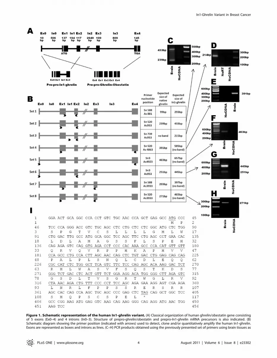
Figure 1. Schematic representation of the human In1-ghrelin variant. (A) Classical organization of human ghrelin/obestatin gene consistingof 5 exons (Ex0–4) and 4 introns (In0–3). Structure of prepro-ghrelin/obestatin and prepro-In1-ghrelin mRNA precursors is also indicated. (B)Schematic diagram showing the primer position (indicated with arrows) used to detect, clone and/or quantitatively amplify the human In1-ghrelin.Exons are represented as boxes and introns as lines. (C–H) PCR products obtained using the previously presented set of primers using brain tissues as
In1-Ghrelin Variant in Breast Cancer
PLoS ONE | www.plosone.org 4 August 2011 | Volume 6 | Issue 8 | e23302

region (Fig. 1B, set-3), thus resulting in a single band of the
expected size and sequence (215bp; Fig. 1D). In contrast, no bands
were amplified in non-reverse transcribed and non-cDNA control,
demonstrating that the band observed was not the consequence of
genomic or external contamination, respectively. In order to study
if other exons, different to Ex1-2, are present in the novel human
In1-ghrelin mRNA variant, we selected primers located in Ex0, 3,
and 4 (Fig. 1B, sets4–8 and Fig. 1E–H) and combined these with
primers in Ex1 and 2. PCR products obtained with these
combinations of primers revealed that human In1-ghrelin variant
comprises Ex0, Ex1, In1 and Ex2, but lacks Ex3 and Ex4. This
novel sequence of the In1-ghrelin variant was submitted to
Genbank (accession #: GU942497).
Since In1-variant and native-ghrelin share the Ex0 and 1, it
seems reasonable to assume that the human In1-ghrelin variant
shares the same start codon as native-ghrelin, located in the Ex1.
Assuming that translation of In1-ghrelin variant mRNA would
encode a 117aa protein (prepro-In1-ghrelin), sharing the first 36aa
of native-prepro-ghrelin, including the signal peptide (24aa:
MPSPGTVCSLLLLGMLWLDLAMA) and the first 12aa of the
mature native-ghrelin, which includes the putative acylation site at
Ser3. However, as is the case of the mouse In2-ghrelin variant
[26], the reading frame of the human In1-ghrelin variant would be
altered by the retention of intron 1, thereby encoding a completely
novel C-terminal tail, where the obestatin would not be produced.
Interestingly, we have also identified the In1-ghrelin variant in a
primate model (Olive baboon, Papio anubis) (Fig. S1) using a similar
strategy to that explained for human but with primers designed
using Macaca mulata, Pan troglodytes and human ghrelin gene
sequences, as these genomes are highly similar. Comparisons of
human and baboon In1-ghrelin and mouse In2-ghrelin variants
show high inter-specific homology (at the mRNA level and at the
level of the predicted protein) (Fig. S1).
Using a specific software to predict prepro-peptide cleavage sites
(ProP-1.0 Server), we identified various putative cleavage sites (for
example, RH (63/64) and KA (42/43)) located in the human
prepro-In1-ghrelin, which exhibit a similar cleavage score to the
cleavage sites located in the prepro-ghrelin/obestatin precursor,
whose processing generates ghrelin and obestatin. Although, it
would be predicted that the first 12aa of native-ghrelin would be
retained in a peptide encoded by In1-ghrelin transcript, use of
three commercially available antibodies directed against this
epitope [Rabbit Anti-Human-Ghrelin, Alpha Diagnostic, San
Antonio, TX (Cat. # GHS11-S), Chicken Anti-Human-Ghrelin,
GeneTex, Inc., Irvine, CA (Cat. # GTX15861, USA) and Anti-
GHRL (ab1), Sigma] failed to detect either native ghrelin or In1-
ghrelin proteins by western (data not shown).
mRNA expression levels of In1-ghrelin variant in humantissues
In order to analyze the presence and abundance of In1-ghrelin
variant in human tissues, we used the primer set 3 shown in Fig. 1B,
and table S2 in qrtRT-PCR to exclusively amplify this transcript. As
template, we used a commercial panel of human tissues, where each
sample (tissue) is a pooling of several samples coming from various
individuals, and therefore represents an average expression level in
this particular tissue. Results of qrtRT-PCR showed that In1-ghrelin
variant was expressed in the 22 tissues analyzed. However, the
expression levels were quite variable (Fig. 2A), with In1-ghrelin
variant mRNA being abundantly expressed in thymus, testis, kidney,
stomach, uterus or brain (as previously reported [35]). We also
measured the expression levels of native-ghrelin, GOAT, GHSR1a
and GHSR1b, in the same human samples and the results are shown
in Fig. 2B and table S3. Interestingly, we observed that expression
levels of native-ghrelin and In1-ghrelin variant did not share a
common tissue-specific expression pattern (R2 = 20,041) (Fig. 2C). In
contrast, when comparing the expression levels of each of the ghrelin-
related transcript to GOAT, we found a strong correlation between
mRNA levels of In1-ghrelin variant and GOAT (R2 = 0,921; Fig. 2D)
in all the tissues analyzed, while there was no significant correlation
between native-ghrelin and GOAT (R2 = 20,025). Finally, our data
showed that mRNA expression levels of both ghrelin receptors
(GHSR1a and R1b) did not correlate with native or In1-variant
ghrelin in the human tissues analyzed (data not shown).
In1-ghrelin variant is overexpressed in breast cancersamples
qrtRT-PCR results showed that In1-ghrelin variant is expressed
in normal mammary glands and, surprisingly, the expression levels
of In1-ghrelin variant were found to be 8-fold up-regulated in a
series of 40 sporadic invasive ductal breast carcinomas classified as
high grade tumors (G3) compared with control samples
(p = 0,0042) (Fig. 3A; table S4). The expression of In1-ghrelin
variant was not correlated with the presence of ER or PR in this
series but showed a trend to be correlated with Her2neu presence,
so that the expression level of In1-ghrelin is numerically, albeit
non-significantly (p = 0.075), higher in Her2neu tumors compared
with luminal and basal tumors (data not shown).
Similarly, the mRNA levels of GOAT were up-regulated in breast
cancer samples (Fig. 3A), and were significantly correlated with the
expression levels of In1-ghrelin variant (R2 = 0.655; p#0.001;
Fig. 3B). In striking contrast, expression levels of native-ghrelin did
not differ between normal mammary gland and breast cancer
samples (Fig. 3A; table S4). Likewise, mRNA levels of GHSR1a were
low in both normal and tumoral breast samples (Fig. 3A; table S4),
and no significant differences were found between those experimental
groups. Interestingly, while expression of GHSR1b was not detected
in normal mammary gland samples, it was highly expressed in breast
cancer samples (Fig. 3A; table S4). Moreover, whereas GHSR1a
mRNA levels do not correlate with native-ghrelin or In1-ghrelin
variant levels (Fig. 3B), we found that expression levels of GHSR1b
parallel those of In1-ghrelin (R2 = 0.403; p = 0.049; Fig. 3B) but not
those of native-ghrelin (data not shown). As expected, the expression
levels of two key tumoral markers of cell proliferation, Ki67 and
cyclin-D3, were highly up-regulated in breast cancer samples as
compared to their matched controls (p,0.01; Fig. 3C). Interestingly,
the levels of expression of In1-ghrelin variant and GOAT levels
showed a trend to or were positively correlated with the expression of
Ki67 (p = 0.069 and p = 0.109, respectively) and cyclin-D3 (p = 0.009
and p = 0.059, respectively) in breast cancer samples (Fig. 3D).
In1-ghrelin variant increases the proliferation rate ofMDA-MB-231 cells and its expression is regulated byghrelin and tamoxifen
To investigate the functional consequences of In1-ghrelin
variant expression and the regulation of In1-ghrelin transcript, we
template, separated on agarose gel and stained with ethidium bromide. In addition, non-RT RNA samples and non-DNA controls were run to controlfor genomic DNA or exogenous contamination, respectively. (I) Nucleotide and amino acid sequences of In1-ghrelin. In1 sequence and the putativestart and stop codons are underlined. Exon:Ex, Intron:In, Base pair:bp.doi:10.1371/journal.pone.0023302.g001
In1-Ghrelin Variant in Breast Cancer
PLoS ONE | www.plosone.org 5 August 2011 | Volume 6 | Issue 8 | e23302

Figure 2. Tissue distribution of the human In1-ghrelin variant. (A) Quantitative (qrtPCR) expression levels of In1-ghrelin using a commercialpanel of total RNA from various human tissues obtained from Clontech, where each tissue sample is a pool of multiple individuals and represents amean expression level in this particular tissue (only a single sample of each tissue is provided). (B) Quantitative (qrtPCR) expression levels of ghrelin,GOAT, GHSR1a and GHSR1b using the same commercial panel of total RNA from various human tissues obtained from Clontech. (C) Correlationbetween In1-ghrelin and native ghrelin expression in human tissues (G) Correlation between In1-ghrelin and GOAT expression in human tissues.doi:10.1371/journal.pone.0023302.g002
In1-Ghrelin Variant in Breast Cancer
PLoS ONE | www.plosone.org 6 August 2011 | Volume 6 | Issue 8 | e23302

employed an in vitro approach using a breast cancer cell line,
MDA-MB-231, which expresses In1-ghrelin variant, GOAT and
GHSR1b transcripts at high levels while virtually lack the
expression of native-ghrelin and GHSR1a (table S4), a pattern
that is reminiscent of that observed in primary breast cancers. In
vitro kinetic proliferation assays using MDA-MB-231 cells
transfected with In1-ghrelin variant or empty vector (mock-
control) indicated that cells over-expressing In1-ghrelin variant
have a higher proliferative rate compared with mock-transfected
cells (Fig. 4A). Specifically, cells that over-express In1-ghrelin
variants exhibited a shorter doubling time than mock-transfected
cells (1.21 days vs. 1.43 days, respectively) these differences being
progressively more significant at 2, 3, and 4 days after
transfection (p#0.05, p#0.01, and p#0.001, respectively). Of
note, expression levels of the native-ghrelin and In1-ghrelin
variant transcripts were divergently regulated, in that treatment
(24 h) with AG or UAG increased native-ghrelin mRNA levels,
while In1-ghrelin variant levels were reduced, although the
negative impact of UAG did not reach statistical significance
(Fig. 4B). Tamoxifen treatment tended to increase native-ghrelin
expression, whereas it significantly reduced In1-ghrelin variant
mRNA levels. Conversely, estradiol (24 h) did not alter the
expression levels of native-ghrelin or In1-ghrelin variant (Fig. 4B).
It should be noted that expression levels of GOAT, GHSR1a and
GHSR1b were not significantly altered in MDA-MB-231 with
these treatments (data not shown).
Figure 3. In1-ghrelin variant in human breast samples. (A) Quantitative expression levels of the ghrelin-axis in normal (n = 4) and breast cancersamples (n = 40). (B) Correlations between In1-ghrelin and ghrelin, GOAT, GHSR1a/1b expression in breast cancer samples. (C) Quantitative expressionlevels of Ki67 and cyclin-D3 in normal and breast cancer samples. (D) Correlation values between In1-ghrelin and GOAT with Ki67 or cyclin-D3expression in breast cancer samples. Values are shown as the mean 6 S.E.M (** P,0.01; indicate differences from controls).doi:10.1371/journal.pone.0023302.g003
In1-Ghrelin Variant in Breast Cancer
PLoS ONE | www.plosone.org 7 August 2011 | Volume 6 | Issue 8 | e23302

Discussion
Results of the current study confirm, as indicated previously in
Gahete et al. [35], and, for the first time demonstrate, that the
human ghrelin gene, similar to that reported in the mouse gene
[26], can retain In1, one type of alternative splicing likely
associated with the failure of intron detection which occurs more
frequently in short introns flanked by weak splice sites [29].
Specifically, human In1-ghrelin variant contains Ex0, Ex1, In1
and Ex2 but lacks Ex3 and Ex4. Based on the fact that the In1-
variant and native-ghrelin share the Ex0 and 1, we postulate that
both share the same start codon, located in the Ex1. In this case,
the first 36aa of the prepro-In1-ghrelin, including the signal
peptide, and the first 12aa of the mature native-ghrelin, including
the putative acylation site at Ser3 and the residues found to be
necessary for acylation (Gly1 and Phe4) [4], would be identical to
that encoded by the native-prepro-ghrelin mRNA. However, as
occurred with the orthologous mouse In2-ghrelin variant, the
reading frame of the human In1-ghrelin variant would be altered
by the intron retention, thus encoding a completely novel C-
terminal tail where obestatin would not be produced. In particular,
the novel prepro-In1-ghrelin would possess a C-terminal tail with
various putative cleavage sites, which exhibit a similar cleavage
score to the cleavage sites located in the prepro-ghrelin/obestatin
precursor whose processing generates ghrelin and obestatin,
suggesting that different peptides could be processed from the
In1-ghrelin variant. Interestingly, baboon ghrelin gene also
undergoes processes of In1-retention which, coupled to the fact
that In1-ghrelin variants are highly conserved across species
(human, baboon and mouse) suggest that relevant functional
parallels of this novel transcript may have occurred through
mammalian evolution.
There is increasing evidence that the GHRL gene is very
complex and can be regulated at multiple levels [22,36]. In fact,
several peptides are generated by alternative splicing of the ghrelin
gene or by post-translational modifications [14,16,21,37]. Our
data demonstrate that an In1-ghrelin variant transcript is
processed from the GHRL gene, similar to the In2-ghrelin variant
previously reported in the mouse [26], and it is expressed in a wide
variety of human tissues, suggesting this novel variant could be
physiologically relevant and exert important biological actions in
humans. Interestingly, expression levels of human native-ghrelin
Figure 4. Effect and expression regulation of In1-ghrelin variant in MDA-MB-231 cells in vitro. (A) Proliferation kinetics of MDA-MB-231transfected with In1-ghrelin or control-mock. (B) Regulation of native-ghrelin and In1-ghrelin expression in MDA-MB-231. The data represent themeans 6 SEM. Asterisks (* P,0.05, ** P,0.01, *** P,0.001) indicate differences from corresponding controls.doi:10.1371/journal.pone.0023302.g004
In1-Ghrelin Variant in Breast Cancer
PLoS ONE | www.plosone.org 8 August 2011 | Volume 6 | Issue 8 | e23302

and In1-ghrelin variant were not correlated in the normal tissues
studied, indicating that the production of both variants derived
from the same gene could be differentially regulated in human
tissues. Indeed, splice site recognition depends on several factors,
such as mRNA regulatory elements or protein factors [38], which
could modulate the expression of intron retaining and intron
spliced isoform in a tissue-dependent manner [39,40]. Important-
ly, we observed that expression levels of In1-ghrelin, but not of
native-ghrelin, were strongly correlated with GOAT mRNA levels
in human tissues. Because translation of In1-ghrelin variant would
be likely to generate a prepro-peptide containing the same first
12aa than native-ghrelin, including the target Serine-3 octanoy-
lated by GOAT, and the residues found to be necessary for its
acylation (Gly1 and Phe4), it is reasonable to suggest that In1-
ghrelin variant may be a primary substrate for GOAT in humans.
This notion is supported by our recent results showing that mouse
In2-ghrelin variant may also be a primary substrate for GOAT in
mice [27], since its expression clearly parallels changes in the
expression of GOAT in the pituitary of several mouse models
analyzed (for example, fasting, obesity, knockout models). Thus,
future studies should aim to unequivocally prove if the putative
novel protein of the human In1-ghrelin variant can be acylated by
GOAT.
Of note, our results revealed that In1-ghrelin variant may play a
relevant role in human breast cancer. Indeed, In1-ghrelin variant
expression was strongly up-regulated (8-fold) in ductal breast
cancer samples compared with normal breast tissue. In line with
this, a previous high-throughput sequencing analysis using tissue
samples obtained from excised human breast tumors [41]
identified an ORF expressed sequence tags (EST) (GenBank:
BF929001.1) that shares 99% nucleotide sequence homology with
the human In1-ghrelin variant identified herein. Since the study of
Dias-Neto et al. focused on central portions of expressed coding
regions by PCR amplifications, the clone homologous to In1-
ghrelin variant (BF929001.1) contains the complete In1 of human
ghrelin gene, but not the complete 59- and 39-regions. These
results strongly suggest that the partial sequence obtained by Dias-
Neto [41] corresponds to the novel human In1-ghrelin variant
reported herein.
This report is also the first to show that the GOAT enzyme is
strikingly overexpressed in breast cancer tissues compared with
normal human mammary gland. Similar to that observed for
normal tissues, absolute copy numbers of In1-ghrelin, but not of
native-ghrelin, show a marked, positive correlation with GOAT
expression in breast cancer, reinforcing the contention that In1-
ghrelin variant represents the primary substrate for GOAT
enzyme in this pathology. Furthermore, our results also unveiled
a remarkable upregulation of GHSR1b, which positively corre-
lated with In1-ghrelin expression, while GHSR1a and native-
ghrelin were not altered in tumor samples. This latter finding is
consistent with previous immunohistochemistry results showing
very low levels of native-ghrelin in the normal breast epithelium,
with moderate elevation in breast cancer samples [7]. The
potential pathophysiological role of GHSR1b overexpression is
both unknown and intriguing; although this truncated receptor
can act as a dominant negative, by binding and internalizing
GHSR1a [42], the disproportionately high levels of GHSR1b
relative to GHSR1a (60-fold) found in human breast cancer
samples suggest this receptor may serve a different role in this
pathology. In line with this, GHSR1b has been shown to acts as a
neuromedin U receptor in lung cancer by heterodimerizing with
neurotensin receptor 1 [13]. Nonetheless, our current data may
offer important clinical information since it provides primary
evidence for the differential expression of In1-ghrelin, GOAT and
GHSR1b in breast cancer tissues, which may represent novel
diagnostic/prognostic markers and therapeutic targets for this
pathology.
Human breast cancer MDA-MB-231 cells represent a suitable
model to study In1-ghrelin function, as their expression patterns of
the ghrelin-axis mimic those of breast cancer samples. Interest-
ingly, expression of native-ghrelin and In1-ghrelin were oppositely
regulated by AG and UAG in this cell model, providing, to our
knowledge, the first evidence that ghrelin (AG and UAG) can
regulate its own synthesis (native-ghrelin and In1-ghrelin tran-
scripts). Thus, both AG and UAG increased the markedly low
levels of expression of native-ghrelin in MDA-MB-231 cells, while
decreasing the high levels of In1-ghrelin mRNA in these cancer
cells, suggesting the existence of an auto-regulatory loop in this cell
type. Most importantly, tamoxifen was also able to reduce In1-
ghrelin mRNA levels while tended to increase native-ghrelin
expression in MDA-MB-231 cells, an observation with potential
patho-physiological implications since tamoxifen is currently a
primary line of therapy for the treatment of estrogen receptor (ER)
positive breast cancers and the ghrelin axis has been reported to
exert relevant actions in cancer cells [7]. Although tamoxifen is an
ER blocker and MDA-MB-231 is an estrogen-independent breast
cancer cell line, several studies have reported effects of tamoxifen
and other ER modulators such as raloxifene in ER-negative breast
cancer cell lines via activation of several intracellular signaling
pathways (protein kinase C, phospholipase D) through ER-
independent mechanisms [43,44].
An additional line of support for a patho-physiologically
relevant role of In1-ghrelin in breast cancer arises from the
positive association in breast cancer samples between In1-ghrelin
expression and Ki67 and cyclin-D3, two markers linked to
proliferation and ductal breast tumor grade [45]. Nevertheless, we
should introduce the caveat that despite showing a strong
significant correlation, this does not necessarily indicates a causal
relationship for these two parameters. However, the plausible
association between In1-ghrelin expression and increased prolif-
eration is supported by our observation that In1-ghrelin
overexpression in MDA-MB-231 cells causes a robust increase in
proliferation rate, suggesting that blockade of In1-ghrelin variant
may have some therapeutic benefit.
In conclusion, our current results provide novel evidence
supporting that, as suggested by previous studies, the ghrelin
system is functionally present in cancer cells [7,9,46248].
Furthermore, by demonstrating the relevant overexpression of
novel components of the ghrelin system in this pathology,
specifically the newly identified In1-ghrelin variant but also
GOAT and GHSR1b, our study provides novel avenues to
investigate the precise pathophysiological role and potential
clinical implications of this family in breast cancer.
Supporting Information
Figure S1 mRNA and putative protein sequence com-parison of human (Homo sapiens) and baboon (Papioanubis) In1-ghrelin variants, and the orthologous mouse(Mus musculus) In2-ghrelin variants. (A) Alignment of the
mRNA coding region (CDS) sequences of the In1-ghrelin and In2-
ghrelin vaiants. Nucleotide sequences of the Exon(Ex) 1 of the In1-
ghrelin and Ex2 of the In2-ghrelin variants that are shared with
native-ghrelin are shown in boxes. CDS of baboon In1-ghrelin
and mouse In2-ghrelin variants are shorter than human CDS
because their mRNAs contain a stop codon (TAG; represented in
shaded grey) located in the sequence of In1 or In2, respectively,
while the stop codon of the human In1-ghrelin variant is located
In1-Ghrelin Variant in Breast Cancer
PLoS ONE | www.plosone.org 9 August 2011 | Volume 6 | Issue 8 | e23302

inside the Ex2 sequence. Therefore, human In1-ghrelin variant
mRNA is not fully represented (nucleotides from 172 to 348 are
not represented). Asterisks (*) indicate conserved nucleotides across
species, which represent a 72% homology among human, baboon
and mouse sequences. Of note, the homology between human and
baboon In1-ghrelin variant mRNAs is 96%. (B) Alignment of
putative In1-ghrein and In2-ghrelin variants protein sequences.
Similar to that shown with nucleotide CDS sequences, human
In1-ghrelin variant amino acid (aa) sequece is not completely
represented (aa from 56 to 116 are not represented). The first 24
aa represents the signal peptide sequences in the three species.
Asterisks (*) indicate conserved aa across species (16/24; 67%
homology) while (#) indicate aa belonging to the same group (7/
24) which results in a 96% homology (23/24 aa) among the
human, baboon and mouse sequences. Boxes represent the aa
sequence of In1-ghrelin and In2-ghrelin variants shared with
native ghrelin. Conserved aa of the In1-ghrelin and In2-ghrelin
variants are highlighted in dark grey, aa belonging to the same
group are highlighted in light grey, while aa not shared across
species are represented in white (not highlighted). 18 of 24 aa of
the human, baboon and mouse In1-ghrelin or In2-ghrelin variants
are identical or belong to the same group (75% homology). Of
note, 22 out of 24 aa are conserved between human and baboon
In1-ghrelin variants (92% homology). The novel sequence of the
baboon In1-ghrelin variant was submitted to Genbank (accession
#: HM048926).
(PPT)
Table S1 Primers used to identify and clone human In1-ghrelin isoform.(PPTX)
Table S2 Sequences and product sizes of primers usedin qrt-PCR.(PPTX)
Table S3 Absolute mRNA copy number of ghrelin axiscomponents in human tissues included in the commer-cial panel of total RNA from Clontech.(PPTX)
Table S4 Expression level of ghrelin axis in breasttissues and cell lines.(PPTX)
Author Contributions
Conceived and designed the experiments: MDG AJMF RDK GMB RML
JPC. Performed the experiments: MDG JCC MHR. Analyzed the data:
MDG JCC MHR AJMF RML. Contributed reagents/materials/analysis
tools: RDK GMB RML JPC. Wrote the paper: MDG AJMF RDK GMB
RML JPC.
References
1. Kojima M, Hosoda H, Date Y, Nakazato M, Matsuo H, et al. (1999) Ghrelin is agrowth-hormone-releasing acylated peptide from stomach. Nature 402(6762): 656–60.
2. Lago F, Gonzalez-Juanatey JR, Casanueva FF, Gomez-Reino J, Dieguez C,
et al. (2005) Ghrelin, the same peptide for different functions: player orbystander? Vitam Horm 71: 405–32.
3. Gutierrez JA, Solenberg PJ, Perkins DR, Willency JA, Knierman MD, et al.
(2008) Ghrelin octanoylation mediated by an orphan lipid transferase. Proc NatlAcad Sci U S A 105(17): 6320–5.
4. Yang J, Brown MS, Liang G, Grishin NV, Goldstein JL (2008) Identification of
the acyltransferase that octanoylates ghrelin, an appetite-stimulating peptidehormone. Cell 132(3): 387–96.
5. Horvath TL, Diano S, Sotonyi P, Heiman M, Tschop M (2001) Minireview:
ghrelin and the regulation of energy balance–a hypothalamic perspective.Endocrinology 142(10): 4163–9.
6. van der Lely AJ, Tschop M, Heiman ML, Ghigo E (2004) Biological,
physiological, pathophysiological, and pharmacological aspects of ghrelin.Endocr Rev 25(3): 426–57.
7. Jeffery PL, Murray RE, Yeh AH, McNamara JF, Duncan RP, et al. (2005)
Expression and function of the ghrelin axis, including a novel preproghrelinisoform, in human breast cancer tissues and cell lines. Endocr Relat Cancer
12(4): 839–50.
8. Lanfranco F, Baldi M, Cassoni P, Bosco M, Ghe C, et al. (2008) Ghrelin and
prostate cancer. Vitam Horm 77: 301–24.
9. Leontiou CA, Franchi G, Korbonits M (2007) Ghrelin in neuroendocrine organs
and tumours. Pituitary 10(3): 213–25.
10. Nikolopoulos D, Theocharis S, Kouraklis G (2009) Ghrelin’s role ongastrointestinal tract cancer. Surg Oncol 19(1): e2–e10.
11. Barzon L, Pacenti M, Masi G, Stefani AL, Fincati K, et al. (2005) Loss of growth
hormone secretagogue receptor 1a and overexpression of type 1b receptortranscripts in human adrenocortical tumors. Oncology 68(4-6): 414–21.
12. Carraro G, Albertin G, Abudukerimu A, Aragona F, Nussdorfer GG (2004)
Growth hormone secretagogue receptor subtypes 1a and 1b are expressed in thehuman adrenal cortex. Int J Mol Med 13(2): 295–8.
13. Takahashi K, Furukawa C, Takano A, Ishikawa N, Kato T, et al. (2006) The
neuromedin U-growth hormone secretagogue receptor 1b/neurotensin receptor1 oncogenic signaling pathway as a therapeutic target for lung cancer. Cancer
Res 66(19): 9408–19.
14. Zhu X, Cao Y, Voogd K, Steiner DF (2006) On the processing of proghrelin to
ghrelin. J Biol Chem 281(50): 38867–70.
15. Takahashi T, Ida T, Sato T, Nakashima Y, Nakamura Y, et al. (2009)
Production of n-octanoyl-modified ghrelin in cultured cells requires prohormone
processing protease and ghrelin O-acyltransferase, as well as n-octanoic acid.J Biochem 146(5): 675–82.
16. Garg A (2007) The ongoing saga of obestatin: is it a hormone? J Clin Endocrinol
Metab 92(9): 3396–8.
17. Zhang JV, Ren PG, Avsian-Kretchmer O, Luo CW, Rauch R, et al. (2005)
Obestatin, a peptide encoded by the ghrelin gene, opposes ghrelin’s effects on
food intake. Science 310(5750): 996–9.
18. Karaoglu A, Aydin S, Dagli AF, Cummings DE, Ozercan IH, et al. (2009)
Expression of obestatin and ghrelin in papillary thyroid carcinoma. Mol CellBiochem 323(1-2): 113–8.
19. Tsolakis AV, Grimelius L, Stridsberg M, Falkmer SE, Waldum HL, et al. (2009)Obestatin/ghrelin cells in normal mucosa and endocrine tumours of the
stomach. Eur J Endocrinol 160(6): 941–9.
20. Volante M, Rosas R, Ceppi P, Rapa I, Cassoni P, et al. (2009) Obestatin inhuman neuroendocrine tissues and tumours: expression and effect on tumour
growth. J Pathol 218(4): 458–66.
21. Seim I, Collet C, Herington AC, Chopin LK (2007) Revised genomic structure
of the human ghrelin gene and identification of novel exons, alternative splice
variants and natural antisense transcripts. BMC Genomics 8: 298.
22. Seim I, Herington AC, Chopin LK (2009) New insights into the molecular
complexity of the ghrelin gene locus. Cytokine Growth Factor Rev 20(4):297–304.
23. Hosoda H, Kojima M, Matsuo H, Kangawa K (2000) Purification andcharacterization of rat des-Gln14-Ghrelin, a second endogenous ligand for the
growth hormone secretagogue receptor. J Biol Chem 275(29): 21995–2000.
24. Yeh AH, Jeffery PL, Duncan RP, Herington AC, Chopin LK (2005) Ghrelinand a novel preproghrelin isoform are highly expressed in prostate cancer and
ghrelin activates mitogen-activated protein kinase in prostate cancer. ClinCancer Res 11(23): 8295–303.
25. Jeffery PL, Duncan RP, Yeh AH, Jaskolski RA, Hammond DS, et al. (2005)
Expression of the ghrelin axis in the mouse: an exon 4-deleted mouse proghrelinvariant encodes a novel C terminal peptide. Endocrinology 146(1): 432–40.
26. Kineman RD, Gahete MD, Luque RM (2007) Identification of a mouse ghrelingene transcript that contains intron 2 and is regulated in the pituitary and
hypothalamus in response to metabolic stress. J Mol Endocrinol 38(5): 511–21.
27. Gahete MD, Cordoba-Chacon J, Salvatori R, Castano JP, Kineman RD, et al.
(2010) Metabolic regulation of ghrelin O-acyl transferase (GOAT) expression in
the mouse hypothalamus, pituitary, and stomach. Mol Cell Endocrinol 317(1-2):154–60.
28. Seim I, Carter SL, Herington AC, Chopin LK (2009) The proximal first exonarchitecture of the murine ghrelin gene is highly similar to its human orthologue.
BMC Res Notes 2(1): 85.
29. Sakabe NJ, de Souza SJ (2007) Sequence features responsible for intron
retention in human. BMC Genomics 8: 59.
30. Sarrio D, Palacios J, Hergueta-Redondo M, Gomez-Lopez G, Cano A, et al.(2009) Functional characterization of E- and P-cadherin in invasive breast
cancer cells. BMC Cancer 9: 74.
31. Duran-Prado M, Gahete MD, Martinez-Fuentes AJ, Luque RM, Quintero A,
et al. (2009) Identification and characterization of two novel truncated but
functional isoforms of the somatostatin receptor subtype 5 differentially presentin pituitary tumors. J Clin Endocrinol Metab 94(7): 2634–43.
32. Martinez-Fuentes AJ, Moreno-Fernandez J, Vazquez-Martinez R, Duran-Prado M, de la Riva A, et al. (2006) Ghrelin is produced by and directly activates
corticotrope cells from adrenocorticotropin-secreting adenomas. J Clin En-docrinol Metab 91(6): 2225–31.
In1-Ghrelin Variant in Breast Cancer
PLoS ONE | www.plosone.org 10 August 2011 | Volume 6 | Issue 8 | e23302

33. Taboada GF, Luque RM, Bastos W, Guimaraes RF, Marcondes JB, et al. (2007)
Quantitative analysis of somatostatin receptor subtype (SSTR1-5) geneexpression levels in somatotropinomas and non-functioning pituitary adenomas.
Eur J Endocrinol 156(1): 65–74.
34. Cordoba-Chacon J, Gahete MD, Duran-Prado M, Pozo-Salas AI,Malagon MM, et al. (2010) Identification and characterization of new functional
truncated variants of somatostatin receptor subtype 5 in rodents. Cell Mol LifeSci 67(7): 1147–63.
35. Gahete MD, Rubio A, Cordoba-Chacon J, Gracia-Navarro F, Kineman RD,
et al. (2010) Expression of the ghrelin and neurotensin systems is altered in thetemporal lobe of Alzheimer’s disease patients. J Alzheimers Dis 22(3): 819–28.
36. Soares JB, Leite-Moreira AF (2008) Ghrelin, des-acyl ghrelin and obestatin:three pieces of the same puzzle. Peptides 29(7): 1255–70.
37. Seim I, Amorim L, Walpole C, Carter S, Chopin LK, et al. (2009) Ghrelin gene-related peptides: multi-functional endocrine/autocrine modulators in health and
disease. Clin Exp Pharmacol Physiol 37(1): 125–31.
38. Matlin AJ, Clark F, Smith CW (2005) Understanding alternative splicing:towards a cellular code. Nat Rev Mol Cell Biol 6(5): 386–98.
39. Galante PA, Sakabe NJ, Kirschbaum-Slager N, de Souza SJ (2004) Detectionand evaluation of intron retention events in the human transcriptome. Rna
10(5): 757–65.
40. Ledee DR, Chen J, Tonelli LH, Takase H, Gery I, et al. (2004) Differentialexpression of splice variants of chemokine CCL27 mRNA in lens, cornea, and
retina of the normal mouse eye. Mol Vis 10: 663–7.41. Dias Neto E, Correa RG, Verjovski-Almeida S, Briones MR, Nagai MA, et al.
(2000) Shotgun sequencing of the human transcriptome with ORF expressedsequence tags. Proc Natl Acad Sci U S A 97(7): 3491–6.
42. Leung PK, Chow KB, Lau PN, Chu KM, Chan CB, et al. (2007) The truncated
ghrelin receptor polypeptide (GHS-R1b) acts as a dominant-negative mutant of
the ghrelin receptor. Cell Signal 19(5): 1011–22.
43. Boyan BD, Sylvia VL, Frambach T, Lohmann CH, Dietl J, et al. (2003)
Estrogen-dependent rapid activation of protein kinase C in estrogen receptor-
positive MCF-7 breast cancer cells and estrogen receptor-negative HCC38 cells
is membrane-mediated and inhibited by tamoxifen. Endocrinology 144(5):
1812–24.
44. Eisen SF, Brown HA (2002) Selective estrogen receptor (ER) modulators
differentially regulate phospholipase D catalytic activity in ER-negative breast
cancer cells. Mol Pharmacol 62(4): 911–20.
45. Wong SC, Chan JK, Lee KC, Hsiao WL (2001) Differential expression of p16/
p21/p27 and cyclin D1/D3, and their relationships to cell proliferation,
apoptosis, and tumour progression in invasive ductal carcinoma of the breast.
J Pathol 194(1): 35–42.
46. Cassoni P, Papotti M, Ghe C, Catapano F, Sapino A, et al. (2001) Identification,
characterization, and biological activity of specific receptors for natural (ghrelin)
and synthetic growth hormone secretagogues and analogs in human breast
carcinomas and cell lines. J Clin Endocrinol Metab 86(4): 1738–45.
47. Jeffery PL, Herington AC, Chopin LK (2002) Expression and action of the
growth hormone releasing peptide ghrelin and its receptor in prostate cancer cell
lines. J Endocrinol 172(3): R7–11.
48. Jeffery PL, Herington AC, Chopin LK (2003) The potential autocrine/paracrine
roles of ghrelin and its receptor in hormone-dependent cancer. Cytokine Growth
Factor Rev 14(2): 113–22.
In1-Ghrelin Variant in Breast Cancer
PLoS ONE | www.plosone.org 11 August 2011 | Volume 6 | Issue 8 | e23302


