Embed Size (px)
Citation preview
�������� ����� ��
A randomısed prospectıve trıal of a novel devıce for measurıng perforatıonsıze durıng inlay ‘Butterfly’ Myrıngoplasty
Sabri Baki EREN MD, Selahattin TUGRUL MD, Berke OZUCER MD,Remzi DOGAN MD, Orhan OZTURAN MD
PII: S0196-0709(14)00042-8DOI: doi: 10.1016/j.amjoto.2014.02.007Reference: YAJOT 1348
To appear in: American Journal of Otolaryngology–Head and Neck Medicine and Surgery
Received date: 14 January 2014Accepted date: 16 February 2014
Please cite this article as: EREN Sabri Baki, TUGRUL Selahattin, OZUCERBerke, DOGAN Remzi, OZTURAN Orhan, A randomısed prospectıve trıal of anovel devıce for measurıng perforatıon sıze durıng inlay ‘Butterfly’ Myrıngoplasty,American Journal of Otolaryngology–Head and Neck Medicine and Surgery (2014), doi:10.1016/j.amjoto.2014.02.007
This is a PDF file of an unedited manuscript that has been accepted for publication.As a service to our customers we are providing this early version of the manuscript.The manuscript will undergo copyediting, typesetting, and review of the resulting proofbefore it is published in its final form. Please note that during the production processerrors may be discovered which could affect the content, and all legal disclaimers thatapply to the journal pertain.

ACC
EPTE
D M
ANU
SCR
IPT
ACCEPTED MANUSCRIPT
A RANDOMISED PROSPECTIVE TRIAL OF A NOVEL DEVICE FOR
MEASURING PERFORATION SIZE DURING INLAY ‘BUTTERFLY’
MYRINGOPLASTY
Sabri Baki EREN1, MD; Selahattin TUGRUL
1, MD; Berke OZUCER
1, MD; Remzi
DOGAN2, MD; Orhan OZTURAN
1, MD, Professor
1Department of Otorhinolaryngology, Bezmiâlem Vakıf University, Fatih, Istanbul, Turkey.
2Department of Otorhinolaryngology, Bayrampasa State Hospital, Bayrampasa, Istanbul,
Turkey.
Corresponding Author
Berke Ozucer, MD
Address: Bezmialem Vakif University, Medical Faculty
Department of Otorhinolaryngology, Fatih, Istanbul, Turkey.
Phone number: +905309635939
Fax: +902125332326
E-mail: [email protected]
Funding source, financial disclosures: None
Conflict of interest: None

ACC
EPTE
D M
ANU
SCR
IPT
ACCEPTED MANUSCRIPT
ABSTRACT
Aim: This study introduces a new device to faciliate perforation size measurement during
“butterfly” myringoplasty. The purpose of this study is to evaluate the use of ‘otological
compass’ on short-term results of inlay cartilaginous ‘butterfly’ tympanoplasty technique in
adult patients.
Study Design: Prospective, randomized, controlled, blinded
Patients and Methods: This study included 25 patients who underwent inlay cartilage
myringoplasty. All operations were performed under general anesthesia by the same surgeon
with a microscope-assisted approach. The patients were divided randomly and consecutively
into two groups: Group 1 (n=12) had perforation dimensions and shape measured using the
Otologic compass (OC) and the control group (n=13) had perforation measured by means of a
Fisch elevator. The duration of surgery, number of trials for correct placement of the cartilage
graft, results and complications of the surgery were evaluated and compared.
Results: The mean follow-up duration was six months. Groups were similar in terms of age
and perforation diameters (p>0.05). Average number of cartilage shaping before satisfactory
graft fitting was significantly fewer in the OC group: 1.1 ± 0.3 and 2.2 ± 0.6 trials for OC and
control groups, respectively (p<0.001). Mean duration of preparation and satisfactory graft
fitting was 9.6±4.2 minutes in the OC group whereas it was 18.1 ± 5.2 minutes for the control
group. Operative duration was significantly shorter in the OC group (p<0.001). At the end of
the follow-up period, successful closure occured 91.7% and 84.6% patients in the OC and
control groups, respectively (p>0.05). The mean preoperative to postoperative three-tone air-
bone gap improved 7.9 dB and 9.0 dB in OC and control groups, respectively (p>0.05).
Conclusion: This study shows that OC presents as a useful tool that expedites and refines
butterfly myringoplasty procedure. The number of cartilage shaping prior to satisfactory graft
fitting revealed significantly better results: almost all surgeries in the OC group were
complete after a single cartilage shaping attempt.

ACC
EPTE
D M
ANU
SCR
IPT
ACCEPTED MANUSCRIPT
INTRODUCTION
The main objective of tympanic membrane perforation (TMP) repair is total and definitive
closure of the perforation and hearing improvement. Myringoplasty is the operation to fulfill
these aims. In 1998, Eavey introduced cartilage, perichondrium butterfly inlay tympanoplasty
technique for closure of small to medium sized central perforations[1]. This technique does
not necessitate tympanomeatal flap elevation, can be carried out under local anesthesia,
minimal scarring associated with transcanal approach and postoperative care and follow-up is
not complex and can be easily and cost-effectively applied transcanally compared to onlay
and underlay tympanoplasty techniques[1-4].
There are two main challenges associated with transcanal “butterfly” inlay myringoplasty:
precise evaluation of the perforation size and meticulous preparation of an accurately-fitting
cartilage graft. These two factors are determining factors for obtaining successful surgical
outcomes. The current article presents the results of 25 consecutive transcanal inlay
cartilaginous myringoplasties and introduces a novel device, namely Otologic Compass
(OC)(patent pending), for correct close-up measuring TMP size. Aim of the present study is
to evaluate the effect of OC on precise graft preparation and positioning by means of
operative time, number of trials for graft positioning and operative outcome.
PATIENTS AND METHODS
This study included patients of older than 15 years old, with TMP caused by mesotympanic
chronic otitis media. A local antibiotic and steroid treatment was performed to eradicate
preoperative inflammation of the tympanic cavity. Both microscope and endoscope-assisted

ACC
EPTE
D M
ANU
SCR
IPT
ACCEPTED MANUSCRIPT
otoscopy (Karl Storz rigid 0° degree endoscope, 4 mm diameter) was carried outdetermined
the features of the perforation. Patient selection and exclusion criteria were as detailed in
Table 1. All operations were performed under general anesthesia by the same surgeon with
microsope-assisted transmeatal approach.
Surgical Technique
Following local Adrenaline and Lidocaine infiltration to tragal cartilage and four quadrants of
the ear canal, cartilage with both-sided perichondrium was harvested from the tragus.
Squamous epithelium were removed from the margins of the TMP with a circular knife or
pick. The patients were divided randomly and consecutively into two groups: OC group
(n=12) had perforation dimensions and shape measured using the OC (Figure 1) and the
control group (n=13) had perforation measured with Fisch elevator. Gelfoam soaked with
Adrenalin was left in the ear canal. A tragal perichondrium-cartilage graft was shaped after
removing of the perichondrium at the medial part of the graft. The graft was shaped with a
#11 blade with 0.5 mm longer than the diameters of the perforation. Graft was cut into shape
of butterfly wings by cleaving the cartilage rim parallel to two layers of perichondrium with a
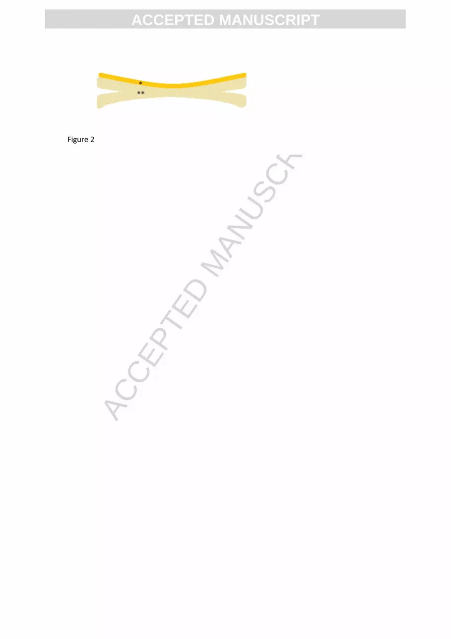
razor blade (Figure 2). The graft was then through the canal, in such a way that circular
groove of the graft grasps the anterior edge of perforation initially and then inserted with one
wing of butterfly above and one below of the posterior margins of the perforation. Graft was
poked medially with a pick and sucked with an aspirator to ensure satisfactory fitting. Tragal
incision was sutured and trans-tragal suture was placed for preventing any possible hematoma
on harvesting site. Patients were discharged on first postoperative day and prescribed with
antibiotic (Sipro solution drop 0.3% Ciprofloxacin, Bilim, Turkey) and steroid (Onadron
solution drop, dexamethasone, I.E Ulagay, Turkey) ear drops as well as systemic oral

ACC
EPTE
D M
ANU
SCR
IPT
ACCEPTED MANUSCRIPT
Cephalosporin antibiotics. Patients were examined initially on first week and 2 months and 6
months after surgery.
Peroperative and Postoperative Evaluations
Duration of the surgery was measured to the nearest minute as the time passed between
perforation measurement and satisfactory graft placement. Number of trials for satisfactory
fitting of the cartilage graft was also noted. At sixth postoperative month; surgical success
rates were evaluated via microscope in a blinded manner. Pure tone audiometry was also
carried out at the sixth postoperative month visit in a blinded manner. The average on three
frequencies (500, 1000 and 2000 Hz) of hearing thresholds in air and bone conduction and the
Air Bone Gap (ABG) have been calculated. Improvements in ABG were calculated and
compared. Results were statistically analyzed using the nonparametric Mann Whitney U test
for comparison of means and categorical data was analyzed with non-parametric Fisher’s
exact test. P values smaller than 0.05 were accepted as statistically significant.
RESULTS
A total of 25 patients underwent transcanal inlay cartilage ‘butterfly’ myringoplasty between
September 2012 and March 2013. The mean age of OC group was 29.9±11.5 years, the
control group had a similar mean age of 30.3±12.5 (p>0.05). The mean follow-up duration
was six months. Groups were also similar in terms of mean perforation diameters
(p>0.05)(Table 2). Average number of cartilage shaping before satisfactory graft fitting was
significantly fewer in the OC group: 1.1±0.3 and 2.2±0.6 trials for OC and control group,
respectively(p<0.001). Mean duration before satisfactory graft fitting was 9.6±4.2 minutes in
ACC
EPTE
D M
ANU
SCR
IPT
ACCEPTED MANUSCRIPT
the OC group whereas it was 18.1±5.2 minutes for the control group. Operative duration was
significantly shorter in the OC group (p<0.001). At the end of the follow-up period,
successful closure occured 91.7% and 84.6% patients in the OC and control groups,
respectively (p>0.05). The mean improvement of ABG after the operation was 7.9 dB and 9.0
dB in the OC and control groups, respectively(p>0.05).
DISCUSSION
Butterfly myringoplasty technique does not necessitate tympanomeatal flap elevation,
therefore allows a shorter surgerical time when possible. It is practical and can be carried out
even under local anesthesia. Transcanal approach also offers a cosmetically desirable result,
therefore should be preferred when possible [5]. Postoperative care is easy and follow-up is
short and cost-effectively applied transcanally compared to conventional myringoplasty with
tympanomeatal flep elevation [1-4].
There are two main challenges associated with transcanal “butterfly” inlay myringoplasty:
precise evaluation of the perforation size and meticulous preparation of an accurately-fitting
cartilage graft. These two factors are determining factors for obtaining satisfactory surgical
outcomes. A learning curve and experience is requisite just like any other surgical technique.
Unlike other conventional myringoplasty techniques; butterfly myringoplasty requires
meticulous preparation of the edges just like exact sizing of the graft. The rim of the cartilage
graft must be cleaved. This cleaveage must be continous all around the graft like a rim for a
perfect fitting. Tragal cartilage is very thin, especially in females, and this procedure was done
with utmost care. This was established with the aid of a razor blade under the microscope.
ACC
EPTE
D M
ANU
SCR
IPT
ACCEPTED MANUSCRIPT
Lateral perichondrium covering the tragal cartilage acted as a safety major when this ‘rim’
around the corners of the graft was imperfect.
Results of the current study shows OC presents a useful tool that expedites and refines
butterfly myringoplasty procedure. Surgeries conducted with the aid of the OC were
significantly quicker (Table 2). The number of cartilage shaping before satisfactory graft
fitting revealed significantly better results: all surgeries – except one- in the OC group were
complete after a single cartilage shaping (Table 2). Although not significant, successful
operative outcome rate was also higher compared to the control group. OC group
demonstrated 91.7% successful closure of TMPs; which was concordant with other authors
including Ghanem et al., Couloignier et al., and Mauri et al.who have reported 92%, 71% and
85% closure rates, respectively [6-8].
In butterfly myringoplasty, a precise fitting is important for successful fixation of the graft. A
large graft can be hard to fit in the perforation site, or a smaller graft can be unstable and
result with an unsuccessful operative outcome. In one of the cases operated in the control
group; size of the cartilage graft was prepared too small. Secondary cartilage harvesting was
required for a stable graft fitting. This prolonged the operative duration as well as causing
extra morbidity to the patient. Bearing these kind of complications in mind, a forbearing
surgeon gradually scales down the size of the cartilage a numerous times before precisely
securing of the graft to the perforation site. This prolongs the duration of the surgery. OC can
be very useful especially to the inexperienced surgeon that has a hard time sizing the graft
after measuring the perforation with different zooming settings of the microscope. OC is now
a routinely used instrument in our otologic armamentarium for ‘butterfly’ myringoplasty
procedures.

ACC
EPTE
D M
ANU
SCR
IPT
ACCEPTED MANUSCRIPT
CONCLUSION
Results of the current study shows OC presents as a useful tool that expedites and refines
butterfly myringoplasty procedure. The number of cartilage shaping before satisfactory graft
fitting revealed significantly better results: almost all surgeries in the OC group were
complete after a single cartilage shaping. We suggest routine use of this instrument during
butterfly myringoplasty procedures.
Acknowledgements
The authors would like to acknowledge audiometrist Kadriye Budak and Senem Ozturk’s for
their diligent audiometric assessments.

ACC
EPTE
D M
ANU
SCR
IPT
ACCEPTED MANUSCRIPT
REFERENCES
1. Eavey RD. Inlay tympanoplasty: cartilage butterfly technique. Laryngoscope. 1998
May;108(5):657-61.
2. Mauri M, Lubianca Neto JF, Fuchs SC. Evaluation of inlay butterfly cartilage tympanoplasty: a
randomized clinical trial. Laryngoscope. 2001 Aug;111(8):1479-85.
3. Lubianca-Neto JF. Inlay butterfly cartilage tympanoplasty (Eavey technique) modified for adults.
Otolaryngol Head Neck Surg. 2000 Oct;123(4):492-4.
4. Hod R, Buda I, Hazan A, Nageris BI. Inlay "butterfly" cartilage tympanoplasty Am J
Otolaryngol. 2013 Jan-Feb;34(1):41-3. doi: 10.1016/j.amjoto.2012.08.004. Epub 2012
Sep 10.
5. Coskun BU, Cinar U, Seven H, Ugur S, Dadas B (2006) The effects of the incision types in
myringoplasty operations on cosmetics. Eur Arch Otorhinolaryngol 263:820–822.
6. Ghanem M.A., Monroy A., Alizadeh F.S. et al. (2006) Butterfly cartilage graft inlay
tympanoplasty for large perforations. Laryngoscope 116, 1813–1816
7. Couloignier V., Baculard F., El Bakkouri W. et al. (2005) Inlay butterfly cartilage tymnpanoplasty
in children. Otol. Neurotol. 26, 247–251.
8. Mauri M., Neto J.F.L. & Fuchs S.C. (2001) Evaluation of inlay butterfly cartilage
tympanoplasty: a randomized clinical trial. Laryngoscope 111, 1479–1485.

ACC
EPTE
D M
ANU
SCR
IPT
ACCEPTED MANUSCRIPT
TABLE LEGEND
Table 1. Study inclusion and exclusion criteria
Table 2. Comparison of operative duration and number of cartilage shaping before
satisfactory graft fitting (minutes)
Table 3. Postoperative functional and hearing results
FIGURE LEGEND
Figure 1. Otologic Compass measuring the size of the graft
1a.Supero-inferior diameter of the cartilage is measured to the nearest milimeters
1b. Antero-posterior diameter of the cartilage is measured to the nearest milimeters
1c. Cartilage graft is meticulously shaped under the microscope accordingly
Figure 2. Per-operative installation (Diagram showing saggital section of the composite graft)
*perichondrium, ** tragal cartilage

ACC
EPTE
D M
ANU
SCR
IPT
ACCEPTED MANUSCRIPT
Inclusion Criteria Exclusion Criteria
Central perforations, Patients with septum deviation,
Perforations limited to one or two quadrants of
the TM, Marginal perforation,
Manubrium mallei was not exposed, Atrophied TM or retraction pocket
TMP margins without inversion or atrophy All edges of the perforation are not seen
under microscope
Healthy middle ear mucosa and dry ears
ABG was lower than 30 dBs,
No clinical or radiological sign of cholesteatom
Table 1

ACC
EPTE
D M
ANU
SCR
IPT
ACCEPTED MANUSCRIPT
Average size of
perforation
Number of Cartilage
Shaping before
satisfactory graft fitting
Operative Duration before
satisfactory graft fitting
OC Group 3.0 ± 0.6 1.1 ± 0.3 9.6 ± 4.2 minutes
Control Group 3.1 ± 0.6 2.2 ± 0.6 18.1 ± 5.2 minutes
TOTAL 3.0 ± 0.6 1.7 ± 0.7 14.0 ± 6.4 minutes
p<0.001
Table 2

ACC
EPTE
D M
ANU
SCR
IPT
ACCEPTED MANUSCRIPT
Preoperative
ABG
Postoperative
ABG
Improvement
in ABG Successful Closure
OC Group 17.5 ± 1.3 9.2 ± 2.8 7.9 ± 2.6 91.7 % (11/12)
Control Group 18.2 ± 2.8 9.4 ± 2.4 9.0 ± 1.5 84.6 % (11/13)
TOTAL 17.8 ± 2.1 9.3 ± 2.6 8.5 ± 2.2 88 % (22/25)
p>0.05, ABG: Air-bone gap
Table 3

ACC
EPTE
D M
ANU
SCR
IPT
ACCEPTED MANUSCRIPT
Figure 1
ACC
EPTE
D M
ANU
SCR
IPT
ACCEPTED MANUSCRIPT
Figure 2


