Embed Size (px)
Citation preview

A RETROVIRAL CHIMERIC CAPSID PROTEIN REVEALS THEROLE OF THE N- TERMINAL β-HAIRPIN IN MATURE COREASSEMBLY
Juliana R. Cortines1,2, Eric B. Monroe1, Sebyung Kang1,3, and Peter E. Prevelige Jr.1,*
1 Department of Microbiology, University of Alabama at Birmingham, Birmingham, AL, 352943 School of Nano-Biotechnology and Chemical Engineering, Ulsan National Institute of Scienceand Technology (UNIST), Ulsan Metropolitan City, 689-798, Korea
AbstractThe Human Immunodeficiency Virus (HIV) is an enveloped virus, constituted by 2 monomericRNA molecules which encode for 15 proteins. Among these, are the structural proteins that aretranslated as the Gag polyprotein. In order to become infectious, HIV must undergo a maturationprocess mediated by the proteolytic cleavage of Gag to give rise to the isolated structural proteinsmatrix (MA), capsid (CA), nucleocapsid (NC) as well as p6 and the spacer peptides SP1 and SP2.Upon maturation, the N-terminal 13 residues from CA fold into a β-hairpin, which is stabilizedmainly by a salt bridge between Pro1 and Asp51. Previous reports have shown that non-formationof the salt bridge, which potentially disrupts proper β-hairpin arrangement, generates non-infectious virus or aberrant cores. To date, however, there is no consensus on the role of the β-hairpin. In order to shed light in this subject, we have generated mutations in the hairpin region toexamine what features would be crucial for the β-hairpin’s role in retroviral mature coreformation. These features include the importance of the proline at the N-terminus, the amino acidsequence, and the physical structure of the β-hairpin itself. The presented experiments providebiochemical evidence that β-hairpin formation plays an important role in regard to CA proteinconformation required to support proper mature core arrangement. Hydrogen/deuterium exchangeand in vitro assembly reactions illustrated the importance of the β-hairpin structure, its dynamics,and its influence on the orientation of helix 1 for the assembly of the mature CA lattice.
Keywordsretroviral assembly; HIV-1 capsid protein; β-hairpin formation; in vitro assembly of HIV capsid;hydrogen/deuterium exchange mass spectrometry
IntroductionHuman Immunodeficiency Virus type 1 (HIV-1) enveloped viral particles bud from infectedcells as immature virions composed of approximately 5000 radially arranged copies of the
© 2011 Elsevier Ltd. All rights reserved.*Address correspondence to: Peter E. Prevelige, Jr. 845 19th St S, BBRB 414, Birmingham, AL, 35294. Tel.: +1-205-975-5327 Fax:+1-205-975-5479 [email protected] address: Department of Molecular and Cell Biology, University of Connecticut, Storrs, CT, 06269Publisher's Disclaimer: This is a PDF file of an unedited manuscript that has been accepted for publication. As a service to ourcustomers we are providing this early version of the manuscript. The manuscript will undergo copyediting, typesetting, and review ofthe resulting proof before it is published in its final citable form. Please note that during the production process errors may bediscovered which could affect the content, and all legal disclaimers that apply to the journal pertain.
NIH Public AccessAuthor ManuscriptJ Mol Biol. Author manuscript; available in PMC 2012 July 22.
Published in final edited form as:J Mol Biol. 2011 July 22; 410(4): 641–652. doi:10.1016/j.jmb.2011.03.052.
NIH
-PA Author Manuscript
NIH
-PA Author Manuscript
NIH
-PA Author Manuscript

55 kDa gag polyprotein.1 The gag polyprotein is composed of six distinct domains withmatrix (MA) at the N-terminus followed by capsid (CA), spacer peptide 1 (SP1),nucleocapsid (NC), spacer peptide 2 (SP2), and p6 at the C-terminus. The N-terminus of gagis myristoylated and is anchored to the viral envelope of budding virions with the C-terminus of gag positioned towards the center of the particle (Figure 1b). While the MA andNC domains do not display any organized quaternary structure in the immature virion,tomographic reconstructions suggest that the CA forms a discontinuous, broken hexamericlattice.2,3
To become infectious, HIV virions must undergo a maturation process following budding.This process is mediated by the viral protease and results in a pronounced morphologicalchange in the virion, as ~2,500 CA monomers rearrange into a conical, centralized core. Theviral protease cleaves the gag polyprotein into the individual structural and spacer domainsdescribed above. The separation of the C-terminus of CA from SP1 is believed to free itfrom a postulated SP1 α-helical bundle. This then allows the rearrangement of the C-terminal domain (CTD) of CA into its mature position to form an intrahexamer,intermolecular interaction with an adjacent N-terminal domain (NTD).4–6 While the N-terminal 13 residues of CA are extended in the immature form and during maturation, theseresidues refold into a β-hairpin following the completion of Gag processing.7–10 Theresulting infectious HIV-1 capsids are organized as fullerene cones based on a hexamericlattice encapsidating the NC-RNA complex (Figure 1b).1,11–14
The β-hairpin structure is maintained by a salt bridge between the N-terminal proline residue1 and the aspartate at position 51 and further strengthened by a hydrogen bonding networkaround and within the hairpin (Figure 1a).6,10,15 Mutational analyses suggest that the saltbridge is essential for proper core assembly and viral infectivity.9,16–18 Recent highresolution crystal structures of the hexameric lattice show no evidence for any intersubunitinteractions that include the β-hairpin.6 Although it appears that the formation of the N-terminal β-hairpin is central to maturation, the molecular basis for this requirement remainsunclear.
To address the role the salt bridge, the amino acid sequence, and the β-hairpin structure playin mature core formation, we generated several substitutions in the β-hairpin region ofHIV-1 capsid protein. The mutants examined included a deletion of the N-terminal prolineto preclude the salt bridge formation, introduction of a flexible glycine-serine linker in lieuof wild-type residues 3–13 and the grafting of the MLV hairpin sequence (residues 2–15)onto the HIV-1 capsid protein sequence (Figure 1c). In vitro assembly reactions, hydrogen/deuterium exchange mass spectrometry and in vivo studies were able to shed light on therole of the hairpin structure on retroviral assembly. Through these experiments, we showthat mutations that inhibit or strain β-hairpin formation prevent in vitro assembly while theinsertion of the MLV β-hairpin sequence enhanced assembly. Additionally, the MLV β-hairpin was found to stabilize the CA assembly and modulate the orientation/stability ofhelix 1 in CA monomers, both of which may strengthen and/or facilitate the formation of themature CA lattice. We also propose that the β-hairpin modulates the orientation of helix 1,which is integral to the assembly of the hexameric lattice.
ResultsCA protein secondary structure and stability of β-hairpin mutants
Amino acid substitutions (Figure 1c) were generated in the hairpin region of the HIV CAprotein to investigate the role of the N-terminal β-hairpin on mature core formation. Thesemutations included the deletion of the N-terminal proline to abolish the formation of thePro1-Asp51 salt bridge, insertion of a glycine-serine loop in positions 3–13
Cortines et al. Page 2
J Mol Biol. Author manuscript; available in PMC 2012 July 22.
NIH
-PA Author Manuscript
NIH
-PA Author Manuscript
NIH
-PA Author Manuscript

(GGSGGSGGSGG) to increase the flexibility of the β-hairpin region, and replacement ofthe WT β-hairpin sequence with that from MLV, a structurally similar virus. In the case ofthe Pro1 deletion, an additional mutation (I2V) was inserted in order to result in thetrimming of the initiating methionine. The extent of conformational disturbances induced bythese mutations on CA monomer secondary structure and stability was monitored by circulardichroism (CD) spectroscopy. Compared to wild type, the hairpin mutations did notdramatically alter the secondary structure indicating that the proteins maintained thepredominantly helical content associated with HIV-1 CA (Figure 2). All four proteins alsodisplayed similar melting temperatures, of approximately 55 °C, suggesting similar thermalstability across the mutants (Figure 2b). However, small variations in the melting curves ofthe CA proteins indicate that subtle structural variations are likely present. ΔPro1 I2V andGly-Ser showed less cooperative unfolding in the melting experiments compared to wildtype. This suggests that deletion of the N-terminal proline or the substitution of the β-hairpinwith a flexible loop disrupted the formation of the β-hairpin.
Introduction of mutations in the hairpin region of HIV capsid protein alters assemblyThe in vitro assembly of WT CA protein can be triggered by the addition ofNaCl9,12,14,19–23 and the kinetics of this assembly process may be monitored by anassociated time-dependent increase in turbidity.23,24 This approach was employed toinvestigate the assembly activity of the HIV-1 CA hairpin mutants. Invariably, eachmutation altered the observed assembly dynamics. Deletion of the N-terminal proline(ΔPro1 I2V) inhibited assembly, requiring an increase in protein concentration in order toobserve an increase in turbidity (80 μM vs. 32 μM for WT), while the substitution of the β-hairpin structure with a flexible glycine-serine loop prevented assembly even at elevatedconcentrations (162 μM, Figure 3). In marked contrast, grafting the MLV hairpin sequenceto the HIV CA protein significantly enhanced the rate of assembly compared to WT. Theassembled structures of WT CA, ΔPro1 I2V and MLV:HIV were visualized by electronmicroscopy (Figure 4a–c). Both WT and the MLV:HIV fusion construct showedpredominantly ordered tubular structures, with diameters ranging from 30 to 50 nm (Figure4a, c). Deletion of the N-terminal proline resulted in the formation of amorphous aggregatesat increased protein concentrations (Figure 4b). The Gly-Ser mutant is assembly deficient;no conditions were found where the mutant would assemble into WT-like tubes. As the Gly-Ser mutant retains the WT hairpin length and Pro1, these results suggest that the proline atN-terminus of CA is necessary, but not sufficient to support proper mature lattice formation.
In vitro assembly reactions of HIV CA display a lag phase followed by a sigmoidal increasein rate, characteristic of a nucleation-limited reaction. The MLV:HIV CA fusion proteindisplayed an enhanced rate of assembly as compared to WT, with significant shortening ofthe lag phase (40 seconds vs. 60 second for WT) at a protein concentration of 32 μM, whichsuggests that the nucleation step in the fusion construct is likely more efficient (Figure 3).Upon purification of the MLV:HIV fusion construct, approximately half of the protein wasfound to precipitate in phosphate buffer in the absence of NaCl. Examination of theprecipitate by electron microscopy revealed tubular structures, identical to those resultingfrom assembly reactions at lower protein concentrations in the presence of NaCl (Figure 5a).Based on the studies from Douglas et al., the addition of sodium chloride is necessary toshield the N-terminal charges and thus prevent charge-charge repulsion that would otherwiseinhibit CA in vitro assembly.24 We therefore carried out assembly reactions of both theMLV:HIV fusion construct and WT CA in different NaCl concentrations. When the log ofthe rate of assembly is plotted versus NaCl concentration for WT and fusion protein, theslopes of the linear fits are not parallel, suggesting different shielding needs (Figure 5b). Therates of assembly for the two proteins do, however, converge at high salt concentration
Cortines et al. Page 3
J Mol Biol. Author manuscript; available in PMC 2012 July 22.
NIH
-PA Author Manuscript
NIH
-PA Author Manuscript
NIH
-PA Author Manuscript

where shielding is most effective, suggesting the difference in assembly rate is due toelectrostatic interactions.24,25
While the structures formed upon polymerization of WT and MLV:HIV CA appear to bemorphologically similar in nature, we could not discount the possibility that the hairpin graftaffected the protein-protein interaction interfaces necessary for assembly of the WT tubes.To assay for such a variation, WT and MLV:HIV were co-assembled in vitro. Nucleation-limited reactions display a non-linear dependence of the rate on protein concentration. Forexample, doubling the concentration of wild type CA results in a roughly ten-fold increasein the rate of in vitro assembly. If WT and the MLV:HIV fusion proteins are incapable ofco-assembly and equimolar amounts are mixed, the assembly rate would be expected tocorrespond to the sum of the contributions from the individual proteins. On the other hand, ifthe proteins are able to co-assemble, the rate of assembly would increase to a rateapproximately equal to the sum of their respective concentrations. Assembly reactions ofeither the WT or the MLV:HIV fusion protein alone at 14 μM displayed little assemblywhereas a mixture containing 14 μM of each protein displayed significant assembly (Figure6a). This result strongly suggests that the two proteins are capable of interacting during therate limiting step of polymerization, as no assembly would otherwise be observed if they didnot interact. Furthermore, the rate of assembly of the equimolar mixture of both proteins isintermediate between that observed for the wild type protein and MLV:HIV fusion proteinsat 28 μM, suggesting that each species retains its intrinsic assembly properties in the mixedassembly reaction. Electron microscopic images of the products from the mixed assemblyreactions demonstrated that tubes similar to those produced with only WT CA or theMLV:HIV fusion are formed (Figure 4d). This indicates that the product of assembly is verysimilar whether WT CA, MLV:HIV, or mixtures of the two proteins are used in theassembly reaction.
To verify that both WT CA and MLV:HIV fusion proteins were incorporated into theproducts of the co-assembly, the tubes were pelleted, separated from the supernatant andanalyzed by electrospray time-of-flight mass spectrometry (ESI-TOF MS). In the massspectrum, a doublet was observed in the charge state envelope with signals corresponding toeach protein being present at approximately a 1:1 ratio (Figure 6b). While not strictlyquantitative, the ionization efficiency of both WT CA and MLV:HIV CA proteins areexpected to be very similar owing to the high sequence similarity between the two forms.Therefore, the mass spectrum may provide a rough measure of the relative extent ofincorporation of each protein in the assembled tubes. Deconvolution of the charge statedistributions yielded masses corresponding to both the calculated masses of WT andMLV:HIV CA. Although not purely quantitative, this data indicates that both the WT andMLV:HIV fusion proteins are assembled into tubes at roughly equal efficiencies undermixed assembly conditions.
As heteropolymers of WT and MLV:HIV fusion proteins may be formed under typicalassembly conditions, specific inter- and intramolecular interactions crucial to assembly arenot likely to involve the β-hairpin. This suggests that the increased assembly kineticsobserved for the MLV:HIV fusion protein is a result of structural modulation beyond the β-hairpin region.
H/D exchange Mass Spectrometry (HDX MS) identifies regions of structural/stabilityvariation
While the β-hairpin does not appear to be involved in any intermolecular interactions in themature CA lattice,6 allosteric effects may be induced by the substitution of the β-hairpin. Inorder to investigate the effect of replacing the β-hairpin of HIV-1 CA with that from MLV,both monomers and assemblies of the WT and MLV:HIV fusion constructs were examined
Cortines et al. Page 4
J Mol Biol. Author manuscript; available in PMC 2012 July 22.
NIH
-PA Author Manuscript
NIH
-PA Author Manuscript
NIH
-PA Author Manuscript

by Hydrogen/Deuterium exchange mass spectrometry (HDX MS). The use of HDX MSallows for the interrogation of protein dynamics and structural interactions both in the β-hairpin region and elsewhere within the protein, either or both of which may be modulatedby the replacement of the β-hairpin.
The exchange plots for a peptide which comprises residues 1–22 of the CA proteins containsboth the β-hairpin and the N-terminal portion of helix 1 are shown in Figure 7a. Whilenotable protection is observed upon assembly, this is likely due to the strong intermolecularinteractions formed within helix 1 and is in line with previously published HDX studies ofthe HIV-1 CA lattice.7,20 The variations in exchange patterns between the WT CA andMLV:HIV fusion protein in both the monomer (open symbols) and assembled states (solidsymbols) suggest the presence of structural or dynamic variations between the two proteins.In the case of the monomers, we interpret this subtle, but statistically significant variation toindicate an increase in the stability of the β-hairpin in the MLV:HIV fusion protein.
In the case of the assembled proteins, the difference in exchange patterns is larger than thatof the monomers. At late times the increased exchange protection in the mutant relative tothe wild type suggests an increased stabilization from intermolecular interaction for themutant protein. Examination of the B-MLV26 and N-MLV15 CA hexameric crystal structureindicate possible intermolecular interactions between β-hairpins of adjacent subunits. Theseinteractions are not present in the WT HIV-1 CA hexamer crystal structure.6 The protectionobserved for the 1–22 peptide on the assembled MLV:HIV fusion protein lattice versus WTis consistent with the presence of an intermolecular interaction similar to that found in the B-and N-MLV structures. Such intermolecular interactions would be expected to increase theprotection in the MLV:HIV fusion assembly slightly and, if the interactions are highlystable, show increased protection at long time periods of exchange. In nearly all otherdetected peptides, no significant variations were observed between WT CA and theMLV:HIV fusion samples. Several peptides showed increased protection upon assembly(such as CA 30–40 shown in Figure 7b) consistent with previously reported results from WTCA in vitro assemblies20 and HIV mature viral like particles.7
In the unassembled form, the peptide that covered the C-terminal portion of helix 1 and partof helix 2 (CA 23–40), exchanged more rapidly in the WT protein than in the fusion protein.This difference was not evident in the assembled samples (Figure 7c). As no statisticallysignificant variation in exchange between fusion and WT proteins was observed for theoverlapping peptide spanning CA residues 30–40, the difference in protection in CA 23–40is confined to residues 23–29. The variation in exchange between the monomeric proteinssuggest that the identity of the β-hairpin affects the orientation of helix 1 prior to assembly.However, the identical exchange patterns in the assembled proteins suggest that theorientation of helix 1 is not mediated by the β-hairpin following assembly. As the variationin the exchange pattern is only observed for CA 23–29 in the monomers and not in theassembled lattice, it appears that the MLV β-hairpin has the ability to modulate theorientation or stability of helix 1 compared to the WT β-hairpin.
Fusion construct only marginally reduces virus infectivityThe consequence of changes to the viral core stability induced by the MLV mutation wouldbe expected to be reflected in viral infectivity, as variations in core stability have beenshown to affect infectivity.27 To test this, the MLV β-hairpin region was introduced into thepNL4-3 proviral DNA. Human kidney 293T cells were transfected with either WT orMLV:HIV proviral DNAs to produce infectious virions which were purified as described inMaterial and Methods. The presence of mature virions was confirmed by Western Blot (datanot shown). The MLV:HIV fusion virus had a 10-fold decrease in viral yield compared toWT and a 6-fold reduction in infectivity. While the MLV:HIV fusion virus resulted in some
Cortines et al. Page 5
J Mol Biol. Author manuscript; available in PMC 2012 July 22.
NIH
-PA Author Manuscript
NIH
-PA Author Manuscript
NIH
-PA Author Manuscript

deficiencies in both viral yield and infectivity, the production of viral particles shows thatsome level of conformational plasticity is accepted by the capsid protein, allowing for theformation of mature, infectious particles following the replacement of the β-hairpin.
DiscussionRetroviral capsid proteins are capable of assembling into mature cores in the absence ofcellular factors9,12–14,18–24,28 and much previous work has gone into understanding thestructural basis of this capability. One core structural determinant of mature CA proteins hasbeen identified as the N-terminal β-hairpin.9,15,18,29 In HIV-1, M-PMV, RSV, HTLV andMLV, this β-hairpin is stabilized by the formation of a salt bridge between an N-terminalproline and an aspartate located in helix 3.6,8,9,15,16,30,31 Modifications and N-terminalextensions of CA that block β-hairpin formation results in the production of spherical,immature-like particles.18,32 Mutations to the N-terminal proline’s proton-accepting partner(aspartate) to alanine, asparagine, glutamine, or glutamate produce similar defects in regardto maturation, morphology and infectivity.9,16,29,33,34 While these experiments highlight theimportance of β-hairpin formation in the production of mature, infectious virions, structuralstudies of the mature HIV-1 CA lattice show no evidence for the presence of anyintermolecular interactions involving the β-hairpin6 or any significant alteration to subunitstructure.9 By deleting the N-terminal proline, replacing the WT β-hairpin with a flexibleGly-Ser loop, and grafting the β-hairpin from MLV, we have been able to examine thestructural/dynamic implications of the β-hairpin and its components on HIV-1 CA.
In this work, we showed that deletion of the N-terminal proline in HIV-1 CA resulted inaberrant in vitro assembled structures, in good agreement with previous data and that thepresence of an N-terminal proline alone is not sufficient to support assembly of the maturecapsid lattices; whether the proline in the Gly-Ser mutant forms the salt bridge is unknown.A plausible explanation is that the deletion of the proline, which abrogates the formation ofthe salt bridge, results in a more random conformation of the N-terminal hairpin residues, assuggested by NMR experiments9 and that this, in turn, interferes with assembly of themature lattice. It has been suggested that formation of the hairpin may destabilize immatureinteractions and serve as a trigger/scaffold for proper capsid protein rearrangement of the N-terminal domain.10,35 While this is at odds with the observation that, despite cleavage ofMA and CA, β-hairpin formation does not occur until the final cleavage CA-SP1 event hasoccurred,7 it does not preclude a contributing role for the hairpin residues in destabilizingthe immature form. Further support for the notion that flexibility in the hairpin residues canhelp disrupt mature lattice formation is found in the fact that while the Gly-Ser mutant wasincapable of assembly despite being properly folded and having a stability similar to wildtype CA, Gross et al18 showed that deletion of the first 13 residues of HIV capsid protein(CAΔ13) allows for the formation of tubular structures in vitro, even in the absence of thesalt bridge. A unifying explanation for these observations could be that these 13 flexibleresidues, if present, must be folded to prevent steric clashes.
While the Gly-Ser and ΔPro1 I2V mutants inhibited or largely abolished assembly, graftingthe β-hairpin from a structurally similar virus, MLV, to HIV-1 CA enhanced in vitroassembly rates and reduced the level of salt needed to allow assembly. Placing the MLVhairpin into the pNL4-3 proviral DNA generated a mild, 6-fold decrease in infectivity whichis similar to reported reductions in infectivity from hairpin mutations Q4A, Q7/Q9A andQ13A.27,36 The ability to form mixed assemblies of the MLV:HIV fusion protein and WTCA, combined with the maintenance of a level infectivity, suggest that the feature(s) thatmake the fusion construct more amenable to assembly are not dramatically different fromthat of the WT CA.
Cortines et al. Page 6
J Mol Biol. Author manuscript; available in PMC 2012 July 22.
NIH
-PA Author Manuscript
NIH
-PA Author Manuscript
NIH
-PA Author Manuscript

The application of HDX MS to monomers and assemblies of WT HIV-1 CA and theMLV:HIV highlight several intriguing variations in protein structure and dynamics. The vastmajority of the exchange patterns from detected peptides did not vary between WT and theMLV:HIV fusion proteins although notable variations were observed for the β-hairpin andhelix 1. These variations provide explanations for the differing in vitro assembly andinfectivity characteristics of the WT and MLV:HIV fusion constructs.
While no variation in the exchange plots were observed for the peptide CA 23–40 betweenthe WT CA and MLV:HIV fusion in the assembled lattices, the dynamics of exchange werevaried in the monomers with WT CA being slightly more protected through the intermediatetime points of exchange. As an overlapping peptide (CA 30–40) does not change betweenthese two proteins, the difference in protection may be mapped to residues 23–29 and thus,helix 1. The variation in the exchange dynamics indicates that the MLV:HIV fusion hairpinmodulates the orientation or dynamics of helix 1 in the monomeric proteins. This alterationof the local environment of helix 1, which forms intermolecular interactions along the sixfold axis of symmetry in hexamers, provides a mechanism for the increased assemblydynamics observed for the MLV:HIV fusion construct. In this regard, the orientation ofhelix 1 in the MLV:HIV fusion appears to be more amenable to assembly. Thisinterpretation is consistent with results from HDX MS experiments of virions where a slightdifference in packing of helix 1 was observed in immature and mature virions. Helix 1 wasnotably destabilized during maturation, suggesting a presence of a transition state betweenthe immature and mature lattices.7 Additionally, the β-hairpin was found to form onlyfollowing complete gag processing and was postulated to serve as a locking mechanism tostabilize CA in the mature lattice. The increased exchange in the MLV:HIV fusionmonomers suggests that helix 1 is either more solvent exposed or slightly less stable than forWT monomers, either of which may aid in nucleating assembly. A comparison betweencrystal structures of the mature NTD with or without the β-hairpin showed a displacement ofhelix 1 of ~0.7 Å between the two forms.35 As mixed assemblies of the MLV:HIV fusionand WT CA were able to form and no change in H1 exchange was observed in theassembled state, we can assume that the structural variations in helix 1 observed in themonomeric form are abolished in the assembled hexameric lattice, likely as a result of thenumerous intermolecular interactions present in the lattice.6
The peptide that contains the N-terminal β-hairpin in the MLV:HIV fusion protein is notablymore protected than WT in both the monomer and assembled forms. In the monomer, thevariation is indicative of the formation of a more stable β-hairpin than that of WT. As thepeptides exchange to the same extent after ~15 minutes, no large scale structural variationsare present in the monomers. The CA 1–22 peptide in the MLV:HIV fusion was also moreprotected than WT in the assembled form. The increased protection, however, extended tolong periods of exchange, suggesting the formation of a stable interaction beyond thosepresent in WT. Close examination of the published B-MLV26 and N-MLV15 hexamericcrystal structures suggest the possible presence of an intermolecular interaction involving β-hairpins from adjacent subunits. In the B-MLV structure,26 a hydrogen-bonding interactionis likely present between Arg3 and Tyr12 and is consistent with the reduction of theMLV:HIV assembly’s dependence on salt as Arg3 is expected to be charged at the pHvalues used in these studies. As variations in core stability have been shown to affectinfectivity,27 the additional intermolecular interaction, thus stabilizing the lattice, would beexpected to change the infectivity of the virions containing the MLV:HIV CA fusion proteinfrom virions relative to WT CA, as we have observed.
Our results clarify the importance and role of the N-terminal β-hairpin in regard to theHIV-1 mature CA lattice. The β-hairpin appears to provide stability for an otherwiseunstructured region that arises during gag processing enroute to the production of mature
Cortines et al. Page 7
J Mol Biol. Author manuscript; available in PMC 2012 July 22.
NIH
-PA Author Manuscript
NIH
-PA Author Manuscript
NIH
-PA Author Manuscript

components. This region also likely modulates and strains the orientation and/or dynamicsof helix 1 during viral maturation prior to the formation of the mature β-hairpin. Togetherour results indicate not only that the N-terminal β-hairpin affects the orientation andassembly dynamics of CA, but also provides evidence that the β-hairpin stabilizes theorientation of CA in the mature lattice while allowing for dynamic motions of helix 1 duringmaturation in vivo as the CA enables the formation of a lattice that is morphologicallydistinct from the immature lattice.
Material and MethodsPlasmid mutagenesis
According to the prediction of the web-based program TermiNator,37 deletion of proline 1followed by an isoleucine would not result in the N-terminal trimming of the initiating Metduring heterologous protein expression in E. coli. To circumvent this matter, we introducedan HIV-subtype conserved substitution, I2V along with the N-terminal proline deletion toobtain proper cleavage of methionine. Mass spectrometry showed that ~70% of theexpressed protein was properly processed, resulting in valine as the first amino acid of theΔP1 I2V protein. A glycine-serine loop mutation was prepared by replacing amino acids 3–13 with the repetitive sequence GGSGGSGGSGG. Both ΔP1 I2V and Gly-Ser substitutionswere introduced into the wild-type pNL4-3 HIV-1 CA sequence on a pET17b vector withthe QuickChange Mutagenesis protocol (Agilent, Santa Clara, CA).
The MLV:HIV fusion CA protein was generated through PCR amplification to create afragment containing both NdeI and BamHI restriction sites. The NdeI and BamHI restrictionenzyme sites were then used to move the MLV β-hairpin encoding fragment into thepNL4-3/pET17b CA vector. All mutagenesis insertions were confirmed by DNA sequencing(Heflin Center, University of Alabama at Birmingham).
The MLV:HIV fusion virus was prepared with the proviral pNL4-3 plasmid as a templateand 35 rounds of exponential PCR amplification. Primers for PCR amplification weregenerated to complement either the capsid protein (forward primer) or the matrix protein(reverse primer). These primers also contained a non-complimentary sequence thatcorresponded to the MLV hairpin sequence. Introduction of the mutation was confirmed byDNA sequencing Heflin Center (University of Alabama at Birmingham).
Protein expression and purificationProteins were expressed and purified as previously described.23 Briefly, E. coli BL21 cellscontaining plasmids encoding for WT HIV CA protein or the various β-hairpin CA mutantswere grown to an O.D. of 0.6–0.8 and induced with 0.4 mM IPTG for 4 hours. Cells wereharvested and the pellets frozen at −80 °C overnight. Pellets were then resuspended in 50mM Tris, 5 mM β-mercaptoethanol, pH 8.4 with 0.01% (w/v) lysozyme, 1 mM PMSF, 1 μg/mL DNase and RNase. Cells were lysed by freeze-thaw cycles in a dry ice-ethanol bathalternating with a water bath set at 37 °C. Cellular debris was pelleted by centrifugation andthe protein of interest was precipitated on ice with 20% ammonium sulfate for 1 hour. Theprecipitate was recovered by centrifugation at 20,000 ×g and the pellet was resuspended at 4°C in 50 mM Tris, 5 mM β-mercaptoethanol, pH 8.4. The soluble material was dialyzedagainst 25 mM Tris, pH 8.4 and centrifuged to separate insoluble matter. The supernatantwas loaded in a Q-Sepharose (GE Healthcare, Piscataway, NJ) and the protein eluted with alinear NaCl gradient from 0–500 mM. Purified protein fractions were pooled and dialyzedagainst 50 mM sodium phosphate buffer, pH 8.0 and stored at −80 °C until needed.
Cortines et al. Page 8
J Mol Biol. Author manuscript; available in PMC 2012 July 22.
NIH
-PA Author Manuscript
NIH
-PA Author Manuscript
NIH
-PA Author Manuscript

Circular DichroismProtein samples were analyzed by circular dichroism using an AVIV model 620S (AvivBiomedical, Lakewood, NJ). Spectra were obtained at wavelengths from 190 to 260 nm in atemperature controlled unit at 20 °C with a 0.1 cm pathlength cuvette. Proteins were dilutedin sodium phosphate buffer, pH 8.0 to a final concentration of 5 μM. All spectra wereaveraged over five independent measurements. Protein CD signals were converted to molarellipticity, [θ], by the formula:
Where θ is the CD signal in millidegrees, Mr is the protein molecular weight, c is the proteinconcentration in mg/mL and l is the pathlength in cm. Melting curves were obtained usingthe same protein conditions across temperatures ranging from 20–90 °C with an incubationat each temperature of 30 seconds.
In vitro assembly and data analysisAssembly reactions were triggered by the rapid dilution of 50 mM sodium phosphate, 4 MNaCl, pH 8.0 to a protein solution to reach the desired NaCl concentration for assembly.Protein polymerization was followed by an increase in turbidity over time, monitored on aBeckman DU640 spectrometer (Beckman Coulter, Brea, CA) at 350 nm every 20 or 30seconds for up to 1 hour. The elapsed time between the addition of NaCl and the first opticaldensity measurement was approximately 20 seconds. The log phase of each reaction was fitto a linear equation using Sigma Plot (Systat Software Inc., San Jose, CA) to examine theassembly kinetics of each reaction.
Electron MicroscopyProducts of the assembly reactions were blotted on copper or nickel formvar-coated grids for2 minutes and negatively stained with 1% uranyl acetate. Grids were observed using a Titantransmission electron microscope (FEI, Hillsboro, OR) operating with an acceleratingvoltage of 60 or 80 kV.
Hydrogen/Deuterium Exchange Mass Spectrometry (HDX MS)Monomers of the HIV-1 WT CA and MLV:HIV fusion construct were prepared for HDXMS in 50 mM sodium phosphate, pH 8.0. To examine the products of assembly, HIV-1 WTCA or MLV:HIV fusion proteins were allowed to assemble for 1 hour at room temperatureand then purified from monomers by centrifugation. Hydrogen-deuterium exchange wasinitiated by diluting ~2 μg of sample tenfold into deuterated 50 mM sodium phosphatebuffer, pD ~7.6 with or without 2 M NaCl. Samples were exchanged for up to 48 hours and,after selected periods of time, the exchange reaction was quenched by the addition of formicacid and guanidine hydrochloride to 1% and 1.5 M final concentration respectively. Sampleswere then either frozen immediately (monomers) or digested with pepsin for 1 minute priorto rapid freezing (assemblies) in liquid nitrogen and stored at −80°C. Immediately prior toanalysis, samples were quickly thawed and, in the case of the monomers, digested with 1mg/mL pepsin for 1 min. Samples were injected onto a C4 trap (Microm BioResources, Inc,Auburn, CA), rapidly eluted by a 5–95% acetonitrile gradient, and detected with a hybridion trap-FT ICR mass spectrometer (LTQ-FT, Thermo Finnigan, Waltham, MA).Exchanged peptides were identified via exact mass measurements with the FT-ICR MS inexchange experiments and are based on peptide identifications determined in parallelexperiments of unexchanged proteins by MS/MS sequencing.
Cortines et al. Page 9
J Mol Biol. Author manuscript; available in PMC 2012 July 22.
NIH
-PA Author Manuscript
NIH
-PA Author Manuscript
NIH
-PA Author Manuscript

The extent of deuterium incorporation at each time point was determined by calculating thecentroid of the isotopic distribution of each peptide using the HD Desktop softwarepackage38 and comparing these values to those from non-exchanged samples. Exchangeplots were represented as the number of deuterons incorporated versus time. These plotswere then fit to a sum of exponentials derived from the exchange rate expression:39
where D is the observed number of deuterons incorporated at time t and N is the totalnumber of protons which exchanged within the time domains of the experiment. A and Bcorrespond to the number of exchangeable sites that are observed to exchange with a rateconstant k1 >1.0 min−1 and k2 <1.0 min−1 respectively. The regression values for A and Band the associated standard error calculated from the fits were used to calculate a p-value viaa two-tailed t-test in order to strengthen the determination of variance as observed in theexchange profiles for individual peptides across the constructs.
Virus productionHuman kidney 293T cells were transfected with wild type or MLV:HIV pNL4-3 proviralDNAs using a ratio of Fugene:DNA (Roche, Indianapolis, IN) of 3:1 or 6:1 for 72 h at 37 °Cin a 5% carbon dioxide atmosphere. Supernatants containing the virus progeny wasaspirated, cellular debris was separated by centrifugation and the virus-containingsupernatant was stored at −80 °C until needed. To examine viral infectivity, JC53.bl reportercells were infected with either HIV-1 WT or MLV:HIV and infectivity was measured aspreviously described.40 Infectivity was also measured using a p24 antigen ELISA kit asdescribed by the manufacturer (Beckman Coulter, Brea, CA). Virus samples were furtherpurified for Western blotting by centrifuging the semi-purified virus stocks through a 20%sucrose cushion at 64,000×g for 2 hours. Pellets were ressuspended in STE buffer at 4 °Covernight.
Western Blotting of infectious particlesHIV-1 WT and MLV:HIV viruses were inactivated by the addition of 10x Laemmli bufferand boiled for 5 minutes prior to separation with a 12.5% SDS-PAGE gel. Separatedproteins were transferred to a PVDF membrane using a wet system for 2 hours at 100 V at 4°C. Following transfer, the membrane was blocked for 1 hour with 4% non-fat milk dilutedin PBS (GIBCO, Carlsbad, CA), and blotted with either anti-HIV-2 1343 (rabbit, with cross-reactivity to gag and its processing products) or anti-CA ARF (goat, anti-HIV-1 gag)overnight at 4 °C, under gentle shaking. Excess antibody was removed by washing themembrane with PBS/milk and then incubated with anti-rabbit-IgG-HRP or anti-goat-IgG-HRP (Southern Biotech, Birmingham, AL) for 1 hour. Chemiluminescence was detected byincubating the blotted membrane with ECL (GE Healthcare, Piscataway, NJ) for 2 minutesand exposing the membrane to film.
AcknowledgmentsThe authors would like to thank Cynthia Rodenburg for cloning assistance, Dr. Carolyn Teschke for critical reviewof the data and Dr. Matthew Renfrow for technical support of the UAB Biomedical FT-ICR MS laboratory. Thiswork was supported by NIH grant R01AI044626 to PEP and fellowship support to EM (F32GM087994).
Cortines et al. Page 10
J Mol Biol. Author manuscript; available in PMC 2012 July 22.
NIH
-PA Author Manuscript
NIH
-PA Author Manuscript
NIH
-PA Author Manuscript

References1. Briggs JA, Simon MN, Gross I, Krausslich HG, Fuller SD, Vogt VM, Johnson MC. The
stoichiometry of Gag protein in HIV-1. Nat Struct Mol Biol. 2004; 11:672–5. [PubMed: 15208690]2. Briggs JA, Riches JD, Glass B, Bartonova V, Zanetti G, Krausslich HG. Structure and assembly of
immature HIV. Proc Natl Acad Sci U S A. 2009; 106:11090–5. [PubMed: 19549863]3. Wright ER, Schooler JB, Ding HJ, Kieffer C, Fillmore C, Sundquist WI, Jensen GJ. Electron
cryotomography of immature HIV-1 virions reveals the structure of the CA and SP1 Gag shells.EMBO J. 2007; 26:2218–26. [PubMed: 17396149]
4. Lanman J, Lam TT, Emmett MR, Marshall AG, Sakalian M, Prevelige PE Jr. Key interactions inHIV-1 maturation identified by hydrogen-deuterium exchange. Nat Struct Mol Biol. 2004; 11:676–7. [PubMed: 15208693]
5. Ganser-Pornillos BK, Cheng A, Yeager M. Structure of full-length HIV-1 CA: a model for themature capsid lattice. Cell. 2007; 131:70–9. [PubMed: 17923088]
6. Pornillos O, Ganser-Pornillos BK, Kelly BN, Hua Y, Whitby FG, Stout CD, Sundquist WI, Hill CP,Yeager M. X-ray structures of the hexameric building block of the HIV capsid. Cell. 2009;137:1282–92. [PubMed: 19523676]
7. Monroe EB, Kang S, Kyere SK, Li R, Prevelige PE Jr. Hydrogen/deuterium exchange analysis ofHIV-1 capsid assembly and maturation. Structure. 2010; 18:1483–91. [PubMed: 21070947]
8. Gitti RK, Lee BM, Walker J, Summers MF, Yoo S, Sundquist WI. Structure of the amino-terminalcore domain of the HIV-1 capsid protein. Science. 1996; 273:231–5. [PubMed: 8662505]
9. von Schwedler UK, Stemmler TL, Klishko VY, Li S, Albertine KH, Davis DR, Sundquist WI.Proteolytic refolding of the HIV-1 capsid protein amino-terminus facilitates viral core assembly.EMBO J. 1998; 17:1555–68. [PubMed: 9501077]
10. Tang C, Ndassa Y, Summers MF. Structure of the N-terminal 283–residue fragment of theimmature HIV-1 Gag polyprotein. Nat Struct Biol. 2002; 9:537–43. [PubMed: 12032547]
11. Freed EO. HIV-1 gag proteins: diverse functions in the virus life cycle. Virology. 1998; 251:1–15.[PubMed: 9813197]
12. Li S, Hill CP, Sundquist WI, Finch JT. Image reconstructions of helical assemblies of the HIV-1CA protein. Nature. 2000; 407:409–13. [PubMed: 11014200]
13. Ganser BK, Li S, Klishko VY, Finch JT, Sundquist WI. Assembly and analysis of conical modelsfor the HIV-1 core. Science. 1999; 283:80–3. [PubMed: 9872746]
14. Ganser-Pornillos BK, von Schwedler UK, Stray KM, Aiken C, Sundquist WI. Assembly propertiesof the human immunodeficiency virus type 1 CA protein. J Virol. 2004; 78:2545–52. [PubMed:14963157]
15. Mortuza GB, Haire LF, Stevens A, Smerdon SJ, Stoye JP, Taylor IA. High-resolution structure ofa retroviral capsid hexameric amino-terminal domain. Nature. 2004; 431:481–5. [PubMed:15386017]
16. Abdurahman S, Youssefi M, Hoglund S, Vahlne A. Characterization of the invariable residue 51mutations of human immunodeficiency virus type 1 capsid protein on in vitro CA assembly andinfectivity. Retrovirology. 2007; 4:69. [PubMed: 17903253]
17. Fitzon T, Leschonsky B, Bieler K, Paulus C, Schroder J, Wolf H, Wagner R. Proline residues inthe HIV-1 NH2-terminal capsid domain: structure determinants for proper core assembly andsubsequent steps of early replication. Virology. 2000; 268:294–307. [PubMed: 10704338]
18. Gross I, Hohenberg H, Huckhagel C, Krausslich HG. N-Terminal extension of humanimmunodeficiency virus capsid protein converts the in vitro assembly phenotype from tubular tospherical particles. J Virol. 1998; 72:4798–810. [PubMed: 9573245]
19. Gross I, Hohenberg H, Krausslich HG. In vitro assembly properties of purified bacteriallyexpressed capsid proteins of human immunodeficiency virus. Eur J Biochem. 1997; 249:592–600.[PubMed: 9370371]
20. Lanman J, Lam TT, Barnes S, Sakalian M, Emmett MR, Marshall AG, Prevelige PE Jr.Identification of novel interactions in HIV-1 capsid protein assembly by high-resolution massspectrometry. J Mol Biol. 2003; 325:759–72. [PubMed: 12507478]
Cortines et al. Page 11
J Mol Biol. Author manuscript; available in PMC 2012 July 22.
NIH
-PA Author Manuscript
NIH
-PA Author Manuscript
NIH
-PA Author Manuscript

21. Campbell S, Vogt VM. Self-assembly in vitro of purified CA-NC proteins from Rous sarcomavirus and human immunodeficiency virus type 1. J Virol. 1995; 69:6487–97. [PubMed: 7666550]
22. Ehrlich LS, Agresta BE, Carter CA. Assembly of recombinant human immunodeficiency virustype 1 capsid protein in vitro. J Virol. 1992; 66:4874–83. [PubMed: 1629958]
23. Lanman J, Sexton J, Sakalian M, Prevelige PE Jr. Kinetic analysis of the role of intersubunitinteractions in human immunodeficiency virus type 1 capsid protein assembly in vitro. J Virol.2002; 76:6900–8. [PubMed: 12072491]
24. Douglas CC, Thomas D, Lanman J, Prevelige PE Jr. Investigation of N-terminal domain chargedresidues on the assembly and stability of HIV-1 CA. Biochemistry. 2004; 43:10435–41. [PubMed:15301542]
25. Schreiber G, Fersht AR. Rapid, electrostatically assisted association of proteins. Nat Struct Biol.1996; 3:427–31. [PubMed: 8612072]
26. Mortuza GB, Dodding MP, Goldstone DC, Haire LF, Stoye JP, Taylor IA. Structure of B-MLVcapsid amino-terminal domain reveals key features of viral tropism, gag assembly and coreformation. J Mol Biol. 2008; 376:1493–508. [PubMed: 18222469]
27. Forshey BM, von Schwedler U, Sundquist WI, Aiken C. Formation of a human immunodeficiencyvirus type 1 core of optimal stability is crucial for viral replication. J Virol. 2002; 76:5667–77.[PubMed: 11991995]
28. Campbell S, Rein A. In vitro assembly properties of human immunodeficiency virus type 1 Gagprotein lacking the p6 domain. J Virol. 1999; 73:2270–9. [PubMed: 9971810]
29. Macek P, Chmelik J, Krizova I, Kaderavek P, Padrta P, Zidek L, Wildova M, Hadravova R,Chaloupkova R, Pichova I, Ruml T, Rumlova M, Sklenar V. NMR structure of the N-terminaldomain of capsid protein from the mason-pfizer monkey virus. J Mol Biol. 2009; 392:100–14.[PubMed: 19527730]
30. Cornilescu CC, Bouamr F, Carter C, Tjandra N. Backbone (15)N relaxation analysis of the N-terminal domain of the HTLV-I capsid protein and comparison with the capsid protein of HIV-1.Protein Sci. 2003; 12:973–81. [PubMed: 12717020]
31. Cornilescu CC, Bouamr F, Yao X, Carter C, Tjandra N. Structural analysis of the N-terminaldomain of the human T-cell leukemia virus capsid protein. J Mol Biol. 2001; 306:783–97.[PubMed: 11243788]
32. Nandhagopal N, Simpson AA, Johnson MC, Francisco AB, Schatz GW, Rossmann MG, Vogt VM.Dimeric rous sarcoma virus capsid protein structure relevant to immature Gag assembly. J MolBiol. 2004; 335:275–82. [PubMed: 14659756]
33. Leschonsky B, Ludwig C, Bieler K, Wagner R. Capsid stability and replication of humanimmunodeficiency virus type 1 are influenced critically by charge and size of Gag residue 183. JGen Virol. 2007; 88:207–16. [PubMed: 17170453]
34. Wildova M, Hadravova R, Stokrova J, Krizova I, Ruml T, Hunter E, Pichova I, Rumlova M. Theeffect of point mutations within the N-terminal domain of Mason-Pfizer monkey virus capsidprotein on virus core assembly and infectivity. Virology. 2008; 380:157–63. [PubMed: 18755489]
35. Kelly BN, Howard BR, Wang H, Robinson H, Sundquist WI, Hill CP. Implications for viral capsidassembly from crystal structures of HIV-1 Gag(1–278) and CA(N)(133–278). Biochemistry. 2006;45:11257–66. [PubMed: 16981686]
36. von Schwedler UK, Stray KM, Garrus JE, Sundquist WI. Functional surfaces of the humanimmunodeficiency virus type 1 capsid protein. J Virol. 2003; 77:5439–50. [PubMed: 12692245]
37. Frottin F, Martinez A, Peynot P, Mitra S, Holz RC, Giglione C, Meinnel T. The proteomics of N-terminal methionine cleavage. Mol Cell Proteomics. 2006; 5:2336–49. [PubMed: 16963780]
38. Pascal BD, Chalmers MJ, Busby SA, Griffin PR. HD desktop: an integrated platform for theanalysis and visualization of H/D exchange data. J Am Soc Mass Spectrom. 2009; 20:601–10.[PubMed: 19135386]
39. Englander SW, Kallenbach NR. Hydrogen exchange and structural dynamics of proteins andnucleic acids. Q Rev Biophys. 1983; 16:521–655. [PubMed: 6204354]
40. Wei X, Decker JM, Liu H, Zhang Z, Arani RB, Kilby JM, Saag MS, Wu X, Shaw GM, Kappes JC.Emergence of resistant human immunodeficiency virus type 1 in patients receiving fusion inhibitor(T-20) monotherapy. Antimicrob Agents Chemother. 2002; 46:1896–905. [PubMed: 12019106]
Cortines et al. Page 12
J Mol Biol. Author manuscript; available in PMC 2012 July 22.
NIH
-PA Author Manuscript
NIH
-PA Author Manuscript
NIH
-PA Author Manuscript

41. Ganser-Pornillos BK, Yeager M, Sundquist WI. The structural biology of HIV assembly. CurrOpin Struct Biol. 2008; 18:203–17. [PubMed: 18406133]
Cortines et al. Page 13
J Mol Biol. Author manuscript; available in PMC 2012 July 22.
NIH
-PA Author Manuscript
NIH
-PA Author Manuscript
NIH
-PA Author Manuscript

Figure 1.Schematics of HIV-1 capsid protein (CA), virions and inserted hairpin mutations. (a)Cartoon representation of the mature HIV-1 CA protein monomer. Substituions generatedfor this work are confined between amino acid numbers 1 through 15; Pro1 and Asp51 areshown in stick form with an asterisk to denote the Pro1-Asp51 hydrogen bond. The relativelocation of the two structural domains (NTD and CTD), the β-hairpin, and the CypA bindingloop are also depicted. (b) Cartoons of immature and mature HIV-1 virions showing the Gagpolyprotein arranged radially in immature virions with the MA attached to the interior of theviral membrane with CA and NC extended towards the center of the virion. Followingmaturation, MA remains attached to the viral membrane while CA has collapsed to form thecondensed, conical core encapsidating the viral RNA-NC complex. (c) Amino acidsubstitutions in the β-hairpin region of HIV capsid protein and their abbreviations withmutated regions underlined including wild-type (WT) CA protein sequence, grafted MLV β-hairpin (MLV:HIV), Glycine-Serine (Gly-Ser) replacement of the β-hairpin, and the deletionof the N-terminal proline, with substitution of the isoleucine to a valine, a conservedsubstitution among other HIV strains, at position 2 (ΔP1 I2V). (Panels a and b adapted fromGanser-Pornillos, et al.41)
Cortines et al. Page 14
J Mol Biol. Author manuscript; available in PMC 2012 July 22.
NIH
-PA Author Manuscript
NIH
-PA Author Manuscript
NIH
-PA Author Manuscript

Figure 2.Secondary structure analysis and melting curves of capsid protein mutants. (a) CircularDichroism spectra of (●) WT, (△) ΔP1 I2V, (◇) Gly-Ser and (□) MLV:HIV. (b) Meltingcurves of (●) WT, Tm=55.2 °C, (△) ΔP1 I2V, Tm=55.1 °C, (◇) Gly-Ser, Tm=52.8 °C, and(□) MLV:HIV, Tm=54.8 °C.
Cortines et al. Page 15
J Mol Biol. Author manuscript; available in PMC 2012 July 22.
NIH
-PA Author Manuscript
NIH
-PA Author Manuscript
NIH
-PA Author Manuscript

Figure 3.Assembly reactions of wild-type and β-hairpin variants. Samples were analyzed at the notedprotein concentrations: (●) WT, 32 μM, (■) MLV:HIV, 32 μM, (△) ΔPro1 I2V, 40 μM, (▲)ΔPro1 I2V, 80 μM, (◇) Gly-Ser, 162 μM.
Cortines et al. Page 16
J Mol Biol. Author manuscript; available in PMC 2012 July 22.
NIH
-PA Author Manuscript
NIH
-PA Author Manuscript
NIH
-PA Author Manuscript

Figure 4.Electron micrographs of the products of selected assembly reactions. (a) WT capsid protein,(b) ΔP1 I2V, (c) MLV:HIV, (d) MLV:HIV and WT mixed assembly. WT was assembled at400 μM in the presence of 1 M NaCl. ΔP1 I2V (80μM) and MLV:HIV (32 μM) sampleswere assembled in the presence of 1.95 M NaCl. For the mixed assembly, 14 μM MLV:HIVand 14 μM WT were co-assembled in 1.95 M NaCl. Grids were stained with 1% uranylacetate. Scale bars = 100 nm.
Cortines et al. Page 17
J Mol Biol. Author manuscript; available in PMC 2012 July 22.
NIH
-PA Author Manuscript
NIH
-PA Author Manuscript
NIH
-PA Author Manuscript

Figure 5.Dependence of WT HIV and MLV:HIV assembly on NaCl. (a) A representative micrographof MLV:HIV assembly in the absence of NaCl. Scale bar = 100 nm. (b) Assembly reactionswere performed for HIV WT CA (●) or MLV:HIV CA (■) in different salt concentrations,ranging from 1.5 to 2.3 M NaCl. The rate of assembly of each reaction was determined byfitting the log phase of the assembly. Resulting values showed a varied reliance on saltbetween the proteins.
Cortines et al. Page 18
J Mol Biol. Author manuscript; available in PMC 2012 July 22.
NIH
-PA Author Manuscript
NIH
-PA Author Manuscript
NIH
-PA Author Manuscript
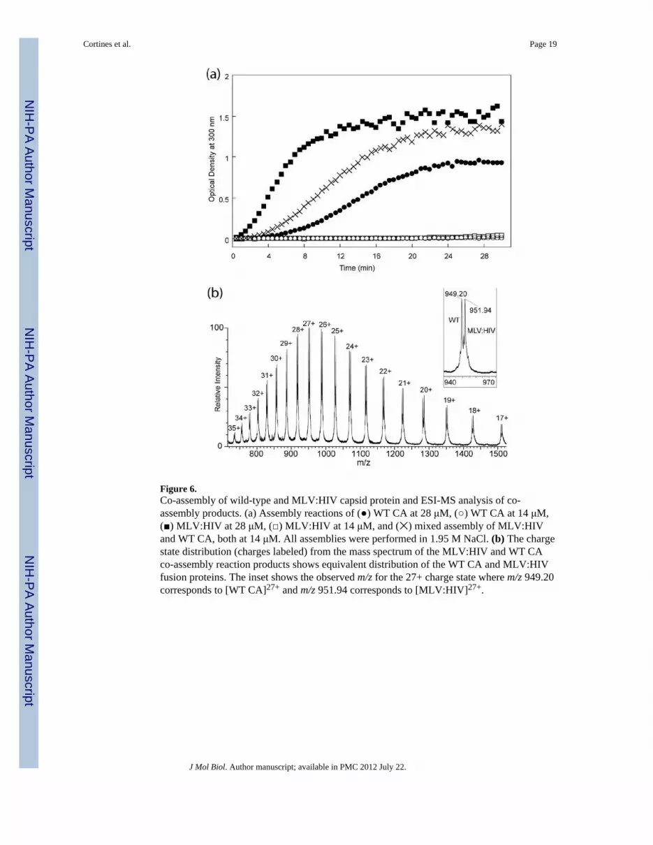
Figure 6.Co-assembly of wild-type and MLV:HIV capsid protein and ESI-MS analysis of co-assembly products. (a) Assembly reactions of (●) WT CA at 28 μM, (○) WT CA at 14 μM,(■) MLV:HIV at 28 μM, (□) MLV:HIV at 14 μM, and (✕) mixed assembly of MLV:HIVand WT CA, both at 14 μM. All assemblies were performed in 1.95 M NaCl. (b) The chargestate distribution (charges labeled) from the mass spectrum of the MLV:HIV and WT CAco-assembly reaction products shows equivalent distribution of the WT CA and MLV:HIVfusion proteins. The inset shows the observed m/z for the 27+ charge state where m/z 949.20corresponds to [WT CA]27+ and m/z 951.94 corresponds to [MLV:HIV]27+.
Cortines et al. Page 19
J Mol Biol. Author manuscript; available in PMC 2012 July 22.
NIH
-PA Author Manuscript
NIH
-PA Author Manuscript
NIH
-PA Author Manuscript

Figure 7.HDX peptide profiles for WT and MLV:HIV analyzed by mass spectrometry. HD exchangeplots for peptides covering the N-terminal 40 amino acids of monomeric (open symbols) andassembled (closed symbols) MLV:HIV (□, ■) and WT CA (○, ●). (a) The exchange plot forresidues 1–22 show a decrease in exchange for MLV:HIV in both monomeric andassembled forms while (b) the peptide containing amino acids 30–40 show no variationbetween the proteins. The increased protection in the assembled states is due tointermolecular interactions of helix 2 in the assembled lattice. (c) The 23–40 peptide showsdifferent exchange patterns between monomeric and assembled capsid proteins, which canbe explained by a change in the orientation of helix 1 in the monomeric form but not in theassembled state.
Cortines et al. Page 20
J Mol Biol. Author manuscript; available in PMC 2012 July 22.
NIH
-PA Author Manuscript
NIH
-PA Author Manuscript
NIH
-PA Author Manuscript


