Embed Size (px)
Citation preview

Antiviral Inhibition of the HIV-1 Capsid Protein
Chun Tang1, Erin Loeliger1, Isaac Kinde1, Samson Kyere1, Keith Mayo2
Eric Barklis2, Yongnian Sun3, Mingjun Huang3 andMichael F. Summers1*
1Howard Hughes MedicalInstitute and Department ofChemistry and BiochemistryUniversity of MarylandBaltimore County, 1000 HilltopCircle, Baltimore, Maryland21250-5398, USA2Vollum Institute andDepartment of MicrobiologyOregon Health & ScienceUniversity, 3181 SW SamJackson Park Road, PortlandOregon 97201-3098, USA3Achillion Pharmaceuticals300 George Street, New HavenCT 06511, USA
During the assembly stage of the human immunodeficiency virus (HIV)replication cycle, several thousand copies of the viral Gag polyproteinassociate at the cell membrane and bud to form an immature, non-infec-tious virion. Gag is subsequently cleaved by the protease, which liberatesthe capsid proteins for assembly into the polyprotein shell of the centralcore particle (or capsid) of the mature virus. Viral infectivity is criticallydependent on capsid formation and stability, making the capsid protein apotentially attractive antiviral target. We have identified compounds thatbind to an apical site on the N-terminal domain of the HIV-1 capsid pro-tein and inhibit capsid assembly in vitro. One compound, N-(3-chloro-4-methylphenyl)-N0-{2-[({5-[(dimethylamino)-methyl]-2-furyl}-methyl)-sul-fanyl]ethyl}urea) (CAP-1), is well tolerated in cell cultures, enabling invivo antiviral and mechanistic studies. CAP-1 inhibits HIV-1 infectivity ina dose-dependent manner, but does not interfere with viral entry, reversetranscription, integration, proteolytic processing, or virus production,indicating a novel antiviral mechanism. Significantly, virus particles gen-erated in the presence of CAP-1 exhibit heterogeneous sizes and abnormalcore morphologies, consistent with inhibited CA–CA interactions duringvirus assembly and maturation. These findings lay the groundwork forthe development of assembly inhibitors as a new class of therapeuticagents for the treatment of AIDS.
q 2003 Published by Elsevier Science Ltd
Keywords: HIV-1; antiviral inhibitors; capsid assembly; nuclear magneticresonance*Corresponding author
Introduction
Currently available drugs for the treatment ofhuman immunodeficiency virus (HIV) infectiontarget the reverse transcriptase (RT) and protease
(PR) enzymes, two of fifteen proteins encoded bythe viral genome. These drugs are marginally effec-tive when administered independently due to therapid emergence of resistant strains that areselected under conditions of incomplete viralsuppression.1 Although sustained reductions inviral load can be achieved when inhibitors areused in appropriate combinations (highly affectiveanti-retroviral therapy, HAART),1,2 inadequate sup-pression due to poor compliance, resistance, andinteractions with other drugs or diet is a significantproblem that limits the effectiveness of HAARTtherapy for many patients and can lead to thespread of drug-resistant strains.1–5 Inhibition ofother viral or cellular components may providethe best approach for attacking viral resistance.1
One potential target that has yet to be exploited isthe capsid protein (CA). CA is initially synthesizedas a domain within a 55 kDa Gag precursorpolyprotein. Approximately 4,000 copies of Gagassemble at the plasma membrane and bud toform an immature virus particle.6 Subsequent to
0022-2836/03/$ - see front matter q 2003 Published by Elsevier Science Ltd
E-mail address of the corresponding author:[email protected]
Abbreviations used: CA, capsid protein (p24); CAP-1,N-(3-chloro-4-methylphenyl)-N0-{2-[({5-[(dimethyl-amino)-methyl]-2-furyl}-methyl)-sulfanyl]-ethyl}urea;CAP-2, 1-(4-(N-methylacetamido)-phenyl)-3-(4-methyl-3-nitrophenyl)urea; CTD, carboxy-terminal domain; CV,cell viability; Gag, precursor polyprotein (p55); Gag283,283-residue N-terminal fragment of the HIV-1 Gagpolyprotein; HAART, highly active anti-retroviraltherapy; HBV, hepatitis B virus; Dd, chemical shiftchange induced upon ligand binding; HIV-1, humanimmunodeficiency virus type-1; HSQC, heteronuclearsingle quantum coherence; IU, infectious units; Kd,dissociation equilibrium constant; MA, matrix protein(p17); NMR, nuclear magnetic resonance; NTD, amino-terminal domain; PR, protease; RT, reverse transcriptase.
doi:10.1016/S0022-2836(03)00289-4 J. Mol. Biol. (2003) 327, 1013–1020
budding, CA is liberated by proteolytic cleavage ofGag, which triggers a conformational change thatpromotes assembly of the capsid particle.7–10 Twocopies of the viral genome and enzymes essentialfor infectivity become encapsidated in the central,cone shaped capsid of the mature virion.
Several recent studies have shown that propercapsid assembly is critical for viral infectivity.8,9,11–13
Mutations in CA that inhibit assembly are
lethal,8,9,11,12 and mutations that alter capsid stabilityseverely attenuate replication,13 making the capsidprotein an attractive potential antiviral target. Anti-viral agents have been developed that bind to thecapsid proteins of picornaviruses (including thehuman coxsackie, rhino- and polioviruses) and sup-press infectivity by inhibiting disassembly of the cap-sid shell,14 and inhibitors of hepatitis B virus (HBV)capsid assembly have recently been identified.15
Although similar strategies were anticipated forHIV more than a decade ago,16 inhibitors of capsidassembly or disassembly have yet to be identified,possibly due to the significant structural differencesbetween the retroviral (predominantly helical) andnon-retroviral (predominantly b-sheet) proteins.7,17,18
Results and Discussion
Identification of a ligand binding site on the N-terminal domain of CA
In an attempt to identify agents that inhibit func-tions of the capsid protein, we computationally
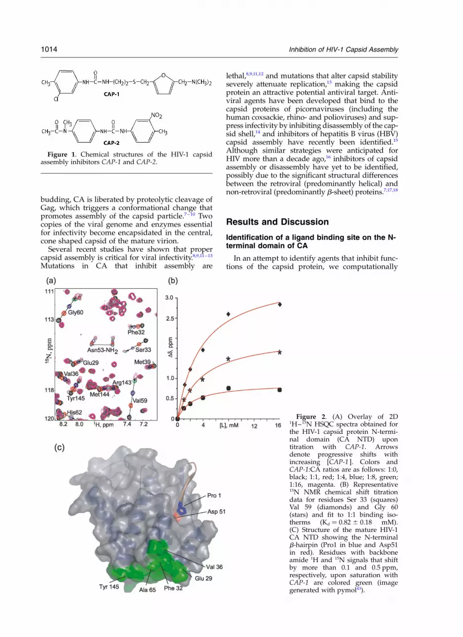
Figure 1. Chemical structures of the HIV-1 capsidassembly inhibitors CAP-1 and CAP-2.
Figure 2. (A) Overlay of 2D1H–15N HSQC spectra obtained forthe HIV-1 capsid protein N-termi-nal domain (CA NTD) upontitration with CAP-1. Arrowsdenote progressive shifts withincreasing [CAP-1 ]. Colors andCAP-1:CA ratios are as follows: 1:0,black; 1:1, red; 1:4, blue; 1:8, green;1:16, magenta. (B) Representative15N NMR chemical shift titrationdata for residues Ser 33 (squares)Val 59 (diamonds) and Gly 60(stars) and fit to 1:1 binding iso-therms (Kd ! 0.82 ^ 0.18 mM).(C) Structure of the mature HIV-1CA NTD showing the N-terminalb-hairpin (Pro1 in blue and Asp51in red). Residues with backboneamide 1H and 15N signals that shiftby more than 0.1 and 0.5 ppm,respectively, upon saturation withCAP-1 are colored green (imagegenerated with pymol43).
1014 Inhibition of HIV-1 Capsid Assembly

screened public-domain chemical libraries for com-pounds that might bind to clefts on the surface ofthe capsid protein and tested for binding usingnuclear magnetic resonance (NMR) titration spec-troscopy. Screening efforts focused mainly on a“b-hairpin cleft” that is exposed on the surface ofthe N-terminal domain (NTD) of the immaturecapsid protein but becomes occupied by residuesof a b-hairpin that forms after proteolytic cleavageof Gag.10 Compounds from public domain chemi-cal libraries were screened using DOCK-4.0,19 and40 compounds with good theoretical bindingproperties (binding energy ,226 kCal/mol, Con-tact Score ,240; further details to be reportedelsewhere) were experimentally tested for bindingto the intact CA protein, the NTD of CA, and a283 residue fragment of the immature Gag poly-protein (Gag283).10 Although we have yet to identifyagents that bind to the b-hairpin cleft, this processled to the fortuitous discovery of compounds thatbind to a different site that also appears to beimportant for capsid assembly.
Representative 1H–15N HSQC NMR dataobtained upon titration of the mature NTD withN-(3-chloro-4-methylphenyl)-N0-{2-[({5-[(dimethyl-amino)-methyl]-2-furyl}-methyl)-sulfanyl]-ethyl}-urea (CAP-1, Figure 1) are shown in Figure 2A.Although most signals were unaffected by thetitrations, a subset of signals shifted as a functionof increasing CAP-1 concentration, indicating site–specific binding. The chemical shift changes fit to1:1 binding isotherms and afforded an equilibriumdissociation constant (Kd) of 0.82 ^ 0.18 mM at35 8C, Figure 2B. Significantly tighter binding wasobserved for a second compound, 1-(4-(N-methyl-acetamido)phenyl)-3-(4-methyl-3-nitrophenyl)urea(CAP-2, Figure 1; Kd ! 52 ^ 27 mM)). In both cases,the CA residues perturbed by binding (1HN
Dd . 0.1 ppm; 15N Dd . 0.5 ppm; Glu29, Lys30,Ala31, Phe32, Ser33, Glu35, Val36, Val59, Gly60,Gly61, His62, Gln63, Ala65, Met144 and Tyr145)are located at or near the apex of a helical bundle(helices 1, 2, 3, 4 and 7), Figure 2C. Essentiallyidentical results were obtained for titrations withGag283 and intact CA, indicating that the bindingsite remains accessible in Gag-like constructs con-taining the native N-terminal matrix (MA) and C-terminal capsid (CTD) domains, and that bindingis insensitive to the maturation state of the protein.
Inhibition of capsid assembly in vitro
In the absence of other viral constituents, HIV-1CA can assemble into tubes with structural fea-tures that resemble mature cores.8,20–22 Tube for-mation leads to increases in sample turbidity thatcan be monitored spectrophotometrically,23 andthis assay was used to probe for potential inhibi-tory effects of the CAP compounds on in vitro cap-sid assembly. As shown in Figure 3, dissolution ofnative HIV-1 CA into assembly buffer (50 mMphosphate buffer, pH 8.0, 2.5 M NaCl, 0.04% v/vDMSO) led to an increase in absorbance at an
initial rate of 204 ^ 36 mOD/min (determinedfrom the initial slope and reported as the mean ^standard deviation from three experiments). Asexpected, compounds tested that do not bind CAdid not affect the rate of assembly. However, theassembly rate was diminished in a dose-dependentmanner by both CAP-1 and CAP-2, with the moretightly binding CAP-2 having a more pronouncedeffect. As shown in Figure 3, the initial assemblyrates in the presence of CAP-1 decreased to 93 ^ 3and 67 ^ 16 mOD/min at CAP-1: CA ratios of 1:1and 2:1, respectively. For comparison, assemblyrates in the presence of the more tightly bindingCAP-2 decreased to 81 ^ 2 and 39 ^ 11 mOD/minat CAP-2: CA ratios of 0.5:1 and 1:1, respectively.These data confirm that CA-binding compoundscan inhibit capsid assembly in vitro, and that therelative efficacy of assembly inhibition is depen-dent on the affinity of the ligands for the CAprotein.
Inhibition of viral infectivity
The CAP compounds were tested for toxicityand antiviral activity using HIV-1 producing, latentinfected U1 cells. This assay allows assessment ofantiviral effects on late phase replication events.24
Although CAP-2 was too cytotoxic for in vivoevaluations, CAP-1 was non-toxic under the con-ditions employed, and its application led to dose-dependent reductions in supernatant infectivity,Figure 4A. At 100 mM CAP-1, the U1 cells werefully viable, but infectivity was reduced by ca.95% relative to untreated samples, Figure 4A,B. To
Figure 3. Turbidity assay results showing the effects ofCA-binding compounds on in vitro capsid assembly.Compound: CA ratios and initial rates (milliopticaldensity units (mOD) per minute, reported as the mean ^standard deviation from three experiments) are asfollows: red, 0.2 ml DMSO control (204 ^ 36 mOD/min);magenta, 1:1 bis-(2-thiomethylbenzimidazole)methane(does not bind CA) (309 ^ 4 mOD/min); yellow, 1:1CAP-1 (93 ^ 3 mOD/min); brown, 2:1 CAP-1(67 ^ 16 mOD/min); green, 0.5:1 CAP-2 (81 ^ 2 mOD/min); blue, 1:1 CAP-2 (39 ^ 11 mOD/min).
Inhibition of HIV-1 Capsid Assembly 1015

determine if CAP-1 affects viral gene expressionand particle production, reverse transcriptase (RT)activity and CA (p24) levels were measured forthe supernatants after pelleting and removal ofthe cells. As shown in Figure 4B, both the CAlevels and RT activities were unaffected by CAP-1,indicating that antiviral activity is not due to inhib-ited virus production. In addition, the p24 (CA)levels observed in Western data obtained for trea-ted and untreated samples were very similar,Figure 5, indicating that CAP-1 does not signifi-cantly affect proteolytic processing of Gag. Consist-ent with this finding, CAP-1 did not affect in vitroprotease activity. The p24 Western data also indi-cate a reduction in the intracellular levels of Gag(p55) as a function of increasing levels of CAP-1,whereas the levels of the Gag cleavage productsp24, p41 remained relatively unaffected. Althoughfurther studies are warranted, these findingssuggest that CAP-1 promotes the intracellulardegradation of the full-length Gag polyprotein.Quantitation of gp120 was also obtained for thesupernatant using antibodies against gp120. No
differences were observed between the treatedand untreated samples, indicating that CAP-1 doesnot inhibit the synthesis or viral incorporation ofthe envelope glycoprotein (data not shown).
Antiviral activity was also tested in a second cel-lular assay using MAGI cell cultures. As observedin the U1 assay, treatment of infected, virus produ-cing MAGI cells with CAP-1 led to dose-dependentreductions in virus particle infectivity, with infec-tious units dropping by nearly two log units toless than 2% of the untreated levels at 100 mMCAP-1, Figure 4B. No reductions in viral RTactivity were observed, providing further evidencethat antiviral activity is not due to inhibited virusproduction. Virus production was also unaffectedby pre-incubation of either MAGI cells or virusparticles with CAP-1, indicating that CAP-1 is notvirucidal and does not directly inhibit early phaseevents. These data collectively indicate that theantiviral activity of CAP-1 is due to the inhibitionof a late-phase viral event that is different fromevents targeted by other anti-HIV agents currentlyunder investigation or in clinical use.
Figure 5. Capsid protein (p24)Western blot assays showing rela-tive concentrations of intra- andextracellular (viral) CA in the free(24 kDa band), Gag polyprotein(55 kDa band) and partially pro-cessed states as a function of addedCAP-1. As controls, results obtainedupon incubation with the proteaseinhibitor Nelfinavir (at low dose,for observation of proteolytic inter-mediates), and without addition oftumor necrosis factor (TNF,required for induction) are alsoshown.
Figure 4. (A) Viral infectivity(diamonds), cell viability (CV,squares) and virus production (p24levels, triangles) from latentinfected U1 cultures as a functionof added CAP-1. CAP-1 does notinhibit cell growth or virus pro-duction, but the particles producedare poorly infectious. (B) Cell viabi-lity (CV), virus particle associatedreverse transcriptase activity (RT)and p24 (CA) levels, and infectiousunits (IU) obtained upon incubationof infected U1 and MAGI cells for72 hrs with 100 mM CAP-1. Valuesare reported as percentages relativeto untreated cells.
1016 Inhibition of HIV-1 Capsid Assembly
Inhibition of Capsid Assembly in vivo
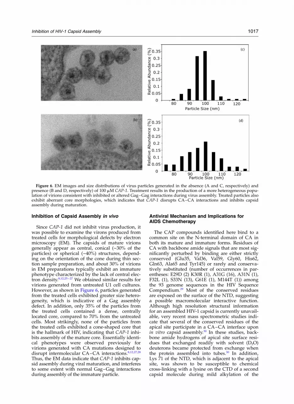
Since CAP-1 did not inhibit virus production, itwas possible to examine the virons produced fromtreated cells for morphological defects by electronmicroscopy (EM). The capsids of mature virionsgenerally appear as central, conical (~30% of theparticles) or spherical (~40%) structures, depend-ing on the orientation of the cone during thin sec-tion sample preparation, and about 30% of virionsin EM preparations typically exhibit an immaturephenotype characterized by the lack of central elec-tron density.8,12,25–27 We obtained similar results forvirions generated from untreated U1 cell cultures.However, as shown in Figure 6, particles generatedfrom the treated cells exhibited greater size hetero-geneity, which is indicative of a Gag assemblydefect. In addition, only 35% of the particles fromthe treated cells contained a dense, centrallylocated core, compared to 70% from the untreatedcells. Most strikingly, none of the particles fromthe treated cells exhibited a cone-shaped core thatis the hallmark of HIV, indicating that CAP-1 inhi-bits assembly of the mature core. Essentially identi-cal phenotypes were observed previously forvirions generated with CA mutations designed todisrupt intermolecular CA–CA interactions.8,12,27,28
Thus, the EM data indicate that CAP-1 inhibits cap-sid assembly during viral maturation, and interferesto some extent with normal Gag–Gag interactionsduring assembly of the immature particle.
Antiviral Mechanism and Implications forAIDS Chemotherapy
The CAP compounds identified here bind to acommon site on the N-terminal domain of CA inboth its mature and immature forms. Residues ofCA with backbone amide signals that are most sig-nificantly perturbed by binding are either strictlyconserved (Glu35, Val36, Val59, Gly60, His62,Gln63, Ala65 and Tyr145) or rarely and conserva-tively substituted (number of occurrences in par-entheses: E29D (2) K30R (1), A31G (16), A31N (1),F32L (1), S33N (13), G61E (1), M144T (1)) amongthe 93 genome sequences in the HIV SequenceCompendium.29 Most of the conserved residuesare exposed on the surface of the NTD, suggestinga possible macromolecular interactive function.Although high resolution structural informationfor an assembled HIV-1 capsid is currently unavail-able, very recent mass spectrometric studies indi-cate that several of the conserved residues of theapical site participate in a CA–CA interface uponin vitro capsid assembly.30 In these studies, back-bone amide hydrogens of apical site surface resi-dues that exchanged readily with solvent (D2O)deuterons became protected from exchange whenthe protein assembled into tubes.30 In addition,Lys 71 of the NTD, which is adjacent to the apicalsite, was shown to be susceptible to chemicalcross-linking with a lysine on the CTD of a secondcapsid molecule during mild alkylation of the
Figure 6. EM images and size distributions of virus particles generated in the absence (A and C, respectively) andpresence (B and D, respectively) of 100 mM CAP-1. Treatment results in the production of a more heterogeneous popu-lation of virions consistent with inhibited or altered Gag–Gag interactions during virus assembly. Treated particles alsoexhibit aberrant core morphologies, which indicates that CAP-1 disrupts CA–CA interactions and inhibits capsidassembly during maturation.
Inhibition of HIV-1 Capsid Assembly 1017

tubes. These results collectively indicate that resi-dues of the apical site of the NTD participate in anintermolecular CA(NTD)–CA(CTD) interfaceupon in vitro capsid formation.30 In combinationwith our present findings, these data provide com-pelling evidence that the CAP compounds functionmechanistically by inhibiting intermolecular CA–CA interactions necessary for proper capsidassembly.
Residues Trp23 and Val59 exhibit significantchemical shift changes upon CAP ligand binding,despite the fact that they are buried betweenhelices 1, 2 and 3 of the CA monomer. It is there-fore likely that the assembly inhibitors alter thelocal structure of the capsid protein, and maythereby either competitively inhibit CA–CAinteractions or promote the formation of astructurally distorted capsid shell. Binding-induced distortions would also explain why thecomputational screening procedure did notidentify the apical site for high affinity ligandbinding, since the protocol employed does notallow for protein flexibility.19,31,32 Binding-inducedprotein conformational changes are not uncommonand have confounded computational approachesto structure-based drug design.32 For example,non-nucleoside inhibitors of HIV-1 reversetranscriptase interact within a binding-inducedcavity that is not present in the native, unligandedprotein.33
Mutagenesis studies from several laboratorieshave shown that HIV-1 infectivity is highly sensi-tive to mutations in CA and to the stability of thecapsid.8,9,11–13 Particles with aberrant capsid mor-phologies are non-infectious,12 and those withcores that are normal in appearance but havealtered stability (both elevated and reduced, dueto mutations in CA) exhibit severely attenuatedinfectivity.13 As such, agents that marginally affectcapsid assembly or stability have the potential tobe potent inhibitors of viral replication. Ourstudies indicate that inhibition of capsid assemblydoes not require ligands with exceptionally highaffinity for CA. This is likely due to the high localconcentration of Gag molecules in assembled vir-ions (~14 mM), which favors binding by ligandswith even modest affinities such as CAP-1. Thus,conservatively assuming that cytosolic drug con-centrations in the budding virus and cells areequal (100 mM), the percentage of viral CAP-1 mol-ecules bound to CA can be estimated by standardmass action calculations (viral diameter D !96 nm; intraviral volume ! pD3/6 ! 463,250 nm3;4,000 Gag molecules/virion; [Gag] ! 14 mM;[CAP-1] ! 100 mM; Kd ! 820 mM ! ([CA]2[CA:CAP-1])([CAP-1]2 [CA:CAP-1])/[CA:CAP-1],which affords a value for the concentration ofbound CAP-1 ([CA:CAP-1 ]) of 94 mM. This indi-cates that 94% of the CAP-1 molecules in imma-ture virions (100 mM dose) should be bound toGag, and that binding to as few as ~25 moleculesof Gag per virion are sufficient to inhibit coreassembly during viral maturation.
The Western data presented here also suggestthat CAP-1 promotes the intracellular degradationof the Gag precursor protein. Gag normally func-tions by recruiting cellular factors associated withthe vacuolar protein sorting and degradationmachinery for assistance in budding,34–37 and it isplausible that the binding of CAP-1 to the capsiddomain of Gag could alter this process and lead tothe targeting of greater quantities of Gag for pro-teasomal degradation.
The present results provide proof of principle forthe development of antiviral inhibitors of HIV-1capsid assembly. Since compounds with higheraffinity for cytosolic Gag will be concentrated inassembling viruses, and compounds with greateraffinity for CA are more potent inhibitors of invitro assembly, it should be possible to rationallydesign agents with increased potency.
Materials and Methods
Sample Preparation and NMR Spectroscopy
DNA encoding the CA NTD (residues 1 through 151)was amplified from HIV-1 cDNA plasmid pNL–4-338
and an oligonucleotide encoding a C-terminal hexa-histi-dine tag was appended to the gene. The DNA wasinserted into a p11a expression vector (Novagen, Madi-son, WI), and the protein product was purified by cobaltaffinity chromatography (Clontech, Palo Alto, CA);MWcalc ! 17523.0 daltons, MWobs ! 17523.10 ^ 0.44daltons (electrospray Mass Spectrometry). The plasmidfor the full-length, native capsid protein was kindly pro-vided by Dr. W. I. Sundquist (Utah), and the proteinwas purified as described.23 NMR spectra were assignedusing conventional triple resonance methods.39 Bindingisotherms from 1H–15N NMR HSQC titration exper-iments were calculated with ORIGIN 7.0 software(MicroCal, Northampton, MA). CAP-1 was from May-bridge (HTS 02911; Cornwall, England) and CAP-2 andbis-(2-thiomethylbenzimidazole)methane were fromSigma–Aldrich (S85,367-4 and RCL S4,007-0, respect-ively; Milwaukee, WI).
In Vitro Assembly
Turbidity assays were performed at 21 8C using aBeckman DU650 spectrophotometer operating at 350 nmwavelength. Concentrated ligand in DMSO (0.2 ml) wasadded to a 250 ml aqueous solution containing the capsidprotein ([CA] ! 60 mM; [NaH2PO4] ! 50 mM; pH 8.0).Particulates were removed by centrifugation, and capsidassembly was initiated by addition of a concentratedNaCl solution (5 M, 250 ml). Spectral measurementswere made every 10 s, following a short initial delay toallow sample equilibration. Relative assembly rateswere estimated from initial slopes of the plots of absor-bance versus time.
In Vivo Infectivity
U1 cells (5 £ 105 cells/ml) were mixed with TNF-a(10 ng/ml, Sigma) for activation of HIV virion pro-duction and treated with CAP-1 at different concen-trations. Cultures were harvested 72 hrs after treatment.
1018 Inhibition of HIV-1 Capsid Assembly

Cell viability was measured using the MTS cell prolifer-ation assay (CellTiter 96 Aqueous One Solution Cell Pro-liferation Assay, Promega, Madison, WI). Supernatantswere collected, the cell debris removed by low speedcentrifugation, and the particles in the supernatantspelleted by microcentrifugation. Infectious units associ-ated with the particles were measured as describedpreviously40 except that b-Gal activities were measuredusing a Tropix Gal-Screen detector system (Applied Bio-systems, Foster City, CA). Particle-associated RT activi-ties were determined as described.41 Cell lysates andpelleted particles were subjected to SDS–PAGE analysisas described previously42 using AIDS patient sera (AIDSResearch and Reference Regent Program, NIAID, NIH).Quantitative p24 (CA) assays were performed with theHIV-1 p24 Antigen Capture ELISA kit (AIDS vaccineprogram, FCRDC/SAIC/NCI, Frederick, MD). Quanti-tative gp120 assays were performed with the HIV-1gp120 Antigen Capture ELISA Kit (Advanced Bio-technologies Inc., Columbia, MD). MAGI cells werewashed after viral adsorption (HIV-1RF) with PBS andwere fed fresh media containing CAP-1 at various con-centrations. Seventy-two hours post infection, the culturesupernatants were harvested and pre-cleared. Virus par-ticles present on the supernatants were collected bymicrocentrifugation and particle-associated RT activityand infectivity were subsequently measured.
Electron Microscopy
Treated (CAP-1) and untreated virus-producing U1cells were pelleted, washed in phosphate-buffered saline(PBS), and resuspended in at least ten cell pellet volumesof fixative (100 mM sodium cacodylate, pH 7.2, 2.5% glu-taraldehyde, 1.6% paraformaldehyde, 0.5% picric acid).Cells were fixed for 24–48 h, after which fixative wasremoved, and cells were washed twice in PBS, and thenpelleted in eppendorf centrifuge tubes. Washed cellpellets were post-fixed 1 h in 1% osmium tetroxide plus0.8% potassium ferricyanide in 100 mM sodium cacody-late, pH 7.2. After thorough rinsing in water, cells werepre-stained in 4% uranyl acetate 1 h, thoroughly rinsed,dehydrated, infiltrated overnight in 1:1 acetone:Epon812, infiltrated 1 h with 100% Epon 812 resin, andembedded in the resin. After polymerization, 60–80 nmthin sections were cut on a Reichert ultramicrotome,stained 5 min in lead citrate, rinsed, post-stained 30 minin uranyl acetate, rinsed and dried. EM was performedat 60 kV on a Philips CM120/Biotwin equipped with a1024 £ 1024 Gatan multiscan CCD, and images were col-lected at original magnifications of 26,880 £ –36,960 £ ,corresponding to resolutions of 8.9 and 6.5 A/pixel,respectively. For each sample, 4 separate EM grids wereviewed, and at least 47 images were collected, corre-sponding to a minimum total area of 35 micron2.
Acknowledgements
This work was supported by NIH grant AI30917(M.F.S.). I.K. is a Meyerhoff Undergraduate Scholarsupported by HHMI Biological Sciences Initiativeand NIH MARC funding, and E.L. is supportedby a UMBC Presidential Undergraduate Scholar-ship. We thank David King (HHMI, U.C. Berkeley)for mass spectral measurements.
References
1. Richman, D. D. (2001). HIV chemotherapy. Nature,410, 995–1001.
2. Pillay, D., Taylor, S. & Richman, D. D. (2000). Inci-dence and impact of resistance against approvedantiretroviral drugs. Rev. Med. Virol. 10, 231–253.
3. Mansky, L. M., Pearl, D. K. & Gajary, L. C. (2002).Combination of drugs and drug-resistant reversetranscriptase results in a multiplicative increase ofhuman immunodeficiency virus type 1 mutant fre-quencies. J. Virol. 76, 9253–9259.
4. Coffin, J. (1995). HIV population dynamics in vivo:implications for genetic variation, pathogenesis, andtherapy. Science, 267, 483–489.
5. Kuritzkes, D. R. (1996). Clinical significance of drugresistance in HIV-1 infection. AIDS, 10, S27–S33.
6. Vogt, V. personal communication7. Gitti, R. K., Lee, B. M., Walker, J., Summers, M. F.,
Yoo, S. & Sundquist, W. I. (1996). Structure of theamino-terminal core domain of the HIV-1 capsid pro-tein. Science, 273, 231–235.
8. von Schwedler, U. K., Stemmler, T. L., Klishko, V. Y.,LI, S., Albertine, K. H., Davis, D. R. & Sundquist,W. I. (1998). Proteolytic refolding of the HIV-1 capsidprotein amino-terminus facilitates viral core assem-bly. EMBO J. 17, 1555–1568.
9. Gross, I., Hohenberg, H., Juckhagel, C. & Krausslich,H.-G. (1998). N-terminal extension of humanimmunodeficiency virus capsid protein converts thein vitro assembly phenotype from tubular to spheri-cal particles. J. Virol. 72, 4798–4810.
10. Tang, C., Ndassa, Y. & Summers, M. F. (2002). Struc-ture of the N-terminal 283-residue fragment of theimmature HIV-1 Gag polyprotein. Nat. Struct. Biol.9, 537–543.
11. Tang, S., Murakami, T., Agresta, B. E., Campbell, S.,Freed, E. O. & Levin, J. G. (2001). Human immuno-deficiency virus type 1 N-terminal capsid mutantsthat exhibit aberrant core morphology are blockedin initiation of reverse transcription in infected cells.J. Virol. 75, 9357–9366.
12. Reicin, A. S., Ohagen, A., Yin, L., Hoglund, S. & Goff,S. P. (1996). The role of Gag in human immuno-deficiency virus type 1 virion morphogenesis andearly steps of the viral life cycle. J. Virol. 70, 8645–8652.
13. Forshey, B. M., von Schwedler, U., Sundquist, W. I. &Aiken, C. (2002). Formation of a human immuno-deficiency virus type 1 core of optimal stability iscrucial for viral replication. J. Virol. 76, 5667–5677.
14. Smith, T. J., Kremer, M. J., Luo, M., Vriend, G., Arnold,E., Kamer, G., Rossmann, M. G., McKinlay, M. A.,Diana, G. D. &Otto, M. J. (1986). The site of attachmentin human rhinovirus 14 for antiviral agents that inhibituncoating. Science, 233, 1286–1293.
15. Deres, K., Schroder, C. H., Paessens, A., Goldmann,S., Hacker, H. J., Weber, O., Kramer, T., Niewohner,U., Pleiss, U., Stoltefuss, J., Graef, E., Koletzki, D.,Masantschek, R. N. A., Reimann, A., Jaeger, R.,Grob, R., Beckermann, B., Schlemmer, K.-H., Haebich,D. & Rubsamen-Waigmann, H. (2003). Inhibition ofhepatitis B virus replication by drug-induceddepletion of nucleocapsids. Science, 299, 893–896.
16. Rossmann, M. G. (1988). Antiviral agents targeted tointeract with viral capsid proteins and a possibleapplication to human immunodeficiency virus. Proc.Natl. Acad. Sci. U.S.A. 85, 4625–4627.
17. Gamble, T. R., Vajdos, F., Yoo, S., Worthylake, D. K.,Houseweart, S. M., Sundquist, W. I. & Hill, C. P.
Inhibition of HIV-1 Capsid Assembly 1019

(1996). Crystal structure of human cyclophilin Abound to the amino-terminal domain of HIV-1 cap-sid. Cell, 87, 1285–1294.
18. Gamble, T. R., Yoo, S., Vajdos, F. F., von Schwedler,U. K., Korthylake, D. K., Wang, H., McCutcheon,J. P., Sundquist, W. I. & Hill, C. P. (1997). Structureof the carboxyl-terminal dimerization domain of theHIV-1 capsid protein. Science, 278, 849–853.
19. Ewing, T. J. A. & Kuntz, I. D. (1997). Critical evalu-ation of search algorithms for automated moleculardocking and database screening. J. Comp. Chem. 18,1175–1189.
20. Ehrlich, L. S., Agresta, B. E., Agresta, A. & Agresta,C. C. (1992). Assembly of recombinant humanimmunodeficiency virus type 1 capsid protein invitro. J. Virol. 66, 4874–4883.
21. Gross, I., Hohenberg, H. & Krausslich, H.-G. (1997).In vitro assembly properties of purified bacteriallyexpressed capsid proteins of human immuno-deficiency virus. Eur. J. Biochem. 249, 592–600.
22. Gross, I., Hohenberg, H., Wilk, T., Wiegers, K.,Grattinger, M., Muller, B., Fuller, S. & Krausslich,H.-G. (2000). A conformational switch controllingHIV-1 morphogenesis. EMBO J. 19, 103–113.
23. Lanman, J., Sexton, J., Sakalian, M. & Prevelige, P. E.,Jr. (2002). Kinetic analysis of the role of intersubunitinteractions in human immunodeficiency virus type1 capsid protein assembly in vitro. J. Virol. 76,6900–6908.
24. Engelman, A., Englund, G., Orenstein, J. M., Martin,M. A. & Craigie, R. (1995). Multiple effects ofmutations in human immunodeficiency virus type 1integrase on viral replication. J. Virol. 69, 2729–2736.
25. Gelderblom, H. R. (1991). Assembly and Mor-phology of HIV: Potential Effect of Structure onViral Function. AIDS, 5, 617–637.
26. Hoglund, S., Ofverstedt, L.-G., Nilsson, A.,Lundquist, P., Gelderblom, H., Ozel, M. & Skoglund,U. (1992). Spatial Visualization of the Maturing HIV-1 Core and Its Linkage to the Envelope. AIDSResearch and Human Retroviruses, 8, 1–7.
27. Kong, L. B., An, D., Ackerson, B., Canon, J., Rey, O.,Chen, I. S. Y., Krogstad, P. & Steward, P. L. (1998).Cryoelectron microscopy examination of humanimmunodeficiency virus type-1 virions withmutations in the cyclophilin A binding loop. J. Virol.72, 4403–4407.
28. Dorfman, A. T., Bukovsky, A., Ohagen, A. S.,Hoglund, H. & Gottlinger, G. (1994). Functionaldomains of the capsid protein of human immuno-deficiency virus type 1. J. Virol. 68, 8180–8187.
29. Kuiken, C. L., Foley, B., Hahn, B., Marx, P. A.,McCutchan, F., Mellors, J., Mullins, J. I., Wolinksy, S.& Korber, B. (2001). HIV Sequence Compendium 2001,Theoretical Biology and Biophysics Group, Los Alo-mos National Laboratory, Los Alomos.
30. Lanman, J., Lam, T. T., Barnes, S., Sakalian, M.,Emmett, M. R., Marshall, A. G. & Prevelige, P. E., Jr.(2003). Identification of novel interactions in HIV-1capsid protein assembly by high-resolution massspectrometry. J. Mol. Biol. 325, 759–772.
31. Lin, J.-H., Perryman, A. L., Schames, J. R. &McCammon, J. A. (2002). Computational drugdesign accommodating receptor flexibility: Therelaxed complex scheme. J. Am. Chem. Soc. 124,5632–5633.
32. Arkin, M. R., Randal, M., DeLano, W. L., Hyde, J.,Luong, T. N., Oslob, J. D., Raphael, D. R., Taylor, L.,Wang, J., McDowell, R. S., Wells, J. A. & Braisted,A. C. (2003). Binding of small molecules to an adap-tive protein-protein interface. Proc. Natl. Acad. Sci.USA, 100, 1603–1608.
33. Rodgers, D. W., Gamblin, S. J., Harris, B. A., Ray, S.,Culp, J. S., Hellmig, B., Woolf, D. J., Debouck, C. &Harrison, S. C. (1995). The structure of unligandedreverse transcriptase from the human immuno-deficiency virus type 1. Proc. Natl Acad. Sci. USA, 92,1222–1226.
34. Garrus, J. E., von Schwedler, U. K., Pornillos, O. W.,Morham, S. G., Zavitz, K. H., Wang, H. E., Wettstein,D. A., Stray, K. M., Cote, M. L., Rich, R. L., Myszka,D. G. & Sundquist, W. I. (2001). Tsg101 and thevacuolar protein sorting pathway are essential forHIV-1 budding. Cell, 107, 55–65.
35. Schubert, U., Ott, D. E., Chertova, E. N., Welker, R.,Tessmer, U., Princiotta, M. F. et al. (2000). Proteasomeinhibition interferes with Gag polyprotein proces-sing, release, and maturation of HIV-1 and HIV-2.Proc. Natl. Acad. Sci. USA, 97, 13057–13062.
36. Strack, B., Calistri, A., Accola, M. A., Palu, G. &Gottlinger, H. G. (2000). A role for ubiquitin ligaserecruitment in retrovirus release. Proc. Natl. Acad.Sci. USA, 97, 13063–13068.
37. Patnaik, A., Chau, V., & Wills, J. W. (2000). Ubiquitinis part of the retrovirus budding machinery. Proc.Natl. Acad. Sci. USA, 97, 13069–13074.
38. Adachi, A., Gendelman, H. E., Koenig, S., Folks, T.,Willey, R., Rabson, A. & Martin, M. A. (1986). Pro-duction of acquired immunodeficiency syndrome-associated retrovirus in human and nonhuman cellstransfected with an infectious molecular clone.J. Virol. 59, 284–291.
39. Kay, L. E., Ikura, M., Tschudin, R. & Bax, A. (1990).Three-dimensional triple-resonance NMR spec-troscopy of isotopically enriched proteins. Journal ofMagnetic Resonance, 89, 496–514.
40. Kimpton, J. & Emerman, M. (1992). Detection ofreplication-competent and pseudotyped HIV with asensitive cell line on the basis of activation of an inte-grated b-galactosidase gene. J. Virol. 66, 2232–2239.
41. Huang, M., Orenstein, J. M., Martin, M. A. & Freed,E. O. (1995). p6Gag is required for particle pro-duction from full-length human immunodeficiencyvirus type 1 molecular clones expressing protease.J. Virol. 96, 6810–6818.
42. Huang, M., Zensen, R., Cho, M. & Martin, M. A.(1998). Construction and characterization of a tem-perature-sensitive human immunodeficiency virustype 1 reverse transcriptase mutant. J. Virol. 72,2047–2054.
43. DeLano Scientific. (2002). The PyMOL moleculargraphics system. DeLano, W. L.
Edited by P. Wright
(Received 28 January 2003; received in revised form 24 February 2003; accepted 25 February 2003)
1020 Inhibition of HIV-1 Capsid Assembly


