Embed Size (px)
Citation preview

A Review: Potential Usage of Cellulose Nanofibers (CNF)for Enzyme Immobilization via Covalent Interactions
Safwan Sulaiman & Mohd Noriznan Mokhtar &
Mohd Nazli Naim & Azhari Samsu Baharuddin &
Alawi Sulaiman
Received: 3 July 2014 /Accepted: 17 November 2014 /Published online: 27 November 2014# Springer Science+Business Media New York 2014
Abstract Nanobiocatalysis is a new frontier of emerging nanosized material support inenzyme immobilization application. This paper is about a comprehensive review on cellulosenanofibers (CNF), including their structure, surface modification, chemical coupling forenzyme immobilization, and potential applications. The CNF surface consists of mainly –OH functional group that can be directly interacted weakly with enzyme, and its binding canbe improved by surface modification and interaction of chemical coupling that forms a strongand stable covalent immobilization of enzyme. The knowledge of covalent interaction forenzyme immobilization is important to provide more efficient interaction between CNFsupport and enzyme molecule. Enzyme immobilization onto CNF is having potential forimproving enzymatic performance and production yield, as well as contributing toward greentechnology and sustainable sources.
Keywords Nanobiocatalysis . Chemical modification . Chemical coupling . Surfacemodification . Enzyme immobilization . Nanostructuredmaterials . Cellulose nanofibers (CNF)
Introduction
There are high expectations for nanotechnology to play an important role in biotechnologyfield. Nanobiotechnology, a branch of nanotechnology involved with biological and biochem-ical elements, is based on work at the atomic and molecular level such as creation andutilization of materials, devices, and even a system (in range of 1 to 100 nm) to fabricatestructures by combining the biological materials especially in integration of physical sciences,molecular engineering, biology, chemistry, genomics, and proteomics [1–3]. Development of
Appl Biochem Biotechnol (2015) 175:1817–1842DOI 10.1007/s12010-014-1417-x
S. Sulaiman :M. N. Mokhtar (*) :M. N. Naim :A. S. BaharuddinDepartment of Process and Food Engineering, Faculty of Engineering, Universiti Putra Malaysia,43400 Serdang, Selangor, Malaysiae-mail: [email protected]
A. SulaimanFaculty of Plantation and Agrotechnology, Universiti Teknologi MARA, 40450 Shah Alam, Selangor,Malaysia

nanobiotechnology leads to a better perspective in enzyme technology and has a goodsignificance toward commercialization of enzymes, as well as a further innovation in improve-ment of catalytic performance [4].
Nanobiocatalysis, a common approach used in nanobiotechnology can be defined as aprocess based on incorporation of enzymes onto nanostructured material. Enzymes areremarkable biocatalysts and have been used in the industrial biotechnology because of theirinteresting characteristics, such as green chemistry, substrate and product specificity, and easeof preparation [5]. The catalytic activity of enzymes implies that they accelerate reactions bydecreasing the activation energy [6]. In biocatalyst fields, major objectives are to stabilize andto recover the enzymes, since they are too costly in the current market. Achieving goodstability, enzyme separation, recovery, and life-time cycle rate are successful keys in enzymaticproduction and commercialization [7]. Native enzymes are commonly applied as industrialbiocatalysts due to their higher enzyme activity. Unfortunately, native enzymes often lacklong-term stability in operational conditions and pose difficulties for recovery and reuse [8].For example, capillary gel electrophoresis can be used for separation between enzyme andproduct [9], but it requires high energy consumption, is expensive, and is not applicable(difficult and time consuming) for large-scale operation.
Enzyme immobilization is a special technique to solve enzymatic problems (especially inthe application of native enzyme), such as stability, reusability, and decline of activity due toinhibition either by medium or products [10]. There are two main reasons why enzymeimmobilization has the potential to overcome those limitations in an effective way. First,immobilization facilitates separating between enzyme molecule and product, and thus itprovides a reliable and efficient condition toward reaction technology [11]. Second, immobi-lization improves enzyme reusability, since insoluble enzymes are much easier in recyclingcompared with soluble enzymes [12]. There are several immobilization techniques such asentrapment, encapsulation, adsorption, cross-linking, and covalent binding.
In practice, immobilization of enzyme would cause lower activity or retention activity dueto inactivation or denaturation of enzyme, which cannot be completely avoided. However, thelevel of enzyme inactivation or denaturation depends on the types of enzyme immobilizationtechniques, structured support, medium conditions (e.g., buffer solution, pH, and temperature),and reaction during preparation of enzyme immobilization [13]. Therefore, the best casescenario to minimize the loss of enzyme activity and to overcome the poor retention is byselecting a proper immobilization technique, supporting selection and optimum immobiliza-tion conditions, in order to preserve the enzyme activity as close as to its original performancelevel by not changing the chemical nature or reactive groups in the enzyme’s binding site.Other reasons are to ensure biomolecule stability and to minimize cost [14, 15].
This present paper provides a comprehensive review of the covalent immobilizationtechnique of enzymes onto cellulose nanofiber (CNF) support. Unlike other techniques(adsorption, entrapment, encapsulation, cross-linking), covalent binding has been demonstrat-ed as the most stable interaction for immobilization of enzyme molecules to the support [16].In order words, covalent binding will minimize the leakage of enzyme from its support into thereaction medium [7]. Covalent immobilization of enzyme also provides an unlimited contactbetween enzyme molecules and substrates, since there has no barrier between them andlocalization of enzyme onto surface support, which attributes to the stability of a biocatalystsystem [17]. Therefore, a lot of approaches can be applied to derive this method, depending onthe type of support materials and substrates that are used in the covalent immobilization ofenzyme [18].
In addition, stability affects covalent interaction due to the fact that an immobilized enzymeis not able to undergo any additional intramolecular process, whereas the soluble enzyme is
1818 Appl Biochem Biotechnol (2015) 175:1817–1842

able to undergo aggregation and interaction via a hydrophobic interface [19]. Greater stabilityof covalent immobilization of enzyme also induces higher resistance to temperature, decom-position, pH, and organic solvents in several cases. However, the extent of these improvementsalso depends on the detailed characteristics of enzyme and support that are usually dictated inimmobilization system. Covalent binding is relatively a complicated procedure due to additionof a chemical coupling reagent, especially one that involves nanosized support materials [20].In normal cases, the immobilization leads to lower residual enzyme activity as mentionedpreviously due to the tendency of some of amino acid residues, which are essential for catalyticactivity to form covalent linkage onto the nanostructured support [21]. Sometimes, the centerof active site of enzyme could be modified or altered through chemical coupling reaction, sincemost chemical coupling agents are toxic and very reactive [7], leading to inactivation of theenzyme molecule. Thus, appropriate selection of type and method of a chemical couplingagent needs consideration. Therefore, in-depth study of chemical coupling in covalent inter-action does not only help in minimizing all these problems but also increases the efficiency ofenzymatic system that could be applied for commercialization purposes [22].
This article also summarizes in detail the relationship among chemical couplings in theinteraction between support and enzyme molecules for covalent immobilization. Chemicalcoupling agents such as ligands and spacer arms are very important in covalent interaction,which can improve binding efficiency, provide a greater mobility, and minimize sterichindrance. The use of nanofibers is potentially advantageous in this covalent immobilizationof enzyme. In fact, CNF provide a great potential to be interacted, modified, or activated withfunctional groups by the use of specific reagents to render them more suitable for enzymeimmobilization. Natural type of CNF will be discussed as a support due to their goodmechanical properties and characteristic in providing functional group on the surface forimmobilization purpose, as good as polymer-based nanofibers.
Current Trend in Enzyme Immobilization
A number of research findings have been published, especially for the application of nano-structured materials in biocatalysts systems. Figure 1 shows the cumulative publications aboutenzymes immobilization from year 2000 to 2013. There is a growing publication in enzymeimmobilization onto nanostructure materials as well as publication in general enzyme immo-bilization. The study on enzyme immobilization onto nanostructured material increaseddrastically from 2004, and then the number increased steadily year by year due to a greaterdevelopment on nanostructure materials, which then particularly contributed to the improve-ment of enzyme immobilization performance and production yield.
Cellulose Nanofibers as a Natural Nanostructure Support
As discussed previously, the performance of enzyme immobilization strongly depends on theproperties of support, such as material type, composition, structure, and mechanical properties[12]. Better support properties provide a good mechanical strength, which can contribute tostability and reusability. The use of nanosized supports in enzyme immobilization is not onlyto enhance the stability and reusability of immobilized enzyme but also to overcome lowerimmobilized enzyme activity due to the presence of high surface area per volume ratio [23]. Inorder words, high surface area of nanosized support provides a high number of functionalgroup on the surface support. Thus, CNF have more chance of interacting with enzymemolecules [4, 24–27]. A unique behavior of nanoscale support would distinguish them from
Appl Biochem Biotechnol (2015) 175:1817–1842 1819

traditional immobilized systems. Development of nanoscale biocatalyst system would show abenefit for both enzyme and nanosized support, since the size of enzyme molecule is already inthe nanoorder.
Nanoporous silica, nanotubes, nanoparticles, nanofibers, nanocomposite, and nanosheetsare types of nanostructured supports. Nanosized support provides opportunities for greatefficiency in manipulation of the nanoscale environment, making possible new innovations,and offering promise for various advanced technologies in the enzyme immobilization field[28]. However, some nanostructured materials have their own limitations. For example,nanoporous silica usually confines enzyme molecules on its inner porous surfaces, and thisinhibits mass diffusion of substrate or product and results in lower enzymatic production [29,30]. Non-porous nanoparticles and nanotubes are remarkable with respect of providing aminimum mass transfer limitation, but their recycling and dispersion are more difficult [12].Nanocomposites could damage or block the reactive functional groups and possibly affect thestability of multipoint interaction of enzymes [31]. On the contrary, nanofibers have a greatpotential as a solution for those problems that can be prepared as non-porous supports andremain easy to be recycled or reused. Nanofiber support normally has sufficient dimension(diameter and length) in certain range of distribution, which provides a wide area forinteraction of immobilized enzyme, compared with other types of nanostructured supports.The use of a nanofiber support is a promising approach for enzyme immobilization, and itoffers unique capabilities, as described previously. Nanofibers has attracted continuous oper-ation of enzymatic process especially in membrane application and other potential applicationsdue to high enzyme loading and its efficiency [29, 32].
Natural resources can be generally described as rigid and partially crystalline objects. Theyare composed of cellulosic micro-fibrils reinforced with amorphous lignin, hemicellulose,waxes, and several water-soluble compounds. Cellulose is the main constituent in the mostabundant organic compound on earth, especially within wood and natural fibers (kenaf, palm,
0
500
1000
1500
2000
2500
3000
2000 2001 2002 2003 2004 2005 2006 2007 2008 2009 2010 2011 2012 2013
Enzymeimmobilization
Enzymeimmobilized onnanostructuredmaterial
Fig. 1 Cumulative annual number of English scientific publication-based articles, reviews, and book chapterssince 2000 according to the keywords used to describe “enzyme immobilization,” “immobilized enzyme,” and“nanostructured for enzyme immobilization.” Data analysis by Scopus scholar search system (December 2013)
1820 Appl Biochem Biotechnol (2015) 175:1817–1842

cotton, hemp, flax, etc.). Almost 65–70 % of cellulose compound is contained in plant fibersand comprised C, H, and O elements [33]. Cellulose is insoluble in water. The poor solubilitycan attribute to strong intra- and inter-molecular hydrogen bonds within and among individualchains [34]. As a result, the cellulose fibers present a moderately hydrophobic surface at itsexterior, and this feature renders it as a promising material for enzyme immobilization. Inaddition to the hydroxyl group on the cellulose surface, it provides an ideal site for partici-pating in covalent bonding of enzyme immobilization [35].
The basic chemical structures of cellulose play a central role in determining the natureof cellulosic nanomaterials. Cellulose consists of linear chains of homopolysaccharidecomposed of β-D-glucopyranose units linked together by β-1-4-linkages [35–37], with adegree of polymerization (DP) of approximately 10,000 for cellulose chain in nature and15,000 for native cellulose cotton [38], as illustrated in Fig. 2. Each monomer unit hasthree hydroxyl groups, which make possible the formation of a dense system of well-ordered hydrogen bonds, which is especially evident in the crystalline packing ofcellulose [39, 41]. These hydrogen bonds give cellulose a stable structure, with nomelting point and lack solubility in typical aqueous solution. Krässig [42] reported thatthe presence of hydroxyl groups located in the amorphous regions were found to behighly reactive and accessible, while for those present in compact crystalline regionsshowed much lower accessibility. Thus, it provides a cellulose chains at high axialstiffness [43]. Stiffness allows an enzyme to separate itself from the matrix surfaceeffectively by minimizing any steric hindrance, such that the enzyme can retain essen-tially its full activity while in the immobilized state. The approaches for the enzyme andsupport ratio depend on the experimental work and enzyme loading onto the surfacesupport, which can be expected in 100 U/g support [44].
The production of cellulose nanofibers (CNF) and their application in enzyme immobili-zation have gained increase attention due to their green support, high strength and stiffness,low weight, low cost, biocompatibility, biodegradability, and renewability [45]. Furthermore,several researchers have reported that CNF consist of high cellulose composition, highcrystallization, superior directional strength, and low coefficient of thermal expansion, whichexhibit good mechanical properties of support [46–50]. High mechanical properties couldmake CNF a preferable support for this application, to increase enzymatic performancebecause of its immobilized enzyme stability. Distribution of similar size of CNF support givesnumerous advantages in enzyme immobilization, which are listed in Table 1.
CNF can be isolated either by using top-down or bottom-up approach method. The top-down approach is defined as a preparation method in which nanosized fibers can be generatedby stripping down a complex multicellular living plant organism [57]. This term is usedespecially when the support is produced from natural resources. The bottom-up approach
Fig. 2 Chemical structure of cellulose [39, 40]
Appl Biochem Biotechnol (2015) 175:1817–1842 1821

refers to the assembly process of material from a small molecule into a more complex structureor the production of a new nanofiber structure [58]. For example, electrospinning method canbe used to electrospun nanofibers from cellulose solutions, which are initially in a solidnanoparticle form [2, 59–61]. This term merely involves the use of polymer material as asupport in enzyme immobilization.
Top-down approach technique mainly involves the use of mechanical treatments [48,50, 62–64], chemical treatments [65–69], biological treatments [70–72], or a combina-tion of two or more of these methods, to remove the plant cell constituents other thancellulose, such as hemicellulose, lignin, pectin, minerals, oils, and other extractivecompounds. However, isolated CNF also participate in aggregation of nanofibers witha random distribution due to complicated multi-layer nanofiber structure and inter-fibrillar hydrogen bonds [73]. The structural images of isolated CNF using TEM areshown in Fig. 3, based on different natural resources.
Table 1 Advantages of CNF properties in enzyme immobilization
Factors Advantages References
Surface area to volume ratio It provides high surface area to volume ratio for enzymeinteraction. High surface area would increase the numberof functional groups on the surface CNF support, whichimproves the covalent binding efficiency.
[4, 23, 51]
Enzyme activity Surface modification of CNF support benefits the stabilityof enzymatic system, which potentially increases theenzyme activity.
[52]
Enzyme loading It offers high enzyme loading due to their high relativesurface area, perhaps giving better results in biocatalyticactivity and stability.
[28]
Functional groups CNF possesses a large number of specific surface functionalgroups compared with micro-sized fibers. Therefore, moreenzymes will interact to achieve higher enzyme loading inimmobilization.
[53]
Flow rate It behaves as a stable, monodispersed in aqueous suspensioneventhough exhibiting the Brownian motion. Based onStokes–Einstein theory, diffusivity, and mobility, thenanostructured materials should be smaller than thosenative enzymes to achieve higher enzymatic activity.
[28, 29]
Mass transfer effects High surface area and interconnectivity of CNF supportprovide low mass transfer limitation, which results inhigh activity and stability of enzyme performance.
[12, 20, 28, 54]
Microporous membrane-likestructure
Non-woven of CNF support distribution forms a microporousmembrane-like structure which improves the mass transferrate of substrate.
[52]
Easily recovery The enzyme is easily recovery from reaction mediacompared to freely suspended enzymes.
[11]
Design of enzymaticbioreactor
Immobilization is a fundamental platform for developmentof bioreactors and biosensors. CNF have high flexibility;they can be easily prepared as membrane or fixed-bedsystem. Some factors especially high flow rate and smallpressure drop in CNF membrane represent the mostpromising advantages in enzyme immobilization comparedwith traditional immobilized enzymes bioreactor andfixed-bed bioreactors.
[32, 55, 56]
1822 Appl Biochem Biotechnol (2015) 175:1817–1842

CNF have been categorized into three main types, as follows: microfibrillated cellulose(MFC), nanocrystals cellulose (NCC), and bacterial nanocellulose (BNC). Fibrils, which canbe described as relatively long and very thin pieces of cellulosic material, have been used invarious applications [76–78]. In nature, cellulose has been found as assemblies of individualcellulose chain-forming fibers and does not occur as an isolated individual molecule. Micro-fibrils or several elementary fibrils are defined as larger units of cellulose molecules. MFCconsists of an assemblage of fibrils that are partly separated from each other [35, 36]. There areseveral terms in nomenclatures that are synonymous to common types of cellulose nanofibers,as described in Table 2.
Surface Modification of CNF Support
The support to be used for enzyme immobilization must present a high external surface area toachieve a good geometrical conjugation with the enzyme [87]. CNF support is attractive to theenzyme because of its support characteristic under inert physiological conditions and its abilityto adapt into hydrophilic or hydrophobic conditions, depending on the applications needed[39, 88]. Non-porosity of CNF support allows high accessibility onto the active site, low
Fig. 3 The transmission electron microscope (TEM) images of isolated CNF with different natural sources: akenaf [66], b bamboo [48], c wood [74], and d hemp [75]
Appl Biochem Biotechnol (2015) 175:1817–1842 1823

Tab
le2
The
differentterm
inologiesof
CNFfamily
Typesof
cellu
lose
Synonym
nomenclature
Descriptio
nAverage
size
References
Microfibrillated
cellulose
(MFC
)Cellulose
microfibril,
microfibrils,
nanofibrils,and
nanofibrillated
cellu
lose
(NFC
)
Produced
throughdelaminationof
fibers
pulp
bymechanicaltreatm
enteither
before
orafterchem
icalor
enzymatic
treatm
ent.
Diameter,5
–60nm
;length,
severalmicrometer
(μm)
[63,
79–82]
Nanocrystallin
ecellu
lose
(NCC)
Cellulose
nanocrystals,crystallites,
whiskers,androdlikecellu
lose
microcrystals
Form
edby
acid
hydrolysisandoften
follo
wed
byultrasonictreatm
ent,
inorderto
removean
amorphous
sections
ofcellulose
source.K
nown
aswhiskers,consistof
rodlike
cellu
lose
crystals.S
imilarto
MFC
size,b
utlim
itedin
flexibility,aswell
asdo
notcontainam
orphousregions
butinsteadexhibitelongated
crystalline
rodlikeshapes.
Diameter,5
–70nm
;length,
100–600nm
(from
natural
plants),100nm
toseveral
micrometers(from
tunicates,
algae,andbacteria)
[2,3
8,41,8
3–85]
Bacterialnanocellu
lose
(BNC)
Bacterialcellulose,m
icrobial
cellu
lose,and
biocellulose
Synthesizedby
aerobicbacteria.F
ormed
apolymer
andnanomaterialsby
biotechnologicalprocessfrom
low-m
olecular-w
eightsugar
( D-glucose)andalcohols.
Diameter:20–100
nm[86]
1824 Appl Biochem Biotechnol (2015) 175:1817–1842
diffusion resistance, and easy recoverability as well as potential applicability for continuousoperations [23, 32, 59, 89, 90].
Many factors such as moisture absorption, quality variations, poor compatibility, surfacehydrophilicity/hydrophobicity, rigidity, and durability may affect the behavior of mechanicalproperties of CNF support, which make a less attractive toward covalent interaction. Bymanipulating the CNF properties and characteristics using surface modification treatment, itwould maintain or enhance the immobilized enzyme conformation and its apparent activity.Moreover, the appropriate surface modification treatment of CNF can increase the availabilityof hydroxyl groups, change the degree of crystallinity, and break hydrogen bonds, whichincrease the reactivity of cellulose [91].
Chemical treatment method is often used to perform surface modification of CNF support,compared with UV oxidation and plasma method [92]. For example, chemical treatment(mercerization) is a well-known treatment for surface modification of CNF. Chemical reagentssuch as acidified sodium chlorite (NaClO2) and sodium hydroxide (NaOH) are often used totreat the structure of CNF to increase the surface roughness [2], providing a platform withreactive functional group for higher interaction in this application. In fact, the treatment willremove other matrix substances covering the surface fibers (e.g., lignin, hemicellulose, pectin,wax, and oil) to obtain high purity in morphological size and composition of CNF [49].
Surface hydrophilicity or hydrophobicity of the CNF support is one of the most importantcriteria in covalent interaction. Hydrophilicity or hydrophobicity characteristic of CNF maychange the conformation structural in enzyme interaction due to reduction of free energy in anenzymatic process. In a few cases, a hydrophobic surface of support also shows an untightenedbond onto accessible hydrophobic group of amino acids due to complimentary unfolding of thehydrophobic core toward the surface. However, lipase is known as an exception enzyme; lipaseactivity increases when it is immobilized on hydrophobic supports [93]. This phenomenon isbecause of hydrophobic “lid” on the surface enzyme in open position state, which ables contactwith a hydrophobic surface support. Open position of the lid may lead toward the increase ofenzyme activity comparedwith the free enzyme [94].Meanwhile, a hydrophilic surfacemay resultin loss of enzyme conformation because of competitive hydrogen bonding between hydrophilicamino group and aqueous solvent [95]. Some researchers have reported that hydrophilic supports(e.g., cellulose, agarose, chitosan, dextran, alginate, gelatin, and collagen) have high retentionactivity, but they can be degraded under harsh chemical reaction conditions especially duringchemical coupling preparation [96]. However, these supports can be manipulated to be moresuitable in retaining immobilized enzyme activity, depending on their interface characteristic; theycan be rendered as either hydrophobic or hydrophilic surface [97–99]. Unfortunately, hydrophobicinteractions between amino acid group and support surface may cause dehydration of the enzyme.This may cause a shift in the enzyme structure, leading to loss of enzymatic activity [100].
Surface modification of CNF support is also able to create the surface interfacial thatconsists of hydrophilic enzyme molecules onto hydrophobic support [101]. This technique willreduce the complimentary unfolding toward hydrophobic-side group in amino acids, as well asit will provide a suitable interface for retaining enzyme activity [97]. Moskovitz and Srebnik[186] reported that by using computational modeling, they suggested that a hydrophilic chainon a hydrophobic surface can be used to preserve the activity of an immobilized enzyme. Anearly patent related to the invention technique for enzyme immobilization through covalentbond on a cellulose support was claimed by Monsan [187].
Numerous studies have been published related to functional groups on the surface oflignocellulosic materials. The types of the functional groups can be identified by using Fouriertransform infrared (FTIR) spectroscopy analysis. On the CNF surface, there is a high surfacedensity of –OH functional groups, which are evident in the region of absorbance around 3400
Appl Biochem Biotechnol (2015) 175:1817–1842 1825

to 3300 cm−1, corresponding to intra- and inter-molecular hydrogen bonding [65, 66]. The –OH group can be a potential target functional group for the interaction of covalent binding.The rest of the functional groups in CNF support are listed in Table 3.
Chemical Modification of Covalent Immobilization of Enzyme onto CNF SupportSurface
Basically, covalent binding is a well-established mechanism, based on the chemical reaction ofside chain attachment between active amino acid group on the enzyme surface and activefunctionality that attached onto the CNF support surface. Interaction of covalent binding canbe achieved either by the activation of surface functional group on CNF support and/orenzyme molecule before binding preparation. Often, the activated of surface functional groupon CNF support provides more efficient interaction with enzyme during immobilization [7].Only in a few cases is the control mode of binding or the number of bonds onto reactive groupof enzyme molecules is necessary, as well as the surface modification of enzyme moleculesshould be considered first before binding to the support, which represents a complex andreverse covalent interaction method [110]. This present study only focuses on the activation ofsurface functional group of CNF support, as discussed previously in surface modificationsection and followed by the detail on chemical modification by using chemical coupling agent.
Covalent immobilization of enzyme onto CNF support belongs to one of the fundamentalnatures which does not only reflect on the inactivation of enzyme active site but also possiblyon the irreversible binding reaction, alteration of the reactive group of enzyme (because ofchemical modification), and misdirection of enzyme orientation [7, 111]. However, thesedrawbacks can be avoided if a proper handling in chemical modification pretreatment isconsidered. Well-defined covalent immobilization will provide an oriented and reproducibleimmobilization, as well as avoid enzyme denaturation [112]. Understanding the fundamentalmechanism based on covalent interaction provides a key to maintain and enhance theenzymatic performance in the covalent immobilization of enzyme.
There are two main requirements that should be met in order to achieve a good covalentinteraction; (1) choice of biocompatible structure that possesses a necessary functional group
Table 3 Details of peak assignment for surface functional groups of CNF that have a potential for covalentbinding
Surface functional groups Component FTIR wavelength(cm−1)
References
–OH Cellulose 3400–3300 [65, 66]
–CH stretching in aromatic ring andalkanes
Lignin, cellulose 2900–2800 [102, 103]
–C=O stretching from acetyl group Hemicellulose and lignin 1800–1700 [104, 105]
–OH Adsorbed by water 1650–1600 [106]
–CH2 symmetric bending Pectin, lignin, and hemicellulose 1435–1425 [66, 107]
–C=C, aromatic groups Pectin, lignin, and hemicellulose 1435–1425 [108]
–CH, −CO, aromatic ring Polysaccharide and cellulose 1380–1320 [106]
–CO stretching of acetyl groups Lignin 1240–1230 [107]
–COC, anti-symmetric bridge Cellulose and hemicellulose 1161–1159 [109]
–CH, symmetric to polysaccharides Polysaccharide and lignin 899–895 [46]
1826 Appl Biochem Biotechnol (2015) 175:1817–1842

and able to recognize the target group that present on the external enzyme surface, and (2)availability of the specific functional group of the enzyme [14]. Choosing a biocompatiblesupporting material is essential. In most cases, a native material does not meet all the needs ofenzyme immobilization and therefore it requires further functionalization steps. In other words,it is necessary to identify and/or to introduce any different functional group onto the supportsurface (e.g., −NH2, −COOH, −OH, and etc.).
The functionalization step (chemical modification) can be achieved by using chemicalcoupling agents with different lengths and types to form spacer arm or ligand or both. Thistechnique required two mutual reactive functional groups on CNF support surface and enzymeside, to form linkage either essential or non-essential in covalent immobilization of enzyme [113].Chemical coupling is defined as a “smart” linker that represents a high affinity for the specificgroup of enzyme [14, 114]. It was designed as a sandwich-like structure (support-specific linkerenzyme), which forms a thin layer on CNF support surface in successive independent stepsseparated by washing of excess activation agents. Thus, it will permit a very strong bindingbetween the target enzyme molecules and CNF support at a specific point of reactive group. Thereactive group acts as a platform for the interaction between two end-terminals of the chemicalcoupling, which shows a more robust way to create bio-functionalized linkage with the aminogroup of enzyme [115]. Most chemical coupling agents contain at least one different functionalgroup that could co-react with the organic phase [114]. The covalent interaction to constructligand and spacer arm–ligand is illustrated in Fig. 4.
Fig. 4 Illustration of the interaction of chemical coupling agents in covalent immobilization: a immobilizedenzyme through ligand and b immobilized enzyme through both spacer arm and ligand
Appl Biochem Biotechnol (2015) 175:1817–1842 1827

A ligand can be described as an interaction between surface chemistry of enzyme and support orspacer arm via covalent binding [116]. The ligand provides a promising site to bait the targetenzyme molecule [117]. The ligand can be arranged to construct a direct linkage between reactivegroup of enzyme molecule and reactive group of support, as illustrated in Fig. 4a. However, thereare some cases in which ligand weakly binds with the functional group on the surface support. Forexample, the best ligand may be selected due to its high suitability for enzyme immobilization, butthe ligand is not suitable for the support (e.g., ligand does not bind or weakly binds on the surface ofCNF). Due to this, enzyme will potentially leach out, which results in low enzyme loading activityand poor reusability. Therefore, to improve this covalent binding, the selected suitable spacer arm isrequired to construct the linkage between support and ligand (as shown in Fig. 4b).
Spacer arms act as indirect linkages, and they involve different types of chemical couplingsthat do not react to the target reactive groups of the enzyme. The spacer arm functions as analiphatic linear chain structure that consists of bi-functional amine groups. The amine group isone of the functional group of terminals that interact with the surface functional group located onthe support, whereas another terminal would bind to the functional group of the ligand [115]. Itacts as an anchor with one other side of functional group to guarantee flexibility to the targetligand and facilitate the accessibility to the target enzyme molecules onto the functional group ofligand [18]. The role of the spacer arm is not only to present a function to dissociate the ligandfrom the surface functional group of the support but also to reduce the steric hindrance when theone side terminal group of spacer arm is bound to the ligand [12]. In fact, a spacer arm provides agreater degree of mobility to the coupled biocatalysts so the activity could be higher than thatdirectly bound to the nanofibers surface, under certain circumstances [118].
Chemical coupling via spacer arm and/or ligand could form either a multipoint or single-point interaction through the functional groups of enzyme. Single-point interaction offers highligand flexibility and easy to access into the enzyme functional group. In fact, single-pointcoupling interaction provides a better site recognition, but generally they are not strong enough,compared with multipoint interactions [119]. A multipoint covalent interaction promotes arigidification of the immobilized enzyme structure by attachment on highly activated CNFsupport through a spacer arm and ligand, as illustrated in Fig. 5 [31]. Multipoint attachment alsoresults in a strong and secure interaction, but it can impede the accessibility between ligand-spacer arm and CNF support [120, 121]. It can also prevent various residual interactions such asprotein extraction, aggregation, and any undesirable contact with a hydrophobic interface. Forexample, a hydrophobic interface of gas bubble might act as an enzyme inhibitor, but it cannot
Single point
Mul -point
CNF support
Fig. 5 Illustration of the multipoint interaction of chemical coupling of covalent immobilization of enzyme
1828 Appl Biochem Biotechnol (2015) 175:1817–1842

inactivate the enzymatic process [92]. The relative distances among all residues involved inthese covalent bindings should be close to each other in order to achieve multipoint interaction.Thus, there are a possible number of reactive amino acid sites that can react onto the surfacefunctional groups of CNF, resulting in a strong interaction among them. Multipoint interactioncould reduce any conformation changes in enzyme inactivation such as heat, organic solvents,and extreme pH values, thus greatly enhancing enzyme stability [10]. However, the formationof multipoint covalent bond also resulted in inactivation of the enzyme due to the conformationchange from native to distortion state [122].
This covalent interaction should be carried out under a compatible micro-environmentmedium (e.g., buffer solution, pH, and temp) to perform an optimum reaction of chemicalcoupling process. Chemical coupling process depends on the presence of reactive group withinthe enzyme molecule. This approach requires the addition of a single chemical coupling agent,acts as ligand (e.g. cyanogen bromide, carbodiimide, aminoalkylethoxysilane, isothiocyanate,epichlorohydrin, etc.) into the reactive group of the support or modification of supportbackbone using multi-chemical coupling agents, acting as spacer arm ligands to produce anactivated group [21]. These cases are desirable to generate reactive group on the support thatwill react on the amino group on the surface of enzyme molecule. In other words, this processwill generate strong electrophilic group on the support that will react with nucleophilic groupon the protein molecule, which depends on the nature of these groups [18, 123].
Chemical coupling through covalent immobilization of enzyme involves many types ofreaction procedures such as diazotation, Schiff’s base formation, thiol-disulfide, peptide bond,amidation reaction, amino bond, and alkylation [124]. For example, Schiff’s base formation isa reversible reaction, and immobilization could not happen at a neutral pH value [125] becausethe amine groups of lysine residue on the enzyme surface at that pH are protonated andconsequently non-reactive. However, at alkaline pH (pK around 10), most of them areunprotonated and reactive enough to undergo immobilization reaction [122, 126].
A primary functional group of amino acid is located at the outer layer of the enzyme structure, asan interaction point for immobilization. The functional groups that are most often involved incovalent binding are amine group (−NH2) of lysine, carboxyl group (−COOH) of aspartic acid orglutamic acid, and thiol group (−SH) of cysteine [127]. Both amine and thiol are good nucleophilicgroups, while carboxylic acid groups can be activated to be reactive toward nucleophiles [128, 129].
The amine group (−NH2) is the most used moiety for covalent immobilization of enzymes.Amine group of lysine is present in most proteins and frequently is located on the surface ofenzyme molecules [130]. Lysine is a reactive nucleophilic group, which reacts toward electro-philic agents (e.g., aldehydes, epoxy, cyanogens bromide, tosyl, etc.), and it provides a goodstability for the coupling interaction [126, 131]. For example, theN-hydroxysuccinimide (NHS)ester is one of the chemical coupling agents that are often used to react with amine group byforming a stable peptide bond between lysine and a carboxylic acid group of the support [132].Ester is also a good electrophilic group, which can react with amine to form amide [133]. Otherexamples of chemical coupling agent to react with amine groups include aldehyde [134],cyanogen bromide [111], epoxide [135], sulfonyl chloride [136], and isothiocynate [137].
The thiol group (−SH) is more nucleophilic (pKa ∼8 for cysteine) compared with the aminegroup (pKa ∼10.5 for side chain of lysine) [138]. Generally, cysteine often reacts faster thanlysine due to selective modification of cysteine over lysine residue. Cysteine is relatively rare inenzyme and mostly appears in oxidized bridges. Reducing agents such as dithiothreitol andtris(2-carboxyethyl)phosphine are required to expose the enzyme to thiol group [138]. Cysteineis an interesting tag in immobilization reaction because the reaction occurs in random orientation.
Covalent immobilization through carboxylic group (−COOH) may be an alternative toamine-based immobilization procedure, because aspartic and glutamic acid constitute major
Appl Biochem Biotechnol (2015) 175:1817–1842 1829
potential sites of reaction on the surface activation groups of protein [111]. Carboxylic groupreacts slowly with amine group using chemical coupling agent such as carbodiimide orcarbonyldiimidazole by producing an ester bond, to activate the protein binding reaction[131]. However, this coupling strategy has the disadvantage of causing rapid cross-linkingof enzyme molecules [135].
There are also other functional groups in amino acids such as aldehyde and tyrosine. Forexample, an aldehyde group is good if it is reacted with guanidino functional group located inarginine and imidazole functional group located in histidine [115]. However, hydroxyl group(−OH) of serine or threonine in amino acid on enzyme molecule is not reactive enough. Theonly reactive hydroxyl group is from tyrosine, and it is located near to active sites of enzyme[139]. Tyrosine is relatively rare on enzyme surface and can be genetically introduced withoutchanging the overall charge state or redox sensitivity [140]. The phenolic ring of tyrosine wasreported as being extremely reactive in a diazo-coupling reaction, and the hydroxyl group inamino acid reacts as an excellent nucleophile at alkaline pH [141]. A medium with an optimumalkaline pH would increase the reaction rate of chemical coupling toward nucleophilicitygroup [16]. Targeting tyrosine for modification or immobilization therefore needs carefulconsideration.
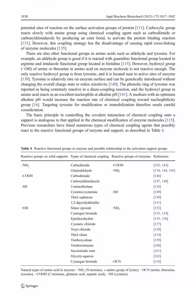
The basic principle in controlling the covalent interaction of chemical coupling onto asupport is analogous to that applied in the chemical modification of enzyme molecules [115].Previous researchers have listed numerous types of chemical coupling agents that possiblyreact to the reactive functional groups of enzyme and support, as described in Table 4.
Table 4 Reactive functional groups in enzyme and possible relationship to the activation support groups
Reactive groups on solid supports Types of chemical coupling Reactive groups of enzymes References
–NH2 Carbodiimide –COOH [142, 143]
Glutaraldehyde −NH2 [134, 144, 145]
–COOH Carbodiimide [146]
Carbonyldiimidazole [147, 148]
–SH 2-iminothiolane [118]
Cysteine/cystamine –SH [149]
Thiol sepharose [150]
2,2-dipyridyldisulfur [151]
−OH Silane epoxide −NH2 [152]
Cyanogen bromide [153, 154]
Epichlorohydrin [155, 156]
Cyanuric chloride [157]
Tosyl chloride [158]
Thiol silane [114]
Triethoxysilane [159]
Trichlorotriazine [160]
Succinimide ester [161]
Glyoxly-agarose [162]
Cyanogen bromide –OCN [118]
Natural types of amino acid in enzyme: −NH2 (N-terminus, ε-amino group of lysine), −OCN (serine, threonine,tyrosine), −COOH (C-terminus, glutamic acid, aspartic acid), −SH (cysteine).
1830 Appl Biochem Biotechnol (2015) 175:1817–1842

Chemical Coupling of Reactive Hydroxyl Group (−OH) on CNF Support
CNF mainly have hydroxyl group (−OH) on the surface of the support. Since the hydroxylgroup is known as a weak electrophilic group, the functionalization step is required. Forexample, cyanogen bromide is often used as a chemical coupling agent for the activation of –OH support group. When the –OH group on CNF support is activated, it is able to interactcovalently to the reactive group from enzyme molecule through N-terminus or amine aminogroup of lysine [141]. In fact, the interaction between enzyme and CNF support throughchemical coupling is potentially susceptible to hydrolytic cleavage, which would potentiallycause enzyme leaching, unless by performing multipoint covalent interaction.
The interaction through chemical coupling agent is extremely popular in covalent immo-bilization especially in the lab scale. Most of the use of chemical coupling agents such ascyanogen bromide, cyanuric chloride, carbodiimide, and others are extremely toxic. Someother factors such as degradation rate and microbial contamination need also to be considered,since the CNF support is based on natural polysaccharides. The mechanism of the hydroxylgroup on the support with coupling agent (interaction via ligand and spacer arm-ligand) andsubsequent enzyme immobilization is shown in Fig. 6.
Potential Applications of Immobilized Enzyme on Nanofibers
Progress in various studies of enzyme immobilization has led to the increase ofpotential applications in the field of enzyme biotechnology. Recently, more attentionhas been paid to the regulation of the microenvironment for the enzyme on a support inorder to obtain significant stabilization of the immobilized enzyme and development ofnew supports [163]. New developments of nanobiocatalysis have revived the enzymeimmobilization field by successfully stabilizing the activities of various enzymes anddemonstrating their potential applications in various fields such as biosensors, medicaland clinical antibiotic production, protein digestion in proteomic analysis, antifouling,food industry, biofuel cells, and bioremediation. Since the CNF support is a natural-based cellulose biomaterial, types of enzyme used to immobilize toward CNF supportmust not include those involved in degradation of cellulose elements, especially“cellulase” enzyme. In addition, CNF possess almost similar properties and benefitswith other nanofibers, as a support for enzyme immobilization application. Table 5shows the summary of the potential applications in covalent immobilization enzymeonto nanofibers support, which also can be applied using CNF support.
Conclusions and Future Perspectives
CNF have a huge potential to be applied in nanobiotechnology especially in enzymeimmobilization applications. Enzyme immobilization onto a natural-based support,especially CNF is has the potential for improving enzymatic performance and produc-tion yield, as well as contributing toward green technology and sustainable sources. TheCNF surface consists mainly of an –OH functional group that can be directly interactedweakly with enzyme, and its binding can be improved by surface modification andinteraction of chemical coupling that forms a strong and stable covalent immobilizationof enzyme. Application of CNF in nanobiocatalyst has also the potential to be appliedin wide applications, including biosensors, medical diagnostics, pharmaceuticals, food,and agriculture industries.
Appl Biochem Biotechnol (2015) 175:1817–1842 1831
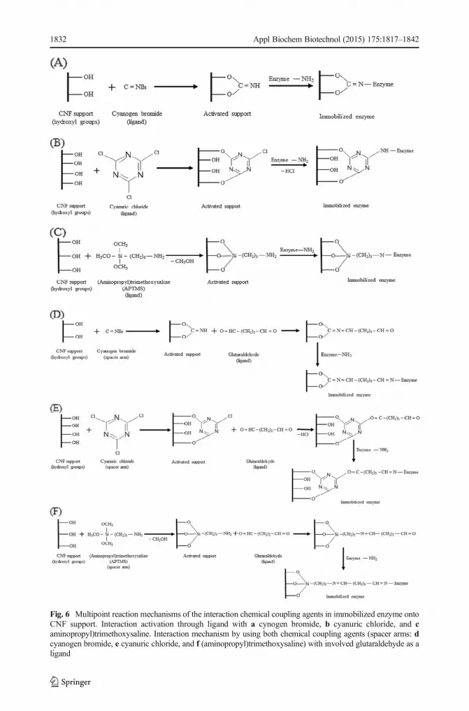
Fig. 6 Multipoint reaction mechanisms of the interaction chemical coupling agents in immobilized enzyme ontoCNF support. Interaction activation through ligand with a cynogen bromide, b cyanuric chloride, and caminopropyl)trimethoxysaline. Interaction mechanism by using both chemical coupling agents (spacer arms: dcyanogen bromide, e cyanuric chloride, and f (aminopropyl)trimethoxysaline) with involved glutaraldehyde as aligand
1832 Appl Biochem Biotechnol (2015) 175:1817–1842

Tab
le5
Potentialapplications
ofim
mobilizedenzymes
onto
nanofiberssupports
Applications
Descriptio
nsReferences
Biosensor
Devicethathascapabilityto
detectbiologicalspeciesselectivity.
[164]
Sensor
thatresponds
tolowconcentrations
ofanalytes
andisableto
discriminateam
ongspeciesaccordingto
theim
mobilizedmolecules
onsurface.
[164]
Itsperformancesandcapabilitiesin
high
specificity
andsensitivity,low
cost,rapid
response,com
pactsize
anduser
friendlin
ess,makebiosensorsan
importanttool
inchem
icalandbiologicaldetection.
[165]
Biosensorsbasedon
immobilizedenzymes
have
good
operationalandstoragestability,h
ighsensitivity,h
ighselectivity,shortresponse
time,andhigh
reproducibility.
[113]
Functions
ofnanofibersforim
mobilizedenzymein
biosensorareto
maintainits
structure,retain
itsbiologicalactiv
ityafterim
mobilizatio
n,andto
remain
tightly
boundto
surface.
[164]
Widelyused
inmajor
applications
inpharmaceutical,m
edicaltreatm
ent,food,and
agricultu
ralindustries.
[166]
Forexam
ple,covalent
interactionof
l-cysteine
(l-cys)onto
modifiedpoly(dially
dimethylammonium)chloride
(PDDA)/gold
nanoparticles(PDDA-A
u)has
been
used
forim
mobilizatio
nof
glucoseoxidase(G
OD),representedas
third-generatio
nof
glucosebiosensor.
[167]
Medicalandclinical
Immobilizedenzymes
have
been
appliedto
specifictargetdrug
delivery,diseasediagnosis,andmolecular
imaging.
[168]
Advantagesin
developm
entin
diagnosis,treatm
ent,andpreventio
nof
diseaseforthefuture
interm
sof
time,accuracy
andreliability,sensitivity,easeto
handlin
g,andlow
costcomparedwith
conventio
naldetectionmethods.
[169]
Major
applicationin
medicaldueto
immobilizedenzymeistheenzymaticelectrode,which
allowsthedetectionforacomplex
samplematrixsuch
asblood,
serum,u
rine,and
food
with
minim
umof
samplepretreatment.
[170,1
71]
Polymerasechainreactio
n(PCR)andsurfaceplasmon
resonance(SPR
)werereported
asrecent
technology
inclinicalapplication,
which
allowsthe
qualitativ
eandquantitativemeasurementof
biom
olecular
interactionwith
outrequired
labelin
gprocedures.
[172]
Piliariketal.introducedan
efficientSP
Rtechniquebasedon
asingle-m
odepolarizatio
n-maintaining
opticalfiberforbiosensors.P
olarization-maintaining
fiberhasbeen
foundto
give
morestableandaccuratemeasurementscomparedwith
micro-prism
fiber-SP
Randpolished-endfiberSP
R[173]
Forexam
ple,im
mobilizing
amonoclonalantim
yoglobin
antib
odytowardchem
icalcouplin
gof
1-ethyl-3-(3-dim
ethylaminopropyl)carbodiim
ide(EDC)/
N-H
ydroxysuccinim
ide(N
HS)
show
edan
effectiveresultatpH
4comparedwith
directim
mobilizatio
ntechnique.
[133]
Antibiotic
productio
nIm
plem
entatio
nsof
biologicalenzymes
areim
portantin
antib
ioticscomparedto
fine
chem
icalprocesses.
[164]
Forexam
ple,im
mobilizedof
penicillinG
acylaseto
cephalexin
byconversion
of7-am
ino-3-deacetoxy-cephalosporanicacid
(7-A
DCA)hadbeen
reported
with
∼85%
conversion
of7-ADCAto
cephalexin
andwith
reusearound
10cycles
underoptim
umconditions.
[174]
Proteindigestionin
proteomicanalysis
Currently
used
becauseof
itsability
toexpoundtheproteinmolecules
andto
regulatepathwaysof
cells
inthedevelopm
entof
noveldrug.
[175]
Allproteins
includingprotease
aredigested
bytrypsinbefore
theiranalysis,and
trypsiniscrucialfortheproteinidentificationin
thesample.
[176]
Optim
izetheproteinidentification,
butalso
canreduce
thetim
eof
proteindigestion
[177]
[178]
Appl Biochem Biotechnol (2015) 175:1817–1842 1833

Tab
le5
(contin
ued)
Applications
Descriptio
nsReferences
Immobilizedtrypsinonto
hydrolysisof
beta-lactoglobulin
(BLG)throughthepresence
ofaspacer
arm
todegradetheallergenicproteinin
bovine
milk
andto
createsafe
milk
products,especially
infood
applications.
Antifoulin
gDefined
asan
adsorptio
nof
proteins
onsurfaces,w
hich
representsasignificantproblem
forabio-device
system
andmedicalpurpose.
[4]
Severaltypesof
antifoulin
gagentssuch
asproteases,subtilisin,
chym
otrypsin,p
ronase,trypsin,p
epsin,
andpapain
wereconsidered.
[179]
Approachin
nanobiocatalystwhich
effectivelyreducestheproteinbindingonto
thesurface,perhapsin
increasing
enzymestability.
[4]
Contributeto
long-lastin
gapplications
which
preventtheattachmentof
barnaclesandmicroorganism
son
thesurfaceof
medicalim
plants,b
iosensors,
andmem
branes.
[4]
Forexam
ple,proteaseshave
been
immobilizedonto
antifoulin
gmoleculestructured
toreduce
theproteinbindingon
thesurfaceandpreventany
contam
ination.
[179]
Food
industry
Immobilizedenzymes
provideagreatvaluein
thefood
processing
especially
inproductio
nof
high-fructosecorn
syrupandtrans-free
oils.
[180]
High-fructose
corn
syrup(H
FCS)
canbe
produced
byusingim
mobilized
D-glucose/xyloseisom
erase(D-xyloseketolisom
erase;EC5.3.1.5).
Productio
nof
non-metabolizablesugars,colored
productsandreducedsw
eetnessoccurbasedon
thereactio
nof
conversion
starch
toHFC
S,allowed
thereactio
nprocessunderam
bienttemperature
andpH
,formed
ahigh
concentrationof
fructose.
[181]
The
reactionof
immobilizedlip
ase(EC3.1.1.3)
couldenzymatically
change
triacylglycerolsin
alow
aqueousenvironm
entwith
soyoilandfree
fatty
acid
(oleicacid),resultedin
theproductio
nof
soyoilwith
ahigher
oleicacid
contentwith
improved
oxidativestability.W
ater
levelisamainfactors
tocontrolthereactio
nprocess.Sy
nthesisof
triacylglycerolswith
new
fatty
acid
profileshappen
afterhydrolysisreactio
nprocessof
fatty
acidsfrom
thetriacylglycerolmolecules
andfinally,o
ilneed
tobe
deodorized
toremovetheresidualfree
fatty
acids.Thisim
mobilizationof
lipaseisvery
convenient
andsimpletechniqueto
produceatrans-free
fatty
acid
comparedto
partialhydrogenationandinter-esterificatio
ntechniques.
[180]
Enzym
aticbio-fuel
cells
Enzym
e-basedbiofuelcells
generatetheconvertedchem
icalenergy
into
electricalenergy,w
hich
hasbeen
used
asapower
source
forlow-pow
ered
sensors,
medicalim
plants,and
communicationdevices.
[182]
Workby
oxidizingtheenzymeproducingelectrons,protons,andotherbyproductsattheanodeelectrodecellviaaconcentrationgradient.
[183]
Nanofibersfabricatetheenzymeelectrodes
thatmeetboth
high
power
density
andlong
lastingin
stability
requirem
ents.
[183]
Improvingenzymegeom
etricalsurfacearea
ofelectrodes
forenzymaticbio-fuelcells
with
increase
oftheenzyme-loadingratein
activ
esites.
[4]
Manoetal.reportedthatim
mobilizedglucoseoxidaseandbilirubin
oxidaseonto
two7μm
electrodecouldoperateto
generateelectricity
with
outusing
proton
exchange
mem
braneas
inaconventio
nalfuelcell.
[184]
Bioremediation
Biodegradationem
ployingnanobiocatalysttechnology
hasprom
ised
greateffectivenessagainstcontam
inated
soilor
water
andalso
toxicpollutants
comparedwith
conventionalmethods.
[164]
Forexam
ple,im
mobilizedperoxidasesfrom
M.charantia
onto
nanostructured
supportshadbeen
foundto
beeffectivein
decolorizing
reactivetextile
dyes
with
50%
inretainingtheactiv
ityof
anenzymeandreusability
upto
morethan
10cycles.
[185]
1834 Appl Biochem Biotechnol (2015) 175:1817–1842

Acknowledgments The authors gratefully acknowledge the financial support by ScienceFund Research Grantfrom MOSTI (grant no. 02-01-04-SF1469) through the Universiti Putra Malaysia.
References
1. Jain, K.K. (2005). The role of nanobiotechnology in drug discovery.DrugDiscovery Today, 10(21), 1435–1442.2. Hubbe, M. A., Rojas, O. J., Lucia, L. A., & Sain, M. (2008). Cellulosic nanocomposites: a review.
BioResources, 3(3), 929–980.3. Srilatha, B. (2011). Nanotechnology in agriculture. Journal of Nanomedicine Nanotechnology, 2(7), 1–5.4. Kim, J., Grate, J. W., & Wang, P. (2008). Nanobiocatalysis and its potential applications. Trends in
Biotechnology, 26(11), 639–646.5. Datta, S., Christena, L. R., & Rajaram, Y. (2013). Enzyme immobilization: an overview on techniques and
support materials. 3. Biotech, 3(1), 1–9.6. Berg, J. M., Tymoczko, J. L., & Stryer, L. (2002). Enzymes accelerate reactions by facilitating the
formation of the transition state. In W. H. Freeman (Ed.), Biochemistry (5th ed., Vol. 8.3). New York.7. Cao, L. (2006). Covalent enzyme immobilization. In Carrier-bound immobilized enzymes (pp. 169–316).
Wiley-VCH Verlag GmbH & Co. KGaA.8. Xie, T., Wang, A., Huang, L., Li, H., Chen, Z., Wang, Q., & Yin, X. (2009). Recent advance in the support
and technology used in enzyme immobilization. African Journal of Biotechnology, 8(19), 4724–4733.9. Wu, D., & Regnier, F. E. (1993). Native protein separations and enzyme microassays by capillary zone and
gel electrophoresis. Analytical Chemistry, 65(15), 2029–2035.10. Mateo, C., Palomo, J. M., Fernandez-Lorente, G., Guisan, J. M., & Fernandez-Lafuente, R. (2007).
Improvement of enzyme activity, stability and selectivity via immobilization techniques. Enzyme andMicrobial Technology, 40(6), 1451–1463.
11. Tischer, W., & Wedekind, F. (1999). Immobilized enzymes: methods and applications. Topics in CurrentChemistry, 200, 95–126.
12. Wang, Z. G.,Wan, L. S., Liu, Z.M., Huang, X. J., &Xu, Z. K. (2009). Enzyme immobilization on electrospunpolymer nanofibers: an overview. Journal of Molecular Catalysis B: Enzymatic, 56(4), 189–195.
13. Lalonde, J., & Margolin, A. (2008). Immobilization of enzymes. In K. Drauz & H. Waldmnn (Eds.),Enzyme catalysis in organic synthesis (2nd ed., pp. 163–184). Weinheim, Germany: Wiley-VCH VerlagGmbH.
14. Andreescu, S., Bucur, B., & Marty, J.-L. (2006). Affinity immobilization of tagged enzymes. In J. M.Guisan (Ed.), Immobilization of enzymes and cells (Vol. 22, pp. 97–106). Humana Press.
15. Fei, G., Ma, G.-H., Wang, P., & Su, Z.-G. (2010). Enzyme immobilization, biocatalyst featured withnanoscale structure. Encyclopedia of industrial biotechnology (pp. 1–26). John Wiley & Sons, Inc.
16. Grunwald, P. (2009). Biocatalysis: Biochemical Fundamentals and Applications (pp. 231–288). ImperialCollege Press.
17. Flickinger, M. C., & Drew, S. W. (1999). Fermentation, biocatalysis and bioseparation. Encyclopedia ofbioprocess technology. New York, USA: John Wiley & Sons.
18. Górecka, E., & Jastrzębska, M. (2011). Immobilization techniques and biopolymer carriers. Food Scienceand Biotechnology, 75(1), 65–86.
19. Betancor, L., Fuentes, M., Dellamora-Ortiz, G., López-Gallego, F., Hidalgo, A., Alonso-Morales, N.,Mateo, C., Guisán, J. M., & Fernández-Lafuente, R. (2005). Dextran aldehyde coating of glucose oxidaseimmobilized on magnetic nanoparticles prevents its inactivation by gas bubbles. Journal of MolecularCatalysis B: Enzymatic, 32(3), 97–101.
20. Kim, J., Grate, J. W., & Wang, P. (2006). Nanostructures for enzyme stabilization. Chemical EngineeringScience, 61(3), 1017–1026.
21. Brena, B., González-Pombo, P., & Batista-Viera, F. (2006). Immobilization of enzymes: a literature survey.In J. M. Guisan (Ed.), Immobilization of enzymes and cells (3rd ed., Vol. 1051, pp. 15–30). Humana Press.
22. Pessela, B. C. C., Dellamora-Ortiz, G., Betancor, L., Fuentes, M., Guisán, J. M., & Fernandez-Lafuente, R.(2007). Modulation of the catalytic properties of multimeric β-galactosidase from E. coli by using differentimmobilization protocols. Enzyme and Microbial Technology, 40(2), 310–315.
23. Wang, P. (2006). Nanoscale biocatalyst systems. Current Opinion in Biotechnology, 17(6), 574–579.24. Wang, P., Dai, S., Waezsada, S. D., Tsao, A. Y., & Davison, B. H. (2001). Enzyme stabilization by covalent
binding in nanoporous sol‐gel glass for nonaqueous biocatalysis. Biotechnology and Bioengineering, 74(3),249–255.
Appl Biochem Biotechnol (2015) 175:1817–1842 1835

25. Kim, B. C., Nair, S., Kim, J., Kwak, J. H., Grate, J. W., Kim, S. H., & Gu, M. B. (2005). Preparation ofbiocatalytic nanofibres with high activity and stability via enzyme aggregate coating on polymer nanofibres.International Journal of Nanotechnology, 16(7), S382.
26. Huang, X.-J., Yu, A.-G., Jiang, J., Pan, C., Qian, J.-W., & Xu, Z.-K. (2009). Surface modification ofnanofibrous poly(acrylonitrile-co-acrylic acid) membrane with biomacromolecules for lipase immobiliza-tion. Journal of Molecular Catalysis B: Enzymatic, 57(1–4), 250–256.
27. Lee, J., Lee, Y., Youn, J. K., Na, H. B., Yu, T., Kim, H., Lee, S.-M., Koo, Y.-M., Kwak, J. H., & Park, H. G.(2008). Simple synthesis of functionalized superparamagnetic magnetite/silica core/shell nanoparticles andtheir application as magnetically separable high-performance biocatalysts. Small, 4(1), 143–152.
28. Verma, M., Barrow, C., & Puri, M. (2013). Nanobiotechnology as a novel paradigm for enzyme immobi-lisation and stabilisation with potential applications in biodiesel production. Applied Microbiology andBiotechnology, 97(1), 23–39.
29. Jia, H., Zhu, G., Vugrinovich, B., Kataphinan, W., Reneker, D. H., & Wang, P. (2002). Enzyme-carryingpolymeric nanofibers prepared via electrospinning for use as unique biocatalysts. Biotechnology Progress,18(5), 1027–1032.
30. Jia, H. (2011). Enzyme-carrying electrospun nanofibers. In P. Wang (Ed.), Methods in Molecular Biology(2011th ed., Vol. 743, pp. 205–212). Humana Press.
31. Gianfreda, L., & Scarfi, M. R. (1991). Enzyme stabilization: state of the art. Molecular and CellularBiochemistry, 100(2), 97–128.
32. Nair, S., Kim, J., Crawford, B., & Kim, S. H. (2007). Improving biocatalytic activity of enzyme-loadednanofibers by dispersing entangled nanofiber structure. Biomacromolecules, 8(4), 1266–1270.
33. Chawla, K. K. (2005). Fibrous materials (p. 312). Cambridge University Press.34. Belgacem, M. N., & Gandini, A. (2008). Surface modification of cellulose fibres. In B. Mohamed (Ed.),
Monomers, polymers and composites from renewable resources (pp. 385–400). Amsterdam: Elsevier.35. Brinchi, L., Cotana, F., Fortunati, E., & Kenny, J. M. (2013). Production of nanocrystalline cellulose from
lignocellulosic biomass: technology and applications. Carbohydrate Polymers, 94(1), 154–169.36. Habibi, Y., Lucia, L. A., & Rojas, O. J. (2010). Cellulose nanocrystals: chemistry, self-assembly, and
applications. Chemical Reviews, 110(6), 3479–3500.37. Siqueira, G., Bras, J., & Dufresne, A. (2010). Cellulosic bionanocomposites: a review of preparation,
properties and applications. Polymers, 2(4), 728–765.38. Samir, M. A. A., Alloin, F., & Dufresne, A. (2005). Review of recent research into cellulosic whiskers, their
properties and their application in nanocomposite field. Biomacromolecules, 6(2), 612–626.39. Cunha, A., & Gandini, A. (2010). Turning polysaccharides into hydrophobic materials: a critical review.
Part 1. Cellulose. Cellulose, 17(5), 875–889. doi:10.1007/s10570-010-9434-6.40. Lavoine, N., Desloges, I., Dufresne, A., & Bras, J. (2012). Microfibrillated cellulose—its barrier properties
and applications in cellulosic materials: a review. Carbohydrate Polymer, 90(2), 735–764.41. John, M. J., & Thomas, S. (2008). Biofibres and biocomposites. Carbohydrate Polymer, 71(3), 343–364.42. Krässig, H. A. (1994). Cellulose: structure, accessibility and reactivity. Journal of Polymer Science Part A:
Polymer Chemistry, 32(12), 367.43. Eichhorn, S. J., Dufresne, A., Aranguren, M., Marcovich, N. E., Capadona, J. R., Rowan, S. J., Weder, C.,
Thielemans, W., Roman, M., & Renneckar, S. (2010). Review: current international research into cellulosenanofibres and nanocomposites. Journal of Materials Science, 45(1), 1–33.
44. Tischer, W., & Kasche, V. (1999). Immobilized enzymes: crystals or carriers? Trends in Biotechnology,17(8), 326–335.
45. Siró, I., & Plackett, D. (2010). Microfibrillated cellulose and new nanocomposite materials: a review.Cellulose, 17(3), 459–494.
46. Karimi, S., Tahir, P. M., Karimi, A., Dufresne, A., & Abdulkhani, A. (2014). Kenaf bast cellulosic fibershierarchy: a comprehensive approach from micro to nano. Carbohydrate Polymer, 101, 878–885.
47. Jonoobi, M., Harun, J., Tahir, P. M., Shakeri, A., SaifulAzry, S., & Makinejad, M. D. (2011).Physicochemical characterization of pulp and nanofibers from kenaf stem. Materials Letters, 65(7),1098–1100.
48. Chen, W., Yu, H., Liu, Y., Hai, Y., Zhang, M., & Chen, P. (2011). Isolation and characterization of cellulosenanofibers from four plant cellulose fibers using a chemical-ultrasonic process. Cellulose, 18(2), 433–442.
49. Shi, J., Shi, S. Q., Barnes, H. M., & Pittman, C. U., Jr. (2011). A chemical process for preparing cellulosicfibers hierarchically from kenaf bast fibers. BioResources, 6(1), 879–890.
50. Chen, W., Yu, H., Liu, Y., Chen, P., Zhang, M., & Hai, Y. (2011). Individualization of cellulose nanofibersfrom wood using high-intensity ultrasonication combined with chemical pretreatments. CarbohydratePolymer, 83(4), 1804–1811.
51. Gupta, M. N., Kaloti, M., Kapoor, M., & Solanki, K. (2011). Nanomaterials as matrices for enzymeimmobilization. Artificial Cells Blood Substitute, 39(2), 98–109.
1836 Appl Biochem Biotechnol (2015) 175:1817–1842

52. Kumar, P., Gupta, A., Dhakate, S. R., Mathur, R. B., Nagar, S., & Gupta, V. K. (2013). Covalentimmobilization of xylanase produced from Bacillus pumilus SV-85S on electrospun polymethyl methac-rylate nanofiber membrane. Biotechnology and Applied Biochemistry, 60(2), 162–9.
53. Yang, D., Paul, B., Xu, W., Yuan, Y., Liu, E., Ke, X., Wellard, R. M., Guo, C., Xu, Y., Sun, Y., & Zhu, H.(2010). Alumina nanofibers grafted with functional groups: a new design in efficient sorbents for removalof toxic contaminants from water. Water Research, 44(3), 741–750.
54. Kim, J., Jia, H., & Wang, P. (2006). Challenges in biocatalysis for enzyme-based biofuel cells.Biotechnology Advances, 24(3), 296–308.
55. Sotowa, K.-I., Takagi, K., & Sugiyama, S. (2008). Fluid flow behavior and the rate of an enzyme reaction indeep microchannel reactor under high-throughput condition.Chemical Engineering Journal, 135(0), 30–36.
56. Song, J., Kahveci, D., Chen, M., Guo, Z., Xie, E., Xu, X., Besenbacher, F., & Dong, M. (2012). Enhancedcatalytic activity of lipase encapsulated in PCL nanofibers. Langmuir, 28(14), 6157–6162.
57. Zhang, S. (2003). Fabrication of novel biomaterials through molecular self-assembly. NatureBiotechnology, 21(10), 1171–1178.
58. Smith, K. H., Tejeda-Montes, E., Poch, M., &Mata, A. (2011). Integrating top-down and self-assembly in thefabrication of peptide and protein-based biomedical materials. Chemical Society Reviews, 40(9), 4563–4577.
59. Bhardwaj, N., & Kundu, S. C. (2010). Electrospinning: a fascinating fiber fabrication technique.Biotechnology Advances, 28(3), 325–347.
60. Laurencin, C., Kumbar, S., Nukavarapu, S., James, R., & Hogan, M. (2008). Recent patents on electrospunbiomedical nanostructures: an overview. Recent Patents on Biotechnology, 1(1), 68–78.
61. Cengiz, F., Krucińska, I., Gliścińska, E., Chrzanowski, M., & Göktepe, F. (2009). Comparative analysis ofvarious electrospinning methods of nanofibre formation. Fibres & Textiles in Eastern Europe, 72(1), 13–19.
62. Aziz, S. H., & Ansell, M. P. (2004). The effect of alkalization and fibre alignment on the mechanical andthermal properties of kenaf and hemp bast fibre composites: part 1—polyester resin matrix. CompositesScience and Technology, 64(9), 1219–1230.
63. Cheng, Q., Wang, S., Rials, T., & Lee, S.-H. (2007). Physical and mechanical properties of polyvinylalcohol and polypropylene composite materials reinforced with fibril aggregates isolated from regeneratedcellulose fibers. Cellulose, 14(6), 593–602.
64. Tischer, P. C. S. F., Sierakowski, M. R., Westfahl, H., & Tischer, C. A. (2010). Nanostructural reorgani-zation of bacterial cellulose by ultrasonic treatment. Biomacromolecules, 11(5), 1217–1224.
65. Jonoobi, M., Harun, J., Tahir, P. M., Zaini, L. H., SaifulAzry, S., & Makinejad, M. D. (2010). Characteristicof nanofibers extracted from kenaf core. BioResources, 5(4), 2556–2566.
66. Jonoobi, M., Harun, J., Shakeri, A., Misra, M., & Oksman, K. (2009). Chemical composition, crystallinity,and thermal degradation of bleached and unbleached kenaf bast (Hibiscus cannabinus) pulp and nanofibers.BioResources, 4(2), 626–639.
67. Isogai, A., Saito, T., & Fukuzumi, H. (2011). TEMPO-oxidized cellulose nanofibers. Nanoscale, 3(1), 71–85.68. Melone, L., Altomare, L., Alfieri, I., Lorenzi, A., De Nardo, L., & Punta, C. (2013). Ceramic aerogels from
TEMPO-oxidized cellulose nanofibre templates: synthesis, characterization, and photocatalytic properties.Journal of Photochemistry and Photobiology A, 261, 53–60.
69. Eichhorn, S. J. (2011). Cellulose nanowhiskers: promising materials for advanced applications. Soft Matter,7(2), 303–315.
70. Henriksson, M., Henriksson, G., Berglund, L. A., & Lindström, T. (2007). An environmentally friendlymethod for enzyme-assisted preparation of microfibrillated cellulose (MFC) nanofibers. European PolymerJournal, 43(8), 3434–3441.
71. Pääkkö, M., Ankerfors, M., Kosonen, H., Nykänen, A., Ahola, S., Österberg, M., Ruokolainen, J., Laine, J.,Larsson, P. T., Ikkala, O., & Lindström, T. (2007). Enzymatic hydrolysis combined with mechanicalshearing and high-pressure homogenization for nanoscale cellulose fibrils and strong gels.Biomacromolecules, 8(6), 1934–1941.
72. Hayashi, N., Kondo, T., & Ishihara, M. (2005). Enzymatically produced nano-ordered short elementscontaining cellulose Iβ crystalline domains. Carbohydrate Polymer, 61(2), 191–197.
73. Shibakami, M., Tsubouchi, G., Nakamura, M., & Hayashi, M. (2013). Polysaccharide nanofiber made fromeuglenoid alga. Carbohydrate Polymer, 93(2), 499–505.
74. Chen, W., Yu, H., Li, Q., Liu, Y., & Li, J. (2011). Ultralight and highly flexible aerogels with long celluloseI nanofibers. Soft Matter, 7(21), 10360–10368.
75. Wang, B., Sain, M., & Oksman, K. (2007). Study of structural morphology of hemp fiber from the micro tothe nanoscale. Applied Composite Materials, 14(2), 89–103.
76. Dufresne, A. (2000). Dynamic mechanical analysis of the interphase in bacterial polyester/cellulosewhiskers natural composites. Composite Interfaces, 7(1), 53–67.
77. Dalmas, F., Chazeau, L., Gauthier, C., Cavaillé, J.-Y., & Dendievel, R. (2006). Large deformationmechanical behavior of flexible nanofiber filled polymer nanocomposites. Polymer, 47(8), 2802–2812.
Appl Biochem Biotechnol (2015) 175:1817–1842 1837

78. Marcovich, N. E., Auad, M. L., Bellesi, N. E., Nutt, S. R., & Aranguren, M. I. (2006). Cellulose micro/nanocrystals reinforced polyurethane. Journal of Materials Research, 21(4), 870–881.
79. Bhatnagar, A., & Sain, M. (2005). Processing of cellulose nanofiber-reinforced composites. Journal ofReinforced Plastics and Composites, 24(12), 1259–1268.
80. Andresen, M., & Stenius, P. (2007). Water-in-oil emulsions stabilized by hydrophobized microfibrillatedcellulose. Journal of Dispersion Science Technology, 28(6), 837–844.
81. Andresen, M., Stenstad, P., Moretro, T., Langsrud, S., Syverud, K., Johansson, L. S., & Stenius, P. (2007).Nonleaching antimicrobial films prepared from surface-modified microfibrillated cellulose.Biomacromolecules, 8(7), 2149–2155.
82. Svagan, A. J., Samir, M. A., & Berglund, L. A. (2007). Biomimetic polysaccharide nanocomposites of highcellulose content and high toughness. Biomacromolecules, 8(8), 2556–2563.
83. Rodriguez, N. L. G., de Thielemans, W., & Dufresne, A. (2006). Sisal cellulose whiskers reinforcedpolyvinyl acetate nanocomposites. Cellulose, 13(3), 261–270.
84. Oksman, K., Mathew, A. P., Bondeson, D., & Kvien, I. (2006). Manufacturing process of cellulosewhiskers/polylactic acid nanocomposites. Composites Science and Technology, 66(15), 2776–2784.
85. Petersson, L., & Oksman, K. (2006). Biopolymer based nanocomposites: comparing layered silicates andmicrocrystalline cellulose as nanoreinforcement. Composites Science and Technology, 66(13), 2187–2196.
86. Kose, R., Mitani, I., Kasai, W., & Kondo, T. (2011). “Nanocellulose” as a single nanofiber prepared frompellicle secreted by gluconacetobacter xylinus using aqueous counter collision. Biomacromolecules, 12(3),716–720.
87. Mateo, C., Palomo, J. M., Fuentes, M., Betancor, L., Grazu, V., López-Gallego, F., Pessela, B. C. C.,Hidalgo, A., Fernández-Lorente, G., Fernández-Lafuente, R., & Guisán, J. M. (2006). Glyoxyl agarose: afully inert and hydrophilic support for immobilization and high stabilization of proteins. Enzyme andMicrobial Technology, 39(2), 274–280.
88. Kosaka, P. M., Kawano, Y., El Seoud, O. A., & Petri, D. F. (2007). Catalytic activity of lipase immobilizedonto ultrathin films of cellulose esters. Langmuir, 23(24), 12167–12173.
89. Lee, K., Ki, C., Baek, D., Kang, G., Ihm, D.-W., & Park, Y. (2005). Application of electrospun silk fibroinnanofibers as an immobilization support of enzyme. Fiber Polymer, 6(3), 181–185.
90. Agarwal, S., Greiner, A., & Wendorff, J. H. (2009). Electrospinning of manmade and biopolymernanofibers—progress in techniques, materials, and applications. Advanced Functional Materials, 19(18),2863–2879.
91. Hu, Z., Foston, M., & Ragauskas, A. J. (2011). Comparative studies on hydrothermal pretreatment andenzymatic saccharification of leaves and internodes of alamo switchgrass. Bioresource Technology,102(14), 7224–7228.
92. Talbert, J. N., & Goddard, J. M. (2012). Enzymes on material surfaces. Colloids and Surfaces, B:Biointerfaces, 93, 8–19.
93. Petkar, M., Lali, A., Caimi, P., & Daminati, M. (2006). Immobilization of lipases for non-aqueoussynthesis. Journal of Molecular Catalysis B: Enzymatic, 39(1–4), 83–90.
94. Shah, S., Solanki, K., & Gupta, M. N. (2007). Enhancement of lipase activity in non-aqueous media uponimmobilization on multi-walled carbon nanotubes. Chemistry Central Journal, 1, 30.
95. You, C.-C., De, M., Han, G., & Rotello, V. M. (2005). Tunable inhibition and denaturation of alpha-chymotrypsin with amino acid-functionalized gold nanoparticles. Journal of the American ChemicalSociety, 127(37), 12873–12881.
96. Rejikumar, S., & Devi, S. (2009). Hydrolysis of lactose and milk whey using a fixed-bed reactor containingβ-galactosidase covalently bound onto chitosan and cross-linked poly(vinyl alcohol). International Journalof Food Science & Technology, 36(1), 91–98.
97. Fang, F., & Szleifer, I. (2002). Effect of molecular structure on the adsorption of protein on surfaces withgrafted polymers. Langmuir, 18(14), 5497–5510.
98. Pessela, B. C. C., Fernandez-Lafuente, R., Fuentes, M., Vián, A., García, J. L., Carrascosa, A. V., Mateo,C., & Guisán, J. M. (2003). Reversible immobilization of a thermophilic β-galactosidase via ionicadsorption on PEI-coated Sepabeads. Enzyme Microbial Technology, 32(3–4), 369–374.
99. Lei, Z., Bi, S., & Yang, H. (2007). Chitosan-tethered the silica particle from a layer-by-layer approach forpectinase immobilization. Food Chemistry, 104(2), 577–584.
100. Mungikar, A. A., & Forciniti, D. (2004). Conformational changes of peptides at solid/liquid interfaces: aMonte Carlo study. Biomacromolecules, 5(6), 2147–59.
101. Goddard, J. M., & Hotchkiss, J. H. (2007). Polymer surface modification for the attachment of bioactivecompounds. Progress in Polymer Science, 32(7), 698–725.
102. Khalil, H. P. S. A., Ismail, H., Rozman, H. D., & Ahmad, M. N. (2001). The effect of acetylation oninterfacial shear strength between plant fibres and various matrices. European Polymer Journal, 37(5),1037–1045.
1838 Appl Biochem Biotechnol (2015) 175:1817–1842

103. Ashori, A., Babaee, M., Jonoobi, M., & Hamzeh, Y. (2014). Solvent-free acetylation of cellulose nanofibersfor improving compatibility and dispersion. Carbohydrate Polymers, 102, 369–75.
104. Tserki, V., Zafeiropoulos, N. E., Simon, F., & Panayiotou, C. (2005). A study of the effect of acetylation andpropionylation surface treatments on natural fibres. Composite Part A-Applied Science, 36(8), 1110–1118.
105. Sgriccia, N., Hawley, M. C., & Misra, M. (2008). Characterization of natural fiber surfaces and natural fibercomposites. Composite Part A-Applied Science, 39(10), 1632–1637.
106. Nacos, M. K., Katapodis, P., Pappas, C., Daferera, D., Tarantilis, P. A., Christakopoulos, P., & Polissiou, M.(2006). Kenaf xylan—a source of biologically active acidic oligosaccharides. Carbohydrate Polymer, 66(1),126–134.
107. Troedec, M. L., Sedan, D., Peyratout, C., Bonnet, J. P., Smith, A., Guinebretiere, R., Gloaguen, V., &Krausz, P. (2008). Influence of various chemical treatments on the composition and structure of hempfibres. Composite Part A-Applied Science, 39(3), 514–522.
108. Adebajo, M. O., Frost, R. L., Kloprogge, J. T., & Kokot, S. (2006). Raman spectroscopic investigation ofacetylation of raw cotton. Spectrochimica Acta Part A, 64(2), 448–453.
109. Rehman, N., Miranda, M., Rosa, S. L., Pimentel, D., Nachtigall, S. B., & Bica, C. D. (2014). Cellulose andnanocellulose from maize straw: an insight on the crystal properties. Journal of Polymers and theEnvironment, 22(2), 252–259.
110. Gemeiner, P., Štefuca, V., & Báleš, V. (1993). Biochemical engineering of biocatalysts immobilized oncellulosic materials. Enzyme and Microbial Technology, 15(7), 551–566.
111. Rusmini, F., Zhong, Z., & Feijen, J. (2007). Protein immobilization strategies for protein biochips.Biomacromolecules, 8(6), 1775–1789.
112. Rao, S. V., Anderson, K. W., & Bachas, L. G. (1998). Oriented immobilization of proteins. MikrochimicaActa, 128(3–4), 127–143.
113. Sassolas, A., Blum, L. J., & Leca-Bouvier, B. D. (2012). Immobilization strategies to develop enzymaticbiosensors. Biotechnology Advances, 30(3), 489–511.
114. Owen, M. J. (2002). Chapter 9—coupling agents: chemical bonding at interfaces. In M. Chaudhury & A. V.Pocius (Eds.), Adhesion science and engineering (pp. 403–431). Amsterdam: Elsevier Science B.V.
115. Redeker, E. S., Ta, D. T., Cortens, D., Billen, B., Guedens, W., & Adriaensens, P. (2013). Proteinengineering for directed immobilization. Bioconjugate Chemistry, 24(11), 1761–1777.
116. Cunha, A. G., Fernandez-Lorente, G., Bevilaqua, J. V., Destain, J., Paiva, L. M., Freire, D. M., Fernandez-Lafuente, R., & Guisan, J. M. (2008). Immobilization of Yarrowia lipolytica lipase—a comparison ofstability of physical adsorption and covalent attachment techniques. Biotechnology and AppliedBiochemistry, 146(1–3), 49–56.
117. Hodneland, C. D., Lee, Y. S., Min, D. H., & Mrksich, M. (2002). Selective immobilization of proteins toself-assembled monolayers presenting active site-directed capture ligands. Proceedings of the NationalAcademy of Science, 99(8), 5048–5052.
118. Huang, Q., Li, D., Kang, A., An, W., Fan, B., Ma, X., Ma, G., Su, Z., & Hu, T. (2013). PEG as a spacer armmarkedly increases the immunogenicity of meningococcal group Y polysaccharide conjugate vaccine.Journal of Controlled Release, 172(1), 382–389.
119. Zhang, J., & Kováč, P. (1999). Studies on vaccines against cholera. Synthesis of neoglycoconjugates fromthe hexasaccharide determinant of Vibrio cholerae O:1, serotype Ogawa, by single-point attachment or byattachment of the hapten in the form of clusters. Carbohydrate Research, 321(3–4), 157–167.
120. Filho, M., Pessela, B. C., Mateo, C., Carrascosa, A. V., Fernandez-Lafuente, R., & Guisán, J. M. (2008).Immobilization–stabilization of an α-galactosidase from Thermus sp. strain T2 by covalent immobilizationon highly activated supports: selection of the optimal immobilization strategy. Enzyme and MicrobialTechnology, 42(3), 265–271.
121. Cowan, D. A., & Fernandez-Lafuente, R. (2011). Enhancing the functional properties of thermophilicenzymes by chemical modification and immobilization. Enzyme andMicrobial Technology, 49(4), 326–346.
122. Rodrigues, R. C., Godoy, C. A., Filice, M., Bolivar, J. M., Palau-Ors, A., Garcia-Vargas, J. M., Romero, O.,Wilson, L., Ayub, M. A. Z., Fernandez-Lafuente, R., & Guisan, J. M. (2009). Reactivation of covalentlyimmobilized lipase from Thermomyces lanuginosus. Process Biochemistry, 44(6), 641–646.
123. Murty, V. R., Bhat, J., & Muniswaran, P. K. A. (2002). Hydrolysis of oils by using immobilized lipaseenzyme: a review. Biotechnology and Bioprocess Engineering, 7(2), 57–66.
124. Elnashar, M. M. M. (2011). The Art of Immobilization using biopolymers, biomaterials andnanobiotechnology. In M. M. M. Elnashar (Ed.), Biotechnology of Biopolymers (p. 1). InTech.
125. Poppe, J. K., Costa, A. P. O., Brasil, M. C., Rodrigues, R. C., & Ayub, M. A. Z. (2013). Multipoint covalentimmobilization of lipases on aldehyde-activated support: Characterization and application intransesterification reaction. Journal of Molecular Catalysis B: Enzymatic, 94, 57–62.
126. Mateo, C., Abian, O., Bernedo, M., Cuenca, E., Fuentes, M., Fernandez-Lorente, G., Palomo, J. M., Grazu,V., Pessela, B. C. C., Giacomini, C., Irazoqui, G., Villarino, A., Ovsejevi, K., Batista-Viera, F., Fernandez-
Appl Biochem Biotechnol (2015) 175:1817–1842 1839

Lafuente, R., & Guisán, J. M. (2005). Some special features of glyoxyl supports to immobilize proteins.Enzyme and Microbial Technology, 37(4), 456–462.
127. Quirk, R. A., Chan, W. C., Davies, M. C., Tendler, S. J., & Shakesheff, K. M. (2001). Poly(L-lysine)-GRGDS as a biomimetic surface modifier for poly(lactic acid). Biomaterials, 22(8), 865–872.
128. Foley, T. L., & Burkart, M. D. (2007). Site-specific protein modification: advances and applications.Current Opinion in Chemical Biology, 11(1), 12–19.
129. Wu, P., Shui, W., Carlson, B. L., Hu, N., Rabuka, D., Lee, J., & Bertozzi, C. R. (2009). Site-specificchemical modification of recombinant proteins produced in mammalian cells by using the geneticallyencoded aldehyde tag. Proceedings of the National Academy of Science, 106(9), 3000–3005.
130. Brady,D.,& Jordaan, J. (2009). Advances in enzyme immobilization.Biotechnology Letters, 31(11), 1639–1650.131. Mateo, C., Grazu, V., Palomo, J. M., Lopez-Gallego, F., Fernandez-Lafuente, R., & Guisan, J. M. (2007).
Immobilization of enzymes on heterofunctional epoxy supports. Nature Protocols, 2(5), 1022–1033.132. Wang, B., Guo, C., Zhang, M., Park, B., & Xu, B. (2012). High-resolution single-molecule recognition
imaging of the molecular details of ricin-aptamer interaction. The Journal of Physical Chemistry. B,116(17), 5316–5322. doi:10.1021/jp301765n.
133. Pei, Z., Anderson, H., Myrskog, A., Duner, G., Ingemarsson, B., & Aastrup, T. (2010). Optimizingimmobilization on two-dimensional carboxyl surface: pH dependence of antibody orientation and antigenbinding capacity. Analytical Biochemistry, 398(2), 161–168.
134. Zhu, J., & Sun, G. (2012). Lipase immobilization on glutaraldehyde-activated nanofibrous membranes forimproved enzyme stabilities and activities. Reactive and Functional Polymers, 72(11), 839–845.
135. Jonkheijm, P., Weinrich, D., Schroder, H., Niemeyer, C. M., & Waldmann, H. (2008). Chemical strategiesfor generating protein biochips. Angewandte Chemie International Edition in English, 47(50), 9618–9647.
136. Hernandez, K., & Fernandez-Lafuente, R. (2011). Control of protein immobilization: coupling immobili-zation and site-directed mutagenesis to improve biocatalyst or biosensor performance. Enzyme andMicrobial Technology, 48(2), 107–122.
137. Sletten, E. M., & Bertozzi, C. R. (2009). Bioorthogonal chemistry: fishing for selectivity in a sea offunctionality. Angewandte Chemie, International Edition, 48(38), 6974–98.
138. Baslé, E., Joubert, N., & Pucheault, M. (2010). Protein chemical modification on endogenous amino acids.Chemistry and Biology, 17(3), 213–227.
139. Gauthier, M. A., & Klok, H.-A. (2008). Peptide/protein-polymer conjugates: synthetic strategies and designconcepts. Chemical Communications (London), 0(23), 2591–611.
140. Joshi, N. S., Whitaker, L. R., & Francis, M. B. (2004). A three-component Mannich-type reaction forselective tyrosine bioconjugation. Journal of the American Chemical Society, 126(49), 15942–15943.
141. Novick, S., & Rozzell, J. D. (2005). Chapter 16—immobilization of enzymes by covalent attachment. In J.Barredo (Ed.), Microbial Enzymes and Biotransformations (Vol. 17, pp. 247–271). Humana Press.
142. Arica, M. Y., Yavuz, H., Patir, S., & Denizli, A. (2000). Immobilization of glucoamylase onto spacer-armattached magnetic poly(methylmethacrylate) microspheres: characterization and application to a continuousflow reactor. Journal of Molecular Catalysis B: Enzymatic, 11(2–3), 127–138.
143. Kang, G., Yu, H., Liu, Z., & Cao, Y. (2011). Surface modification of a commercial thin film compositepolyamide reverse osmosis membrane by carbodiimide-induced grafting with poly(ethylene glycol) deriv-atives. Desalination, 275(1–3), 252–259.
144. Ferreira, L., Ramos, M. A., Dordick, J. S., & Gil, M. H. (2003). Influence of different silica derivatives inthe immobilization and stabilization of a Bacillus licheniformis protease (Subtilisin Carlsberg). Journal ofMolecular Catalysis B: Enzymatic, 21(4–6), 189–199.
145. Migneault, I., Dartiguenave, C., Bertrand, M. J., & Waldron, K. C. (2004). Glutaraldehyde: behavior inaqueous solution, reaction with proteins, and application to enzyme crosslinking. Biotechniques, 37(5),790–806.
146. Delvaux, M., & Demoustier-Champagne, S. (2003). Immobilisation of glucose oxidase within metallicnanotubes arrays for application to enzyme biosensors. Biosensors and Bioelectronics, 18(7), 943–951.
147. Alila, S., Ferraria, A. M., do Rego Botelho, A. M., & Boufi, S. (2009). Controlled surface modification ofcellulose fibers by amino derivatives using N,N′-carbonyldiimidazole as activator. Carbohydrate Polymer,77(3), 553–562.
148. Chen, H., Guo, Z., & Jia, L. (2012). Preparation and surface modification of highly dispersed nano-ZnOwith stearic acid activated by N,N′-carbonyldiimidazole. Materials Letters, 82, 167–170.
149. Nobs, L., Buchegger, F., Gurny, R., & Allémann, E. (2003). Surface modification of poly(lactic acid)nanoparticles by covalent attachment of thiol groups by means of three methods. International Journal ofPharmaceutics, 250(2), 327–337.
150. Hu, W., Tedesco, S., Faedda, R., Petrone, G., Cacciola, S. O., O’Keefe, A., & Sheehan, D. (2010). Covalentselection of the thiol proteome on activated thiol sepharose: a robust tool for redox proteomics. Talanta,80(4), 1569–1575.
1840 Appl Biochem Biotechnol (2015) 175:1817–1842
151. Hermanson, G. T.,Mallia, A. K., & Smith, P. K. (1992). Immobilized affinity ligand techniques. Academic Press.152. Petri, A., Gambicorti, T., & Salvadori, P. (2004). Covalent immobilization of chloroperoxidase on silica gel and
properties of the immobilized biocatalyst. Journal of Molecular Catalysis B: Enzymatic, 27(2–3), 103–106.153. Jurado, L. A., Mosley, J., & Jarrett, H. W. (2002). Cyanogen bromide activation and coupling of ligands to
diol-containing silica for high-performance affinity chromatography: optimization of conditions. Journal ofChromatography. A, 971(1–2), 95–104.
154. Wilchek, M., & Miron, T. (2003). Oriented versus random protein immobilization. Journal of Biochemicaland Biophysical Methods, 55(1), 67–70.
155. Tümtürk, H., Aksoy, S., & Hasırcı, N. (2000). Covalent immobilization of α-amylase onto poly(2-hydroxyethyl methacrylate) and poly(styrene −2-hydroxyethyl methacrylate) microspheres and the effectof Ca2+ ions on the enzyme activity. Food Chemistry, 68(3), 259–266.
156. Santos, J. C., Paula, A. V., Nunes, G. F. M., & de Castro, H. F. (2008). Pseudomonas fluorescens lipaseimmobilization on polysiloxane–polyvinyl alcohol composite chemically modified with epichlorohydrin.Journal of Molecular Catalysis B: Enzymatic, 52–53, 49–57.
157. Schaubroeck, D., De Baets, J., Desmet, T., Dubruel, P., Schacht, E., Van Vaeck, L., & Van Calster, A.(2010). Surface modification of an epoxy resin with polyamines via cyanuric chloride coupling. AppliedSurface Science, 256(21), 6269–6278.
158. Khalafi-Nezhad, A., Parhami, A., Soltani Rad, M. N., & Zarea, A. (2005). Efficient method for the directpreparation of amides from carboxylic acids using tosyl chloride under solvent-free conditions. TetrahedronLetters, 46(40), 6879–6882.
159. Matte, C. R., Nunes, M. R., Benvenutti, E. V., da Schöffer, J. N., Ayub, M. A. Z., & Hertz, P. F. (2012).Characterization of cyclodextrin glycosyltransferase immobilized on silica microspheres viaaminopropyltrimethoxysilane as a “spacer arm”. Journal of Molecular Catalysis B: Enzymatic, 78, 51–56.
160. Moreno, J. M., & Sinisterra, J. V. (1994). Immobilization of lipase from Candida cylindracea on inorganicsupports. Journal of Molecular Catalysis, 93(3), 357–369.
161. Bhatia, S. K., Cooney, M. J., Shriver-Lake, L. C., Fare, T. L., & Ligler, F. S. (1991). Immobilization ofacetylcholinesterase on solid surfaces: chemistry and activity studies. Sensors and Actuators, B, 3(4), 311–317.
162. Megías, C., Pedroche, J., del Mar Yust, M., Alaiz, M., Girón-Calle, J., Millán, F., & Vioque, J. (2006).Immobilization of angiotensin-converting enzyme on glyoxyl-agarose. Journal of Agricultural and FoodChemistry, 54(13), 4641–4645.
163. Singh, R. K., Tiwari, M. K., Singh, R., & Lee, J.-K. (2013). From protein engineering to immobilization:promising strategies for the upgrade of industrial enzymes. International Journal of Molecular Sciences,14(1), 1232–1277.
164. Alzohairy, M. A., & Khan, A. A. (2010). Recent advances and applications of immobilized enzymetechnologies: a review. Research Journal Biological Sciences, 5(8), 565–575.
165. Amine, A., Mohammadi, H., Bourais, I., & Palleschi, G. (2006). Enzyme inhibition-based biosensors forfood safety and environmental monitoring. Biosensors and Bioelectronics, 21(8), 1405–1423.
166. Leung, A., Shankar, P. M., & Mutharasan, R. (2007). A review of fiber-optic biosensors. Sensors andActuators, B, 125(2), 688–703.
167. Wang, Y., Li, Z., Wang, J., Li, J., & Lin, Y. (2011). Graphene and graphene oxide: biofunctionalization andapplications in biotechnology. Trends in Biotechnology, 29(5), 205–12.
168. Fakruddin, M., Hossain, Z., & Afroz, H. (2012). Prospects and applications of nanobiotechnology: amedical perspective. International Journal of Nanotechnology, 10(1), 31.
169. Moghimi, S. M., Hunter, A. C., & Murray, J. C. (2005). Nanomedicine: current status and future prospects.FASEB Journal, 19(3), 311–330.
170. D’Orazio, P. (2003). Biosensors in clinical chemistry. Clinica Chimica Acta, 334(1), 41–69.171. Malhotra, B. D., & Chaubey, A. (2003). Biosensors for clinical diagnostics industry. Sensors and Actuators,
B, 91(1), 117–127.172. Hoa, X. D., Kirk, A. G., & Tabrizian, M. (2007). Towards integrated and sensitive surface plasmon
resonance biosensors: a review of recent progress. Biosensors and Bioelectronics, 23(2), 151–160.173. Piliarik, M., Homola, J., Maníkova, Z., & Čtyroký, J. (2003). Surface plasmon resonance sensor based on a
single-mode polarization-maintaining optical fiber. Sensors and Actuators, B, 90(1–3), 236–242.174. Maladkar, N. K. (1994). Enzymatic production of cephalexin. Enzyme and Microbial Technology, 16(8),
715–718.175. Aebersold, R., & Mann, M. (2003). Mass spectrometry-based proteomics. Nature, 422(6928), 198–207.176. Fan, J., Shui, W., Yang, P., Wang, X., Xu, Y., Wang, H., Chen, X., & Zhao, D. (2005). Mesoporous silica
nanoreactors for highly efficient proteolysis. Chemistry, 11(18), 5391–5396.177. Shui, W., Fan, J., Yang, P., Liu, C., Zhai, J., Lei, J., Yan, Y., Zhao, D., & Chen, X. (2006). Nanopore-based
proteolytic reactor for sensitive and comprehensive proteomic analyses. Analytical Chemistry, 78(14),4811–4819.
Appl Biochem Biotechnol (2015) 175:1817–1842 1841

178. Yamamoto, S., Imamura, A., Susanti, I., Hori, K., Tanji, Y., & Unno, H. (2005). Effect of spacer length onbeta-lactoglobulin hydrolysis by trypsin covalently immobilized on a cellulosic support. Food andBioproducts Processing, 83(1), 61–67.
179. Chen, J., Wang, L., & Zhu, Z. (1992). Preparation of enzyme immobilized membranes and their self-cleaning and anti-fouling abilities in protein separations. Desalination, 86(3), 301–315.
180. Walsh, M. K. (2007). Immobilized enzyme technology for food applications. In R. Rastall (Ed.), Novelenzyme technology for food applications (pp. pp. 60–84). Cambridge: Woodhead Publishing Limited.
181. Kaneko, T., Takahashi, S., & Saito, K. (2000). Characterization of acid-stable glucose isomerase fromStreptomyces sp., and development of single-step processes for high-fructose corn sweetener (HFCS)production. Bioscience, Biotechnology, and Biochemistry, 64(5), 940–947.
182. Willner, I., Yan, Y.-M., Willner, B., & Tel-Vered, R. (2009). Integrated enzyme-based biofuel cells—areview. Fuel Cells, 9(1), 7–24.
183. Ha, S.,Wee, Y., &Kim, J. (2012). Nanobiocatalysis for enzymatic biofuel cells. Topics in Catalysis, 55(16–18),1181–1200.
184. Mano, N., Mao, F., & Heller, A. (2003). Characteristics of a miniature compartment-less glucose-O2 biofuelcell and its operation in a living plant. Journal of the American Chemical Society, 125(21), 6588–94.
185. Akhtar, S., & Husain, Q. (2006). Potential applications of immobilized bitter gourd (Momordica charantia)peroxidase in the removal of phenols from polluted water. Chemosphere, 65(7), 1228–1235.
186. Moskovitz, Y., & Srebnik, S. (2005). Mean-field model of immobilized enzymes embedded in a graftedpolymer layer. Biophysical Journal, 89(1), 22–31.
187. Monsan, P. (1981). Enzymes immobilized on a solid support containing cellulose and lignin. United StatesPatent.
1842 Appl Biochem Biotechnol (2015) 175:1817–1842


