Embed Size (px)
Citation preview

HAL Id: tel-01137883https://tel.archives-ouvertes.fr/tel-01137883
Submitted on 31 Mar 2015
HAL is a multi-disciplinary open accessarchive for the deposit and dissemination of sci-entific research documents, whether they are pub-lished or not. The documents may come fromteaching and research institutions in France orabroad, or from public or private research centers.
L’archive ouverte pluridisciplinaire HAL, estdestinée au dépôt et à la diffusion de documentsscientifiques de niveau recherche, publiés ou non,émanant des établissements d’enseignement et derecherche français ou étrangers, des laboratoirespublics ou privés.
Non-covalent interactions in natural productsImene Bayach
To cite this version:Imene Bayach. Non-covalent interactions in natural products. Biochemistry [q-bio.BM]. Université deLimoges, 2014. English. �NNT : 2014LIMO0050�. �tel-01137883�

2
À la mémoire de mon père Said BAYACH,
À ma chère maman Souad Gouja,
À mes frères, sœurs et beaux-frères

3
“La réflexion est la bougie du cœur. Si elle le quitte, le cœur n'aura plus de lumière."
“Reflection is the lamp of the heart. If it departs, the heart will have no light.”
Abd Allah Ibn Alawi Attas

4
Acknowledgments
La présente thèse a été effectuée dans le laboratoire de Biophysique de la Faculté de Pharmacie de
l’Université de Limoges appartenant au Laboratoire de Chimie des Substances Naturelles (LCSN) affilié
à l’école Doctorale Gay Lussac ED-523 et financée par l’Association Djerbienne en France (ADF). Ce
travail n’aurait pas été possible si je n’avais pas l’aide de plusieurs personnes envers lesquelles je suis
reconnaissante.
Je remercie tout d’abord très sincèrement mon directeur de thèse Dr Patrick Trouillas pour ses
qualités scientifiques et humaines. J’estime avoir de la chance de faire ma thèse sous ta direction. Je te
remercie fortement de m’avoir former à la chimie théorique avec tant de gentillesse, de patience et de
compréhension. Je m’excuse aussi de tous les moments difficiles par lesquels j’ai passé et qui ont
influencé mon attitude et mes comportements. Je te remercie pour les différentes aides pendant ma thèse
et pendant la correction de mon manuscrit ainsi que les efforts extraordinaires que t’as effectué.
Je tiens également à remercier Prof. Jean-Frédéric F. Weber (UiTM Puncak Alam, Malaisie) qui
m’a proposé cette thèse et m’a donné l’opportunité de travailler sur son projet (Oligostilbenoids) dont il
tient à coeur. Je vous remercie profondément de m’avoir choisi pour ce projet parmi vos étudiants, de
m’avoir fais confiance et d’avoir cru en moi. Je vous remercie pour toutes les discussions scientifiques ou
générales et de vos conseils pendant mon séjour en Malaisie. Je vous remercie également pour vos
encouragements, votre soutien, vos critiques toujours constructifs. Malgré les difficultés, le manque
d’échange, l’échec de la cotutelle, j’ai toujours un énorme respect pour vous. J’apprécie toutes les qualités
humaines et scientifiques que vous possédez. Je vous remercie aussi de m’avoir présenter Dr Patrick
Trouillas, c’est grâce à vous que j’ai pu connaitre une personnelle exceptionnelle comme lui. Merci aussi
à Mme Aida pour ces divers aides et sa gentillesse.
Je remercie fortement Prof Juan Carlos Sancho-García (Université d’Alicante, Espagne) pour
avoir accepté d’être rapporteur de cette thèse mais aussi pour m’avoir accepter dans son laboratoire pour
des séjours scientifiques et m’avoir beaucoup aider. Je te remercie beaucoup de ta gentillesse, de tes
conseils et ton aide inconditionné.
Je remercie sincèrement Dr Frédéric Castet (Université de Bordeaux 1) pour avoir accepté d’être
rapporteur de ces travaux. Je vous remercie également de m’avoir accueilli dans votre laboratoire au
cours de ma 2ème année de thèse.
I would like to thank sincerely Dr. Zoran Marković (University of Novi Pazar, Serbie) for having
5
agreed to judge this work. I will be pleased of knowing you.
Mes sincères remerciements vont également à Prof Jean-Luc Duroux (Université de Limoges)
d’avoir accepté de juger ce travail.
Dans notre Labo, je remercie mon collègue Dr Gabin Fabre, pour ses différents aides notamment
“informatiques", pour ses qualités humaines et pour sa bonne humeur. Je te souhaite encore plus de
brillance et beaucoup de succès pour ta dernière année de thèse. Mes encouragements amicaux à Tahani
Ossman pour la suite de sa thèse. Je te remercie sincèrement pour ton engagement dans le scan de ma
thèse lors des dernières corrections et des différents services que tu m’as rendu. Je remercie également
mon ami Michal Biler pour ses qualités humaines et sa bonne ambiance. Je suis très sincèrement ravie de
t’avoir connu et je te souhaite beaucoup de brillance sur le sillon de la réussite. Je remercie également Dr
Claude Calliste, pour ses discussions et sa bonne sympathie pendant les pauses café dont je suis rarement
présente. Je remercie par ailleurs Florent, Thomas, Nicolas et Assia qui ont été présents au début de ma
thèse. Je remercie également tous les membres de l’équipe LCSN, en particulier Amandine Bonet et
Olivier Rezazgui pour leur amitié, Pr Vincent sol, Dr Rachida Zarrouki et Mme Adeline Riguad pour
leur différents aides et surtout leur compréhension et gentillesse.
Je remercie également mon amie Dr Laura Rustituoni pour la bonne ambiance qu’elle a crée au
labo pendant son séjour à Limoges, pour son amitié, son accueil chaleureux à Milano et tous les bons
moments. Je t’ai connu pendant une courte période mais ton ouverture d’esprit, ta sympathie et tes
qualités humaines font de toi une personne originale dont je suis ravie de connaître. Mes salutations vont
à Francisco et Delphy ☺
From Malaysia, I want to thank all my friends from Atta-ur-Rahman Research Institute for Natural
Product Discovery, especially Zuhra and Fatimah whom I consider my sisters. I am really very pleased
to know you. I also thank my friends Dr Rasha, Salwa, Najwa, Dr Mahdi and Dr Anouar.
Par ailleurs, j’exprime ma gratitude envers l’association Djerbienne en France pour le financement
d’une partie importante de ma thèse. Je remercie notamment le comité sociale et en particulier Mr Faouzi
Madani, Mr Hedi Raged et Mme Chedlia Ragued, Mr Jamel Ouersighni, Mr Taoufik Gouja, Mr
Monir Gouja, Mr Mongi Ghandri, Mr Ahmed chououi et Mr Soufien Mestaoui de tous leurs efforts
« bénévoles » et exceptionnels à mon égard. Mers remerciements vont également à tous les autres
comités, les personnels ainsi que tous les membres y compris les étudiants pour l’accueil familial et la
bonne ambiance au sein du siège de l’association.
Je remercie toute ma famille à Paris qui m’a toujours supporté et accueillie avec tant de chaleur

6
familiale. Je cite particulièrement Mme Chedlia Ben Maamar, Mr Aroussi Bayach, Mr Chokri
Bayach et mes cousins et cousines notamment Najet, Sarah et Maher pour leur générosité,
encouragements et soutiens financier et moral. Mes remerciements vont également à mes amies
parisiennes Hena (et sa famille), Raja (son mari et son frère), Jamila (et sa famille), Karamoko
(maintenant Canadien), mon cousin Jamel BAYACH et sa femme Houda et à tous mes amis, cousins et
cousines partout.
Finalement, je dédie cette thèse à ma chère maman Souad Gouja qui compte tout pour moi, rien
n’est plus cher que toi maman, je te remercie profondément de ta confiance, de ta patience, de toutes les
valeurs que tu m’as transmis, de ton amour inconditionné, ton soutien, impossible d’exprimer ma
reconnaissance et ma gratitude envers toi, que Dieu te préserve de tout mal, je m’excuse que je n’ai pas
pu être souvent à tes cotés. À la mémoire de mon père Saïd Bayach (que Dieu lui accorde Sa Grâce et
Sa Miséricorde et l’accueille en son vaste Paradis), papa qui nous a quitté vite sans voir un des rêves de sa
fille se réaliser, papa qui m’a toujours fais confiance et qui a laissé de côté les vielles coutumes de la
région pour que je puisse suivre mes études universitaires dans un pays étranger, cette thèse est à ton
honneur même si tu n’es plus parmi nous. Un grand merci à mes sœurs Olfa, Boutheina et Sondes et
mes frères Radhouen et Hatem qui m’ont toujours fais confiance et soutenus, à mes trois beaux-frères,
mes chers neveux et nièces (Malek, Aziz, Maram, Rayen et Rayssen) et à toute la grande famille
BAYACH de la ville de poterie à l’ile de Djerba.

7
Table of contents
Acknowledgments ............................................................................................................................................... 4
Acronyms ............................................................................................................................................................. 10
Introduction ....................................................................................................................................................... 14
Chapter 1 – Natural products ........................................................................................................................ 17
1. Flavonoids ...................................................................................................................................................... 19 1.1. Structures and diversity ..................................................................................................................................... 19 1.2. Distribution ............................................................................................................................................................ 21 1.3. Biosynthesis ............................................................................................................................................................ 21 1.4. Biological roles and activities ........................................................................................................................... 22 1.4.1. Antioxidant activity ............................................................................................................................................................ 23 1.4.2. Anti−inflammatory activity ............................................................................................................................................. 24 1.4.3. Antimicrobial activity ........................................................................................................................................................ 24 1.4.4. Other biological activities ................................................................................................................................................ 25
2. Stilbenoids ...................................................................................................................................................... 25 2.1. Structures and diversity ..................................................................................................................................... 26 2.1.1. Stilbenes .................................................................................................................................................................................. 26 2.1.2. Oligostilbenes ........................................................................................................................................................................ 27 2.1.3. Bibenzyls ................................................................................................................................................................................. 28 2.1.4. Bisbibenzyls ........................................................................................................................................................................... 28 2.1.5. Phenanthrenoids ................................................................................................................................................................. 29 2.1.6. Other Stilbenoids ................................................................................................................................................................. 29
2.2. Distribution ............................................................................................................................................................ 30 2.3. Biosynthesis ............................................................................................................................................................ 30 2.4. Biological roles and activities ........................................................................................................................... 34 3.4.1. Antioxidant activity ............................................................................................................................................................ 34 3.4.2. Anti−Inflammation and Immunomodulating Activity ......................................................................................... 34 2.4.3. Anti−Microbial activity ..................................................................................................................................................... 35 3.4.4. Anticancer/antitumor activity ....................................................................................................................................... 37 3.4.5. Other biological activities ................................................................................................................................................ 38
Chapter 2 – Non‐covalent interactions ...................................................................................................... 41
1. A general definition of non‐covalent interactions ............................................................................ 41 1.1. vdW Interactions ................................................................................................................................................... 42 1.2. Hydrogen bond ...................................................................................................................................................... 44 1.3. π‐stacking ............................................................................................................................................................... 44
2. Non‐covalent bonding in nature ............................................................................................................. 46 2.1. Proteins .................................................................................................................................................................... 46 2.2. DNA ............................................................................................................................................................................ 48 2.3. Copigmentation ..................................................................................................................................................... 50 2.4. Natural graphite .................................................................................................................................................... 52 2.5. Non‐covalent interactions in synthetic compounds ................................................................................. 53
Chapter 3 – Theoretical methods ................................................................................................................ 56
1. Basic concepts and general view on methods of calculation ........................................................ 56
1.1. The Born−Oppenheimer approximation ...................................................................................................... 56 1.2. The Hartree‐Fock approximation ................................................................................................................... 57 1.2.1. The Slater determinant ..................................................................................................................................................... 57 1.2.2. The Hartree−Fock approximation ............................................................................................................................... 58

8
1.2.3. Restricted and unrestricted Hartree−Fock formalisms ...................................................................................... 59 1.3. Roothaan‐Hall equations .................................................................................................................................... 60 1.4. Basis sets .................................................................................................................................................................. 61 1.4.1. Usual basis sets ..................................................................................................................................................................... 62 1.4.2. Basis Set Superposition Error (BSSE) .................................................................................................................... 63
1.5. Post Hartree‐Fock methods ............................................................................................................................... 64 1.5.1. Coupled−cluster (CC) methods ...................................................................................................................................... 64 1.5.2. Perturbation Theory (PT) ................................................................................................................................................ 65 1.5.3. Møller‐Plesset (MP) methods ........................................................................................................................................ 66 1.5.4. SCS−MP2 methods .............................................................................................................................................................. 67
1.6. Semi‐empirical methods .................................................................................................................................... 68
2. Density functional theory (DFT) ............................................................................................................. 68 2.1. Thomas and Fermi ................................................................................................................................................ 68 2.2. Hohenberg and Kohn theorems ....................................................................................................................... 69 2.3. Kohn−Sham ............................................................................................................................................................. 70 2.3. Exchange−correlation functionals .................................................................................................................. 71 2.3.1. Local density approximation .......................................................................................................................................... 71 2.3.2. Generalized gradient approximation .......................................................................................................................... 72 2.3.3. Hybrid functionals .............................................................................................................................................................. 73
3. Further developments of DFT .................................................................................................................. 73 3.1. Self− interaction error ......................................................................................................................................... 74 3.2. Dispersion−corrected functionals .................................................................................................................. 74 3.2.1. DFT−D ....................................................................................................................................................................................... 74 3.2.2. DFT−D2 .................................................................................................................................................................................... 75 3.2.3. DFT−D3 .................................................................................................................................................................................... 76 3.2.4. DFT−NL .................................................................................................................................................................................... 77
4. Time−Dependent DFT (TD−DFT) ........................................................................................................... 77
Chapter 4 – Methodology ............................................................................................................................... 80
π−Stacked polyphenolic dimers: A case study using dispersion−corrected methods ............. 80
1. Introduction ................................................................................................................................................... 81
2. Theoretical methods ................................................................................................................................... 82 2.1. Modeling dispersion effects .............................................................................................................................. 82 2.2. Technical details ................................................................................................................................................... 84
3. Results and discussion ............................................................................................................................... 86 3.1. Optimized geometry of the complex .............................................................................................................. 86 3.2. Reference data ....................................................................................................................................................... 86 3.3. Assessment of DFT−based dispersion corrections ................................................................................... 88
4. Conclusions .................................................................................................................................................... 92
Chapter 5 – Methodological assessments of dispersion between prototypes of polyphenol
non‐covalent complexes ................................................................................................................................. 95
Oligostilbenoids from the heartwood of N. heimii: role of non−covalent association in their
biogenesis ........................................................................................................................................................... 95
1. Introduction ................................................................................................................................................... 96
2. Results and Discussion ............................................................................................................................... 96
3. Conclusion .................................................................................................................................................... 116
4. Method Section ........................................................................................................................................... 117

9
Chapter 6 – Antioxidants .............................................................................................................................. 122
Section I. Antioxidant properties of phenolic Schiff bases: Structure activity relationship and
mechanism of action ...................................................................................................................................... 123
1. Introduction ................................................................................................................................................. 125
2. Methodology ................................................................................................................................................ 128
3. Results and discussion ............................................................................................................................. 130
5. Conclusion .................................................................................................................................................... 142
Section II. Atomistic description of collaborative antioxidant effects between vitamins E, C
and natural polyphenols in lipid−bilayer membranes ...................................................................... 143
1. Results/Discussion .................................................................................................................................... 144 1.1. Association energies .......................................................................................................................................... 144 1.2. Critical analysis of the association Gibbs energy evaluation ............................................................... 145
2. Methods ......................................................................................................................................................... 146 " Quantum mechanics calculations ................................................................................................................. 146
4. Conclusion .................................................................................................................................................... 147
Chapter 7 – Tuning optical properties of chalcone derivatives ...................................................... 149
1. Introduction ................................................................................................................................................. 149
2. Methodology ................................................................................................................................................ 150
3. π−π non−covalent chalcone dimers ................................................................................................... 152 3.1. 3D arrangements issued from X‐ray crystal structures ........................................................................ 152 3.2. Non−restrained conformational analysis .................................................................................................. 154
4. Optical properties ...................................................................................................................................... 159 4.1. Monomers 1 and 2 .............................................................................................................................................. 159 4.2. Non−covalent complexes ................................................................................................................................. 160
5. Conclusion .................................................................................................................................................... 168
Conclusion ......................................................................................................................................................... 171
Annex .................................................................................................................................................................. 196

10
Acronyms
BDE Bond Dissociation Enthalpy
BSSE Basis Set Superposition Error
CC Coupled Cluster
CC2 Second−order approximate Coupled Cluster singles and doubles
CD Circular Dichroism
COSMO COnductor−like Screening Model
CT Charge Transfer
DFT Density Functional Theory
DFT-D Dispersion−corrected DFT
DNA DesoxyriboNucleic Acid
ES Excited State
ES-CT Excited State Charge Transfer
GEA Gradient Expansion Approximation
GGA Generalized Gradient Approximation
GS Ground State
GTO Gaussian−Type Orbital
HAT H−Atom Transfer
HF Hartree−Fock
HK Hohenberg−Kohn
HOMO Highest Occupied Molecular Orbital
HSV Herpes Simplex Virus
IP Ionization Potential
IR Infrared
KS Kohn−Sham
LCAO Linear Combination of Atomic Orbital
LDA Local Density Approximation

11
LDL Low−Density Lipoprotein
LPS Lipopolysaccharide
LSDA Local Spin Density Approximation
LUMO Lowest Unoccupied Molecular Orbital
MI Molecular Interaction
MO Molecular Orbital
MP Møller−Plesset
MP1 First−Order Møller−Plesset
MP2 Second−Order Møller−Plesset
MP3 Third−Order Møller−Plesset
MP4 Fourth−Order Møller−Plesset
MP5 Fifth−Order Møller−Plesset
NLNnon−Local
NMR Nuclear Magnetic Resonance
PAL Phenylalanine Ammonia Lyase
PCET Proton−Coupled Electron Transfer
PCM Polarizable Continuum Model
PT Perturbation Theory
QM Quantum Mechanics
RHF Restricted Hartree−Fock
ROHF Restricted Open−Shell Hartree−Fock
ROS Reactive−Oxygen−Species
SCF Self Consistent Field
SCS Spin−Component−Scaled
SHS Chalcone Synthase
SOMO Single Occupied Molecular Orbital
SOS Scaled−Opposite-Spin
SPLET Sequential Proton Loss Electron Transfer

12
STO Slater−Type Orbital
STS Stilbene Synthase
TD Time−Dependent
UHF Unrestricted Open−Shell Hartree-Fock
UV/Vis Ultraviolet/Visible
vdW van der Waals
XC Exchange−Correlation

13

14
Introduction
Technological progress makes our lives much easier and more comfortable. Particularly, since the
invention of the computer in the 20th century, the world has been revolutionized opening an unimaginable
number of uses that have globally improved life. Moreover, the revolution of computers has allowed
scientists to develop Quantum Mechanics (QM), a powerful tool that may support experimentalists either
chemists or biologists. This has allowed rationalizing many scientific issues. For example, in the research
on natural products, one of the most difficult and prevalent issues concerns the determination of
stereochemistry in the design of new compounds. In fact, CD (Circular Dichroism) either ECD
(Electronic Circular Dichroism) or VCD (Vibrational Circular Dichroism) is fast becoming a major
method when NOE contacts are unobserved or ambiguous in NMR spectroscopy. However, to determine
absolute configuration, CD requires similar compounds to be used for comparison purpose, while there
are not always available. At this stage, QM may provide fruitful supports. Calculations of chemical shifts
and coupling constants have reached high accuracy, which may also contribute to distinguish different
isomers.
Another area in which QM becomes handy concerns synthetic/biosynthetic mechanisms. QM allows
testing various hypothesized reaction mechanisms. Many other related properties can be accurately
evaluated. Whereas the fields of application of QM are large, this PhD focuses on the role of this tool at
describing non−covalent interactions, a crucial type of interactions that exist everywhere in Nature.
Non-covalent interactions make fascinating molecular architectures and designs with more or less
complex inter− and/or intra−molecular arrangements. These interactions provide “artistic” touch and
original conception for molecules, which may in turns provide different biological and pharmacological
actions.
This thesis focuses on non-covalent interactions between natural polyphenols. These compounds
constitute a well−known family of natural products found in fruit, vegetables, spices and beverages made
from plants like (herbal) teas, wines and fruit juices. They are very trendy for their antioxidant properties
that decrease oxidative stress and consequences such as skin aging.
Chapter 1 describes two main families of polyphenols, namely flavonoids and stilbenoids focusing on
their biosynthetic pathways, their diverse and fascinating chemical structures as well as their
pharmaceutical roles and biological activities. Interestingly, these polyphenols can form non−covalent
complexes with various applications.

15
Chapter 2 rationalizes this type of interactions briefly reminding the different types of non−covalent
interactions in terms of their physical−chemical particularities. The importance of these interactions is
then exemplified by different examples showing their implications and contributions in Nature.
Chapter 3 details basic concepts of quantum chemistry in an attempt to introduce the different
methods of calculations used in the present work.
The theoretical strategies to evaluate non-covalent interactions are still under development. In
Chapter 4, which is a published article, we proposed a new parameterization for the B3P86−NL
functional, in which dispersive interactions are accounted by a non-local approach.
In our work, we provide three new applications of non-covalent interactions in polyphenols. First,
non−covalent complexation between oligostilbenoids, isolated from Neobalanocarpus heimii, a common
tree found in abundance in the rainforests of Malaysia, has been studied. Malaysia offers probably the
greatest plant biodiversity in the world, perhaps higher than the Amazonian rainforest. Chapter 5, a
submitted article, shows how non−covalent association may drive regio− and stereo−selectivity of
oligomerisation reactions. This QM study is aiming at a support to synthesis of new active agents for the
pharmaceutical, cosmetic and food industries.
Second, Chapter 6 shows how non-covalent interactions may influence antioxidant properties i) in
pre-reaction complexes before hydrogen and electron transfers from the polyphenol to the free radical;
and ii) in stable non−covalent antioxidant complexes, which may favor antioxidant regeneration.
Third, Chapter 7 consists at elucidating the role of non−covalent interactions in the modulation of
optoelectronic properties of molecules derived from natural polyphenols. These molecules that efficiently
absorb light can be applied to the bioinspired design of new photovoltaic cells.

16

17
Chapter 1 – Natural products
Natural products are all compounds derived from natural sources including all plant tissues, marine
organisms and microorganisms. Many natural compounds exhibit pharmacological or biological activities
and may also inspire the design of new drugs. Their diverse biological activities and medicinal potentials
have increasingly attracted the attention of scientists over the past decades. Even if chemical and
biological tools have shed light on many biological issues regarding natural compounds, many others are
still open. In particular, biosynthesis of many natural polyphenols (e.g., flavonoids and stilbenoids) is still
not fully elucidated. Flavonoids and stilbenoids are two important groups of natural polyphenols, widely
distributed in the plant kingdom and present in human diet. This chapter presents these two families of
polyphenols, which derive from the same precursors but differ in the further steps of the biosynthesis.
Both stilbene synthase (STS) and chalcone synthase (CHS) are related type III Polyketide synthase
(PKS) enzymes that catalyze the formation of identical linear tetraketide intermediate from a
CoA−tethered phenylpropanoid starter and three molecules of malonyl−CoA. Each enzyme catalyzes the
cyclization of this intermediate but using different cyclization mechanisms to produce different chemical
scaffolds for a variety of plant products (Fig. 1). This is a key step for which the synthetized tetraketide is
further folded differently depending on the enzyme and subjected to aldol or Claisen condensation to
yield either stilbene or chalcone. CHS, ubiquitous in the plant kingdom, catalyzes a C6→C1 Claisen
condensation to form the core chalcone scaffold of all natural flavonoids. STS enables divergence from
CHS, and instead catalyzes a C2→C7 aldol condensation that forms the stilbene backbone of resveratrol
and related antifungal phytoalexins (Fig. 3).

18
Figure 1. Major branch pathways of flavonoid biosynthesis.
The exact mechanisms of action of these enzymes are still unknown but many pathways have been
hypothetized.[1]
NH2HOOC HOOC HOOC
OH
CoASOC
OH
COOH
COSCoA
+ 3
phenylalanine cinnamic acid p-coumaric acid 4-coumaroyl-CoA malonyl-CoA
PAL C4H 4CL
General phenylpropanoid pathway
STSCHS
O
O
HO
OH
O
O
HO
ROH
O
O
HO
ROCH3HO
OHO
O
OCH3
CHI
flavanone
IFS
isoflavone
12'H
IFR
VR
DMID
isoflavonoids
HO
O
OH
OH
chalcone
O
O
HO
OH
OH
O
O
HO
OH
OH
OH
O
O
HO
OH
OH
OH
R
R'
O
O
HO
OH
OH
OH
R
R'
CHI
naringenin
O
O
HO
OH
OH
OH
eriodictyol
O
O
HO
OH
OH
R
R'
F3H
dihydrokaempferol
F3'H
F3'H
F3'5'H
F3H
OMT
UFGT
RT
leucoanthocyanidins
DFR
FLSFLS
flavonols
O
O
Rha-O
OH
Glc-O
O-Glc-O-Rha
R
R'
flavonol glycosides
RTUFGT
stilbene
IFS
FS1
flavones
flavonesFS1
3-OH-anthocyanidins
LDOX LC
R
O
O
HO
OH
OH
OH
R
R'
flavanols
proanthocyanidins
DFR flavan-4-ols
phlobaphenes
anthocyanins
OGlc-O
OH
O-Glc
O-Glc-O-Rha
OCH3
OCH3
OH
OH
HO

19
1. Flavonoids
Flavonoids constitute the largest and the most studied family of polyphenols with more than 8000
different substances found in virtually all plants. They are responsible for many plant colors covering all
visible spectrum. In oriental medicine, plants rich in flavonoids have been used for centuries e.g.,
scultellaria root, cornus fruit, licorice, and green tea are examples of flavonoid containing plant foods
widely used in oriental medicine.
1.1. Structures and diversity
All flavonoids have a common chemical skeleton. They are generally made of two aromatic rings, each
containing at least one hydroxyl, which are connected through a three-carbon "bridge" within a six-
member heterocyclic ring (Fig.2).
Figure 2. Flavonoids basic structure.
Flavonoids are divided into subclasses according to aromaticity of the heterocyclic ring, oxidation
state and functional groups of the heterocyclic ring. The major subgroups are flavones, flavonols,
flavanones, flavanonols, flavanols, chalcones and dihydrochalcones, isoflavones, anthocyanins and
anthocyanidins (Table 1). The individual compounds of each subclass are characterized by specific
hydroxylation and conjugation patterns.
Natural flavones are characterized by the presence of a keto group at C4 and a 2,3−double bond
(Table 1). They include apigenin, luteolin, tangeritin, chrysin, 6−hydroxyflavone, baicalein, scutellarein
and wogonin.* All compounds quoted in the manuscript are referenced in the Annex section.
Flavonols are flavones having a hydroxyl group at C3 (Table 1). They are widely distributed in fruits
as well as vegetables. Their diversity derives from the different positions of the phenolic OH−groups. The
most common are quercetin, kaempferol, rhamnazin, pachypodol and myricetin.
* All compounds quoted in the manuscript are referenced in the Annex section.
1
4
1'
4'
5
O
A B
C
6
7
8
2'
3'
6'
5'2
3

20
Flavanones are characterized by the presence of a keto group at C4 and an asymmetric carbon at C2
(Table 1). They are generally glycosylated by a disaccharide at C7 to give flavanone glycosides. The most
common are naringenin, eriodictyol and hespertin.
Flavanonols, also called dihydroflavonols, exhibit two asymmetric carbons, C2 and C3, with a
hydroxyl group at C3 (Table 1). The most common examples are aromanderin and taxifolin.
Flavanols (not to be confused with flavonols), also called flavan−3−ols, are derivatives of flavans and
are characterized by a 2,3 single bond and the absence of the keto group at C4 (Table 1). Catechin and
epicatechin are the most widely distributed flavanols and they are partly responsible for the beneficial
effects of green tea. This class includes also epicatechin gallate, epigallocatechin, epigallocatechin
gallate, proanthocyanidins, theaflavins and thearubigins.
Chalcones or chalconoids are open−chain flavonoids, in which the two aromatic rings are linked by
an α,β-unsaturated carbonyl system (Table 1). Chalcones therefore exist as two stereoisomers (E and Z)
according to the arrangement of substituents around the central double bond. Benzylideneacetophenone is
the parent member of the chalcone series. The transformation of chalcone to flavanone is possible and
catalysed by the chalcone isomerase.
Isoflavones are widely studied, mainly, for their pseudo-estrogenic properties. They are isomers of
flavones, with an almost identical structure, the only difference being the position of the phenyl group,
which is bonded at C3 instead of C2 for the flavones (Table 1). Almost exclusively, isoflavones are
produced by the members of the Fabaceae family i.e., Leguminosae or bean.
Anthocyanidins and anthocyanins are characterized by the presence of a cationic charge (Table 1).
Anthocyanidins are common plant pigments and typically not found as free aglycones. Most of them are
partly responsible for color variation in fruit and flowers. The five main anthocyanidins are cyanidin,
delphinidin, pelargonidin, malvidin, peonidin and petunidin. Anthocyanins are the glycosides of
anthocyanidins, they are water−soluble vacuolar pigments that may appear red, purple, or blue depending
on pH. They are found in many fruits and vegetables including purple cabbage, beets, blueberries,
cherries, raspberries and purple grapes. They occur in all tissues of higher plants, including leaves, stems,
roots, flowers and fruits.

21
Table 1. Chemical structures of the principal flavonoid subclasses.
Flavanols
Flavones
Chalcones
Flavanones
Isoflavones
Anthocyanins
Flavanonols
Flavonols
Anthocyanidins
1.2. Distribution
Virtually all fruits, vegetables, herbs and spices contain flavonoids. Beverages made from plants e.g.,
wines, tea and fruit juices also contain a wide variety of flavonoids.[2] Berries have high content of
anthocyanins. Black raspberries, for example, may contain up to 100 milligrams of anthocyanins per
ounce.† Green tea has high content of catechins, reaching 1 g per cup. Skin of fruits is known to contain
high concentration of flavonoids. Flavonoids can be colored but also colorless, thus being less noticeable
in food. Orange flavonoids can be found in the white pulpy portion inside the skin.
1.3. Biosynthesis
The discovery of the first enzyme involved in the phenylpropanoid pathway for flavonoid biosynthesis
was achieved in 1961 by Koukol and Conn.[3] Later, it was demonstrated, using radioactively labeled
compounds, that flavonoids were originated from acetate units and a phenylpropanoid intermediate
derived from the shikimic acid pathway.[4] In short, ring A is formed by head−to−tail condensation of
three acetate units and ring B as well as C2, C3 and C4 atoms from a phenylpropanoid precursors
(Fig.1).[5] The primary enzyme specific for the flavonoid pathway is CHS that produces chalcone
scaffolds from which all flavonoids derive (Fig. 3). Although the central pathway for flavonoid
† 1 ounce ≈ 28 g.
1
4
1'
4'
5
O
OH
1
4
1'
4'
O
O
5
O
α
β 1
4
1'
4'
1
4
1'
4'
5
O
O
1
4
1'
4'
5
O
O
1
4
1'
4'
5
O
Ogly
1
4
1'
4'
5
O
O
OH
1
4
1'
4'
O
O
OH5
1
4
1'
4'
5
O

22
biosynthesis is conserved in plants, depending on the species, a group of enzymes, such as isomerases,
reductases, hydroxylases, and several Fe2+/2−oxoglutarate−dependent dioxygenases modify the basic
flavonoid skeleton, leading to the different flavonoid subclasses.[6] Finally, tranferases modify the
flavonoid backbone with sugars, methyl groups and/or acyl moieties, thus modulating the physiological
activity of the resulting flavonoids by altering their solubility, reactivity and interaction with cellular
targets.[7], [8]
Figure 3. Type III Polyketide Synthase Enzymes and Tetraketide Cyclization
1.4. Biological roles and activities
Whereas nearly all organisms possess antioxidant defense and repair systems to protect them against
oxidative damage leading to cancer, aging, atherosclerosis, ischemic injury, inflammation and
neurodegenerative diseases, these systems may fail at preventing all oxidative damages.[9] Food
antioxidant-containing diets may contribute to be used to help the human body to reduce oxidative
stress.[10] Also the use of antioxidant food supplements have been envisaged, but such a use is
controversy, beneficial effects being not systematically proved [11] and negative (pro−oxidant) effects
being suggested in case of mega doses of antioxidants.[12]
Flavonoids have been extensively studied for their biological roles (in plants) and activities (in
mammals). Many analyses have focused on the understanding of their roles in i) plant−microbe
interactions, ii) protection against ultraviolet (UV) light, iii) (red, purple, orange, yellow, blue) plant
pigmentation and iv) implication in male fertility process. These studies show importance of chemical
flavonoid structures to rationalize a wide range of activities, but also highlight the flavonoid pathway as a
paradigm for the study of the evolution of plant metabolism.[13] Consequently, these secondary plant
metabolites have gained much attention, especially in their potential role to explain some of the human
RS
O
O
OH
O
S
CoA
CoA
R
OOOO
S
R
O
OO
OH
CoA
3 X
+
OH
R
HO
HO
O
HO
HO
R
Type III PKSCTAS
C5oxy->C1 lactone
Linear tetraketide intermediate
STS C2->C7 aldol
CHS C6->C1Claisen CO2
Chalcone Stilbene
1 2 5 67
Tetraketide lactone
R = OH or H

23
health benefits associated diets rich in fruit and vegetables including apples, apricots, blueberries, pears,
raspberries, strawberries, black beans, cabbage, onions, parsley, pinto beans and tomatoes.[14]
1.4.1. Antioxidant activity
Most of flavonoids exhibit antioxidant activities.[15] As antioxidants, they can participate in
neutralization of reactive−oxygen−species (ROS) overproduction, subsequently preventing related cell
damages. Flavonoids are powerful in vitro antioxidants, being able to scavenge many free radical species.
While flavonoids may exert their cell structure protection through a variety of mechanisms, one of their
potent effects may be through their ability to increase levels of glutathione, a powerful antioxidant, as
suggested by various research studies. The capacity of flavonoids to act as antioxidants, depends mainly
upon their molecular structures. The position of hydroxyl groups and other features in the chemical
structure of flavonoids plays a key role on their free radical scavenging activities. Quercetin, the most
abundant dietary flavonol found in its glycoside form, is a potent antioxidant because exhibiting most of
structural requirements for an effective free radical scavenging capacity.[15]
The capacity of prenylated flavonoids to inhibit LDL (low-density lipoprotein) oxidation induced by
copper was evaluated as an indicator of antioxidant activity. It was a comparative study of quercetin (a
flavonol), genistein (the major isoflavone in soy), chalconaringenin (a non−prenylated chalcone),
naringenin (a non-prenylated flavanone) and vitamin E (Fig. 4). The prenylchalcones and prenylflavones
are effective in preventing LDL oxidation, the prenylchalcones having generally greater antioxidant
activity than the prenylflavanones. Xanthohumol, the major prenylchalcone in hops and beer, is more
powerful than vitamin E or genistein, whereas xanthohumol was less potent than quercetin. The potency
of xanthohumol as an antioxidant is clearly increased when combined with an equivalent amount of
vitamin E.
The prenyl group plays an important role in the antioxidant activity of certain flavonoids. Indeed, a
flavonoid chalcone (chalconaringenin) and a flavanone (naringenin) without prenyl groups act as
pro−oxidants, i.e. they promote rather than limit LDL oxidation. However, adding a prenyl group to these
flavonoids counteracted their pro−oxidant activities. This observation that prenyl groups are important in
conferring antioxidant activity to certain flavonoids may lead to the discovery or synthesis of novel
prenylated flavonoids as preventive or therapeutic agents against human diseases associated with free
radicals.

24
Figure 4. Examples of antioxidant flavonoids.
1.4.2. Anti− inflammatory activity
The natural response of the human organism to external aggression is continuously regulated to prevent
over−activation of the immune system and unwanted immune responses. Many types of cells involved
with the immune system including T cells, B cells, NK cells, mast cells, and neutrophils have been shown
to alter their behavior in the presence of flavonoids. Many investigations have proved anti−inflammatory
activities of many flavonoids.[16] Flavonols (quercetin, rutin and morin) and flavanones (hesperetin and
hesperidin) were investigated in acute and chronic inflammation animal models.[17] Only flavanones
were effective on neurogenic inflammation induced by xylene. Quercetin represents the most important
compound in reducing paw edema induced by carrageenan.[17] Paradkar et al. demonstrated that diet rich
in the isoflavones daidzin, glycitin, genistein and their glucosides, modulate the inflammatory reaction in
the mouse intestine and liver after LPS (lipopolysaccharide) injection.[18] Genistein (isoflavone)
constitutes one of the most studied among a great variety of natural flavonoids in different models of
inflammation. Its anti-inflammatory effect can be mediated by inhibition of the tyrosine kinase signaling
cascade.[19] Besides, other flavonoids were effective in preventing adjuvant arthritis in the rat. Daily
intraperitoneal administration of rutin, quercetin and hesperidin, inhibited both acute and chronic phases
in this experimental model of inflammation, with rutin being the most active compound in the chronic
phase.[20]
1.4.3. Antimicrobial activity
" Antifungal activity
Flavonoids have been proposed for use against fungal pathogens because of their widespread ability to
inhibit spore germination of plant pathogens. For example, galangin, a flavonol commonly found in
O
OH
OH
HO
O
HO
OH
OCH3
OCH3OH
OHH3CO
O
OH
OH
H3CO
O
OH
OCH3
OCH3
O
O
HO
HO
OHGenistein Isoxanthohumol Xanthohumol Quercetin
Antioxidant
potency

25
propolis samples has been shown to inhibit Aspergillus tamarii, A. flavus, Cladosporium
sphaerospermum, Penicillium digitatum and Penicillium italicum effects.[21]
" Antiviral activity
Several research groups have investigated the relationship between flavonoid structure and inhibitory
activity against HIV−1 and related enzymes.[22]–[25] Flavonoids also exhibit activity against other
viruses. For example, quercetin, morin, rutin, dihydroquercetin, dihydrofisetin, leucocyanidin,
pelargonidin chloride and catechin possess activity against up to seven types of viruses, including herpes
simplex virus (HSV), respiratory syncytial virus, poliovirus and Sindbis virus.[23], [24]
" Antibacterial activity
The antibacterial activity of flavonoids is being increasingly documented. For example, flavonoid−rich
plant extracts from Hypericum, Capsella and Chromolaena have been reported to possess antibacterial
activities.[25], [26] Many other phytochemical preparations with high flavonoid content have also been
reported to exhibit antibacterial activity.[27]–[29]
1.4.4. Other biological activities
Flavonoids have been reported to possess many other useful properties, including oestrogenic activity,
enzyme inhibition, antiallergic activity, vascular activity and cytotoxic antitumor activity.[31]
2. Stilbenoids
This section is more detailed than that on flavonoids because of i) the huge number of articles in
literature dedicated to flavonoids compared to stilbenoids as well as previous theses from our laboratory
and ii) the particular interest of oligostilbenoid molecules in this present thesis.
Stilbenoids constitute a family of polyphenols known for their structural complexity and for their diverse
biological activities. They occur with a limited and heterogeneous distribution in the plant kingdom. The
plant family Vitaceae is one of the richest sources of stilbenes, together with other families, such as
Dipterocarpaceae, Gnetaceae and Fabaceae. Stilbenoids were isolated from the plants for the first time in
1899 [31] but only named as it in 1980 by Gorham.[32]

26
Their chemical diversity and their bioactivities are attracting increasing interest in particular due to
resveratrol (i.e., 3,5,4’−trihydroxystilbene), known for promising biological activities. Currently, the
number of stilbenoids isolated from plants exceeds 1000 molecules against ca. 300 in 1995 and only 100
in 1980. A large number of these compounds have been investigated mainly for their roles in plant
resistance to fungal pathogens but also for their diverse biological activities.
2.1. Structures and diversity
Over the last few years, more than 800 novel stilbenoids have been identified.[33] While the constituent
unit is simple, their structures emphasize chemical diversity through different alternatives and various
oligomeric features. Their structures are made of 1,2−diphenylethylene or 1,2−diphenylethane moieties.
According to their structural characteristics, stilbenoids can be classified into five groups including
stilbenes, oligostilbenes, bibenzyls, bisbibenzyls and phenanthrenes.
2.1.1. Stilbenes
The essential structural stilbene skeleton includes two aromatic rings linked by a methylene bridge. Due
to the double bond (i.e., no free rotation allowed), stilbene may have either trans or cis configuration. The
trans−(E) configuration is often common among naturally occurring stilbenes, but the cis−(Z)
configuration has also been encountered. From this relatively simple structure, hydroxyl groups may be
substituted together with sugar, methyl, methoxy and other moieties. More than 120 stilbenes and their
glycosides have been isolated and classed as spermatophytes‡.[34] Stilbenes represent the most widely
investigated small stilbenoids, due to their bioactivities, especially resveratrol and its analogues.[35]
Table 2. Chemical structures of a few examples for all five subclasses of stilbenoids
A B C D E
Resveratrol Artocarbene Corsifuran C Idenburgene Gnetofuran A
‡ Spermatophytes are plants that produce seeds.
OH
OHHO
OHO
HO
OH
O
H3CO
OCH3
OCH3HO
OCH3O
OH
HO
O
HO
OCH3
HO
OCH3

27
One can classify stilbenes according to their chemical structures into subclasses A−E (Table 2), namely
A) simple stilbenes (e.g., resveratrol) that contain molecules having oxygen functions on the aromatic
rings and including the methylenedioxy derivatives and glycosides; B) prenylated and geranylated
stilbenes (e.g., artocarbene) independently of the substitution position and including cyclized derivatives;
C) aryl benzofuran derivatives (e.g., corsifuran C); D) carbon substituted stilbenes other than
C−glycosides and those of groups B and C (e.g., idenburgene); as well as E) other hybrid stilbenes (e.g.,
gnetofuran A).
2.1.2. Oligostilbenes
Stilbene oligomers or oligostilbenes display high chemical diversities as a result of homogeneous or
heterogeneous coupling between monomeric stilbenes. Coggon et al. reported in 1965 the first naturally
occurring resveratrol tetramer, namely (-)−hopeaphenol.[36] Currently, the octamer vateriaphenol A
isolated from the stem bark of Vateria indica, which belong to the Dipterocarpaceae family, is the most
condensed naturally occurring oligomer. In 1993, Sotheeswaran and Pasupathy proposed to classify
oligostilbenes into two groups, namely Group A that includes at least on five−membered oxygen
heterocycle (usually the trans−2−aryl−2,3−dihydrobenzofuran moiety), and Group B that contains no
oxygen heterocycle.[37] Sotheeswaran et al. suggested that all naturally occurring resveratrol oligomers
from group A were formed from resveratrol through the dimer ε−viniferin (see annex). It is worth noting
that (+)−ε−viniferin occurs only in Vitaceae, whereas (-)−ε−viniferin is found in Dipterocarpaceae,
Cyperaceae, Gnetaceae and Leguminosae.
The Sotheeswaran’s classification is restricted to resveratrol oligomers. Thus, Lin et al. has extended
the definition and has proposed that oligomeric stilbenes may be divided into five major groups according
to constituent units, each group being split into two subgroups according to Sotheeswaran’s scheme.
Recently, many novel oligomers have been discovered, and the newly discovered stilbene oligomers have
been classified according to their degree of polymerization, namely as dimers (e.g., viniferin), trimers
(e.g., caraphenol A), tetramers (e.g., gnemonol B) and higher oligomers (Fig. 5).
Figure 5. Oligostilbene examples.
O
HO
HO
OH
OH
OH
H
H
(+/-)-ε-viniferin
O
O
O OH
HO
HO
OH
HO
OH
H
H
H
H
caraphenol A
O
HO
OH
H
H
HO
HO
O
HO
HO
OH
H
H
H
HO
OH
HO
gnemonol B

28
Structural identifications and characterizations of oligostilbenes by NMR is sometimes a challenging task
due to complex structures, with confusing stereochemistry due to huge possibilities of diastereoisomers,
epimers, enantiomers and conformers (e.g., gneafricanin F, upunaphenols I and J, see annex).[38], [39]
The basic units of stilbene oligomers are resveratrol, isorhapontigenin, piceatannol, oxyresveratrol,
rhapontigenin, gnetol and pterostilbene. The various stilbene oligomers may combine in either homo− or
hetero−polymerization processes, giving rise to huge chemical diversity from only a few monomer units.
2.1.3. Bibenzyls
One of the richest sources of bibenzyls is liverwort (a group of plants belonging to the bryophytes).
According to their structures, bibenzyls are classified into four groups (Table 3) as follow, A) simple
bibenzyls having oxygenated functions on the aromatic rings, including methylenedioxy derivatives and
glycosides (e.g., bulbophyllin); B) prenylated, geranylated and farnesylated bibenzyls (e.g., glepidotin D);
C) 4−hydroxybenzyl substituted bibenzyls (e.g., shanciguol); and D) other bibenzyls (e.g., lespedezol
H).[31]
Table 3. Examples of bibenzyls structures: groups A-D.
A B C D
Bulbophyllin
Glepidotin D
Shanciguol
Lespedezol H
2.1.4. Bisbibenzyls
Bisbibenzyls are usually distributed in liverworts and were rarely discovered in other species. Indeed, the
first occurrence of bisbibenzyls other than from liverworts was perrottetin H that was isolated from a
pteridophyte, Hymenophyllum barbatum. It is a derivative of cyclic bisbibenzyl, which was isolated from
the liverwort Jubula japonica. The occurrence of bisbibenzyls in both pteridophytes and liverworts is
very important to understand evolutionary of terrestrial green plant spores.
Due to their structural diversity and biological activities, bisbibenzyls have attracted much interest
from chemists for synthesis. Gorham has identified nine types of bisbibenzyls based on different modes
of cyclization.[31] Other highlights in bisbibenzyl research must be mentioned including elucidation of
HO
HO
H3COO
O OH
OHO
HO
OH
OH
OH
OH OH
HO
OO
HO
OH

29
absolute configuration of isoplagiochins C and D and structurally related bazzanins, the first linkage
found between a bibenzyl and a phenanthrene.[31]
2.1.5. Phenanthrenoids
Phenanthrenoids are usually abundant in Orchidaceae, Juncaceae, Stemonaceae and liverworts. They may
occur also in Euphorbiaceae, Dioscoreaceae and Ulmaceae. Phenanthrenoids are classified according to
their structure into five subclasses, namely phenanthrenes, 9,10−dihydrophenanthrenes, dimeric
phenanthrenoids, phenanthrene alkaloids and other phenanthrenoids.[31]
Figure 6. Examples of phenanthrene structures.
2.1.6. Other Stilbenoids
In addition to the structures mentioned above, there are other stilbenoids with specific structural features
so do not allow classification into the above five types. They are oligostilbenes related derivatives or
hybrids conjugated with flavonoids and lignans.
Figure 7. Examples of other stilbenoid structures.
One example is laevifonol, an ε−viniferin ascorbic acid hybrid isolated from the heartwood of Shorea
laeviforia, which possesses a five membered lactone ring (Fig. 7). Another example is hemsleyanol E
H3CO
H3CO H3CO
OCH3
H3CO OH OCH3
OH
Callosuminin Crotoflavol
O
OH
H3CO
OCH3
HOCH3
Agrostophylloxidin
O
O
O
O
HO
O
OH
HO
OH
H
H
H
H OH
HO
H
OH
Laevifonol
OOH
OH
HO
HO
H
HO
OH
Hemsleyanol E
OO
OHOH
H
H
H3CO
HO
HO
OH
OHH
H
Gnetoflavanols A
O
OHHO OCH3
OH
OCH3
HO
HOH
H
Cararosin A
O
HH
H
H
OHHO OCH3
OH
OCH3
HO
HO
Maackoline R
30
isolated from the stem bark of Shorea hemsleyana (Dipterocarpaceae). It is the stereoisomer
(+)−parviflorol, which was initially isolated from the bark of Hopea parviflora (Fig. 7). This subclass
contains also flavonostilbenes (e.g., gnetoflavanols, Fig. 7), resulting from coupling between flavanone
and stilbene. Stilbenolignans (e.g., maackoline R (optically inactive) or cararosin A (optically active, Fig.
7) are phenolic compounds formed from stilbenoids (e.g. stilbene) and lignans (e.g. phenylpropanoid).
2.2. Distribution
The interest of stilbenoids is related to the increasing number of new molecules; for instance, between
1995 and 2008, 125 new monomeric stilbenes and 275 new oligomeric stilbenes were reported.
Monomeric and oligomeric stilbenes have mainly been found in the species of twenty−three families e.g.,
Cyperaceae, Dipterocarpaceae, Gnetaceae, Iridaceae, Leguminosae (Fabaceae), Moraceae, Orchidaceae
and Polygonaceae. Stilbene oligomers have been found mainly in Dipterocarpaceae, Gnetaceae, Vitaceae,
Cyperaceae, Leguminosae, Moraceae, Welwitschiaceae, Umbelliferae, Iridaceae, Celastraceae,
Paeoniaceae and Haemodoraceae families. Especially, Dipterocarpaceae, Vitaceae and Gnetaceae
constitute a large number of oligostilbenes. The Dipterocarpaceae was the richest source of new
oligomeric stilbenes.
2.3. Biosynthesis
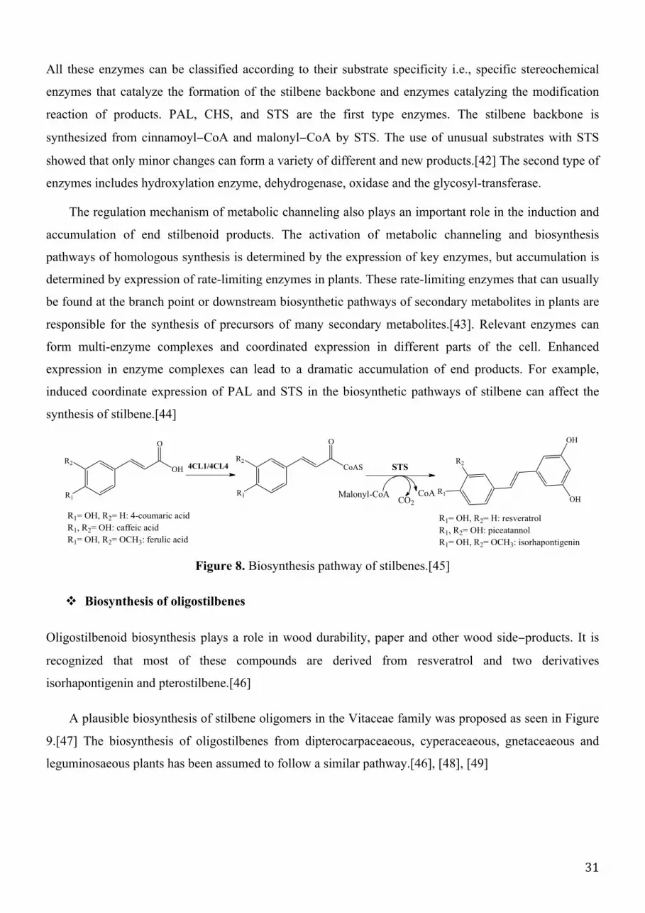
" Biosynthesis of stilbenes
Over the past years, great efforts have been dedicated at rationalization of stilbenoid biosynthesis.
Sotheeswaran studied the biosynthetic pathway of stilbenes in 1993, where ε−viniferin was recognized as
an important intermediate involved in the biosynthesis of stilbenes containing benzofuran moieties.[37]
Indeed, the biosynthesis of simple stilbenes has been well characterized, in which the last step is
catalyzed by STS, a key enzyme in the biosynthetic pathway. STS catalyzes the sequential
decarboxylative addition of three acetate units from malonyl-CoA to a p−coumaroyl−CoA precursor
molecule derived from phenylalanine through the general phenylpropanoid pathway (Fig.8).[40] For
instance, STS condenses three molecules of malonyl−CoA and one molecule of p−coumaryl−CoA to
form resveratrol. It must be reminded that CHS can catalyze the formation of chalcone by
p−coumaroyl−CoA and malonyl−CoA via the intramolecular cyclization and aromatization of the
resulting linear phenylpropanoid tetraketide.[41] Different types of enzymes are involved in the
biosynthetic pathway of stilbenes e.g., phenylalanine ammonia lyase (PAL), 4−coumarate−CoA ligase
(4CL), cinnamate 4−hydroxylase (C4H), pinosylvin methoxy-transferase (PMT) in combination to STS.
31
All these enzymes can be classified according to their substrate specificity i.e., specific stereochemical
enzymes that catalyze the formation of the stilbene backbone and enzymes catalyzing the modification
reaction of products. PAL, CHS, and STS are the first type enzymes. The stilbene backbone is
synthesized from cinnamoyl−CoA and malonyl−CoA by STS. The use of unusual substrates with STS
showed that only minor changes can form a variety of different and new products.[42] The second type of
enzymes includes hydroxylation enzyme, dehydrogenase, oxidase and the glycosyl-transferase.
The regulation mechanism of metabolic channeling also plays an important role in the induction and
accumulation of end stilbenoid products. The activation of metabolic channeling and biosynthesis
pathways of homologous synthesis is determined by the expression of key enzymes, but accumulation is
determined by expression of rate-limiting enzymes in plants. These rate-limiting enzymes that can usually
be found at the branch point or downstream biosynthetic pathways of secondary metabolites in plants are
responsible for the synthesis of precursors of many secondary metabolites.[43]. Relevant enzymes can
form multi-enzyme complexes and coordinated expression in different parts of the cell. Enhanced
expression in enzyme complexes can lead to a dramatic accumulation of end products. For example,
induced coordinate expression of PAL and STS in the biosynthetic pathways of stilbene can affect the
synthesis of stilbene.[44]
Figure 8. Biosynthesis pathway of stilbenes.[45]
" Biosynthesis of oligostilbenes
Oligostilbenoid biosynthesis plays a role in wood durability, paper and other wood side−products. It is
recognized that most of these compounds are derived from resveratrol and two derivatives
isorhapontigenin and pterostilbene.[46]
A plausible biosynthesis of stilbene oligomers in the Vitaceae family was proposed as seen in Figure
9.[47] The biosynthesis of oligostilbenes from dipterocarpaceaeous, cyperaceaeous, gnetaceaeous and
leguminosaeous plants has been assumed to follow a similar pathway.[46], [48], [49]
O
OH
R1= OH, R2= H: 4-coumaric acid
R1, R2= OH: caffeic acid
R1= OH, R2= OCH3: ferulic acid
4CL1/4CL4
R1
O
CoAS STS
OH
OHR1
R1= OH, R2= H: resveratrol
R1, R2= OH: piceatannol
R1= OH, R2= OCH3: isorhapontigenin
Malonyl-CoAR1
R2R2
CO2
CoA
R2

32
Figure 9. Plausible biosynthesis of stilbene dimers.
Figure 10. Plausible biosynthesis of examples of stilbene tetramers.
O
HO
HO
OH
O
OH
OH
HO
OHHO
HO
HO
HO OH
OH
OH
OH
OH
OHHO
HO
HO
OH
OH
a
b
c
H
H
b
bb
c
ac
b
c
c
a
(+)-ampelopsin B
(-)-ampelopsin D
(+)-ampelopsin F
O
HO
HO
OH
OH
OH
O
HO
HO
OH
OH O
HO
HO
OH
OHox
(+)-ε-viniferin [A] [B]
(+)-vitisin A
(+)-vitisin D
O OOH
O
HO
HO
OH
HO
O
HO
HO
OH
OH
O
O
O
HO
HO
OH
OH O
O
HO
OH
OH
OH
O
O
[A]+[B][B]+[B]
OH
O
HO
OH
OH
OH
O
OH
O
HO OH
HO
O
HO
HO
OH
OH
O
O
HO
OH
OH
HO
OH
(-)-vitisin B
O
OH
O
O
O
OH
OH
HO
HO
HO
OH
HO
O
OH
O
O
O
HO
HO
HO
HO
HO
OH
OH
(+)- and (-)-hopeaphenol (+)-viniferol A

33
The attempts to rationalize oligostilbene biomimetic synthesis have not provided convincing mechanisms
to explain reaction selectivity and the apparent discrepancies between all results.[50]–[54] Snyder et al.
elegantly described stereo-control of resveratrol oligomer synthesis by the introduction of a novel reagent
of bromination.[55] Moreover, Velu et al. explained the formation of different oligostilbenoids and
argued on their regio− and stereo−selective biosynthesis.[56], [57] It appears that the biosynthesis of most
components of dipterocarpaceae is driven by non−covalent interactions (see chapter 5).[58]
" Biosynthesis of bisbibenzyls
The biosynthesis of marchantin A was investigated using thallus tissue of Marchantia polymorpha.
Experiments demonstrated that rings A and C of marchantin were derived from the benzene ring of
L−phenylalanine through cinnamic acid (clinnamate) and p−coumaric acid. Dihydro−p−coumaric acid is
an intermediate of the marchantin biosynthesis. The phenylpropane or polymalonate pathway using
dihydro-p-coumaric acid and acetate or malonate was proposed to understand biosynthesis of bibenzyl
monomers, which have been confirmed as the constituent elements leading to marchantin. Therefore, the
bibenzyls are coupled in a unique manner.[59] The cell suspension cultures of Marchantia polymorpha
analysis revealed the presence of two specific cytochromes P450 enzymes having different roles. The first
catalyzes coupling of two lunularic acid molecules to form marchantin C together with CO2 release. The
second hydroxylates marchantin C to marchantin A (Fig. 11). The polyketide synthase (PKS) is a key
enzyme that uses dihydro-4-coumaroyl-CoA as starter and performs three condensation reactions marked
by colors. This is followed by a STS−type ring-folding with retention of the terminal carboxyl group that
is removed in the standard STS-type reaction. The first identified product in vivo is prelunularic acid,[60]
for which the biosynthesis requires an additional reduction step through a polyketide reductase (PKR) i.e.,
it reduces carbonyl group to hydroxyl group.[61]
Figure 11. Biosynthesis pathway of two simple bisbibenzyls.
CoA-S
O
OH
OH
O O
HO
OH
OH
O O
OH
OH
OH
OH
OH
OH
HO
OH
O
OH
OOH
OH
O
OH
OOH
dihydro-4-coumaroyl-CoA
PKSPKR
prelunularic acid lunularic acid lunularin
2 X lunularin marchantin C marchantin A
P450 P450
CO2
aromatization

34
2.4. Biological roles and activities
Due to their structural diversity, stilbenoids exhibit a multiple of biological roles and activities that are
listed in this section, as exhaustively as possible.
3.4.1. Antioxidant activity
Most of stilbenoids possess antioxidant activities thanks to the phenolic groups. Resveratrol is a major
stilbenoid antioxidant, with adequate structural feature for efficiency. Resveratrol analogs have widely
been studied for their antioxidant properties, and some were found more active than resveratrol. Several
resveratrol derivatives from Yucca periculosa have showed inhibitory activity on crocin bleaching
induced by alkoxyl radicals and DPPH scavenging activities. Many natural stilbene glycosides have also
significant antioxidant activities e.g., (Z)-astringin and (E)- and (Z)- resveratrol-4'-O-β-D-
glucopyranoside, 2,3,4',5-tetrahydroxystilbene-2-O-β-D-glucopyranoside. [62]
Different oligostilbenes have exhibited antioxidant activities, namely gneafricanins A, longusol A
and bisisorhapontigenin B. Bisisorhapontigenins E−G and 13-b-methoxy bisisorhapontigenin G have also
highlighted potent antioxidant activity comparable to vitamin E.[31]
Other stilbenoids for which activities were much stronger than vitamin E, such as the
flavonostilbenes gnetoflavanols A, E and F, have showed inhibition of lipid peroxidation and superoxide
produced within a xanthine−xanthine oxidase system.
Tyrosinase (a copper−containing enzyme) inhibitors are antioxidants; they are widely used in
dermatological treatments and also applied in cosmetics (e.g., as skin whitening agent). Some stilbenoids
have been reported to possess tyrosinase inhibition activity e.g., the oxyresveratrol, exerting inhibition on
murine tyrosinase. Resveratrol oligomers from dipterocarpaceae plants have also been reported to have
inhibitory effects on murine tyrosinase activity. The double bond in the stilbene skeleton was critical for
inhibition.[63]
3.4.2. Anti−Inflammation and Immunomodulating Activity
Different stilbenes e.g., resveratrol and piceatannol as well as their derivatives have been regarded as
potent anti−inflammatory agents and still attract attention.[64]–[68] Stilbene derivatives e.g., from
Artoccarpus dadah showed inhibitory effects against COX-1 and COX-2.[69]
(+)-α-Viniferin from the root of Carex humilis is an inhibitor of prostaglandin H2 synthase 3-4-fold,
35
stronger than resveratrol. Moreover, it has displayed a significant anti−inflammatory activity on
carrageenin−induced paw edema in mice due to inhibitory effect on the release of prostanoids and NO.
[70], [71] Viniferin and miyabenol C exhibited protein kinase C (PKC) inhibitory activity at low
micromolar concentrations.
There are many bibenzyls and bisbibenzyls that have exhibited anti−inflammatory activities.
Bibenzyls from the tubers of Gymnadenia conopsea, (including the structure gymconopin D) were found
to inhibit the antigen-induced degranulation in RBL-2H3 cells.[72] Some bisbibenzyls have exhibited
anti−inflammatory activities through inhibition of LPS-induced nitric oxide synthase (NOS) e.g.,
marchantin A involved in the inhibition of LPS-induced Inducible nitric oxide synthase (iNOS)
Messenger Ribonucleic acid (mRNA) expression, is considered as the most potent. The presence of
phenolic hydroxyls and saturation at C−7, C−8 and/or C−7'/C−8' are required for an efficient inhibition of
NO production.[73]
The phenanthrenoidsgymconopin B and blestriarene A isolated from Gymnadenia conopsea were
found to inhibit the antigen−induced degranulation in RBL-2H3 cells. Otherwise, stemanthrene A and D
showed strong inhibition of leukotriene biosynthesis (IC50=8.5 and 4.8µM, respectively). Another
example is dendrochrysanene from the stems of Dendrobium chrysanthum, which was proved to suppress
mRNA level of TNF−α, IL8, ILI0, and iNOS in murine peritoneal macrophages stimulated by LPS.[74]
Other stilbenoids, e.g., artocapol C, have inhibited the release of histamine and β−glucuronidase from
mast cell degranulation in concentration-dependent way. It has also displayed inhibitory effect on the
formyl−peptide−simulated superoxide anion formation in neutrophils.[75] Racemosol and
10−O−demethylracemosol as well as their synthetic derivatives have exhibited potent in vitro inhibitor
activities against COX-l and COX-2.
2.4.3. Anti−Microbial activity
As other stilbenoids, resveratrol has antifungal and antibacterial effects, as well as antiviral effects against
HIV−1 (human immunodeficiency virus), HSV−1 (herpes simplex virus) and human
cytomegalovirus.[66], [76], [77]
" Phytoalexin function
Many stilbenoids behave as phytoalexins that are antimicrobial substances synthesized by plants, which
accumulate rapidly in microorganisms.[40] Their actions derive from multiple defense reactions. For

36
example, it was shown that transfer of two genes from grapevine (VST1 and VST2) to tobacco increase
resistance of the latter to various diseases including infection by Botrytis cinerea.[78] Thereafter, a series
of gene transfers were conducted in many plants such as rice, tomato, wheat, papaya.[79], [80]
" Antifungal
Many stilbenoids act as antifungal i.e., they selectively eliminate fungal pathogens from a host with
minimal toxicity. Stemofurans displayed antifungal activity against four microfungi, namely Alternaria
citri, Fusarium avenaceum, Pyricularia grisea and Botrytis cinerea. Stemofuran B showed the highest
activity against the four pathogenic fungi (EC50=1.4 µg/ml against Pyricularia grisea), but only weak
effects against C. herbarum. Stemofuran E and stilbostemin B showed high activities, while
stemanthrenes showed negligible inhibition.[81]
The 3−hydroxy−4'-methoxybibenzyl isolated from the liverwort Frullania muscicola Steph showed
potent inhibitory effects against certain fungi, especially Trichophyton rubrum and Trichophyton
gypseum. The 3,4−dimethoxy−4−hydroxybibenzyl from the same plant exhibited moderate inhibition.[82]
Bazzanin B, isoplagiochin D and bazzanin S are three bisbibenzyls from the liverwort Bazzania
trilobata showing antifungal activity against a variety of phytopathogenic fungi, in particular Pyricularia
oryzae. The antifungal activity against Trichophyton mentagrophytes was also reported for marchantin C,
neomarchantins A. Plagiochin E, neomarchantin A and 13,13'−O−isoproylidenericcardin D against
Candida albicans. [82], [83]
Stilbene tetramers may prove to be valuable agents against, rice-blast disease. For example
carasiphenol D inhibited the mycelial growth of Pyricularia oryzae by curling and swelling effect.[84]
On the other hand, dimeric stilbenes (trans and cis ε−viniferins) have showed higher antifungal activity
than resveratrol. There are many other examples reported antifungal activities of resveratrol dimers, such
as cyphostemmins A and B.[85]
" Antibacterial
Many stilbenoids are reported as antibacterials. For example, combretastatin B5 from Combretum woodii
leaf powder displayed significant activity on S. aureus but with lower activity toward P. aeruginosa and
E. faecalis and slight activity towards E. coli. Stilbenes isolated from Rumex hymenosepalus, Mexican
medicinal plants, showed moderate antimycobacterial activities.[86] The prenylated stilbene isolated from
the aerial parts of Artocarpus integer showed antibacterial activity against Bacillus subtilis. Lakoochins A
and B demonstrated antimycobacterial activity. The bisbibenzyls marchantin C, neomarchantin A and B

37
showed moderate activity against Bacillus subtilis. Gnetin E, parthenocissin A, ε−viniferin, α−viniferin,
hemsleyanols B and D, vaticanol B and davidiol A inhibited the growth of MRSA (methicillin-resistant
Staphylococcus aureus, a type of staph bacteria). Hemsleyanol D was found to be the most effective
compound i.e., Minimal Inhibitory Concentration (MIC) of 2µg/ml vs. 1 µg/ml of vancomicin.[87]
Hopeaphenol A and vaticaphenol A displayed moderate inhibitory activity against MRSA and
methicillin-resistant Mycobacterium smegmatis.[88]
The antibacterial activity pattern of the two photosensitizers, dehydroeffusol and juncusol against
methicillin-resistant and sensitive S. aureus and B. subtilis was similar to that of their antifungal
activity.[89]
" Antivirus
The main anti−virus activity researches concerning stilbenoids focuse on HIV. For instance, glepidotin D,
which is isolated from the leaves of Glycyrrhiza lepidota, is considered as the compound responsible for
the antiviral activity.[90] Dimers scirpusin A and B (oligostilbenes) were isolated from Caragana rosea
with EC50 values of 10 and 7 µg/ml against HIV-1 III-B, respectively.[91] The dimer balanocarpol and
the tetramer dibalanocarpol from the leaves of Hopea malibato exhibited very modest HIV-inhibitory
activity (EC50 of 46 and 20 µg/ml, respectively). However, malibatols A and B from the same plant were
cytotoxic to the host cells. Riccardin C and F were weakly active against HIV-1 reverse transcriptase.[92]
3.4.4. Anticancer§/antitumor
** activity
To find new chemopreventive agents from stilbenoids has attracted much attention. The different groups
of stilbenes are reported to be potentially important cancer chemoprotective agents, being able to inhibit
cellular events associated with carcinogenesis, including tumor initiation, promotion, and progression.
Stilbenes, especially resveratrol and its analogs play a role in the prevention of carcinogenesis. Their
effects on human cancer cell lines (and on animal models) have been carried out extensively.[93]–[95]
Combretastatin A−4 is seen as a promising tubulin inhibitor targeting tumor vasculature to induce rapid
and selective vascular dysfunction in tumors. The combretastatin A−4 phosphate (fosbretabulin), a
prodrug of combretastatin A-4, is now in phase II of clinical trials.[96], [97] Since Jang et al. published
the anticancer effects of resveratrol, there have been a few studies on their potential therapeutic roles in
the prevention cancer, as well as of the corresponding oligomers.[98], [99] Anticancer effects of
stilbenoids are cytotoxicity, proliferation inhibition and apoptosis of tumor cells.
§ anticancer is an activity restricted to clinical effect on humans ** antitumor would also refer to biological effect on animal models, such as xenografts

38
Many oligostilbenes have been tested for their antitumor effects. For example, vaticanol C
(resveratrol tetramer) have induced cytotoxicity in different cell lines and have exhibited growth
suppression in colon cancer cell lines at low concentration via apoptosis induction.[100], [101] A
noticeable progress of oligostilbene anticancer activities comes from the discovery of their inhibitory
activity of DNA Topoisomerase II (topoisomerase II inhibitors are used as anticancer drugs). For
instance, nepalensinols isolated from the stem of Kobresia nepalensis (Cyperaceae) have presented potent
inhibitory effect on topoisomerase II; some even stronger than etoposide or daunorubicin. Particularly,
nepalensinol B has exhibited the most potent activity (~1000 times stronger than etoposide).[102], [103]
Different bibenzyls (e.g., 3,3'-dihydroxy-2',6'-bis(4−hydroxybenzyl)-5−methoxybibenzyl and
3',5−dihydroxy−2-(4−hydroxybenzyl)−3−methoxybibenzyl from Bletilla striata) and bisbibenzyls (e.g.,
neomarchantins A and B and marchantin C) have also showed anticancer activity as well as some
phenanthrenoids (e.g., 4,7-dihydroxy-2-methoxy-9,10-dihydrophenanthrene and 2-methoxy-3,4,7-
trihydroxy−phenanthrene).[104], [105] Many other stilbenoids have exhibited antitumor activity, for
example, racemosol and demethylracemosol exerted cytotoxicity against KB and breast cancer cell
lines.[106]
3.4.5. Other biological activities
Stilbenoids have different other activities including phytotoxicity;[107], [108] ecdysteroid antagonistic
activity;[109]–[111] 5−α−Reductase inhibitory activity; [112], [113] estrogenic/antiestrogenic activities;
[65], [114], [115] neuroprotective activity; [116], [117] antiplatelet activity;[118], [119] antidiabetic
activity;[120]–[122] hepatoprotective and hepatotoxic activities; [123] spasmolytic activity;[124]–[126]
aromatase inhibitory activity (e.g., ε−viniferin); human cytochrome P450 enzyme inhibitory activity (e.g.,
bibenzyls moscatilin and gigantol from Dendrobium nobile, trans− and cis−ε−viniferin, gnetin H,
suffruticosols A and B); antimutagenic activity; and stimulation of osteopath growth activity (e.g.,
karakinos A−C, leachianol C and cararosinol A from the roots of Caragana sinica).[127]–[129]
In conclusion, flavonoids and stilbenoids have attracted increasing interest due to their shown or
assumed beneficial health effects. There is no doubt concerning their importance, however there are a lot
of confusions and misunderstanding in the related literature and in their use for cosmetic, food and
pharmaceutical applications. In vivo, food intake is not systematically well documented e.g., quid
inapetant effects. Also, many in vitro studies lead authors to overestimate the real effects on human, as for
anticancer or antitumor activities. Because the related epidemiological studies are delicate to conduct (too
many crossed parameters and too low population), there is no clear evidence of beneficial effects of
polyphenol−rich foods. In particular in vivo antioxidant activities from polyphenol−rich diets is still under

39
debate.[130] Thereby, care must be systematically given before claiming polyphenols actions. Nowadays,
virtually no polyphenol−derived drugs are clinically available.

40

41
Chapter 2 – Non-covalent interactions
Nature designs fantastic supramolecular architectures and provides fascinating molecules that are
stabilized by mixture of covalent and non−covalent interactions. It is necessary to well understand and
rationalize the role of these interactions; here we are mainly interested in non-covalent interactions.
To better define what are non−covalent molecular assemblies and their importance in modern
molecular sciences, this chapter provides i) physical-chemical definition of the different chemical
bonding and ii) a series of examples existing in Nature.
1. A general definition of non-covalent interactions
In covalent bonds, atoms share their electrons that belong to common molecular orbitals. The optimum
intermolecular distance is usually shorter than 2 Å. Also the angles formed between covalently bonded
atoms are specific and characteristic, providing molecules definite and predicable shapes. Non−covalent
bonds do not strictly share electrons from the two atoms involved in the bond. This implies more
conformational variability inherent to dispersive and electromagnetic interactions between atoms or
molecules non-covalently linked. Understanding the nature of non−covalent interactions is more delicate
and theoretical calculations have been repeatedly in conflict with experimental data. The basic principles
of non-covalent interactions were formulated by Pauling in the 1930’s.[131], [132] However, many
developments have been required and still nowadays the theoretical evaluation must be carefully
conducted to agree with experiments.
An important feature of non−covalent association is the contribution not only of classical Coulomb
electrostatic interaction but also of multipole−electric interactions. Non−covalent bonds are often called
‘weak bonds’, which can be inadequate as these bonds can maintain supramolecular assemblies.
Anyway, non−covalent interactions are weak compare to covalent bonding with stabilizing energies often
lower than -30 kJ.mol-1 per fragment. Consequently, according to the environment, these bonds may be
continuously formed and broken. Adding many non−covalent contributions in extended complexes (up to
100 atoms) may stabilize conformations. Non−covalent interactions are driven by electrostatic and vdW
(van der Waals) forces. Whereas electrostatic interactions are extensively taught in basic physic and
chemistry lessons, vdW do not systematically deserve deep attention. Therefore, we propose to detail the
latter interaction.

42
1.1. vdW Interactions
vdW Interactions are attractive and repulsive forces between molecular entities other than electrostatic
interactions. They are short−range and weak interactions. They were first introduced by Johannes D. van
der Waals for real gas properties i.e. correcting ideal gas by accounting for intermolecular interactions.
The theory appeared also applicable to liquids and solids, in which vdW forces may also participate in
molecule linkage. There exist vdW solids in which the packing between molecules is insured by vdW
forces. They have lower melting points and are softer compare to classical solid formed only by ionic,
covalent and metallic bonds.
vdW Forces are issued from dipole − dipole interactions either permanent dipole − permanent dipole,
permanent dipole − induced dipole and instantaneous dipole − instantaneous dipole (dispersion). The
three-vdW contributions are also classified as orientation (Keesom), induction (Debye) and dispersion
(London) interactions, respectively. The first contribution is calculated as the interaction between two
permanent dipoles m1 and m2:
� � = −�!!�!
!
3 4���!!�!��
! (��. 1)
The Debye interaction is developed between a permanent dipole and an induced dipole. The
corresponding interacting energy, which include polarizabilities, is given by:
� � = −�!!�!" + �!
!�!"
4���!!�!
(��. 2)
The dispersion term was first rationalized by Fritz London[133] in 1937. vdW Forces possible
between all types of atoms and molecules regardless existence of permanent dipoles. In fact, even
non−polar and uncharged atoms and molecules, exert attractive forces on each other due to their atomic
polarizability (�!). There are repulsive or attractive contributions. Indeed, there exists a temporary
attractive force between atoms and molecules, resulting from quantum electron fluctuations. The
dispersion contribution is fully rationalized from a quantum point of view but a classical interpretation is
easily provided as well, as the electron motion induce instantaneous dipoles, so that the interaction is
often described in terms of instantaneous dipole − instantaneous dipole or instantaneous dipole − induced
dipole. In a first approximation, the attractive dispersion energy was shown to vary as −!!
!! , where �! was
given for two dissimilar and non−polar molecules by:

43
�! =3�!"
!�!"
!�!�!
2 4��!! �! + �!
(��. 3)
where �! is the first ionization potential of each molecule i, �!! is the polarizability, and � is the distance
between both molecules. Note that this expression was obtained in a classical way and does not account
explicitly for the quantum fluctuation and quantum mechanic theory. In the perturbative quantum scheme,
electron fluctuation can be described as a sum over single excitations. Consequently the dispersion energy
is given by:
�!"#$ = −�� �� �� �� − �� ��
�! + �! − �! − �!!"!"
(��. 4)
where the sum is over all possible single excitation � → � (on fragment A) and excitation � → � (on
fragment B), �� �� is a two−electron integral and � the corresponding orbital energy.
The dispersion energy can also be given as:
� � = −�!
�!+�!
�!+�!"
�!"+⋯ (��. 5)
There exists also a repulsive term, when molecules are too close to each other, for which no exact
form is known. Variation in !
!! is used with n=12 to form, as a first-order approximation, the
Lennard−Jones potential:
� � =�!"
�!"−�!
�! (��. 6)
The total attractive vdW energy that includes the three possible contributions (Keesom, Debye and
London) is expressed as:
�!"# � = −�!"#
�!= −
1 + 2 + 3
�! (��. 8)
= −
�!!�!" + �!
!�!" +
�!!�!
!
3��+3�!"�!"ℎ�!�!
2 �! + �!
4��!!�!
The equation (��. 8) has two main limits since it i) assumes that all atoms and molecules have only one
ionization potential and so one absorption frequency and ii) fails at prediction of interactions between
molecules in solvent.

44
1.2. Hydrogen bond
H-bonds play a key role in chemistry, physics and biology. For instance, they are responsible for the
structure and properties of an essential compound for life, namely water. Furthermore, H−bonds also play
a key role in determining the shapes, properties and functions of biomolecules like proteins and DNA.
Molecules may establish intra−or inter−molecular H−bond. H−bonding has been discussed in literature
for almost a century due to its high importance.[134]–[136] H−bond may represent the strongest and the
most common type of non−covalent interactions.
H−bond is an attractive interaction between two fragments (atoms, groups, molecules) in a structural
arrangement where one hydrogen of one fragment, covalently linked to a sufficiently electronegative
atom (e.g., N, O or F), is non−covalently linked to an electron−rich atom of the other fragment.[137]
Figure 3. Example of H-bond (represented by a dotted line) between two molecules of water.
H−bonds can be strong if the two atom of both fragments (donor and acceptor, respectively) are collinear.
The length of hydrogen bonds is typically less than 3.0 Å but strongly depends on the chemical
neighboring.[138]
1.3. π-stacking
π−Stacking (also called π−π interaction) constitute one of the most fascinating non−covalent interactions
and corresponds to the non−covalent attraction between aromatic rings when they are oriented favorably.
As it is known, to be aromatic, molecule must have i) a cyclic and planar arrangement of p orbitals and ii)
(4n+2) electrons in the p system.
π-Stacking interactions are caused by intermolecular overlapping of p−orbitals in π−conjugated
systems, thus they become stronger as the number of π−electrons increases. There is three general types
of π−stacking arrangements namely displaced (or slip stacked), edge−to−face (or T−shape) and sandwich
(Fig. 4), the latter being less favorable due to electrostatic repulsion.

45
Figure 4. π−π interactions of benzene.
The attractive interaction between the π−systems of both partners is partly driven by electrostatics
and dispersion, when both π systems have very similar electron densities. In case of different electron
density (i.e., one is electron−rich and the other electron−deficient systems), induction interactions
becomes important.[139], [140]
The study of the interaction between two benzene rings has been intense over the past decades both
experimentally and theoretically. Experiments consider that attraction greatly affects the interaction of
phenyl rings in solution or other environments. [141] For the benzene dimer, the T−shape conformer is
the most stable but is nearly isoenergetic to the parallel−displaced conformation. Consequently, the
isolated benzene dimer is highly flexible and both forms may coexist. Substitution effect influences edge-
to-face aromatic interaction, the electron density at the para position is an important stabilizing factor for
the axially substituted aromatic systems. The stabilization by substitution of an aromatic ring is
significantly affected by the electrostatic energy, while the dispersion energy is mostly canceled out by
the exchange repulsion.[142] As a consequence, the stabilization (or destabilization) by substitution is
governed mainly by electrostatics. Furthermore, the aromatic system facially substituted depends on the
electron donating ability, which is responsible for the electrostatic energy as well as the dispersion
interaction and exchange repulsion. The dispersion energy is the most dominating interaction in both axial
and facial substitutions, however it is almost canceled by the exchange repulsion in the axial substitution.
Whereas in the facially substituted aromatic systems, together with the exchange repulsion it augments
the electrostatic energy.[143] As an amazing electrochemically controllable nanomechanical application,
using the flipping/flapping motion from edge−to−face to face−to−face, a molecular flipper has been
designed.[144] Electrochemically and photochemically active π systems may be utilized to maximize
control of this interconversion. While both T−shaped and displaced-stacked structures of the benzene
dimer are almost isoenergetic, the latter π−stacked structure tends to be more favored with aromatic
N−heterocyclic rings except for the tetraazine dimer.[145]
displaced edge-to-face sandwich

46
2. Non-covalent bonding in nature
Non−covalent interactions are key factors of numerous natural three−dimensional architectures. Among
many other examples, non−covalent interactions participate in supramolecular arrangements of proteins,
DNA, natural graphite. A less long− term studied process, in which these interactions are major actors, is
the copigmentation association between natural polyphenols that modulate optical properties on e.g.
berries, wine and flowers.
2.1. Proteins
The primary structure of proteins is driven by covalent bonds (peptide backbone), whereas disulfide
bonds and non-covalent interactions (H−bond, hydrophobic effect, electrostatics and vdW) drive the
secondary structure. This allows appreciating how proteins work, in their wide variety according to
specific domains. Further non−covalently−driven folding and reorganization lead to tertiary structure.
Secondary and tertiary structures reflect the most (thermodynamic) stable conformation of the protein,
resulting from non-covalent interactions between the various amino acid side−chains (intramolecular) and
with the surrounding water molecules (intermolecular). The solvent accessible surface of protein may also
be involved in non−covalent interactions with other molecules, including other proteins. Protein−protein
interactions result in the quaternary structure (Fig. 5).
Figure 5. (a) Primary, (b) secondary, (c) tertiary and (d) quaternary structures of protein.
" H-bonds
H−bonds influence the three-dimensional structure of proteins. It results from the contribution of both
polar amino acid side−chains and peptide bonds (carbonyl oxygen and amide proton). Forming H−bond is
usually energetically favorable in these cases. Even if H−bonds are thermodynamically weaker
(a) (b) (d)(c)

47
(∆H∼ 10−20 kJ/mol) than covalent bonds (∆H ∼ 400 kJ/mol), they become important since proteins
contain a large number of donors and acceptors.
Intermolecular H−bonding with solvent usually weakens intramolecular H−bonds, therefore inside
protein hydrophobic cavities, where solvent is excluded, H−bonds tend to be rather important. Similar
considerations are important for the transmembrane portion of membrane proteins.
Figure 6. Example of H−bonds (green dotted line) in proteins.
" Electrostatics
The particularity of electrostatic interactions compared to the other types present in proteins is that these
interactions are effective at relatively long ranges, especially in environments of low dielectric constant
(ε). Electrostatic interactions result either from ionizable residue side−chains or from free ionized groups
of the amino− and carboxy−termini. Polar groups and secondary structural elements can also contribute to
the electrostatic ion−dipole or dipole-dipole interactions. Ionic interactions are weakened by high salt
concentrations inducing competition between interactions. Electrostatic interactions are much stronger in
nonpolar environments (ε 2−5) than in aqueous solvent (ε=78), the strength of the interaction being
inversely proportional to ε. The interactions between oppositely charged groups usually contribute to
about 20 kJ/mol per interaction to protein folding under cellular conditions.
Figure 7. An example of electrostatic interactions.
" Van der Waals
The other important bonding type that helps stabilizing protein structure is vdW. vdW Interactions
contribute to the strength of hydrophobic effects because non−polar atoms are prone to vdW interactions.
For instance, in the coiled−coil protein (Fig. 8), involved in the regulation of gene expression, there are
donor
acceptor
C O
O
H3N CH2H2CAspartate Lysine

48
Heptad repeats (i.e. repeated every seven residues) forming alpha helices. vdW Interactions occurrence
depends on hydrophobicity of residues, for instance if these repeating residues are hydrophobic, such as
leucine, vdW interaction will be formed to stabilize this protein structure.
Figure 8. The GCN4 leucine zipper structure: a classical (parallel and left−handed homodimer)
example of a coiled coil protein (leucine residues in red).
" Hydrophobic effect
Hydrophobic effects are often considered as a specific interaction, it is however definitely not an
interaction by itself. It results from the combination of other interactions, where non−polar side chains
interact with each other to avoid direct interaction with water. It is a major driving force for protein
folding.
2.2. DNA
As for proteins, non-covalent interactions play a central role in stabilizing DNA structure. Again all types
of interactions (ionic, H−bonds, base-stacking, hydrophobic interactions and vdW) have a role in DNA
structure, however i) H−bonds between two adjacent bases from opposite strands (forming a
Watson−Crick pair), and ii) base−stacking interactions between two bases within the same strand are
major components. Non−covalent interactions involved in DNA structure, such as H−bonding are well
understood by high−level DFT calculations.[146]
" Hydrogen Bonding
The DNA double helix is stabilized by H−bonds between the opposite strands. Each type of the four
nucleic bases (adenine (A), cytosine (C), guanine (G) and thymine (T)) has the capacity to make H−bond
with its complementary base i.e. purines form H-bonds with pyrimidines A−T and C−G. This
arrangement of two nucleotides binding together across the double helix is called a base pair.

49
Interestingly, using modified DNA bases increases the DNA duplex stability. For instance, the
2−aminoamine−T which include three H−bonds (i.e., one more than A−T base pairs) stabilizes the DNA
duplex by increasing the melting temperature (Tm) by about 3°C per substitution compared to A−T base
pair (Fig. 9). The other example is the G−G−clamp (four H−bonds), in which Tm is increased by 18°C
compared to G−C (three H-bonds, Fig. 9).
Moreover, slight variations in the DNA sequence can induce dramatic effects on the stability of the
DNA duplex. For instance, errors at the DNA replication stage induce mutations in the base sequence that
lead to relatively unstable duplexes. This instability is rectified through proofreading enzymes that
recognize the mutation and replace it by the correct nucleotide.
Figure 9. Role of H−bonds between bases in stabilizing DNA.
" Base-stacking interactions
As for H−bonds, the individual base stacking forces are weak but become a major stabilizing factor when
they are cumulated over several thousand base pairs. In nature, base−stacking interactions within nucleic
acid duplexes are partially intra− and partially inter−strands.
The degree of stabilization provided by base−stacking depends on the DNA sequence. Base-stacking
interactions and therefore DNA duplex stability increase with salt concentration, because the high
concentration of salt hides the destabilizing charge repulsion between the two negatively charged
phosphodiester backbones. Divalent cations (e.g., Mg2+) are more stabilizing than monovalent ions (e.g.,
Na+). In addition, some metal ions bind specific loci on the DNA duplex.
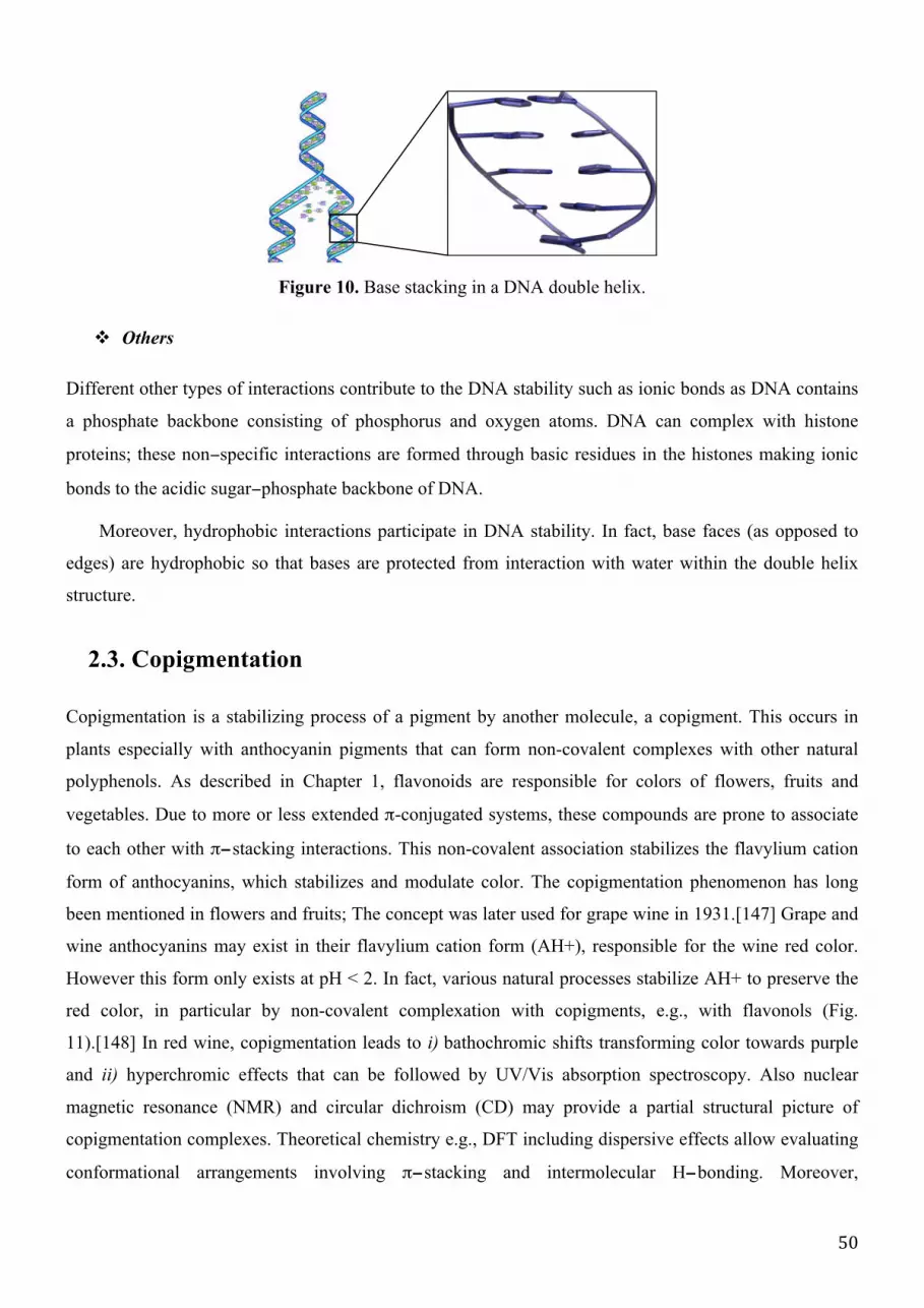
50
Figure 10. Base stacking in a DNA double helix.
" Others
Different other types of interactions contribute to the DNA stability such as ionic bonds as DNA contains
a phosphate backbone consisting of phosphorus and oxygen atoms. DNA can complex with histone
proteins; these non−specific interactions are formed through basic residues in the histones making ionic
bonds to the acidic sugar−phosphate backbone of DNA.
Moreover, hydrophobic interactions participate in DNA stability. In fact, base faces (as opposed to
edges) are hydrophobic so that bases are protected from interaction with water within the double helix
structure.
2.3. Copigmentation
Copigmentation is a stabilizing process of a pigment by another molecule, a copigment. This occurs in
plants especially with anthocyanin pigments that can form non-covalent complexes with other natural
polyphenols. As described in Chapter 1, flavonoids are responsible for colors of flowers, fruits and
vegetables. Due to more or less extended π-conjugated systems, these compounds are prone to associate
to each other with π−stacking interactions. This non-covalent association stabilizes the flavylium cation
form of anthocyanins, which stabilizes and modulate color. The copigmentation phenomenon has long
been mentioned in flowers and fruits; The concept was later used for grape wine in 1931.[147] Grape and
wine anthocyanins may exist in their flavylium cation form (AH+), responsible for the wine red color.
However this form only exists at pH < 2. In fact, various natural processes stabilize AH+ to preserve the
red color, in particular by non-covalent complexation with copigments, e.g., with flavonols (Fig.
11).[148] In red wine, copigmentation leads to i) bathochromic shifts transforming color towards purple
and ii) hyperchromic effects that can be followed by UV/Vis absorption spectroscopy. Also nuclear
magnetic resonance (NMR) and circular dichroism (CD) may provide a partial structural picture of
copigmentation complexes. Theoretical chemistry e.g., DFT including dispersive effects allow evaluating
conformational arrangements involving π−stacking and intermolecular H−bonding. Moreover,

51
copigmentation has been rationalized by using TD−DFT, showing that the observed bathochromic shift
was attributed to the formation of a charge transfer excited state (ET−CT) (see Chapter 3). The
copigmentation of C:Q [3−O−Methylcyanidin (anthocyanin pigment) and quercetin (copigment)]
complex was totally elucidated and there optical properties understood from a molecular orbital analysis
(Fig. 12).[149]
Wine color has been of great importance to food industry since color influences consumer
satisfaction. The experimental quantification of phenolic composition in wine, as well as evaluation of
their physicochemical properties, is still challenging.[150], [151] Theoretical chemistry may appear as a
new analytical tool to support rationalization of chemical and optical features of wine polyphenols. This
molecular understanding may help food industry to achieve optimum balance of wine pigmentation. One
can also envisage new opportunities for designing new dyes for food and cosmetics.
Figure 11. Chemical structures of some important examples of wine pigments and copigments.
O
OH
OH
HO O
OGlu
OH
HO
O
O
O
O
OHHO
OH
OGlu
O
R'3
OH
R'5
R'3
OH
R'5
R'3
OH
R'5
HO
R
OGlu
R'3
OH
R'3
R'3
OH
R'5
OGlu
HO
R'
Anthocyanidins Anthocyanins: Oenin: R'3, R'5=OCH3 Chalcones
Pyranoanthocyanins Vinylpyranoanthocyanins
R'3
OH
HO
O
OHO
OH
OH
OH
OH
O
R'3
OH
R'5
OH
O
HO
OHPhenolic acids Flavan-3-ols Flavanols
O
OH
O
O
R'3
R'5
OGlu
Anionic quinonoid bases A-
Copigments
R= COOHR= phenols, catechol, guaiacolR= catechin
R'= phenols, catechin
R'3= H or R'5= OH

52
Figure 12. MO correlation diagram of quercetin, complex C: Q and 3−O−methylcyanidin.[149]
2.4. Natural graphite
Natural graphite is a multilayer, each single planar layer (graphene) being constituted by carbon atoms
arranged in a hexagonal lattice. The interlayer distance is 0.335 nm (Fig. 13), stabilized by face−to−face
π−stacking interactions. It has two known forms, alpha (hexagonal) and beta (rhombohedral), that have
very similar physical properties, except that the graphene layers stack slightly differently. In each unit,
carbon atoms are covalently bonded and π electrons are highly mobile which explains the high electrical
and thermal conductivity as well as the black color of graphite. Layers are non−covalent bonded, which
explain cleavage and low hardness. It is used for refractories, batteries, steelmaking, expanded graphite,
brake linings, foundry facings and lubricants.[152], [153] Furthermore, natural graphite is an essential
source of graphene.[154]
Figure 13. Different views of layer stacking forming the graphite structure.
OHO
OH
OCH3
OH
OH
OHO
OH O
OH
OH
OH
Quercetin Complex C:Q 3-O-methylcyanidin

53
2.5. Non-covalent interactions in synthetic compounds
The understanding of different natural (biological) processes is fundamental and increasingly developed
through the understanding of non−covalent interactions. This guides the development and design of
synthetic structures based on non-covalent interactions such as micelles, micro-emulsions, foldamer (Fig.
14) that allows protein folding, nucleic acids, polysaccharides and other molecules into predictable and
well−defined conformations.
Figure 14. Non−covalent interactions stabilizing two synthetic structures: (a)†† rotaxane (blue) with
cyclobis(paraquat−p−phenylene) macrocyle (green) and (b)‡‡ foldamer.
The non−covalent interactions constitute a major driving force for supramolecular self−assemblies,
therefore its control is crucial for designing and synthesizing novel organic nanostructures.[155]
" Materials science
Many non-covalent synthetic polymers have been developed. For instance, conjugated polymers have
attracted much interest over the last three decades due to their large application in flexible, lightweight
and low-cost electronic devices. They are used in organic light-emitting diodes (OLEDs), field−effect
transistors (FETs), plastic lasers and photovoltaic cells.
Furthermore, these materials, when key structural requirements are met, may combine the mechanical
properties of polymers with the electrical and optical properties of functional organic molecules. They
exhibit specific properties such as solution processability, large charge transporting capabilities as well as
broad optical absorption properties.[156]
†† Eur. J. Org. Chem. 1998 (11): 2565–2571. ‡‡ Helvetica Chimica Acta 2003, 86 (5): 1598.
(a) (b)

54
At the end of the 20th century, the new concept emerged of supramolecular polymers, which are
self−assembled small molecules held together by reversible non−covalent interactions, such as hydrogen
bonds, metal−ligand complexation and π−stacking.
" Catalysis
Non-covalent interactions are important in catalysis, binding reactants into conformations suitable for
reaction and decreasing transition state energies. Template-directed synthesis is a special case of
supramolecular catalysis. Encapsulation systems such as micelles and dendrimers are also used in
catalysis to create microenvironments suitable for reactions.[157]
" Medicine
The importance of non-covalent complexation is also appreciated by the development of new
pharmaceutical candidates through the understanding of the interactions at a drug-binding site mostly
controlled by this type of interactions. The field of drug delivery has also made important advances as a
result of supramolecular chemistry providing encapsulation and targeted release mechanisms.[158]
Furthermore, non-covalent systems have been designed to disrupt protein-protein interactions that are
crucial to cellular function.[157], [159]
" Green and organic chemistry
Understanding of non-covalent interactions has also application in green chemistry by proceeding within
solid state directed by non-covalent bonding, thus decreasing the use of solvents during synthesis.[157]
" Others
Non-covalent complexation often allows developing of new functions that cannot exist in single molecule
(fragment). These functions also include magnetic properties, light response, self−healing polymers,
synthetic ion channels and molecular sensors. Moreover, these interactions have been applied to e.g.,
develop high-tech sensors, processes to treat radioactive waste and contrast agents for Computed Axial
Tomography (CAT) scans.[157]
To conclude, it is evident that the role of non-covalent interactions in Nature is crucial. They are
responsible for stabilizing and maintaining many natural and synthetic molecules. In other words, some
original (supra)molecular assemblies “made in Nature” would not be the same without non−covalent
interactions.

55

56
Chapter 3 – Theoretical methods
1. Basic concepts and general view on methods of calculation
1.1. The Born−Oppenheimer approximation
The resolution of the Schrödinger equation[160] is only possible for mono−electron systems, namely
hydrogen atom or the helium monocation. The exact solution of many-particle equation is not possible, in
this case, using approximations is mandatory. Born and Oppenheimer proposed a first
approximation[161], [162], based on which the motion of electrons and nuclei can be considered
separately. This is allowed because electrons, being lighter than nuclei, are much faster. Therefore, when
considering the motion of electrons, we can consider nuclei as immobile, and the electronic wave function
depends only on electron coordinates. That does mean also that the kinetic energy of nuclei is zero when
electrons are moving in a field of nuclei. The total wave function of the system is divided into its
electronic and nuclear components:
Ψ!"!#$ = Ψ!"!#$%&'(# Ψ!"#$%&' (��. 1)
The electronic Schrödinger equation for a molecular system is:
�!Ψ! = �!Ψ! (��. 2)
where Ψ! is the electronic wave function, �! is the electronic energy and �! is the electronic Hamiltonian
operator given by:
�! = −1
2∇!!
!
!!!
−�!
�!"
+1
�!"
!
!!!
!
!!!
!
!!!
!
!!!
(��. 3)
The total energy (�) is the sum of the electronic energy (�!) and the constant nuclear repulsion energy
between nuclei:
� = �! +�!�!
�!"
!
!!!
!
!!!
(��. 4)
This first approximation gave rise to the quantum chemistry or quantum mechanics (QM) methods. The

57
second side within the Born−Oppenheimer approximation[163] describing explicitly the motion of nuclei
and in which atoms are balls and bonds are springs, gave rise to molecular mechanics (MM) methods.
1.2. The Hartree-Fock approximation
1.2.1. The Slater determinant
According to the molecular orbital (MO) theory, each electron is described as belonging to a spin−orbital
wave function � (�!):
��� �! = � �! � �!
� �! = � �! � �! (��. 5)
where � (�!) is the spatial orbital, �(�!) and �(�!) are the spin Eigen−function corresponding to spin
up and spin down, respectively, �! and �! correspond to spatial and spin coordinates, respectively.
The total wave function Ψ of a poly−electronic system (N electrons), can elegantly be given by a Slater
determinant, which satisfies the anti-symmetry principle[164] and is solution of the Schrödinger equation:
Ψ �!, �!,… , �! =1
�!
�! �! ⋯ �! �!⋯ ⋱ …
�! �! … �! �!
(��. 6)
where !
!! is the normalization factor, χi is the individual spin orbital as a function of spatial and spin
coordinates of electron i. If there are two identical lines i.e., identical electrons, the determinant equals
zero. This satisfies the Pauli principle, which states that two electrons cannot have both the same spin at
the same spatial location. Also, the sign of the Slater determinant changes when two lines are exchanged;
this satisfies the non−distinguishability of electrons.
When introducing the Salter determinant into the total energy expression, it is written as the sum of
three variable terms plus one constant:
� = �!
!
!!!
+ �!" − �!" + �!
!
!!!
!
!
(��. 7)
where:
- �! is the kinetic energy and the potential energy of each electron moving in the field of nuclei:

58
�! = �!"!#$
!"#$= ��!�! 1 −
1
2∇!!−
�!
�!"
!
!!!
!
!!!
�! 1 (��. 8)
- �!" is the classical Coulomb repulsion between electrons in the i and j spin−orbitals:
�!"
!"
= �!"!#$
!"#$"%&= ��!��!�!(1)�!(2)
1
�!"
�!(1)�!(2)
!"
(��. 9)
- �!" is the non−classical (purely quantum) exchange interaction, which describes correlation between
electrons with parallel spin:
�!"
!"
= ��!��!�!(1)�!(2)1
�!"!"
�! 2 �! 1 (��. 10)
- �! is a constant that represents the repulsion potential between nuclei.
1.2.2. The Hartree−Fock approximation
The Hartree−Fock (HF) approximation consists in replacing the complicated many electron problem by a
one-electron problem. In fact, in N−electron systems, the motions of all electrons are correlated. Hartree
suggested to exchange the influence of all N−1 electrons on a single electron by a mean potential field. In
other words, every electron behaves as being in this potential field, which is due to the other electrons.
Each electron is described by its own wave function Ψ!:
�!Ψ! = ℇ!Ψ! (��. 11)
Within the Hartree approximation, the N−electron Hamiltonian is defined as the sum of all
one−electron Hamiltonians, �!:
� = �!
!
!!!
= −1
2∇!!−
�!
�!"
!
!!!
+ �!
!
!!!
(��. 12)
The N−electron wave function as defined by Hartree is not anti−symmetrical, it is the product of the
one−electron wave functions:
Ψ!" = Ψ!Ψ!…Ψ! (��. 13)
59
Fock considered the wave function as a Slater determinant. Thus, within the Hartree-Fock
approximation the exchange contribution was included:
�!"#$
+ �!(�!)− �!(�!
!
!!!
!
!!!
) �!(�!) = ℇ!"�!
!
(�!) (��. 14)
where �!"#$ is the core potential for electron i, �! is the Coulomb repulsion operator and �! is the
exchange operator. The term in brackets is the so-called Fock operator (�!):
�! = −1
2∇!!−
�!
�!"
!
!!!
+ �!!" (��. 15)
where the one-electron equation can be written as:
�!�! = ℇ!�! (��. 16)
ℇ! is the energy of a given spin-orbital �!.
The HF Hamiltonian is defined for N−electron systems by:
�!"
= �! = �!!!"#$
+ �!(�)− �!(�)
!
!!!
!
!
!
!
!
!
(��. 17)
where �!!!!"# is the monoelectronic core operator that includes kinetics and electron−nuclei interactions.
1.2.3. Restricted and unrestricted Hartree−Fock formalisms
The HF equations must be solved within a SCF (self−consistent field) procedure. The guessed wave
function is a Slater determinant chosen as a trial wave function and it is introduced in the equations to
calculate a first approximation of the energy and a new wave function. This is used for a new loop until
self-convergence is reached, namely no significant difference in energy and gradient.
According to the variational principle, the total energy obtained from the HF approximation is
overestimated compared to the energy of the exact solution of the Schrödinger equation. The missing part
is mainly the electron correlation energy, which is not taken into account with the HF approach.
Depending on the number of electrons, the approaches used for the resolution of the HF equation are
different. For systems with pair number of electrons, restricted Hartree−Fock (RHF) is used, in which,
electrons with α− and β−spins must occupy the same spatial orbitals �!(�!). The advantage of this

60
approach is that the wave function is an eigenfunction of �! operator.
The corresponding energy is given by:
� = 2 �!!!"#$
!!
!!!
+ (2�!" − �!"
!!
!!!
!!
!
) (��. 18)
However, the constraint of two electrons with α and β spins occupying the same spatial orbital
ignores the correlation between two electrons with opposed spins (spin correlation). This is of particular
importance for systems having odd number of electrons in their outer shells, e.g., radicals. In this case
either restricted open−shell Hartree−Fock (ROHF) or unrestricted open−shell Hartree−Fock (UHF) can be
used. The advantage of the UHF is that there is no imposed restrains on the spatial nature of orbitals
�! because it takes the correlation between electrons with opposed spins into account. Nevertheless, the
corresponding wavefunction is not an eigenfunction of �! . This unrestricted set is written as:
�! = �!!� �
�!!! = �!
!�(�)
(��. 19)
1.3. Roothaan-Hall equations
The most popular strategy to make the direct solution of the HF equations more practical was developed
by Roothaan and Hall.[165], [166] They proposed to describe each MO as a linear combination of atomic
orbitals (LCAO-MO) approximation:
�! = �!"�!
!
!!!
(��. 20)
where �! are the atomic orbitals and �!" are the linear coefficients that must satisfy the variational
theorem:
��
��!"= 0 (��. 21)
The insertion of these atomic orbitals into the HF equation leads to the following form:
�!"�!" = �!�!"�!"
!!
(��. 22)

61
where �!" are the terms of the Fock matrix:
�!" = ��!�! 1 �! 1 �! 1 (��. 23)
�! = ℎ! + 2�!
!!
!!!
�! − �! �! (��. 24)
�!" is the overlap between two atomic orbitals �! and �!. When the orbitals are located on different
atoms, this overlap is different from zero:
�!" = ��!�!(1)�!(1) (��. 25)
The current form of the Roothaan−Hall equations is written as follows:
�!"�!"
!
= ℰ!�!"�!"
!
(��. 26)
or in its matrix form:
�� = ��� (��. 27)
where C is a matrix containing the coefficients �!" and E is a diagonal matrix describing orbital energies:
� =
�!! �!"⋯ �!!
�!"⋮
�!!⋮
… �!!⋮
�!! �!!⋯ �!!
� =
ℰ! 0 ⋯ 0
0 ℰ! ⋯ 0
⋮ ⋮ ⋮
0 0 ⋯ ℰ!
The solvation is then a matter of solving an eigenvalue equation. However, again, since F depends on
its own solution (through the orbitals), the process must be done self−consistently (iteratively). So, for a
given nuclei geometry, one provides a guess C matrix, then to enter in a SCF procedure.
1.4. Basis sets
The description of the atomic orbitals is achieved by the basis set, which has a huge impact on the quality
of the results; so that it should be carefully chosen.

62
1.4.1. Usual basis sets
In the LCAO theory, the MOs �! are written as:
�! = �!"�!
!
!!!
(��. 28)
and atomic orbitals (�!) are described by Slater−type orbital (STO) functions:
�!"# = � �,�, � �!!" (��. 29)
The resolution of HF equations with �!"# is a complex mathematical issue, that can be simplified by
replacing the STO by Gaussian-type orbital (GTO) functions:
�!"# = � �,�, � �!!!!
(��. 30)
A proper description of the Slater orbitals requires at least three Gaussian functions and the precision
increases with the number of Gaussian functions. Each orbital �! is thus replaced by a linear combination
of (primitive) Gaussian functions. The basis sets, in which each contraction is developed by n primitives
1 ≤ � ≤ 6 are called the STO−nG basis sets. The most famous is the STO−3G.[167]
The minimal basis set does not provide accurate results because it describes only occupied atomic
orbitals. Therefore, to improve accuracy, virtual orbitals are added to the description. Also core and
valence electrons can be described by two different sets of Gaussian function. If the number of
contractions is doubled for valence electrons, the basis set is called a double−dzeta. The mathematical
expression is given by two contractions and allows more flexibility in the description of atomic orbitals.
By the same way, there exist the triple- and quadruple−dzeta.
To improve further the accuracy, polarized functions can be added to the basis set. It consists to add
p− and d−type contraction on hydrogen and heavy atoms, respectively.
Adding diffuse primitives improves the description far from the nucleus. These basis sets are indicated
with "+ " and "+ +" for heavy and hydrogen atoms, respectively.

63
Figure 1. Example of a Pople’s basis set with attribution
Other basis sets than Pople-type basis sets exist, namely natural orbital basis sets also called
correlation consistent basis sets (Dunning−type),[168] e.g., cc−pVDZ, cc−pVTZ and cc−pVQZ, where
“cc” mean that we introduce correlation consistent basis sets, p designates polarization function and the
last three letters describe valence electrons i.e., valence double or triple or quadruple-dzeta. The prefix
“aug” is added when introducing diffuse functions.
1.4.2. Basis Set Superposition Error (BSSE)
The basis functions of atoms from two molecules may overlap when the two atoms are close to each other
(e.g., in non−covalent dimers). Each fragment somehow borrows the functions from the other fragment,
thus artificially increasing basis set description, and consequently inadequately improving the calculated
properties, such as energy. This issue can be solved by using the counterpoise approach (CP). In CP, one
calculates the BSSE by re-performing all calculations using the mixed basis sets, through introducing
"ghost orbitals", and then subtracts this error a posteriori from the uncorrected energy:
ΔΕ!"#$!"
�� = �!"!"
�� − �!"!"
� − �!"!"(�) + �!"
!� − �!
!(�) + �!"!� − �!
!(�) (��. 31)
where �!"!"
� and �!"!"(�) are the energies of two given parteners A and B, respectively as obtained in
the AB complex geometry with the AB basis set, �!"!� and �!"
!� are the energies of A and B,
respectively as obtained in the AB complex geometry with the A and B basis sets, respectively.
BSSE is reduced when using large basis sets. In conclusion, there are some criteria to make the best
choice of basis set, e.g.,
# Double−dzeta at least
# Including polarization of heavy atoms
# Including diffuse functions for anions and systems stabilized by H-bonding
# Accounting for BSSE correction when necessary

64
1.5. Post Hartree-Fock methods
The HF method fails at describing the dissociation of molecules into open-shell fragments. For example,
the phenol BDE (bond dissociation enthalpy) obtained by HF methods is 49.5 kcal.mol-1 while the
experimental value is about 87 kcal.mol-1. To reach the exact energy (E), the missing part of the
correlation energy (Ecorr) must be added to the HF energy (EHF). This correlation contribution mainly
describes the correlation motion of electrons having antiparallel spins. The HF−derived methods that
account for Ecorr are called post−HF methods namely configuration interaction, coupled cluster, and
perturbation theory.
1.5.1. Coupled−cluster (CC) methods
The coupled−cluster approach uses an exponential operator for the wave function:
Ψ = �!Φ! (��. 32)
in which the exponential operator can be transformed into a Taylor expansion:
�!= 1+ Τ+
1
2!Τ!+⋯+
1
�!Τ!=
Τ!
�!
!
!!!
(��. 33)
where Τ is the cluster operator obtained as the sum of operators that generate N-excited determinants, i.e.,
Τ! and Τ! mono− and bi−excited, respectively:
Τ = Τ! !
!!! (��. 34)
Τ!�! = �!!
!
!
!
!
�!! (��. 35)
Τ!�! = �!"!"
!
!!!
!
!!!
�!"!" (��. 36)
where �!! and �!"
!" are the amplitudes, O and V are the number of occupied and virtual orbitals,
respectively.
The operator Τ is divided into Τ! and Τ! within the (CCSD) model and the energy is given by:

65
�!!"# = Ψ! � Ψ! + Ψ! � Τ!Ψ! + Ψ! � Τ! +Τ!!
2Ψ! (��. 37)
The first term corresponds to the HF energy �!" , the second term equals zero according to the
Brillouin’s theorem and the third term is the CC correlation energy �!!"#!"# :
�!!"#!"#
= �!"!"+ �!
!�!!− �!
!�!!
!,!,!,!
Ψ! � Ψ! (��. 38)
Currently, the CCSD(T) method allows reaching high accuracy and providing the best results
concerning ground-state calculations. However, the corresponding computational time is dramatically
increased with respect to other methods (following a N7 law, N being the number of basis functions). It is
thus limited to rather small molecular systems containing ca. twenty heavy atoms. Nevertheless, this
method is often used as a reference to validate e.g., “single point” energy calculation.
Figure 2. CC methods ranking and corresponding operators.
1.5.2. Perturbation Theory (PT)
The perturbation theory consists in adding a correcting contribution to the Hamiltonian operator. In a
general way, the expression of a physical property is given as a perturbation series:
A = A! + ℰ!A! + ℰ
!�! +⋯+ ℰ
!�! (��. 39)
where A! is the first approximation (e.g. HF solution) and �! is the higher-order terms. The first order
perturbation is given by:
A ≈ A! + ℰ!A! (��. 40)
CCD
CCSD
CCSD(T)
CCSDT
accuracy
cost (time)

66
1.5.3. Møller-Plesset (MP) methods
The perturbation theory is developed for treating an n electrons system in which the HF solution appears
as the zero−order approximation. The Møller−Plesset (MP) correlation energy is the perturbation to the
total energy given as:
�� = � − �! =1
2
1
�!"!!
− �!!"
!
(��. 41)
From ��. 41 that includes the HF Hamiltonian, the energy can be written as the sum of the HF energy
(�!") and the electron−electron term (�!!). The first−order Møller−Plesset (MP1) method provides the
HF energy:
�!"! = �!
(!)+ �
!
(!)= �!" (��. 42)
where �!
(!)= −�!!
The development of the higher approximation involves only calculations based on a definite one-
body problem.[169] The second−order Møller−Plesset (MP2) energy [170], [171] is written as:
�!
(!)=
�!"1
�!"!!! �!
!
�!
!− �
!
!
!"##$%&'#
!"#$%"&'&"()*
=�� ��
!
ℇ! − ℇ! + ℇ! − ℇ!!!!!!!
(��. 43)
where a and b are the spin-orbitals occupied by electrons 1 and 2 for the reference (ground state)
configuration, while r and s are the spin-orbitals occupied by electrons 1 and 2 in the excited
configuration, �! is the double-electron wave function.
It must be reminded that the MP method does not use the variational principle and so it is possible to
have: �!" < �!"#$%. While MP2 provides highly accurate results with respect to experimental data, these
methods are expensive in terms of computational time particularly when using large basis sets. The
molecule size is limited to ca. fifty atoms. In the case of using small basis sets, the accuracy is prone to be
limited. The other limitations of these methods consist in the bad description of dispersion interactions.
Even with the third−order Møller−Plesset (MP3) method, the results do not improved while the
computational time dramatically increased.[172] MP4 significantly improved the accuracy, but again the
computational time dramatically increases as a function of N7.[173] The fifth order Møller−Plesset (MP5)
method is rarely used because of its cost.[174]

67
1.5.4. SCS−MP2 methods
Recently, MP2 has been improved by developing new methods; the most common are the
spin−component-scaled (SCS) methods. The MP2 correlation energy is expressed as:
�!,!"! = �!
(!)=1
2(�↑↑ + �↓↓)+ �↑↓ (��. 44)
where �↑↓ corresponds to the energy of spin-paired and (�↑↑ + �↓↓) to the spin-unpaired double-excited
electron systems.
The SCS−MP2 method was parameterized by Grimme in 2003 to correct the underestimation of the
spin-paired energy and the overestimation of the spin-unpaired energy. The SCS−MP2 correlation energy
is given by:
�!!"!!!"!
= �!
(!)= �
↑↑1
2�↑↑+1
2�↓↓
+ �↑↓�↑↓ (��. 45)
where �↑↑ and �↑↓ are the scaling factors for spin−unpaired and spin−paired electron contributions,
respectively.
In 2004, Jung et al. proposed to improve SCS−MP2 by neglecting the �↑↑ and changing the �↑↓
parameter to 1.3. This improvement led to the scaled−opposite−spin−MP2 (SOS−MP2) method.[175]
SCS−MP2 methods can also be parameterized for specific chemical purposes e.g., to describe long-range
interactions by using SCS−MI−MP2 and SCS−S66−MP2 methods as proposed by Hobza’s group. There
have already been a number of studies of the SCS approach for the calculation of non-covalent
interactions.[176]–[178] The different �↑↑ and �↑↓ values are given in Table 1.
Table 1. �↑↑ and �↑↓ component factors for SCS−MP2−type methods.
Method MP2 SCS-MP2 SOS-MP2 SCS-MI-MP2* SCS-S66-MP2*
�↑↑ 1.00 1.20 1.30 0.17 0.14
�↑↓ 1.00 0.33 0.00 1.75 1.88
* cc-pVTZ basis set

68
1.6. Semi-empirical methods
To simplify the HF and related methods and to decrease computational time, semi−empirical methods
have been developed. These methods offered the possibility to treat large molecules in reduced time by
adding several approximations (e.g., neglecting some integrals for certain long−range−interactions,
choosing the minimal basis set and using fitted parameters for bi−electronic integrals). Due to these
approximations, some properties are not well described (e.g., non−covalent interactions) and will not be
considered in this work. There are a variety of semi-empirical methods. The most common is AM1
(Austin Model 1), which has been developed in 1985 and still give reliable results for a very efficient
computational time, as compared to HF and DFT. There are also CNDO (Complete Neglect of
Differential Overlap), INDO (Incomplete neglect of Differential Overlap), MNDO (Medium Neglect of
Differential Overlap) and PM3 (third parameterization of MNDO).
The difference between semi−empirical and Hartree−Fock methods is that i) the parameterization of
the former includes all thermochemical corrections to yield heat of formation at T=300K, while the latter
do not, and ii) the semi−empirical parameterization which refers to experimental data methods could
recover some part of electron correlation effects only for the ground states and not for the transition or
excited states.
2. Density functional theory (DFT)
While it was originally used only in solid−state physics, density functional theory (DFT) has more
recently become popular in various fields of chemistry and biology.
2.1. Thomas and Fermi
While post−HF methods allow accuracy, only rather small molecules can be studied. DFT has been
developed to treat bigger systems at a lower computational cost. The wave function does not have any
physical meaning in its native form but its square module corresponds to the electron density. The DFT
formalism has proposed to replace the wave function by the electron density as a variable, which can be
experimentally measured (e.g., by X-ray diffraction).
The electron density � �! defines the probability of finding one electron at a given location �!. It is
described by three coordinates, and the expression for one electron is:

69
� �! = � … �∗�!, x!… �! � �!, �!,… �! ��!��!…��! (��. 46)
Thomas and Fermi proposed to write the total energy as a function of �. The energy of the system is
so a functional i.e., a function of a function:
� � = �!" � + �!" � + �!! � (��. 47)
The model proposed by Thomas and Fermi was accurate for solid−state physics but it totally failed at
describing chemical properties due to the local approximation inherent to this formalism. Hohenberg and
Kohn proposed two theorems to improve DFT.
2.2. Hohenberg and Kohn theorems
" The first theorem
This theorem states that the external potential �!"# is determined, within a trivial additive constant, by
the electron density.[179] In other words, all physical properties can be obtained from � e.g., the external
potential, the Hamiltonian H and the wave function. The electronic energy is given by:
� � = �!"# � + � � + �!! � = � � + �!"# � ��� + �!! � (��. 48)
where �!"# corresponds to the interaction between nuclei and electrons in absence of any other external
field, this is system-dependent:
�!"# � = �!" � = −�!
�!"
!
!!!
!
!!!
(��. 49)
� � and �!! � correspond to the kinetic energy and the potential energy of the electron-electron
interaction, respectively; they are not system−dependent.
" The second theorem
The second theorem is the variational principle for the energy functional. From the first theorem, the
exact electron density �! of the ground state corresponds to the exact energy �!. Thus, for a trial � the
corresponding energy E is such as:
�! ≤ �

70
the energy is given by:
E ρ � = �!! ρ � + �!"# � ρ � �� (��. 50)
�!" � � is the universal system-independent HK functional given by:
�!" ρ � = � ρ � + �!! ρ � (��. 51)
The electron−electron interaction can be split into the Coulomb � and the non−classical and
correlation �!"# contributions, the �!" expression becomes:
�!" � � = � ρ � + � ρ � + �!"# ρ � (��. 52)
where the Coulomb term is given by:
� ρ � =1
2
ρ �! ρ �!
�!"
��!��! (��. 53)
2.3. Kohn−Sham
The electron−electron interaction is not known at this stage and Kohn and Sham proposed to calculate the
kinetic energy introducing a non-interacting electron system:
�!" = �! −1
2∇! �!
!
!
(��. 54)
and the total energy is given by:
� � = �!" � + �!" � + �!! � + �! � + �!"# � (��. 55)
where �! � is a correction added to the kinetic energy, which is combined to the non−classical
interaction �!"# � to form the exchange−correlation energy �!":
�!" � = �! � + �!"# � = � � − �!" � + �!! � − �!! � (��. 56)
The equations can be written with the one-electron operator �!!" within the non−interacting system
given by:
�!!"�! = �!Ψ! (��. 57)

71
�!!"= −
!
!∇!+ �!"" � (��. 58)
where �!"" � is the effective potential:
�!"" � = �!" � +� �!
�!"
��! + �!" � (��. 59)
2.3. Exchange−correlation functionals
The only unknown term in the total energy expression is the exchange-correlation functional �!" , which
contains the non−classical contribution of the electron−electron interaction and the correction to the
kinetic energy. Different approaches were proposed to describe this functional.
2.3.1. Local density approximation
The local density approximation (LDA) is based on the concept of the uniform gas of electrons, i.e., the
jellium model, in which the electron density is defined locally, that is at a certain r. The corresponding
exchange-correlation energy is:
�!" � = � � �!" � � �� (��. 60)
where �!" � � is the exchange−correlation energy per electron, which is usually divided into the
exchange and correlation contributions:
�!" � � = �! � � + �! � � (��. 61)
The most known LDA functional is SVWN, in which the exchange part was described by the Slater
exchange obtained from the HF approximation, and the correlation part was suggested by Vosko, Wilk
and Nusair. The LDA formalism overestimates the correlation energy and underestimates the exchange
energy. It is accurate only for homogenous systems. A better description is obtained when separated both
� and � contributions (�! ,�!), corresponding to the local spin-density approximation (LSDA), for which
the energy is:
�!" �! ,�! = � � �!" �! � ,�! � �� (��. 62)
When the number of α−electrons is equal to the β−electrons, the energies obtained from LSDA and
LDA must equals, considering the spin polarization function:

72
� � =(1+ �)
!! + (1− �)
!! − 2
2!! − 2
(��. 63)
where � � =!!!!!
!!!!!
LSDA is not accurate for heterogeneous systems, but it is accurate for e.g., conductor and
semi−conductor systems.
2.3.2. Generalized gradient approximation
To improve the LDA description and decrease the local character, the density gradient is included to treat
also non-homogenous systems. GEA (gradient expansion approximation) was the first attempt, where the
exchange-correlation energy is:
�!" � � = � � �!"!"# � � �� + �
!"
!,!� �
! ,� �!
∇� �!
(�(�)!)!!
∇� �!
(�(�)!)!!
�� +⋯!!
(��. 64)
The improvement given by GEA had not appeared sufficient to describe non-homogenous systems.
An improved version was then proposed, namely GGA (generalized gradient approximation), for which
the exchange-correlation functional is given by:
�!"!!" �,∇!,∇
!� = �!"!!" �! ,�! ,∇�! ,∇�! �� (��. 65)
The GGA functionals were empirically tested by comparison to experimental data, e.g., G2 test
(atomization energies of 50 small molecules), and provided much accurate results. Two types of GGA
exchange functionals were developed. First, those fitted on the exchange energy of rare gas, as the first
popular GGA exchange functional, developed by Becke (B) or e.g., FT97, O, PW, mPW. Second, those
mathematically developed and based on rational function expansions, as the one developed by Perdew (P)
or e.g., P86, LG, P, PBE (PBE is an improvement of PW86), mPBE. These functionals do not include any
empirically optimized parameters. Besides, the correlation functionals are empirically built. The
corresponding mathematical expressions are rather complicated and cannot be understood with simple
physical rules e.g., LYP (Lee Yang Parr), P86 and PW91. The exchange−correlation GGA functionals are
the combinations of both exchange and correlation e.g., BLYP. The further improvements (meta−GGA
functionals) were to include the second derivative of the density or the Laplacian (��. 66) e.g., B95, B98,
ISM, KCIS, PKZB, tHCTH, TPSS and VSXC.

73
�!"!!!!" �,∇!,∇
!� = �!"!!!!" �,∇�,∇!� �� (��. 66)
2.3.3. Hybrid functionals
The main limitation of GGA and meta−GGA is that the exchange contribution is overestimated, while the
HF−theory provides the exact exchange �!. The original idea of the hybrid functionals was to use the
exact HF−exchange and DFT−correlation, and so to write the exchange−correlation functional as:
�!" = �!!"+ �!
!" (��. 67)
This approximation is accurate only for atoms and totally failed to describe molecules. As an
improvement, the hybrid functionals include one part of the exact HF exchange, the rest being of
DFT−type; the correlation contribution is purely DFT:
�!" = ��!!"+ 1− � �!"
!"# (��. 68)
For example, this functional is called half−and−half (HandH) when a = 0.5, meaning that the
combination is 50% of HF−exchange and 50% of DFT-exchange. There are virtually an infinity of
functionals that can be built based on this principle. One of the most used is B3-type[180] in which the
exchange correlation functional is described by three parameters:
�!"!!= �!"
!"#+ � �!"
!"− �!
!"#+ ��!
!+ ��!
!"!" (��. 69)
where a, b and c are known as: a = 0.20, b = 0.72 and c = 0.81.
The hybrid−meta GGA functionals were accurate to describe specific properties, e.g., delocalized
systems and kinetics of reactions. As an example, the results obtained with the MPWB1K functional,
which was parameterized to describe kinetics, is in agreement with many experimental data, e.g., H atom
transfer reactions.
3. Further developments of DFT
For many chemical systems, DFT methods provide an adequate compromise between accuracy and
computational time. Among the most important drawbacks of DFT is that this method introduce
self−interaction error and fails at describing non−covalent interactions.

74
3.1. Self− interaction error
Within the HF theory, for a single-electron system, the Coulomb term �!" is exactly cancelled by the
exchange term �!" , which is not the case with DFT functionals that introduce an artificial interaction of
one electron with itself. This is the SIE (self−interaction error), which may influence also many−electron
systems. Perdew and Zunger[181] proposed to correct this error by using the SIC (Self−Interaction
Correction) procedure, which basically ensure equality between �!" and �!", according to:
�!"!"# �! ,�! = �!" �
! ,�! − � �!!+ �!" �!
!, 0
!!
(��. 70)
where the second sum runs over α− and β−spins
3.2. Dispersion−corrected functionals
The classical vdW interaction is described by the Lennard−Jones−type potential, which is expressed for a
M−atom system as:
�!"# = �!"(�!,!"
�!"!"−�!,!"
�!"!)
!
!!!
!!!
!
(��. 71)
where �!" and �!,!" are atom−couple dependent parameters, �!" is the inter−atomic distance, !
!!"!"
and
!
!!"!
are the repulsive and attractive terms, respectively.
Regular DFT functionals do not properly account for dispersion, which is crucial to describe
non−covalent interactions e.g., H−bonding and π−stacking. Over the past decade, various solutions have
been proposed to account for the dispersive contribution. The main improvements were based on the
explicit introduction of �!! (i.e., DFT−D and DFT−D2), �!!and �!!" (i.e., DFT−D3) decay functions.
3.2.1. DFT−D
To improve the DFT energy, Grimme proposed to simply add a carefully parameterized dispersion
correction to the KS−DFT energies.[182] Therefore, the total energy is written as:
�!"#!! = �!"# + �!"#$ (��. 72)

75
The dispersion correction is given by:
�!"#$ = −�!
�!
!"
�!"!
!
!!!
!!!
!
�!"# �!" (��. 73)
where �! is a scaling factor that is a functional−dependent parameter that must be adjusted to reach
accuracy; �!
!" is the dispersion coefficient of a given ij atom pair; �!"# is the damping function that avoid
near−singularities for small interatomic distances and double−counting effects of correlation at
intermediate distances:
�!"# � =1
1+ �!!
!
!!!!
(��. 74)
where � indicates the slope of the damping function and �! is the sum of the respective vdW radii of both
atoms involved.
The DFT−D methods is not only used for molecular complexes; other workers extended this
correction to periodic systems.[183], [184]
3.2.2. DFT−D2
The DFT−D2 dispersion correction is given by the same equation as for DFT−D, but it differs by the
slope of the damping function (d) and the atomic and atom-paired dispersion coefficients (Table 2).
Table 2. Comparison between DFT−D and DFT−D2 methods.
Parameter DFT-D DFT-D2
�!! Ref.[185] 0.05��!
!�!!∗
�!!"
2�!!�!!
�!
!+ �
!
! �
!
!�!
!
� 23 20
*N equals 2, 10, 18, 36 and 54 for atoms from rows 1-5 of the periodic table, �!! and �!
! are the ionization potential and static dipole polarizabilities of atom A.
The scaling factor �! is a key parameter in this formalism. It must be carefully parameterized in
agreement with high−level post−HF methods and/or experimental data when available.

76
3.2.3. DFT−D3
Grimme [186] has improved his previous model by including a three-body term and higher order
dispersion terms. A significant change in this so-called DFT−D3 method is that the �! dispersion
coefficients are dependent on the molecular structure, which accounts for subtle effects. The new
formalism of the dispersion energy is written as the sum of the two− (�!) and three−body (�!) terms:
�!"#$ = �! + �! (��. 75)
The two−body term includes the higher nth
−order classical dispersion terms:
�! = �!
!!!,!,…
!
!!!
!!!
!
�!!"
�!"
!�!"#,! (��. 76)
Differently from the empirically derived interpolation formula of DFT−D2, the atom-pair dispersion
coefficients �!!" are calculated from the geometrical average of atomic quantities �!
! and �!! . These
coefficients are obtained by using multiple expansion of the atomic density.
Chai and Head−Gordon have developed the damping function used in DFT−D3, which is
numerically stable for all nth−orders. Nevertheless, other functions may be used as the (BJ) function:[187]
�!"#,! � =1
1+ 6 ��!,!�!"#
!!! (��. 77)
where �!,! is the nth−order−dependent scaling factor of the cutoff radii and �! is the steepness factor.
The three−body terms is expressed as:
�! =�!!"#(3����!����!����! + 1)
(�!"�!"�!")!
�!"#,(!)(!!"#)
!
!!!
!!!
!!!
!!!
!!!
!
(��. 78)
where �!, �! and �! are the internal angles of the triangle formed by the AB, BC and AC segments.
�!!"# is the geometrical average of the previously defined atom-pair dispersion coefficients:
�!!"#
≅ − �!
!"�!
!"�!
!" (��. 79)
DFT−D3 is accurate for complex systems for the high-order dispersive term n=8, however it provides

77
unstable results with higher-order term as n=10.
3.2.4. DFT−NL
The dispersion energy can be directly obtained from a NL (non−local) correlation functional, which
inherently accounts for this contribution.[188]–[190] The total energy is then �!"#!! = �!"! + �!", �!"
being a correction covering long−ranged interactions as those between fluctuating induced local dipoles;
it is given by:
�!" = ���(�) � � +1
2��′�(�!)Φ(�, �!) (��. 80)
where Φ(�, �!) depends on two electron coordinates simultaneously, including non-locality.
Using the specific construction called VV10[190] for the non-local � �!, �! kernel. Note that in the
NL−approach, a short−range attenuation functional-dependent parameter dubbed b; � � is required to
efficiently couple the total correlation energy to any particular exchange form used. Note also that a
(more costly) double integration is required for �!", which implies the use of an additional numerical grid
on top of the grid used for the local exchange–correlation functional; however, thanks to recent
techniques this step is not a bottleneck for real calculations.[191]
The DFT−NL allows high accuracy in studying not only non−covalent interactions, but it is readily
applicable to a large range of chemical elements and therefore particularly recommended for more
general applications.[192] However, it requires more computational times compared to regular DFT
functionals.
4. Time−Dependent DFT (TD−DFT)
While the description of the potential energy surface of ground states (GS) can be achieved by classical
quantum methods (HF, DFT, semi−empirical), the potential energy surface of ES (excited state) is usually
badly reproduced. The challenge of modern quantum chemistry is to provide an accurate description of
ES as well as for GS. Different methods were developed (e.g., time−dependent HF and time−dependent
configuration interaction). Most of the methods have proposed to solve the problem within the
perturbation theory, in which the perturbation is a time-dependent excitation. The time−dependent DFT
(TD−DFT) has been relatively recently developed. Most of their uses are achieved within a linear
response theory, which is enough to reproduce the behavior of most of the physical systems (e.g.,

78
UV/visible absorption).
Runge and Gross have established a generalized Hohenberg−Kohn−Sham formalism within a
time−dependent formalism.[193] Their idea consists on proving that for two different external potentials
differing by a time-dependent constant �! � ≠ �! � + � � , the corresponding time−dependent densities
�! �, � and �! �, � are different. The Runge−Gross theorem, which is the time-dependent equivalence
of the HK theorem, states that as soon as � �, � is defined, all observables can be obtained.
The time−dependent KS equations are:
−∇!
2+ �!" �, � Φ!
!"�, � = �
�
��Φ!
!"�, � (��. 81)
in which:
�!" �, � = �!"# �, � + �!!"#"$$ �, � + �!" �, � (�q. 82)
where �!"# �, � is the external potential, �!!"#"$$ �, � is the classical electrostatic interaction between
electrons and �!" �, � is the exchange-correlation potential.
The time-dependent KS equation can thus be written as:
−1
2∇!+ �!"# � +
�′
� − �′��
!+ �!" � �! �, � = �ℏ
�
���! �, � (��. 83)
TD−DFT allows characterizing ESs and subsequently UV/Vis absorption wavelengths and oscillator
strengths.

79

80
Chapter 4 – Methodology
Note for readers: This article has been published in Chemical Physics Letters (I. Bayach et al., Chem.
Phys. Lett., vol. 578, pp. 120–125, Jul. 2013). My contribution concerns the calculations of the
association energies of the oligostilbenoid complex.
The figure and reference formats and positions are sometimes modified compared to the original article
to keep homogeneity of the thesis.
π−Stacked polyphenolic dimers: A case study using
dispersion−corrected methods
I. Bayach a,d, J.C. Sancho-García b,c,*, F. Di Meo a, J.-F.F. Weber d, P. Trouillas a,c,e
Abstract: The accuracy of dispersion-corrected calculations (DFT−D2, DFT−D3 and DFT−NL) is
assessed here, with large basis sets (def2−QZVP) to avoid incompleteness effects, for the most stable
structure of a real-world polyphenol dimer chosen as an appropriate model. Natural polyphenols form
such complexes with π−stacking playing a key-stabilizing role. Our benchmark calculations predict its
existence favored by 22–24 kcal/mol with respect to the isolated monomers, mainly driven by both π−π
and H−bonding interactions. The adequate comparison of lower-cost DFT−based methods allowed
bracketing their expected accuracy. These results thus pave the way towards reliable studies of
challenging aggregation processes of natural products.
a LCSN, Faculté de Pharmacie, Université de Limoges, 87025 Limoges, France
b Departamento de Química Física, Facultad de Ciencias, Universidad de Alicante, 03080 Alicante, Spain
c Laboratory for Chemistry of Novel Materials, University of Mons, 7000 Mons, Belgium
d Atta-ur-Rahman Research Institute for Natural Product Discovery, Faculty of Pharmacy, Universiti Teknologi MARA,
42300 Puncak Alam, Malaysia
e Department of Physical Chemistry, Faculty of Science, Palacky University, 771 46 Olomouc, Czech Republic
* Corresponding author at: Departamento de Química Física, Facultad de Ciencias, Universidad de Alicante, 03080 Alicante, Spain.

81
1. Introduction
Polyphenols sensu lato [194] (e.g., lignans and lignins, chalconoids and flavonoids, condensed and
hydrolysable tannins, phlorotannins, depsides, stilbenoids, curcuminoids, anthraquinoids, etc.) constitute
one of the most important groups of natural products, with some 105 defined structures.[195] They have
been isolated from all plant organs (e.g., bark, wood, roots, leaves, flowers, fruit, and seeds) in which they
may accumulate in substantial amounts. Therefore, they are quite abundant in human diets (e.g., fruit,
vegetables, spices and beverages) exhibiting various potential health benefits (see for example [196],
[197]). In order to fully rationalize and increase these beneficial effects, particular attention is paid to the
chemical properties involved in e.g., (i) their biological properties and (ii) their biomimetic syntheses.
Non−covalent complexes have been suggested in last decades to play an important role in these chemical
properties. The high π−delocalization observed in the polyphenol backbone would allow monomer
self−association, while the presence of OH substituents allows additional formation of strong
intermolecular H-bonds. For example, non−covalent interactions in polyphenols lead to complexes
involved in many key natural processes including: (i) plant color persistence;[198], [199] and (ii) regio−
and stereo−selective biogenetic reactions (see for example [200]). However, the definite role of these
weak interactions in polyphenol compounds is still under scrutiny; theoretical predictions are thus of
(expectedly) high value for the rationalization of these processes and they appear as the only current way
to provide a direct molecular visualization of these non−covalent dimers in solution. The theoretical
results thus appear issued from a ‘computational microscope’ supporting the indirect experimental
visualization (e.g., bathochromic shift observed in UV/Vis absorption measurements).
A small yet emblematic group of polyphenols is that of oligostilbenoids, biogenetically deriving from
the oligomerisation of polyhydroxylated stilbene precursors. The large variety of their chemical structures
allows for a broad range of biological activities including antibacterial, antioxidant, anti−fungal and
anti−inflammatory properties.[201] Oligostilbenoid polymerization is a typical case where the importance
of non−covalent interactions has been recently highlighted: regio− and stereo−selective synthesis is
largely driven by the ability of these compounds to self−assembly in solution prior to the oxidative
initiation stage.[48], [202] The extended π−delocalization of ε−viniferin derivative (Figure 1) is known to
allow long−range interactions in solution,[202], [203] as it is indeed confirmed by NMR−based
dynamical and structural studies.[204] Therefore, if theory aims at describing a complete picture of these
interactions and corresponding supramolecular association at the molecular scale, the treatment of weak
interactions is mandatory. Note also that regarding the large size of the (if any) dimer involved precludes

82
the use of methods that unfavorably scale with system size. Thus, Density Functional Theory (DFT)
becomes the favored and more judicious choice here, also keeping in mind that such methodology should
be used for large series of compounds similar in size, as a predictive tool in the near future. Taken into
account these issues, the present study deals with structure and energetics of the non−covalent dimer of
ε−viniferin, which is used as a prototype to tackle as accurately as possible these effects leading to
dimerization. Various DFT functionals including dispersive effects are used and the associated
association energies are compared to reference results. To present the achievements towards the above
goals, the manuscript is structured as follows: Section 2 presents the different DFT−based theoretical
methods able to largely deal with non−covalent interactions at both intra− and intermolecular levels.
Section 3 reports the careful application of these methods concomitantly with their benchmarking. This
would then allow reaching predictive yet robust conclusions about the stability of this kind of complexes,
and complementarily shedding light about the possible routes followed in their reactivity.
Figure 1. Chemical structure of the studied compound. The hydrogen atoms and corresponding C–H
bonds have been omitted for clarity. Figure created with ChemDraw.
2. Theoretical methods
2.1. Modeling dispersion effects
Dispersion physics arises from locally induced interactions, be them intra− or intermolecular, after the
response of the electronic cloud in one region to the presence of instantaneous and fluctuating charge
densities in another.[205] In other words, whenever polarizable electronic clouds are present in two
spatially separated but interacting fragments or subsystems, even if weakly overlapping, these correlated
dipole–dipole interactions might clearly drive self−assembly or supramolecular organization. To account
for these interactions is a real challenge for any theoretical method currently in use. A purely ab initio
treatment would therefore imply the use of energy magnitudes depending simultaneously of properties at

83
two separate points in space r and r’. This is one of the reasons why classical Møller–Plesset perturbation
theory truncated at second order (MP2) is able to partly capture the physics behind these interactions. It
has been considered as the pioneering yet simplest theoretical method to be applied within this context.
Contrarily to this, unmodified or poorly fitted DFT−based functionals completely fails due to the
short−sight treatment of matter imposed by the dependence of common (semi−local) functionals on the
density (�) and its gradient (∇�) on r exclusively. Here we briefly review the most common DFT−based
dispersion methods currently applied [206] to overcome this undesired but generalized drawback.
Due to the difficulty to self−consistently introduce the dispersion energy (�!) into the computational
treatment, one normally adds this contribution to the electronic energy in a post self−consistent
way:�!"#!! = �!"# + �! , the density thus remains unaffected upon the dispersion treatment. The
modeling of �! term is based on the well−known pairwise additivity of effects between atoms A and B
belonging to weakly overlapping fragments:
�! = −[�!!"
�!"
!−�!!"
�!"
!−�!"!"
�!"
!"… ]
!
!!!
(1)
where �!!"
are interatomic dispersion coefficients and �!" is the distance between the two atoms
involved. The simplest approach, coined as D2 [207] truncates the expansion at first order providing the
1=R6 attractive term as found in the classical Lennard−Jones potential. This term is however weighted
introducing a functional−dependent parameter (�!) to efficiently couple both terms, �!"# and �!:
�!! = −�!
�!!"
�!"
!� �!" ,
!
!!!
(2)
also relying on a damping function, � �!" , to efficiently and more physically switch from the infinite
separate limit to distances belonging to the binding region.[208] This correction has been successfully
applied for complexes of the most interest,[177], [209], [210] although a more sophisticated correction
(D3) has been recently introduced to overcome some known limitations of the latter.[211] In this case the
correcting term is given by:
�!! = − �!
�!!"
�!"
!
!!!,!
!
!!!
�! �!" , (3)
expanding the former series and introducing now nth−order dispersion coefficients allowing a better
respond to changes in chemical environment. The mathematical form imposed to the damping function

84
introduces two new parameters, �!,!, to be defined for each value of n,
�! �!" =1
1+ 6(�!"
�!,!�!!")!!
, (4)
with the ratio �!!"=
!!!"
!!!"
. Note that the dumping function reduces to a simpler form for DFT−D2 and
that more details about the form and (expectedly) negligible influence of other devised damping functions
can be found in Ref.[212].
Interestingly, there is a recent renewed interest [213]–[215] to obtain the dispersion energy directly
from the electron density through a non−local (NL) correlation functional, which inherently account for
this contribution. The total energy is now �!"#!! = �!"# + �!", with �!" being a correction covering
mostly long−ranged interactions between these instantaneous and fluctuating induced local dipoles:
�!" = ��� � � +1
2��
!� �! � �, �! (5)
using the specific construction called VV10 [216] for the � �, �! kernel. Note that in the NL−approach a
short−range attenuation functional−dependent parameter dubbed b, � = �(�) is required to efficiently
couple the total correlation energy to any particular exchange form used. Note also that a (more costly)
double integration is required for �!", which implies the use of an additional numerical grid on top of the
grid used for the local exchange−correlation functional; however, thanks to recent techniques [217] this
step is not a bottleneck for real calculations.
2.2. Technical details
The choice of the exchange–correlation functionals BP86 and B3P86 is motivated by two features: (i) the
excellent performance shown by the latter model for bond dissociation energies [218]–[220] and
optoelectronic properties [221] of many different polyphenols; and (ii) the lower computational cost of
the parent non−hybrid model (BP86), which would allow applications to larger real−world systems and
large series, due to the pervasive trade−off between accuracy and computational resources. The related
parameters of Eqs. (2)–(4) are reported in Table 1. Whereas these are taken from Grimme’s work for
BP86 (−D2 or −D3), we have recently extended the B3P86 model (−D2) in this way.[198] The
attenuation parameter b required for the use of both models together with Eq. (5), BP86−NL and
B3P86−NL, is also assessed here for the first time (to the best of our knowledge) in order to obtain (vide

85
infra) its optimum value (Table 1).
All calculations were performed with the ORCA program [222] with the built in def2−xVP family of
basis sets, unless otherwise noticed, i.e., the hierarchy def2−SVP, def2−TZVP, and def2−QZVP. The cost
of the calculations was reduced in all cases by the use of the ‘resolution of the identity’ (RI) [223] and/or
the ‘chain−of−spheres’ (COSX) [224] algorithms, for Coulomb or exchange integrals, respectively; note
that the largest calculations performed here involve 7500 primitive basis functions. Concerning numerical
grids for integration, to be on the safer side, their size was always made larger than hardwired defaults:
grid4 and grid6 for computing �!"# and �!", respectively. Due to the (expected) flatness of potential
energy curves around the equilibrium geometry, we also imposed larger than default thresholds for the
optimization algorithm.
The intermolecular interaction or association energy (Δ�) was calculated as the difference between
the energy of the complex (�!") and those of the free monomers I and II (�!"#!! and �!"#!!!). A
negative value for Δ� thus implies the existence of the complex with respect to the pair of isolated
monomers.
The basis set superposition error (BSSE), which introduces a spurious (over stabilizing the dimer
formation) energetic � ���� contribution to � was calculated by the standard counterpoise method,
giving thus rise to:
Δ� = �!"!"
�� − �!"#!!!"#!!
��� − � − �!"#!!!!"#!!!
��� − �� − Δ� ���� , (6)
in which:
� ���� = �!"#!!!"
�� − �!"#!!!"
��� − � + �!"#!!!!"
�� − �!"#!!!!"
��� − �� (7)
where �!
!(�) is the energy of fragment P calculated at the optimized geometry of Q and with the basis set
of R. Note how this procedure always implies an extra significant computational effort, which must be
considered as a limit for further extensions of this methodology to large series of compounds. On the
other hand, the BSSE can be nearly minimized, and thus the contribution Δ� ���� ≅ 0, by applying a
very large basis set, the def2−QZVP here; we will thus compare the results of both approaches to estimate
the results at the Complete Basis Set (CBS) limit.

86
Table 1. List of parameters entering into the dispersion-corrected methods employed.
Method S6 Sr,6 S8 Sr,8 b
BP86−D2 1.050 1.100 - - -
B3P86−D2 0.780 1.100 - - -
BP86−D3 1.000 1.139 1.683 1.000 -
BP86−NL - - - - 4.4
B3P86−NL - - - - 5.1
3. Results and discussion
3.1. Optimized geometry of the complex
The geometry of the complex has been calculated at the BP86−D2, BP86−D3, and B3P86−D2 levels with
the def2−SVP basis set; the geometry is not expected to significantly change upon use of larger basis sets.
Figure 2 shows two views of the head−to−tail complex formed, no matter the theoretical level employed:
(i) a large π−stacking of the backbone upon resulting monomer interaction; (ii) a release of steric
hindrance caused by the phenolic moieties acting as substituents after adopting (almost) perpendicular
positions with respect to the central backbone; and (iii) the strong directionality and force of some
intermolecular hydrogen bonds � − �⋯�, occurring within the two heads or at the tails; to name just a
few interesting facts. The dimer intermolecular distance, defined as the closest distance between carbon
atoms of the central backbone belonging to both monomers, is around 2.99, 3.02, and 2.98 Å, at the
BP86−D2, BP86−D3, and B3P86−D2 levels, respectively. These features clearly show how the choice of
the exchange–correlation functional is of relatively little importance to describe the geometrical features,
once a proper correction for dispersion is considered. Whereas the backbone of isolated monomers is
almost completely planar, the strong intermolecular interactions (mainly H−bonds) slightly bent the
monomers in the complex geometry. These hydrogen bonds located at the edges of the backbone reduce
the intermolecular distance to values that are normally repulsive (the sum of C–C van der Waals radii is
3.5 Å); this effect has also recently been detected with halogenated polycyclic aromatic complexes.[225]
3.2. Reference data
The size (N) of the system tackled here precludes the use of highly sophisticated yet very costly ab initio

87
methods like CCSD(T) or some of its variants,[226] despite recent progress [227] to reduce its formal
dependence with size: O(N7). The so-called Spin−Component−Scaled (SCS)§§ MP2 method [228] was
used as the current base−line to deal with dispersion effects. Note that despite being a method scaling as
O(N5), which dramatically alleviates the computational cost with respect to CCSD(T) this (general
purpose) method is known to provide remarkable accuracy for a wide variety of covalent and
non−covalent interacting molecular systems,[229] including para−diiodobenzene [230] or anthracene
[231] dimers extracted from crystalline structures and paracyclophane derivatives,[232] and will be thus
used as reference in the following. Note that all these single−point calculations were performed here at
the BP86−D3 optimized geometry for both the complex and the isolated monomers. As expected, the
calculated SCS−MP2 interaction energy decreases, upon augmenting the size of basis sets: -21.8, -18.4,
and -16.2 kcal/mol with the def2−SVP, def2−TZVP, and def2−QZVP basis sets, respectively. SCS-MP2
has been shown to slightly underestimate non-covalent association energies;[233] thus, within the
SCS−MP2−D2 corrected method (�! = 0.6 , obtained with a large TZVPP basis set [233]), the
combination is somewhat considered as an artifact dropping the values to -24.3 and -22.1 kcal/mol with
the def2−TZVP and def2−QZVP basis sets, respectively. However, the high stability of these complexes
is confirmed with a new SCS−MP2 version, namely SCS−S66−MP2 [234] specifically suited for
non−covalent interactions, which is additionally known to become very accurate in the description of
strong hydrogen bonds. A -26.5 kcal/mol energy of complexation was obtained with the def2−TZVP
basis set, which serves to firmly bracket the reference value and to validate afterwards the DFT−based
approximations employed.
Figure 2. Optimized structure of the studied dimer from perpendicular (right) and side (left) views. Figure created with VMD.
§§ This method scales differently the contribution to correlation energy arising from opposite- or same-spin contribution.

88
3.3. Assessment of DFT−based dispersion corrections
Further reduction of computational time and associated resources will necessarily proceed through the use
of DFT−based approaches scaling as much as O(N4), e.g. B3P86, or even as O(N3), e.g. BP86. The
association energies obtained with BP86−D2, BP86−D3, and B3P86−D2 are compared to the reference
data in Figure 3. Note first that the complex is predicted to be unbound without these −D2 or −D3
corrections, independently of the functional employed. Note also that the calculations with the large
def2−TZVP and def2−QZVP basis sets were done at the def2−SVP respective optimized geometries. The
association energies are always largely affected by the BSSE: the energy decreases by 7–8 kcal/mol when
going from the def2−SVP to the def2−TZVP, and only by 1.5–2.0 kcal/mol upon extension to the nearly
saturated def2−QZVP basis set. The use of the counterpoise correction, see Eq. (7), brings the results
close to those achieved by using the large def2−QZVP basis set, which can be thus considered near to the
(unknown) CBS limit. As a matter of example, the counterpoise−corrected BP86−D3 association energies
are -15.8, -18.5, and -18.6 kcal/ mol, with the def2−SVP, def2−TZVP, and def2−QZVP, respectively, as
compared to the values obtained without BSSE correction, i.e., -28.0, -20.9, and -19.1 kcal/mol for the
three basis set, respectively. The underlying BSSE can be thus estimated to be -12.2 (unacceptable), -2.4
(mildly acceptable), and -0.5 (within the ‘chemical accuracy’ range) kcal/mol, for the def2−SVP,
def2−TZVP or def2−QZVP basis sets, respectively. This trend is similar to all the functionals tested here.
Note also that this way to calculate the BSSE is believed to slightly overestimate its effect, and some
authors even propose to scale it down by a factor between 0.5 and 1.0.[235], [236] This is the reason why
in the following we will estimate (if any) the CBS limit as the average between the def2−QZVP values
with and without the counterpoise−correction. As a matter of example, the BP86−D3/CBS result will be
thus -18.8 ± 0.2 kcal/mol, being the error bar the difference of each method with respect to its averaged
value.

89
Figure 3. BSSE−uncorrected association energies (in kcal/mol) at several dispersion−corrected DFT
levels, and with the sequence of def2−xVP (x = T,Q) basis sets. Figure created with Xmgrace.
The �! parameter entering into the B3P86−D2 form was originally assessed with the cc−pVTZ basis
set for a non−covalent polyphenol dimer,[198] i.e., with very similar interaction than the present dimer.
The effect of using a particular family (cc−pVxZ or def2−xVP) of basis sets has been also investigated
here as a by-product. The cc−pVTZ value is -14.5 kcal/mol, which reduces to -11.2 kcal/mol after the
corresponding counterpoise correction, which is compared to -13.4 (or -11.4 when BSSE−corrected) and
−11.7 kcal/mol with the def2−TZVP or def2−QZVP, respectively. The �! parameterization is thus not
expected to be significantly influenced by this basis set issue and would not significantly affect the
association energies.
We also recognize at this stage that the corrections discussed so far are pairwise additive, which
might be related to a slight tendency of BP86−D3 towards overbinding. A way to evaluate the (repulsive)
amplitude of the three−body contribution is through the function [237]:
�!!!"#$ = �!!"#
3����!����!����! + 1
�!"�!"�!"
, (8)
where ABC are all the atom triples, �! are the internal angles of the triangle formed by �!"−�!"−�!" ,
and the coefficient is approximated by �!!"#
= − �!
!"�!
!"�!
!" . While this contribution is believed to be
negligible for small complexes, around 2% of the association energy of the benzene dimer,[238] it might
become crucial for larger systems, around 25% for two graphene layers.[237] In the present case, this
contribution amounts for 1.2 kcal/mol (which is 6–7% of the association energy if we take for instance
the BP86−D3/CBS result of -18.8 kcal/mol as reference for estimating the weight of this 3−body

90
correction). This correction thus reduces the difference between the BP86−D2 and BP86−D3 results and
appears not negligible. However, it is not expected to significantly influence the conclusions about the
relative performance of methods and the derived association energies.
The relevance of the BP86−NL and B3P86−NL models was also assessed for the present
non−covalent polyphenol dimer. In this case, the exchange–correlation functional is defined by:
�!" � = �! �(�) + �!!"#$! �(�) + �!
!"!!!"#$! � � ,� �! (9)
where �! is the B and B3 exchange parts, respectively, and �!!"#$! (�!
!"!!!"#$!) is P86 (VV10) in both
functionals. Again the sequence of def2−xVP basis sets was used. The initial value imposed to the b
parameter for an efficient coupling of non−local correction with the exchange–correlation part was the
available values for related models,[239] i.e., b=3.5 (b=4.0) for BP86 (B3P86). Note that for the cases
known up to now (BLYP vs. B3LYP and revPBE vs. revPBE0) the value of b turns to be always lower
for pure than for hybrid methods, as it should be upon a careful inspection of the whole function
� = �(�) entering into Eq. (5). We then accordingly modified it in a systematic way to check the
influence on the association energies. Table 2 presents the corresponding association energies, leading to
the following conclusions: (i) going across the sequence def2−SVP/def2−TZVP/def2−QZVP largely
reduces the BSSE in line with the observations made before; and (ii) the association energies depend on b
values, B3P86 always providing larger stabilization energies than BP86 at same b−value. Figure 4
exemplifies the amplitude of this variation for both BP86−NL and B3P86−NL models when the large
def2−QZVP basis set is employed.

91
Table 2. Association energies (in kcal/mol) at several non−local dispersion−corrected DFT levels, with
the sequence of def2−xVP basis sets, as a function of the attenuation parameter.
Method Basis set ΔE
(kcal/mol)
b = 3.5 b = 4.0 b = 4.5 b = 5.0 b = 5.5
BP86−NL def2−SVP -38.5 -32.3 -27.4 -23.5
def2−TZVP -32.0 -25.5 -20.5 -16.4
def2−QZVP -29.9 -23.5 -18.5 -14.4
B3P86−NL def2−SVP -35.6 -30.8 -26.9 -23.6
def2−TZVP -28.6 -23.6 -19.6 -16.3
def2−QZVP -26.7 -21.7 -17.7 -14.4
In the hope to obtain a refined value of b for the use of this correction for polyphenol compounds,
three systems (benzene–benzene, benzene–methanol, phenol–phenol, see Figure 5) were correspondingly
selected. They are representative of the leading p–p, p–OH, and OH–OH interactions, respectively, as
found in non−covalent polyphenol dimers. Note that very accurate CCSD(T)/CBS results are available in
the literature for the association energy of these complexes of moderate size,[240], [241] which will thus
serve to guarantee the lowest possible deviation with respect to any benchmark thought. To do that, the
Mean Absolute Deviation (MAD) is reduced as much as possible for association energies calculated at the
BP86−NL and B3P86−NL levels, according to the b values and with respect to the CCSD(T)/CBS
association energies taken as reference. Note also that the large def2−QZVP basis set is used here to
avoid any spurious BSSE. The following optimum b=4.4 and b=5.1 values were found, providing a MAD
lower than 0.2 kcal/mol in both cases with respect to the CCSD (T)/CBS results. We thus now predict
with these optimized b values, and again with the def2−QZVP basis set, association energies of -19.4 and
-17.0 kcal/mol, with the BP86−NL and B3P86−NL models respectively, which are very close to the
benchmark SCS−MP2−based results (Figure 3).

92
Figure 4. BSSE−uncorrected association energies (in kcal/mol) at several non−local dispersion−corrected DFT levels, with the def2−QZVP basis set, as a function of the attenuation parameter. Figure created with
Xmgrace.
Figure 5. Selected interacting dimers for benchmarking. From top to bottom: Benzene–Benzene,
Benzene–Methanol, and Phenol–Phenol. Figure created with VMD.
4. Conclusions
The association or interaction energy of a large real−world non−covalent polyphenol dimer has been
elucidated by dispersion−corrected DFT methods using several flavors. First, benchmark calculations at
improved second−order perturbation theory were performed to adequately bracket the stabilizing energy
gained when the two monomers self−associate to form the complex. The use of large basis sets, up to the
def2−QZVP, leads to sufficiently converged results. Interestingly, we estimate at the complete basis set
limit (within an expected error bar or about 0.2 kcal/mol) complexation energies of -17.6 kcal/mol at both
the BP86−D2 and BP86−B3 levels, the latter after taking into account the 3−body interactions, and of
−11.5 kcal/mol at the B3P86−D2 level. When these functionals combine in a purely ab initio fashion with
a correlation correction (the VV10 functional) and with an optimum value for the attenuation
b−parameter entering into this non−local functional, the values are -19.4 and -17.0 kcal/mol (BP86−NL
and B3P86−NL, respectively). All these schemes seem to slightly underestimate the

93
SCS−S66−MP2−based results although, however, it is satisfying to see that, even being fairly different in
both the underlying density functional and the way in which they incorporate the missing dispersion
forces, they predict enough stabilization energy to anticipate the existence of this kind of complexes
almost independently of expected thermal or environmental conditions. Thus, a practical yet accurate
combination of DFT−D2/ DFT−D3 searches along potential energy hypersurfaces, thanks to rapid
evaluation of gradients with these levels and their moderate scaling with size, together with refinements
employing DFT−NL might constitute a valid strategy for further future studies.

94

95
Chapter 5 – Methodological assessments of dispersion between
prototypes of polyphenol non-covalent complexes
Note for readers: This article is under minor revisions in Chemistry−An Asian Journal. It consists on an
experimental and theoretical study of the biosynthesis of oligostilbenoids. I only participate to the
theoretical elucidation. The entire manuscript is incorporated in this PhD to keep consistency.
The figure and reference formats and positions are sometimes modified compared to the original article
to keep homogeneity in the thesis.
Oligostilbenoids from the heartwood of N. heimii: role of
non−covalent association in their biogenesis
Bayach, I.,[a],# Manshoor, N.,[b], # Sancho García,[c] J.C., Choudhary, M. I.,[d] Trouillas, P.,[e,f,g],* and Weber, J. F. F.[b],*
Abstract: Four new oligostilbenes, including one dimer and three tetramers of resveratrol, i.e. heimiols
B-E (1-4) were isolated from the heartwood of Neobalanocarpus heimii (Dipterocarpaceae), together with
thirteen known resveratrol oligomers (5-17). Examination of structural diversity of the isolated
oligostilbenes led to hypothesise their biogenetic origin through a small number of versatile chemical
pathways. These hypotheses are strongly supported by computational calculations (based on the density
functional theory, DFT) that were performed to rationalize conformational rearrangements and thus
provide insights into the mechanism of oligostilbenoid biosynthesis. Non−covalent complexes are
believed to drive the regio− and stereo−selectivity of the oligomerisation reactions.
Keywords: Neobalanocarpus heimii • Oligostilbenes • Biosynthesis • DFT(−Dispersive) • Non−covalent
interactions
[a] Imene Bayach: LCSN - Faculté de Pharmacie, Université de Limoges, 2 rue du Dr Marcland, 87000 Limoges, France
[b] Dr. Nurhuda Manshoor and Prof. Dr. Jean-Frédéric F.Weber: Atta-ur-Rhaman Institute for Natural Product Discovery
(AuRIns), Faculty of Pharmacy, Universiti Teknologi MARA, 42300, Bandar Puncak Alam, Selangor, Malaysia. E-mail:
[c] Prof. Dr Muhamad Iqbal Choudhary: H. E. J. Research Institute of Chemistry, International Center for Chemical and
Biological Sciences, University of Karachi, Karachi, Pakistan.

96
[d] Dr. J.C. Sancho-García: Departamento de Química Física, Facultad de Ciencias, Universidad de Alicante, 03080 Alicante,
Spain
[e] Dr. Patrick Trouillas: INSERM, UMR-S850, Faculty of Pharmacy, 2, Rue du Dr Marcland, 87025 Limoges Cedex,
France. E-mail: [email protected]
[f] Regional Centre of Advanced Technologies and Materials, Department of Physical Chemistry, Faculty of Science,
Palacký University, tř. 17 listopadu 12, 771 46 Olomouc, Czech Republic
[g] Laboratoire de Chimie des Matériaux Nouveaux, Université de Mons, Place du Parc 20, B-7000 Mons, Belgium
* Corresponding author # These authors contributed equally
1. Introduction
Oligostilbenes are major secondary metabolites of plants belonging to the Dipterocarpaceae family.
About one third of oligostilbenes ever isolated were from dipterocarps,[242] which makes them relevant
chemical markers for the family. They are found in various plant parts (the leaves, barks, woods and
seeds), but were obtained most often from the heartwood. Most oligostilbenes found in this family
include at least one oxygenated heterocyclic ring, usually in the form of a
trans−2−aryl−2,3−dihydrobenzofuran ring.[243] In a preceding paper, we reported the characterization of a
novel dimeric stilbene and four known oligostilbenoids from the heartwood of Neobalanocarpus
heimi.[244] Further investigation of the methanol extract led to the isolation of four new resveratrol
oligomers along with thirteen known oligostilbenes. We thus report herein the structural elucidation of a
new stilbene dimer, namely heimiol B (1), and three tetramers, heimiols C (2), D (3) and E (4). After a
complete description of the elucidated chemical structures, the biogenesis of these compounds is
proposed as a result of close examination of the heartwood extract composition and recent insights in
oligostilbene biosynthetic mechanisms.[203] The hypotheses based on supramolecular arrangements are
elegantly supported by DFT (density functional theory) calculations, augmented by dispersive corrections
(DFT−D) to properly account for non−covalent interactions.
2. Results and Discussion
Compounds 1-4 (Figure 1) are four new oligostilbenes that were isolated from the methanol extracts of
the heartwood of Neobalanocarpus heimii by repeated chromatography on C18 silica−bonded columns.

97
" Heimiol B
Heimiol B (1) was obtained as a light brown residue. It exhibited a [M−H]- ion at m/z 469.1288 in
HR−ESI−TOF−MS compatible with the molecular formula C28H22O7, suggesting 1 to be an oxidative
dimer of resveratrol. This was supported by the 13C−NMR spectrum (Table 1), which showed
twenty−eight signals, assignable to four sp3 and twenty−four sp2−hybridized carbons. The 1H−NMR
spectrum of 1 (Table 1) displayed two sets of ortho−coupled aromatic hydrogen signals in AA’BB’ spin
systems at d 7.34, 7.03, 6.75 and 6.71 (2 H, each d, J = 8.7 Hz), assignable to two independent
4−hydroxyphenyl groups, two sets of meta−coupled aromatic hydrogen signals at d 6.36, 6.52, 6.24 and
6.08 (l H each, d, J = 2.1 Hz), assignable to two di−substituted resorcinol moieties, and four olefinic
hydrogen signals at d 5.13, 4.90, 4.61 and 4.22 (l H each, d, J = 2.0 Hz). The HMBC cross peaks between
methine protons and aromatic carbons (H−7a/C−1a, H−7a/C−9a, H−8a/C−9a, H−8a/C−10a, H−8a/C−14a,
H−7b/C−1b, H−7b/C−2(6)b, H−7b/C−9b, H−8b/C−1b, H−8b/C−9b, H−8b/C−10b and H−8b/C−14b) allowed
establishment of connections between aromatic rings and their corresponding benzylic methine groups as
described in Figure 2.
O
HO
HO
OH
OH
OH
HO
H
H
H
H
7a7b
8b
8a
1a
4a
3a
2a
13a
12b
5a
6a
14b
12a
11a
10a
9b
4b1b
14a
13b
11b
10b
O O
OH HO
HOOH
OHHO
OHHO
HO OH
H
H
HH
H
H
H
H
O
7a
7b
8b
8a
4a
11a
9a10a
12a
13a
3a
14a
2b
3b
4b
11b
9b10b
1b
2a
O
OH
HOOH
H
H
H
H
O
HH
HO
OHOH
HO
OH
OH
HO
H
H
2a
4a
12a
14b
14a
1b
3a
1a
6a
7a
9a
13b
10a
13a
8a
5a
4b
7b
8b
11b
10b
9b
12c
2c
12b
3c
4c
7c
8c
11c
10c
9c
5c
14c
13c
11d 4d
9d 2d
12d
7d
10d 3d
8d
1d
O
OH
HO
HO
OH
OH
O
OH
OH
OH
HO
HO
HH
H
H
H
H
H
H
13d
14d
4d
2d 3d
9d
10d12d
11d
8d
7d14c
9c
1d
7c
8c
11c13c
10c
12c
3c 4c
1c
1 : H-7b = α
5 : H-7b = β2
3 : H-7a=α H-8a=β H-8c=α
6 : H-7a=β H-8a=α H-8c=β
4 : H-7d=β H-8d=α
7 : H-7d=α H-8d=β
7a
8a
7b
8b
O O
OH HO
HOOH
OH HO
OHHO
HO OH
H
H
HH
H
H
H
H
7d
8d
7c
8c
8 : H-7b=β H-8b=α H-7c=α H-8c=β
9 : H-7b=α H-8b=β H-7c=β H-8c=α
10 :H-7b=β H-8b=α H-7c=β H-8c=α
11 :H-7b=α H-8b=α H-7c=β H-8c=α
O
OH
HOOH
H
H
H
H
H
HO
HO
H
H
OH
O
OH
OH
HO
H
H
HO
12
HO
O
OHHO
OH
H
OH
H
H
H
HO
7a
7b
8b
8a
13 : H-7b=α
14 : H-7b=β
HO
O
OHHO
OH
H
H
H
HO
H
H
H
OH
OH
OH
7a
8a
7b
8b7c 8c
15 : H-8b=α
16 : H-8b=β
OH
HO OH
H
H
H
HO
OH
HO
H
H
H
OH
OH
HO
17
D2
2a
4a
12a
14b
14a
1b
3a
1a
6a
7a
9a
13b
10a
13a
8a
5a
4b
7b
8b
11b
10b
9b
12c
12b
4c
7c
8c
11c
10c
9c
1c
13c
11d
4d
9d
12d
7d
8d
1d
7a
8a
7b
8b
7c 8c
HO
O
HO
HO
OH
OH
18
7b
8b
11b
2b

98
Figure 1. Oligostilbenes isolated from Neobalanocarpus heimii heartwood.
The HMBC spectrum also displayed H−7a/C−10b, H−8a/C−9b, H−8a/C−10b, H−8a/C−11b, H−7b/C−9a,
H−7b/C−10a, H−7b/C−11a and H-8b/C-10a correlations indicating linkages between both resveratrol units
of the dimer. Such correlations were similar to those obtained with ampelopsin A (14). However, the JH-H
of 2.0 Hz for the benzylic hydrogen at the 7a position was incongruent with the presence of a
dihydrofuran ring. The 13C signals at d 81.4 (C−7a) and 81.5 (C−8b) correspond to benzylic carbons
bearing an oxygen atom. The excess of only one oxygen atom in the molecule suggested that both
carbons were connected to the same oxygen atom, forming an ether linkage between C−7a and C−8b. The
long−range correlation H−7b/C−9a indicated a linkage between C−7b and C−10a. Such correlations
establish an oxygenated seven−membered ring. The cross peaks H−7a/C−10b, H−8a/C−9b, H−8a/C−10b
and H−8a/C−11b showed the presence of a bridged framework in the molecule similar to that of heimiol A.
Based on NOESY experiment, correlations between H−7a/H−8a and H−7b/H−8b were observed, which
indicate cis configurations. Further assessment of NOE correlations revealed cross peaks between H−7a
and H−8b, indicating syn orientation. The HMBC and NOE correlations and relative stereochemistry of 1
are shown in Figure 2. Based on this structure a conformational analysis was obtained at the DFT level
(see Figure 2 and Method section for computational details). No intra H−bonding was observed in the
most stable conformer.
Figure 2. Selected HMBC and NOESY correlations and 3D computed most stable conformation of 1.
" Heimiol C
Heimiol C (2) was obtained as a dark brown amorphous solid. It exhibited a pseudomolecular [M+Na]+
ion peak at m/z 947.2679 in ESI−MS. On the basis of HR−MS data, the molecular formula of 2 was
determined to be C56H44O13, compatible with a tetramer of resveratrol composed of four resveratrol units
with an additional oxygen atom. However, both the 1H and 13C−NMR spectra of compound 2 (Table 1)
showed half the signals corresponding to the above molecular formula. This observation indicated that
compound 2 is a symmetrical molecule with an additional oxygen atom being located along the axis of
O
HO
HO
OH
OH
OH
HO
H
H
H
H
7a
7b
8b
8a
1a
4a
3a
2a
13a
12b
5a
6a
14b
12a
11a
10a
9b
4b1b
14a
HMBC
13b
11b
O
HO
HO
OH
OH
OH
HO
H
H
H
H
H
H
H
H
7a
7b
8b
8a
4a
3a
2a
13a
12b
5a
6a
11b
12a
11a
10a
9b
4b1b
14a
10b
NOESY
13b
9a
9a

99
symmetry. This suggested the presence of an ether linkage between both symmetrical moieties. The
1H−NMR and COSY spectra exhibited the signals of two sets of ortho−coupled aromatic protons in the
form of AA’BB’ systems at d 6.91, 6.68 (4 H each, d, J = 8.4 Hz) and d 6.67, 6.54 (4 H each, d, J = 8.4
Hz). They were assignable to four 4−hydroxyphenyl groups. Signals from a set of meta−coupled aromatic
protons from a 1,2,3,5−tetra−substituted benzene ring were observed at d 6.06 and 6.83 (2 H each, d, J =
1.9 Hz). The 1H−NMR spectrum also included a signal at d 6.20 (2 H, t, J = 2.0 Hz). From the splitting
pattern and chemical shift, the signal was assignable to the proton of a resorcinolic B ring. However, in
the COSY spectrum, that signal did not show any correlation.
Table 1. 1H− and 13C−NMR spectral data for compounds 1-4.
No 1 2 3 4
1H 13C 1H 13C 1H 13C 1H 13C 1a 133.4 131.5 134.2 134.7 2a, 6a 7.34 (d, 8.7) 128.7 6.91 (d, 8.4) 128.3 6.92 (d, 8.5) 127.2 7.10 (d, 8.5) 127.2 3a, 5a 6.75 (d, 8.7) 115.0 6.68 (d, 8.4) 114.8 6.68 (d, 8.5) 116.1 6.73 (d, 8.5) 116.4 4a 158.1 157.1 158.1 157.9 7a 4.90 (d, 2.0) 81.5 5.10 (d, 7.3) 94.3 5.44 (d, 4.3) 94.0 5.29 (d, 1.3) 93.9 8a 4.61 (d, 2.0) 46.2 3.42 (d, 7.3) 54.7 4.10 (d, 4.3) 57.3 4.29 (d, 1.3) 57.1 9a 142.8 146.5 147.9 148.5 10a 116.2 5.90 [a] (br s) 106.0 [b] 119.8 6.27 (d, 1.8) 106.5 [c] 11a 156.2 158.4 159.0 162.8 12a 6.08 (d, 2.1) 102.4 6.20 (t, 2.0) 101.2 5.94 (d, 1.9) 93.5 6.06 (t, 1.8) 102.1 13a 156.8 158.4 162.8 14a 6.24 (d, 2.1) 110.4 5.90 [a] (br s) 106.0 [b] 6.13 (d, 1.9) 109.2 6.28 (d, 2.0) 106.6 1b 136.8 130.9 136.6 138.7 [d] 2b, 6b 7.03 (d, 8.7) 130.8 6.67 (d, 8.4) 127.9 7.16 (d, 8.5) 130.8 6.77 (d, 8.5) 129.3 [e] 3b, 5b 6.71 (d, 8.7) 115.1 6.54 (d, 8.4) 114.8 6.73 (d, 8.5) 115.5 6.59 (d, 8.5) 115.4 [f] 4b 157.8 156.5 156.0 155.9 7b 4.22 (d, 2.0) 53.6 5.19 (dd, 6.9, 2.6) 84.1 3.72 (d, 1.9) 52.7 4.28 (br s) 50.6 8b 5.13 (d, 2.0) 82.9 3.59 (dd, 6.9, 2.6) 56.4 3.64 (d, 1.9) 62.1 4.02 (br d, 6.8) 60.5 9b 140.7 136.7 144.8 144.6 10b 121.3 121.8 123.4 116.0 11b 153.0 160.7 161.6 163.2 12b 6.36 (d, 2.1) 102.4 6.06 (d, 1.9) 95.7 6.10 (s) 96.3 6.16 (s) 96.8 13b 156.8 158.4 154.1 155.4 14b 6.36 (d, 2.1) 104.9 6.83 (d, 1.9) 105.6 118.7 126.5 1c 130.9 133.0 138.6 [d] 2c, 6c 6.67 (d, 8.4) 127.9 6.81 (d, 8.5) 131.2 6.75 (d, 8.5) 129.2 [e] 3c, 5c 6.54 (d, 8.4) 114.8 6.54 (d, 8.5) 115.2 6.56 (d, 8.5) 114.8 [f] 4c 156.5 156.4 155.9 7c 5.19 (dd, 6.9, 2.6) 84.1 4.00 (d, 7.0) 48.7 4.22 (br s) 50.3 8c 3.59 (dd, 6.9, 2.6) 56.4 4.62 (d, 7.0) 39.6 4.07 (br d, 6.8) 60.7 9c 136.7 143.5 143.9 10c 121.8 113.8 119.2 11c 160.7 157.9 162.5 12c 6.06 (d, 1.9) 95.7 6.31 (d, 1.6) 101.5 6.19 (s) 97.0 13c 158.4 157.8 155.4 14c 6.83 (d, 1.9) 105.6 4.45 (d, 1.6) 103.0 126.5 1d 131.5 133.4 129.1 2d, 6d 6.91 (d, 8.4) 128.3 7.10 (d, 8.5) 128.4 7.01 (d, 8.5) 128.9 3d, 5d 6.68 (d, 8.4) 114.8 6.79 (d, 8.5) 116.0 6.60 (d, 8.5) 115.4 [f] 4d 157.1 157.7 157.3 7d 5.10 (d, 7.3) 94.3 5.94 (d, 6.0) 85.2 5.69 (d, 7.0) 91.5 8d 3.42 (d, 7.3) 54.7 4.49 (d, 6.0) 49.7 4.43 (d, 7.0) 53.5 9d 146.5 147.0 143.1 10d 5.90 [a] (br s) 106.0 [b] 6.37 (br s) 129.6 5.87 (d, 2.0) 107.5 11d 158.4 159.6 160.0 12d 6.20 (t, 2.0) 101.2 6.23 (t, 2.0) 102.1 6.31 (t, 2.0) 102.2 13d 158.4 159.6 160.0 14d 5.90 [a] (br s) 106.0 [b] 6.37 (br s) 129.6 6.24 (d, 2.0) 106.5 [c]
1H and 13C NMR spectra were recorded in acetone-d6 at 400 and 100 MHz, respectively, unless otherwise stated. [a] NMR experiment at 60°C (500 MHz). [b] Signal not detected when spectrum recorded at RT; assignment made through 1H-13C cross peaks in HMQC experiment at 60°C (500 MHz). [c], [d], [e], [f] assignments respectively interchangeable.
Furthermore, the integration of signals observed in the 1H−NMR spectrum did account for only
eleven aromatic protons, instead of the expected thirteen (Figure 3a). It had been previously observed that
aromatic protons H−10(14) of a free resorcinol moiety resonate either as a broad signal [245] or just as a
hump [246], [247] and this was explained by steric hindrance restricting the rotation of this ring. These

100
authors recorded 1H spectra at -20°C and/or -40°C in order to block the molecules in their different stable
conformers and could observe these protons as two independent broad singlets. An opposite strategy
consists of heating up the sample and facilitating the free rotation of that problematic ring, which would
lead to a well−resolved averaged signal. A 1H−NMR spectrum of 2 was recorded at 60°C and a broad
signal was observed at d 5.90 (Figure 3b); a 1D−TOCSY experiment, also performed at 60°C, allowed to
obtain correlation between that broad signal and the signal at d 6.20 (Figure 3c). The 1H−NMR and COSY
spectra also displayed two sets of coupled benzyl methine protons resonating at d 5.10 and 3.42 (2 H each,
d, J = 7.3 Hz) on one hand, and d 5.19, 3.59 (2 H each, dd, J = 7.0, 2.6 Hz) on the other hand. The
corresponding methine carbons were assigned from HMQC experiments.
In the HMBC spectrum, cross peaks observed between the methine protons and aromatic carbons
allow the establishment of connections between the aromatic rings and their corresponding benzylic
methine groups (Figure 4). The only correlation that connected the two resveratrol units (of one of the
symmetric half) was H−8a/C−11b. This correlation, together with the deshielding of C−7a and the
coupling constant of 7.3 Hz, suggested linkages through a dihydrobenzofuran ring. The HMBC cross
peak H−8b/C−8b indicated the linkage (C−8b/C−8b’) between both halves of the tetramer. The deshielding
effect on methine carbon C−7b was compatible with the presence of an oxygen atom. This observation
suggested the presence of an ether linkage between C−7b and C−7b’. The linkages between the two
equivalent units of compound 2 resembled to those already observed for tricuspidatol A, a resveratrol
dimer.[248] Indeed, the chemical shifts and coupling constants of the methine protons of the
tetrahydrofuran ring of both compounds were found quite similar. Consequently, compound 2 was
suggested to be a tricuspidatol A derivative having two additional symmetrically attached resveratrol
units.
Figure 3. NMR spectra for 2. [a] 1H−NMR experiment at RT; [b] 1H−NMR experiment at 60°C; [c] 1D
TOCSY experiment at 60°C, irradiation at d 5.90.
[a]
[b]
[c]
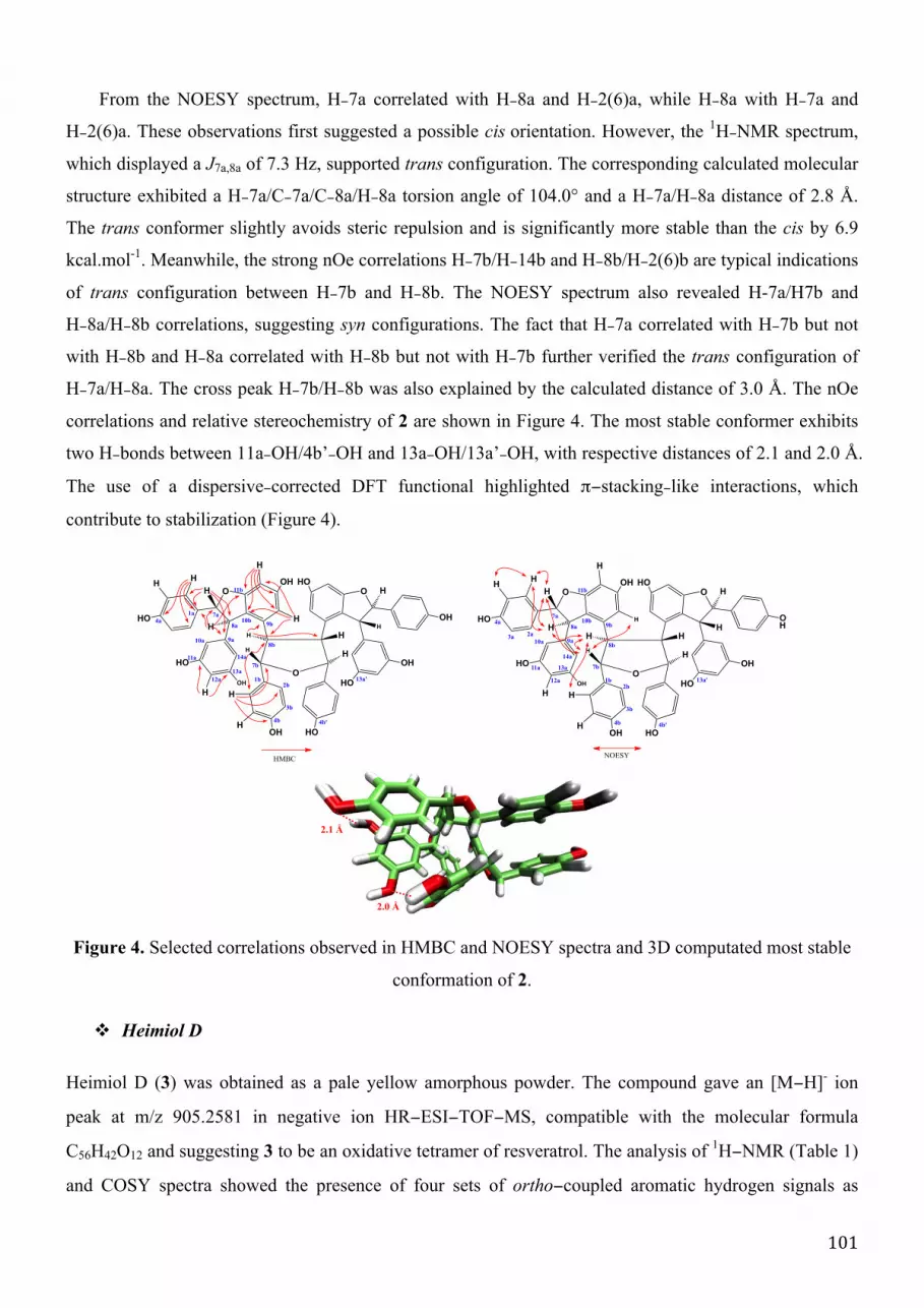
101
From the NOESY spectrum, H−7a correlated with H−8a and H−2(6)a, while H−8a with H−7a and
H−2(6)a. These observations first suggested a possible cis orientation. However, the 1H−NMR spectrum,
which displayed a J7a,8a of 7.3 Hz, supported trans configuration. The corresponding calculated molecular
structure exhibited a H−7a/C−7a/C−8a/H−8a torsion angle of 104.0° and a H−7a/H−8a distance of 2.8 Å.
The trans conformer slightly avoids steric repulsion and is significantly more stable than the cis by 6.9
kcal.mol-1. Meanwhile, the strong nOe correlations H−7b/H−14b and H−8b/H−2(6)b are typical indications
of trans configuration between H−7b and H−8b. The NOESY spectrum also revealed H-7a/H7b and
H−8a/H−8b correlations, suggesting syn configurations. The fact that H−7a correlated with H−7b but not
with H−8b and H−8a correlated with H−8b but not with H−7b further verified the trans configuration of
H−7a/H−8a. The cross peak H−7b/H−8b was also explained by the calculated distance of 3.0 Å. The nOe
correlations and relative stereochemistry of 2 are shown in Figure 4. The most stable conformer exhibits
two H−bonds between 11a−OH/4b’−OH and 13a−OH/13a’−OH, with respective distances of 2.1 and 2.0 Å.
The use of a dispersive−corrected DFT functional highlighted π−stacking−like interactions, which
contribute to stabilization (Figure 4).
Figure 4. Selected correlations observed in HMBC and NOESY spectra and 3D computated most stable
conformation of 2.
" Heimiol D
Heimiol D (3) was obtained as a pale yellow amorphous powder. The compound gave an [M−H]- ion
peak at m/z 905.2581 in negative ion HR−ESI−TOF−MS, compatible with the molecular formula
C56H42O12 and suggesting 3 to be an oxidative tetramer of resveratrol. The analysis of 1H−NMR (Table 1)
and COSY spectra showed the presence of four sets of ortho−coupled aromatic hydrogen signals as
7a
7b
8b
8a
O O
OH HO
HOOH
OH HO
OHHO
HO OH
H
H
H
HH
H
H
H
O
HMBC
HH
H
H
H
H
H
4a
11a
9a10a
12a
13a
1a
14a
2b
3b
4b
11b
9b10b
1b 13a'
4b'
O O
OH HO
HOOH
OH HO
OH
HO
HO OH
H
H
H
H
H
H
H
H
O
NOESY
HH
H
H
H
H
H
7a
7b
8b
8a4a
11a
9a10a
12a
13a
3a
14a
2b
3b
4b
11b
9b10b
1b
2a
4b'
13a'
2.1 Å
2.0 Å

102
pseudo doublets typical of AA’BB’ systems at d 6.92, 6.68 (2 H each, d, J = 8.5 Hz), 7.16, 6.73 (2 H
each, d, J = 8.5 Hz), 6.81, 6.54 (2 H each, d, J = 8.5 Hz) and 7.10, 6.79 (2 H each, d, J = 8.5 Hz),
assignable to four independent 4−hydroxy−phenyl groups. Two sets of meta−coupled aromatic hydrogen
signals were observed at d 6.13, 5.94 (l H each, d, J = 1.9 Hz) and 6.31, 4.45 (l H each, d, J = 1.6 Hz),
assignable to two di−substituted resorcinol moieties. A signal from an aromatic proton of a
penta−substituted benzene ring was observed at d 6.10 (1 H, s). Further analysis of the 1H−NMR
spectrum revealed one set of signals due to a 3,5−dihydroxyphenyl group at d 6.37 (2 H, br s) and 6.23 (1
H, t, J = 2.0 Hz). Four olefinic hydrogen signals at d 5.44, 4.10 (l H each, d, J = 4.3 Hz), and 5.94, 4.49 (1
H, each d, J = 6.0 Hz) were attributed to two diaryl−dihydrobenzofuran moieties. A spin system formed
of four aliphatic methine protons linearly attached is identified from the COSY spectrum at d 3.72 (1 H,
d, J = 1.9 Hz), 3.64 (1 H, d, J = 1.9 Hz), 4.00 (1 H, d, J = 7.0 Hz) and 4.62 (1 H, d, J = 7.0 Hz). In the
HMBC spectrum, significant cross peaks H−2(6)a/C−7a, H−8a/C−9a, H−8a/C−10a, H−2(6)b/C−7b,
H−8b/C−9b, H−8b/C−10b, H−7c/C−9c, H−8c/C−9c, H−8c/C−10c, H−2d(6d)/C−7d, H−7d/C−1d,
H−7c/C−19d, H−8d/C−1d, H−8d/C−9d and H−8d/C−10d established connections between the aromatic
rings and their corresponding benzylic methine groups (Figure 5). Long−range correlations H−7a/C−10b,
H−7a/C−11b, H−8a/C−9b, H−8a/C−10b, H−8a/C−11b, H−7d/C−11c and H−8d/C−9c suggested that the
resveratrol units A and D form a dihydrofuran ring with rings B and C, respectively. The HMBC
spectrum also showed correlations, namely H−7b/C−1c, H−7b/C−7c, H−8b/C−1c, H−7c/C−14b, and
H−8c/C−14b, suggesting connection between the resveratrol units B and C, through the C−8b/C−7c
linkage. This connection was previously not detected from the COSY spectrum as no cross peak between
H−7c and H−8b could be observed. The relative stereochemistry was determined from the NOESY
spectrum. The lack of correlation between H−7a/H−8a and H−7d/H−8d was a preliminary indication of
trans orientations. Further evidence was obtained from the correlations between H−2(6)a and H−7a (but
not H−8a), and H−2(6)d and H−8b (but not H−7a), see Figure 5. Such cross peaks suggested
configurations of both chiral carbons 7d and 8d to be S. Some nOes were also observed between
H−2(6)b/H−7c, H−7b/H−2(6)c, H-8c/H-8d and H−7d/H−12c indicated their respective syn orientations.
The J values for the H−7b/H−8b and H−7c/H−8c correlations were 1.9 and 7.0 Hz, respectively,
suggesting cis conformation. From the assignment on the dihedral angle of the respective protons, it may
be concluded that the configurations for C−7b, C−8b, C−8c and C−7c were R, R, S and R respectively.
The corresponding 3D structure was optimized at the DFT level (Figure 5). Neither H−bonding nor
π−stacking interaction exist in the most stable conformer of 3.
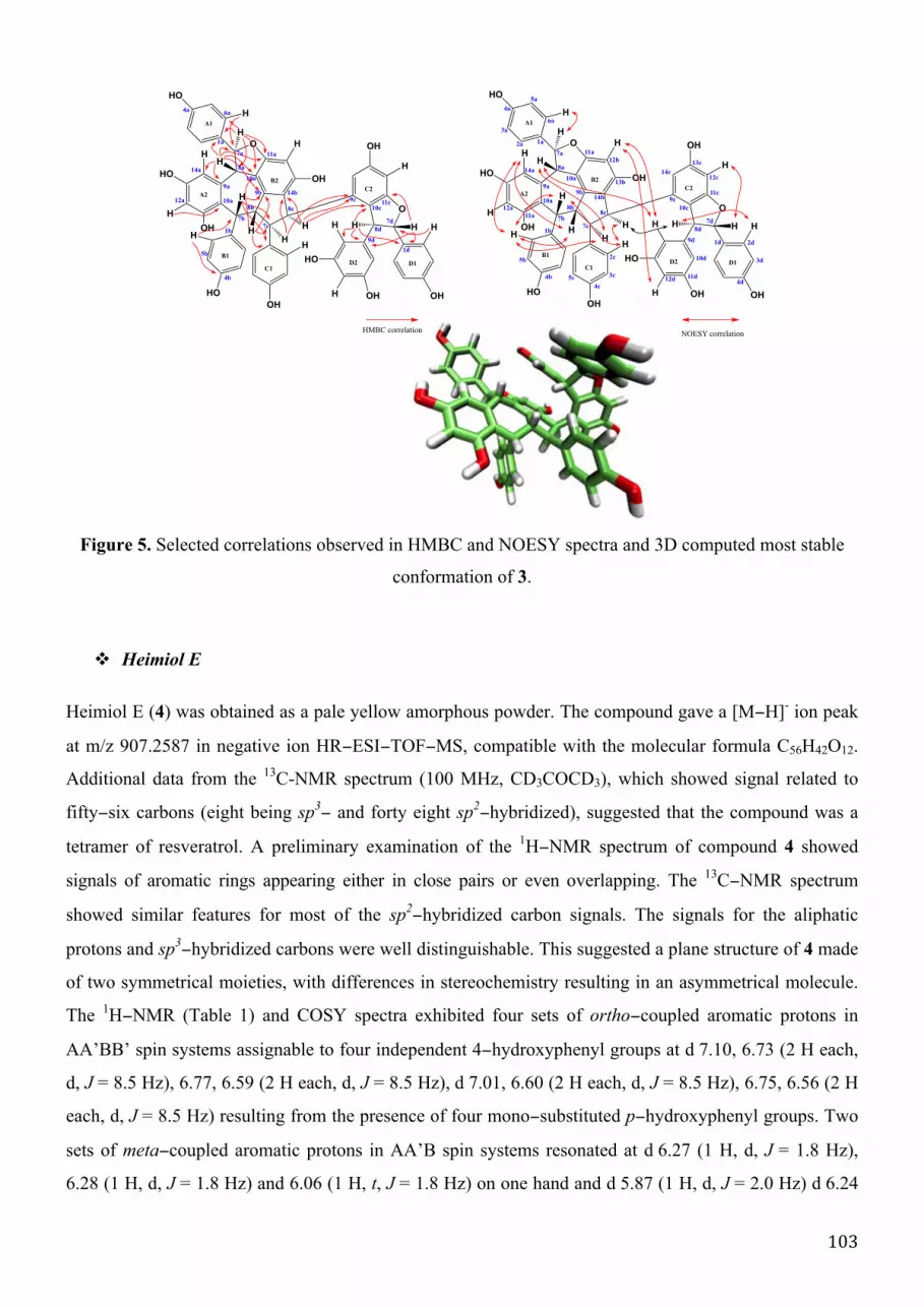
103
Figure 5. Selected correlations observed in HMBC and NOESY spectra and 3D computed most stable
conformation of 3.
" Heimiol E
Heimiol E (4) was obtained as a pale yellow amorphous powder. The compound gave a [M−H]- ion peak
at m/z 907.2587 in negative ion HR−ESI−TOF−MS, compatible with the molecular formula C56H42O12.
Additional data from the 13C-NMR spectrum (100 MHz, CD3COCD3), which showed signal related to
fifty−six carbons (eight being sp3− and forty eight sp2
−hybridized), suggested that the compound was a
tetramer of resveratrol. A preliminary examination of the 1H−NMR spectrum of compound 4 showed
signals of aromatic rings appearing either in close pairs or even overlapping. The 13C−NMR spectrum
showed similar features for most of the sp2−hybridized carbon signals. The signals for the aliphatic
protons and sp3−hybridized carbons were well distinguishable. This suggested a plane structure of 4 made
of two symmetrical moieties, with differences in stereochemistry resulting in an asymmetrical molecule.
The 1H−NMR (Table 1) and COSY spectra exhibited four sets of ortho−coupled aromatic protons in
AA’BB’ spin systems assignable to four independent 4−hydroxyphenyl groups at d 7.10, 6.73 (2 H each,
d, J = 8.5 Hz), 6.77, 6.59 (2 H each, d, J = 8.5 Hz), d 7.01, 6.60 (2 H each, d, J = 8.5 Hz), 6.75, 6.56 (2 H
each, d, J = 8.5 Hz) resulting from the presence of four mono−substituted p−hydroxyphenyl groups. Two
sets of meta−coupled aromatic protons in AA’B spin systems resonated at d 6.27 (1 H, d, J = 1.8 Hz),
6.28 (1 H, d, J = 1.8 Hz) and 6.06 (1 H, t, J = 1.8 Hz) on one hand and d 5.87 (1 H, d, J = 2.0 Hz) d 6.24
O
OH
HOOH
H
H
H
H
H
O
HH
HO
OH
OH
HO
OH
OH
HO
H
H
A1
A2
D2
C2
D1
B2
C1
B1
H
H
H
H
H
H
H
HMBC correlation
H
H
4a
12a
14b
14a
1b
1a
6a
7a
9a
10a
8a
4b
7b
8b
11a
10a
9b
5b
7c
8c
11c10c
9c
9d
7d
8d
1d
O
OH
HOOH
H
H
H
H
H
O
HH
HO
OH
OH
HO
OH
OH
HO
H
H
A1
A2
D2
C2
D1
B2
C1
B1
H
H
H
H
H
H
H
NOESY correlation
H
H
2a
4a
12a
14b
14a
1b
3a
1a
6a
7a
9a13b
10a
11a
8a
5a
4b
7b
8b
11a
10a
9b
12c
5b2c
12b
3c
4c
7c
8c
11c
10c
9c
5c
14c
13c
11d4d
9d2d
12d
7d
10d 3d
8d
1d

104
(1 H, d, J = 2.0 Hz) and 6.31 (1 H, t, J = 2.0 Hz), suggesting the presence of two independent
2,5−dihydroxyphenyl groups. The protons at positions 10 and 14 of the resorcinol rings resonated at
different chemical shifts thus indicating a strong steric hindrance preventing the rings free rotation.
Finally, the aromatic proton signals of two penta−substituted benzene rings were observed at d 6.16 and
6.19 (1 H, each br s). Two pairs of aliphatic hydrogen signals at d 5.29 and 4.29 (1 H each, d, J = 1.3 Hz)
and 5.69 and 4.43 (1 H each, d, J = 7.0 Hz) respectively suggested the presence of a dihydrobenzofuran
group bearing 4−hydroxyphenyl and 3,5−dihydroxyphenyl groups characteristic of oligostilbenes
biosynthesized from resveratrol derivatives. The other two pairs of aliphatic 1H−NMR signals, at d 4.07,
4.02 (1 H each, d, J = 6.8Hz) and 4.28, 4.22 (1 H each, br s) showed correlations in COSY as one spin
system. In the HMBC spectrum, correlations between the methine protons and the aromatic carbons, as
shown in Figure 6, allowed the establishment of connections between these aromatic rings and their
corresponding benzylic methine group. Cross peaks H−7a/C−10b, H−7a/C−11b, 8a/C−10b, 8a/C−11b and
8d/C−10c, 8d/C−11c indicated the formation of dihydrofuran rings between the resveratrol units A and B
as well as between units D and C. The H−7b/C−8c, H−7b/C−9c, H−7b/C−10c, H−7b/C−11c, H−8b/C−8c,
H−8b/C−10c, H−7c/C−8b, H−7c/C−9b, H−7c/C−10b, H−7c/C−11b, H−8c/C−8b and H−8c/C−10b
correlations suggested C−7b/C−10c, C−8b/C−8c and C−7c/C−10b linkages, thus establishing the
connections between resveratrol units B and C. In addition, COSY assignments showed that H−7b, H−8b,
H−8c and H−7c are linearly connected. The splitting patterns of the 1H−NMR signals confirmed the
linkage between C−8b and C−8c. The connections between the resveratrol units B and C are symmetrical.
This was supported by the fact that the protons and carbons of both resveratrol units B and C resonated at
similar chemical shifts, some of them being almost overlapped and became indistinguishable. The
NOESY spectrum showed cross peaks H−2(6)a/H−7a, H−2(6)a/H−8a and H−7a/H−8a (Figure 6).
Theoretically, these observations, together with the coupling constant value (J = 1.3 Hz) between H−7a
and H−8a, match a cis conformation. However, the only configuration compatible with the J value of 10.5
Hz and NOE correlations more corresponds to trans (7a=a and 8a=b). Another significant NOE
correlation H−7d/H−8d, together with a coupling constant of 7.0 Hz corresponded to a trans conformer of
the second dihyrofuran ring. Cross peaks H−7b/H−10a and H−7d/H−12c were compatible with syn
conformations. The H−8a/H−7b and H−8d/H−8c correlations suggested the conformation of H−8a/H−7b
and H−8d/H−8c to be syn. The H−7a/H−8a, H−7a/H−2(6)a, H−7b/H−8b, H−7b/H−2(6)b, H−7c/H−8c,
H−7c/H−2(6)c and H−7d/H−8d, H−7d/H−2(6)d correlations are explained by the close proximity of the
protons. This is supported by the distances between the corresponding protons in the 3−dimensional
molecular conformation, which were less than 3 Å. The HMBC and NOE correlations, as well as relative

105
stereochemistry of 4 is as shown in Figure 6. Both trans and cis conformers were optimized, and the
cis−conformer appeared much more stable (-36.5 and -31.1 kcal.mol-1.for H−a and H−b, respectively).
Figure 6. Selected correlations observed in HMBC and NOESY spectra and 3D computed most stable
conformation of 4.
" Other known oligostilbenes and stereoselectivity of the oxidative biosynthesis
In addition to the above four new compounds, thirteen known compounds were isolated and their
structures identified according to their spectral data and in agreement with those previously published.
These known compounds include heimiol A (5),[244] vaticaphenol A (6),[245] ampelopsin H (7),[249]
hopeaphenol (8),[250] isohopeaphenol (9),[251] hopeaphenol A (10), isohopeaphenol A (11),[252]
hemsleyanol D (12),[253] balanocarpol (13),[256] ampelopsins A (14) and C (15),[259] vaticanol A
(16)[246] and copalliferol A (17).[255] An HPLC analysis of the extract shows that virtually
all−significant peaks can be assigned to the above listed compounds (Figure 7).
Figure 7. HPLC analysis of the extract.
A thorough examination of the overall oligostilbenoid composition of N. heimii wood raised many
questions regarding the fascinating variety of chemical structures and biosynthesis of the above
oligostilbenes, which is driven by stereo− and regio−selective processes. It clearly appears that all isolated
compounds originate from resveratrol. As noted by previous workers,[243] the composition of this
dipterocarp, like all members of this family, is dominated by derivatives that include a benzodihydrofuran
NOESY
O
OH
HO
HO
OH
OH
O
OH
OH
OH
HO
HO
HH
H
H
H
H
A1
A2
D2
C2D1
B2
C1
B1
H
H
H
H
H
H
HH
H
H
HH
H H
H
H
H
H
HMBC
O
OH
HO
HO
OH
OH
O
OH
OH
OH
HO
HO
HH
H
H
H
H
A1
A2
D2
C2D1
B2
C1
B1
H
H
H
H
H
H
H
H
H
H
HH
H H
H
H
H
H 2.1A"
7d
8d
7c
8c
10c
7a
8a
8c
7c
7a
476
9
15
8
12
11916
1 4 5
5 17 13
106
moiety. Even though we did not isolate ε−viniferin (18), it is their most likely precursor. Sotheeswaran, in
his seminal review,[37] proposed a biosynthetic scheme for the generation of hopeaphenol. This scheme
however is not without several flaws; one of them being that it does not address the issue of the
stereoselectivity of the dimerization. The stereo−control of resveratrol oligomer synthesis was proposed
following a unique three−stage design. This was discussed mainly in terms of functionalization, namely
positional control through the introduction of a novel reagent of bromination.[55] In previous papers,[48],
[203] we have proposed that the outcome of the stilbene dimerization depends on how species would
approach toward each other within a pre−reaction complex, i.e. head−to−head (H/H) or head−to−tail
(H/T) as well as approaching through their Re/Si or Re/Re (Si/Si) faces. Applying this concept
to ε−viniferin as a substrate would explain the formation of most components of this plant extract. The
starting point of such an oxidatively induced process is electron transfer from the polyphenol to an
oxidative system, usually an oxidase in plants. The electron transfer occurs from either the neutral
polyphenol or its deprotonated form. In the former case a radical cation is formed, which is highly
unstable and undergoes heterolytic bond dissociation to form a phenoxy radical. The whole process is
called SET−PT (sequential electron transfer – proton transfer). The latter case depends on the pKa of the
molecule and the pH of the environment. In this instance the deprotonated polyphenol is somehow
activated to favour electron transfer. This mechanism is called SPLET (sequential proton loss electron
transfer). A phenoxy radical is formed here also. Disregarding the mechanism, a hydrogen atom transfer
(HAT) occurs from one of the OH group to the oxidative system. Calculation of the bond dissociation
enthalpy (BDE) of the different OH groups of stilbene derivatives shows that HAT occurs from the 4−OH
group to form the corresponding phenoxy radical, for which the spin density is delocalized over the
molecule with high values at C−1, C−3, C−5, C−8 and O−4 (Figure 8). While we are aware of debates on
whether the stilbene dimerization occurs through radical−radical (as proposed by Stekhan [48], [203]
following Barton’s general mechanism for phenolic oxidative coupling [256]) or radical−to−neutral
molecule reaction (as we proposed recently [57], [257]), we have chosen here to use the former
hypothesis for simplicity purpose. This radical−radical reaction has been rationalized for the dimerization
of flavonolignan derivatives on the basis of quantum chemical calculations .[258] It is shown that the
radical−radical formation is relatively fast but, in order to be thermodynamically favourable, the whole
dimerization mechanism requires subsequent molecular re-arrangements to form stable products. If one
would consider the group of tetrameric species formed by hopeaphenol, isohopeaphenol, hopeaphenol A,
isohopeaphenol A, one would recognized that they differ by the stereochemistry at C−8b and 8c in one
hand and C−7b and 7c in the other hand. The determinism for C−8b and 8c will be discussed first.

107
" Determinism at carbon C-8
Many oligostilbenoids reported in Figure 1 require, as a primary event, bond formation at C−8. The
regioselectivity of this atom is not explained by the spin density distribution of the phenoxy radical
(Figure 8), which exhibits five sites (C−8, C−6, C−4, C−2 and O−3) for further reactions following
oxidation, like covalent bond formation. As already shown for stilbene dimerization, the regioselective
dimerization of ε−viniferin units can be driven by the formation of non−covalent pre−reaction complexes
that may occur in solution. The long−range interaction driving such association was assessed with
quantum calculations describing dispersive forces, namely DFT−D calculations (see the Method section).
Considering all possible supramolecular arrangements, H/H and H/T with four possible approaches Re/Re,
Si/Si, Re/Si and Si/Re, eight geometries were obtained (Scheme 1). The differences in association energies
between the various complexes are not of any significance (Table 2), indicating that all possibilities may
occur in solution with an equal Boltzmann distribution. In these non−covalent complexes, intermolecular
distances were around 3.5 Å, which is typical of π−stacking interactions. Yet, π−stacking is not the only
driving force for such association, as H−bonding also plays a crucial role in stabilizing these non−covalent
pre-reaction complexes (Scheme 1). The formation of these non−covalent complexes appears the only
logical rationale for the stereoselectivity determinism at the C−8 of oligostilbenoid structures.[259]
Figure 8. Spin density distribution of the phenoxy radical.
Table 2. Association energies (kcal.mol-1) of the non-covalent pre−reaction complexes between two
ε−viniferin units for the different alignments as obtained by COSMO−B3P86−D2(s6=0.780)/def2−QZVP.
Non-covalent pre-reaction
complexes
[H/H]Re/Re [H/H]Si/Si [H/H]Si/Re [H/H]Re/Si [H/T]Si/Re [H/T]Re/Si [H/T]Re/Re [H/T]Si/Si
EAssociation -15.7 -19.8 -15.4 -15.0 -16.8 -10.0 -16.9 -10.9
[H/H]: head−to−head alignment; [H/T]: head−to−tail alignment.
0.34
0.39
0.28
0.22
0.29

108
From the [H/H]Si/Si and [H/T]Si/Si complexes, radical alignments 19 and 20 are obtained, respectively
(Scheme 1). Atoms C−8b and 8c would readily establish a bond leading to the dimeric intermediate 21.
This intermediate has both C−8b and 8c in S−configuration as in hopeaphenol (8). Conversely, a Re/Re
approach (alignments 22 and 23, see Scheme 1) leads to an inverted configuration for these carbon atoms
(intermediate 24) as in isohopeaphenol (9). From Re/Si approach, alignments 25−28 (Scheme 1) are
formed, all leading to a single intermediate (29), which would account for the formation of both
hopeaphenol A (10) and isohopeaphenol A (11).

109
Scheme 1. Oxidation and initial step of ε−viniferin (18) dimerization: {a} non−covalent complexation; {b}
oxidation; {c} C−C bond formation.
OH
O
HO
HO
OH
OH
O
OHO
HO
OH
OH
H
O
OOH
OH
HO
HO
H
18
27
A1
A2
B2
B1
[H/H]Si/Si
{c}
{a}
{b}
7b8b
26
H
O
OH
OH
OH
HO
O
O
HO
HO
OH
OH
H
O[H/H]Re/Si
O
O
OH
OH
HO
HO
H
O
O
HO
HO
OH
OH
H7b 7c
8b 8c
O
OOH
OH
HO
HO
H
28O
OHO
HO
OH
OH
H
[H/T]Re/Si
[H/T]Si/Re
25
O
O
HO
HO
OH
OH
HH
O
OH
OH
OH
HO
O[H/H]Si/Re
O OH
O
H
HO
O
H
OH
OH
OHHO
HO
OH
O
23
[H/T]Si/Si
O
O
OH
OH
HO
HO
H
O
O
HO
HO
OH
OH
H
(S) (S)
24
O
O
OH
OH
HO
HO
H
O
O
HO
HO
OH
OH
H
(S)
(R)
29
22
O
O
HO
HO
OH
OH
H
O
O
OH
OH
HO
HO
H
19
[H/H]Re/Re
O OH
O
H
O
HO
O
H
OH
OH
HOHO
HO
OH
20
[H/T]Re/Re
O
O
OH
OH
HO
HO
H
O
O
HO
HO
OH
OH
H
(R) (R)
21
{b}
{b}
{b}
{b}
{b}
{b}
{b}
{c}
{c}
{c}
{c}
{c}
{c}
{c}

110
Scheme 2. Role of non−covalent interactions in determining the stereochemistry at C−7b of oligostilbenes
with a dibenzocycloheptane moiety. ® represents either an OH group or an ε−viniferyl moiety.
R
O
O
HO
HO
OH
OH
H 7b
8b
20
R
O
O
HO
HO
OH
OH
H 7b
8b
R
O
HO
OH
H
O
HOO
H
H
O
HO
OH
O
O
HO
H
H
H
R
A1
A2
B2
B1
A1
A2
B2
B1
22
20b
22a
22b
8
9
R
O
HO
OH
H
O
O
HO
H
H
3.4 Å
R
O
O
HO
HO
OH
OH
H7b
8b
25
A1
A2
B2
B1
25b
7b
8b
O O
OH HO
HOOH
OH
OH
OHHO
HO OH
H
H
H
HH
H
H
H
7c
8c
31
4.1 Å
3.6 Å
11
R
O
HO
OH
H
HO
O
H
O
H
3.6 Å
R
O
HO
OH
H
HO
O
H
O
H
20a 30
3.3 Å
O O
OH HO
OHOH
OH
OH
OHHO
HO OH
H
H
HH H
H
H
H
R
O
HO
OH
H
HO
O
H
O
H
25a
103.7 Å
R
R
R
R
R
R
7c
8c8b
7bA2
B1
A4
B3

111
The stability (given by the difference in Gibbs energy between the intermediate and the
corresponding pair of phenoxy radicals) of the three intermediates (21, 24 and 29) was evaluated
by quantum calculations in the DFT formalism. The ΔG of formation of these intermediates were
-16.4, -18.5 and -21.8 kcal.mol-1 for 21, 24 and 29, respectively, as obtained by
B3P86−D2/def2−SVP, which confirm that all compounds are thermodynamically favoured, i.e.
the feasibility of each process mainly depends on the stability of the respective non−covalent
complexes. Therefore the ratio of the different products depends on both the relative
thermodynamic stability of these intermediates, and the stability of the corresponding molecular
approaches, the latter being driven by long−range interactions (π−stacking and H−bonding) and
steric hindrance towards a given approach.
" Further reactions following the primary C−8/C−8 bonding event
Once the C−8 configuration is set, the subsequent intermediates continue reacting according to
intramolecular re-arrangements. Concerning C−7b and C−7c configurations, the key parameter is
the re-arrangement of rings A2 and B1 on one hand and B3 and A4 (the corresponding rings in
the other monomer) on the other hand (Scheme 2). Carbon C−8 of intermediates 21, 24 and 29 is
sp3−hybridized and the changes of the torsion angle C−7/C−8/C−9/C−10 lead to conformational
re-arrangements determining the C−7 stereochemistry. All intermediates are stabilized with
π−stacking interactions between resorcinol ring A2 and semiquinone ring B1 holding them
nearly parallel (Scheme 2). In addition, some intermediates are further stabilized by H−bonds
with interatomic distances of ca. 2.0 Å. In these conformations, the C−7 atom of one unit is
facing the resorcinol ring of the other unit, thus favouring the formation of the seven−membered
rings of 8, 9, 10, and 11 (Scheme 2). Ring closure follows an intra SN1 nucleophilic substitution
from the carbon in para position to the OH group of the resorcinol ring onto C−7 of the other
unit (Scheme 2). The nucleophilic attack is favoured since i) these two atoms are relatively close
to each other in the intermediates (distance around 3.5 Å); and ii) the former atom is
electron−rich with an atomic charge of -0.5, while the C−7 carries an atomic charge of -0.06.***
According to the intermediate and molecular re-arrangement, one of the four isomers is formed.
*** A similar chemical pathway is proposed to explain the formation of 13 and 14. In this case, the semiquinone intermediate comes from
the phenoxy radical formed after oxidation of ε‐viniferin, which then catch one hydrogen atom from its neighboring environment.

112
In principle two other hypothetic compounds (30 and 31) could be formed (Scheme 2). However,
detailed quantum mechanics−based calculations showed these two compounds to be
energetically much less favoured (Table 3) and therefore in much lower occurrence if any in
plants. The four identified compounds (8-11) exhibited Gibbs energies of formation (∆�!!) of -
25.3, -29.4, -22.1 and -17.3, respectively, indicating high stability (Table 3).
Table 3. ΔE of formation (kcal.mol-1) of 8−11 and 26−27 as obtained by COSMO−B3P86−D2
(s6=0.780)/QZVP
8 9 10 11 30 31
ΔE -64.7 -60.7 -60.1 -61.6 -41.2 -31.0
ΔG -25.3 -29.4 -22.1 -17.3 - -
It is remarkable that only isohopeaphenol A (11) includes a moiety where H−7b and H−8b
are in cis configuration, while hopeaphenol (8), isohopeaphenol (9) and hopeaphenol A (10) only
include trans configurations. At the same time, balanocarpol (13) with a 7b−8b cis configuration
is far more abundant that its diastereoisomer ampelopsin A (14), which displays a trans
configuration. This is probably related to the bulkiness of the substituent at C−8b, which would
favour or disfavour the relative positioning of the rings A2 and B1 in intermediates 21, 24 and 29
(Scheme 1). Indeed, DFT calculations indicated 13 to be more stable than 14 by 3.7 kcal.mol-1.
From intermediate 21, another chemical pathway is allowed and rationalizes the
biosynthesis of heimiol C (2). The molecular arrangement of 21 indeed exhibits short distances
of around 3 Å between rings A2 and B1 as well as C1 and D2 (Scheme 3). In this case, the
nucleophilic attack on the quinone methide group does not originate from the neighbouring
resorcinol ring, but from a water molecule interacting with C−8b and C−8c. DFT calculations
confirmed the stability of intermediate 32 since the hydrolysis reaction exhibits ∆�!! of -3.4
kcal.mol-1. Similar processes are expected from the other intermediates 24 and 29, which would
probably yield other stereoisomers of 2. Even if this is still hypothetical (the compounds have not
been observed yet), there are no particular molecular restraints forbidding the formation of these
compounds.

113
Scheme 3. Biosynthetic route to 2.
Biosynthetic intermediates 21 and 29 (from [H/T]Si/Si and [H/T]Si/Re complexes, respectively)
are the precursors of 4 and 7. Through a mechanism similar to that described above, the quinone
methide groups are subjected to nucleophilic attack from the resorcinol rings, though located on
the opposite moieties (Scheme 4).
Scheme 4. Biosynthetic route to 7.
The feasibility of this mechanism was here as well confirmed by DFT, which showed that 4
and 7 were more stable than their corresponding intermediates (21 and 29) according to their
energies of formation of -42.7 and -44.3 kcal.mol-1, respectively.
H
O
H
HH
O
HO
H
H
OH
H
HH
O
O
H
H
O
O
H
HH
O
O
OH HOOHHO
OHHO
8b
7b7c
8c
21
OH
OH
HOOH
HO
HO
2
32
3.3 Å3.2 Å
29 7H
O
O
H
HH
O
OH
OH
8b
7b
7a
8a
OH
OH
HO
OH
HO
HO
OH

114
" Other pathways
Hemsleyanol D (12) is likely to derive also from intermediate 29 deriving itself from the
[H/T]Re/Si alignment (Scheme 1). In this case, both carbon atoms C−7b and C−7c are subjected to
nucleophilic attack from the neighbouring resorcinol rings. Indeed, the 3D conformation allows
C−7b and C−7c to be attacked by C−10a and C−14b, respectively (Scheme 5). This reaction is
thermodynamically favoured, exhibiting a -44.3 kcal.mol-1 Gibbs energy of formation.
Scheme 5. Biosynthetic route to 12.
Compounds 3, 6, 15 and 16 are obviously biosynthesized through a different mechanism.
Even if less thermodynamically favoured than from the 4−OH group of the stilbenoid moiety,
HAT from one resorcinolic OH group of resveratrol is thermodynamically feasible, leading to a
quinoid radical that would combine with another radical originating from 4−OH oxidation (32,
Scheme 6). The spin density distributions on both radicals allow various further linkages,
including C−8a/C−10b bond formation. As a result, the first established inter−stilbene bond
would be C-8a/C−10b (33). DFT calculation confirmed that 33 was the most stable intermediate
by 2.1 kcal.mol-1 compared to the other possible dimers obtained by radical−radical dimerization.
The bond formation breaks the planarity of the ring. The dimerization process is thus
thermodynamically feasible only if followed by tautomerism with the adjacent OH group.[257]
Next, the methide (C−7a) would be attacked by the 11−OH group leading eventually to
ε−viniferin (18), or by C−8b, thus yielding intermediates 34 and 35. The rigidity of the
cyclopentane ring does not allow the quinone methide to be subjected to any intramolecular
nucleophilic attack necessary for re-aromatization of ring A2. Therefore, intermediate 35 is ready
29
12
H
O
O
H H
O
HO
HO
8a
7b
8bHO
HOOH
OH
O
O
HO
H
H
3.3 Å
3.6 Å
10a
1.8 Å
7c
14b
14b
10a

115
to react with the B−ring of another resveratrol unit followed by a new cycle of intramolecular
phenolic oxidative coupling leading to 3, 6, 15 and 16. The structure of copalliferol A (17) seems
to vindicate these biosynthetic hypotheses. The only structural difference between 17 and 15 is
the C−7a/C−2b instead of C−7a/O−11b bond (Figure 1). This C−7a/C−2b bond is very unusual
and reflects the high reactivity of the quinone methide intermediate that would result from the
addition of a resveratrol unit onto 35. DFT calculation confirmed that 17 is much more stable
than 16, itself more stable than 15 based on the relative energies of -5.6 and -10.5 kcal.mol-1,
respectively.
Scheme 6. Biosynthetic route to intermediates 3, 6, 15 and 16.
Finally, the biosynthesis of 1 and 5 can also be understood by the nucleophilic attack of the
methide (C−8b) of 33 with a water molecule, leading to intermediate 36 (Scheme 7). A second
nucleophilic attack onto C−7a leads to isochroman 37. Depending on the orientation of the
quinone methide moiety and its nucleophilic attack from C−10a, two diastereoisomers can be
obtained, which correspond to heimiol A or B. The formation of both 1 and 5 was confirmed by
HO
OH
O
32
33
34
35
3
6
15
16
O
O OH
OH
OH
HO
O
HO OH
O
OH
HO
H
H
OH
HO
HO
OH
HO
O
3.4 Å
4b
8a
10bHO
O
OH
8a
10b
4b
32: spin density distribution
7a
2b
11b
7b
8b
7b
7a 7a
4a
4a
4a
4b
11a11a
11b
10b10b
8a8a
8b
10b
8a
11b

116
their low Gibbs energies of formation, i.e., -31.1 and -32.5 kcal.mol-1, respectively.
Scheme 7. Biosynthetic route to 1 and 5.
3. Conclusion
Besides providing a series of new and structurally provocative compounds, namely heimiols
B−E, the study of the oligostilbenes from the heartwood of N. heimii provided a series of
derivatives characterized by a high consistency in their plausible biogenesis. In this respect, the
dominant features consist of i) electron abstraction from the frontier orbitals of the polyphenol
and the subsequent (or concomitant) proton loss from an OH group of a suitable precursor; ii) the
combination of species aligned in specific supramolecular arrangements due to π−stacking and
H−bonding interactions; and iii) the intra− or inter−molecular bond formation as a result of
radical−radical reactions or nucleophilic substitutions, followed by the quenching of quinone
methide intermediates. Electron transfer from the monomer or certain intermediates must be
enzymatically controlled whereas the other steps of the whole process most probably result in
chemical re-arrangements, thermodynamically favoured. Supramolecular self−assembly of units
through non−covalent bonding is confirmed to be a significant driving force in some key−steps
of the stereoselective oligostilbene biosynthesis. These molecular assemblies formed prior to
oxidation provide powerful indications for the targeted stilbenoid oligomer synthesis.
Non−covalent trimers were also envisaged (Figure 9) explaining the origin of stilbenoids of
higher degrees of polymerization. This work sheds new light on the biogenesis of a number of
natural product classes. A proper understanding of oligostilbenoid biosynthesis may provide
more refined approaches towards biomimetic syntheses, which are likely to become a most
HO OH
OH
OH
OH
OO H
H
HO OH
O
OH
OH
O
OH
HO OH
O
OH
OH
OH
O
a
b
a
b
5
1
32
3637
10a10b 8a
7a8b
7b7a

117
practical approach to the generation of series of derivatives and analogues of some
representatives endowed with interesting bioactivities.
Figure 9. Predicted 3D geometry of a non-covalent trimer. Here we have provided a comprehensive (bio)synthetic mechanism of different
oligostilbenoids isolated from Neobalanocarpus heimii., whereas the enzyme was not taken into
account. We believe that the enzyme mainly initiate oxidation but has a minor role in the further
steps oligomerisation. Therefore, the active site of the corresponding (yet unknown) enzyme is
most probably at the surface, relatively flexible and weakly specific. We are confident that our
work is a first crucial step to get in-depth into the knowledge of the biosynthetic oligomerisation
of stilbenes.†††
4. Method Section
General Procedures. The following instruments were used: optical rotation, JASCO J-715 CD
Spectropolarimeter, 1D− and 2D−NMR spectra, BRUKER 300, 400 and 500 (chemical shifts
given in d values); TOF−MS spectra, Agilent 6200 Series; C−18 Column, Genesis (Jones
chromatography), Synergi Hydro−RP (Phenomenex), Hypersil (Thermo) and Chromolith
(Merck).
††† This section in italic was not included in the published paper.
1.8 Å
2.0 Å
3.3 Å
3.2 Å

118
Plant material. Heartwood of Neobalanocarpus heimii was collected in Malaysia, see[244].
Methods of calculation. Geometries and energies of all compounds were obtained using the
B3P86 functional. This hybrid functional provided most of the geometrical conformation of
stand−alone oligostilbenoids at the DFT level while B3P86−D2 was used as this method includes
dispersive corrections and allows high accuracy in the description of non−covalent interactions.
This method was used systematically with s6 = 0.780 as recently re-parameterized for phenolic
derivatives.[184] The def2−SVP and QZVP basis sets were used for geometry optimization and
energy calculations, respectively. All calculations were performed either with Gaussian 09 [260]
or ORCA.[222]
The association energy (EAssociation, Table 2) between fragments A and B was calculated as
follow:
�!""#$%&'%#( = �!" − (�! + �!)
where (�!") is the energy of the non−covalent complex, (�!) and (�!) are the energies of
isolated fragments A and B, respectively.
The solvent effect was accounted implicitly, in which the solute is embedded into a shape
adapted cavity surrounded by a structure less continuum. Conformational analysis and
interaction energies were carried out using PCM (Polarizable Continuum Model) [261] or
COSMO (COnductor−like Screening Model).[262] Methanol was selected as being the solvent
used to extract compounds and to measure their physical−chemical properties.
Extraction and isolation. Crude methanol extract (15 g) from the heartwood of N. Heimii was
fractionated by semi−preparative chromatography on C18 and eluted by gradient elution of water
and MeCN (84:16 to 77:23 in 7 minutes, followed by 77:23 to 66:34 in 18 minutes) to yield 57
fractions. Fraction 18 (RT =11.2 min) was further analysed by HPLC on a Chromolith column,
eluted by isocratic elution of H2O/MeOH (84:16) to yield compounds 1 (31.0 mg) and 5 (116.8
mg). Compound 2 (5.3 mg) was obtained together with compound 11 (131.9 mg) from
purification of fraction 29 (RT =15.5 min) over a Genesis C18 column, eluted by isocratic elution
of H2O/MeOH (77:23). Fraction 40 (RT =20.2 min) was further processed over a Synergi
Hydro−RP (Phenomenex) column (eluted by H2O/MeOH, 75:25) and yielded compounds 3 (14.3

119
mg), 8 (9.1 mg) and 15 (29.9 mg). Fraction 51 (RT =25.5 min) yielded compounds 4 (5.2 mg) and
17 (7.6 mg) from purification over a Genesis C18 column eluted by H2O/MeOH (75:25).
Heimiol B (1). Light brown amorphous powder; negative ion HR−TOF−MS [M−H]- m/z
469.1288 (calc. for C28H22O7: 471.1444). 1H− and 13C−NMR data are listed in Table 1. HMBC
correlations are as in Figure 2 (selected) and H−2(6)a/C-4a, H−3(5)a/C−4a, H−12a/C−10a,
H−12a/C−13a, H−14a/C−10a, 14a/C−13a, H−2(6)b/C−4b, H−3(5)b/C−4b, H−7b/C−1b,
H−7b/C−2(6)b, H−7b/C−9b, H−8b/C−1b, H−8b/C−9b, H−8b/C−10b, H−8b/C−14b H−12b/C−10b,
H−12b/C−11b, H−12b/C−13b, H−14b/C−10b, H−14b/C−13b, H−7b/C−10a and H−7b/C−11a.
NOESY correlations, see Figure 2.
Heimiol C (2). Dark brown amorphous powder; negative ion HR−TOF−MS [M+Na]+ m/z
947.2679 (calc. for C56H44O13: 925.2860). 1H and 13C NMR data are listed in Table 1. HMBC
correlations are as in Figure 4 (selected) and H−2(6)a/C−6(2)a, H−2(6)a/C−1a, H−2(6)a/C−4a,
H−2(6)a/C−7a, H−3(5)a/C−5(3)a, H−2(6)b/C−6(2)b, H−2(6)b/C−4b, H−3(5)a/C−1a, H−3(5)a/C−4a,
H−3(5)b/C−1b, H−3(5)b/C−4b, H−3(5)b/C−5(3)b, H−12b/C−10b, H−12b/C−11b, H−12b/C−13b,
H−12b/C−14b, H−14b/C−10b, H−14b/C−12b and H−14b/C−13b. NOESY correlations, see Figure
4.
Heimiol D (3). Pale yellow amorphous powder; negative ion HR−TOF−MS [M−H]- m/z
905.2581 (calc. for C56H42O12: 907.2755). 1H and 13C NMR data are listed in Table 1. HMBC
correlations are as in Figure 5 (selected) and H−2(6)a/C−6(2)a, H−2(6)a/C−4a, H−3(5)a/C−1a,
H−3(5)a/C−2(6)a, H−3(5)a/C−4a, H−7a/C−1a, H−7a/C−2(6)a, H−7a/C−8a, H−7a/C−9a, H−8a/C−1a,
H−8a/C−7a, H−8a/C−10a, H−12a/C−10a, H−14a/C−10a, H−14a/C−11a, H−2(6)b/C−6(2)b,
H−2(6)b/C−3(5)b, H−2(6)b/C−4b, H−3(5)b/C−1b, H−3(5)b/C−5(3)b, H−3(5)b/C−4b, H−7b/C−1b,
H−7b/C−2(6)b, H−7b/C−9b, H−8b/C−10b, H−8b/C−7c, H−2(6)c/C−6(2)c, H−2(6)c/C−1c,
H−3(5)c/C−1c, H−3(5)c/C−5(3)c, H−3(5)c/C−4c, H−7c/C−10b, H−7c/C−8b, H−8c/C−7b, H−8c/C−7c,
H−8c/C−10b, H−12c/C−10c, H−12c/C−11c, H−2(6)d/C−6(2)d, H−2(6)d/C−3(5)d, H−2(6)d/C−4d,
H−3(5)d/C−1d, H−3(5)d/C−5(3)d, H−3(5)d/C−4d, H−7d/C−2(6)d, H−7d/C−1d, H−8d/C−11(13)d,
H−8d/C−10c and H−12d/C−11(13)d. NOESY correlations, see Figure 5.
Heimiol E (4). Pale yellow amorphous powder; positive ion HR−TOF−MS [M+H]+ m/z
907.2587 (calc. for C56H42O12: 907.2755). 1H and 13C NMR data are listed in Table 1 and 2.

120
HMBC correlations are as in Figure 6 (selected) and H−2(6)a/C−6(2)a, H−2(6)a/C−4a,
H−2(6)a/C−7a, H−3(5)a/C−1a, H−3(5)a/C−5(3)a, H−3(5)a/C−4a, H−7a/C−2(6)a, H−7a/C−8a,
7a/C−9a, 8a/C−1a, 8a/C−7a, 8a/C−9a, H-8a/C−10a, H−10a/C−11a, H−14a/C−13a, H−2(6)b/C−6(2)b,
H−2(6)b/C−4b, H−2(6)b/C−7b, H−3(5)b/C−1b, H−3(5)b/C−5(3)b, H−3(5)b/C−4b, H−7b/C−1b,
H−7b/C−2(6)b, H−7b/C−8b, H−7b/C−9b, H−7b/C−13c, H−7b/C−14c, H−8b/C−1b, H−8b/C−7b,
H−8b/C−9b, H−8b/C−10b, H−8b/C−14b, H−8b/C−9c, H−12b/C−10b, H−12b/C−11b, H−12b/C−13b,
H−12b/C−14b, H−12b/C−7c, H−2(6)c/C−6(2)c, H−2(6)c/C−4c, H−2(6)c/C−7c, H−3(5)c/C−1c,
H−3(5)c/C−5(3)c, H−3(5)c/C−4c, H−7c/C−1c, H−7c/C−2(6)c, H−7c/C−8c, H−7c/C−13b,
H−7c/C−14b, H−8c/C−1c, H−8c/C−7c, H−8c/C−9c, H−8c/C−10c, H−8c/C−11c, H−8c/C−14c,
H−8c/C−9b, H−12c/C−10c, H−12c/C−11c, H−12c/C−13c, H−12c/C−14c, H−12c/C−7b,
H−2(6)d/C−6(2)d, H−2(6)d/C−4d, H−2(6)d/C−7d, H−3(5)d/C−1d, H−3(5)d/C−5(3)d, H−3(5)d/C−4d,
H−7d/C−8d, H−7d/C−2(6)d, H−7d/C−9d, H−8d/C−7d, H−8d/C−9d, H−8d/C−10d, H−10d/C−11d,
H−10d/C−12d, H−12d/C−11d, H−12d/C−13d, H−12d/C−14d, H−14d/C−12d and H−14d/C−13d.
NOESY correlations, see Figure 6.

121
122
Chapter 6 – Antioxidants
The plant kingdom provides the largest number of antioxidants. There are mainly terpenoids (e.g.
vitamin E, vitamin C) and polyphenols (e.g., flavonoids, stilbenoids, lignans, Schiff bases), and
in a less extent alkaloids (e.g., caffeine, theobromine). The large variety of the chemical
structures of polyphenols allows many important biological activities, including antioxidant
activity. Over the past two decades, many works have been dedicated to theoretical elucidation
of the free radical scavenging capacity of polyphenols.[263]–[268], [268]
QM allows calculating different physico−chemical parameters related to the antioxidant
activity including OH−BDE (the major descriptor of the antioxidant activity), IP (Ionization
Potential), ETE (Electron Transfer Energy) from deprotonated forms, and electronic and spin
density distributions. The use of hybrid DFT functionals provides an accurate description of the
structure-activity relationships of free radical scavenging, in agreement with experiments.[269]–
[274] The aim of the theoretical calculations is to accurately predict activity of new compounds,
as a crucial stage for pharmaceutical, cosmetic and food industries. The free radical scavenging
by a polyphenol antioxidant proceeds by H atom transfer through four possible mechanisms (see
section I).
This chapter includes two sections. Section I describes antioxidant properties of phenolic
Schiff bases and section II describes non−covalent association of antioxidants in lipid−bilayer
membranes.

123
Note for readers: This article has been published in the Journal of Computer-Aided Molecular
Design (Anouar et al., J Comput Aided Mol Des, 10.1007/s10822-013-9692-0). My contribution
concerns the study of the SPLET mechanism. The entire manuscript is incorporated in this PhD
to keep consistency.
The figure and reference formats and positions are sometimes modified compared to the original
article to keep homogeneity in the thesis.
Section I. Antioxidant properties of phenolic Schiff bases:
Structure activity relationship and mechanism of action
El Hassane Anouara,*, Salwa Raweha,b, Imene Bayacha,c, Muhammad Tahaa, Mohd Syukri Baharudina ,
Florent Di Meod, Mizaton Hazizul Hasanb, Aishah Adamb, Nor Hadiani Ismaila,e, Jean-Frédéric F.
Webera,b, Patrick Trouillasd,f-g*
Abstract: Phenolic Schiff bases are known for their diverse biological activities and ability to
scavenge free radicals. To elucidate (i) the structure−antioxidant activity relationship of a series
of thirty synthetic derivatives of 2−methoxybezohydrazide phenolic Schiff bases and (ii) to
determine the major mechanism involved in free radical scavenging, we used density functional
theory (DFT) calculations (B3P86/6−31+(d,p)) within polarizable continuum model. The results
showed the importance of the bond dissociation enthalpies related to the first (BDE) and second
(BDEd) hydrogen atom transfer (intrinsic parameters) for rationalizing the antioxidant activity. In
addition to the number of OH groups, the presence of a bromine substituent plays an interesting
role in modulating the antioxidant activity. Theoretical thermodynamic and kinetic studies
demonstrated that the free radical scavenging by these Schiff bases mainly proceeds through
proton−coupled electron transfer (PCET) rather than sequential proton loss electron transfer
(SPLET), the latter mechanism being only feasible at relatively high pH.
Keywords: Schiff bases, Antioxidant, DFT, kinetics, free−radical−scavenging, BDE,
structure−activity−relationship.

124
aAtta-ur-Rahman Institute For Natural Product Discovery (RiND), Universiti Teknologi MARA, 42300 Bandar
Puncak Alam, Selangor D. E, Malaysia.
bFaculty of Pharmacy, Universiti Teknologi MARA, Puncak Alam Campus, Malaysia.
cLCSN - Faculté de Pharmacie, Université de Limoges, 2 rue du Dr Marcland, 87000 Limoges, France
dINSERM, UMR-S850 / Faculté de Pharmacie, Université de Limoges, 2 rue du Dr Marcland, 87000 Limoges,
France
eFaculty of Applied Sciences, Universiti Teknologi MARA, 40450 Shah Alam. Malaysia.
fLaboratoire de Chimie des Matériaux Nouveaux, Université de Mons, Place du Parc, 20, B-7000, Mons, Belgium
gRegional Center of Advanced Technologies and Materials, Department of Physical Chemistry, Faculty of Science,
Palacký University, 17. Listopadu 1192/12, 77146 Olomouc, Czech Republic
Corresponding author:
Atta-ur-Rahman Institute for Natural Product Discovery (RiND), Universiti Teknologi MARA Kampus
Puncak Alam, 42300 Bandar Puncak Alam, Selangor D. E, Malaysia.
Tel: +603 3258 4771; fax:+603 3258 4770
E-mail addresses: (E. Anouar), [email protected] (P. Trouillas).

125
1. Introduction
Natural and synthetic phenols including flavonoids, phenolic acids, stilbenoids and curcuminoids
have been described as powerful antioxidants, being able to efficiently scavenge free
radicals.[218], [275], [276] Schiff bases form an important class of synthetic phenolic
compounds, substituted by a hydrazone moiety. They are known for their various fields of
application including inorganic chemistry, biological and analytical chemistry.[277], [278]
Several studies focused on their biological activities as antibacterial,[279]–[282] anticancer,[278]
and antifungal activities.[283], [284] Recently, we reported the antileishmanial activity of the
series of synthetic Schiff bases studied in the present work (Fig. 1).[285] Schiff bases also
showed potent antioxidant activity to scavenge free radicals. In two recent studies, we reported
the antioxidant activity of acylhydrazide and 2,4,6−trichlorophenylhydrazine Schiff bases as
DPPH radical and super oxide anion scavengers.[286], [287] Free radical scavenging capacity of
(poly)phenols is generally attributed to the hydrogen atom lability of the OH groups attached to
aromatic rings (Ar);[269], [271], [288], [289] however in some other antioxidants, NH and SH
groups may provide labile hydrogen.[290]–[296] Antioxidants (ArX-H) may scavenge free
radicals (R•) by H atom transfer through one of the four mechanisms:
(i) HAT (hydrogen atom transfer) and PCET (proton-coupled electron transfer)
ArX−H + R• → ArX• + R−H (eq. 1)
This is the direct HAT, which is purely governed by the homolytic bond dissociation
enthalpy (BDE) of X−H; note that the lower the BDE, the more important the role of the
corresponding XH group in the antioxidant activity. PCET is distinguished from the pure HAT
as it involves several molecular orbitals in an H−bonding pre−reaction complex.[297]–[300] The
proton transfer is coupled to the electron transfer that occurs from a lone pair of the antioxidant
to the SOMO (singly occupied molecular orbital) of the free radical.
ii) ET−PT (Electron Transfer − Proton Transfer) or SET−PT (Sequential ET−PT)
ArX−H + R• → ArXH+• + R- → ArX• + R−H (eq. 2)

126
The first step of this reaction leads to the formation of the radical cation ArXH+•, which
easily undergoes heterolytic dissociation of X−H bond leading to the same final products than
those yielded by PCET. The first step is mainly governed by the ionization potential (IP) of the
antioxidant.
iii) SPLET (Sequential Proton−Loss−Electron−Transfer)
ArX−H → ArO- + H+ (eq. 3)
ArX- + R• → ArX• + R- (eq. 4)
R- + H+ → RH (eq. 5)
In this mechanism, a proton is lost prior to electron transfer from the subsequent anion to the free
radical. SPLET is strongly favoured under alkaline conditions, which may help in the proton loss
of the first step.[301], [302]
iv) AF (Adduct Formation)
ArX−H + R• → [ArXH−R]• → metabolites or ArX• + RH (eq. 6)
This mechanism is relatively specific to •OH free radicals. It has been observed in radiolytic
solutions. The radical may add on double bonds and aromatic rings. This mechanism is not
considered in this work. As often shown in the literature, intrinsic parameters including BDE, IP
and ETE (electron transfer enthalpy) rationalize free radical scavenging capacities [269], [271],
[288], [303] as the thermodynamic balance of the first three mechanisms is the same (ΔGPCET =
ΔGET-PT = ΔGSPLET). However to tackle the mechanisms of action, kinetic parameters should be
considered.[268], [271], [296], [301], [304], [305]
In the present study, we elucidate the structure−antioxidant activity relationship for a series
of 30 synthetic derivatives of 2−methoxybezohydrazide phenolic Schiff bases (Fig. 1). The
Schiff bases differ from the nature of the R− (aromatic) moiety attached to the nitrogen atom
engaged in the –N=C double bond (Fig. 1). The compounds can be subdivided into three classes,
namely (i) compounds 1-25, where the aromatic ring is a benzene substituted with different OH,

127
OCH3, halogen, COOCH3 and NO2 groups, (ii) compounds 26−28, in which R is an
unsubstituted pyridine attached at the three different possible positions and (iii) compounds
29−30, in which R is an unsaturated five−membered ring containing one heteroatom. The
positive or negative contributions of different descriptors (e.g, number and position of OH
groups, BDE, double (BDEd), IP, spin density distribution, intramolecular hydrogen bonds,
presence of electron donating or withdrawing groups, and structural parameters) have been
examined. The elucidation of the mechanism of action is based on a combined experimental and
theoretical approach. For the theoretical approach, transition state and Marcus−Levich−Jörtner
theories were used to evaluate atom (PCET mechanism) and electron transfer (SPLET
mechanism), respectively.
Monohydroxylated
1: R2 = OH 2: R3 = OH 3: R4 = OH Dihydroxylated
4: R2 = R3 = OH 5: R2 = R4 = OH 6: R2 = R5 = OH 7: R3 = R4 = OH 8: R3 = R5 = OH Trihydroxylated
9: R2 = R4 = R6 = OH 10: R3 = R4 = R5 = OH
Monomethoxylated
11: R3 = OCH3 12: R4 = OCH3
Dimethoxylated
13: R3 = R4 = OCH3 14: R3 = R5 = OCH3
1-OH and 1-OCH3
15: R2 = OH, R4 = OCH3 16: R2 = OH, R5 = OCH3 17: R3 = OH, R4 = OCH3
Halogenated
18: R3 = Br, R4 = OH 19: R3 = Br, R4 = Cl 20: R4 = F 21: R4 = Cl 22: R2 = I, R3 = OH, R4 = OCH3
Other substitution
23: R4 = COOCH3 24: R4 = NO2
No substitution
25: Ri = H (i = 2 to 6)
26 27 28
29 30
Figure 1. Structures of the synthesized Schiff bases.
OCH3
NH
O
N
R2
R3
R4
R5
R6
1
23
4
56
OCH3
NH
O
N
N OCH3
NH
O
N
N
OCH3
NH
O
N
N
OCH3
NH
O
N
O
OCH3
NH
O
N
S

128
2. Methodology
" Experimental synthesis
The N−benzylidene−2−methoxybenzohydrazide phenolic derivatives were synthesized by
refluxing mixtures of 2 mmol 2−methoxy−benzohydrazide and different aldehydes in methanol
and in the presence of catalytical amount of acetic acid for 3 hours, according to the previously
described method.[285] The reaction progress was monitored by thin layer chromatography
(TLC); after completion, the solvent was evaporated under vacuum to afford the crude products,
which were further re-crystallized in methanol. Needle−like pure products in good to excellent
(0.40g (78%) - 0.58 (92%)) yields were obtained.[285]
" DPPH free radical scavenging capacity
The antioxidant capacity was evaluated as the capacity of the 30 different compounds to
scavenge the 1,1−diphenyl−2−picrylhydrazil (DPPH) free radical, following the Blois’
method.[306] The reaction mixture contains 5 µL of test sample (1 mM in DMSO) and 95 µL of
DPPH (Sigma, 300 µM) in ethanol; it was placed into a 96-well microtiter plate and incubated at
37º C for 30 min. The UV/Vis light absorbance was measured at 515 nm on microtiter plate
reader (Molecular Devices, CA, and USA). The percentage of free radical scavenging was
determined with respect to DMSO control. Following free radical scavenging vs. compound
concentration, IC50 was measured, which represent the concentration of compounds that
scavenge 50% of DPPH radicals (i.e., 50% of absorbance at 515 nm). Propyl gallate was used as
a positive control. All chemicals used were of analytical grade (Sigma, USA).
" General computational details
Geometry optimization and frequency calculations have been carried out using density functional
theory (DFT) as implemented in Gaussian09.[260] To elucidate the structure−antioxidant
activity relationship of polyphenols, the hybrid functional B3P86 appears reliable.[221], [270],
[273]a Increasing the number of polarization and diffuse functions have no significant effects on
theoretical results, especially on BDEs and IPs,[270], [271] therefore the calculations were
129
performed with the 6−31+G(d,p) basis set. The ground states (GSs) geometries were confirmed
by the absence of any imaginary frequency, while for the transition states (TSs) of PCET one
imaginary frequency was obtained, which corresponds to H atom transfer. It has been shown that
the B3P86 hybrid functional underestimates activation energy (ΔG#) especially for hydrogen
atom transfer reactions [274], [307]; in this course, MPWB1K appears more recommended even
if overestimation are sometimes observed.[308], [309] The kinetic calculations of the PCET
mechanism were performed with both (U)B3P86/6−31+G (d,p) and MPWB1K/6−31+G(d,p)
methods.
Solvent effects were taken into account implicitly using the polarizable continuum model
(PCM). In PCM models, the substrate is embedded into a shape−adapted cavity surrounded by a
dielectric continuum characterized by its dielectric constant (εMeOH = 32.613).[261] Using an
explicit solvent were investigated by other authors,[310] confirming that PCM succeeded in
providing a reasonably accurate description of BDE, the main descriptor of antioxidant capacity.
Hybrid models (i.e., one or two molecules in the surrounding of the OH groups + PCM) were
also tested for quercetin, an antioxidant, showing only slight differences in terms of BDE when
compared to pure PCM calculations, while computational time was dramatically increased.[288]
" Pre-reaction complexes, descriptions of non-covalent interactions and electron transfer
kinetics
The rate constants of electron transfer (i.e., for ET−PT and SPLET mechanism) were evaluated
within the Marcus−Levich−Jortner (kLJ-Marcus) formalism according the following expression:
�!"!!"#$%&
=4π
!
h∙ V!"
!1
4πλ!k!T∙
S!!
ν!!∙ exp (−S) ∙ exp −
ΔG°+ λ! + ν′ℏ ω!
4λ!k!T!!
where ΔG° is the Gibbs energy difference of ET reactions (i.e., eq. (2) or (4) for SET−PT and
SPLET, respectively), λS is the outer−shell reorganisation energy attributed to the solvent, �!" is
the electronic coupling, S is the Huang−Rhys factor and ν’ is the vibrational quantum number.
The sum runs over all effective vibrational modes, namely in our case aromatic C−C, phenolic
C−O and C−N bond stretching. These bonds are the most probable reaction coordinates involved

130
in electron transfer. ΔG°, λS, S were calculated following the previous study on free radical
scavenging of quercetin.[301] V!" was calculated from the pre−reaction complex geometries.
They were calculated following the Farazdel et al. approach with a def2−TZVP basis set:[311]
V!" =
H!" − S!"(E! + E!)
2
1− S!"
!
where H!" is the total reactant−product interaction energy, SRP is the reactant−product overlap,
ER and EP are the electronic energies of reactants and products, respectively.
Pre−reaction complexes are of major importance in ET mechanisms since they drive the
bimolecular mechanisms. These complexes involved either H−bond or [ν−π] dispersive
interactions, which are poorly described by classical hybrid functionals. Dispersion−corrected
DFT−D is a successful approach to circumvent the use of expensive post−HF methods.[211],
[312], [313] The B3P86−D2 functional was recently re-parameterized, reaching accuracy to
evaluate dispersion effects in the non−covalent complexes involving natural polyphenols.[184]
The geometries of the pre−reaction complexes were obtained at the B3P86−D2/def2−TZVPP
level of theory for all possible H-bond and [ν−π] complexes, i.e., either towards the OH groups
or towards aromatic and N−N p−bonds. All calculations were carried out using Gaussian09,[260]
Orca 2.8.1 [222] and NWCHEM 6.1.1 [314] packages for PCET, SPLET and electronic coupling
calculations, respectively. When available, the RIJCOSX (resolution of identity and
chain−of−sphere) approximations were used, allowing large speed−up of calculations for a
minimal error.[224]
3. Results and discussion
" Experimentally-based (DPPH scavenging) structure activity relationship
As usually observed for polyphenols, at least one phenolic OH group is required to provide an
active compound (namely, 1−10, 15−18, 22 as seen in Table 1); the other compounds without
OH group are inactive (namely, 11−14, 19−21, 23−30). The position of OH groups is of crucial

131
importance to modulate antioxidant capacity. Based on the measured IC50 values (Table 1), the
active phenolic Schiff bases can be divided into three classes, depending on the number and
position of the OH groups.
(i) One OH−group compounds (compounds 1-3, IC50 ranging from 0.6 to 1.1 µg/mL) - In this
case, compound 3 with a para−OH group is a stronger antioxidant than 1 having an ortho−OH
group, being as well more active than 2 with a meta−OH group.
(ii) Two OH−group compounds (compounds 4-8, IC50 ranging from 0.2 to 0.9 µg/mL) - The
following order in terms of antioxidant activity is observed 7 ≈ 4 < 5 < 8 ≈ 6. In the active
phenolic Schiff bases 7 and 4, the two OH groups are ortho to each other, which makes the
compounds particularly active (IC50 about 0.2 µg/mL). This positive contribution of the catechol
moiety is in agreement with the general literature on the structure−antioxidant activity
relationship of polyphenols.[270], [271], [275], [288] Compound 5, having two OH groups meta
to each other, one being in para with respect to the N=C double bond is less active. Compounds
6 and 8, having two OH groups meta to each other (no OH group at C4), are even much less
active (Table 1). This highlights the importance of the para substitution in the benzene moiety.
(iii) Three OH−group compounds (compounds 9-10) - Compound 10 is a stronger antioxidant
than 9, is particularly due to the adjacent three OH groups.
The main trend observed here is that compounds of the first class are less active than those
of the other two classes. Moreover the presence of two OH groups ortho to each other and one
OH group para is of crucial importance to increase the activity. An additional third OH group is
not mandatory to increase the antioxidant capacity, as already observed for myricetin with
respect to quercetin.[275]
Another interesting feature derived from this series of compounds concern the effect of
bromination and methoxylation of phenolic ring. To emphasize this effect on the antioxidant
activity, compound 7 (3−OH, 4−OH) is compared to both 17 (3−OH, 4−OCH3), and 18 (3−Br,
4−OH). The methoxylation significantly reduces the antioxidant activity (Table 1), while
interestingly the bromine at C3 makes compound 18 as active as 7 (having the active catechol
moiety).

132
Table 1 BDEs, IPs, spin density on the oxygen atom from which the hydrogen atom is removed,
as obtained with PCM−B3P86/6−31+G (d,p), and experimental IC50 of the 30 Schiff bases.
Schiff base BDEd (Kcal/mol)
IP (eV) Spin density* IC50 (µg/mL) NH 2-OH 3-OH 4-OH 5-OH 6-OH
1 96.5 83.8 - - - - 6.4 0.27 0.90± 0.045 2 97.7 - 87.1 - - - 6.6 0.37 1.1 ± 0.05 3 95.7 - 83.2 - - 6.3 0.32 0.65± 0.045 4 96.7 77.5 79.7 - - 6.5 0.31 0.22± 0.045 5 94.7 82.4 - 82.2 - - 6.2 0.25 0.34 ± 0.045 6 96.4 78.8 - 82.2 - 6.3 0.31 0.92 ± 0.045 7 95.8 - 78.8 76.1 - - 6.2 0.25 0.20± 0.045 8 97.9 - 86.6 87.4 - 6.6 0.34 0.91± 0.0045 9 94.8 83.9 - 83.0 - 84.9 6.1 0.23 0.35± 0.045
10 95.7 80.2 72.3 81.68 - 6.2 0.27 0.30 ± 0.045 11 97.7 - - - - - 6.6 0.50 No activity 12 95.6 - - - - - 6.3 0.46 >> 2 13 95.6 - - - - - 6.2 - >> 2 14 97.7 - - - - - 6.6 0.50 >> 2 15 94.7 84.1 - - - - 6.2 0.28 0.50 ± 0.071 16 97.2 79.2 - - - - 6.2 0.31 1.01± 0.045 17 95.5 86.0 - - - 6.2 0.33 0.8 ± 0.002 18 96.6 - - 82.2 - - 6.4 0.26 0.22 ± 0.0045 19 99.0 - - - - - 6.7 0.53 >> 2 20 97.4 - - - - - 6.6 0.49 >> 2 21 97.7 - - - - - 6.6 0.49 >> 2 22 - - - - - - - - 1.63 ± 0.21 23 98.6 - - - - - 6.8 0.45 >> 2 24 99.6 - - - - - 7.0 0.50 >> 2 25 97.5 - - - - - 6.6 0.51 >> 2 26 98.4 - - - - - 6.9 0.57 >> 2 27 98.5 - - - - - 6.8 0.51 >> 2 28 99.4 - - - - - 7.0 0.54 >> 2 29 94.9 - - - - - 6.4 0.45 >> 2 30 95.1 - - - - - 6.4 0.44 >> 2
*Spin density of the active OH site
" Theoretical rationalization
The free radical scavenging capacity has been extensively correlated to O−H BDEs, rationalized
by spin density distribution and stability of the phenoxyl radical (ArX• with X=O) formed after
HAT. In principle, N−H BDE may also correlate with the free radical scavenging capacity, since
the hydrogen of these groups may be labile according to the chemical neighboring. The inactive
compounds (11−14, 19−21, 23−30) having no OH group, and thus no O−H BDE, could have
only been active due to the NH group. However all N−H BDEs of this series of compounds are
higher than 90 kcal/mol (Table 1), making these compounds inefficient to scavenge DPPH.
Indeed, the BDE of DPPH−H is ca. 80 kcal/mol,[273], [315] thus for these compounds the
thermodynamic balance of eq. 1 is positive with DPPH.
133
In the first class of active compounds (1-3), the low antioxidant activity of 2 is in agreement
with the relatively high BDE of the ortho−2−OH group (87.1 kcal/mol) compared to those of the
meta−3−OH and para−4−OH groups (83.8 and 83.1 kcal/mol, respectively). The BDE is related
to the capacity of the phenoxy radical (ArO•) formed after HAT (eq. 1) to stabilize by
π−conjugation. The higher BDE obtained for the 2−OH group is attributed to a lower spin
density delocalization in the corresponding ArO• (Fig. 2a-middle) compare to the better
π−conjugation observed when HAT occurs at 3−OH (Fig. 2a-left and right). Regarding both
compounds 1 and 3, the BDEs are similar (difference in BDE ca. 0.5 kcal/mol as seen in Table
1), while the latter compound is more active as free radical scavenger (Table 1). This shows that
BDE cannot be the only descriptor to fully rationalize slight antioxidant activities. In this case,
the spin density distribution better explain this difference i.e., ArO• obtained after HAT from 3
exhibits a better electron delocalization (Fig. 2a). In next section this is even better rationalized
with another secondary descriptor.
In the second class of active compounds (4−8), the role of the catechol moiety (compounds 4
and 7) is rationalized by the low BDE values, namely 76.1 and 77.5 kcal/mol for 2−OH (4) and
4−OH (7), respectively. The low BDEs obtained in the catechol moiety is attributed to the
stabilization of the corresponding ArO• by spin delocalization (Fig. 2b) and intramolecular
H−bonding. In compound 7, the 4−OH group has a lower BDE (76.1 kcal/mol) than the 3−OH
group (78.8 kcal/mol), in agreement with the better spin density delocalization when HAT occurs
from the former group (Fig. 2b). Compare to 4 and 7, the Schiff bases 5, 6 and 8 exhibit higher
BDEs, as correlated by the higher IC50 values (Table 1). This again exemplified the importance
of the catechol moiety in the antioxidant activity.
In the third class, the Schiff base 10 is more active than 9. This is attributed to the low BDE
of 4−OH in 10 (72.3 kcal/mol) (Table 1). The decrease in free radical scavenging capacity
observed from 7 to 17 (after methylation of 4−OH) is simply attributed to the loss of the most
active group (having the lowest BDE and no delocalization of spin density (Fig. 2c-left)). When
methylation occurs on the other OH groups, the effects on BDE is lower and the free radical
scavenging capacity is not significantly reduced (Table 1).

134
Figure 2. Spin density distribution of the AO• obtained after first HAT transfer from (a) 2−OH,
3−OH, 4−OH groups for compounds 1, 2 and 3, respectively; (b) 2−OH, 4−OH, 2-OH, 3(4)-OH
and 3−OH groups for compounds 4, 5, 6, 7 and 8, respectively; (c) 2-OH and 4-OH for
compounds 17 and 18, respectively.
The substitution of position 3 by a bromine (compound 18) slightly decreases the 4−OH
BDE compare to compound 3, having only one OH group at C4 (Table 1) and increases spin
delocalization in the related phenoxy radical compare to compound 7 (Fig. 2c-right). This agrees
perfectly with a relatively good free radical scavenging capacity (Table 1), showing the role of
bromine substitution in enhancing the antioxidant activity.
" Double BDE Analysis
The free radical scavenging capacity can be rationalized by HAT from one of the active OH
group, but it has also been explained that a second HAT may occurs from active compounds (to
(a)
(b)
(c)
2-OH (17) 4-OH (18)
2-OH (4) 4-OH (5) 2-OH (6) 3-OH (7) 4-OH (7) 3-OH (8)
2-OH (1) 3-OH (2) 4-OH (3)

135
scavenge a second free radical). This has been rationalized by the double BDE (BDEd)
descriptor. In case, the calculated BDEs have similar values, BDEd may appear as an efficient
descriptor to rationalize slight variations of the antioxidant activity. BDEd appeared particularly
adequate to differentiate the antioxidant activity of a series of synthetic oligomers of
guaiacol.[271] BDEd values of the second and third classes (two and three OH groups,
respectively) of Schiff bases are reported in table 2. To highlight the potent role of this
descriptor, compounds 8 and 17 were considered. After HAT from 3−OH (having very similar
BDEs in both compounds), the BDEds from the NH group are 113.3 and 100.6 kcal/mol (Table
2), respectively, which agrees with the better activity for the latter compound. Another example
concerns compounds 1 and 3, for which the O−H BDEs are very similar (difference lower than
0.8 kcal/mol, as seen in Table 1) while the N−H BDEd difference is 2.6 kcal/mol in favor of
compound 3 (Table 2), in agreement with the better antioxidant activity of 3.
Table 2 BDEd and IPd of Schiff bases, as calculated with PCM−B3P86/6−31+G(d,p).
Schiff base BDEd (Kcal/mol)
IC50 (µg/mL) NH 2-OH 3-OH 4-OH 5-OH 6-OH IPd (eV)
1-(2-OH) 88.7 - - - - - 6.5 0.90± 0.045 2-(3-OH) 112.1 - - - - - 7.1 1.1 ± 0.05 3-(4-OH) 86.1 - - - - - 6.4 0.65± 0.045 4-(2-OH) 91.8 - 76.7 - - - 6.5 0.22± 0.045 5-(4-OH) 84.9 94.1 - - - - 6.4 0.34 ± 0.045 6-(2-OH) 90.7 - - - 68.8 - 6.2 0.92 ± 0.045 7-(4-OH) 87.3 - 75.9 - - - 6.2 0.20± 0.045 8-(3-OH) 113.3 - - - 107.9 - 7.1 0.91± 0.0045 9-(4-OH) 84.6 95.0 - - - 83.6 6.4 0.35± 0.045
10-(4-OH) 88.1 - 76.6 - 79.1 - 6.2 0.30 ± 0.045 15-(2-OH) 86.3 - - - - - 6.4 0.50 ± 0.071 16-(2-OH) 91.8 - - - - - 6.1 1.01 ± 0.045 17-(3-OH) 100.6 - - - - - 6.3 0.8 ± 0.002 18-(4-OH) 86.8 - - - - - 6.4 0.22 ± 0.0045 22-(3-OH) - - - - - - - 1.63 ± 0.21
" Mechanism of free radical scavenging
The capacity of antioxidant to scavenge free radicals depends on intrinsic parameters as BDE but
also on the nature of the free radical itself. In terms of thermodynamics, the radical scavenging
depends on the Gibbs energy of eq. 1. Here we focus on DPPH and a prototype of peroxy radical
(CH3OO•). The free radical scavenging of DPPH usually correlate relatively well with that of
peroxy radicals even if both radicals are chemically different.[303] The thermodynamic balance
is calculated for both reactions:

136
ArO−H + DPPH• → ArO• + DPPH−H (eq. 1’)
ArO−H + CH3OO• → ArO• + CH3OO−H (eq. 1’’)
Table 3. Gibbs energies of the HAT and ET processes (ΔGHAT and ΔGET) of Schiff bases with
the DPPH and CH3OO• free radical, as obtained with B3P86/6−31+G (d,p).
(a) First HAT
Schiff base
HAT mechanism ET mechanism ΔGHAT with DPPH ΔGHAT with CH3OO• ΔGET with DPPH ΔGET with CH3OO• Gas Solvent Gas Solvent Gas Solvent Gas Solvent
1 2.9 7.5 -1.6 -0.5 88.0 26.3 143.1 45.7 2 7.1 10.8 2.6 2.8 91.3 29.4 146.5 48.7 3 4.1 6.8 -0.4 -1.2 85.8 23.8 141.0 -1.2 4 -4.1 1.5 -8.6 -6.5 87.4 26.2 142.6 45.6 5 0.9 5.7 -3.5 -2.3 81.9 21.9 137.1 41.3 6 -1.3 2.3 -7.8 -5.6 85.4 24.1 140.5 43.5 7 -4.6 0.2 -9.1 -7.9 84.4 21.7 139.5 41.1 8 6.2 10.3 1.7 2.3 91.2 29.2 146.4 48.6 9 2.7 6.6 -1.8 -1.4 79.1 21.4 134.2 40.8
10 -8.1 -2.7 -12.6 -10.7 83.6 23.5 138.7 42.9 11 15.8 20.5 11.3 12.5 89.4 28.8 144.5 48.1 12 14.5 18.5 10.0 10.4 82.8 - 137.9 - 13 14.8 18.2 10.3 10.2 79.2 20.7 143.4 40.4 14 15.9 20.4 11.4 12.4 87.6 28.1 142.7 47.5 15 7.4 7.7 -0.7 -0.4 79.7 20.0 134.8 39.3 16 -1.2 0.0 -5.7 -8.0 83.5 21.0 138.6 40.4 17 5.4 9.3 0.8 1.3 81.3 20.1 136.4 39.4 18 14.9 6.0 10.5 -2.1 88.4 25.9 143.5 39.8 19 16.0 21.0 11.5 13.0 95.7 32.3 150.8 51.6 20 15.8 20.4 11.3 12.4 93.7 30.0 148.8 49.4 21 16.0 20.7 11.5 12.7 93.5 30.5 148.6 49.8 22 - - - - - - - - 23 16.3 21.3 11.8 13.3 95.6 33.2 150.7 52.5 24 17.1 21.2 12.6 14.2 104.2 37.4 159.3 56.8 25 15.8 20.3 11.4 12.4 92.9 30.2 148.0 49.5 26 14.6 20.8 10.1 12.8 97.3 35.2 152.4 54.6 27 16.3 21.2 11.8 13.2 98.1 33.7 153.2 53.0 28 16.2 21.7 11.8 13.7 103.3 38.2 158.4 57.6 29 13.7 18.0 9.2 10.0 88.5 24.5 143.6 43.8 30 13.6 18.2 9.1 10.2 88.5 25.0 143.7 44.3
(b) Second HAT 1 7.0 11.9 1.0 -1.6 91.8 28.0 146.9 42.0 2 33.5 35.2 27.5 21.7 106.9 42.5 161.9 56.5 3 10.2 9.4 4.2 -3.9 90.3 25.7 145.4 39.7 4 -0.4 -0.5 -6.4 -14.0 92.0 29.5 147.0 43.4 5 18.8 18.1 12.8 4.6 88.7 25.0 143.7 38.9 6 -12.5 -7.6 -18.5 -21.0 84.5 21.4 139.5 35.4 7 -0.8 -0.6 -6.8 -14.1 85.8 23.4 140.8 37.3 8 32.3 30.3 26.3 16.9 107.1 43.9 162.1 57.8 9 5.3 9.3 -0.8 -4.2 87.6 26.7 142.6 40.6
10 -0.5 0.1 -6.5 -13.4 85.3 21.2 140.3 35.2 15 5.5 9.7 -0.5 -3.7 88.7 27.5 143.7 41.5 16 10.1 15.7 4.1 2.3 81.7 22.1 136.7 36.1 17 21.6 24.2 15.6 10.8 87.1 25.1 142.1 39.1 18 3.9 9.8 -2.1 -3.6 90.4 26.5 145.4 40.5

137
In order to tackle the mechanism of action of free radical scavenging, the kinetics of the
limiting step should be evaluated. Here only the kinetics of CH3OO• scavenging was evaluated.
Regarding the large Gibbs energy of the ET step in the SET−PT process (higher than 40 kcal/mol
as seen in Table 4), this mechanism appears unlikely, as usually described in the literature.[271]
The competition may occur between PCET and SPLET.
Table 4. Free energies of activation (ΔG#) and rate constants (kTST, kTST/W and kTST/ST) of HAT
mechanism of the active Schiff bases with the CH3OO (R•) free radical, as obtained with hybrid
functionals B3P86 and MPWB1K.
B3P86 N° of OH in aromatic ring Compound ΔG# KTST KTST/W KTST/ST
1-OH 1 12.26 6.5 x10³ 2.9 x10⁴ 6.5 x10³ 2 15.09 5.5 x10 2.2 x10² 5.5 x10 3 11.99 1.0 x10⁴ 5.2 x10⁴ 1.0 x10⁴
2-OH
4 10.73 8.6 x10⁴ 2.3 x10⁵ 8.6 x10⁴ 5 11.47 2.5 x10⁴ 1.2 x10⁵ 2.5 x10⁴ 6 9.39 8.3 x10⁵ 3.3 x10⁶ 8.3 x10⁵ 7 8.82 2.2 x10⁶ 9.3 x10⁶ 2.2 x10⁶ 8 14.36 1.9 x10² 1.1 x10³ 1.9 x10²
3-OH 9 11.55 2.2 x10⁴ 1.0 x10⁵ 2.2 x10⁴
10 11.53 2.2 x10⁴ 3.9 x10⁴ 2.2 x10⁴ MPWB1K
1-OH 1 22.44 2.3x10⁻⁴ 1.0 x10⁻³ 2.3 x10⁻⁴ 2 23.76 2.5 x10⁻⁵ 1.0 x10⁻⁴ 2.5 x10⁻⁵ 3 23.20 6.4 x10⁻⁵ 3.2 x10⁻⁴ 6.4 x10⁻⁵
2-OH
4 22.00 4.8 x10⁻⁴ 1.3 x10⁻³ 4.8 x10⁻⁴ 5 22.26 3.1 x10⁻⁴ 1.5 x10⁻³ 3.1 x10⁻⁴ 6 17.97 4.3 x10⁻¹ 1.7 x10 4.3 x10⁻¹ 7 18.10 3.5 x10⁻¹ 1.5 x10 3.5 x10⁻¹ 8 24.16 1.3 x10⁻⁵ 7.1 x10⁻⁵ 1.3 x10⁻⁵
3-OH 9 21.09 2.2 x10⁻³ 1.1 x10⁻² 2.2 x10⁻³
10 20.02 1.4 x10⁻² 2.3 x10⁻² 1.4 x10⁻²
The former mechanism is described by the transition state theory. In the transition state, the
hydrogen atom is approximately located between the phenoxyl and peroxyl radicals (Fig. 3).

138
Fig. 3. Geometries of the PCET transition states between the Schiff bases 4 (left) and 7 (right),
and the peroxyl radical CH3OO•.
As explained above the B3P86 hybrid functional underestimates Gibbs energy of activation,
while MPWB1K give a better description of PCET.[274], [307] Therefore the values reported in
Table 5 only provide a range. In this process, tunneling appears crucial thus rate constants are
more relevant than ΔG# in order to discuss on PCET. The rate constant of the most active
compounds (e.g. compound 7) is ranging from 10-1 to 106 M-1.s-1 (Table 4), probably within an
intermediate value. Compounds having only one OH group (1-3) are 103 less active than 7.
Interestingly, BDE correlates with ΔG# (R2=0.99) when the two most active compounds are
excluded (Fig. 4). Regarding these two compounds, the intramolecular H−bonding may strongly
influence kinetics.
Table 5. Structural parameters of the transition state of active Schiff bases and the peroxyl
CH3OO• obtained with B3P86 functional.
Distance (Å) Angle (Degree) Torsional angle (Degree) Schiff base Ca-Oa Oa-Ha Ha-Or Or-Or Ca-Oa-Ha Oa-Ha-Or Ha-Or-Or Or-Or-Cr Ca-Oa-Ha-Or Oa-Ha-Or-Or Ca-Oa-Or-Or
1 1.302 1.181 1.204 1.378 120 169 105 110 180 -180 0 2 1.311 1.161 1.214 1.378 115 167 104 109 94 -102 -10 3 1.304 1.139 1.257 1.380 119 170 105 109 -179 179 0 4 - - - - - - - - - - - 5 1.305 1.127 1.275 1.381 119 170 106 109 -180 180 0 6 1.313 1.103 1.307 1.376 118 169 106 109 180 -180 0 7 1.314 1.110 1.295 1.370 118 171 106 109 -180 180 0 8 1.304 1.173 1.212 1.380 120 170 105 109 167 -170 -3 9 1.305 1.126 1.278 1.382 118 170 106 109 -178 178 0
10 - - - - - - - - - - - a: refers to the atom of antioxidant, r: refers to atom of peroxyl radical

139
The mechanism of antioxidant activity of polyphenols is known to be dependent on
pH,[274], [302] due to the capacity of these compounds to deprotonate, leading to an anion
having a better capacity to release an electron to the free radical (SPLET mechanism as
described in eq. 3-5). SPLET is investigated on the most active compound studied here (7). In
this case, only the two OH groups of the catechol moiety (3−OH and 4−OH) can be
deprotonated. From our calculations, the 4−OH group appears slightly more acidic than 3−OH
by 2.6 kcal/mol (Table 6); here we quote 7− [4H+] and 7− [3H+] the deprotonated form of 7 at
C4 and C3, respectively.
Figure 4. Correlation between BDE and free activation energy (ΔG#) for peroxyl radical
scavenging by active Schiff bases (1-10).
As expected, IP of the deprotonated form is lower than that of parent molecule by 1.32 and
1.27 eV for 7−[3H+] and 7−[4H+], respectively. No significant difference is observed between
the electron donor capacities of 7−[3H+] and 7−[4H+] (IP = 4.14 and 4.19 eV, respectively).
This highlights that the electron donor capacity of 7 is related to the catechol moiety and not only
one specific OH group.
The electron transfer towards the free radical is expected only if a non−covalent
pre−reaction complex is formed between the antioxidant and the free radical. Two types of
pre−reaction complexes may exist, both involving the O−atom lone pair of the free radical. The
first type involves a H−bond with the OH group of the antioxidant ([HB]−type complex), as
observed in the PCET mechanism; the second involves a [ν−π] dispersive interaction with
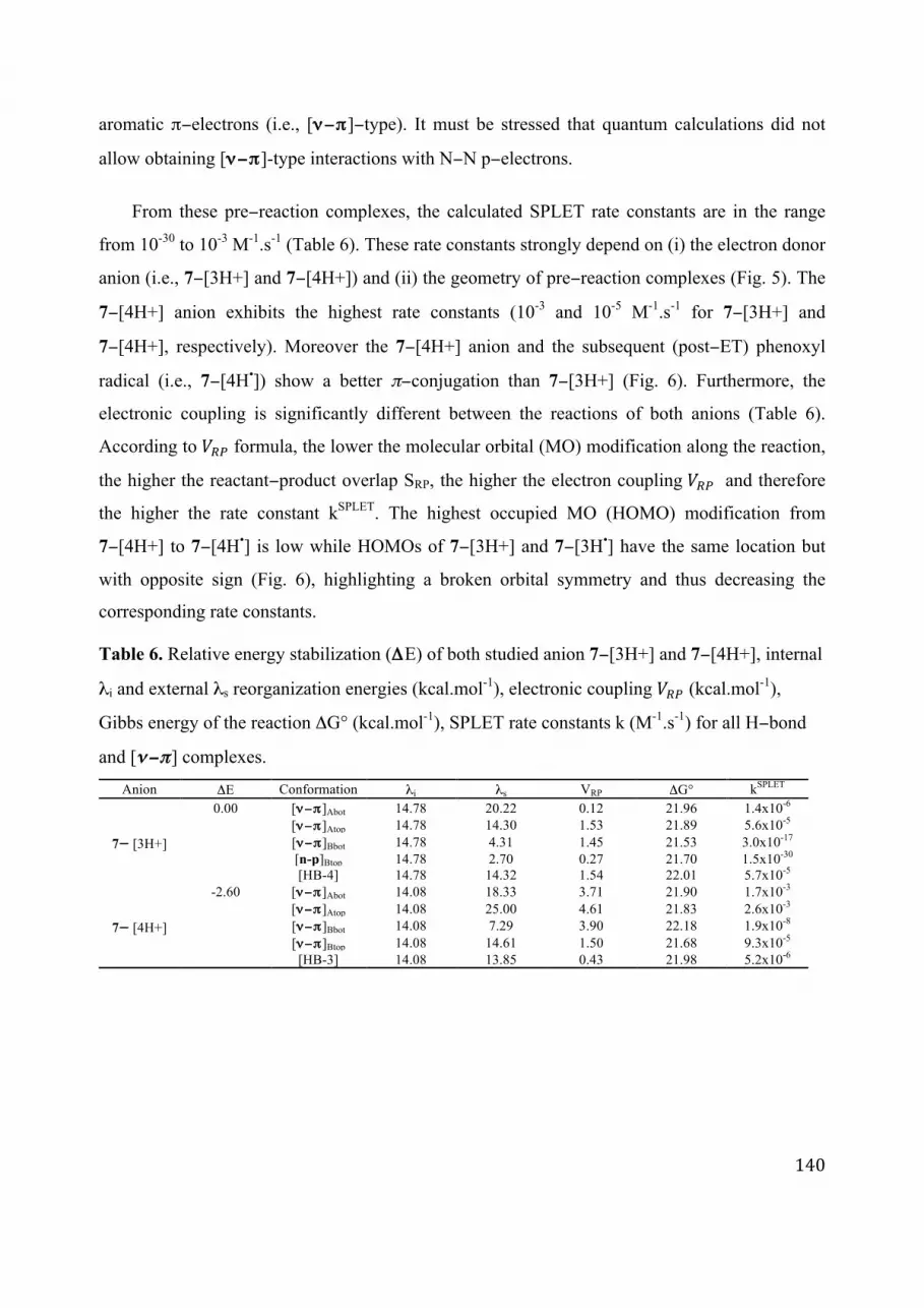
140
aromatic π−electrons (i.e., [ν−π]−type). It must be stressed that quantum calculations did not
allow obtaining [ν−π]-type interactions with N−N p−electrons.
From these pre−reaction complexes, the calculated SPLET rate constants are in the range
from 10-30 to 10-3 M-1.s-1 (Table 6). These rate constants strongly depend on (i) the electron donor
anion (i.e., 7−[3H+] and 7−[4H+]) and (ii) the geometry of pre−reaction complexes (Fig. 5). The
7−[4H+] anion exhibits the highest rate constants (10-3 and 10-5 M-1.s-1 for 7−[3H+] and
7−[4H+], respectively). Moreover the 7−[4H+] anion and the subsequent (post−ET) phenoxyl
radical (i.e., 7−[4H•]) show a better π−conjugation than 7−[3H+] (Fig. 6). Furthermore, the
electronic coupling is significantly different between the reactions of both anions (Table 6).
According to �!" formula, the lower the molecular orbital (MO) modification along the reaction,
the higher the reactant−product overlap SRP, the higher the electron coupling �!" and therefore
the higher the rate constant kSPLET. The highest occupied MO (HOMO) modification from
7−[4H+] to 7−[4H•] is low while HOMOs of 7−[3H+] and 7−[3H•] have the same location but
with opposite sign (Fig. 6), highlighting a broken orbital symmetry and thus decreasing the
corresponding rate constants.
Table 6. Relative energy stabilization (ΔE) of both studied anion 7−[3H+] and 7−[4H+], internal
λi and external λs reorganization energies (kcal.mol-1), electronic coupling �!" (kcal.mol-1),
Gibbs energy of the reaction ΔG° (kcal.mol-1), SPLET rate constants k (M-1.s-1) for all H−bond
and [ν−π] complexes.
Anion ΔE Conformation λi λs VRP ΔG° kSPLET
7− [3H+]
0.00 [ν−π]Abot 14.78 20.22 0.12 21.96 1.4x10⁻⁶ [ν−π]Atop 14.78 14.30 1.53 21.89 5.6x10⁻⁵ [ν−π]Bbot 14.78 4.31 1.45 21.53 3.0x10⁻¹⁷ [n-p]Btop 14.78 2.70 0.27 21.70 1.5x10⁻³⁰ [HB-4] 14.78 14.32 1.54 22.01 5.7x10⁻⁵
7− [4H+]
-2.60 [ν−π]Abot 14.08 18.33 3.71 21.90 1.7x10⁻³ [ν−π]Atop 14.08 25.00 4.61 21.83 2.6x10⁻³ [ν−π]Bbot 14.08 7.29 3.90 22.18 1.9x10⁻⁸ [ν−π]Btop 14.08 14.61 1.50 21.68 9.3x10⁻⁵ [HB-3] 14.08 13.85 0.43 21.98 5.2x10⁻⁶

141
Fig. 1. Optimized geometries of pre-reaction complexes of 7-[4H+]: (a) [HB-3] and (b) [ν−π]Atop.
Figure 6. HOMOs modification along electron abstraction (a) from 7−[3H+] to 7− [3H•] and (b)
from 7− [4H+] to 7-[4H•].
Compare to quercetin, an antioxidant of reference, the SPLET rate constants are
significantly lower.[274] Therefore, the present series of compounds are not expected to be
powerful free radical scavenging by ET process, even from the deprotonated (activated) forms.
The π−delocalization in the Schiff bases studied here is less extended than in quercetin, thus
significantly decreasing the positive energetic contributions (e.g., internal reorganization λi,
ΔG°) along the electron transfer process.
142
5. Conclusion
Based on a series of synthetized Schiff bases, the present study has confirmed the role of the
catechol moiety and the importance of the O−H BDE descriptor. Other minor descriptors
succeeded in explaining slight differences in antioxidant activity, namely spin density
distribution and BDEd. The latter descriptor elegantly rationalizes the role of the NH group that,
according to the chemical structure, may provide a hydrogen atom for a second HAT. Bromine
substitution may slightly enhance antioxidant activity. The free radical scavenging capacity of
these compounds mainly proceeds by PCET rather than SPLET, the latter mechanism being only
feasible at relatively high pH.

143
Note for readers: This article has been submitted and is still under revision. My contribution
concerns the evaluation of the stability of the non-covalent complexes between antioxidants
within lipid-bilayer membranes. Since the article is not yet published, we have only incorporated
the part in which I contributed together with the abstract and a part of the conclusion.
The figures and reference formats and positions are sometimes modified compared to the
original article to keep the homogeneity of the thesis.
Section II. Atomistic description of collaborative antioxidant
effects between vitamins E, C and natural polyphenols in
lipid−bilayer membranes
Gabin Fabre1,2, Imene Bayach1,3, Karel Berka2, Marketa Paloncýová2, Jean-Luc Duroux1, Michal
Otyepka*,2, Patrick Trouillas*,4,2,5
1LCSN EA1069, Faculté de Pharmacie, Université de Limoges, 2 rue de Dr. Marcland, 87025 Limoges, France 2Regional Centre of Advanced Technologies and Materials, Department of Physical Chemistry, Faculty of Science,
Palacký University, tř. 17 listopadu 12, 771 46 Olomouc, Czech Republic 3Atta-ur-Rahman Research Institute for Natural Product Discovery, Faculty of Pharmacy, Universiti Teknologi
MARA, 42300 Puncak Alam, Malaysia 4INSERM UMR-S850, Faculté de Pharmacie, Université de Limoges, 2 rue du Docteur Marcland, 87025 Limoges
Cedex, France 5Laboratoire de Chimie des Matériaux Nouveaux, Université de Mons, Place du Parc 20, B-7000 Mons, Belgium
Abstract: Vitamin E, vitamin C and natural polyphenols (flavonoids and non-flavonoids) are
considered major natural antioxidants capable of preventing damage generated by oxidative
stress. Here, we show the capacity of vitamin E and vitamin C to interact and form non−covalent
complexes within lipid bilayers, close to the membrane/cytosol interface. The formation of such
non-covalent complexes contributes to the synergism existing between these two antioxidants
(vitamin E being regenerated by vitamin C). Quercetin, considered here as a representative
polyphenol, also forms non−covalent complexes with both vitamins C and E. Therefore, in

144
addition to their inherent capacity to scavenge free radicals in membranes, flavonoids forming
such complexes within lipid bilayer membranes can function as enhancers of the regeneration
process
Figure 1. Antioxidant compounds evaluated in this study. The active antioxidant OH groups are
shown in red.
1. Results/Discussion
1.1. Association energies
The strength of the non−covalent intermolecular interaction between antioxidants was evaluated
by using quantum calculations. Computations were made with density functional theory (DFT)
augmented by an empirical dispersion term, allowing high accuracy.[313] After exploring the
conformational space, we analyzed the thirteen, ten, eight and four stable geometries of
quercetin:vitE, quercetin:vitC, vitC:vitE and vitE:vitE complexes, respectively (see Figure 4 for
the most stable geometries per type of complex). The Gibbs energies of association were
obtained with B3P86−D2, recently re-parameterized for polyphenol non−covalent complexes
[316]. To simulate an environment with polarity similar to the inner bilayer, a diethylether−like
polarizable continuum model was used. Different arrangements were observed, namely
head−to−head and head−to−tail. The calculated Gibbs energies of association of the most stable
conformer for each complex were in the range −4.0 to −4.9 kcal.mol-1 (Table 1). The QM

145
calculations confirmed that the association−complexes are stabilized by a combination of
π−stacking (the ring−to−ring distance is around 3.6 Å, which is a typical distance for π−stacking
of aromatic rings [317]) and intermolecular hydrogen bonds (Figure 4). According to this
quantum evaluation, strong attractive forces exist between the three antioxidants, favoring the
formation of non−covalent dimers.
Figure 4. Geometries of the most stable association−complexes as obtained by quantum DFT−D
calculations. (A) quercetin:vitE, (B) vitC:vitE, (C) vitE:vitE and (D) quercetin:vitC.
1.2. Critical analysis of the association Gibbs energy evaluation
The first attempt to evaluate the Gibbs energies of association at the QM level provided
surprisingly positive values. Such positive values are inconsistent with previous findings, as
association enthalpies of -13.7 kcal.mol-1 between two flavonoid derivatives [184] have been
attributed to formation of non−covalent complexes, which have been repeatedly evidenced
experimentally [318], [319]. As in this study the association enthalpies are (favorable) in the
range (Table 1), it seems plausible that the entropy correction to the Gibbs energy was
overestimated as assessed by the statistical thermodynamic apparatus used. The entropy term
consisted of three major components arising from translational, rotational and vibrational modes.
As these contributions were estimated under ideal gas, rigid rotor and harmonic approximations,
one might expect that the loss of translational entropy accompanying formation of the
non−covalent complexes in the lipid bilayer was dramatically overestimated. Therefore, a rough
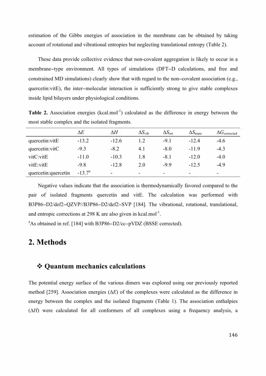
146
estimation of the Gibbs energies of association in the membrane can be obtained by taking
account of rotational and vibrational entropies but neglecting translational entropy (Table 2).
These data provide collective evidence that non-covalent aggregation is likely to occur in a
membrane−type environment. All types of simulations (DFT−D calculations, and free and
constrained MD simulations) clearly show that with regard to the non−covalent association (e.g.,
quercetin:vitE), the inter−molecular interaction is sufficiently strong to give stable complexes
inside lipid bilayers under physiological conditions.
Table 2. Association energies (kcal.mol-1) calculated as the difference in energy between the
most stable complex and the isolated fragments.
∆E ∆H ∆Svib ∆Srot ∆Strans ∆Gcorrected
quercetin:vitE -13.2 -12.6 1.2 -9.1 -12.4 -4.6
quercetin:vitC -9.3 -8.2 4.1 -8.0 -11.9 -4.3
vitC:vitE -11.0 -10.3 1.8 -8.1 -12.0 -4.0
vitE:vitE -9.8 -12.8 2.0 -9.9 -12.5 -4.9
quercetin:quercetin -13.7a - - - - -
Negative values indicate that the association is thermodynamically favored compared to the
pair of isolated fragments quercetin and vitE. The calculation was performed with
B3P86−D2/def2−QZVP//B3P86−D2/def2−SVP [184]. The vibrational, rotational, translational,
and entropic corrections at 298 K are also given in kcal.mol-1. aAs obtained in ref. [184] with B3P86−D2/cc−pVDZ (BSSE corrected).
2. Methods
" Quantum mechanics calculations
The potential energy surface of the various dimers was explored using our previously reported
method [259]. Association energies (∆E) of the complexes were calculated as the difference in
energy between the complex and the isolated fragments (Table 1). The association enthalpies
(∆H) were calculated for all conformers of all complexes using a frequency analysis, a
147
temperature of 298 K and a pressure of 1 atm. Negative values indicated that the association was
thermodynamically favored compared to the pair of isolated compounds. The calculations were
performed with B3P86−D2(s6=0.78)/def2−QZVP//B3P86−D2(s6=0.78)/def2−SVP. This
re−parameterized DFT functional, which includes Grimme’s dispersive term with the �!
parameter adjusted to a value of 0.780, has been validated by high−level SCS−MP2 calculations
and experimental values on polyphenol non−covalent complexes [184], [320]. The COSMO
implicit solvent was used to model diethylether solvation. This solvent was chosen as it exhibits
a dielectric constant of 4.24, which is close to the polarity of phospholipid headgroups. Bond
dissociation enthalpy calculations were performed with B3P86/6−311+G(d,p) as previously
reported [220].
4. Conclusion
Quercetin can readily form strong non−covalent association complexes with vitE and vitC. In
summary, we have presented a molecular picture of the possible intermolecular interaction
between vitE, vitC and polyphenols. This may favour regeneration of vitE and mediation of vitE
regeneration by vitC, thus rationalizing antioxidant synergism between these compounds. This
behaviour may also be observed in membranes.

148
149
Chapter 7 – Tuning optical properties of chalcone
derivatives
Abstract: The conformational feature of non-covalent complexes of two borondifluoride
chalcone derivatives was assessed using DFT-D2. The corresponding optical properties were
analysed based on TD-DFT calculations. As already described in such complexes, the
π−stacking interaction existing between both fragments allowed formation of a new absorption
band corresponding to the S0→ S1 transition. However, this band appears very close to the most
intense band corresponding the S0→ S2 transition.
Keywords: Borondifluoride chalcones • non-covalent complexes • UV/Vis absorption •
Molecular orbitals • DFT-D2/TD-DFT • π−stacking interactions
1. Introduction
Chalcones represent a specific group of polyphenols. They are present in common fruit and
cereals e.g., apples, pears, strawberries, tomatoes and wheat. The most common natural
chalcones are phloretin, phloridzin, chalconaringenin and arbutin. They are key intermediates in
the biosynthesis of flavonoids (shikimate pathway, see chapter 1).
Chalcones are known to exhibit many biological activities.[321] For example, significant
chemoprotective effects have been evidenced.[322]–[327] These activities can be partly
correlated to their antioxidant properties. Among other important properties, chalcones have
antibacterial, antifungal, antiviral, anti-inflammatory and capillary strengthening properties.[328]
Chalcones may be used in novel therapeutic approach in the treatment of inflammatory disorders.
In addition, some chalcones exhibit insulin like activities, which impart to them anti-diabetic
properties.
As π−conjugated systems, chalcones are optically active in the UV/Vis range; most of
natural chalcones being yellowish. They have thus inspired spectroscopic studies aiming at

150
adapting their optical features (by chemical functionalization) to be used as dyes for new
optically active materials. As π−conjugated systems, these compounds have also the capacity to
interact by π−stacking interactions. The chapter aims at describing and rationalizing π−stacking
interactions[329] and optical properties of two chemically−modified chalcones, namely
borondifluoride complexes of 2'−hydroxychalcones (Fig. 1).[330] To achieve these purposes, we
first provide a full conformational analysis that has been followed by the systematic evaluation
of their optical properties with TD−DFT[331] calculations. We have taken advantages of the
expertise on theoretical electronic spectroscopy studies of polyphenols that has been developed
in our lab, mainly using adequate functionals for an accurate description of dispersive
effects.[258], [332], [333] Those two functionalized chalcones, which have similar structures
(i.e., they only differ by the position of the chlorine and bromine atoms, see Fig. 1), are expected
to exhibit interesting optical properties for medical and material applications. They can form
non-covalent complexes in solution mainly driven by π−stacking. The interaction energies and
UV/Vis properties have been investigated for all possible non−covalent complexes.
Figure 1. Chemical structures of both borondifluoride complexes of 2'-hydroxychalcones
1 and 2.
2. Methodology
Over the last years, QM has become an adapted and powerful tool to describe non−covalent
complexes and related optical properties.[334] Within the DFT formalism, classical hybrid
functionals well describe medium size chemical systems. For example, B3P86 is well adapted to
describe thermodynamic and optical properties of chalcones.[335] Nevertheless, as previously
described in this manuscript, hybrid functionals failed at describing charge transfer and
O
B
O
X1
X2
FF
O1: X1=Cl X2=Br
2: X1=Br X2=Cl

151
non−covalent interactions e.g., π−stacking. So including dispersive terms in the energy
calculation is crucial to describe non−covalent complexes. One of the most used approach is the
so-called DFT−D as developed by Grimme (See Chapter 3), which provide good
accuracy/computational time ratio.[312] Following the native DFT−D formalism, the refined
dispersive DFT−D2 method has been widely used.[336] As described in Chapter 3, various
parameterizations and benchmarking were performed for polyphenol derivatives, B3P86−D2
with the parameterized scaling factor s6 = 0.78 has been selected as adequate to perform
geometry optimizations and to evaluate interaction energies. This method was combined with
COSMO (COnductor-like Solvation MOdel) to describe implicitly solvent effect.[262] The
def2−SVP//QZVP basis set was used for the DFT−D2 calculations as it provides good
compromise between accuracy and computational time; in this case the BSSE can be neglected.
Once the ground state (GS) structure of the complexes is studied, the excited−states (ESs)
are described by the use of a range separated hybrid (RSH)−corrected functionals (i.e., ωB97X)
developed in particular to improve charge transfer (CT) description in ESs. This functional was
also corrected to describe non−covalent interactions by including a dispersion term in the
ωB97XD version. Therefore, ωB97XD was used to calculate ES and optical properties within
the TD−DFT framework. In this case, the PCM implicit solvent model is used. The chalcone
derivatives studied in this work are not phenolic compounds, they differ from our previous
methodological studies not from the skeleton but from the chemical functionalization. Therefore,
other functionals were tested on one prototype complex (C1‡‡‡, see section 3.1), including
CAMB3LYP, M062X, MPW1K, with the 6−31+G(d,p) basis set (Fig.2). Solvents with different
polarities (i.e., CCl4 (ε=2.23), CH2Cl2 (ε=8.93) and H20 (ε=78.36)) were used to evaluate solvent
effects and the results were consistently compared to experimental data. All absorption
wavelengths calculated were shifted compared to those obtained experimentally (Fig. 2), and
none of the tested functionals performed better than our previous methodology validated for
phenolic compounds. For all this study, the B3P86 functional and def2−SVP//QZVP basis set
were chosen within the DFT−D2 formalism whereas ωB97XD/6−31+G(d,p) was selected for the
TD−DFT study. It is important to highlight that the choice of the functional does not influence
‡‡‡ C1 is a non−covalent complex between two molecules 1 as identified from crystallography.

152
the MO description and electronic transition assignment.
Figure 2. Theoretical UV/Vis absorption spectra of C1 complex.
3. π−π non−covalent chalcone dimers
This section describes the non−covalent conformations in the solid state and solvent (sections 3.1
and 3.2, respectively).
3.1. 3D arrangements issued from X-ray crystal structures
We first studied the two non−covalent dimers, named C1 and C2, issued from the X−ray crystal
structures of molecules 1 and 2, respectively (Fig. 3). C1 and C2 are the corresponding
arrangements of two stacked units of 1 and 2, respectively. B3P86−D2(s6=0.780)/def2−SVZ
single point calculation confirmed the energetic stability of these two arrangements with
association energies of -18.8 and -19.2 kcal.mol-1 for C1 and C2, respectively, as obtained in the
gas phase (Fig. 5). The stability is mainly attributed to π−stacking interactions between both
partners being distant from each other by 3.5 Å (Fig.3).
λ/nm
f
ωB97XD
M062X
MPW1K
CAMLYP

153
Figure 3. (a) C1 and (b) C2 geometries issued from the crystal structures of molecules 1 and 2,
respectively.
As C1 and C2 corresponded to solid-state geometries, they may exist in a specific restrained
environment. In order to understand how these non−covalent dimers may arrange without these
restrains, both geometries were optimized. After optimization, these geometries were
significantly modified, both partners slid along each other (Fig. 4). Due to the relaxation within
the implicit PCM environment, the optimized structures appeared slightly more stable by
providing slightly lower association energies, e.g., in CCl4, the associations energies are i) -14.7
and-15.0 for the C1 and its corresponding optimized complex, respectively and ii) -14.5 and -
14.9 for C2 and its corresponding optimized complex, respectively (see Table 1).
Figure 4. (a) Complex C1 issued from the crystal structure of 1 and (b) the corresponding
(B3P86−D2/def2−SVP) −Optimized geometry.
(a) (b)
3.5 Å
3.5 Å
horizontal sliding
3.5 Å(a)
(b)3.4 Å

154
Table 1. (B3P86−D2/def2−TZVP) −Association energies of the C1 and C2 complexes in
nonpolar (CCl4) and polar (H2O) solvents, (a) Single Point and (b) Optimization.
Complex C1 C2
(a) (b) (a) (b)
CCl4 -14.7 -15.0 -14.5 -14.9
H2O -8.2 -10.5 -8.0 -10.1
When increasing the polarity of the solvent, the interaction energies are lowered in absolute
value (Table 1 and Fig. 5). Here we are aware that the solvent is taken into account implicitly
therefore loosing the description of specific interactions (H−bonds) and the global hydrophobic
effects. This can be particularly true for polar and protic solvent such as water.
Figure 5. B3P86−D2/def2−TZVP Association energies for C1 and C2 in the gas phase and in
PCM-type different solvents.
3.2. Non−restrained conformational analysis
The orientations of non−covalent association may considerably influence optical properties.
Therefore it appears crucial to explore the entire potential energy hypersurface for the

155
non−covalent dimers, other than the two described above (i.e., the optimized geometries from C1
and C2). One must consider either i) offset−dimers, in which both fragments are slid along each
other within a 1:1 cofacial aromatic rings (Fig. 7 & 8), or ii) aligned−dimers, exhibiting 2:2
cofacial rings thus enhancing π−stacking alignment (Fig. 9). For asymmetric compounds
(asymmetric substitutions), head−to−head and head−to−tail arrangements must be considered as
well as Re/Re and Re/Si approaches. For each chalcone (1 and 2), six offset−dimer geometries
(Fig. 8) and eight aligned−dimer geometries (Fig. 9) were elucidated.
Figure 7. Chemical structures of the different (1) offset− and (2) aligned−dimers.
OB
O
X1
X2
FF
O
O
BO
X2
F
F
X2X1 OB
O
X1
X2
FF
O
OB
O
X1
X2
FF
O
OB
O
X1
X2
F
F
O
O
B O
X2
F
F
X2
X1
OB
O
X1
X2
FF
O
O
B O
X2
F
F
X2
X1
OB
O
X1
X2
FF
O
O
BO
O
F
F
X2X1
OB
O
X1
X2
FF
OOBO
X1
X2
F F
O
OB
O
X1
X2
F
F
O
OBO
O
F
F
X2
X1
OB
O
X1
X2
FF
O
OBO
X1
X2
F F
O
(a) (b) (c) (d)
(e) (f) (g) (h)
OB
O
X1
X2
FF
O
O
B O
O
F
F
X2
X1
OB
O
X1
X2
FF
O
OB
O
X1
X2
FF
O
OB
O
X1
X2
FF
OO
B O
O
F
F
X2
X1
OB
O
X1
X2
FF
O
O
B O
O
F
F
X2
X1
OB
O
X1
X2
FF
OO
BOX2
F
F
X2
X1
OB
O
X1
X2
FF
O
OBO
X1
X2
F F
O OB
O
X1
X2
FF
O
O
BO
X2
F
F
X2
X1
OB
O
X1
X2
FF
O
OBO
X1
X2
F F
O
(a) (b)(c) (d)
(e) (f) (g) (h)
(1)
(2)

156
a§§§ b c d
e f g h
a**** b c d
e f g h
Figure 8. (B3P86−D2(s6 = 0.780)/def2−SVP)−Optimized geometries from the eight possible offset-dimers of 1 [A] and 2 [B].
Starting with eight offset−dimer conformations, the optimization procedure led to two
aligned−dimers conformations (c and e) and to six offset−dimers including the optimized
complex issued from the crystal structure (conformation a) (Fig. 8). Due to better π−stacking
§§§ This orientation corresponds to the optimized conformation of C1 as described in Fig.4. **** This orientation corresponds to the optimized conformation of C2.
[A]
[B]

157
overlap, conformations c and e are more stable than the six offset−dimers (Table 2).
Figure 9. (B3P86−D2/def2−SVP)−Optimized geometries of the eight aligned−dimer from [A] 1 and [B] 2.
While all orientations exhibit considerable negative association energies, there are
significant differences between the various complexes (Table 2). The intermolecular distances
within all non−covalent complexes were around 3.5 Å, typical of π−stacking.
[A]
[B]
a b c d
e f g h
a b c d
e f g h

158
Table 2. (B3P86−D2/def2−QZVP)−Association energies (ΔEint in kcal.mol-1) of the
non−covalent complexes and Boltzmann weights (Dbolt in %) calculated for the different
alignments.
1 2
offset aligned offset aligned
ΔEint Dbolt ΔEint Dbolt ΔEint Dbolt ΔEint Dbolt
a -13.2 0.0 a -19.7 0.0 a -15.0 0.0 a -17.3 0.0
b -9.7 0.0 b -11.6 0.0 b -13.1 0.0 b -13.8 0.0
c -22.6 99.8 c -24.1 37.1 c -22.6 100.0 c -24.5 57.7
d -8.1 0.0 d -19.0 0.0 d -11.2 0.0 d -19.3 0.0
e -18.9 0.2 e -17.6 0.0 e -17.1 0.0 e -18.3 0.0
f -9.8 0.0 f -24.4 62.8 f -10.7 0.0 f -24.3 42.1
g -4.2 0.0 g -17.3 0.0 g -6.4 0.0 g -14.5 0.0
h -17.0 0.0 h -17.0 0.0 h -12,7 0.0 h -20.8 0.1
As expected, the aligned-dimers are much more stable than the offset−dimers. In the former
type of dimers indeed, π−stacking is favored along an extended region. The most stable energies
were obtained for the head−to−tail alignments (Fig. 9). The position for bromine and chlorine
substitution also influences significantly the stabilizing energies. The non−covalent dimers are
particularly stable because they exhibit adequate charge distribution. Nevertheless, as we pointed
out in our recent paper,[337] considering only association energies is not enough to conclude,
ΔG calculations would be required. Therefore all complexes are considered for the following
comprehensive optical analysis.
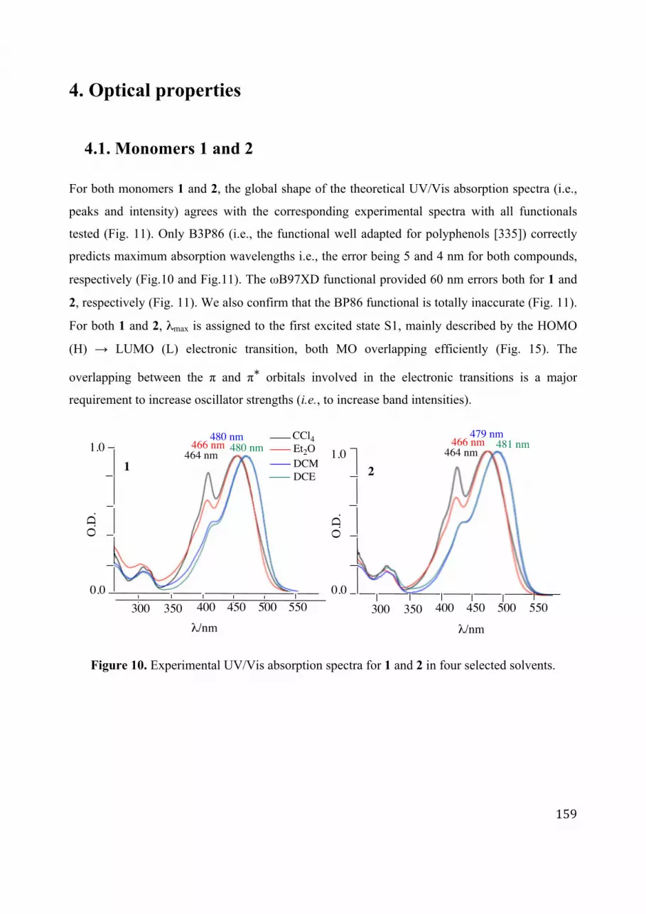
159
4. Optical properties
4.1. Monomers 1 and 2
For both monomers 1 and 2, the global shape of the theoretical UV/Vis absorption spectra (i.e.,
peaks and intensity) agrees with the corresponding experimental spectra with all functionals
tested (Fig. 11). Only B3P86 (i.e., the functional well adapted for polyphenols [335]) correctly
predicts maximum absorption wavelengths i.e., the error being 5 and 4 nm for both compounds,
respectively (Fig.10 and Fig.11). The ωB97XD functional provided 60 nm errors both for 1 and
2, respectively (Fig. 11). We also confirm that the BP86 functional is totally inaccurate (Fig. 11).
For both 1 and 2, λmax is assigned to the first excited state S1, mainly described by the HOMO
(H) → LUMO (L) electronic transition, both MO overlapping efficiently (Fig. 15). The
overlapping between the π and π∗ orbitals involved in the electronic transitions is a major
requirement to increase oscillator strengths (i.e., to increase band intensities).
Figure 10. Experimental UV/Vis absorption spectra for 1 and 2 in four selected solvents.
CCl4
Et2O
DCM
DCE
λ/nm λ/nm
O.D
.
O.D
.
0.0
1.0
0.0
1.0
300 350 400 450 500 550 300 350 400 450 500 550
464 nm
466 nm479 nm
481 nm
2
464 nm
466 nm480 nm
480 nm
1

160
Figure 11. Theoretical UV/Vis absorption spectra in CCl4 with different functionals.
Contrary to what is observed for polyphenols,[335] increasing the polarity of the solvent
induces a slight hypsochromic shift of λmax by 8 nm and 9 nm for 1 and 2, respectively (Fig. 12).
Figure 12. Theoretical UV/Vis absorption spectra in different solvents for 1 and 2.
4.2. Non−covalent complexes
Even if the optimized geometries appeared more stable in a solvent-type environment, the
understanding of optical properties of the X-ray crystal structures (C1 and C2) is a crucial step,
mainly because aiming at a comparison between solvent and solid state behaviors.
The UV/Vis spectra of C1 and C2 looks very similar to that of the corresponding
monomers, with a clear maximum absorption band at 409.26 and 405.90 nm for both frozen
non−covalent dimers C1 and C2, respectively (Fig. 14). Interestingly, in the relaxed
non−covalent dimers, the band is split into two main (and clearly separated) bands (Fig. 14). The
maximum absorption band is actually a new peak resulting from the non−covalent complexation
λ/nm
f
1
ωB97XD
λ/nm
f
2
M062X
B3P86
CAM-B3LYP409 nm
403 nm
411 nm
469 nm
BP86
ωB97XD
B3P86
403 nm
468 nm
567 nm
λ/nm
f
1
CCl4
λ/nm
f
2H2O
CH2Cl2
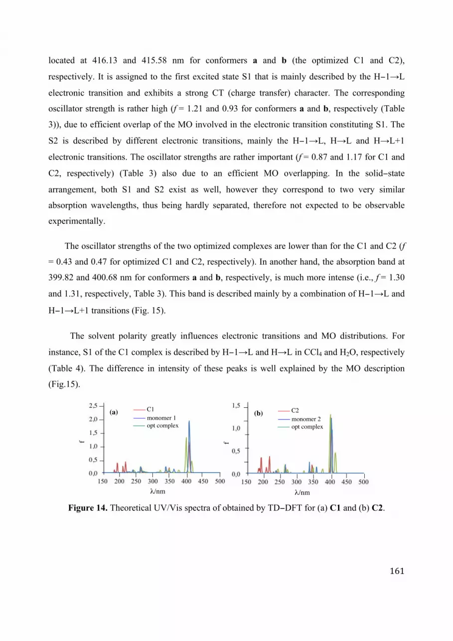
161
located at 416.13 and 415.58 nm for conformers a and b (the optimized C1 and C2),
respectively. It is assigned to the first excited state S1 that is mainly described by the H−1→L
electronic transition and exhibits a strong CT (charge transfer) character. The corresponding
oscillator strength is rather high (f = 1.21 and 0.93 for conformers a and b, respectively (Table
3)), due to efficient overlap of the MO involved in the electronic transition constituting S1. The
S2 is described by different electronic transitions, mainly the H−1→L, H→L and H→L+1
electronic transitions. The oscillator strengths are rather important (f = 0.87 and 1.17 for C1 and
C2, respectively) (Table 3) also due to an efficient MO overlapping. In the solid−state
arrangement, both S1 and S2 exist as well, however they correspond to two very similar
absorption wavelengths, thus being hardly separated, therefore not expected to be observable
experimentally.
The oscillator strengths of the two optimized complexes are lower than for the C1 and C2 (f
= 0.43 and 0.47 for optimized C1 and C2, respectively). In another hand, the absorption band at
399.82 and 400.68 nm for conformers a and b, respectively, is much more intense (i.e., f = 1.30
and 1.31, respectively, Table 3). This band is described mainly by a combination of H−1→L and
H−1→L+1 transitions (Fig. 15).
The solvent polarity greatly influences electronic transitions and MO distributions. For
instance, S1 of the C1 complex is described by H−1→L and H→L in CCl4 and H2O, respectively
(Table 4). The difference in intensity of these peaks is well explained by the MO description
(Fig.15).
Figure 14. Theoretical UV/Vis spectra of obtained by TD−DFT for (a) C1 and (b) C2.
λ/nm
f
(a) C1
monomer 1
opt complex
λ/nm
f
(b) C2
monomer 2
opt complex

162
Table 3. Maximum vertical excitation energies (Emax, eV), absorption wavelengths (λmax, nm),
oscillator strengths (f) and MO descriptions of C1 and C2 and their corresponding optimization
at the PCM (CCl4)−TD−ωB97XD/6−31+G(d,p) level.
Optimization SP
Emax λmax f MO Emax λmax f MO
C1
2.98 416.13 0.43 H→L (49%)
3.03 409.26 1.21 H-1→L (57%)
H→L+1(-36%) H-1→L+1 (29%)
3.10 399.82 1.30 H-1→L (53%)
3.04 407.83 0.87 H→L (-43%)
H-1→L+1 (31%) H→L+1(47%)
C2
2.98 415.58 0.47
H→L (47%)
3.05 405.90 0.93 H-1→L (60%) H→L+1(-37%)
H-1→L (-30%)
3.09 400.68 1.31
H→L (51%)
3.07 403.89 1.17
H→L (-45%)
H-1→L+1 (31%) H→L+1(47%)
H→L (29%)
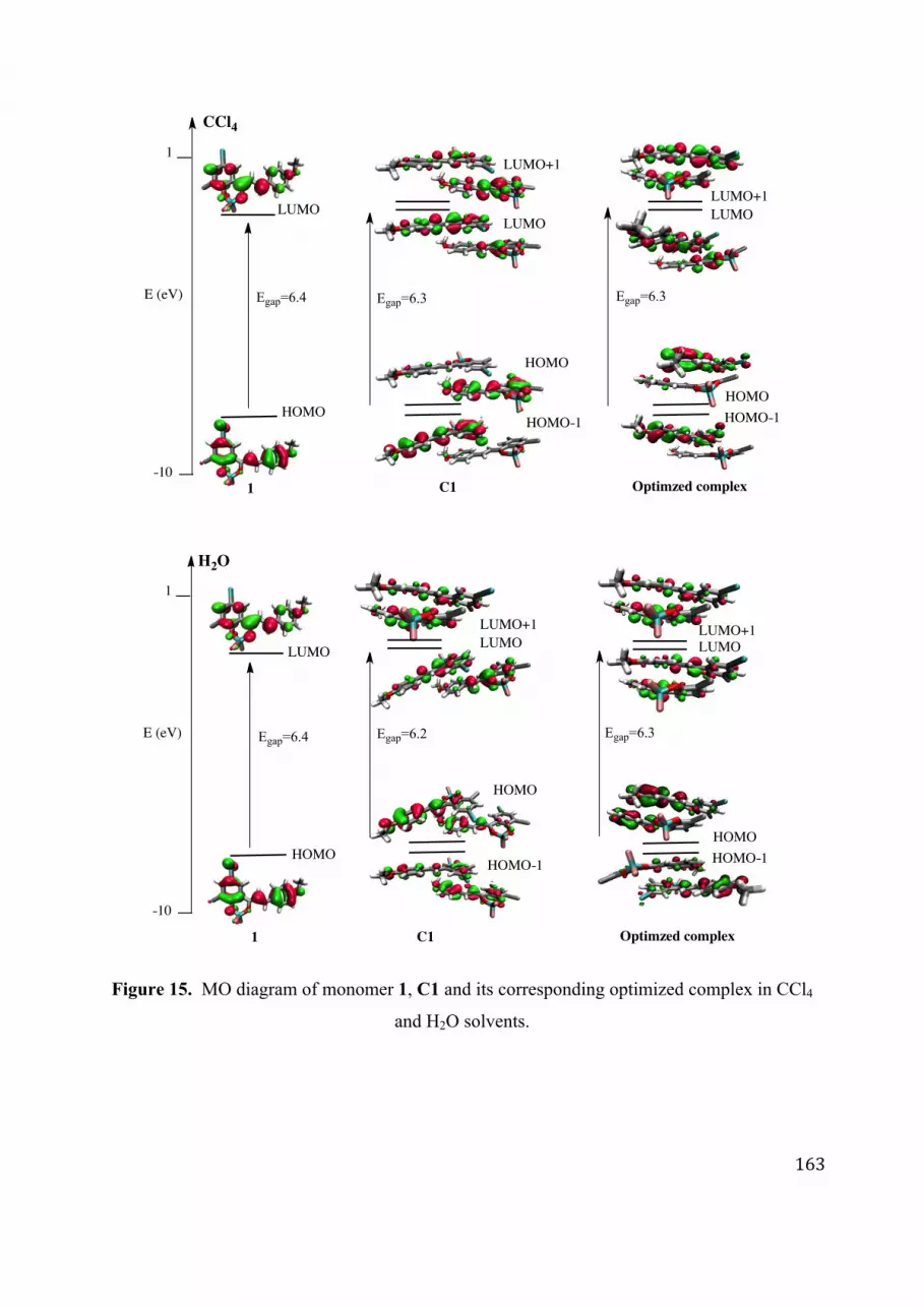
163
Figure 15. MO diagram of monomer 1, C1 and its corresponding optimized complex in CCl4
and H2O solvents.
-10
1
E (eV)
1 C1 Optimzed complex
C1
-10
1
E (eV)
1 Optimzed complex
HOMO
LUMO
HOMO-1
HOMO
LUMO
LUMO+1
Egap=6.4 Egap=6.3 Egap=6.3
LUMO+1
LUMO
HOMO
HOMO-1
HOMO
LUMO
LUMO+1
LUMO
HOMO
HOMO-1
LUMO+1
LUMO
HOMO
HOMO-1
Egap=6.4 Egap=6.2 Egap=6.3
H2O
CCl4

164
Table 4. Emax (eV), λmax (nm), f and MO descriptions of C1 at the PCM−TD−ωB97XD/6-
31+G(d,p) level.
CCl4 H20
OPT SP OPT SP
2.98eV 416.13nm f=0.43 2.99eV 414.86nm f=0.48 3.03eV 409.26nm f=1.21 3.05eV 406.57nm f=2.12
H-2→L+1 (11%) H-2→L+1 (-11%) H-3→L+2 (11%) H-1→L (14%)
H-1→L (27%) H-1→L (45%) H-1→L (57%) H-1→L+1 (-35%)
H→L (49%) H→L (-43%) H-1→L+1 (29%) H→L (54%)
H→L+1 (-36%) H→L+1 (23%) H→L (-20%)
3.10eV 399.82 nm f=1.30 3.10eV 399.48nm f=1.25 3.04eV 407.83nm f=0.87 3.07eV 404.25nm f=0.12
H-1 →L (53%) H-1→L (29%) H-2 → L+1 (-12%) H-2→L+1 (-12%)
H-1 →L+1 (31%) H-1→L+1 (36%) H-2→L+3 (-12%) H-1→L (53%)
H →L (-25%) H→L (41%) H-1→L (-17%) H→L+1 (-39%) H→L+1 (25%) H→L (-43%)
H→L+1 (47%)
The ES description of all conformers was studied as well (Tables 5 and 6). However, MO
diagrams were given for the significant complexes (high Dbolt), i.e., orientations c and f only for
aligned-dimer (Table 2 and Fig. 16−19). As for C1, C2 and their corresponding optimized
complexes, the UV/vis spectra of all other complexes consist of two main absorption bands.
From Tables 5 and 6 and the MO diagrams, these two bands are well understood following the
same explanation as previously. Again, both absorption wavelengths and oscillator strengths are
highly influenced by the conformation, some electronic transitions being interchanged.

165
Table 5. Maximum vertical excitation energies (Emax, eV), absorption wavelengths (λmax, nm), oscillator strengths (f)
and MO descriptions of different complexes from two units of 1 at the PCM (CCl4)−TD−ωB97XD/6−31+G(d,p)
level.
offset−dimers aligned−dimers
Emax λmax f MO Emax λmax f MO
a
2.93 421.75 1.45 H→L 2.85 435.36 0.08 H→L
3.03 409.55 0.34 H−1→L (42%)
3.07 403.25 1.58 H−1→L H→L+1 (45%)
b
3.01 411.88 1.70 H→L 3.00 413.34 0.03 H−1→L
3.08 402.86 0.30 H−1→L (49%)
3.12 397.57 1.75 H−1→L (41%)
H→L+1 (38%) H→L+1 (51%)
c
2.69 460.12 0.00 H→L 2.76 448.75 0.00 H→L
3.09 401.68 1.85 H−1→L (43%)
3.08 402.60 1.80 H−1→L (-44%)
H→L+1 (47%) H→L+1 (47%)
d
2.94 421.10 0.11 H→L 2.90 428.42 0.04 H→L+1
3.04 407.49 1.84 H−1→L (40%)
3.11 398.34 1.67 H−1→L (35%)
H→L+1 (53%) H−1→L+1 (38%)
e
2.90 427.87 0.08 H−1→L 2.80 443.07 0.08 H→L
3.12 397.93 1.61 H→L+1 3.12 397.26 1.71 H−1→L (46%)
H→L+1 (47%)
f
2.95 420.14 2.30 H→L 2.78 446.69 0.00 H→L
3.02 410.32 0.01 H→L+1 3.09 401.11 2.03 H→L+1 (58%)
g
3.02 409.94 0.67 H-1→L (54%) 2.91 426.60 0.11 H→L
H→L (35%) 3.14 394.81 1.67
H−1→L (43%)
3.07 403.52 1.78 H→L+1 H−1→L+1 (48%)
h
3.00 414.60 0.00 H→L 2.85 435.32 0.10 H→L+1
3.04 407.29 2.22 H−1→L (51%)
3.03 408.59 1.77 H-1→L+1 (-31%)
H→L+1 (43%) H→L+1 (55%)

166
Table 6. Maximum vertical excitation energies (Emax, eV), absorption wavelengths (λmax, nm),
oscillator strengths (f) and MO descriptions of different complexes from two units of 2 at the
PCM(CCl4)−TD−ωB97XD/6−31+G(d,p) level.
offset−dimers aligned−dimers
Emax λmax f MO Emax λmax f MO
a
2.91 425.75 0.96 H→L 2.83 438.05 0.07 H→L
3.01 410.18 0.79 H−1→L (32%)
3.10 400.59 1.51 H−1→L (49%)
H→L+1 (54%) H→L+1 (-45%)
b
2.97 416.25 0.48 H→L 2.96 419.47 0.09 H-1→L
3.07 403.42 1.22 H−1→L (45%)
3.13 396.53 1.59 H−1→L (42%)
H→L+1 (48%) H→L+1 (49%)
c
2.72 454.80 0.00 H→L 2.76 449.47 0.00 H→L
3.10 399.15 1.91 H−1→L (36%)
3.09 401.46 1.88 H−1→L (-35%)
H→L+1 (55%) H→L+1 (58%)
d
2.99 415.30 0.35 H→L 2.88 430.69 0.02 H→L
3.02 410.98 1.76 H−1→L (49%)
3.13 396.27 1.75 H−1→L (41%)
H→L+1 (-43%) H→L+1 (49%)
e
2.84 435.15 0.01 H→L 2.77 447.38 0.00 H→L
3.07 403.44 1.74 H→L+1 3.11 398.13 2.08 H→L+1
f
2.95 420.89 2.01 H→L 2.79 445.17 0.00 H→L
3.00 413.27 0.14 H→L+1 3.10 399.60 1.99 H→L+1
g
3.00 412.44 0.47 H−1→L 2.79 443.93 0.04 H→L
3.07 403.34 1.66 H→L+1 3.15 393.09 1.72 H−1→L (47%)
H→L+1 (48%)
h
2.94 421.06 0.00 H→L
2.87 432.68 0.04 H−1→L (49%)
3.04 407.93 2.18 H−1→L (52%) H→L+1 (45%)
H→L+1 (42%) 2.99 414.83 1.68 H→L

167
Figure 16. MO diagram of monomer 1 and complex c.
Figure 17. MO diagram of monomer 2 and complex c.
c
offset- dimersc
aligned-dimers
-10
1
E (eV) Egap=6.4Egap=6.0
L
H H-1
H
L
L+1
1
Egap=6.1
H-1
H
L
L+1
-10
1
E (eV) Egap=6.4Egap=6.1
L
H H-1
H
L
L+1
2 c
offset- dimers
Egap=6.1
H-1
H
L
L+1
c
aligned-dimers

168
Figure 18. MO diagram of monomer 1 and complex f.
Figure 19. MO diagram of monomer 2 and complex f.
5. Conclusion
Quantum chemical calculations have allowed providing a detailed MO picture and thus a
complete spectroscopic understanding of non−covalent association between the complexes of
borondifluoride 2'−hydroxychalcones. We provided a quantum rationalization of UV/Vis spectra
of single molecules and different possible non-covalent arrangements driven by π−stacking. As
expected, the B3P86 functional within the TD-DFT formalism provided accurate results for the
-10
1
E (eV) Egap=6.4Egap=6.2
L
H H-1
H
L
L+1
1 f
offset-dimers
Egap=6.2
H-1
H
L
L+1
f
aligned-dimers
-10
1
E (eV) Egap=6.4 Egap=6.2
L
H H-1
H
L
L+1
2 f

169
monomers. However, even if less accurate for the monomer, the ωB97XD functional was
required to describe CT within the π−stacking complexes.
Within the non-covalent complexes, a new band due to the π−stacking interaction
attributed to the S0→S1 transition (mainly described by a H→L electronic transition) with a CT
character. However, the supramolecular arrangements only slightly influence the MO scheme
since the global shape of the UV/Vis spectrum is conserved. The solvent description influences
the optical properties i.e., solvent polarity induces slight hypsochromic shifts of λmax. The
halogen substituent position poorly influenced the MO description and the subsequent optical
properties.
This computational study of monomer and non-covalent dimers in solution is a first step
towards full rationalization of optical properties of these compounds in the solid phase. In this
case, stacking description and spectral influence is mandatory.

170

171
Conclusion
The purpose of this PhD was to rationalize the role of non−covalent interactions through three
examples covering three fields of applications. First, in chemistry, our results constitute efficient
weapons to help proceeding of supramolecular synthesis. Non−covalent complexes formed prior
to oxidation have been shown to be specific precursors of the regio− and stereo−selective
stilbenoid oligomer synthesis. Supramolecular self−assembly of oligostilbenoids units through
non−covalent bonding is the driving force in some key−steps of the oligostilbene biosynthesis.
QM calculations, carefully including dispersive effects, have brought new understanding for
biogenesis of different classes of natural products. This understanding may provide more refined
approaches to biomimetic syntheses, which are likely to become more practical in the production
of series of new derivatives from lead drugs with enhanced biological activities.
Second, in biochemistry, our work provides a new molecular picture of antioxidants (i.e.,
vitamins and polyphenols) and their antioxidant action in membranes. We demonstrated that
antioxidants form non−covalent complexes that eventually affect their activities. In lipid bilayer
membranes, vitamin E could form stable non−covalent association-complexes with vitamin C
and quercetin, considered as a representative polyphenol, can also form strong non−covalent
association−complexes with either vitamin E or C. We believe that non−covalent complexation
of these antioxidants promotes vitamin E regeneration and mediation of vitamin E regeneration
by vitamin C and polyphenols. These effects due to the non−covalent complexation would be
even more enhanced in larger aggregates possibly forming part nanodomains.
Third, in materials science, DFT−D and TD−DFT have provided a preliminary
spectroscopic understanding of non−covalent complexation of chalcone derivatives through a
detailed molecular orbitals picture. The non−covalent association (3D arrangement) affects
optical properties of these selected chalcones. Solvent and position of halogen substituent (Br or
Cl) also influence the optical properties.

172
This thesis highlights the importance of non−covalent interactions that have increasingly
attracted interest from food and pharmaceutical applications. Theoretical chemistry represents an
accurate tool to describe these non−covalent interactions with practical perspectives.
Thanks to all refinements that have allowed reaching high accuracy, DFT has become
indispensable for Science to improve understanding, especially for natural products. In this work,
the robustness of dispersion−corrected calculations (DFT−D2, DFT−D3 and DFT−NL) was
assessed, with large basis sets (def2−QZVP) to avoid incompleteness effects for one real−world
polyphenol dimer chosen as an appropriate model. Our benchmark study predicts the existence
of natural polyphenols complexes mainly driven by both π–stacking and H−bonding interactions.
The adequate comparison of lower−cost DFT−based methods allowed bracketing their expected
accuracy. These results thus pave the way towards reliable studies of challenging aggregation
processes of natural products. However, theoretical chemistry still has its limits to be enhanced
since the choice of the functional and the basis set is not always obvious and is
system−dependent. The challenging task in QM is still to find the best compromise between
accuracy and computational time.
Theoretical chemistry is a crucial tool to rationalize many other chemical and biological
properties of natural or synthetic compounds, as is the case of the synthetized phenolic Schiff
bases for which the structure–antioxidant activity relationship have been elucidated and have
confirmed the importance of the O–H groups together with the catechol moiety as well as other
minor descriptors in modulating the antioxidant activity. We also demonstrated the role of the
NH group to provide a hydrogen atom for a second HAT according to the chemical structure and
the bromine substitution to slightly enhance antioxidant activity. Moreover, this part of the work
allowed determining the major mechanism involved in free radical scavenging using DFT
calculations by confirming that the free radical scavenging by these Schiff bases mainly proceeds
through atom transfer (PCET) rather than electron transfer (SPLET).

173
BIBLIOGRAPHY
[1] M. B. Austin, M. E. Bowman, J.-L. Ferrer, J. Schröder, and J. P. Noel, “An Aldol Switch Discovered in Stilbene Synthases Mediates Cyclization Specificity of Type III Polyketide Synthases,” Chem. Biol., vol. 11, no. 9, pp. 1179–1194, Sep. 2004.
[2] T. Iwashina, “The Structure and Distribution of the Flavonoids in Plants,” J. Plant Res., vol. 113, no. 3, pp. 287–299, Sep. 2000.
[3] J. Koukol and E. E. Conn, “The metabolism of aromatic compounds in higher plants. IV. Purification and properties of the phenylalanine deaminase of Hordeum vulgare,” J. Biol. Chem., vol. 236, pp. 2692–2698, Oct. 1961.
[4] H. Grisebach and S. Kellner, “studies on the mechanism of transformation of 4,2',4',6'-tetrahydroxychalcone into taxifoline in Chamaecyparis obtusa,” Z. Für Naturforschung Teil B Chem. Biochem. Biophys. Biol., vol. 20, pp. 446–450, May 1965.
[5] M. L. Falcone Ferreyra, S. P. Rius, and P. Casati, “Flavonoids: biosynthesis, biological functions, and biotechnological applications,” Front. Plant Sci., vol. 3, Sep. 2012.
[6] S. Martens, A. Preuss, and U. Matern, “Multifunctional flavonoid dioxygenases: flavonol and anthocyanin biosynthesis in Arabidopsis thaliana L,” Phytochemistry, vol. 71, no. 10, pp. 1040–1049, Jul. 2010.
[7] D. Bowles, J. Isayenkova, E.-K. Lim, and B. Poppenberger, “Glycosyltransferases: managers of small molecules,” Curr. Opin. Plant Biol., vol. 8, no. 3, pp. 254–263, Jun. 2005.
[8] J.-L. Ferrer, M. B. Austin, C. Stewart Jr, and J. P. Noel, “Structure and function of enzymes involved in the biosynthesis of phenylpropanoids,” Plant Physiol. Biochem. PPB Société Fr. Physiol. Végétale, vol. 46, no. 3, pp. 356–370, Mar. 2008.
[9] M. G. Simic, “Mechanisms of inhibition of free-radical processes in mutagenesis and carcinogenesis,” Mutat. Res., vol. 202, no. 2, pp. 377–386, Dec. 1988.
[10] J.-H. Yang, H.-C. Lin, and J.-L. Mau, “Antioxidant properties of several commercial mushrooms,” Food Chem., vol. 77, no. 2, pp. 229–235, May 2002.
[11] S. Hercberg, P. Galan, P. Preziosi, S. Bertrais, L. Mennen, D. Malvy, A.-M. Roussel, A. Favier, and S. Briançon, “The SU.VI.MAX Study: a randomized, placebo-controlled trial of the health effects of antioxidant vitamins and minerals,” Arch. Intern. Med., vol. 164, no. 21, pp. 2335–2342, Nov. 2004.
[12] O. Dangles, “Antioxidant Activity of Plant Phenols: Chemical Mechanisms and Biological Significance,” Curr. Org. Chem., vol. 16, no. 6, pp. 692–714, Apr. 2012.
[13] B. W. Shirley, “Flavonoid biosynthesis: ‘new’ functions for an ‘old’ pathway,” Trends Plant Sci., vol. 1, no. 11, pp. 377–382, Jan. 1996.
[14] G. R. Beecher, “Overview of dietary flavonoids: nomenclature, occurrence and intake,” J. Nutr., vol. 133, no. 10, p. 3248S–3254S, Oct. 2003.
[15] R. J. Williams, J. P. E. Spencer, and C. Rice-Evans, “Flavonoids: antioxidants or signalling molecules?,” Free Radic. Biol. Med., vol. 36, no. 7, pp. 838–849, Apr. 2004.
[16] A. García-Lafuente, E. Guillamón, A. Villares, M. A. Rostagno, and J. A. Martínez, “Flavonoids as anti-inflammatory agents: implications in cancer and cardiovascular disease,” Inflamm. Res. Off. J. Eur. Histamine Res. Soc. Al, vol. 58, no. 9, pp. 537–552, Sep. 2009.

174
[17] A. E. Rotelli, T. Guardia, A. O. Juárez, N. E. de la Rocha, and L. E. Pelzer, “Comparative study of flavonoids in experimental models of inflammation,” Pharmacol. Res. Off. J. Ital. Pharmacol. Soc., vol. 48, no. 6, pp. 601–606, Dec. 2003.
[18] P. N. Paradkar, P. S. Blum, M. A. Berhow, H. Baumann, and S.-M. Kuo, “Dietary isoflavones suppress endotoxin-induced inflammatory reaction in liver and intestine,” Cancer Lett., vol. 215, no. 1, pp. 21–28, Nov. 2004.
[19] W. Duan, I. C. Kuo, S. Selvarajan, K. Y. Chua, B. H. Bay, and W. S. F. Wong, “Antiinflammatory effects of genistein, a tyrosine kinase inhibitor, on a guinea pig model of asthma,” Am. J. Respir. Crit. Care Med., vol. 167, no. 2, pp. 185–192, Jan. 2003.
[20] T. Guardia, A. E. Rotelli, A. O. Juarez, and L. E. Pelzer, “Anti-inflammatory properties of plant flavonoids. Effects of rutin, quercetin and hesperidin on adjuvant arthritis in rat,” Farm. Soc. Chim. Ital. 1989, vol. 56, no. 9, pp. 683–687, Sep. 2001.
[21] A. J. Afolayan and J. J. Meyer, “The antimicrobial activity of 3,5,7-trihydroxyflavone isolated from the shoots of Helichrysum aureonitens,” J. Ethnopharmacol., vol. 57, no. 3, pp. 177–181, Aug. 1997.
[22] Y. M. Lin, H. Anderson, M. T. Flavin, Y. H. Pai, E. Mata-Greenwood, T. Pengsuparp, J. M. Pezzuto, R. F. Schinazi, S. H. Hughes, and F. C. Chen, “In vitro anti-HIV activity of biflavonoids isolated from Rhus succedanea and Garcinia multiflora,” J. Nat. Prod., vol. 60, no. 9, pp. 884–888, Sep. 1997.
[23] R. I. Brinkworth, M. J. Stoermer, and D. P. Fairlie, “Flavones are inhibitors of HIV-1 proteinase,” Biochem. Biophys. Res. Commun., vol. 188, no. 2, pp. 631–637, Oct. 1992.
[24] M. R. Fesen, Y. Pommier, F. Leteurtre, S. Hiroguchi, J. Yung, and K. W. Kohn, “Inhibition of HIV-1 integrase by flavones, caffeic acid phenethyl ester (CAPE) and related compounds,” Biochem. Pharmacol., vol. 48, no. 3, pp. 595–608, Aug. 1994.
[25] J. W. Critchfield, S. T. Butera, and T. M. Folks, “Inhibition of HIV activation in latently infected cells by flavonoid compounds,” AIDS Res. Hum. Retroviruses, vol. 12, no. 1, pp. 39–46, Jan. 1996.
[26] E. M. Jr and C. Kandaswami, “The impact of plant flavonoids on mammalian biology: implications for immunity, inflammation and cancer,” in The Flavonoids, J. B. Harborne, Ed. Springer US, 1994, pp. 619–652.
[27] S. M. Colegate and R. J. Molyneux, Bioactive Natural Products Detection, Isolation, and Structural Determination. CRC Press, 1993.
[28] R. Dall’Agnol, A. Ferraz, A. P. Bernardi, D. Albring, C. Nör, L. Sarmento, L. Lamb, M. Hass, G. von Poser, and E. E. S. Schapoval, “Antimicrobial activity of some Hypericum species,” Phytomedicine Int. J. Phytother. Phytopharm., vol. 10, no. 6–7, pp. 511–516, 2003.
[29] M. S. El-Abyad, N. M. Morsi, D. A. Zaki, and M. T. Shaaban, “Preliminary screening of some Egyptian weeds for antimicrobial activity,” Microbios, vol. 62, no. 250, pp. 47–57, 1990.
[30] J. B. Harborne and C. A. Williams, “Advances in flavonoid research since 1992,” Phytochemistry, vol. 55, no. 6, pp. 481–504, Nov. 2000.
[31] Atta-ur-Rahman, Studies in Natural Products Chemistry. Elsevier, 2008. [32] J. Gorham and S. J. Coughlan, “Inhibition of photosynthesis by stilbenoids,”
Phytochemistry, vol. 19, no. 10, pp. 2059–2064, 1980. [33] H.-J. Z. Kai Xiao, “Stilbenoids: Chemistry and bioactivities,” Stud. Nat. Prod. Chem.,
vol. 34, pp. 453–646, 2008.

175
[34] G. J. Soleas, E. P. Diamandis, and D. M. Goldberg, “The world of resveratrol,” Adv. Exp. Med. Biol., vol. 492, pp. 159–182, 2001.
[35] S. He and X. Yan, “From resveratrol to its derivatives: new sources of natural antioxidant,” Curr. Med. Chem., vol. 20, no. 8, pp. 1005–1017, 2013.
[36] P. Coggon, N. F. Janes, F. E. King, T. J. King, R. J. Molyneux, J. W. W. Morgan, and K. Sellars, “61. Hopeaphenol, an extractive of the heartwood of Hopea odorata and Balanocarpus heimii,” J. Chem. Soc. Resumed, no. 0, pp. 406–409, Jan. 1965.
[37] S. Sotheeswaran and V. Pasupathy, “Distribution of resveratrol oligomers in plants,” Phytochemistry, vol. 32, no. 5, pp. 1083–1092, Mar. 1993.
[38] I. Iliya, Z. Ali, T. Tanaka, M. Iinuma, M. Furusawa, K. ichi Nakaya, J. Murata, and D. Darnaedi, “Stilbenoids from the stem of Gnetum latifolium (Gnetaceae),” Phytochemistry, vol. 61, no. 8, pp. 959–961, Dec. 2002.
[39] T. Ito, Z. Ali, M. Furusawa, I. Iliya, T. Tanaka, K. Nakaya, J. Murata, D. Darnaedi, M. Oyama, and M. Iinuma, “Two novel trimeric resveratrol derivatives from Cotylelobium lanceolatum,” Chem. Biodivers., vol. 2, no. 9, pp. 1200–1216, Sep. 2005.
[40] N. M. Packter, “Biochemistry of stilbenoids: By J Gorham (with contributions by M Tori and Y Asakawa). pp 262. Chapman & Hall, London. 1995. £59 ISBN 0-412-55070-9,” Biochem. Educ., vol. 23, no. 3, pp. 179–179, Jul. 1995.
[41] M. B. Austin, M. E. Bowman, J.-L. Ferrer, J. Schröder, and J. P. Noel, “An aldol switch discovered in stilbene synthases mediates cyclization specificity of type III polyketide synthases,” Chem. Biol., vol. 11, no. 9, pp. 1179–1194, Sep. 2004.
[42] H. Morita, H. Noguchi, J. Schröder, and I. Abe, “Novel polyketides synthesized with a higher plant stilbene synthase,” Eur. J. Biochem. FEBS, vol. 268, no. 13, pp. 3759–3766, Jul. 2001.
[43] H. Shuilin, Z. Jingui, W. Xiaofeng, W. Yanhua, X. Ming, L. Binlian, and L. Ming, “Plant secondary metabolism: Function, regulation and gene engineering,” Chin. J. Appl. Environ. Biol. Zhongguo Ke Xue Yuan Chengdu Sheng Wu Yan Jiu Suo Zhu Ban, vol. 8, no. 5, pp. 558–563, Dec. 2001.
[44] F. Melchior and H. Kindl, “Coordinate- and elicitor-dependent expression of stilbene synthase and phenylalanine ammonia-lyase genes in Vitis cv. Optima,” Arch. Biochem. Biophys., vol. 288, no. 2, pp. 552–557, Aug. 1991.
[45] K. T. Watts, P. C. Lee, and C. Schmidt-Dannert, “Biosynthesis of plant-specific stilbene polyketides in metabolically engineered Escherichia coli,” BMC Biotechnol., vol. 6, no. 1, p. 22, Mar. 2006.
[46] S. S. Velu, I. Buniyamin, L. K. Ching, F. Feroz, I. Noorbatcha, L. C. Gee, K. Awang, I. A. Wahab, and J.-F. F. Weber, “Regio- and stereoselective biomimetic synthesis of oligostilbenoid dimers from resveratrol analogues: influence of the solvent, oxidant, and substitution,” Chem. Weinh. Bergstr. Ger., vol. 14, no. 36, pp. 11376–11384, 2008.
[47] A.- Rahman, Studies in Natural Products Chemistry: Bioactive Natural Products (Part L). Gulf Professional Publishing, 2011.
[48] S. S. Velu, F. Di Meo, P. Trouillas, J.-C. Sancho-Garcia, and J.-F. F. Weber, “Regio- and stereocontrolled synthesis of oligostilbenoids: theoretical highlights at the supramolecular level,” J. Nat. Prod., vol. 76, no. 4, pp. 538–546, Apr. 2013.
[49] K. C. L. Noel F. Thomas, “Tandem Stereospecific Radical Cation-Mediated Syntheses of Oligostilbenoid Dimers,” Front. Nat. Prod. Chem., vol. 1, no. 1, pp. 19–30, 2004.

176
[50] Y. Takaya, K. Terashima, J. Ito, Y.-H. He, M. Tateoka, N. Yamaguchi, and M. Niwa, “Biomimic transformation of resveratrol,” Tetrahedron, vol. 61, no. 43, pp. 10285–10290, Oct. 2005.
[51] Y. Takaya, K.-X. Yan, K. Terashima, Y.-H. He, and M. Niwa, “Biogenetic reactions on stilbenetetramers from Vitaceaeous plants,” Tetrahedron, vol. 58, no. 45, pp. 9265–9271, Nov. 2002.
[52] M. Sako, H. Hosokawa, T. Ito, and M. Iinuma, “Regioselective Oxidative Coupling of 4-Hydroxystilbenes: Synthesis of Resveratrol and ε-Viniferin (E)-Dehydrodimers,” J. Org. Chem., vol. 69, no. 7, pp. 2598–2600, Apr. 2004.
[53] C.-S. Yao, L.-X. Zhou, and M. Lin, “Preparation on Oligostilbenes of Isorhapontigenin by Oxidative Coupling Reaction,” Chem. Pharm. Bull. (Tokyo), vol. 52, no. 2, pp. 238–243, 2004.
[54] W. Li, H. Li, Y. Li, and Z. Hou, “Total Synthesis of (±)-Quadrangularin A,” Angew. Chem. Int. Ed., vol. 45, no. 45, pp. 7609–7611, Nov. 2006.
[55] S. A. Snyder, A. Gollner, and M. I. Chiriac, “Regioselective Reactions for Programmable Resveratrol Oligomer Synthesis,” Nature, vol. 474, no. 7352, pp. 461–466, Jun. 2011.
[56] S. S. Velu, I. Buniyamin, L. K. Ching, F. Feroz, I. Noorbatcha, L. C. Gee, K. Awang, I. A. Wahab, and J.-F. F. Weber, “Regio- and Stereoselective Biomimetic Synthesis of Oligostilbenoid Dimers from Resveratrol Analogues: Influence of the Solvent, Oxidant, and Substitution,” Chem. – Eur. J., vol. 14, no. 36, pp. 11376–11384, Dec. 2008.
[57] S. S. Velu, F. Di Meo, P. Trouillas, J.-C. Sancho-Garcia, and J.-F. F. Weber, “Regio- and Stereocontrolled Synthesis of Oligostilbenoids: Theoretical Highlights at the Supramolecular Level,” J. Nat. Prod., vol. 76, no. 4, pp. 538–546, Apr. 2013.
[58] J. C. S.-G. I. Bayach, “π-Stacked polyphenolic dimers: A case study using dispersion-corrected methods,” Chem. Phys. Lett., vol. 578, pp. 120–125, 2013.
[59] S. Friederich, U. H. Maier, B. Deus-Neumann, Yoshinori Asakawa, and M. H. Zenk, “Biosynthesis of cyclic bis(bibenzyls) in Marchantia polymorpha,” Phytochemistry, vol. 50, no. 4, pp. 589–598, Feb. 1999.
[60] Y. Ohta, S. Abe, H. Komura, and M. Kobayashi, “Prelunularic acid, a probable immediate precursor of lunularic acid. First example of a ‘prearomatic’ intermediate in the phenylpropanoid-polymalonate pathway,” J. Am. Chem. Soc., vol. 105, no. 13, pp. 4480–4481, Jun. 1983.
[61] S. Friederich, M. Rueffer, Y. Asakawa, and M. H. Zenk, “Cytochromes P-450 catalyze the formation of marchantins A and C in Marchantia polymorpha,” Phytochemistry, vol. 52, no. 7, pp. 1195–1202, Dec. 1999.
[62] H. Yang, M. K. Lee, and Y. C. Kim, “Protective activities of stilbene glycosides from Acer mono leaves against H2O2-induced oxidative damage in primary cultured rat hepatocytes,” J. Agric. Food Chem., vol. 53, no. 10, pp. 4182–4186, May 2005.
[63] K. Ohguchi, T. Tanaka, I. Iliya, T. Ito, M. Iinuma, K. Matsumoto, Y. Akao, and Y. Nozawa, “Gnetol as a potent tyrosinase inhibitor from genus Gnetum,” Biosci. Biotechnol. Biochem., vol. 67, no. 3, pp. 663–665, Mar. 2003.
[64] C. Cal, H. Garban, A. Jazirehi, C. Yeh, Y. Mizutani, and B. Bonavida, “Resveratrol and cancer: chemoprevention, apoptosis, and chemo-immunosensitizing activities,” Curr. Med. Chem. Anti-Cancer Agents, vol. 3, no. 2, pp. 77–93, Mar. 2003.

177
[65] B. B. Aggarwal, A. Bhardwaj, R. S. Aggarwal, N. P. Seeram, S. Shishodia, and Y. Takada, “Role of resveratrol in prevention and therapy of cancer: preclinical and clinical studies,” Anticancer Res., vol. 24, no. 5A, pp. 2783–2840, Oct. 2004.
[66] B. B. Aggarwal and S. Shishodia, Resveratrol in Health and Disease. CRC Press, 2005. [67] A. R. Martín, I. Villegas, M. Sánchez-Hidalgo, and C. A. de la Lastra, “The effects of
resveratrol, a phytoalexin derived from red wines, on chronic inflammation induced in an experimentally induced colitis model,” Br. J. Pharmacol., vol. 147, no. 8, pp. 873–885, Apr. 2006.
[68] N. Elmali, I. Esenkaya, A. Harma, K. Ertem, Y. Turkoz, and B. Mizrak, “Effect of resveratrol in experimental osteoarthritis in rabbits,” Inflamm. Res. Off. J. Eur. Histamine Res. Soc. Al, vol. 54, no. 4, pp. 158–162, Apr. 2005.
[69] B.-N. Su, M. Cuendet, M. E. Hawthorne, L. B. S. Kardono, S. Riswan, H. H. S. Fong, R. G. Mehta, J. M. Pezzuto, and A. D. Kinghorn, “Constituents of the bark and twigs of Artocarpus dadah with cyclooxygenase inhibitory activity,” J. Nat. Prod., vol. 65, no. 2, pp. 163–169, Feb. 2002.
[70] S. H. Lee, N. H. Shin, S. H. Kang, J. S. Park, S. R. Chung, K. R. Min, and Y. Kim, “Alpha-viniferin: a prostaglandin H2 synthase inhibitor from root of Carex humilis,” Planta Med., vol. 64, no. 3, pp. 204–207, Apr. 1998.
[71] E. Y. Chung, B. H. Kim, M. K. Lee, Y.-P. Yun, S. H. Lee, K. R. Min, and Y. Kim, “Anti-inflammatory effect of the oligomeric stilbene alpha-Viniferin and its mode of the action through inhibition of cyclooxygenase-2 and inducible nitric oxide synthase,” Planta Med., vol. 69, no. 8, pp. 710–714, Aug. 2003.
[72] H. Matsuda, T. Morikawa, H. Xie, and M. Yoshikawa, “Antiallergic phenanthrenes and stilbenes from the tubers of Gymnadenia conopsea,” Planta Med., vol. 70, no. 9, pp. 847–855, Sep. 2004.
[73] L. Harinantenaina, D. N. Quang, N. Takeshi, T. Hashimoto, C. Kohchi, G.-I. Soma, and Y. Asakawa, “Bis(bibenzyls) from liverworts inhibit lipopolysaccharide-induced inducible NOS in RAW 264.7 cells: a study of structure-activity relationships and molecular mechanism,” J. Nat. Prod., vol. 68, no. 12, pp. 1779–1781, Dec. 2005.
[74] A. M. Pridgeon, P. J. Cribb, M. W. Chase, and F. N. Rasmussen, Genera Orchidacearum Volume 6: Epidendroideae. Oxford University Press, 2014.
[75] Y.-H. Lu, C.-N. Lin, H.-H. Ko, S.-Z. Yang, L.-T. Tsao, and J.-P. Wang, “Novel Anti-Inflammatory Constituents of Artocarpus rigida,” Helv. Chim. Acta, vol. 86, no. 7, pp. 2566–2572, Jul. 2003.
[76] W. Li, Exploration of the Underlying Mechanisms for the Superinduction Effects of Phytoestrogens. ProQuest, 2006.
[77] B. B. Aggarwal and S. Shishodia, Resveratrol in Health and Disease. CRC Press, 2005. [78] R. Hain, H. J. Reif, E. Krause, R. Langebartels, H. Kindl, B. Vornam, W. Wiese, E.
Schmelzer, P. H. Schreier, and R. H. Stöcker, “Disease resistance results from foreign phytoalexin expression in a novel plant,” Nature, vol. 361, no. 6408, pp. 153–156, Jan. 1993.
[79] J. M. Mérillon and K. G. Ramawat, Plant Defence: Biological Control: Biological Control. Springer, 2011.
[80] P. Jeandet, A.-C. Douillet-Breuil, R. Bessis, S. Debord, M. Sbaghi, and M. Adrian, “Phytoalexins from the Vitaceae: biosynthesis, phytoalexin gene expression in transgenic plants, antifungal activity, and metabolism,” J. Agric. Food Chem., vol. 50, no. 10, pp. 2731–2741, May 2002.

178
[81] T. Pacher, C. Seger, D. Engelmeier, S. Vajrodaya, O. Hofer, and H. Greger, “Antifungal Stilbenoids from Stemona collinsae,” J. Nat. Prod., vol. 65, no. 6, pp. 820–827, Jun. 2002.
[82] C. Niu, J.-B. Qu, and H.-X. Lou, “Antifungal Bis[bibenzyls] from the Chinese Liverwort Marchantia polymorpha L.,” Chem. Biodivers., vol. 3, no. 1, pp. 34–40, Jan. 2006.
[83] J. M. Scher, J.-B. Speakman, J. Zapp, and H. Becker, “Bioactivity guided isolation of antifungal compounds from the liverwort Bazzania trilobata (L.) S.F. Gray,” Phytochemistry, vol. 65, no. 18, pp. 2583–2588, Sep. 2004.
[84] P.-H. Ducrot, A. Kollmann, A. E. Bala, A. Majira, L. Kerhoas, R. Delorme, and J. Einhorn, “Cyphostemmins A-B, two new antifungal oligostilbenes from Cyphostemma crotalarioides (Vitaceae),” Tetrahedron Lett., vol. 39, no. 52, pp. 9655–9658, Dec. 1998.
[85] S. Wang, D. Ma, and C. Hu, “Three New Compounds from the Aerial Parts of Caragana sinica,” Helv. Chim. Acta, vol. 88, no. 8, pp. 2315–2321, Aug. 2005.
[86] I. Rivero-Cruz, L. Acevedo, J. A. Guerrero, S. Martínez, R. Bye, R. Pereda-Miranda, S. Franzblau, B. N. Timmermann, and R. Mata, “Antimycobacterial agents from selected Mexican medicinal plants,” J. Pharm. Pharmacol., vol. 57, no. 9, pp. 1117–1126, Sep. 2005.
[87] T. A. Tomoko Nitta, “Antibacterial Activity of Extracts Prepared from Tropical and Subtropical Plants on Methicillin-Resistant Staphylococcus aureus,” J. Health Sci. - J Health SCI, vol. 48, no. 3, pp. 273–276, 2002.
[88] J. R. Zgoda, A. J. Freyer, L. B. Killmer, and J. R. Porter, “Polyacetylene carboxylic acids from Mitrephora celebica,” J. Nat. Prod., vol. 64, no. 10, pp. 1348–1349, Oct. 2001.
[89] F. Hanawa, M. Okamoto, and G. H. N. Towers, “Antimicrobial DNA-binding photosensitizers from the common rush, Juncus effusus,” Photochem. Photobiol., vol. 76, no. 1, pp. 51–56, Jul. 2002.
[90] K. P. Manfredi, V. Vallurupalli, M. Demidova, K. Kindscher, and L. K. Pannell, “Isolation of an anti-HIV diprenylated bibenzyl from Glycyrrhiza lepidota,” Phytochemistry, vol. 58, no. 1, pp. 153–157, Sep. 2001.
[91] G. Yang, J. Zhou, Y. Li, and C. Hu, “Anti-HIV bioactive stilbene dimers of Caragana rosea,” Planta Med., vol. 71, no. 6, pp. 569–571, Jun. 2005.
[92] J. T. Romeo, Phytochemicals in Human Health Protection, Nutrition, and Plant Defense. Springer, 1999.
[93] V. Cardile, L. Lombardo, C. Spatafora, and C. Tringali, “Chemo-enzymatic synthesis and cell-growth inhibition activity of resveratrol analogues,” Bioorganic Chem., vol. 33, no. 1, pp. 22–33, Feb. 2005.
[94] A. Gosslau, M. Chen, C.-T. Ho, and K. Y. Chen, “A methoxy derivative of resveratrol analogue selectively induced activation of the mitochondrial apoptotic pathway in transformed fibroblasts,” Br. J. Cancer, vol. 92, no. 3, pp. 513–521, Feb. 2005.
[95] M. Murias, W. Jäger, N. Handler, T. Erker, Z. Horvath, T. Szekeres, H. Nohl, and L. Gille, “Antioxidant, prooxidant and cytotoxic activity of hydroxylated resveratrol analogues: structure-activity relationship,” Biochem. Pharmacol., vol. 69, no. 6, pp. 903–912, Mar. 2005.
[96] G. R. Pettit, S. B. Singh, M. R. Boyd, E. Hamel, R. K. Pettit, J. M. Schmidt, and F. Hogan, “Antineoplastic agents. 291. Isolation and synthesis of combretastatins A-4, A-5, and A-6(1a),” J. Med. Chem., vol. 38, no. 10, pp. 1666–1672, May 1995.
[97] S. L. Young and D. J. Chaplin, “Combretastatin A4 phosphate: background and current clinical status,” Expert Opin. Investig. Drugs, vol. 13, no. 9, pp. 1171–1182, Sep. 2004.

179
[98] M. Jang, L. Cai, G. O. Udeani, K. V. Slowing, C. F. Thomas, C. W. Beecher, H. H. Fong, N. R. Farnsworth, A. D. Kinghorn, R. G. Mehta, R. C. Moon, and J. M. Pezzuto, “Cancer chemopreventive activity of resveratrol, a natural product derived from grapes,” Science, vol. 275, no. 5297, pp. 218–220, Jan. 1997.
[99] G. J. Soleas, E. P. Diamandis, and D. M. Goldberg, “Resveratrol: a molecule whose time has come? And gone?,” Clin. Biochem., vol. 30, no. 2, pp. 91–113, Mar. 1997.
[100] T. Ito, Y. Akao, H. Yi, K. Ohguchi, K. Matsumoto, T. Tanaka, M. Iinuma, and Y. Nozawa, “Antitumor effect of resveratrol oligomers against human cancer cell lines and the molecular mechanism of apoptosis induced by vaticanol C,” Carcinogenesis, vol. 24, no. 9, pp. 1489–1497, Sep. 2003.
[101] K. Ohguchi, Y. Akao, K. Matsumoto, T. Tanaka, T. Ito, M. Iinuma, and Y. Nozawa, “Vaticanol C-induced cell death is associated with inhibition of pro-survival signaling in HL60 human leukemia cell line,” Biosci. Biotechnol. Biochem., vol. 69, no. 2, pp. 353–356, Feb. 2005.
[102] M. Yamada, K.-I. Hayashi, H. Hayashi, S. Ikeda, T. Hoshino, K. Tsutsui, K. Tsutsui, M. Iinuma, and H. Nozaki, “Stilbenoids of Kobresia nepalensis (Cyperaceae) exhibiting DNA topoisomerase II inhibition,” Phytochemistry, vol. 67, no. 3, pp. 307–313, Feb. 2006.
[103] S. Mishima, K. Matsumoto, Y. Futamura, Y. Araki, T. Ito, T. Tanaka, M. Iinuma, Y. Nozawa, and Y. Akao, “Antitumor effect of stilbenoids from Vateria indica against allografted sarcoma S-180 in animal model,” J. Exp. Ther. Oncol., vol. 3, no. 5, pp. 283–288, Sep. 2003.
[104] A. M. Pridgeon, P. J. Cribb, M. W. Chase, and F. N. Rasmussen, Genera Orchidacearum Volume 6: Epidendroideae. Oxford University Press, 2014.
[105] G.-N. Zhang, L.-Y. Zhong, S. W. A. Bligh, Y.-L. Guo, C.-F. Zhang, M. Zhang, Z.-T. Wang, and L.-S. Xu, “Bi-bicyclic and bi-tricyclic compounds from Dendrobium thyrsiflorum,” Phytochemistry, vol. 66, no. 10, pp. 1113–1120, May 2005.
[106] P. Kittakoop, K. Kirtikara, M. Tanticharoen, and Y. Thebtaranonth, “Antimalarial preracemosols A and B, possible biogenetic precursors of racemosol from Bauhinia malabarica Roxb,” Phytochemistry, vol. 55, no. 4, pp. 349–352, Oct. 2000.
[107] Y. Hernández-Romero, L. Acevedo, M. de los A. Sánchez, W. T. Shier, H. K. Abbas, and R. Mata, “Phytotoxic activity of bibenzyl derivatives from the orchid Epidendrum rigidum,” J. Agric. Food Chem., vol. 53, no. 16, pp. 6276–6280, Aug. 2005.
[108] N. A. Valencia-Islas, R. N. Paul, W. T. Shier, R. Mata, and H. K. Abbas, “Phytotoxicity and ultrastructural effects of gymnopusin from the orchid Maxillaria densa on duckweed (Lemna pausicostata) frond and root tissues,” Phytochemistry, vol. 61, no. 2, pp. 141–148, Sep. 2002.
[109] J. Harmatha and L. Dinan, “Biological activities of lignans and stilbenoids associated with plant-insect chemical interactions,” Phytochem. Rev., vol. 2, no. 3, pp. 321–330, Jan. 2003.
[110] G. Smagghe, Ecdysone: Structures and Functions: Structures and Functions. Springer, 2009.
[111] Y. Meng1, P. C. Bourne, P. Whiting, V. Sik, and L. Dinan, “Identification and ecdysteroid antagonist activity of three oligostilbenes from the seeds of Carex pendula (Cyperaceae),” Phytochemistry, vol. 57, no. 3, pp. 393–400, Jun. 2001.
180
[112] Y. Hirano, R. Kondo, and K. Sakai, “Compounds inhibitory to rat liver 5α-reductase from tropical commercial wood species: resveratrol trimers from melapi (Shorea sp.) heartwood,” J. Wood Sci., vol. 47, no. 4, pp. 308–312, Aug. 2001.
[113] Y. Hirano, R. Kondo, and K. Sakai, “5α-Reductase inhibitory compounds produced by polymerization of resveratrol with horseradish peroxidase,” J. Wood Sci., vol. 48, no. 1, pp. 64–68, Feb. 2002.
[114] S. Sanoh, S. Kitamura, K. Sugihara, and S. Ohta, “Cytochrome P450 1A1/2 mediated metabolism of trans-stilbene in rats and humans,” Biol. Pharm. Bull., vol. 25, no. 3, pp. 397–400, Mar. 2002.
[115] V. Farines, M.-C. Monje, J. P. Telo, E. Hnawia, M. Sauvain, and F. Nepveu, “Polyphenols as superoxide dismutase modulators and ligands for estrogen receptors,” Anal. Chim. Acta, vol. 513, no. 1, pp. 103–111, Jun. 2004.
[116] J. Y. Liu, Y. H. Ye, L. Wang, D. H. Shi, and R. X. Tan, “New Resveratrol Oligomers from the Stem Bark of Hopea hainanensis,” Helv. Chim. Acta, vol. 88, no. 11, pp. 2910–2917, Nov. 2005.
[117] S. H. Sung, S. Y. Kang, K. Y. Lee, M. J. Park, J. H. Kim, J. H. Park, Y. C. Kim, J. Kim, and Y. C. Kim, “(+)-Alpha-viniferin, a stilbene trimer from Caragana chamlague, inhibits acetylcholinesterase,” Biol. Pharm. Bull., vol. 25, no. 1, pp. 125–127, Jan. 2002.
[118] C. Fan, W. Wang, Y. Wang, G. Qin, and W. Zhao, “Chemical constituents from Dendrobium densiflorum,” Phytochemistry, vol. 57, no. 8, pp. 1255–1258, Aug. 2001.
[119] Y.-L. Huang, W.-J. Tsai, C.-C. Shen, and C.-C. Chen, “Resveratrol derivatives from the roots of Vitis thunbergii,” J. Nat. Prod., vol. 68, no. 2, pp. 217–220, Feb. 2005.
[120] A. Soumyanath, Traditional Medicines for Modern Times: Antidiabetic Plants. CRC Press, 2005.
[121] S. Z. Choi, S. O. Lee, K. U. Jang, S. H. Chung, S. H. Park, H. C. Kang, E. Y. Yang, H. J. Cho, and K. R. Lee, “Antidiabetic stilbene and anthraquinone derivatives from Rheum undulatum,” Arch. Pharm. Res., vol. 28, no. 9, pp. 1027–1030, Sep. 2005.
[122] M. Manickam, M. Ramanathan, M. A. Jahromi, J. P. Chansouria, and A. B. Ray, “Antihyperglycemic activity of phenolics from Pterocarpus marsupium,” J. Nat. Prod., vol. 60, no. 6, pp. 609–610, Jun. 1997.
[123] Y. Oshima, K. Namao, A. Kamijou, S. Matsuoka, M. Nakano, K. Terao, and Y. Ohizumi, “Powerful hepatoprotective and hepatotoxic plant oligostilbenes, isolated from the Oriental medicinal plant Vitis coignetiae (Vitaceae),” Experientia, vol. 51, no. 1, pp. 63–66, Jan. 1995.
[124] S. Estrada-Soto, J. J. López-Guerrero, R. Villalobos-Molina, and R. Mata, “Endothelium-independent relaxation of aorta rings by two stilbenoids from the orchids Scaphyglottis livida,” Fitoterapia, vol. 77, no. 3, pp. 236–239, Apr. 2006.
[125] Y. Hernández-Romero, J.-I. Rojas, R. Castillo, A. Rojas, and R. Mata, “Spasmolytic effects, mode of action, and structure-activity relationships of stilbenoids from Nidema boothii,” J. Nat. Prod., vol. 67, no. 2, pp. 160–167, Feb. 2004.
[126] S. Estrada, J. J. López-Guerrero, R. Villalobos-Molina, and R. Mata, “Spasmolytic stilbenoids from Maxillaria densa,” Fitoterapia, vol. 75, no. 7–8, pp. 690–695, Dec. 2004.
[127] S. Yannai, Dictionary of Food Compounds with CD-ROM, Second Edition. CRC Press, 2012.

181
[128] A. Puntumchai, P. Kittakoop, S. Rajviroongit, S. Vimuttipong, K. Likhitwitayawuid, and Y. Thebtaranonth, “Lakoochins A and B, new antimycobacterial stilbene derivatives from Artocarpus lakoocha,” J. Nat. Prod., vol. 67, no. 3, pp. 485–486, Mar. 2004.
[129] B. Piver, F. Berthou, Y. Dreano, and D. Lucas, “Differential inhibition of human cytochrome P450 enzymes by epsilon-viniferin, the dimer of resveratrol: comparison with resveratrol and polyphenols from alcoholized beverages,” Life Sci., vol. 73, no. 9, pp. 1199–1213, Jul. 2003.
[130] O. Dangles, C. Dufour, and G. Fargeix, “Inhibition of lipid peroxidation by quercetin and quercetin derivatives: antioxidant and prooxidant effects,” J. Chem. Soc. Perkin Trans. 2, no. 6, pp. 1215–1222, Jan. 2000.
[131] L. Pauling, “THE NATURE OF THE CHEMICAL BOND. IV. THE ENERGY OF SINGLE BONDS AND THE RELATIVE ELECTRONEGATIVITY OF ATOMS,” J. Am. Chem. Soc., vol. 54, no. 9, pp. 3570–3582, Sep. 1932.
[132] L. Pauling, “THE NATURE OF THE CHEMICAL BOND. APPLICATION OF RESULTS OBTAINED FROM THE QUANTUM MECHANICS AND FROM A THEORY OF PARAMAGNETIC SUSCEPTIBILITY TO THE STRUCTURE OF MOLECULES,” J. Am. Chem. Soc., vol. 53, no. 4, pp. 1367–1400, Apr. 1931.
[133] F. London, “The general theory of molecular forces,” Trans. Faraday Soc., vol. 33, no. 0, p. 8b–26, Jan. 1937.
[134] W. M. Latimer and W. H. Rodebush, “POLARITY AND IONIZATION FROM THE STANDPOINT OF THE LEWIS THEORY OF VALENCE.,” J. Am. Chem. Soc., vol. 42, no. 7, pp. 1419–1433, Jul. 1920.
[135] L. Pauling, The Nature of the Chemical Bond and the Structure of Molecules and Crystals: An Introduction to Modern Structural Chemistry. Cornell University Press, 1960.
[136] G. Gilli and P. Gilli, The Nature of the Hydrogen Bond: Outline of a Comprehensive Hydrogen Bond Theory. Oxford ; New York: Oxford University Press, 2009.
[137] P. Hobza, “2 Theoretical studies of hydrogen bonding,” Annu. Rep. Sect. C Phys. Chem., vol. 100, p. 3, 2004.
[138] T. Steiner and G. R. Desiraju, “Distinction between the weak hydrogen bond and the van der Waals interaction,” Chem. Commun., no. 8, pp. 891–892, Jan. 1998.
[139] A. P. West, S. Mecozzi, and D. A. Dougherty, “Theoretical Studies of the Supramolecular Synthon Benzene · · · Hexafluorobenzene,” J. Phys. Org. Chem., vol. 10, no. 5, pp. 347–350, May 1997.
[140] I. Geronimo, E. C. Lee, N. J. Singh, and K. S. Kim, “How Different are Electron-Rich and Electron-Deficient π Interactions?,” J. Chem. Theory Comput., vol. 6, no. 7, pp. 1931–1934, Jul. 2010.
[141] D. A. Dougherty and D. A. Stauffer, “Acetylcholine binding by a synthetic receptor: implications for biological recognition,” Science, vol. 250, no. 4987, pp. 1558–1560, Dec. 1990.
[142] E. C. Lee, D. Kim, P. Jurecka, P. Tarakeshwar, P. Hobza, and K. S. Kim, “Understanding of assembly phenomena by aromatic-aromatic interactions: benzene dimer and the substituted systems,” J. Phys. Chem. A, vol. 111, no. 18, pp. 3446–3457, May 2007.
[143] E. C. Lee, B. H. Hong, J. Y. Lee, J. C. Kim, D. Kim, Y. Kim, P. Tarakeshwar, and K. S. Kim, “Substituent effects on the edge-to-face aromatic interactions,” J. Am. Chem. Soc., vol. 127, no. 12, pp. 4530–4537, Mar. 2005.

182
[144] H. G. Kim, C.-W. Lee, S. Yun, B. H. Hong, Y.-O. Kim, D. Kim, H. Ihm, J. W. Lee, E. C. Lee, P. Tarakeshwar, S.-M. Park, and K. S. Kim, “An electrochemically controllable nanomechanical molecular system utilizing edge-to-face and face-to-face aromatic interactions,” Org. Lett., vol. 4, no. 22, pp. 3971–3974, Oct. 2002.
[145] W. Wang and P. Hobza, “Theoretical Study on the Complexes of Benzene with Isoelectronic Nitrogen-Containing Heterocycles,” ChemPhysChem, vol. 9, no. 7, pp. 1003–1009, May 2008.
[146] T. van der Wijst, C. F. Guerra, M. Swart, and F. M. Bickelhaupt, “Performance of various density functionals for the hydrogen bonds in DNA base pairs,” Chem. Phys. Lett., vol. 426, no. 4–6, pp. 415–421, Aug. 2006.
[147] G. M. Robinson and R. Robinson, “A survey of anthocyanins. I,” Biochem. J., vol. 25, no. 5, pp. 1687–1705, 1931.
[148] N. Malaj, B. C. De Simone, A. D. Quartarolo, and N. Russo, “Spectrophotometric study of the copigmentation of malvidin 3-O-glucoside with p-coumaric, vanillic and syringic acids,” Food Chem., vol. 141, no. 4, pp. 3614–3620, Dec. 2013.
[149] F. Di Meo, J. C. Sancho Garcia, O. Dangles, and P. Trouillas, “Highlights on Anthocyanin Pigmentation and Copigmentation: A Matter of Flavonoid π-Stacking Complexation To Be Described by DFT-D,” J. Chem. Theory Comput., vol. 8, no. 6, pp. 2034–2043, Jun. 2012.
[150] L. Rustioni, R. Basilico, S. Fiori, A. Leoni, D. Maghradze, and O. Failla, “Grape Colour Phenotyping: Development of a Method Based on the Reflectance Spectrum,” Phytochem. Anal., vol. 24, no. 5, pp. 453–459, Sep. 2013.
[151] H. Mizuno and Vitis, “Effect of anthocyanin composition in grape skin on anthocyanic vacuolar inclusion development and skin coloration,” Vitis, 2006.
[152] Kenji Fukuda, Kazuhiko Kikuya, Kennichi Isono, and Masaki Yoshio, “Foliated natural graphite as the anode material for rechargeable lithium-ion cells,” J. Power Sources, vol. 69, no. 1–2, pp. 165–168, Nov. 1997.
[153] W. Primak and L. H. Fuchs, “Electrical Conductivities of Natural Graphite Crystals,” Phys. Rev., vol. 95, no. 1, pp. 22–30, Jul. 1954.
[154] A. K. Geim and K. S. Novoselov, “The rise of graphene,” Nat. Mater., vol. 6, no. 3, pp. 183–191, Mar. 2007.
[155] K. S. Kim, S. B. Suh, J. C. Kim, B. H. Hong, E. C. Lee, S. Yun, P. Tarakeshwar, J. Y. Lee, Y. Kim, H. Ihm, H. G. Kim, J. W. Lee, J. K. Kim, H. M. Lee, D. Kim, C. Cui, S. J. Youn, H. Y. Chung, H. S. Choi, C.-W. Lee, S. J. Cho, S. Jeong, and J.-H. Cho, “Assembling phenomena of calix[4]hydroquinone nanotube bundles by one-dimensional short hydrogen bonding and displaced pi-pi stacking,” J. Am. Chem. Soc., vol. 124, no. 47, pp. 14268–14279, Nov. 2002.
[156] A. Facchetti, “π-Conjugated Polymers for Organic Electronics and Photovoltaic Cell Applications †,” Chem. Mater., vol. 23, no. 3, pp. 733–758, Feb. 2011.
[157] D. Musmade, “Research Journal of Pharmaceutical, Biological and Chemical Sciences.” [Online]. Available: http://www.academia.edu/797890/Research_Journal_of_Pharmaceutical_Biological_and_Chemical_Sciences. [Accessed: 04-Jul-2014].
[158] J. Mattsson, O. Zava, A. K. Renfrew, Y. Sei, K. Yamaguchi, P. J. Dyson, and B. Therrien, “Drug delivery of lipophilic pyrenyl derivatives by encapsulation in a water soluble metalla-cage,” Dalton Trans., vol. 39, no. 35, pp. 8248–8255, Aug. 2010.
183
[159] N. Bertrand, M. A. Gauthier, C. Bouvet, P. Moreau, A. Petitjean, J.-C. Leroux, and J. Leblond, “New pharmaceutical applications for macromolecular binders,” J. Controlled Release, vol. 155, no. 2, pp. 200–210, Oct. 2011.
[160] E. Schrödinger, “An Undulatory Theory of the Mechanics of Atoms and Molecules,” Phys. Rev., vol. 28, no. 6, pp. 1049–1070, Dec. 1926.
[161] M. Born and R. Oppenheimer, “Zur Quantentheorie der Molekeln,” Ann. Phys., vol. 389, no. 20, pp. 457–484, Jan. 1927.
[162] M. Klein, A. Martinez, and X. P. Wang, “On the Born-Oppenheimer approximation of wave operators in molecular scattering theory,” Commun. Math. Phys., vol. 152, no. 1, pp. 73–95, Feb. 1993.
[163] R. G. Parr and Yang, Density-functional theory of atoms and molecules. New York; Oxford [England]: Oxford University Press ; Clarendon Press, 1989.
[164] W. Koch and M. C. Holthausen, A chemist’s guide to density functional theory. Weinheim; New York: Wiley-VCH, 2001.
[165] C. C. J. Roothaan, “New Developments in Molecular Orbital Theory,” Rev. Mod. Phys., vol. 23, no. 2, pp. 69–89, Apr. 1951.
[166] G. G. Hall, “The Molecular Orbital Theory of Chemical Valency. VIII. A Method of Calculating Ionization Potentials,” Proc. R. Soc. Lond. Ser. Math. Phys. Sci., vol. 205, no. 1083, pp. 541–552, Mar. 1951.
[167] W. J. Hehre, R. F. Stewart, and J. A. Pople, “Self‐Consistent Molecular‐Orbital Methods. I. Use of Gaussian Expansions of Slater‐Type Atomic Orbitals,” J. Chem. Phys., vol. 51, no. 6, pp. 2657–2664, Sep. 2003.
[168] W. Kohn and L. J. Sham, “Self-Consistent Equations Including Exchange and Correlation Effects,” Phys. Rev., vol. 140, no. 4A, pp. A1133–A1138, Nov. 1965.
[169] C. Møller and M. S. Plesset, “Note on an Approximation Treatment for Many-Electron Systems,” Phys. Rev., vol. 46, no. 7, pp. 618–622, Oct. 1934.
[170] C. Møller and M. S. Plesset, “Note on an Approximation Treatment for Many-Electron Systems,” Phys. Rev., vol. 46, no. 7, pp. 618–622, Oct. 1934.
[171] M. Head-Gordon, J. A. Pople, and M. J. Frisch, “MP2 energy evaluation by direct methods,” Chem. Phys. Lett., vol. 153, no. 6, pp. 503–506, Dec. 1988.
[172] J. A. Pople, R. Seeger, and R. Krishnan, “Variational configuration interaction methods and comparison with perturbation theory,” Int. J. Quantum Chem., vol. 12, no. S11, pp. 149–163, Jan. 1977.
[173] R. Krishnan and J. A. Pople, “Approximate fourth-order perturbation theory of the electron correlation energy,” Int. J. Quantum Chem., vol. 14, no. 1, pp. 91–100, Jul. 1978.
[174] K. Raghavachari, J. A. Pople, E. S. Replogle, and M. Head-Gordon, “Fifth order Moeller-Plesset perturbation theory: comparison of existing correlation methods and implementation of new methods correct to fifth order,” J. Phys. Chem., vol. 94, no. 14, pp. 5579–5586, Jul. 1990.
[175] Y. Jung, R. C. Lochan, A. D. Dutoi, and M. Head-Gordon, “Scaled opposite-spin second order Møller–Plesset correlation energy: An economical electronic structure method,” J. Chem. Phys., vol. 121, no. 20, pp. 9793–9802, Nov. 2004.
[176] J. Grant Hill, J. A. Platts, and H.-J. Werner, “Calculation of intermolecular interactions in the benzene dimer using coupled-cluster and local electron correlation methods,” Phys. Chem. Chem. Phys., vol. 8, no. 35, p. 4072, 2006.

184
[177] J. Antony and S. Grimme, “Is spin-component scaled second-order Møller-Plesset perturbation theory an appropriate method for the study of noncovalent interactions in molecules?,” J. Phys. Chem. A, vol. 111, no. 22, pp. 4862–4868, Jun. 2007.
[178] T. Takatani and C. David Sherrill, “Performance of spin-component-scaled Møller-Plesset theory (SCS-MP2) for potential energy curves of noncovalent interactions,” Phys. Chem. Chem. Phys. PCCP, vol. 9, no. 46, pp. 6106–6114, Dec. 2007.
[179] P. Hohenberg and W. Kohn, “Inhomogeneous Electron Gas,” Phys. Rev., vol. 136, no. 3B, pp. B864–B871, Nov. 1964.
[180] S. Grimme, “Improved second-order Møller–Plesset perturbation theory by separate scaling of parallel- and antiparallel-spin pair correlation energies,” J. Chem. Phys., vol. 118, no. 20, pp. 9095–9102, May 2003.
[181] J. P. Perdew and A. Zunger, “Self-interaction correction to density-functional approximations for many-electron systems,” Phys. Rev. B, vol. 23, no. 10, pp. 5048–5079, May 1981.
[182] S. Grimme, “Accurate description of van der Waals complexes by density functional theory including empirical corrections,” J. Comput. Chem., vol. 25, no. 12, pp. 1463–1473, Sep. 2004.
[183] A. D. Becke, “A new mixing of Hartree–Fock and local density‐functional theories,” J. Chem. Phys., vol. 98, no. 2, pp. 1372–1377, Jan. 1993.
[184] F. Di Meo, J. C. Sancho Garcia, O. Dangles, and P. Trouillas, “Highlights on Anthocyanin Pigmentation and Copigmentation: A Matter of Flavonoid π-Stacking Complexation To Be Described by DFT-D,” J. Chem. Theory Comput., vol. 8, no. 6, pp. 2034–2043, juin 2012.
[185] Q. Wu and W. Yang, “Empirical correction to density functional theory for van der Waals interactions,” J. Chem. Phys., vol. 116, no. 2, pp. 515–524, Jan. 2002.
[186] S. Grimme, S. Ehrlich, and L. Goerigk, “Effect of the damping function in dispersion corrected density functional theory,” J. Comput. Chem., vol. 32, no. 7, pp. 1456–1465, May 2011.
[187] R. A. Heaton, J. G. Harrison, and C. C. Lin, “Self-interaction correction for density-functional theory of electronic energy bands of solids,” Phys. Rev. B, vol. 28, no. 10, pp. 5992–6007, Nov. 1983.
[188] M. Dion, H. Rydberg, E. Schröder, D. C. Langreth, and B. I. Lundqvist, “Van der Waals Density Functional for General Geometries,” Phys. Rev. Lett., vol. 92, no. 24, p. 246401, Jun. 2004.
[189] C. Lee, W. Yang, and R. G. Parr, “Development of the Colle-Salvetti correlation-energy formula into a functional of the electron density,” Phys. Rev. B, vol. 37, no. 2, pp. 785–789, Jan. 1988.
[190] S. KÜMMEL and J. P. PERDEW, “Two avenues to self-interaction correction within Kohn—Sham theory: unitary invariance is the shortcut,” Mol. Phys., vol. 101, no. 9, pp. 1363–1368, 2003.
[191] G. Román-Pérez and J. M. Soler, “Efficient Implementation of a van der Waals Density Functional: Application to Double-Wall Carbon Nanotubes,” Phys. Rev. Lett., vol. 103, no. 9, p. 096102, Aug. 2009.
[192] L. Goerigk, “How Do DFT-DCP, DFT-NL, and DFT-D3 Compare for the Description of London-Dispersion Effects in Conformers and General Thermochemistry?,” J. Chem. Theory Comput., vol. 10, no. 3, pp. 968–980, Mar. 2014.

185
[193] E. Runge and E. K. U. Gross, “Density-Functional Theory for Time-Dependent Systems,” Phys. Rev. Lett., vol. 52, no. 12, pp. 997–1000, Mar. 1984.
[194] “Why bother with Polyphenols ? : Groupe Polyphenols.” . [195] N. Authors, “Dictionary of Natural Products on DVD,” 2011. [196] C. C. Tangney and H. E. Rasmussen, “Polyphenols, inflammation, and cardiovascular
disease,” Curr. Atheroscler. Rep., vol. 15, no. 5, p. 324, May 2013. [197] V. Habauzit and C. Morand, “Evidence for a protective effect of polyphenols-containing
foods on cardiovascular health: an update for clinicians,” Ther. Adv. Chronic Dis., vol. 3, no. 2, pp. 87–106, Mar. 2012.
[198] J. S. G. Florent Di Meo, “Highlights on Anthocyanin Pigmentation and Copigmentation: A Matter of Flavonoid π-Stacking Complexation To Be Described by DFT-D,” J. Chem. Theory Comput., vol. 8, no. 6, pp. 2034–2043, 2012.
[199] F. Nave, N. F. Brás, L. Cruz, N. Teixeira, N. Mateus, M. J. Ramos, F. Di Meo, P. Trouillas, O. Dangles, and V. De Freitas, “Influence of a Flavan-3-ol Substituent on the Affinity of Anthocyanins (Pigments) toward Vinylcatechin Dimers and Proanthocyanidins (Copigments),” J. Phys. Chem. B, vol. 116, no. 48, pp. 14089–14099, Dec. 2012.
[200] T. Tanaka, I. Kouno, and G.-I. Nonaka, “Biomimetic Synthesis and Related Reactions of Ellagitannins,” in Biomimetic Organic Synthesis, E. Poupon and B. Nay, Eds. Wiley-VCH Verlag GmbH & Co. KGaA, 2011, pp. 637–675.
[201] J. Gorham, Biochemistry of the Stilbenoids. Springer, 1995. [202] S. S. Velu, N. F. Thomas, and J.-F. F. Weber, “Strategies and Methods for the Syntheses
of Natural Oligomeric Stilbenoids and Analogues,” Curr. Org. Chem., vol. 16, no. 5, pp. 605–662, Mar. 2012.
[203] S. S. Velu, I. Buniyamin, L. K. Ching, F. Feroz, I. Noorbatcha, L. C. Gee, K. Awang, I. A. Wahab, and J.-F. F. Weber, “Regio- and stereoselective biomimetic synthesis of oligostilbenoid dimers from resveratrol analogues: influence of the solvent, oxidant, and substitution,” Chem. Weinh. Bergstr. Ger., vol. 14, no. 36, pp. 11376–11384, 2008.
[204] C. Bonechi, S. Martini, A. Magnani, and C. Rossi, “Stacking interaction study of trans-resveratrol (trans-3,5,4’-trihydroxystilbene) in solution by Nuclear Magnetic Resonance and Fourier Transform Infrared Spectroscopy,” Magn. Reson. Chem. MRC, vol. 46, no. 7, pp. 625–629, Jul. 2008.
[205] J. F. Dobson and T. Gould, “Calculation of dispersion energies,” J. Phys. Condens. Matter, vol. 24, no. 7, p. 073201, Feb. 2012.
[206] J. Klimeš and A. Michaelides, “Perspective: Advances and challenges in treating van der Waals dispersion forces in density functional theory,” J. Chem. Phys., vol. 137, no. 12, p. 120901, Sep. 2012.
[207] S. Grimme, “Semiempirical GGA-type density functional constructed with a long-range dispersion correction,” J. Comput. Chem., vol. 27, no. 15, pp. 1787–1799, Nov. 2006.
[208] G. Yi Liu, “A Universal Damping Function for Empirical Dispersion Correction on Density Functional Theory,” 2009.
[209] S. Grimme, “Density functional theory with London dispersion corrections,” Wiley Interdiscip. Rev. Comput. Mol. Sci., vol. 1, no. 2, pp. 211–228, Mar. 2011.
[210] M. E. Foster and K. Sohlberg, “Empirically corrected DFT and semi-empirical methods for non-bonding interactions,” Phys. Chem. Chem. Phys. PCCP, vol. 12, no. 2, pp. 307–322, Jan. 2010.

186
[211] S. Grimme, J. Antony, S. Ehrlich, and H. Krieg, “A consistent and accurate ab initio parametrization of density functional dispersion correction (DFT-D) for the 94 elements H-Pu,” J. Chem. Phys., vol. 132, no. 15, p. 154104, Apr. 2010.
[212] S. Grimme, S. Ehrlich, and L. Goerigk, “Effect of the damping function in dispersion corrected density functional theory,” J. Comput. Chem., vol. 32, no. 7, pp. 1456–1465, May 2011.
[213] O. A. Vydrov and T. Van Voorhis, “Dispersion interactions from a local polarizability model,” Phys. Rev. A, vol. 81, no. 6, Jun. 2010.
[214] K. Lee, É. D. Murray, L. Kong, B. I. Lundqvist, and D. C. Langreth, “Higher-accuracy van der Waals density functional,” Phys. Rev. B, vol. 82, no. 8, p. 081101, Aug. 2010.
[215] M. Dion, H. Rydberg, E. Schröder, D. C. Langreth, and B. I. Lundqvist, “Van der Waals Density Functional for General Geometries,” Phys. Rev. Lett., vol. 92, no. 24, p. 246401, Jun. 2004.
[216] O. A. Vydrov and T. Van Voorhis, “Nonlocal van der Waals density functional: the simpler the better,” J. Chem. Phys., vol. 133, no. 24, p. 244103, Dec. 2010.
[217] G. Román-Pérez and J. M. Soler, “Efficient Implementation of a van der Waals Density Functional: Application to Double-Wall Carbon Nanotubes,” Phys. Rev. Lett., vol. 103, no. 9, p. 096102, Aug. 2009.
[218] E. Anouar, P. Kosinová, D. Kozlowski, R. Mokrini, J. L. Duroux, and P. Trouillas, “New aspects of the antioxidant properties of phenolic acids: a combined theoretical and experimental approach,” Phys. Chem. Chem. Phys. PCCP, vol. 11, no. 35, pp. 7659–7668, Sep. 2009.
[219] P. Trouillas, C. Fagnère, R. Lazzaroni, C. Calliste, A. Marfak, and J.-L. Duroux, “A theoretical study of the conformational behavior and electronic structure of taxifolin correlated with the free radical-scavenging activity,” Food Chem., vol. 88, no. 4, pp. 571–582, Dec. 2004.
[220] P. Trouillas, P. Marsal, D. Siri, R. Lazzaroni, and J.-L. Duroux, “A DFT study of the reactivity of OH groups in quercetin and taxifolin antioxidants: The specificity of the 3-OH site,” Food Chem., vol. 97, no. 4, pp. 679–688, août 2006.
[221] E. H. Anouar, J. Gierschner, J.-L. Duroux, and P. Trouillas, “UV/Visible spectra of natural polyphenols: A time-dependent density functional theory study,” Food Chem., vol. 131, no. 1, pp. 79–89, Mar. 2012.
[222] F. Neese, “The ORCA program system,” Wiley Interdiscip. Rev. Comput. Mol. Sci., vol. 2, no. 1, pp. 73–78, Jan. 2012.
[223] K. Eichkorn, O. Treutler, H. Öhm, M. Häser, and R. Ahlrichs, “Auxiliary basis sets to approximate Coulomb potentials,” Chem. Phys. Lett., vol. 240, no. 4, pp. 283–290, Jun. 1995.
[224] F. Neese, F. Wennmohs, and A. Hansen, “Efficient and accurate local approximations to coupled-electron pair approaches: An attempt to revive the pair natural orbital method,” J. Chem. Phys., vol. 130, no. 11, p. 114108, Mar. 2009.
[225] J. Řezáč, K. E. Riley, and P. Hobza, “Benchmark Calculations of Noncovalent Interactions of Halogenated Molecules,” J. Chem. Theory Comput., vol. 8, no. 11, pp. 4285–4292, Nov. 2012.
[226] K. E. Riley, M. Pitoňák, P. Jurečka, and P. Hobza, “Stabilization and Structure Calculations for Noncovalent Interactions in Extended Molecular Systems Based on Wave

187
Function and Density Functional Theories,” Chem. Rev., vol. 110, no. 9, pp. 5023–5063, Sep. 2010.
[227] T. Schwabe, “Accurate and fast treatment of large molecular systems: Assessment of CEPA and pCCSD within the local pair natural orbital approximation,” J. Comput. Chem., vol. 33, no. 26, pp. 2067–2072, Oct. 2012.
[228] S. Grimme, “Improved second-order Møller–Plesset perturbation theory by separate scaling of parallel- and antiparallel-spin pair correlation energies,” J. Chem. Phys., vol. 118, no. 20, pp. 9095–9102, May 2003.
[229] T. Schwabe and S. Grimme, “Theoretical thermodynamics for large molecules: walking the thin line between accuracy and computational cost,” Acc. Chem. Res., vol. 41, no. 4, pp. 569–579, Apr. 2008.
[230] C. R. Taylor, P. J. Bygrave, J. N. Hart, N. L. Allan, and F. R. Manby, “Improving density functional theory for crystal polymorph energetics,” Phys. Chem. Chem. Phys. PCCP, vol. 14, no. 21, pp. 7739–7743, Jun. 2012.
[231] J. C. Sancho-García and Y. Olivier, “Reliable DFT-based estimates of cohesive energies of organic solids: The anthracene crystal,” J. Chem. Phys., vol. 137, no. 19, p. 194311, Nov. 2012.
[232] P. Hemberger, A. Bodi, C. Schon, M. Steinbauer, K. H. Fischer, C. Kaiser, and I. Fischer, “A pass too far: dissociation of internal energy selected paracyclophane cations, theory and experiment,” Phys. Chem. Chem. Phys., vol. 14, no. 34, pp. 11920–11929, Aug. 2012.
[233] A. Karton, A. Tarnopolsky, J.-F. Lamère, G. C. Schatz, and J. M. L. Martin, “Highly accurate first-principles benchmark data sets for the parametrization and validation of density functional and other approximate methods. Derivation of a robust, generally applicable, double-hybrid functional for thermochemistry and thermochemical kinetics,” J. Phys. Chem. A, vol. 112, no. 50, pp. 12868–12886, Dec. 2008.
[234] K. E. Riley, J. A. Platts, J. Řezáč, P. Hobza, and J. G. Hill, “Assessment of the Performance of MP2 and MP2 Variants for the Treatment of Noncovalent Interactions,” J. Phys. Chem. A, vol. 116, no. 16, pp. 4159–4169, Apr. 2012.
[235] S. Grimme, C. Mück-Lichtenfeld, and J. Antony, “Noncovalent Interactions between Graphene Sheets and in Multishell (Hyper)Fullerenes,” J. Phys. Chem. C, vol. 111, no. 30, pp. 11199–11207, Aug. 2007.
[236] T. Risthaus and S. Grimme, “Benchmarking of London Dispersion-Accounting Density Functional Theory Methods on Very Large Molecular Complexes,” J. Chem. Theory Comput., vol. 9, no. 3, pp. 1580–1591, Mar. 2013.
[237] O. A. von Lilienfeld and A. Tkatchenko, “Two- and three-body interatomic dispersion energy contributions to binding in molecules and solids,” J. Chem. Phys., vol. 132, no. 23, p. 234109, Jun. 2010.
[238] S. Grimme, “Supramolecular Binding Thermodynamics by Dispersion-Corrected Density Functional Theory,” Chem. – Eur. J., vol. 18, no. 32, pp. 9955–9964, Aug. 2012.
[239] W. Hujo and S. Grimme, “Comparison of the performance of dispersion-corrected density functional theory for weak hydrogen bonds,” Phys. Chem. Chem. Phys., vol. 13, no. 31, pp. 13942–13950, Jul. 2011.
[240] J. Řezáč, K. E. Riley, and P. Hobza, “S66: A Well-balanced Database of Benchmark Interaction Energies Relevant to Biomolecular Structures,” J. Chem. Theory Comput., vol. 7, no. 8, pp. 2427–2438, Aug. 2011.

188
[241] M. S. Marshall, L. A. Burns, and C. D. Sherrill, “Basis set convergence of the coupled-cluster correction, δ[sub MP2][sup CCSD(T)]: Best practices for benchmarking non-covalent interactions and the attendant revision of the S22, NBC10, HBC6, and HSG databases,” J. Chem. Phys., vol. 135, no. 19, p. 194102, 2011.
[242] T. Shen, X.-N. Wang, and H.-X. Lou, “Natural stilbenes: an overview,” Nat. Prod. Rep., vol. 26, no. 7, pp. 916–935, Jun. 2009.
[243] E. . Hakim, S. A. Nanik, S. Atun, Tukiran, S. A. Achmad, Y. M. Syah, and D. J. Lia, “Prosiding Seminar Kimia bersama UKM-ITB ke-5,” pp. 29–41, 2001.
[244] J.-F. F. Weber, I. A. Wahab, A. Marzuki, N. F. Thomas, A. A. Kadir, A. H. A. Hadi, K. Awang, A. A. Latiff, P. Richomme, and J. Delaunay, “Heimiol A, a new dimeric stilbenoid from Neobalanocarpus heimii,” Tetrahedron Lett., vol. 42, no. 29, pp. 4895–4897, Jul. 2001.
[245] E. K. Seo, M. E. Wall, M. C. Wani, H. Navarro, R. Mukherjee, N. R. Farnsworth, and A. D. Kinghorn, “Cytotoxic constituents from the roots of Tovomita brevistaminea,” Phytochemistry, vol. 52, no. 4, pp. 669–674, Oct. 1999.
[246] T. Tanaka, T. Ito, K. Nakaya, M. Iinuma, and S. Riswan, “Oligostilbenoids in stem bark of Vatica rassak,” Phytochemistry, vol. 54, no. 1, pp. 63–69, May 2000.
[247] T. Ito, T. Tanaka, M. Iinuma, I. Iliya, K. Nakaya, Z. Ali, Y. Takahashi, R. Sawa, Y. Shirataki, J. Murata, and D. Darnaedi, “New resveratrol oligomers in the stem bark of Vatica pauciflora,” Tetrahedron, vol. 59, no. 28, pp. 5347–5363, Jul. 2003.
[248] A. P. Lins, J. D. Felicio, M. M. Braggio, and L. C. Roque, “A resveratrol dimer from Parthenocissus tricuspidata,” Phytochemistry, vol. 30, no. 9, pp. 3144–3146, 1991.
[249] Y. Oshima, A. Kamijou, H. Moritani, K.-I. Namao, and Y. Ohizumi, “Vitisin A and cis-vitisin A, strongly hepatotoxic plant oligostilbenes from Vitis coignetiae (vitaceae),” J. Org. Chem., vol. 58, no. 4, pp. 850–853, 1993.
[250] R. Madhav, T. R. Seshadri, and G. B. V. Subramanian, “Chemical components of Shorea talura,” Tetrahedron Lett., vol. 6, no. 31, pp. 2713–2716, 1965.
[251] T. Ito, N. Abe, M. Oyama, and M. Iinuma, “Oligostilbenoids from Dipterocarpaceaeous Plants: A New Resveratrol Tetramer from Vateria indica and the Revised Structure of Isohopeaphenol,” Helv. Chim. Acta, vol. 91, no. 10, pp. 1989–1998, Oct. 2008.
[252] J. R. Zgoda-Pols, A. J. Freyer, L. B. Killmer, and J. R. Porter, “Antimicrobial resveratrol tetramers from the stem bark of Vatica oblongifoliassp. oblongifolia,” J. Nat. Prod., vol. 65, no. 11, pp. 1554–1559, Nov. 2002.
[253] T. I. Toshiyuki Tanaka, “Six New Heterocyclic Stilbene Oligomers from Stem Bark of Shorea hemsleyana,” Heterocycles, vol. 55, no. 4, 2001.
[254] M. N. C. Diyasena, S. Sotheeswaran, S. Surendrakumar, S. Balasubramanian, M. Bokel, and W. Kraus, “Balanocarpol, a new polyphenol from Balanocarpus zeylanicus(trimen) and Hopea jucunda(Thw.)(Dipterocarpaceae),” J. Chem. Soc. [Perkin 1], no. 0, pp. 1807–1809, Jan. 1985.
[255] T. Ito, T. Tanaka, K. Nakaya, M. Iinuma, Y. Takahashi, H. Naganawa, M. Ohyama, Y. Nakanishi, K. F. Bastow, and K.-H. Lee, “A novel bridged stilbenoid trimer and four highly condensed stilbenoid oligomers in Vatica rassak,” Tetrahedron, vol. 57, no. 34, pp. 7309–7321, Aug. 2001.
[256] D. H. R. Barton and P. de Mayo, “27. Sesquiterpenoids. Part VII. The constitution of tenulin, a novel sesquiterpenoid lactone,” J. Chem. Soc. Resumed, no. 0, pp. 142–149, Jan. 1956.

189
[257] P. Košinová, R. Gažák, J.-L. Duroux, R. Lazzaroni, V. Křen, X. Assfeld, and P. Trouillas, “Dimerisation process of silybin-type flavonolignans: insights from theory,” Chemphyschem Eur. J. Chem. Phys. Phys. Chem., vol. 12, no. 6, pp. 1135–1142, Apr. 2011.
[258] F. Di Meo, J. C. Sancho Garcia, O. Dangles, and P. Trouillas, “Highlights on Anthocyanin Pigmentation and Copigmentation: A Matter of Flavonoid π-Stacking Complexation To Be Described by DFT-D,” J. Chem. Theory Comput., vol. 8, no. 6, pp. 2034–2043, Jun. 2012.
[259] I. Bayach, J. C. Sancho-García, F. Di Meo, J.-F. F. Weber, and P. Trouillas, “π-Stacked polyphenolic dimers: A case study using dispersion-corrected methods,” Chem. Phys. Lett., vol. 578, pp. 120–125, juillet 2013.
[260] Frisch, Trucks, Schlegel, Scuseria, Robb, Cheeseman, Scalmani, Barone, Mennucci, Petersson, Nakatsuji, Caricato, Li, Hratchian, Izmaylov, Bloino, Zheng, Sonnenberg, Hada, Ehara, Toyota, Fukuda, Hasegawa, Ishida, Nakajima, Honda, Kitao, Nakai, Vreven, Montgomery, Peralta, Ogliaro, Bearpark, Heyd, Brothers, Kudin, Staroverov, Kobayashi, Normand, Raghavachari, Rendell, Burant, Iyengar, Tomasi, Cossi, Rega, and Millam, “Gaussian 09, Revision A. 1. Wallingford CT: Gaussian,” 2009.
[261] J. Tomasi, B. Mennucci, and R. Cammi, “Quantum mechanical continuum solvation models,” Chem. Rev., vol. 105, no. 8, pp. 2999–3093, Aug. 2005.
[262] S. Sinnecker, A. Rajendran, A. Klamt, M. Diedenhofen, and F. Neese, “Calculation of solvent shifts on electronic g-tensors with the conductor-like screening model (COSMO) and its self-consistent generalization to real solvents (direct COSMO-RS),” J. Phys. Chem. A, vol. 110, no. 6, pp. 2235–2245, Feb. 2006.
[263] Z. M. Ana Amić, “Towards an improved prediction of the free radical scavenging potency of flavonoids: The significance of double PCET mechanisms.,” Food Chem., vol. 152C, pp. 578–585, 2014.
[264] S. R. Jeremić, S. F. Šehović, N. T. Manojlović, and Z. S. Marković, “Antioxidant and free radical scavenging activity of purpurin,” Monatshefte Für Chem. - Chem. Mon., vol. 143, no. 3, pp. 427–435, Mar. 2012.
[265] J. Đ. Zoran Markovi, “DFT study of free radical scavenging activity of erodiol,” 2013. [266] J. M. D. M. Zoran S Marković, “Mechanistic study of the structure-activity relationship
for the free radical scavenging activity of baicalein.,” J. Mol. Model., vol. 17, no. 10, pp. 2575–84, 2011.
[267] S. V. M. Zoran S Marković, “Electrochemical and density functional theory study on the reactivity of fisetin and its radicals: implications on in vitro antioxidant activity.,” J. Phys. Chem. A, vol. 113, no. 51, pp. 14170–9, 2009.
[268] M. E. Alberto, N. Russo, A. Grand, and A. Galano, “A physicochemical examination of the free radical scavenging activity of Trolox: mechanism, kinetics and influence of the environment,” Phys. Chem. Chem. Phys. PCCP, vol. 15, no. 13, pp. 4642–4650, Apr. 2013.
[269] P. Trouillas, P. Marsal, D. Siri, R. Lazzaroni, and J.-L. Duroux, “A DFT study of the reactivity of OH groups in quercetin and taxifolin antioxidants: The specificity of the 3-OH site,” Food Chem., vol. 97, no. 4, pp. 679–688, Aug. 2006.
[270] P. Trouillas, P. Marsal, D. Siri, R. Lazzaroni, and J.-L. Duroux, “A DFT study of the reactivity of OH groups in quercetin and taxifolin antioxidants: The specificity of the 3-OH site,” Food Chem., vol. 97, no. 4, pp. 679–688, Aug. 2006.
[271] E. Anouar, C. A. Calliste, P. Kosinová, F. Di Meo, J. L. Duroux, Y. Champavier, K. Marakchi, and P. Trouillas, “Free radical scavenging properties of guaiacol oligomers: a

190
combined experimental and quantum study of the guaiacyl-moiety role,” J. Phys. Chem. A, vol. 113, no. 50, pp. 13881–13891, Dec. 2009.
[272] E. Anouar, P. Kosinová, D. Kozlowski, R. Mokrini, J. L. Duroux, and P. Trouillas, “New aspects of the antioxidant properties of phenolic acids: a combined theoretical and experimental approach,” Phys. Chem. Chem. Phys. PCCP, vol. 11, no. 35, pp. 7659–7668, Sep. 2009.
[273] P. Košinová, F. Di Meo, E. H. Anouar, J.-L. Duroux, and P. Trouillas, “H-atom acceptor capacity of free radicals used in antioxidant measurements,” Int. J. Quantum Chem., vol. 111, no. 6, pp. 1131–1142, May 2011.
[274] F. Di Meo, V. Lemaur, J. Cornil, R. Lazzaroni, J.-L. Duroux, Y. Olivier, and P. Trouillas, “Free radical scavenging by natural polyphenols: atom versus electron transfer,” J. Phys. Chem. A, vol. 117, no. 10, pp. 2082–2092, Mar. 2013.
[275] C. A. Rice-Evans, N. J. Miller, and G. Paganga, “Structure-antioxidant activity relationships of flavonoids and phenolic acids,” Free Radic. Biol. Med., vol. 20, no. 7, pp. 933–956, 1996.
[276] B. Halliwell, R. Aeschbach, J. Löliger, and O. I. Aruoma, “The characterization of antioxidants,” Food Chem. Toxicol., vol. 33, no. 7, pp. 601–617, Jul. 1995.
[277] P. Vicini, A. Geronikaki, M. Incerti, B. Busonera, G. Poni, C. A. Cabras, and P. La Colla, “Synthesis and biological evaluation of benzo[d]isothiazole, benzothiazole and thiazole Schiff bases,” Bioorg. Med. Chem., vol. 11, no. 22, pp. 4785–4789, Nov. 2003.
[278] M. T. H. Tarafder, A. Kasbollah, N. Saravanan, K. A. Crouse, A. M. Ali, and K. Tin Oo, “S-methyldithiocarbazate and its Schiff bases: evaluation of bondings and biological properties,” J. Biochem. Mol. Biol. Biophys. JBMBB Off. J. Fed. Asian Ocean. Biochem. Mol. Biol. FAOBMB, vol. 6, no. 2, pp. 85–91, Apr. 2002.
[279] A. A. Khalid M. Khan, “Schiff Bases of 3-Formylchromones as Antibacterial, Antifungal, and Phytotoxic Agents (Supplementry Table),” Lett. Drug Des. Amp Discov., vol. 6, no. 5, pp. 363–373, 2009.
[280] Z. H. Chohan, H. Pervez, A. Rauf, K. M. Khan, and C. T. Supuran, “Antibacterial cobalt (II), copper (II), nickel (II) and zinc (II) complexes of mercaptothiadiazole--derived furanyl, thienyl, pyrrolyl, salicylyl and pyridinyl Schiff bases,” J. Enzyme Inhib. Med. Chem., vol. 21, no. 2, pp. 193–201, Apr. 2006.
[281] Z. H. Chohan, H. Pervez, A. Rauf, K. M. Khan, G. M. Maharvi, and C. T. Supuran, “Antibacterial and antifugal mono- and di-substituted symmetrical and unsymmetrical triazine-derived Schiff-bases and their transition metal complexes,” J. Enzyme Inhib. Med. Chem., vol. 19, no. 2, pp. 161–168, Apr. 2004.
[282] A. Kabeer, M. Baseer, and N. Mote, “Synthesis and antimicro- bial activity of some Schiff bases from benzothiazoles.,” Asian J Chem, pp. 496–500, 2001.
[283] Z. H. Chohan, M. Arif, Z. Shafiq, M. Yaqub, and C. T. Supuran, “In vitro antibacterial, antifungal & cytotoxic activity of some isonicotinoylhydrazide Schiff’s bases and their cobalt (II), copper (II), nickel (II) and zinc (II) complexes,” J. Enzyme Inhib. Med. Chem., vol. 21, no. 1, pp. 95–103, Feb. 2006.
[284] Z. Guo, R. Xing, S. Liu, Z. Zhong, X. Ji, L. Wang, and P. Li, “Antifungal properties of Schiff bases of chitosan, N-substituted chitosan and quaternized chitosan,” Carbohydr. Res., vol. 342, no. 10, pp. 1329–1332, Jul. 2007.
[285] M. Taha, M. S. Baharudin, N. H. Ismail, K. M. Khan, F. M. Jaafar, null Samreen, S. Siddiqui, and M. I. Choudhary, “Synthesis of 2-methoxybenzoylhydrazone and evaluation of

191
their antileishmanial activity,” Bioorg. Med. Chem. Lett., vol. 23, no. 11, pp. 3463–3466, Jun. 2013.
[286] K. M. Khan, M. Taha, F. Naz, S. Siddiqui, S. Ali, F. Rahim, S. Perveen, and M. I. Choudhary, “Acylhydrazide Schiff bases: DPPH radical and superoxide anion scavengers,” Med. Chem. Shāriqah United Arab Emir., vol. 8, no. 4, pp. 705–710, Jul. 2012.
[287] K. M. Khan, Z. Shah, V. U. Ahmad, M. Khan, M. Taha, F. Rahim, S. Ali, N. Ambreen, S. Perveen, M. I. Choudhary, and W. Voelter, “2,4,6-Trichlorophenylhydrazine Schiff bases as DPPH radical and super oxide anion scavengers,” Med. Chem. Shāriqah United Arab Emir., vol. 8, no. 3, pp. 452–461, May 2012.
[288] D. Kozlowski, P. Trouillas, C. Calliste, P. Marsal, R. Lazzaroni, and J.-L. Duroux, “Density functional theory study of the conformational, electronic, and antioxidant properties of natural chalcones,” J. Phys. Chem. A, vol. 111, no. 6, pp. 1138–1145, Feb. 2007.
[289] M. Leopoldini, N. Russo, and M. Toscano, “The molecular basis of working mechanism of natural polyphenolic antioxidants,” Food Chem., vol. 125, no. 2, pp. 288–306, Mar. 2011.
[290] L.-F. Wang and H.-Y. Zhang, “A theoretical investigation on DPPH radical-scavenging mechanism of edaravone,” Bioorg. Med. Chem. Lett., vol. 13, no. 21, pp. 3789–3792, Nov. 2003.
[291] L. E. S. Netto, H. Z. Chae, S.-W. Kang, S. G. Rhee, and E. R. Stadtman, “Removal of Hydrogen Peroxide by Thiol-specific Antioxidant Enzyme (TSA) Is Involved with Its Antioxidant Properties TSA POSSESSES THIOL PEROXIDASE ACTIVITY,” J. Biol. Chem., vol. 271, no. 26, pp. 15315–15321, Jun. 1996.
[292] B. Halliwell, “Reactive oxygen species in living systems: Source, biochemistry, and role in human disease,” Am. J. Med., vol. 91, no. 3, Supplement 3, pp. S14–S22, Sep. 1991.
[293] P. Di Mascio, M. E. Murphy, and H. Sies, “Antioxidant defense systems: the role of carotenoids, tocopherols, and thiols,” Am. J. Clin. Nutr., vol. 53, no. 1 Suppl, p. 194S–200S, Jan. 1991.
[294] H. Saito and K. Ishihara, “Antioxidant activity and active sites of phospholipids as antioxidants,” J. Am. Oil Chem. Soc., vol. 74, no. 12, pp. 1531–1536, Dec. 1997.
[295] A. Padmaja, C. Rajasekhar, A. Muralikrishna, and V. Padmavathi, “Synthesis and antioxidant activity of oxazolyl/thiazolylsulfonylmethyl pyrazoles and isoxazoles,” Eur. J. Med. Chem., vol. 46, no. 10, pp. 5034–5038, Oct. 2011.
[296] O. Brede, M. R. Ganapathi, S. Naumov, W. Naumann, and R. Hermann, “Localized Electron Transfer in Nonpolar Solution: Reaction of Phenols and Thiophenols with Free Solvent Radical Cations,” J. Phys. Chem. A, vol. 105, no. 15, pp. 3757–3764, Apr. 2001.
[297] G. A. DiLabio and E. R. Johnson, “Lone pair-pi and pi-pi interactions play an important role in proton-coupled electron transfer reactions,” J. Am. Chem. Soc., vol. 129, no. 19, pp. 6199–6203, May 2007.
[298] E. Hatcher, A. V. Soudackov, and S. Hammes-Schiffer, “Proton-Coupled Electron Transfer in Soybean Lipoxygenase: Dynamical Behavior and Temperature Dependence of Kinetic Isotope Effects,” J. Am. Chem. Soc., vol. 129, no. 1, pp. 187–196, Jan. 2007.
[299] S. Hammes-Schiffer, “Proton-coupled electron transfer: classification scheme and guide to theoretical methods,” Energy Environ. Sci., vol. 5, no. 7, pp. 7696–7703, Jun. 2012.
[300] M. Lingwood, J. R. Hammond, D. A. Hrovat, J. M. Mayer, and W. T. Borden, “MPW1K Performs Much Better than B3LYP in DFT Calculations on Reactions that Proceed by Proton-Coupled Electron Transfer (PCET),” J. Chem. Theory Comput., vol. 2, no. 3, pp. 740–745, 2006.

192
[301] F. Di Meo, V. Lemaur, J. Cornil, R. Lazzaroni, J.-L. Duroux, Y. Olivier, and P. Trouillas, “Free Radical Scavenging by Natural Polyphenols: Atom versus Electron Transfer,” J. Phys. Chem. A, vol. 117, no. 10, pp. 2082–2092, Mar. 2013.
[302] M. Musialik, R. Kuzmicz, T. S. Pawłowski, and G. Litwinienko, “Acidity of hydroxyl groups: an overlooked influence on antiradical properties of flavonoids,” J. Org. Chem., vol. 74, no. 7, pp. 2699–2709, Apr. 2009.
[303] P. Trouillas, P. Marsal, A. Svobodová, J. Vostálová, R. Gazák, J. Hrbác, P. Sedmera, V. Kren, R. Lazzaroni, J.-L. Duroux, and D. Walterová, “Mechanism of the antioxidant action of silybin and 2,3-dehydrosilybin flavonolignans: a joint experimental and theoretical study,” J. Phys. Chem. A, vol. 112, no. 5, pp. 1054–1063, Feb. 2008.
[304] C. Iuga, J. R. Alvarez-Idaboy, and N. Russo, “Antioxidant Activity of trans-Resveratrol toward Hydroxyl and Hydroperoxyl Radicals: A Quantum Chemical and Computational Kinetics Study,” J. Org. Chem., vol. 77, no. 8, pp. 3868–3877, Apr. 2012.
[305] M. Leopoldini, S. G. Chiodo, N. Russo, and M. Toscano, “Detailed Investigation of the OH Radical Quenching by Natural Antioxidant Caffeic Acid Studied by Quantum Mechanical Models,” J. Chem. Theory Comput., vol. 7, no. 12, pp. 4218–4233, Dec. 2011.
[306] M. S. Blois, “Antioxidant Determinations by the Use of a Stable Free Radical,” Nature, vol. 181, no. 4617, pp. 1199–1200, Apr. 1958.
[307] S. G. Chiodo, M. Leopoldini, N. Russo, and M. Toscano, “The inactivation of lipid peroxide radical by quercetin. A theoretical insight,” Phys. Chem. Chem. Phys. PCCP, vol. 12, no. 27, pp. 7662–7670, Jul. 2010.
[308] Y. Zhao and D. G. Truhlar, “Hybrid Meta Density Functional Theory Methods for Thermochemistry, Thermochemical Kinetics, and Noncovalent Interactions: The MPW1B95 and MPWB1K Models and Comparative Assessments for Hydrogen Bonding and van der Waals Interactions,” J. Phys. Chem. A, vol. 108, no. 33, pp. 6908–6918, Aug. 2004.
[309] I. Tejero, N. González-García, À. González-Lafont, and J. M. Lluch, “Tunneling in Green Tea: Understanding the Antioxidant Activity of Catechol-Containing Compounds. A Variational Transition-State Theory Study,” J. Am. Chem. Soc., vol. 129, no. 18, pp. 5846–5854, May 2007.
[310] M. Guerra, R. Amorati, and G. F. Pedulli, “Water effect on the o-h dissociation enthalpy of para-substituted phenols: a DFT study,” J. Org. Chem., vol. 69, no. 16, pp. 5460–5467, Aug. 2004.
[311] A. Farazdel, M. Dupuis, E. Clementi, and A. Aviram, “Electric-field induced intramolecular electron transfer in spiro .pi.-electron systems and their suitability as molecular electronic devices. A theoretical study,” J. Am. Chem. Soc., vol. 112, no. 11, pp. 4206–4214, May 1990.
[312] S. Grimme, “Accurate description of van der Waals complexes by density functional theory including empirical corrections,” J. Comput. Chem., vol. 25, no. 12, pp. 1463–1473, Sep. 2004.
[313] S. Grimme, “Semiempirical GGA-type density functional constructed with a long-range dispersion correction,” J. Comput. Chem., vol. 27, no. 15, pp. 1787–1799, Nov. 2006.
[314] M. Valiev, E. J. Bylaska, N. Govind, K. Kowalski, T. P. Straatsma, H. J. J. Van Dam, D. Wang, J. Nieplocha, E. Apra, T. L. Windus, and W. A. de Jong, “NWChem: A comprehensive and scalable open-source solution for large scale molecular simulations,” Comput. Phys. Commun., vol. 181, no. 9, pp. 1477–1489, Sep. 2010.

193
[315] L. R. Mahoney, G. D. Mendenhall, and K. U. Ingold, “Calorimetric and equilibrium studies on some stable nitroxide and iminoxy radicals. Approximate oxygen-hydrogen bond dissociation energies in hydroxylamines and oximes,” J. Am. Chem. Soc., vol. 95, no. 26, pp. 8610–8614, Dec. 1973.
[316] Y. Kimura and A. Ikegami, “Local dielectric properties around polar region of lipid bilayer membranes,” J. Membr. Biol., vol. 85, no. 3, pp. 225–231, Oct. 1985.
[317] K. E. Riley and P. Hobza, “Noncovalent interactions in biochemistry,” Wiley Interdiscip. Rev. Comput. Mol. Sci., vol. 1, no. 1, pp. 3–17, 2011.
[318] J. M. Dimitrić Marković, J. M. Baranac, and T. P. Brdarić, “Electronic and infrared vibrational analysis of cyanidin-quercetin copigment complex,” Spectrochim. Acta. A. Mol. Biomol. Spectrosc., vol. 62, no. 1–3, pp. 673–680, Nov. 2005.
[319] P. Figueiredo, M. Elhabiri, K. Toki, N. Saito, O. Dangles, and R. Brouillard, “New aspects of anthocyanin complexation. Intramolecular copigmentation as a means for colour loss?,” Phytochemistry, vol. 41, no. 1, pp. 301–308, Jan. 1996.
[320] D. Marquardt, J. A. Williams, J. J. Kinnun, N. Kučerka, J. Atkinson, S. R. Wassall, J. Katsaras, and T. A. Harroun, “Dimyristoyl Phosphatidylcholine: A Remarkable Exception to α-Tocopherol’s Membrane Presence,” J. Am. Chem. Soc., vol. 136, no. 1, pp. 203–210, Jan. 2014.
[321] “Bioavailability of chalcones Arbutin,” Docstoc.com. [Online]. Available: http://www.docstoc.com/docs/47364588/Bioavailability-of-chalcones-Arbutin. [Accessed: 09-Apr-2014].
[322] S.-J. Won, C.-T. Liu, L.-T. Tsao, J.-R. Weng, H.-H. Ko, J.-P. Wang, and C.-N. Lin, “Synthetic chalcones as potential anti-inflammatory and cancer chemopreventive agents,” Eur. J. Med. Chem., vol. 40, no. 1, pp. 103–112, Jan. 2005.
[323] H. Forejtníková, K. Lunerová, R. Kubínová, D. Jankovská, R. Marek, R. Kareš, V. Suchý, J. Vondráček, and M. Machala, “Chemoprotective and toxic potentials of synthetic and natural chalcones and dihydrochalcones in vitro,” Toxicology, vol. 208, no. 1, pp. 81–93, Mar. 2005.
[324] Y. L. Hsu, P. L. Kuo, W. S. Tzeng, and C. C. Lin, “Chalcone inhibits the proliferation of human breast cancer cell by blocking cell cycle progression and inducing apoptosis,” Food Chem. Toxicol., vol. 44, no. 5, pp. 704–713, May 2006.
[325] T. Akihisa, H. Tokuda, D. Hasegawa, M. Ukiya, Y. Kimura, F. Enjo, T. Suzuki, and H. Nishino, “Chalcones and Other Compounds from the Exudates of Angelica keiskei and Their Cancer Chemopreventive Effects,” J. Nat. Prod., vol. 69, no. 1, pp. 38–42, Jan. 2006.
[326] T. Akihisa, H. Tokuda, M. Ukiya, M. Iizuka, S. Schneider, K. Ogasawara, T. Mukainaka, K. Iwatsuki, T. Suzuki, and H. Nishino, “Chalcones, coumarins, and flavanones from the exudate of Angelica keiskei and their chemopreventive effects,” Cancer Lett., vol. 201, no. 2, pp. 133–137, Nov. 2003.
[327] M. L. Go, X. Wu, and X. L. Liu, “Chalcones: An Update on Cytotoxic and Chemoprotective Properties,” Curr. Med. Chem., vol. 12, no. 4, pp. 483–499, Feb. 2005.
[328] B. S. Publishers, Current Medicinal Chemistry. Bentham Science Publishers, 1999. [329] S. Grimme, “Do Special Noncovalent π–π Stacking Interactions Really Exist?,” Angew.
Chem. Int. Ed., vol. 47, no. 18, pp. 3430–3434, Apr. 2008. [330] A. D’Aléo, D. Gachet, V. Heresanu, M. Giorgi, and F. Fages, “Efficient NIR-Light
Emission from Solid-State Complexes of Boron Difluoride with 2′-Hydroxychalcone Derivatives,” Chem. – Eur. J., vol. 18, no. 40, pp. 12764–12772, Oct. 2012.

194
[331] E. Runge and E. K. U. Gross, “Density-Functional Theory for Time-Dependent Systems,” Phys. Rev. Lett., vol. 52, no. 12, pp. 997–1000, Mar. 1984.
[332] E. H. Anouar, J. Gierschner, J.-L. Duroux, and P. Trouillas, “UV/Visible spectra of natural polyphenols: A time-dependent density functional theory study,” Food Chem., vol. 131, no. 1, pp. 79–89, Mar. 2012.
[333] M. Millot, F. Di Meo, S. Tomasi, J. Boustie, and P. Trouillas, “Photoprotective capacities of lichen metabolites: A joint theoretical and experimental study,” J. Photochem. Photobiol. B, vol. 111, pp. 17–26, Jun. 2012.
[334] J. Fabian, “TDDFT-calculations of Vis/NIR absorbing compounds,” Dyes Pigments, vol. 84, no. 1, pp. 36–53, Jan. 2010.
[335] E. H. Anouar, J. Gierschner, J.-L. Duroux, and P. Trouillas, “UV/Visible spectra of natural polyphenols: A time-dependent density functional theory study,” Food Chem., vol. 131, no. 1, pp. 79–89, Mar. 2012.
[336] S. Grimme, “Semiempirical GGA-type density functional constructed with a long-range dispersion correction,” J. Comput. Chem., vol. 27, no. 15, pp. 1787–1799, Nov. 2006.
[337] G. Fabre, I. Bayach, K. Berka, M. Paloncyova, J.-L. Duroux, M. Otyepka, and P. Trouillas, “Atomistic description of collaborative antioxidant effects between vitamins and natural polyphenols in lipid-bilayer membranes,” 2014.

195
Annex

196
Annex
Flavonoids
1. 6−hydroxyflavone
2. apigenin
3. baicalein
4. catechin
5. chrysin
6. daidzin
7. dihydrofisetin (fustin)
8. dihydroquercetin
9. galangin
10. genistein
11. glycitein
12. hesperetin
13. hesperidin
14. leucocyanidin
15. luteolin
16. morin
17. naringenin
18. pelargonidin (taxifolin)
19. quercetin
20. rutin
21. scutellarein
22. tangeritin
23. vitamin E
24. wogonin
25. Xanthohumol
1 2 3 4

197
5 6 7 8
9 10 11 12
13 14 15 16
17 18 19 20
21 22 23 24
25
Stilbenoids
26. (-)−hopeaphenol
27. (+)−parviflorol
28. 13,13'−O−isoproylidenericcardin D
29. 3−hydroxy−4'-methoxybibenzyl

198
30. astringin
31. balanocarpol
32. Bazzanin B
33. bazzanin S
34. blestriarene A
35. carasiphenol D
36. corsifuran C 37. cyphostemmins A
38. cyphostemmins B
39. davidiol A
40. dibalanocarpol
41. glepidotin D
42. gnetofuran A
43. gymconopin D
44. hemsleyanol E
45. isoplagiochin D
46. isorhapontigenin
47. Lakoochins A
48. Lakoochins B
49. lespedezol H
50. marchantin A
51. miyabenol C
52. oxyresveratrol
53. parthenocissin A
54. piceatannol
55. pterostilbene
56. resveratrol
57. rhapontigenin
58. Riccardin C
59. scirpusin B
60. shanciguol
61. stemanthrene A
62. stemanthrene D
63. upunaphenols I
64. upunaphenols J
65. vaticanol C
66. α-viniferin
67. ε-viniferin

199
26 27 28 29
30 31 32 33
34 35 36 37
38 39 40 41
OH
OCH3
O
OH
HO
OH
HO
OH
H
H
H
HO
Cl
OH
OH
OHOH
Cl
OH
HO
OH
Cl
OH
H3CO
HO
OH
OCH3
OH
OH
O
O
O
OH
OH
HO
H
H
OH
OH
HOO
OCH3
H3CO
OHHO
OH
OH
OH
HO
HO
OH
OH
OH
OH
OH
O
OH
HO
HO
HO
HO
OH
HO
OHH
H
H
H
H
H

200
42 43 44 45
46 47 48 49
50 51 52 53
54 55 56 57
58 59 60 61
OH
HO
O
HO
OH
OCH3OCH3
OHH3CO
OCH3
OH
HO
O
OH
HO
OH
HO
HOOH
OHOH
HO
OHO
OCH3
OCH3
OHO
OH
OH
HO
OH O
O
OH
OH
OHO
OH
OH
OH
HO
OH
HO
OH
OH
OH
HO
OH
O
OHO
HO

201
62 63 64
65 66 67
OH
OHO
O
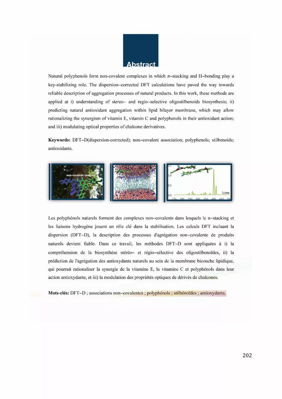
202


