Embed Size (px)
Citation preview
A Sterol-Binding Protein IntegratesEndosomal Lipid Metabolism withTOR Signaling and Nitrogen SensingCarl J. Mousley,1,5,* Peihua Yuan,1 Naseem A. Gaur,2 Kyle D. Trettin,1 Aaron H. Nile,1 Stephen J. Deminoff,3
Brian J. Dewar,4 MaxWolpert,1 Jeffrey M. Macdonald,4 Paul K. Herman,3 Alan G. Hinnebusch,2 and Vytas A. Bankaitis1,5,*1Department of Cell and Developmental Biology, Lineberger Comprehensive Cancer Center, University of North Carolina School of Medicine,
Chapel Hill, NC 27599-7090, USA2Laboratory of Gene Regulation and Development, Eugene Shriver National Institute of Child Health and Human Development,National Institutes of Health, 6 Center Drive, Bethesda, MD 20892, USA3Department of Molecular Genetics and Microbiology, The Ohio State University, Columbus, OH 43210, USA4Department of Biomedical Engineering, University of North Carolina School of Medicine, Chapel Hill, NC 27599-7575, USA5These authors contributed equally to this work
*Correspondence: [email protected] (C.J.M.), [email protected] (V.A.B.)
DOI 10.1016/j.cell.2011.12.026
SUMMARY
Kes1, and other oxysterol-binding protein super-family members, are involved in membrane and lipidtrafficking through trans-Golgi network (TGN) andendosomal systems. We demonstrate that Kes1represents a sterol-regulated antagonist of TGN/endosomal phosphatidylinositol-4-phosphate sig-naling. This regulation modulates TOR activation byamino acids and dampens gene expression drivenby Gcn4, the primary transcriptional activator of thegeneral amino acid control regulon. Kes1-mediatedrepression of Gcn4 transcription factor activity ischaracterized by nonproductive Gcn4 binding to itstarget sequences, involves TGN/endosome-derivedsphingolipid signaling, and requires activity of thecyclin-dependent kinase 8 (CDK8) module of theenigmatic ‘‘large Mediator’’ complex. These datadescribe a pathway by which Kes1 integrates lipidmetabolism with TORC1 signaling and nitrogensensing.
INTRODUCTION
Golgi and endosomes are dynamic organelles that combine
physical properties of stable compartments with features
consistent with a cycle of self-organized renewal and maturation
(Glick and Nakano, 2009). These compartments are key
membrane sorting stations and active sites of intracellular
signaling. How Golgi/endosomal trafficking and signaling func-
tions are coordinated is not understood, but a lipid signaling
interface is an attractive mechanism given the established link
between lipid metabolism and membrane trafficking in these
compartments. Lipid-exchange proteins play important roles
in coordinating lipid metabolism with the actions of protein
702 Cell 148, 702–715, February 17, 2012 ª2012 Elsevier Inc.
components of the membrane trafficking machinery in the
trans-Golgi network (TGN) and the endosomal membrane
system (Bankaitis et al., 2010; Graham and Burd, 2011).
Trafficking through the yeast TGN and endosomal systems
is regulated by two lipid-exchange proteins that execute
opposing activities (Cleves et al., 1991; Fang et al., 1996).
The phosphatidylinositol (PtdIns)/phosphatidylcholine (PtdCho)
transfer protein Sec14 is the pro-trafficking member of this
pair. Sec14 acts as a coincidence sensor that couples PtdCho
metabolism with PtdIns-4-phosphate production (PtdIns-4-P)
(Schaaf et al., 2008; Bankaitis et al., 2010). PtdIns-4-P is an
essential potentiator of membrane trafficking in the TGN/endo-
somal system (Hama et al., 1999; Rivas et al., 1999; Graham
and Burd, 2011). The Sec14 antagonist is Kes1/Osh4, one of
the seven yeast members of the oxysterol-binding protein
(OSBP) superfamily (Fang et al., 1996). Kes1 dampens PtdIns-
4-P signaling in TGN/endosomes (Li et al., 2002; Fairn et al.,
2007; Stefan et al., 2011). Why cells engineer a Sec14/Kes1
‘‘tug of war’’ into the TGN/endosomal trafficking design remains
unclear.
Kes1 is a PtdIns-4-P and sterol-binding protein. PtdIns-4-P
binding is a biologically important Kes1 activity and is required
for Kes1 homing to TGN/endosomal membranes (Li et al.,
2002). With regard to sterol binding, Kes1 is suggested to serve
as a diffusible sterol carrier (Im et al., 2005; Schulz and Prinz,
2009). Carrier models are confronted with the rapid rate at which
sterol shuttles between membranes via nonvesicular pathways,
however (Mesmin and Maxfield, 2009), and measured rates for
Kes1-mediated sterol transport in vitro are too slow to account
for such rapid flux. New data argue that OSBPs have no role
in sterol transport (Georgiev et al., 2011). This controversy under-
scores our lack of knowledge regarding how Kes1 translates
sterol-binding activity to TGN/endosomal trafficking functions,
and howKes1 coordinates its dual PtdIns-4-P and sterol-binding
activities.
Herein, we report that Kes1 integrates multiple aspects of
lipid metabolism in distal stages of the secretory pathway with
TORC1 and nitrogen signaling. Kes1 couples TGN/endosomal
sterol and PtdIns-4-P status with sphingolipid (SL) signaling
from this endomembrane system. We show that Kes1-regulated
SL-signaling in TGN/endosomal membranes attenuates activi-
ties of TORC1 and Gcn4, a primary transcription factor for
control of amino acid homeostasis. Finally, we report that
SL-regulated attenuation of Gcn4 activity requires a functional
CDK8 module of the ‘‘large Mediator’’ complex, which regulates
RNA polymerase II holoenzyme activity. These results introduce
new conceptual frameworks for interpreting how Kes1, and
other Kes1-like OSBPs, link membrane trafficking and lipid
signaling with cell proliferation and transcriptional programs
that respond to nitrogen stress.
RESULTS
Kes1 Defective in Sterol Binding RetainsBiological ActivityAssignment of Kes1 as a TGN/endosomal trafficking ‘‘brake’’
derives from loss-of-function phenotypes recorded in complex
genetic backgrounds (e.g., ‘‘bypass Sec14’’) (Cleves et al.,
1991; Fang et al., 1996; Li et al., 2002). The effects of a genuine
trafficking brake should also be apparent in otherwise wild-type
(WT) genetic backgrounds. Because Kes1 is a nonessential
protein in vegetative cells, the function was probed in cells
producing excess Kes1. The effects of acute elevations in
Kes1 expression were monitored in WT yeast using an inducible
system where KES1 and kes1 alleles of interest were placed
under control of a doxycycline (Dox)-repressible promoter. Three
Kes1 derivatives were expressed in parallel: (1) a biologically
inactive kes1R236E,K242E,K243E triple mutant (kes13E) unable to
target to TGN/endosomal membranes because it is defective
in PtdIns-4-P binding (Li et al., 2002); (2) the sterol-binding
mutant kes1Y97F (Im et al., 2005); and (3) a second putative
sterol-binding mutant (kes1T185V). The T185V substitution, like
Y97F, is predicted to disrupt the ordered water chain that
stabilizes sterol binding within the Kes1 lipid-binding pocket
(Figure S1A available online).
Kes1 and its mutant derivatives were further characterized.
[3H]-Cholesterol binding to Kes1 and kes13E was saturable
(apparent Kd z 0.5–0.8 mM) and specific on the basis of its
sensitivity to competition by unlabeled cholesterol (Figures
S1B and S1C). In agreement with structural data (Im et al.,
2005), the saturation binding data demonstrated both Kes1-
and kes13E-bound [3H]-cholesterol in an �1:1 stoichiometry
(Bmax = 1.2 pmol sterol bound/pmol protein). The Y97F and
T185V substitutions each diminished specific cholesterol binding
to the extent that saturation binding was not attainable. We
estimate the binding affinities to be >703 weaker than those
measured for Kes1 and kes13E (Figure S1C). Gel filtration
and circular dichroism assays confirmed that kes1Y97F and
kes1T185V, like Kes1, were well folded monomeric proteins
(Figures S1D and S1E).
Introduction of PDOX::KES1, PDOX::kes1Y97F, PDOX::kes1
T185V,
or PDOX::kes13E vectors into yeast did not impair cell growth
when the host yeast cells were cultured under noninducing
conditions (in Dox-replete media). Induced Kes1 expression by
Dox withdrawal severely inhibited cell growth, while kes13E
expression had no such detrimental effect (Figure 1). Unexpect-
edly, expression of the purportedly nonfunctional kes1Y97F
and kes1T185V mutants also arrested growth of WT yeast
(Figure 1A). The inducible PDOX expression system elevated
protein levels �5-fold (relative to endogenous Kes1) following
Dox withdrawal (Figure 1B), indicating that the inhibitory effects
of kes1Y97F and kes1T185V were not results of excessive ex-
pression relative to Kes1 or kes13E. Toxicity of kes1Y97F and
kes1T185V did not require strongly enhanced production. Yields
of WT yeast transformants per unit DNA were reduced �100-
fold for YCp(kes1Y97F) and YCp(kes1T185V) plasmids relative to
YCp(KES1) and YCp(kes13E). These YCp vectors drive only
modest constitutive expression of the Kes1 derivatives (2-fold
relative to endogenous Kes1) (Figure S1F). These data indicate
that kes1Y97F and kes1T185V are more potent inhibitors of cell
growth than is Kes1, a result that is incongruent with reports
that loss of sterol binding inactivates Kes1 (Im et al., 2005; Schulz
and Prinz, 2009). Although OSBPs are reported to function in
concert with FFAT receptors (VAPs) (Stefan et al., 2011), Kes1-
evoked arrest was indifferent to combinatorial ablation of VAP
structural genes (SCS2, SCS22).
Kes1 is unique among the yeast OSBPs in its privileged
interaction with Sec14-dependent pathways in TGN/endosomal
trafficking (Fang et al., 1996). This specificity is reflected in the
growth-inhibition assay. High-level expression of Hes1 (Osh5,
the protein most similar to Kes1), the sterol-binding-defective
hes1Y97F, or Osh7 did not compromise cell proliferation.
Lipid Binding and Kes1 Associationwith TGN/EndosomesTwo lines of evidence show that sterol binding controls Kes1
association with TGN/endosomes. First, whereas Kes1-GFP
adopted both diffuse cytosolic and punctate distributions in
cells, kes1Y97F-GFP localized predominantly to punctate struc-
tures (Figure 1C). These compartments were identified as
TGN/endosomes because they load with FM4-64 under condi-
tions where the tracer marks endocytic compartments (Fig-
ure 1D). Fractionation analyses also show increased kes1Y97F
membrane association (Figure S2A). In addition, our finding
that challenge of cells with a sterol synthesis inhibitor (micona-
zole) provoked Kes1-GFP superrecruitment to punctate
compartments loaded with FM4-64 (Figure 1E) also suggests
that sterol binding releases Kes1 from TGN/endosomes.
Enhanced association of Kes1 and kes1Y97F with TGN/endo-
somes also requires PtdIns-4-P-binding activity and robust
Pik1-dependent PtdIns-4-P synthesis. Introduction of kes13E
PtdIns-4-P-binding defects into the context of kes1Y97F had
the effect of (1) abrogating recruitment of the kes1Y97F sterol-
binding-defective mutant to TGN/endosomes (Figure 1G),
and (2) relieving kes1Y97F-mediated growth arrest (Figure 1H).
Moreover, shift of pik1-101ts yeast to 37�C, a condition nonper-
missive for Pik1-mediated PtdIns-4-P production, released
kes1Y97F-GFP from TGN/endosomal membranes (Figure 1F).
Kes1 Restricts PtdIns-4-P AvailabilityEnhanced Kes1 recruitment to TGN/endosomes interfered with
localization of the GOLPH3-GFP PtdIns-4-P sensor to this
membrane system. In agreement with Wood et al. (2009),
Cell 148, 702–715, February 17, 2012 ª2012 Elsevier Inc. 703
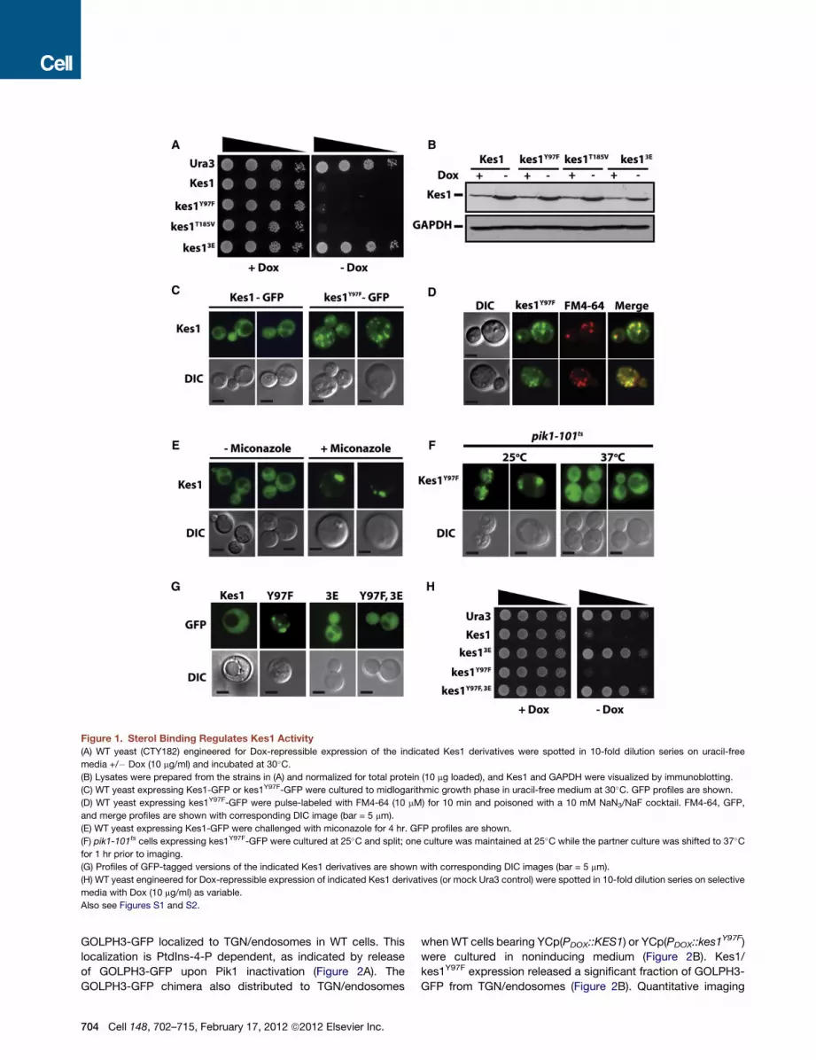
Figure 1. Sterol Binding Regulates Kes1 Activity
(A) WT yeast (CTY182) engineered for Dox-repressible expression of the indicated Kes1 derivatives were spotted in 10-fold dilution series on uracil-free
media +/� Dox (10 mg/ml) and incubated at 30�C.(B) Lysates were prepared from the strains in (A) and normalized for total protein (10 mg loaded), and Kes1 and GAPDH were visualized by immunoblotting.
(C) WT yeast expressing Kes1-GFP or kes1Y97F-GFP were cultured to midlogarithmic growth phase in uracil-free medium at 30�C. GFP profiles are shown.
(D) WT yeast expressing kes1Y97F-GFP were pulse-labeled with FM4-64 (10 mM) for 10 min and poisoned with a 10 mM NaN3/NaF cocktail. FM4-64, GFP,
and merge profiles are shown with corresponding DIC image (bar = 5 mm).
(E) WT yeast expressing Kes1-GFP were challenged with miconazole for 4 hr. GFP profiles are shown.
(F) pik1-101ts cells expressing kes1Y97F-GFP were cultured at 25�C and split; one culture was maintained at 25�C while the partner culture was shifted to 37�Cfor 1 hr prior to imaging.
(G) Profiles of GFP-tagged versions of the indicated Kes1 derivatives are shown with corresponding DIC images (bar = 5 mm).
(H) WT yeast engineered for Dox-repressible expression of indicated Kes1 derivatives (or mock Ura3 control) were spotted in 10-fold dilution series on selective
media with Dox (10 mg/ml) as variable.
Also see Figures S1 and S2.
GOLPH3-GFP localized to TGN/endosomes in WT cells. This
localization is PtdIns-4-P dependent, as indicated by release
of GOLPH3-GFP upon Pik1 inactivation (Figure 2A). The
GOLPH3-GFP chimera also distributed to TGN/endosomes
704 Cell 148, 702–715, February 17, 2012 ª2012 Elsevier Inc.
whenWT cells bearing YCp(PDOX::KES1) or YCp(PDOX::kes1Y97F)
were cultured in noninducing medium (Figure 2B). Kes1/
kes1Y97F expression released a significant fraction of GOLPH3-
GFP from TGN/endosomes (Figure 2B). Quantitative imaging

Figure 2. Kes1 and PtdIns-4-P Homeostasis
(A) WT and pik1-101ts cells expressing GOLPH3-GFP were cultured in uracil-free medium at 25�C and shifted to 37�C for 60 min prior to imaging. Corresponding
DIC images are shown at bottom (bar = 5 mm).
(B) WT yeast (CTY8-11Ca) expressing GOLPH3-GFP and indicated Kes1 derivatives were incubated +/� Dox (10 mg/ml) for 18 hr. GOLPH3-GFP (top) and DIC
image (bottom) profiles are shown (bar = 5 mm).
(C) WT yeast (CTY182) engineered for Dox-repressible expression of Kes1 derivatives were radiolabeled to steady state at 30�C with 20 mCi/ml [3H]-Ins +/� Dox
as indicated. Fractional incorporation of [3H]-Ins into the deacylated PtdIns-3-P, PtdIns-4-P, and PtdIns(4,5)P2 species is indicated (n = 4). Error bars represent
standard deviation.
(D) WT yeast (CTY182) expressing the Kes1 derivatives were cultured +/� Dox. Cells were pulse-radiolabeled with [35S]-amino acids (15 min). After 30 min of
chase, immunoprecipitated CPY species were analyzed by SDS-PAGE and autoradiography. p2 CPY and mCPY are indicated.
(E) WT yeast (CTY182) expressing Kes1 species (�Dox) and corresponding nonexpressing controls (+Dox), were examined for GFP-Snc1 distribution. DIC
images are shown at bottom (bar = 5 mm).
Also see Figure S2.
Cell 148, 702–715, February 17, 2012 ª2012 Elsevier Inc. 705

analyses recorded 3-fold reductions in puncta fluorescence
intensity relative to cytosol under those conditions (n = 150,
p = 0.0009 and p = 0.0003). [3H]-Inositol labeling showed no
diminution of bulk PtdIns-4-P, or bulk levels of other phosphoi-
nositides, in the face of Kes1/kes1Y97F expression (Figure 2C).
We conclude that Kes1 and kes1Y97F sequester PtdIns-4-P
without stimulating its degradation.
Kes1 Impairs TraffickingThe ability of Kes1 to bind PtdIns-4-P suggests that Kes1
interferes with interaction of this phosphoinositide with its
prosecretory effectors. Several independent assays demon-
strate that enhanced Kes1 activity impairs TGN/endosomal
dynamics. Pulse-radiolabeling experiments show that carboxy-
peptidase Y (CPY) trafficking to the vacuole was inhibited
by Kes1, kes1Y97F, or kes1T185V (Figure 2D). Trafficking of the
Snc1 v-SNARE and the bulk endocytic tracer FM4-64 were
also compromised by Kes1, kes1Y97F, or kes1T185V (Figure 2E).
Normally, FM4-64 is internalized from the plasma membrane
into endosomal compartments within 7.5 min of chase, and
a significant fraction of the cell-associated FM4-64 is detected
in the vacuole by that time point. The nonvacuolar FM4-64
pool chases from endosomes to vacuoles during the remainder
of the time course (Figure S2C). FM4-64 trafficking was interrup-
ted in cells with enhanced Kes1, kes1Y97F, or kes1T185V activities;
>80% and >40% of cells presented solely punctate endosomal
profiles after 15 and 30 min of chase. By 30 min, only 5% of
the Kes1-, kes1Y97F-, or kes1T185V-expressing cells exhibited
vacuolar labeling profiles (Figure S2C).
Trafficking defects were recorded for the general amino
acid permease Gap1 (Figure S2D), and the defects in uptake
of [35S]-amino acids observed for yeast with enhanced Kes1/
kes1Y97F activity were also consistent with defects in amino
acid permease trafficking to the plasmamembrane (Figure S2B).
Kes1 Induces AutophagyKes1/kes1Y97F-induced membrane trafficking defects notwith-
standing, electron microscopy failed to record the typical
accumulation of cargo-engorged TGN/endosomes. Instead,
intravacuolar vesicles (diameter �350 nm) were observed in
>60% of cells expressing Kes1/kes1Y97F (Figure 3A). These
morphologies report that Kes1/kes1Y97F-arrested cells were
engaged in autophagy while bathed in nutrient-sufficient
medium. Two other phenotypes support this diagnosis. First,
we measured autophagic import from the cytoplasm of a modi-
fied alkaline phosphatase zymogen (Pho8D60) into the vacuole
lumen, where Pho8D60 is activated (Noda et al., 1995).
Pho8D60 activity was elevated 3.8- and 4.2-fold in the face of
excess Kes1 and kes1Y97F (Figure 3B). This enhancement was
abrogated by atg1D, an allele which blocks autophagy at an
early stage (Figure S3A). Second, the Atg18 subunit of the
preautophagosome was recruited to a juxtavacuolar location
in Kes1/kes1Y97F-arrested cells in a manner similar to that
evoked by NH4+ starvation (Figures S3B and S3C).
Kes1/kes1Y97F-mediated growth arrest is not accompanied
by loss of viability, even after 20 days, indicating cell quiescence.
Survival requires active autophagy, however. Arrested cells
rapidly lose viability if they are incompetent for initiating auto-
706 Cell 148, 702–715, February 17, 2012 ª2012 Elsevier Inc.
phagy (atg1Dmutants) or if vacuolar protease activity is compro-
mised (Figures S3D and S3E). Interestingly, Kes1/kes1Y97F-
arrested atg1D cells accumulated cargo-engorged TGN/
endosomes in the cytoplasm, while interference with vacuolar
degradative functions resulted in membrane accretion in the
vacuole lumen (Figure S3F). These observations indicate that
the affected compartments are cleared by autophagy in Kes1/
kes1Y97F-arrested cells and degraded in the vacuole, which
explains our initial failure to observe these structures by electron
microscopy.
Kes1 Impairs Amino Acid HomeostasisThe autophagy phenotype suggested metabolic imbalances
in cells with elevated Kes1 activity. A metabolomic signature
for Kes1/kes1Y97F-arrested yeast was established by profiling
methanol-soluble small molecules by 1H-NMR (Figure S4A).
Unsupervised principal component analysis (PCA) deconvoluted
the NMR spectra, and PCA score plots revealed informative
variances in the first principal component (PC1) (Figure 3C).
These variances clustered along compact regions of the PC1
axis and accounted for 51% of the total variance in the data
set. The second principal component (PC2) further distinguished
Kes1- from kes1Y97F-expressing cells. Variances were again
confined to narrow windows of the PC2 axis (Figure 3C). The
discriminating methanol-soluble analytes were identified.
Shown in Figure 3D is a comparison of selected metabolites
from Kes1/kes1Y97F-arrested cells and mock controls. Pools of
60% of the assignable amino acids were reduced at least
2-fold in the arrested cells. Most prominent were diminutions
in Arg, Asn, Asp, Glu, Gln, Thr, and Trp pools (Figure S4B).
Amino Acid Resuscitation of Arrested CellsA concentrated Asn/Glu/Gln/Arg (NEQR) cocktail rescued
growth and amino acid pools of Kes1/kes1Y97F-arrested cells
(Figure 3E)—even though these cells are genetically NEQR
prototrophs. Yet the trafficking defects associated with en-
hanced Kes1/kes1Y97F activity remained unresolved. EM
analyses demonstrated that NEQR-rescued cells, unlike Kes1/
kes1Y97F-arrested cells, accumulated cargo-engorged TGN/
endosomes typically associated with trafficking defects through
this system (Figure S4C).
The NEQR data demonstrate that Kes1/kes1Y97F-induced
arrest derives from amino acid deficiencies rather than from
trafficking defects per se. The nature of the amino-acid-
homeostatic problem is the focus of the remainder of this
work. Several lines of evidence demonstrate that TORC1 activity
is inversely proportional to potency of the Kes1 trafficking
brake. First, phosphorylation of the TORC1 substrate Atg13
was reduced by Kes1/kes1Y97F expression (Figure 3F). This
effect was accompanied by phospho-eIF2a accumulation—
a hallmark of reduced TORC1 activity (Zaman et al., 2008).
Second, genome content assays scored a G1 block in Kes1/
kes1Y97F-arrested cells (Figure S4D). This block exhibits acti-
vated Rim15 signatures; a diagnosis of predisposal for entry
into G0. Third, sec14-1ts yeast (harbor diminished activity for
the protrafficking Sec14) were more rapamycin sensitive than
isogenic WT strains. Increased rapamycin sensitivity was sup-
pressed by kes1D (Figure 3G). Furthermore, rapamycin-treated

Figure 3. Kes1 and Amino Acid Homeostasis
(A) WT yeast cells (CTY182) producing Kes1 derivatives (� Dox) and corresponding nonexpressing controls (+ Dox; 10 mg/ml) were analyzed by EM.
Representative images are shown (bar = 1 mm). Intravacuolar vesicular profiles (diameter �350 nm) are highlighted by arrows.
(B) The ALP-expressing PHY2433 strain (TN124; Noda et al., 1995) was induced (�Dox), or not (+ Dox; 10 mg/ml), for Kes1 or kes1Y97F expression. Cultures were
maintained at an OD600nm < 0.2 throughout, harvested, and assayed for ALP activity. Error bars represent standard deviation.
(C) PCA scores plot distinguishing the 1H-NMR metabolic profiles of WT yeast (CTY182) expressing Kes1 or kes1Y97F (� Dox), or not (+ Dox).
(D) Targeted PCA of individual metabolites was performed for each condition. Reductions in Arg, Asn, Asp, Glu, Gln, Thr, and Trp pools were the major
contribution to the variance between conditions of excess Kes1/kes1Y97F and mock controls.
(E) WT yeast (CTY182) transformed with YCp(URA3), YCp(PDOX::KES1), or YCp(PDOX::kes1Y97F) were spotted in 10-fold dilution series on media with Dox
(10 mg/ml) and amino acids (NEQR, 0.2% w/v).
(F) atg13D yeast cotransformed with YEp(PCUP1::HA-ATG13), and either YCp(PDOX::KES1) or YCp(PDOX::kes1Y97F) was cultured in media containing 10 mg/ml
Dox. KES1 or kes1Y97F expression was induced for 7 hr (� Dox), or not (+ Dox; 10 mg/ml), and cells were challenged with CuSO4 (100 mM for 1 hr) to induce
HA-Atg13 expression. HA-Atg13 species were visualized by immunoblotting with anti-HA antibodies.
(G) WT (CTY182), sec14-1ts (CTY1-1A), and sec14-1ts kes1D (CTY159) yeast were spotted in 10-fold dilution series on media with or without 7.5 nM rapamycin
and grown at 30�C.(H) WT (CTY8-11Ca) and kes1D (CTY2039) yeast were spotted in 10-fold dilution series on media with or without 10 nM rapamycin and grown at 37�C.Also see Figures S3, S4, and S5.
Cell 148, 702–715, February 17, 2012 ª2012 Elsevier Inc. 707

kes1D cells accumulated less phospho-eIF2a than did isogenic
WT cells (Figure S5A), and both sec14-1 ts kes1D, and WT
kes1D yeast showed increased rapamycin resistance relative
to WT controls (Figures 3G and 3H). By contrast, a 2-fold eleva-
tion in KES1 expression rendered a sec14-1ts cki1D ‘‘bypass
Sec14’’ mutant hypersensitive to rapamycin (Figure S5B). In all
cases, the relative drug sensitivities reflected the magnitude of
rapamycin-induced phospho-eIF2a accumulation in these
strains (see below).
Amino acids promote TORC1 signaling by potentiating Gtr1
and Gtr2 GTPase activities (Binda et al., 2009), and neither casa-
mino acids nor NEQR revived growth of Kes1/kes1Y97F-arrested
gtr1D or gtr2D yeast (Figure S5C). NEQR resuscitated growth of
Kes1/kes1Y97F-arrested spo14D and scs2D scs22D mutants.
These data demonstrate that neither phospholipase D nor
FFAT receptors (VAPs) are required for the NEQR effect, but
that Gtr-mediated activation of TORC1 is.
Kes1- and Gcn4-Dependent Transcriptional RegulationThe general amino acid control (GAAC) pathway is a major
mechanism for amino acid homeostasis. The GAAC is acti-
vated by Gcn2-mediated phosphorylation of eIF2a, a modi-
fication that reduces eIF2a activity and promotes translation
of the Gcn4 transcription factor ORF (Hinnebusch, 1997).
The sensing component of the GAAC is engaged in Kes1/
kes1Y97F-arrested yeast, as evidenced by phospho-eIF2a
accumulation (Figures 4A and S5D). Yet downstream induction
of the GAAC fails, as measured by induced HIS4 and ARG1
transcription (Figures 4B and S6A). In contrast, Gcn2 induced
Gcn4-independent expression of ARO9 and ARO10 trypto-
phanase genes (Chen et al., 2009; Staschke et al., 2010) �7-
fold in Kes1/kes1Y97F-arrested cells (Figure S6B). This induc-
tion likely contributed to the 50-fold reductions in Trp pools
in those cells.
To assess Gcn4 status under conditions of enhanced Kes1
activity, we employed a sensitized system where sec14-1ts
cki1D cells arrest in response to 2-fold elevations in Kes1 level
(Fang et al., 1996). Kes1-dependent increases in phospho-
eIF2a were reproduced in that genetic background (Fig-
ure S6C). However, translational derepression of GCN4 was
severely diminished (Figure S6D). The consequences are
growth defects in minimal media supplemented with the His
analog 3-aminotriazole (3-AT) (Figure S6E). Consistent data
were also obtained from otherwise WT cells subjected to
Kes1/kes1Y97F growth arrest. Reduced Gcn4 accumulation
was observed in those cells under conditions where Gcn4
production is normally induced by 3-AT (Figure 4C). Yet bypass
of Gcn4 translational control (Gcn4c) failed to activate the
GAAC in Kes1/kes1Y97F-arrested cells (Figures 4D and S6F),
despite sustained Gcn4c protein levels (Figure S5F). NEQR
administration revived the GAAC in those cells (Figures 4E
and S6G).
Inactivation of the GAAC without Increased Kes1ExpressionThe data predicted that physiological levels of Kes1 expression
would silence the GAAC upon (1) enhanced Kes1 recruitment
to TGN/endosomes, or (2) imposition of TGN/endosomal traf-
708 Cell 148, 702–715, February 17, 2012 ª2012 Elsevier Inc.
ficking defects by reducing activities of proexocytic factors.
Kes1 load on TGN/endosomes was increased by challenging
yeast with sub-growth-inhibitory levels of the sterol synthesis
inhibitor miconazole. The intoxicated cells failed to respond
appropriately to 3-AT challenge (Figure S7A).
The effects of Kes1/kes1Y97F expression on GAAC activity
were recapitulated in a mutant that expresses normal levels of
Kes1 but is compromised for two factors that promote TGN/
endosomal trafficking (sec14-1ts tlg2D) (Figure 4F). The quies-
cent GAAC was not only revived by kes1D, but was constitu-
tively induced in sec14-1ts tlg2D kes1D mutants (Figure 4F).
This effect was not a general effect of ‘‘bypass Sec14’’ muta-
tions, as cki1D failed to reactivate the GAAC in sec14-1ts
tlg2D yeast.
Kes1 and SL MetabolismThe GAAC defects observed in sec14-1ts tlg2D cells were
accompanied by elevated ceramide, sphingoid base, and sphin-
goid-base-phosphate mass (Figure S7B). Kes1/kes1Y97F-
arrested yeast similarly exhibited increases in dihydro- (DHC)
and phytoceramide (PHC) mass, and dihydro- (DHS) and phy-
tosphingosine (PHS) mass (Figure 5A). Increases were also
measured for the sphingoid base phosphates (Figure 5A).
Incorporation of kes1D into the sec14-1ts tlg2D double mutant
normalized intracellular ceramide, sphingoid base, and sphin-
goid-base-phosphate mass (Figure S7B). These lipidomics
data suggest that SL metabolism links TGN/endosome traf-
ficking status to nuclear transcriptional outcomes.
Sphingosine Feeding Compromises the GAACIf Kes1 extinguishes the GAAC via an SL-signaling mechanism,
alterations in SL mass by means that do not affect cellular
Kes1 levels should also silence the GAAC. WT yeast were
challenged with sub-growth-inhibitory levels of PHS. Histidine
stress was superimposed on this condition by 3-AT challenge.
While cell proliferation was unaffected by individual PHS or
3-AT challenge, both growth inhibition and blunting of the
GAAC transcriptional response were elicited by dual challenge
(Figures 5B, 5C, and S7C). The PHS-mediated inhibition of
cell growth was apparent in the face of constitutive expression
of Gcn4 (Figure 5D), and HIS4 and ARG1 transcription was
strongly diminished under that condition as well (Figures 5E
and S7D). TGN/endosomal trafficking was unperturbed under
these conditions.
Sensitivity of the GAAC to PHS challenge requires Kes1, as
kes1D cells exhibited enhanced resistance to PHS (Figure 5B).
Gcn4-dependent HIS4 and ARG1 transcription was similarly
resistant to dual PHS and 3-AT challenge in kes1D cells (Figures
5C and S7C). Finally, ectopic expression of the yeast Ypc1
phytoceramidase revived the GAAC in the face of Kes1- and
PHS-challenge (Figures S7E and S7F). These data connect SL
metabolism with GAAC activity and implicate ceramide as a
key regulatory lipid.
Nonproductive Binding of Enhancer Elements by Gcn4Chromatin immunoprecipitation experiments interrogated
Gcn4 status on the ARG1 promoter in vivo. Gcn4 binding at
the ARG1 upstream activation sequence (UAS) was induced

Figure 4. Kes1 Attenuates Gcn4-Dependent Activation of the General Amino Acid Control
(A) Lysates were prepared from paired cell cultures induced for Kes1 or kes1Y97F expression (� Dox), or not (+ Dox; 10 mg/ml). Phospho-eIF2a, Kes1, and eIF2a
were visualized by immunoblotting.
(B) Total RNA was prepared from cells (CTY8-11Ca) induced for Kes1 or kes1Y97F expression (�Dox), or not (+ Dox), and challenged with 100mM 3-AT for 2 hr to
induce the GAAC. HIS4, ARG1, and ACT1 expression were surveyed by RT-PCR. Error bars represent standard deviation.
(C) Gcn4, Kes1, and GAPDH were analyzed by immunoblotting of lysates prepared from WT cells (CTY8-11Ca) expressing Kes1 or kes1Y97F (� Dox) or not
(+ Dox). Cells were challenged with 100 mM 3-AT for 2 hr to induce the GAAC.
(D) Total RNA fractions were isolated from WT cells harboring YCp(PDOX::KES1) and cotransformed with YCp (TRP1) or YCp (GCN4C) cultured +/� Dox. HIS4
and ARG1 gene expression was surveyed by RT-PCR, and normalized to ACT1 expression. Undiluted product and a 4-fold dilution of product were analyzed.
Error bars represent standard deviation.
(E) The experiment is as in (D), with the modification that an NEQR cocktail (0.2% w/v) was added to a parallel culture induced for Kes1 expression. HIS4, ARG1,
and ACT1 expression were scored by RT-PCR. Error bars represent standard deviation.
(F) RT-PCR of GAAC target genesARG1 andHIS4, aswell asACT1 control from total RNA fractions prepared fromWT, sec14-1ts tlg2D, sec14-1ts tlg2D kes1D, or
sec14-1ts tlg2D cki1D cells shifted to 37�C for 2 hr to impose the strong sec14-1ts tlg2D-associated TGN/endosomal trafficking block. Error bars represent
standard deviation.
Also see Figure S6.
by 3-AT in both mock-treated yeast and yeast challenged with
15 mM PHS (Figure 6A). Enhanced RNA Polymerase II (Rpb3)
occupancy evoked by 3-AT was dampened by PHS at the
promoter and at 50 and 30 locations in ARG1 coding sequences
(Figure 6B). These results report only modest defects in the
ability of UAS-bound Gcn4 to stimulate assembly of the preini-
tiation complex (PIC) at the ARG1 promoter. Additional levels
of regulation must be engaged downstream of PIC assembly
to account for the potent block of induced ARG1 transcription
by PHS.
Large-Mediator and GAAC RepressionCore Mediator is recruited to promoters by Gcn4, and it plays
a key role in stimulating PIC assembly (Govind et al., 2005).
The CDK8 module of the large Srb-Mediator complex, is
implicated in transcriptional repression and in transcriptional
Cell 148, 702–715, February 17, 2012 ª2012 Elsevier Inc. 709

Figure 5. Sphingolipids Inhibit the GAAC
(A) Quantitative lipidomics of DHC, PHC, DHS, PHS, DHS-1-P, and PHS-1-P in yeast (CTY182) as a function of Kes1 or Kes1Y97F expression. Note the log10scale for the y axis.
(B) WT strain (CTY8-11Ca) and a kes1D derivative were spotted in a 10-fold dilution series on synthetic-complete (SC) media and SC media with 7.5 mM PHS,
with 10 mM 3-AT, or both.
(C) Total RNA fractions were isolated from WT yeast either mock treated or grown in the presence of 7.5 mM PHS for 2 hr. Cells were either mock-treated or
challengedwith 100mM3-AT for 2 hr to induce theGAAC.HIS4,ARG1, andACT1 expressionweremonitored by RT-PCR. Undiluted product and a 4-fold dilution
of product were analyzed. Error bars represent standard deviation.
(D) WT yeast (CTY8-11Ca) expressing Ura3 or Gcn4c were spotted in a 10- fold dilution series on SC media and SC media with 7.5 mM PHS, with 10 mM 3-AT,
or both.
(E) Total RNA fractions were prepared fromWT yeast expressing Ura3 or Gcn4c +/� 15 mM PHS for 2 hr. HIS4 and ARG1 expression were surveyed by RT-PCR
and normalized to ACT1 expression. Undiluted product and a 4-fold dilution of product were analyzed. Error bars represent standard deviation.
Also see Figure S7.
checkpoints (Myers and Kornberg, 2000; Hengartner et al., 1998;
Taatjes, 2010). We found that Gcn4 inactivation in both Kes1-
and PHS-intoxicated yeast required a functional Srb10, the
cyclin-dependent kinase subunit of the CDK8 module. srb10D
yeast were resistant to Kes1/kes1Y97F arrest and to combinato-
rial PHS/3-AT challenge (Figures 6C and 6D). These phenotypes
coincided with rejuvenated Gcn4-dependent HIS4 and ARG1
transcription (Figure 6E). The same results were obtained upon
deletion of the structural genes encoding the other three
components of the CDK8 module (Srb8, 9, and 11). Thus, the
CDK8 module compromises the ability of Mediator to stimulate
GAAC activation in response to altered TGN/endosomal SL
signaling.
710 Cell 148, 702–715, February 17, 2012 ª2012 Elsevier Inc.
DISCUSSION
Herein, we demonstrate that Kes1 uses its sterol- and PtdIns-
4-P-binding activities to integrate multiple pathways of endoso-
mal lipid metabolism with the activity of TORC1-dependent
proliferative pathways and transcriptional control of nitrogen
signaling. This Kes1-regulated pathway provides mechanistic
insights as to why GAAC activation by Gcn4 is sensitive to
trafficking defects involving endosomal compartments (Mous-
ley et al., 2008; Zhang et al., 2008). Finally, these findings
generate conceptual frameworks for interpreting how Kes1,
and other Kes1-like OSBPs, are integrated into eukaryotic cell
physiology.

Figure 6. Gcn4 Binding to Target Genes and Mediator
(A and B) Cells were treated with PHS (15 mM) for 2 hr prior to challenge with 3-AT (10 mM) for 30 min and perfused with formaldehyde. Sonicated chromatin
was precipitated with antibodies against Gcn4 (A) or Rpb3 (B). DNA extracted from immunoprecipitates (ChIP) or starting chromatin (input) was PCR-amplified in
the presence of [32P]-dATPwith primers forARG1UAS (A), TATA element, or sequences from the 50 or 30 end of theARG1 coding sequences (B), and with primers
for an intergenic chromosome V sequence (nonspecific control). PCR products were quantified by phosphorimaging. Ratios of ARG1 ChIP-to-input signals
were normalized against chromosome V control sequence ratios to yield occupancy values. Averages and standard errors from two PCR amplifications for each
of two independent immunoprecipitates from two independent cultures are plotted. Error bars represent standard deviation.
(C) IsogenicWT (CTY1740) and srb10D yeast harboring YCp vectors for Dox-repressible expression of Kes1 were spotted in 10-fold dilutions series onmedia +/�Dox (10 mg/ml) and incubated at 30�C.(D) Isogenic WT (CTY1740) and srb10D yeast were spotted in a 10-fold dilution series on media with 7.5 mM PHS, 10 mM 3-AT, or both 7.5 mM PHS and 10 mM
3-AT.
(E) Isogenic WT (CTY1740) or srb10D yeast were either mock treated, or challenged with 15 mM PHS for 2 hr. The cultures were challenged with 100 mM 3-AT
for 2 hr to induce the GAAC. HIS4, ARG1, and ACT1 expression was monitored by RT-PCR. Undiluted product and a 4-fold dilution of product were analyzed.
Error bars represent standard deviation.
Kes1 Lipid BindingThe data identify Kes1 as a TGN/endosomal ‘‘trafficking brake’’
whose activity depends on PtdIns-4-P binding. The brake is
attenuated by sterol binding which promotes Kes1 disengage-
ment from PtdIns-4-P in TGN/endosomal membranes. We
describe Kes1 as a sterol-regulated rheostat of TGN/endosomal
PtdIns-4-P signaling. The interplay between sterol- and PtdIns-
4-P binding regulates potency of Kes1-mediated inhibition of
PtdIns-4-P-dependent trafficking through this endomembrane
system. We cannot reconcile these results with conclusions
that sterol-binding defects inactivate Kes1 (Im et al., 2005). Our
data indicate that Kes1 couples TGN/endosomal sterol status
with efficacy of PtdIns-4-P signaling as a sterol sensor. Signaling
circuits built on ‘‘tuning’’ principles do not require Kes1 to display
the large capacities for sterol exchange demanded by nonvesic-
ular sterol transfer models.
Kes1 and Nitrogen SignalingA potent Kes1 TGN/endosomal membrane trafficking brake
induces G1/G0 growth arrest associated with diminished
TORC1 activity, induction of autophagy, and depleted amino
acid pools. That amino acid homeostatic defects cause growth
arrest is demonstrated by the Gtr GTPase dependence of
NEQR-mediated resuscitation of cell proliferation, TORC1
Cell 148, 702–715, February 17, 2012 ª2012 Elsevier Inc. 711

activity, and proper amino acid homeostasis in cells with
enhanced Kes1 activity. We write a chain of events where
Kes1-evoked NH4+ starvation compromises TORC1 by attenu-
ating amino acid stimulation of Gtr GTPases.
Asn is a critical component of the NEQR cocktail. We interpret
that Asn essentiality reports NH4+ starvation as a key insult to
Kes1-arrested cells because NEQR failed to rescue Kes1 arrest
in asp1D yeast. The Asp1 asparaginase produces Gln by
transferring NH2 from Asn to Glu. Gln is a key reservoir of bio-
synthetic NH4+ (Wise and Thompson, 2010), yet Gln supplemen-
tation failed to rescue Kes1-mediated growth arrest, suggesting
that metabolic channeling of Asn is crucial. This concept might
be of broad relevance, as the Gcn2-Atf4 pathway (Atf4 is
mammalian Gcn4) supports Asn-dependent tumor survival (Ye
et al., 2010).
Endosomal Signaling and Nuclear OutputsResuscitation of Kes1-arrested cells by NEQR occurred in the
face of unresolved TGN/endosomal trafficking defects, signi-
fying that the trafficking defects are, by themselves, insufficient
to compromise cell proliferation. Growth arrest was driven by
a TGN/endosomal SL-derived signal that dampens TORC1
and Gcn4 activities and leads to an insurmountable nitrogen
deficit. The trafficking defects initiated the chain of events in
two respects. First, they provided the initial insult to cellular
nitrogen status. Amino acid and NH4+permeases were not
efficiently delivered to the plasma membrane under such
conditions, thereby compromising nitrogen acquisition. Second,
Kes1/kes1Y97F-mediated trafficking defects increased SL-signal
potency with downstream consequences, e.g., transcriptional
downregulation of Gap1 (3-fold) and of the major NH4+-
permease Mep2 (8.5-fold).
Activation of autophagy, and proximal stages of the GAAC,
indicated that Kes1-arrested yeast register nitrogen stress.
Yet, the cells failed to execute a productive GAAC because
GCN4 mRNA translation was blunted. The translational defect
might result from Gln deficiency. Gln is an indicator of nitrogen
availability, and nitrogen starvation blocks translation of GCN4
mRNA induced by phospho-eIF2a (Grundmann et al., 2001).
Kes1 signaling also inhibited Gcn4’s activity as a transcription
factor, recapitulating a phenotype we observed in vps mutants
(Zhang et al., 2008). Hence, the transcriptional defects in vps
mutants might also involve Kes1 signaling.
What SL biochemistry underlies themechanism throughwhich
endosomal events influence nuclear Gcn4 activities? While the
nature of the signal remains to be established, an SL-driven
signaling pathway must negotiate the physical separation of
these compartments. A diffusible enzyme (SL-activated kinase
or phosphatase) might be activated on the endosomal surface
and modify nuclear substrates that regulate Gcn4 activity. While
the identities of nuclear effectors are unknown, the CDK module
of large Mediator is an attractive candidate given that functional
silencing of Gcn4 requires Srb10. Whether SLs stimulate Srb10
CDK activity is a key question. Gcn4 might represent the
effector, as we observe an endosome-associated Gcn4 pool
(N.A.G. and A.G.H., unpublished data). Epigenetic effects are
also plausible as histone deacetylases are inhibited by sphingo-
sine-1-P (Nitai et al., 2009).
712 Cell 148, 702–715, February 17, 2012 ª2012 Elsevier Inc.
Kes1 and Homeostatic ControlWhy engineer a Sec14/Kes1 antagonism into the TGN/endoso-
mal trafficking design? We propose that the Sec14/Kes1 tug of
war sets the SL signaling output of a PtdIns-4-P/sterol-regulated
endosomal compartment (Figure 7A). Appropriate tuning of
signaling output is crucial, because the organelle couples
membrane trafficking to local lipid metabolism and, subse-
quently, to global responses. The Kes1 brake on PtdIns-4-P
signaling enhances production of bioactive SLs in an endosomal
compartment monitored by homeostatic control systems (Fig-
ure 7B). Whereas such a design could be used in physiological
contexts (see below), enhanced endosomal SL signaling exerts
significant consequences for cell physiology, as manifested by
the effects we document on TORC1 and GAAC signaling.
Although we focus on the GAAC, the nitrogen control
response (NCR) is also silenced in Kes1-arrested cells. The
NCR controls nitrogen acquisition via GATA transcription
factor-regulated expression of amino acid and NH4+ permeases
(e.g., GAP1, MEP2) (Zaman et al., 2008). NEQR supplementa-
tion, or Srb10 inactivation, also revives the NCR. NCR defects
suggest a mechanism for transcriptional downregulation of
GAP1 and MEP2 in Kes1-arrested cells.
Broader Implications for OSBPs?Kes1-like proteins might be physiological modulators of cell
proliferation programs. Loss of such activities will be scored
under conditions where a trafficking brake needs to be imposed.
In that regard, functional ablation of Kes1 compromises cell
viability under starvation conditions, suggesting that loss of
Kes1 activity hinders adjustment of endosomal lipid signaling
by cells in response to nitrogen deprivation. The cardinal proper-
ties of Kes1-arrested yeast, like those of Sec14-deficient cells,
conform to those of postmitotic cells. Perhaps privileged PtdIns
transfer protein::OSBP pairs coevolved for fine-tuning systems
that couple endosomal lipid signaling with cell proliferation.
Such modalities could guide cell entry into postmitotic states
or maintain postmitotic cell physiology. This concept raises
possibilities for OSBPs flexibly coupling membrane trafficking
flux and organelle maturation to the fine-tuning of transcriptional
programs for organogenesis of tissues populated with postmi-
totic cells.
EXPERIMENTAL PROCEDURES
Yeast Strains and Media
Yeast strain genotypes and plasmids are listed in Tables S1 and S2. Yeast
complex and synthetic complete media and yeast transformation methods
were as described (Cleves et al., 1991; Schaaf et al., 2008). Fine chemicals
were purchased from Sigma Chemical (St. Louis, MO, USA), and restriction
enzymeswere fromNewEnglandBiolabs, Inc. (Ipswich,MA). YCp(PDOX::KES1)
transformants, and mutant derivatives thereof, were maintained in the pres-
ence of 10 mg/ml Dox. To induce expression, cells were washed four times
in ddH2O and resuspended in fresh Dox-free media for a minimum of 8 hr.
Amino acids (Asn, Glu, Gln, and Arg) were added at a final concentration of
0.2% (w/v). YCp(PCUP1-ATG13HA) expression was induced by the addition of
100 mM CuSO4 for 1 hr.
Expression Vectors
To generate YCp(PDOX::KES1), theKES1ORFwas PCR amplified with oligonu-
cleotides clamped with PmeI and PstI restriction sites at the 50 and 30 ends.

Figure 7. Integration of TGN/Endosomal Trafficking Flux to Transcriptional Competence of the Nitrogen Response
(A) Membrane trafficking through a signaling TGN/endosomal compartment is defined by Sec14-regulated production of PtdIns-4-P countered by Kes1-
mediated PtdIns-4-P sequestration. Degree of sequestration is set by PtdIns-4-P-mediated recruitment of Kes1 to TGN/endosomal membranes, and released by
binding of the membrane-bound Kes1 to active sterol.
(B) Kes1-mediated trafficking defects reduce efficiency of amino acid and NH4+ permease delivery to the plasmamembrane. The incipient nitrogen stress results
in reduced TOR activity and engagement of the early stages of the GAAC (phosphorylation of eIF2a). Efficiency of the terminal stages of the GAAC is determined
by potency of Gcn4 antagonism levied by activity of the large SRB Mediator complex.
(C) Potency of a Kes1-mediated trafficking arrest/delay sets the amplitude of TGN/endosomal production of SL available for nuclear signaling. SL* denotes
a sphingolipid with signaling power (e.g., ceramide) produced in TGN/endosomal compartments. The potency of SL* signaling is directly proportional to
Kes1 activity, and inhibits transcription initiation/elongation of genes loaded with Gcn4 and RNA polymerase II via action of the CDK8 module of the large
SRB Mediator complex. SL* signaling foils the GAAC and induces cells to enter a metabolically coherent quiescence with properties of postmitotic cell
physiology.
The 1319 bp fragment was subcloned into pCM189 (Garı et al., 1997). Primer
sequences are available from the authors by request.
Sterol Binding
The sterol:detergent micelle assay of Infante et al. (2007) was used to measure
sterol binding with minor modification. Details are in the Extended Experi-
mental Procedures.
Fluorescence Microscopy
Cells grown at 30�C were incubated with a final concentration of 10 mM
FM4-64 (Molecular Probes) for 10 min. Labeling was stopped with 10 mM
NaN3/NaF. For GFP-Snc1 imaging, cells were cultured at 30�C. Images
were collected with a Nikon E600 microscope equipped with a Princeton
Instruments 512 X 512 back-illuminated frame-transfer CCD camera. Meta-
morph (Universal Imaging Corp.) was used to capture images.
Transmission Electron Microscopy
Yeast were grown to midlogarithmic phase (OD600nm = 0.3). Ten OD600nm units
of cells were isolated, and fixed in 3% glutaraldehyde. Cells were converted
to spheroplasts with zymolyase, stained with 2% osmium tetroxide and 2%
uranyl acetate, dehydrated in a 50%, 70%, 90% ethanol series, and washed
in 100% ethanol and 100% acetone, respectively. Cells were embedded in
Spurr’s resin, and sections were prepared as described (Adamo et al., 2001)
and visualized at 80 kV on an FEI Tecnai 12 electron microscope. Images
were captured using Gatan micrograph 3.9.3 software.
Metabolic Labeling and Immunoprecipitation
Yeast were grown in minimal media lacking methionine and cysteine to mid-
logarithmic phase (OD600nm = 0.5) and radiolabeled with [35S]-amino acids
(Translabel; New England Nuclear; 100 mCi/ml). Chase was initiated by
introduction of unlabeled methionine and cysteine (2 mM each, final concen-
tration) for the specified time, and chase was terminated by addition of
trichloroacetic acid (5% w/v final concentration). Immunoprecipitation of
CPY with rabbit antiserum and resolution by SDS-PAGE and autoradiog-
raphy were performed as previously described (Cleves et al., 1991; Schaaf
et al., 2008).
Determination of LacZ Activity and RT-PCR
Yeast strains were transformed with plasmid p180 (Hinnebusch, 1985) and
LacZ activity assayed (Mousley et al., 2008). For RT-PCR assays, total RNA
was isolated from cells and 1 mg was used to template reverse transcription
to generate cDNA (20 ml final volume). To analyze HIS4, ARG1, or ACT1
expression, PCR was performed using 1 ml of cDNA fraction as template
and specific oligonucleotides as primers. Products were quantified with
ImageQuant (GE Healthcare Life Science).
Autophagy Assays
Alkaline phosphatase assays that monitor delivery of a cytosolic Pho8
phosphatase reporter to the vacuole lumen by autophagy, and the subsequent
proteolytic activation of this reporter in the vacuole, were performed as
described (Stephan et al., 2009).
Cell 148, 702–715, February 17, 2012 ª2012 Elsevier Inc. 713
Sphingolipidomics
Ceramides and sphingoid bases were measured at the Lipidomics Shared
Resource facility at the Medical University of South Carolina using normal-
phase high-perfomance liquid chromatography coupled to atmospheric
pressure chemical mass spectrometry (Bielawski et al., 2006). Neutral lipids
were isolated from yeast extracts generated by pooling three independent
cultures of each yeast strain that had been grown at 30�C. ESI/MS/MS analysis
of ceramides and internal standards were performed on a Thermo Finnigan
TSQ 7000 triple quadruple mass spectrometer operating in a multiple-
reaction-monitoring positive-ionization mode. Calibration curves for quantifi-
cation were constructed by plotting peak area ratios of synthetic standards.
Ceramides were normalized to total organic phosphate.
NMR Spectroscopy and Analysis
Small metabolites were extracted in 50% methanol and reconstituted in
0.5 mM trimethylsilyl-2,20,3,30-tetradeuteropropioninc acid in D2O. 1H NMR
spectra were acquired at 16.4T with a Varian INOVA (700 MHz 1H, Varian
Instruments) equipped with 5 mm indirect cold probe and processed with
ACD/1D NMR Manager (version 12.0; Advanced Chemistry Development,
Inc., Toronto, ON, Canada). Spectra were phase and baseline corrected
and referenced to the TSP peak to 0.00 ppm, and grouped spectra were
data reduced to 250 bins using intelligent bucketing. Principal component
analysis used SIMCA-P (version 11.0; Umetrics, Umea, Sweden) and Pareto
scaling. Individual metabolites were quantified using Chenomx NMR Suite
(version 5.1; Chenomx Inc., Edmonton, Canada), with TSP as concentration
and chemical shift reference (Dewar et al., 2010). Details are described in the
Extended Experimental Procedures.
Online Supplemental Information
Figure S1 describes properties of Kes1 sterol-binding mutants. Figure S2
describes effects of Kes1 and mutant Kes1 on TGN/endosomal trafficking.
Figure S3 documents induction of autophagy by Kes1/kes1Y97F expression
and the role of autophagy in arrested cell viability. Figure S4 shows metabo-
lomics profiling data. Figure S5 shows Kes1 modulation of TORC1 signaling.
Figure S6 documents how Kes1/kes1Y97F inhibits activation of the GAAC.
Figure S7 shows that miconazole-evoked superrecruitment of Kes1 to TGN/
endosomes inactivates the GAAC, that Kes1 regulates sphingolipid metabo-
lism, and that sphingoid-base feeding inactivates the GAAC. Tables S1 and
S2 list yeast strains and plasmids.
SUPPLEMENTAL INFORMATION
Supplemental Information includes Extended Experimental Procedures, seven
figures, and two tables and can be found with this article online at doi:10.1016/
j.cell.2011.12.026.
ACKNOWLEDGMENTS
This paper is dedicated to the memories of Christian Raetz and Eugene
Kennedy. The work was supported by NIH grant GM44530 to V.A.B.; N.G.
and A.G.H. were supported by the Intramural Research Program of the NIH;
S.J.D. and P.K.H. by NIH grant GM65227 awarded to P.K.H.; and B.J.D. and
J.M.M. by NIEHS T32EX007126 and GM075941. K.D.T. acknowledges
support from the UNC William W. and Ida W. Taylor Honors Mentored
Research Fellows Program. We thank Doug Cyr, Scott Moye Rowley, Chris
Burd, and Dan Klionsky for strains and plasmids, Colin Stirling for CPY
antibody, Chris Stefan, Scott Emr, and Maria Cardenas for discussions, and
Lora Yanagisawa, Victoria Hewitt, and Robert Sons for comments on the
manuscript. We acknowledge the UNC Lineberger Comprehensive Cancer
Center Genome Analysis and Nucleic Acids Core facilities.
Received: October 15, 2010
Revised: October 13, 2011
Accepted: December 5, 2011
Published: February 16, 2012
714 Cell 148, 702–715, February 17, 2012 ª2012 Elsevier Inc.
REFERENCES
Adamo, J., Moskow, J.J., Gladfelter, A.S., Viterbo, D., Lew, D.J., and
Brennwald, P.J. (2001). Yeast Cdc42 functions at a late step in exocytosis,
specifically during polarized growth of the emerging bud. J. Cell Biol. 155,
581–592.
Bankaitis, V.A., Mousley, C.J., and Schaaf, G. (2010). The Sec14-superfamily
and mechanisms for crosstalk between lipid metabolism and lipid signaling.
Trends Biochem. Sci. 35, 150–160.
Bielawski, J., Szulc, Z.M., Hannun, Y.A., and Bielawska, A. (2006). Simulta-
neous quantitative analysis of bioactive sphingolipids by high-performance
liquid chromatography-tandem mass spectrometry. Methods 39, 82–91.
Binda, M., Peli-Gulli, M.P., Bonfils, G., Panchaud, N., Urban, J., Sturgill, T.W.,
Loewith, R., and De Virgilio, C. (2009). The Vam6 GEF controls TORC1 by
activating the EGO complex. Mol. Cell 35, 563–573.
Chen, H., Huang, H., Li, X., Tong, S., Niu, L., and Teng, M. (2009). Crystalliza-
tion and preliminary X-ray diffraction analysis of ARO9, an aromatic amino-
transferase from Saccharomyces cerevisiae. Protein Pept. Lett. 16, 450–453.
Cleves, A.E., McGee, T.P., Whitters, E.A., Champion, K.M., Aitken, J.R., Dow-
han, W., Goebl, M., and Bankaitis, V.A. (1991). Mutations in the CDP-choline
pathway for phospholipid biosynthesis bypass the requirement for an essential
phospholipid transfer protein. Cell 64, 789–800.
Dewar, B.J., Keshari, K., Jeffries, R.E., Dzeja, P., Graves, L.M., and Macdon-
ald, J.M. (2010). Metabolic assessment of a novel chronic myelogenous
leukemic cell line and an imatinib resistant subline by H NMR spectroscopy.
Metabolomics 6, 439–450.
Fairn, G.D., Curwin, A.J., Stefan, C.J., and McMaster, C.R. (2007). The
oxysterol binding protein Kes1p regulates Golgi apparatus phosphatidylinosi-
tol-4-phosphate function. Proc. Natl. Acad. Sci. USA 104, 15352–15357.
Fang,M., Kearns, B.G., Gedvilaite, A., Kagiwada, S., Kearns,M., Fung,M.K.Y.,
and Bankaitis, V.A. (1996). Kes1p shares homology with human oxysterol
binding protein and participates in a novel regulatory pathway for yeast
Golgi-derived transport vesicle biogenesis. EMBO J. 15, 6447–6459.
Garı, E., Piedrafita, L., Aldea, M., and Herrero, E. (1997). A set of vectors with
a tetracycline-regulatable promoter system for modulated gene expression in
Saccharomyces cerevisiae. Yeast 13, 837–848.
Georgiev, A.G., Sullivan, D.P., Kersting, M.C., Dittman, J.S., Beh, C.T., and
Menon, A.K. (2011). Osh proteins regulate membrane sterol organization but
are not required for sterol movement between the ER and PM. Traffic 12,
1341–1355. 10.1111/j.1600-0854.2011.01234.x.
Glick, B.S., and Nakano, A. (2009). Membrane traffic within the Golgi appa-
ratus. Annu. Rev. Cell Dev. Biol. 25, 113–132.
Govind, C.K., Yoon, S., Qiu, H., Govind, S., and Hinnebusch, A.G. (2005).
Simultaneous recruitment of coactivators by Gcn4p stimulates multiple steps
of transcription in vivo. Mol. Cell. Biol. 25, 5626–5638.
Graham, T.R., and Burd, C.G. (2011). Coordination of Golgi functions by
phosphatidylinositol 4-kinases. Trends Cell Biol. 21, 113–121.
Grundmann, O., Mosch, H.U., and Braus, G.H. (2001). Repression of GCN4
mRNA translation by nitrogen starvation in Saccharomyces cerevisiae.
J. Biol. Chem. 276, 25661–25671.
Hama, H., Schnieders, E.A., Thorner, J., Takemoto, J.Y., and DeWald, D.B.
(1999). Direct involvement of phosphatidylinositol 4-phosphate in secretion
in the yeast Saccharomyces cerevisiae. J. Biol. Chem. 274, 34294–34300.
Hengartner, C.J., Myer, V.E., Liao, S.-M., Wilson, C.J., Koh, S.S., and Young,
R.A. (1998). Temporal regulation of RNA polymerase II by Srb10 and Kin28
cyclin-dependent kinases. Mol. Cell 2, 43–53.
Hinnebusch, A.G. (1985). A hierarchy of trans-acting factors modulates trans-
lation of an activator of amino acid biosynthetic genes in Saccharomyces
cerevisiae. Mol. Cell. Biol. 5, 2349–2360.
Hinnebusch, A.G. (1997). Translational regulation of yeast GCN4. A window on
factors that control initiator-trna binding to the ribosome. J. Biol. Chem. 272,
21661–21664.

Im, Y.J., Raychaudhuri, S., Prinz, W.A., and Hurley, J.H. (2005). Structural
mechanism for sterol sensing and transport by OSBP-related proteins. Nature
437, 154–158.
Infante, R.E., Abi-Mosleh, L., Radhakrishnan, A., Dale, J.D., Brown, M.S., and
Goldstein, J.L. (2007). Purified NPC1 protein. 1. Binding of cholesterol and
oxysterols to a 1278-amino acid membrane protein. J. Biol. Chem. 283,
1052–1063.
Li, X.M.P., Rivas, M.P., Fang, M., Marchena, J., Mehrotra, B., Chaudhary, A.,
Feng, L., Prestwich, G.D., and Bankaitis, V.A. (2002). Analysis of oxysterol
binding protein homologue Kes1p function in regulation of Sec14p-dependent
protein transport from the yeast Golgi complex. J. Cell Biol. 157, 63–77.
Mesmin, B., and Maxfield, F.R. (2009). Intracellular sterol dynamics. Biochim.
Biophys. Acta 1791, 636–645.
Mousley, C.J., Tyeryar, K., Ile, K.E., Schaaf, G., Brost, R.L., Boone, C., Guan,
X., Wenk, M.R., and Bankaitis, V.A. (2008). Trans-Golgi network and endo-
some dynamics connect ceramide homeostasis with regulation of the
unfolded protein response and TOR signaling in yeast. Mol. Biol. Cell 19,
4785–4803.
Myers, L.C., and Kornberg, R.D. (2000). Mediator of transcriptional regulation.
Annu. Rev. Biochem. 69, 729–749.
Noda, T., Matsuura, A., Wada, Y., and Ohsumi, Y. (1995). Novel system for
monitoring autophagy in the yeast Saccharomyces cerevisiae. Biochem. Bio-
phys. Res. Commun. 210, 126–132.
Rivas, M.P., Kearns, B.G., Xie, Z., Guo, S., Sekar, M.C., Hosaka, K., Kagiwada,
S., York, J.D., and Bankaitis, V.A. (1999). Pleiotropic alterations in lipid metab-
olism in yeast sac1 mutants: relationship to ‘‘bypass Sec14p’’ and inositol
auxotrophy. Mol. Biol. Cell 10, 2235–2250.
Schaaf, G., Ortlund, E., Tyeryar, K., Mousley, C., Ile, K., Woolls, M., Garrett, T.,
Raetz, C.R.H., Redinbo, M., and Bankaitis, V.A. (2008). Functional anatomy of
phospholipid binding and regulation of phosphoinositide homeostasis by
proteins of the Sec14 superfamily. Mol. Cell 29, 191–206.
Schulz, T.A., and Prinz, W.A. (2009). Sterol transport in yeast and the oxy-
sterol binding protein homologue (OSH) family. Biochim. Biophys. Acta
1771, 769–780.
Staschke, K.A., Dey, S., Zaborske, J.M., Palam, L.R., McClintick, J.N., Pan, T.,
Edenberg, H.J., and Wek, R.C. (2010). Integration of general amino acid
control and target of rapamycin (TOR) regulatory pathways in nitrogen assim-
ilation in yeast. J. Biol. Chem. 285, 16893–16911.
Stephan, J.S., Yeh, Y.Y., Ramachandran, V., Deminoff, S.J., and Herman, P.K.
(2009). The Tor and PKA signaling pathways independently target the Atg1/
Atg13 protein kinase complex to control autophagy. Proc. Natl. Acad. Sci.
USA 106, 17049–17054.
Stefan, C.J., Manford, A.G., Baird, D., Yamada-Hanff, J., Mao, Y., and Emr,
S.D. (2011). Osh proteins regulate phosphoinositide metabolism at ER-plasma
membrane contact sites. Cell 144, 389–401.
Taatjes, D.J. (2010). The human Mediator complex: a versatile, genome-wide
regulator of transcription. Trends Biochem. Sci. 35, 315–322.
Wise, D.R., and Thompson, C.B. (2010). Glutamine addiction: a new thera-
peutic target in cancer. Trends Biochem. Sci. 35, 427–433.
Wood, C.S., Schmitz, K.R., Bessman, N.J., Setty, T.G., Ferguson, K.M., and
Burd, C.G. (2009). PtdIns4P recognition by Vps74/GOLPH3 links PtdIns
4-kinase signaling to retrograde Golgi trafficking. J. Cell Biol. 187, 967–975.
Ye, J., Kumanova, M., Hart, L.S., Sloane, K., Zhang, H., De Panis, D.N.,
Bobrovnikova-Marjon, E., Diehl, J.A., Ron, D., and Koumenis, C. (2010). The
GCN2-ATF4 pathway is critical for tumour cell survival and proliferation in
response to nutrient deprivation. EMBO J. 29, 2082–2096.
Zaman, S., Lippman, S.I., Zhao, X., and Broach, J.R. (2008). How Saccharo-
myces responds to nutrients. Annu. Rev. Genet. 42, 27–81.
Zhang, F., Gaur, N.A., Hasek, J., Kim, S.J., Qiu, H., Swanson, M.J., and
Hinnebusch, A.G. (2008). Disrupting vesicular trafficking at the endosome
attenuates transcriptional activation by Gcn4. Mol. Cell. Biol. 28, 6796–6818.
Cell 148, 702–715, February 17, 2012 ª2012 Elsevier Inc. 715


