Embed Size (px)
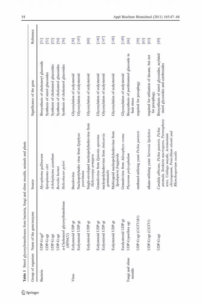
Citation preview

Sterol Glycosyltransferases—The EnzymesThat Modify Sterols
Pankaj Chaturvedi & Pratibha Misra & Rakesh Tuli
Received: 19 November 2010 /Accepted: 22 March 2011 /Published online: 6 April 2011# Springer Science+Business Media, LLC 2011
Abstract Sterols are important components of cell membranes, hormones, signallingmolecules and defense-related biotic and abiotic chemicals. Sterol glycosyltransferases(SGTs) are enzymes involved in sterol modifications and play an important role inmetabolic plasticity during adaptive responses. The enzymes are classified as a subset offamily 1 glycosyltransferases due to the presence of a signature motif in their primarysequence. These enzymes follow a compulsory order sequential mechanism forming aternary complex. The diverse applications of sterol glycosides, like cytotoxic and apoptoticactivity, anticancer activity, medicinal values, anti-stress roles and anti-insect andantibacterial properties, draws attention towards their synthesis mechanisms. Manysecondary metabolites are derived from sterol pathways, which are important in defensemechanisms against pathogens. SGTs in plants are involved in changed sensitivity to stresshormones and their agrochemical analogs and changed tolerance to biotic and abioticstresses. SGTs that glycosylate steroidal hormones, such as brassinosteroids, function asgrowth and development regulators in plants. In terms of metabolic roles, it can be said thatSGTs occupy important position in plant metabolism and may offer future tools for cropimprovement.
Keywords Adaptive response . Brassinosteroids . Cellular homeostasis . Detoxification .
Glycosyltransferases . Hormonal regulation . Insect resistance . Medicinal plant . Sterols stress
Appl Biochem Biotechnol (2011) 165:47–68DOI 10.1007/s12010-011-9232-0
P. Chaturvedi : P. Misra (*) : R. TuliNational Botanical Research Institute (Council of Scientific & Industrial Research), Rana Pratap Marg,Lucknow 226001, Uttar Pradesh, Indiae-mail: [email protected]: [email protected]
Present Address:P. ChaturvediSanjay Gandhi Post Graduate Institute of Medical Sciences, Lucknow, India
Present Address:R. TuliDepartment of Biotechnology, National Agrifood Biotechnology Institute, Mohali 160071, Punjab, India

Introduction
Bacteria, fungi, animals and plants are a valuable source of numerous metabolites, whichare used as pharmaceuticals, agrochemicals, flavours, fragrances, colours, biopesticides andfood additives. The immense heterogeneity of metabolites is a result of diverse modificationmechanisms, which are undergoing inside the organisms. Many of these metabolites aremade, in attempt to adapt rapidly to the external environmental conditions and maintaintheir cellular homeostasis. This adaptability depends on their growth and developmentalstages and evolution of mechanisms that regulate metabolic functions. Studies suggest thatglycosylation is one of the mechanisms by which organisms modify hormones, secondarymetabolites, biotic and abiotic chemicals and toxins in the environment to maintain theircellular homeostasis. The identification of large multigene families of glycosyltransferases(GTs), which recognise these diverse molecules, adds to this fact. This review focuses onglycosyltransferases that recognise small molecules such as sterols especially in plants anddescribes their functions pertaining to their biological activities, structural features andresponse to stress. The sterol glycosyltransferases are grouped into family 1 of the 90distinct families, which describe GTs (http://afmd.cnrs-mrs.fr/CAZY/acc.html). Most of theGT family 1 members are defined by the presence of a carboxyl terminal consensussequence termed as signature motif (Fig. 1), which is involved in the interaction of theenzyme with the activated sugar donor [1–4]. The signature motif can be identified in theenzyme sequences of animals, plants, fungi and bacteria [5, 6]. In Arabidopsis thaliana, thegene family comprises 112 full-length sequences and eight pseudogenes with frame-shiftmutations [3].
The activated sugar donor of plant GTs is commonly UDP-glucose (UDP-Glc) althoughUDP-rhamnose (UDP-Rha), UDP-galactose (UDP-Gal), UDP-xylose (UDP-Xyl) and UDP-glucuronic acid (UDP-GicUA) also act as activated sugars [7–9]. Glycosylation occurs at –OH, –COOH, –NH2, –SH and C–C groups and more than one sugar may be attached [10,11]. The membrane transport systems recognise the glycosyl residues and facilitate thetransport of glycosides, which is reflected by the fact that many glycosylated molecules arefound in vacuole compartments [12]. Studies do show ATP binding cassette (ABC)transporters in the tonoplast membranes, which transport both endogenous and xenobioticglycosides. Glycosides and glycosinolates of small molecules have also been identified inthe apoplast [13, 14]. Diverse glycosylated molecules accumulate in the vacuolar andapoplastic space, and there is also clearance of these molecules. The above points indicatethat GTs are involved in detoxification. It can be concluded that the glycosyltransferasesmodify small lipophilic molecules, which leads to a change in their participation in cellularmetabolism.
Sterol Glycosyltransferases
In contrast to vast literature available on glycosyltransferases of diverse molecules, there arefew reports on sterol glycosyltransferases. Sterols are a part of vast family of isoprenoidsand are precursors of many steroid hormones, defense-related compounds, such as
[FW]-x(2)-[LIVMYA]-[LIMV]-x(4-6)-[LVGAC]-[LVFYA]-[LIVMF]-[STAGCM]-[HNQ]-[STAGC]-G-x(2)-[STAG]-x(3)-[STAGL]-[LIVMFA]-x(4)-[PQR]-[LIVMT]-x(3)-[PA]-x(3)-[PA]-x(3)-[DES]-[QEHN]
Fig. 1 Signature sequence ofglycosyltransferases
48 Appl Biochem Biotechnol (2011) 165:47–68

saponins, signalling molecules and are important structural components of cell membranes[15]. Sterols have been shown to regulate development and gene regulation in Arabidopsis[16]. A group of oxidised sterols called brassinosteroids function as plant growth regulatorsand development in plants [17]. In plant cells, sterols are synthesised primarily in theendoplasmic reticulum using the mevalonate pathway of isoprenogenesis by phenylprecursors from cytosol. Some contribution of the plastid localised 1-deoxy-D-xylulose 5-phosphate (DOXP) pathway of isoprenogenesis has also been suggested [18]. DOXPpathway is an alternative pathway by which plants, bacteria, such as Mycobacterium, orprotozoans synthesise isoprene units such as isopentenyl pyrophosphate and dimethylallylphosphate. These isoprene units lead to the biosynthesis of 2,3-oxidosqualene, which servesas the common progenitor of different classes of sterols. Sterol pathway (Fig. 2a, b) hasbeen extensively reviewed [19].
Sterols occur either in free form or as steryl conjugates.
Steryl Free steryl acylated steryl
Esters sterols glycosides glycosides
The ratio of sterol and their modified counterparts varies when plant adapts toenvironmental conditions, which lays down an area to be explored by looking for enzymesinvolved in sterol modifications. Sterol glycosides have been extensively reviewed byGrille et al. [20], where a number of medicinal, cytotoxic, apoptotic, anti-stress, anti-insect,anti-fungal and anti-cancer properties have been attributed, though in most of the casessterol glycosyltransferase enzymes have not been reported. Sterol glycosides are highlybioactive food components and laboratory mice fed on sterol glycosides lead to eitheramyotrophic lateral sclerosis or Parkinsonism pathologies [21]. Hence, understanding theprocesses involved in sterol glycoside production is of immense human importance. Inaddition, the consumption of seeds of the cycad palm (Cycas micronesica), containing highsterol glycoside levels, has been linked to an unusual human neurological disorder,amyotrophic lateral sclerosis-parkinsonism in people of Guam [22]. SGTs provide a way tolook into diverse sterol modifications, which is helpful in understanding plant mechanisms.
Glycosylation
Most of the higher plant sterols posses a β-OH group at C-3 position and are structurallydiversified through a variety of transformations including desaturation, chain-elongation,cyclisation, esterification, epoxidation, hydroxylation and glycosylation. Amongst them, thecytochrome P450-dependent oxidation and glycosylation represent the most predominanttransformations [23]. Glycosylation not only stabilises the products but also modulates theirphysiological activities and governs intracellular distribution [24]. Glycosylation enhanceswater solubility of otherwise lipophilic membrane sterols and therefore can lead to a changein cellular mobility, fluidity, permeability, hydration and phase behaviour [25]. SGTs catalysethe transfer of sugar molecules on to sterols at different positions such as C-3, C-17 or C-27.
UDP-sugar + sterol sterol-sugar + UDP
Sugar – glucose, galactose, rhamnose, xylose, glucuronic acid
Sterol glycosides with di-, tri- and terta-glucoside residues have also been reportedthough they are rare in occurrence [26]. Sterol glycosides can be acylated,
Appl Biochem Biotechnol (2011) 165:47–68 49

polyhydroxylated or sulphated and the primary alcohol of the carbohydrate unit attachedto acylated sterol glycoside can be further esterified with fatty acids (most common typeof modification).
5 Isopentenyl-pp (IPP) Dimethylallyl-PP (DMAPP)
C10 Geranyl-PP (GPP)
C15 Farnesyl-PP (FPP)
FPPS
Monoterpenes
Fragrant oils
Acetyl-CoA
HMG-CoA
HMGR
Mevalonic Acid
MVA pathway
(Cytosol)
Sesquiterpenes
C20 GGPP DiterpenesGibberellic acidPhytoalexins
TetraterpenesPhytyl moiety of chlorophyll, CarotenoidsTriterpenoids
Saponins
Polyprenoids
Natural rubber
Polyprenyl moiety of various quinones
2X SqualenePhytosterols
Glyceraldehyde-3-P + Pyruvate
1-Deoxy-D-Xylulose-5-P
2-C-Methyl-D-erythritol-4-P
Rohmer DOXP Pathway (Chloroplast)
Acetyl Co-A Acetoacetyl Co-A 3-Hydroxyl-3-methyl-glutaryl-C0-A Mavelonate Mavelonate-5-Phosphate
Mavelonate-5-diphosphate Isopentenyl diphosphate
Dimethyl allyl diphosphate
Flux into chloropast for terpenoid Biosynthesis
Farnesyl diphosphate
Sesquiterpenes
Squalene 2,3-epoxy Squalene
Cycloartenol
24-Methylene-cycloartenol
Obtusifolial
Cycloeucalenol
?
4-α-Methyl fecosterol
24-Methylene lophenol
Citrostadienol
Avenasterol
5-Dehydro episterol Isofucosterol β-sitosterol Stigmasterol
Episterol 5-dehydro avenasterol
24-Methylene cholestrol Campasterol
Brassinosteroids
C
a
b
Fig. 2 a Synthetic pathway of various classes of phytosterols in plants. b The sterol pathway
50 Appl Biochem Biotechnol (2011) 165:47–68

Characteristic Structural Features
Structural information is important in fundamental understanding of protein evolution andunderstanding the catalytic mechanisms. Initially, only three GT1 protein structures wereavailable, and all of those were bacterial GTs involved in antibiotic synthesis [27–29]. Thestructure consisted of two Rossmann folds, each constructed with a central sheet of severalβ strands flanked on either side by α helices.
Results from the co-crystallisation of these proteins with their ligands indicated thatresidues in the N-terminal half of the protein were responsible for acceptor binding,whereas those in the C-terminal half were involved mainly in donor interactions. However,the bacterial sequences are substantially different from those of plant and mammalianenzymes and are not classified in the same subset of family 1, since they lack the 44-aminoacid consensus sequence.
From the plants, two GT1 enzymes have been crystallised, and their 3D structure hasbeen solved independently by two research groups [30, 31]. In both the studies, resultsshow that plant proteins also contain two Rossmann folds and acceptors bind to residues inthe N-terminal half, whereas activated donor sugars bind mainly to the C-terminal region.The UGT71G1 enzyme from Medicago truncatula was co-crystallised with donor UDP-Glcand the structure was resolved at 2.6 Å [31]. The Vitis vinifera GT1 enzyme was co-crystallised with UDP-2-deoxy-2-fluoro-Glc, and the structure was resolved at 1.9 Å. Thestructure of both GTs clearly illustrates the role of the signature motif in activated sugar-donor binding.
Mechanism of Action
Most biochemical reactions involve two substrates, and these are often transfer reactions of onetype or another (including oxidation/reduction reactions). The reaction mechanism may be asequential one, where both substrates bind to the enzyme to form a ternary complex before thefirst product is formed or it may be non-sequential. SGTs have been reported to follow acompulsory-order sequential mechanism forming a ternary complex [32–34]. The analysis ofkinetic mechanism of two purified enzymes from Withania somnifera, one for 3-β hydroxyposition and other for 27-β hydroxyl position of β-sitosterol and testosterone, respectively,was performed. For both enzymes, varying non-saturating concentrations of sterols in thepresence of several sub-optimal concentrations of UDP-glucose and vice versa was used. TheLineweaver–Burk plots (Fig. 3) of initial velocity vs. substrate concentration showed that
a b
0
0.05
0.1
0.15
0.2
0.25
-100 100 300 500
681012
1 / v
(v
=pm
ol/m
in)x
10-3
1 / [β-sitosterol] (1/mM)
0
0.05
0.1
0.15
0.2
0.25
-0.05 0 0.05 0.1 0.15 0.2
0.0020.0040.0060.008
1 / v
(v
=pm
ol/m
in)x
10-3
1 / [UDPG] (1/μM)
Fig. 3 Enzyme kinetics of W. somnifera cytosolic sterol glycosyltransferase
Appl Biochem Biotechnol (2011) 165:47–68 51

lines in the plots converged to the left of the vertical axis in the second quadrant. This is thecharacteristic of an enzyme that acts via the formation of a ternary complex [35].
Thus, both UDP-glucose and sterol substrates are required to bind to the enzyme at the sametime to catalyse the reaction. Ternary complex formation is entirely in agreement with thetheory that UDP-glucuronosyl transferases posses two major functional domains, a theoryproposed as a result of sequence homology studies made on these enzymes [36, 37]. Productinhibition studies suggested that the reaction followed a compulsory order ternary complexmechanism in which UDP-glucose was the first binding substrate. This ternary complexmechanism is in agreement with the limited work carried out on other enzymes [38, 39].
Localisation of SGTs
Both membrane-bound and cytosolic forms of SGTs have been reported. A membrane-bound UDP-glucose:sterol glycosyltransferase has been purified from Avena sativa [40] andexpressed in Escherichia coli [41]. In Calendula officinalis 2-week-old seedlings, UDPG:sterol glycosyltransferase has been reported to be localised in the membrane structures,separated from chloroplast and mitochondria and consisting probably of fragments of Golgiapparatus [15]. A sterol glycosyltransferase UGT80A2 has been found in plasmamembrane, endoplasmic reticulum membrane, golgi vesicles and, occasionally, the vacuolarmembrane tonoplast [39, 41–43]. Glycosyltransferases function in the cytosol, but this mayinvolve association with the cytosolic face of the membrane compartments or locationwithin the multiprotein complexes [44].
Although the GT transfer reactions are cytosolic, the glycoside products that are formedgain access to the membrane bound transport systems that recognise the glycosyl residues,e.g. ATP-binding cassette transporter (ABC) systems implicated in glycosylated xenobiotictransport [45, 46]. Studies have documented the accumulation of glycosylated compoundsin vacuolar compartment [12]. Glycosides and glycosidases of small lipophilic moleculeshave also been identified in the apoplast [13, 14]. In contrast, the human glycosyltransferasehave a signal sequence involved in cotranslational translocation into the rough endoplasmicreticulum, as well as a transmembrane spanning domain and an ER retention signal [47].These enzymes therefore clearly function in the ER, necessitating the transport ofnucleotide sugar from the cytosol into the ER lumen for the transfer reaction [48].
Occurrence of SGTs
SGTs from Bacteria and Viruses
Steryl glycosides are mostly membrane lipids that are synthesised by all plants, most fungi,slime moulds, and some animals [41, 49, 50]. The biosynthesis of sterol glycosides wasfirst reported in Mycoplasma gallinarum, where the cholesteryl glycoside was synthesisedby the transfer of glucose from uridine-5′-diphosphoglucose to membrane bound sterol[51]. The enzyme activity was associated with the membrane and treatment of themembrane to remove endogenous sterol inactivated the enzyme. Sterol glycosides have alsobeen reported from Spiroplasma citri [52], Acholeplasma axanthum [53], Borrelia hermsi[54] and Helicobacter pylori [55]. The presence of sterol glycosides in bacteria indicatesthat these bacteria contain sterol glycosyltransferases.
A cholesterol alpha-glycosyltransferase has been cloned from H. pylori [56]. H. pyloriinfection causes gastric pathology such as peptic ulcers and carcinoma. Cholesterol
52 Appl Biochem Biotechnol (2011) 165:47–68

glycosylation by SGT of H. pylori promotes immune invasion by the pathogen [57]. Whenthe gene coding for this enzyme was deleted, there was loss of cholesteryl glycoside and itsderivatives. The mutant lacking the cholesteryl glycosides showed impaired infection. Thecholesterol-α-glycosyltransferase also showed sequence similarities with bacterial diac-ylglycerol α-glycosyltransferase. Such diacylglycerol α-glycosyl and galactosyl trans-ferases have been identified in Acholeplasma laidlawii, Streptococcus pneuminiae,Deinococcus radiodurans and Thermotoga maritime [58, 59].
Sterol glycosyltransferases have been reported from several baculoviruses, where theseenzymes glycosylate ecdysteroids [60]. Baculoviruses are invertebrate-specific pathogensthat have been described in more than 800 species of insects. The ecdysteroid UDP-glucosyltransferases (EGT) catalyse the conjugation of sugars (glucose or galactose) fromUDP-sugars to ecdysteroid molting hormones, which makes the hormone inactive in theinfected larvae [38]. As many as 27 baculovirus EGTs have been reported, some of whichare given in Table 1. An active EGT disrupts the hormonal balance of the host larvae andprevents insect larvae from moulting. Since this gene plays an important role in theregulation of host development, it can be potential tool for pest management.
SGTs from Fungi and Slime Moulds
Fungal sgts have been reported from Candida bogoriensis [61]; Pythium sylvaticum [62];Sachharomyces cerevisiae, Candida albicans, Pichia pastoris, and slime mould Dictyos-telium discoidium [50]. SGTs play different functional roles in P. pastoris and Yarrowialipolytica [63]. P. pastoris is a methylotropic yeast and a sterol glycosyltransferase has beenshown to be involved in vacuole-dependent selective degradation of peroxisomes(pexophagy) in response to glucose or ethanol. Upon induction of pexophagy, the enzymewas found to reside in proximity with the vacuolar membrane and associated with a novelmembranous structure, essential for the pexophagic process. A mutant, defective in theUGT51 gene was also defective in pexophagy and did not contain the catalytic product ofsterol glycosyltransferase–ergosterol glycoside.
In alkane-utilising yeast Yarrowia lypolytica, the UGT51 enzyme is required forutilisation of decane and not for pexophagy. A mutant defective in UGT51 gene of thisyeast is not defective in pexophagy but is severely affected in the assimilation of decane. Itwas demonstrated that sterol glycoside accumulates in P. pastoris under stress conditions,such as heat shock or excess ethanol [49]. Hard surface contact has been known to benecessary to induce infection structure (appresorium) in many phytopathogenic fungi. Oneof the genes induced by hard surface contact of the conidia of Colletotrichumgloeosporioides chip6 encodes a protein with homology to sterol glycosyltransferases[64]. When expressed in E. coli, this enzyme caused glycosylation of cholesterol.Disruption of this gene causes reduction in virulence on its natural host. Heat stressinduces the glycosylation of membrane sterols in myxamoebae of a true slime mould,Physarum polycephalum [65, 66]. The above examples elucidate that many fungi containsterol glycosides and sterol glycosyltransferases with different functions.
Animal SGTs
SGTs have been reported from snake epidermis [67], chicken epidermis [68], rat liver [69],and humans [70, 71]. Glucuronidation is a major pathway for the inactivation and excretionof both endobiotic compounds such as bilirubin and steroids as well as xenobioticcompounds including drugs, carcinogens and other environmental pollutants [72]. Two rat
Appl Biochem Biotechnol (2011) 165:47–68 53
Tab
le1
Sterolglycosyltransferases
from
bacteria,fungiandslim
emoulds,anim
alsandplants
Group
oforganism
Nam
eof
thegene/enzym
eSou
rce
Significanceof
thegene
Reference
Bacteria
UDP-G
:sgt
aMycoplasm
aga
llinarum
Biosynthesisof
cholesterylglucoside
[51]
UDP-G
:sgt
Spirop
lasm
acitri
Synthesisof
sterol
glucosides
[52]
UDP-G
:sgt
Acholeplasm
aaxan
thum
Synthesisof
cholesterolglucosides
[53]
UDP-G
:sgt
Borrelia
herm
siSynthesisof
cholesterolglucosides
[54]
α-Cholesterol
glycosyltransferase
(HP04
21)
Helicobacterpylori
Synthesisof
cholesterolglucosides
[56]
Virus
Ecdysteroid
UDP-gt
Baculovirus
Glycosylatio
nof
ecdysteroid
[38]
Ecdysteroid
UDP-gt
Nucleopolyhedro
virusfrom
Epiph
yas
postvitta
naGlycosylatio
nof
ecdysteroid
[145
]
Ecdysteroid
UDP-gt
Single-envelopednu
cleopolyhedrov
irus
from
Helicoverpa
armigera
Glycosylatio
nof
ecdysteroid
[60]
Ecdysteroid
UDP-gt
Granu
lovirusfrom
Epino
tiaap
orem
aGlycosylatio
nof
ecdysteroid
[146
]
Ecdysteroid
UDP-gt
nucleopolyhedrov
irus
from
Anticarsia
gemmatalis
Glycosylatio
nof
ecdysteroid
[147
]
Ecdysteroid
UDP-gt
Multicapsidnucleopolyhedrovirus
from
Spodop
tera
frug
iperda
Glycosylatio
nof
ecdysteroid
[148
]
EecdysteroidUDP-gt
Granu
lovirusfrom
Adoxoph
yesorana
Glycosylatio
nof
ecdysteroid
[149
]
Fun
giandslim
emou
lds
UDP-G
:poriferasgt
Physarum
polyceph
alum
Biosynthesisof
poriferasterol
glucosidein
heat
stress
[66]
UDP-G
:sgt
(UGT51
B1)
methanol-utilising
yeastPichiapa
storis
required
forpexo
phagy
[50]
[63]
UDP-G
:sgt
(UGT51)
alkane-utilisingyeastYarrow
ialip
olytica
required
forutilisatio
nof
decane,bu
tno
tforpexo
phagy.
[63]
UDP-G
:sgt
Candida
albicans,Pichiapastoris,Pichia
anom
ala,
Sordaria
macrospora,
Pyrenop
hora
teres,Ustila
gomaydis,Acrem
onium
chrysogenu
m,Penicillium
olsoniian
dRhyncho
sporium
secalis
Biosynthesisof
sterol
glycosides,acylated
sterol
glycosides
andcerebrosides
[49]
54 Appl Biochem Biotechnol (2011) 165:47–68

Tab
le1
(con
tinued)
Group
oforganism
Nam
eof
thegene/enzym
eSou
rce
Significanceof
thegene
Reference
UDP-G
:sgt
Can
dida
bogo
riensis
Glycosylatio
nof
ergosterol
andcholesterol
[61]
UDP-G
:sgt
Pythium
sylvaticum
Glycosylatio
nof
cholesterol
[62]
UDP-G
:sgt
(UGT51,UGT51C1,
UGT51B1,
UGT52)
Saccharomyces
cerevisiae,Candida
albicans,Pichiapa
storis,an
dDictyosteliu
mdiscoideum
Glycosylatio
nof
mostly
themem
branebo
und
lipidscholesterol,sitosterol,andergosterol
[50]
UDP-G
:sgt
(chip6)
Colletotrichum
gloeosporioidesconidia
Hardsurfacecontactindu
cedgene
which
inducesform
ationof
appressorium
.Biosynthesisof
cholesterolglycoside
[64]
Animals
UDP-G
:sgt
Snake
epidermis
Biosynthesisof
glycosylsterol
and
acylglycosylsterol
(cholestrol)
[67]
UDP-G
:sgt
Chicken
epidermis
beta-D
-glucosylsterolsand
6-O-acyl-beta-D
-glucosylsterols
[68]
UDP-glucuronyltransferase
ratliv
erBiosynthesisof
testosterone
andoestrone
glycosides
[69]
UDP-G
:sgt
human
fibroblasts
Glycosylatio
nof
cholesterol
[115,116]
Plants
UDP-G
:sgt
etiolatedoatshoots(Avena
sativa
L.cv
Alfred)
Glycosylatesmem
branesterols:[beta]-sito
sterol,
cholesterol,stigmasterol,andergosterol.
[40]
UDP-G
:sgt
(UGT73
C5)
Arabidopsisthaliana
Glycosylatesbrassinosteroids
which
regulate
grow
thanddevelopm
ent
[137
]
UDP-G
:sgt
(sgt
L1)
With
ania
somnifera
Glcosylates
3beta-hydroxysterolssuch
assitosterol
andstigmasterolwhich
are
mem
branesterols
[34]
Mem
brane-boundUDP-G
:sgt
Solanum
melon
gena
Glycosylates22
-oxy
cholesterol
[84,
85,15
0]
SolanidineUDP-G
:gt
Solanum
tuberosum
Glycosylatessolanidine
andaccumulates
inrespon
seto
injury
[ 86]
SGT2
Solanum
tuberosum
ReduceSGA
accumulation
[151
]
UDP-G
:sgt
(SaG
T4A
)So
lanum
aculeatissimum
Involved
insteroidsaponinbiosynthesis
[87]
UDPG
:ginsenoside
Rd-
Glucosyltransferase
Pan
axginseng
Biosynthesisof
ginsenosideRb 1
from
ginsenosideRd
[93]
UDP-G
:sgt
With
ania
somnifera
Glycosylates27
-beta-OH
steroidal
lacton
eswhich
aremedicinally
important
[33]
Appl Biochem Biotechnol (2011) 165:47–68 55

Tab
le1
(con
tinued)
Group
oforganism
Nam
eof
thegene/enzym
eSou
rce
Significanceof
thegene
Reference
UDP-G
:sgt
With
ania
somnifera
Glycosylates3-beta-O
Hsteroidallactones
which
aremedicinally
important
[32]
UDP-glucuronicacid:soyasapogenol
glucuronosyltransferases
(UGASG
T)
Germinatingseedsof
Glycine
max
[L.]Merr.
Key
enzymein
sapo
ninbiosyn
thesis
[152
]
UDP-G
:sgt
Fruits
ofTribulus
terrestris
Involved
insteroidalsaponinbiosynthesis
which
aremedicinally
importat
[153
]
UDP-G
:sgt
From
Turkish
Astraga
lusspecies
Glycosylatescycloartane-type
triterpenes
which
aremedicinally
important
[154
]
UDP-G
:sterolβ-D
-glucosyltransferase
Etio
latedmaize
coleoptiles
Glycosylatio
nof
mem
branesterols
[24]
UDP-G
:sgt
Arabidopsisthaliana
Glycosylatio
nof
mem
branesterols
[41]
UDP-glucose:saponin
glucosyltransferase
(UGT73
K1,
UGT71
G1)
Medicagotruncatula
UGT73K1with
specificity
forhederagenin
andsoyasapogeno
lsBandE,andUGT71
G1
with
specificity
formedicagenic
acid.
[90]
UDP-G
:saponin
glucosyltransferase
Oat
andtomato
Biosynthesisof
glycosylated
saponins
which
areantim
icrobial
[98]
UDPG
-ginsenoside:Rd
glycosyltransrerase
Pan
axno
toginseng
Biosynthesisof
ginsenosides
[139
]
UDP-glucose
dependentglucoceram
ide
synthase
Gossypium
hurisutum
Biosynthesisof
sterol
glucosides
[88]
CesA
glycosyltransferases
Cottonfibres
Sito
sterol-β-glucoside
[92]
UDP-G
:sgt
UDP-glucose:sterolglycosyltransferase,
UDP-gtUDP-glucosyltransferase
56 Appl Biochem Biotechnol (2011) 165:47–68

liver steroid UDP-glucuronosyltransferases have been purified and their enzymaticmechanism of glucuronidation has been reported [73]. Recently, several human UGTcomplementary DNAs (cDNAs) have been cloned, and on the basis of evolutionarydivergence, have been split into two families termed UGT1 and UGT2 [74].
A single UGT1 gene on choromosome 2 encodes human phenol and bilirubin UGTs [70,71]. The UGT2 gene family, which contains odorant and steroid metabolising isoforms incontrast with UGT1 family, is composed of independent genes [36, 75], which are clusteredon human choromosome 4 [76]. Human bilirubin UGT is involved in ethinyloestradiolglucuronidation, and also it is capable of the glucuronidation of endogenous oestrogens (3-oestradiol estriols) and a diverse variety of xenobiotic compounds (phenols, anthraquinonesand flavones) of which many are found in our diet, e.g. eugenol (cloves), galletes (foodpreservatives), emodin (rhubarb) and naringenin (grape fruit), indicating the functionaldiversity of this enzyme.
Plant SGTs
Apart from membrane sterols, plants contain triterpenoids, steroids or steroidal alkaloids,which exist in numerous glycoforms, regioselectively glycosylated at different positions [23,77–79]. Various membrane-bound UDP-sterol:glycosyltransferases have been studied inplants [24, 40, 80]. Their intracellular localisation in the plasma and vacuolar membranes aswell as in the Golgi apparatus reflects the distribution of sterol glycosides and acylated sterolglycosides in plant cell [24, 81, 82]. A membrane-bound sterol glycosyltransferase has beencloned and characterised from Arabidopsis thaliana and Avena sativa [40]. The enzyme wasexpressed in E. coli, and it exhibited in vitro enzyme activity for membrane associated sterols.In Arabidopsis thaliana, SGT mutants have been generated by insertional inactivation of thegene. Mutations in the UDP-glucose:sterol glucosyltransferase in Arabidopsis thaliana causedtransparent testa phenotype and suberisation defect in seeds [83].
A membrane-bound UDP-glucose:sterol glycosyltransferase from Solanum melongena(eggplant) leaves was partially purified and its specificity as well as molecular and kineticproperties were defined [84, 85]. Among a wide spectrum of 3-OH steroids (i.e. typicalplant sterols, androstane, pregnane and cholestane derivatives, steroidal alkaloids andsapogenins) and triterpenic alcohols, the highest activity was found with 22-oxycholesterol.UDP-glucose appeared to be the best sugar donor. The enzyme preparation was also able toutilise UDP-galactose, TDP-glucose and CDP-glucose as a sugar source for sterolglycosylation, but at distinctly lower rates.
A potato sgt was identified from screening a wound-induced potato cDNA library inyeast and selecting clones on the basis of higher toxicity of a steroidal alkaloid aglyconerelative to its glycoside [86]. In vitro assays with the recombinant Sgt1 indicated that itcould transfer glucose to a number of acceptors with higher activity towards tomatidine andsolasodine as compared with solanidine (Fig. 4). Another Solanum species SGT (SAGT4A)
NCH3
CH3
CH3
CH3
OH
O
OH
HH
H
OH
OH
H OH
OH
NCH3
CH3
CH3
CH3
O
+ UDP-glucose + UDP
Fig. 4 Biosynthesis of γ-chaconine by solanidine UDP-glucose glucosyltransferase
Appl Biochem Biotechnol (2011) 165:47–68 57

catalysed the 3-o-glycosylation of steroidal sapogenins, such as nuatigenin, as well assteroidal alkaloids such as solanidine, using UDP-glucose as donor [87]. In Gossypiumhirsutum, a UDP-glucose-dependent glucoceramide synthase has been reported, which alsosynthesises sterol glucosides in plants [88].
Methyl jasmonate, an elicitor involved in pathogen response, has been shown to inducethe accumulation of saponins in cell suspension culture of Medicago truncatula [89].Transcriptome analysis has revealed a number of potential GT transcripts upregulated inresponse to methyl jasmonate. Two SGTs UGT73K1 and UGT73G1 were functionallycharacterised in vitro from M. truncatula, using UDP-Glc, UDP-Gal or UDP-GlcUA asdonors and a wide variety of saponin aglycone and phenolic acceptors [90]. Both GTs usedUDP-Glc as donor and glycosylated a number of triterpenoids. The sweet honey leaf (Steviarebaudiana) accumulates a mixture of intensely sweet compounds in its leaves (Fig. 5).These are different diterpenoid glycosides in which the extent and regioseletivity ofglycosylation influences the taste perception. The three GTs (UGT74G1, UGT76G1 andUGT85C2) were identified and cloned from Stevia leaves, and their regioselectiveglycosylation of steviol was confirmed through in vitro analysis of the recombinantenzymes [91]. Sterol glucosides can serve as primers for cellulose synthesis in plants, whichshows the importance of SGT’s for plants [92]. Sitosterol-β-glucoside was co-purified withcellulose fragments in herbicide-treated cotton fibres. The three important steps in cellulosesynthesis are glucan initiation, elongation and termination. The glucan polymerisation isinitiated by CesA glycosyltransferases, which use sitosterol-β-glucoside as primer. Inaddition, the herbicides that inhibit cellulose synthesis (2-6-dichlorobenzonitrile) alsopharmacologically inhibit sitosterol-β-glucoside biosynthesis [92].
The purified cytosolic sterol glycosyltransferase that glycosylates C-3β hydroxy groupof ginsenosides has been reported from Panax ginseng [93]. Two sterol glycosyltransferasesspecific for 3β-OH and 27β-OH position have been purified and characterised fromWithania somnifera in our laboratory [32, 33]. A gene coding for 3-β-hydroxy position ofsterols has also been cloned and expressed in E. coli [34].
Functional Significance of SGTs
Role in Biotic Stress—Bacterial and Fungal Resistance
In comparison to antifungal properties of steroidal glycoalkaloids (saponins), there are onlya few reports on antibacterial activity. In potato, the tuber glycoalkaloid content was linkedto resistance to bacterial ring rot [94] or soft rot [95]. Downregulation of a pathogen-responsive tobacco UDP-Glc:phenylpropanoid glycosyltransferase reduces scopoletin
CH3 H
OHO
H
H
OH
CH2
Steviol
Fig. 5 Sweet compoundin Stevia
58 Appl Biochem Biotechnol (2011) 165:47–68

glycoside accumulation, enhances oxidative stress and weakens virus resistance [96].Tomatine was reported to have some antibacterial effects on Gram-positive bacteria thatinfect humans [97]. Avenacosides A and B are well-characterised steroidal saponins in oatplants that have sugar chains at C-3 and C-26 carbons (Fig. 6a, b). They lack antimicrobialactivity but can be converted into their biologically active forms by removing the C-26sugar. The avenacosides glycosylated at C-3 position are active against the oat rootpathogen Gaeumannomyces gramines [98].
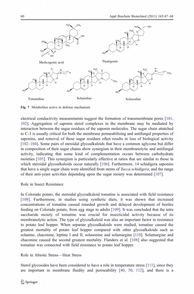
Avenacosides A-1 from oat roots (a triterpenoid saponin) and the steroidal glycoalkaloidsα-tomatine and α-chaconine (from tomato and potato, respectively) contain α-1,2-linked C-rhamnosemolecule at the C-3 position and provide resistance againstGibberella pulicaris [99].The glycosyltransferase SAGT4, involved in steroid saponin biosynthesis in Solanumaculetissimun, catalyses the 3-o-glycosylation of steroidal sapogenins, such as, diosgenin,nuatigenin and tigogenin [87]. The response of this enzyme to wounding stress indicates theinvolvement of SAGT4 in plant defense system. This enzyme also glycosylates steroidalalkaloids such as solanidine, solasodine and tomatidine. The potato GT (SAGT1) identifiedby screening a wound-induced cDNA library in yeast suggested the role of Sgt in stress [86].The enzyme (SAGT1) glycosylated tomatidine, solasodine and solanidine, the metabolitesactive in defense mechanism (Fig. 7). UGT73C5 which glycosylates brassinosteroids inArabidopsis thaliana also glycosylates fungal mycotoxin de-oxynivalenol (DON). Hence, itprovides protection against the pathogen Fusarium by detoxifying DON [100].
The major mechanism of antifungal activity of saponins is apparently due to their abilityto complex with sterols in fungal membranes and cause loss of membrane integrity [101],although the precise mechanism is not fully understood. Electron microscopic analysis and
O
O
H
OHHH
O
H OH
H
O
O
HNMe
Avenacin A-1 (Roots)
Avenacoside A (Leaves)
α-L-ara(1 )
β-D-glu-(1 2)
β-D-glu-(1 4)
β-D-glu(1 2)
α-L-rha(1 4)
OH
O
O
H
H
H
Oβ-D-glu(1 )
( 1)β-D-glu b
a
Figs. 6 a, b Avenacosides from oat plant
Appl Biochem Biotechnol (2011) 165:47–68 59
electrical conductivity measurements suggest the formation of transmembrane pores [101,102]. Aggregation of saponin sterol complexes in the membrane may be mediated byinteraction between the sugar residues of the saponin molecules. The sugar chain attatchedto C-3 is usually critical for both the membrane permeabilising and antifungal properties ofsaponins, and removal of these sugar residues often results in loss of biological activity[102–104]. Some pairs of steroidal glycoalkaloids that have a common aglycone but differin composition of their sugar chains show synergism in their membranolytic and antifungalactivity, indicating that some kind of complementation occurs between carbohydratemoieties [105]. This synergism is particularly effective at ratios that are similar to those inwhich steroidal glycoalkaloids occur naturally [106]. Furthermore, 14 schidigera saponinsthat have a single sugar chain were identified from stems of Yucca schidigera, and the rangeof their anti-yeast activities depending upon the sugar moiety was determined [107].
Role in Insect Resistance
In Colorado potato, the steroidal glycoalkaloid tomatine is associated with field resistance[108]. Furthermore, in studies using synthetic diets, it was shown that increasedconcentrations of tomatine caused retarded growth and delayed development of beetlesfeeding on Colorado potato, from egg stage to adults [109]. It was concluded that the tetrasaccharide moiety of tomatine was crucial for insecticidal activity because of itsmembranolytic action. The type of glycoalkaloid was also an important factor in resistanceto potato leaf hopper. When separate glycoalkaloids were studied, tomatine caused thegreatest mortality of potato leaf hopper compared with other glycoalkaloids such assolanine, chaconine, leptine I and II, solasonine and solamargine [110]. Solamargine andchaconine caused the second greatest mortality. Flanders et al. [108] also suggested thattomatine was connected with field resistance to potato leaf hopper.
Role in Abiotic Stress—Heat Stress
Sterol glycosides have been considered to have a role in temperature stress [111], since theyare important in membrane fluidity and permeability [40, 50, 112], and there is a
N
HH
O
H
HHH
CH3 H
CH3
OH
H
Tomatidine
NH
CH3
O
CH3
CH3
H
CH3H
HH
OH
H
Solasodine
N
CH3
CH3
H
CH3H
HH
OH
CH3
H
H
Solanidine
O
CH3
CH3
H
CH3
HH
OH
H
O
CH3 OH
H
Nuatigenin
CH3
CH3H
OH
CH3 CH3
OH
O
HCH3
HH
OHO
OH
Medicagenic acid
Fig. 7 Metabolites active in defense mechanism
60 Appl Biochem Biotechnol (2011) 165:47–68

phospholipid dependence of UDP-glucose:sterolglycosyltransferase [113]. In comparison tonormal sterols, sterol glycosides and acylated sterol glycosides exchange more slowlybetween the monolayer halves of a bilayer, which could serve to regulate free sterol contentand its distribution [39, 50]. This can provide insight into the role of sterol glycosides in theplasma membrane. Plants adapt to environmental heat stress by synthesising heat shockproteins. It is well known that transcriptional induction of heat shock genes is mediated bythe heat shock transcription factors (HSFs) in eukaryotes [114]. The HSFs are activated bymultimerisation and phosphorylation, bind to the promoters of heat shock genes andstimulate their transcription. The activation of sterol glycosyltransferase and the productionof sterol glycoside are important events in the signal transduction system to induce thesynthesis of heat shock proteins. Heat stress induces rapid glycosylation of membranesterols in myxoamoebae of true slime mould P. polycephalum [66, 115, 116].
When heat shock was given to human foetal lung fibroblast cells, there was a change inthe composition of membrane lipids. There was a heat-induced expression of cholesterylglycoside, which suggests the involvement of steryl glycoside in heat shock responses inmammalian cells. This phenomenon is also followed by activation of calcium-dependentprotein kinase followed by the HSP induction in P. polycephalum cells. The activity ofUDP-glucose:poriferasterol glycosyltransferase also increased rapidly after heat shock. Itwas also observed that HSP 70 was induced when cholesteryl glycosides were added to theculture medium of TIG-3 cells without heat stress. Hence, steryl glycoside is indirectlyconsidered to enhance high temperature tolerance and participate in the activation of HSFs.Recently, two enzymes of SGT gene family from W. somnifera have been characterised[32–34]. Both the enzymes exhibited a rapid in vivo response to high temperature andsalicylic acid treatment, suggesting a physiological role in biotic and abiotic stress.Glycosylation of sterols may alter the distribution of 3β-OH sterols in membrane rafts, andthese may be involved in the transmission of heat shock signals in cells, by change inmembrane functions like fluidity, phase transition, etc. The membrane rafts rich in 3β-OHsterols have been reported in plants [117]. Hence, increase in the level of SGTs during heatstress shows their role in perceiving heat stress.
Cold Stress
The adaptation of plants towards cold stress involves complex phenomena such asmodulating the fluidity of biomembranes and synthesis and accumulation of low molecularweight and high molecular weight cryoprotectants [118–120]. The plasma membrane isregarded as key site of injury during freeze thaw stress in herbaceous plants [111, 121].Alterations that contribute to increased freezing tolerance include increased level of fattyacid desaturase in membrane phospholipids and change in the level and type of membranesterols and cerebrosides [122]. A difference in the proportion of glycosylated versusacylated sterols has been reported [111]. It has been observed that sterol content of theplasma membrane changes in response to environmental conditions and alterations in thesterol compositions of plasma membranes may play a role in cold acclimation process[123]. This is supported by the fact that SGT’s, e.g. UGT80B1 transcript has been slightlyupregulated in cold stress, as shown by gene expression data form micro-array experiments(genome cluster database). The proportion of phospholipids increase from 46.8% to57.1 mol% of the total lipids with increase in the proportion of di-unsaturated species ofphasphatidylcholine and phasphatidylethanolamine [124]. The proportion of cerebrosidesdecreases from 7.3% to 4.3 mol% with decrease in the proportion of free sterols from37.7% to 31.2 mol%. These studies indicate that the level of sterols and their modified
Appl Biochem Biotechnol (2011) 165:47–68 61

counterparts varies in response to cold stress. Hence, Sgts, which bring about themodification of sterols, play an important role in coping up with heat and cold stress.
Some heat shock proteins such as Hsp70 have been reported to function as molecularchaperons during cold stress [125]. Upregulation of Hsp70s at low temperature may berelated to an increased demand for molecular chaperone function at low temperature, andHsp70s could bind unfolded or non-native cold labile proteins. Furthermore, cholesterylglycosides have been reported to enhance the expression of heat shock proteins. Hence, anincrease in the expression of SGTs may be related to the increase in the production of sterolglycosides, which behave as messengers in triggering heat shock proteins, which help theplant in adaptation to heat and cold stress. Treatment with methyl jasmonate and methylsalicylate in tomato and peppers reduced chilling injury, enhanced transcript level of heatshock proteins, PR proteins and alternative oxidase [126]. The same treatment alsoenhances the expression of SGTs, which indicate the involvement of SGTs in stress.Expression of a 70-kDa spinach endoplasmic reticulum heat shock protein during coldacclimation has been reported [127]. Coordinated and non-coordinated expression of thestress 70 family and other molecular chaperones have been reported at high and lowtemperature in spinach and tomato [128]. Increase in the expression of SGTs in cold andheat stress and cholesteryl glycosides triggering the expression of heat shock proteinsindicate that SGTs are playing an important role in both heat and cold stress.
Role of SGTs in Hormonal Regulation
Steroid hormones play a conserved role in regulating development in eukaryotic organismsranging from fungi and plants to insects, vertebrates and mammals. Sterols function asbiosynthetic precursors of steroid hormones, such as glycocorticoids, androgens andestrogens in animals, ecdysteroid in insects, anthridiol and oogoniol in fungi andbrassinosteroids in plants [129, 130]. Sgts that glycosylate steroidal hormones play animportant role in regulating the activity of steroids in plants, mammals and insects.Brassinosteroids (BRs) are plant-specific polyhydroxylated derivatives of 5α-cholestanestructurally similar to cholesterol-derived animal steroid hormones and ecdysteroids frominsects [131, 132]. Like their animal counterparts, BRs (Fig. 8) have been shown to regulategene expression, stimulate cell division and differentiation and modulate reproductivebiology [133].
O
O
H
H
OH
OH CH3
CH3
CH3
CH3OH
OH
CH3
H
H
Brassinolide(Brassinosteroid)
HOH
OH CH3
CH3
CH3
CH3OH
OHCH3
H
O
H
H
CH3
Castasterone(Brassinosteroid)
Fig. 8 Structure of brassinosteroids
62 Appl Biochem Biotechnol (2011) 165:47–68

BRs also mediate growth response unique to the plants, including the promotion of cellelongation in the presence of a complex cell wall and influencing multiple developmentresponses to darkness and light. The phenotypic mutants have been described extensivelyand include extreme dwarfism, altered leaf morphology, reduced fertility or male sterility,delayed senescence and altered vascular development, implicating BRs in all of thesedevelopmental processes [129]. One of the ways by which plants maintain hormonalhomeostasis is by conjugating the hormones with fatty acids, glucose or disaccharides [133,134]. Enzymes involved in glycosylation of plant hormones, such as auxins, cytokinins andabscisic acid, belong to a subset of family 1 UDP-glycosyltransferases (UGTs) defined bythe presence of a C-terminal consensus sequence [10]. Proteins of this class have also beenshown to accept mammalian and insect steroid hormones as substrates. In mammals, thetypical donor sugar is UDP-glucuronic acid, and the conjugation of androgens andestrogens by members of UGT subfamily 2B is thought to regulate hormone activity [135,136]. The UDP-glycolsyltransferase UGT73C5 of Arabidopsis thaliana catalyses 23-Oglycosylation of the BRs brassinolide and catasterone [137]. Transgenic plants over-expressing UGT73C5 displayed BR-deficient phenotypes and contained reduced amount ofBRs. The phenotype, which was already apparent in the seedlings, could be rescued byapplication of BR.
Role in Medicinally Important Plants
Panax ginseng is a medicinal plant that contains diverse ginsenosides, which arecardioprotective, antifatigue, antitumor, hepatoprotective and immunomodulatory. Thegenes involved in the biosynthesis of ginsenosides and other secondary metabolites wereestimated by the treatment of hairy roots with methyl jasmonate and analysis of thetranscript. Among the genes were glycosyltransferase, oxidosqualene cyclase andcytochrome P450 [138]. A glycosyltransferase UDPG-ginsenoside:Rd glycosyltransrerasehas been purified from suspended cells of P. notoginseng [139]. The effect of sugarpositions in ginsenosides on inhibition of Na(+)/K(+) ATPase suggests that the enzymesinvolved in sterol transformation determine the level of pharmacologically importantbioactive metabolites in P. ginseng [140]. W. somnifera, which is equated to ginseng in itsmedicinal properties, is a rich source of a variety of glycosylated steroidal lactones calledwithanosides in roots [141, 142] and leaves [143]. Withanosides are steroidal lactones withone or more glucose units attached to C-3 or C-27 positions. Recently, Ahuja et al. [144]have isolated four glycowithanolides, viz., withanoside IV (WSG-3), withanoside VI(WSG-3A), physagulin D (WSG-P) and withastraronolide (WSC-O), from multiple shootcultures of W. somnifera. A novel sterol glycosyltransferase specific to β-OH position hasrecently been reported from our group [32]. The enzyme showed broad sterol specificity,glycosylating a variety of sterols and steroidal sapogenins with β-OH group at C-17, C-21and C-27 positions. Two Sgts glycosylating sterols at C-3-OH have also been reported fromour group [33, 34]. Increase in the expression of both sgts on salicylic acid treatmentsuggests their possible role in stress. In the light of above points, it can be postulated thatsterols and their modified counterparts play a crucial role in plant defense mechanism, andthey are diversified by sterol modifying enzymes.
This review has emphasised the range of acceptors for Sgts and their regioselectivity.These features provide a basis for application of SGTs in crop plants. These may involve,for example, changed sensitivity to hormones and their agrochemical analogs and changedtolerance to biotic abiotic stresses. Increasing the glycosylation of secondary metabolites,
Appl Biochem Biotechnol (2011) 165:47–68 63

may lead to increased yield of the corresponding glycosides. It may be possible to increasethe flux of a molecule by glycosylating it and removing the product from the reaction mix.In terms of metabolic engineering, the SGTs could prove to have utility in cropimprovement.
Conclusion
The principle feature of Sgts is their ability to glycosylate sterol acceptors in aregioselective manner. Sterols are major components of plant cell membranes and exist assterol glycosides or acylated sterol glycosides. The ratio of sterols and their modifiedcounterparts varies when plant perceives stress conditions like heat and cold. Manysecondary metabolites are derived from the sterol pathway, which are important in defensemechanisms against pathogens. Sterol glycosides such as cholesterol glycosides act asimportant signalling molecules in triggering the expression of heat shock proteins. Sgts thatglycosylate steroidal hormones, such as brassinosteroids, play an important role inregulating the level of hormones in planta. Some Sgts that catalyse the glycosylation ofendogenous substrates also glycosylate foreign acceptors. Many sterols and steroidalmolecules attribute medicinal properties to the plant as in the case of W. somnifera and P.ginseng. This fascinating group of enzymes represent an area to study various aspects ofplant metabolism such as stress, plant defense mechanism and medicinal properties withcoming years to provide still more clarity about these enzymes.
Acknowledgements The authors are thankful to the Council of Scientific and Industrial Research, NewDelhi, Government of India, for providing the financial support under NMITLI scheme and Network Project(NWP-08) and also to the Department of Biotechnology, New Delhi, Government of India, for financial helpthrough the project no. GAP 231225, for functional analysis of SGT genes of Withania somnifera.
References
1. Li, Y., Baldauf, S., Lim, E. K., & Bowles, D. J. (2001). The Journal of Biological Chemistry, 276,4338–4343.
2. Mackenzie, P. I., Owen, I. S., Burchell, B., Bock, K. W., Bairoch, A., Bélanger, A., et al. (1997).Pharmacogenetics, 7, 255–269.
3. Paquette, S., Moller, B. L., & Bak, S. (2003). Phytochemistry, 62, 399–413.4. Ross, J., Li, Y., Lim, E. K., & Bowles, D. J. (2001). Genome Biology, 2, 3004.1–3004.6.5. Kapitonov, D., & Yu, R. K. (1999). Glycobiology, 9, 961–978.6. Vogt, T., & Jones, P. (2000). Trends in Plant Science, 5, 380–386.7. Bowles, D. J., Poppenberger, B., & Vaistij, F. (2006). Annual Review of Plant Biology, 57, 567–597.8. Ikan, R. (444). Naturally occurring glycosides. Chichester: Wiley.9. Jones, P., & Vogt, T. (2001). Planta, 213, 164–174.
10. Lim, E. K., & Bowles, D. J. (2004). The EMBO Journal, 23, 2915–2922.11. Pflugmacher, S. S. H. (1998). Phytochemistry, 49, 507–511.12. Martinoia, E. K. M., Gesser, M., Sanchez-Hernandez, R., & Rea, P. A. (2001). Vacuolar transport of
secondary metabolites and xenobiotics. UK: Sheffield Acad.13. Dietz, K. J., Sauter, A., Wichert, K., Messdaghi, D., & Hartung, W. (2000). Journal of Experimental
Botany, 51, 937–944.14. Samuels, A. L., Rensing, K. H., Douglas, C. J., Mansfield, S. D., Dharmawardhana, D. P., & Ellis, B. E.
(2002). Planta, 216, 72–82.15. Wojciechowski, Z. A., & Van Uon, N. (1975). Acta Biochimica Polonica, 22, 25–38.
64 Appl Biochem Biotechnol (2011) 165:47–68

16. He, J. X., Fujioka, S., Li, T. S., Kang, S. G., Seto, H., Takatsuto, S., et al. (2003). Plant Physiology,131, 1258–1269.
17. Schumacher, K., & Chory, J. (2000). Current Opinion in Plant Biology, 3, 79–84.18. Wanke, M., Skorupinska-Tudek, K., & Swiezewska, E. (2001). Acta Biochemica Polonica, 48, 663–
672.19. Benveniste, P. (2004). Annual Review of Plant Biology, 55, 429–457.20. Grille, S., Zaslawski, A., Thiele, S., Plat, J., & Warnecke, D. (2010). Progress in Lipid Research, 49,
262–288.21. Ly, P. T. T., Singh, S., & Shaw, C. A. (2007). Journal of Neuroscience Research, 85, 231–237.22. Cruz-Aguado, R., & Shaw, C. A. (2009). Neurology, 72, 474–476.23. Haralampidis, K., Trijanowska, M., & Osbourn, A. E. (2002). Advances in Biochemical Engineering/
Biotechnology, 75, 31–49.24. Ullmann, P., Ury, A., Rimmele, D., Benveniste, P., & Bouvier-Nave, P. (1993). Biochimie, 75, 713–719.25. Webb, M. S., Irving, T. C., & Steponkus, P. L. (1995). Biochimica et Biophysica Acta, 1239, 226–238.26. Kojima, M., Ohnishi, M., Ito, S., & Fujino, Y. (1989). Lipids, 24, 849–853.27. Mulichak, A. M., Losey, H. C., Walsh, C. T., & Garavito, R. M. (2001). Structure (Camb.), 9, 547–557.28. Mulichak, A. M., Losey, H. C., Lu, W., Wawrzak, Z., Walsh, C. T., & Garavito, R. M. (2003).
Proceedings of the National Academy of Science, 100, 9238–9243.29. Mulichak, A. M., Lu, W., Losey, H. C., Walsh, C. T., & Garavito, R. M. (2004). Biochemistry, 43,
5170–5180.30. Offen, W., Martinez-Fleites, C., Yang, M., Kiat-Lim, E., Davis, B. G., Tarling, C. A., et al. (2006). The
EMBO Journal, 25, 1396–1405.31. Shao, H., He, X., Achinine, L., Blount, J. W., Dixon, R. A., & Wang, X. (2005). The Plant Cell, 17,
3141–3154.32. Madina, B. R., Sharma, L. K., Chaturvedi, P., Sangwan, R. S., & Tuli, R. (2007). Biochimica et
Biophysica Acta, 1774, 392–402.33. Madina, B. R., Sharma, L. K., Chaturvedi, P., Sangwan, R. S., & Tuli, R. (2007). Biochimica et
Biophysica Acta, 1774, 1199–1207.34. Sharma, L. K., Madina, B. R., Chaturvedi, P., Sangwan, R. S., & Tuli, R. (2007). Archives of
Biochemistry and Biophysics, 460, 48–55.35. Lescovac, V. (2003). Comprehensive enzyme kinetics. Newyork: Kluwer.36. Mackenzie, P. I., & Rodbourn, L. (1990). The Journal of Biological Chemistry, 265, 11328–11332.37. Mackenzie, P. I. (1990). The Journal of Biological Chemistry, 265, 3432–3435.38. Evans, O. P., & O’reilly, D. R. (1998). The Biochemical Journal, 330, 1265–1270.39. Ullmann, P., Bouvier-Nave, P., & Benveniste, P. (1987). Plant Physiology, 85, 51–55.40. Warnecke, D. C., & Heinz, E. (1994). Plant Physiology, 105, 1067–1073.41. Warnecke, D. C., Baltrusch, M., Buck, F., Wolter, F. P., & Heinz, E. (1997). Plant Molecular Biology,
35, 597–603.42. Hartmann-Bouillon, M. A., & Benveniste, P. (1978). Phytochemistry, 17, 1037–1042.43. Yoshida, S., & Uemura, M. (1986). Plant Physiology, 82, 807–812.44. Burbulis, I. E., & Winkel-Shirley, B. (1999). Proceedings of the National Academy of Sciences of the
United States of America, 96, 12929–12934.45. Bartholomew, D. M., Van Dyk, D. E., Lau, S. M. C., O’Keefe, D. P., Rea, P. A., & Viitanen, P. V.
(2002). Plant Physiology, 130, 1562–1572.46. Rea, P. A. (1999). Journal of Experimental Botany, 50, 895–913.47. Radominska-Pandya, A., Czernik, P. J., Little, J. M., Battaglia, E., & Mackenzie, P. I. (1999). Drug
Metabolism Reviews, 31, 817–899.48. Hirschberg, C. B., Robbins, P. W., & Abeijon, C. (1998). Annual Review of Biochemistry, 67, 49–69.49. Sakaki, T., Zahringer, U., Warnecke, D. C., Fahl, A., Knogge, W., & Heinz, E. (2001). Yeast, 18, 679–
685.50. Warnecke, D., Erdmann, R., Fahl, A., Hube, B., Muller, F., Zank, T., et al. (1999). The Journal of
Biological Chemistry, 274, 13048–13059.51. Smith, P. F. (1971). Journal of Bacteriology, 108, 986–991.52. Patel, K. R., Smith, P. F., & Mayberry, W. R. (1978). Journal of Bacterial, 136, 829–831.53. Mayberry, W. R., & Smith, P. F. (1983). Biochimica et Biophysica Acta, 752, 434–443.54. Livermore, B. P., Bey, R. F., & Johnson, R. C. (1978). Infection and Immunity, 20, 215–220.55. Hirai, Y., Haque, M., Yoshida, T., Yokota, K., Yasuda, T., & Oguma, K. (1995). Journal of Bacterial,
177, 5327–5333.56. Lebrun, A. H., Wunder, C., Hildebrand, J., Churin, Y., Zähringer, U., Lindner, U. B., et al. (2006). The
Journal of Biological Chemistry, 281, 27765–27772.
Appl Biochem Biotechnol (2011) 165:47–68 65

57. Wunder, C., Churin, Y., Winau, F., Warnecke, D., Vieth, M., Lindner, B., et al. (2006). NatureMedicine, 12, 1030–1038.
58. Berg, S., Edman, M., Li, L., Wikstrom, M., & Wieslander, A. (2001). The Journal of BiologicalChemistry, 276, 22056–22063.
59. Holzl, G., Zahringer, U., Warnecke, D., & Heinz, E. (2005). Plant & Cell Physiology, 46, 1766–1778.60. Khan, S., Sneddon, K., Fielding, B., Ward, V., & Davison, S. (2003). Virus Genes, 27, 17–27.61. Esders, T. W., & Light, R. J. (1972). The Journal of Biological Chemistry, 247, 7494–7497.62. McMorris, T. C., & White, R. H. (1977). Biochimica et Biophysica Acta, 486, 308–312.63. Stasyk, O. V., Nazarko, T. Y., Stasyk, O. G., Krasovska, O. S., Warnecke, D., Nicaud, J. M., et al.
(2003). Cell Biology, 27, 947–952.64. Kim, Y. K., Wang, Y., Liu, Z. M., & Kolattukudy, P. E. (2002). The Plant Journal, 30, 177–187.65. Murakami-Murofushi, K., & Ohta, J. (1989). Biochimica et Biophysica Acta, 992, 412–415.66. Murakami-Murofushi, K., Nishikawa, K., Hirakawa, E., & Murofushi, H. (1997). The Journal of
Biological Chemistry, 272, 486–489.67. Abraham, W., Wertz, P. W., Burken, R. R., & Downing, D. T. (1987). Journal of Lipid Research, 28,
446–449.68. Wertz, P. W., Stover, P. M., Abraham, W., & Downing, D. T. (1986). Journal of Lipid Research, 27,
427–435.69. Weatherill, P. J., & Burchell, B. (1980). The Biochemical Journal, 189, 377–480.70. Moghrabi, N., Sutherland, L., Wooster, R., Povey, S., Boxer, M., & Burchell, B. (1992). Annals of
Human Genetics, 56, 81–91.71. Ritter, J. K., Chen, F., Sheen, Y. Y., Tran, H. M., Kimura, S., Yeatman, M. T., et al. (1992). The Journal
of Biological Chemistry, 267, 3257–3261.72. Dutton, G. J. (1980). Glucuronidation of drugs and other compounds. Boca Raton: CRC.73. Falany, C. N., Green, M. D., & Tephly, M. D. (1987). The Journal of Biological Chemistry, 262, 1218–
1222.74. Burchell, B., Nebert, D. W., Nelson, D. R., Bock, K. W., Iyanagi, T., Jansen, P. L., et al. (1991). Cell
Biology, 10, 487–494.75. Haque, S. J., Petersen, D. D., Nebert, D. W., & Mackenzie, P. I. (1991). DNA and Cell Biology, 10,
515–524.76. Monaghan, G., Clarke, D. J., Povey, S., See, C. G., Boxer, M., & Burchell, B. (1994). Genomics, 23,
496–499.77. Hostettmann, K., & Marston, A. (1995). Saponins. Cambridge: Cambridge University Press.78. Hostettmann, A. M. K., Hostettmann, A., & Marston, A. (1995). Saponins, saponins chemistry and
pharmacology of natural products. Cambridge: Cambridge University Press.79. Morrissey, J. P., & Osbourn, A. E. (1999). Microbiology and Molecular Biology Reviews, 63, 708–
724.80. Misiak, M., Kalinowska, M., & Wojciechowski, Z. A. (1991). Acta Biochimica Polonica, 38, 43–45.81. Dupéron, R., & Dupéron, P. (1989). Comptes Rendus de l’Academie des Sciences Serie III, 308, 31–34.82. Verhoek, B., Haas, R., Wrage, K., Linscheid, M., & Heinz, E. (1983). Zeitschrift für Naturforschung,
38, 770–777.83. Debolt, S., Scheible, W. R., Schrick, K., Auer, M., Beisson, F., Bischoff, V., et al. (2009). Plant
Physiology, 151, 78–87.84. Potocka, A., & Zimowski, J. (2008). Acta Biochimica Polonica, 55, 127–134.85. Potocka, A., & Zimowski, J. (2008). Acta Biochimica Polonica, 55, 135–140.86. Moehs, C., Friedman, P. A. M., & Belknap, W. (1997). The Plant Journal, 11, 227–236.87. Kohara, A., Nakajima, C., Hashimoto, K., Ikenaga, T., Tanaka, H., Shoyama, Y., et al. (2005). Plant
Molecular Biology, 57, 225–239.88. Hillig, I., Leipelt, M., Ott, C., Zahringer, U., Warnecke, D., & Heinz, E. (2003). FEBS Letters, 553,
365–369.89. Suzuki, H., Naoumkina, M. R. M., Aziz, N., & May, G. (2005). Planta, 220, 696–707.90. Achnine, L., Huhman, D. V., Farag, M. A., Sumner, L. W., Blount, J. W., & Dixon, R. A. (2005). The
Plant Journal, 41, 875–877.91. Richman, A., Swanson, A., Humphrey, T., Chapman, R., & McGarvey, B. (2005). The Plant Journal,
41, 56–67.92. Peng, L. C., Kawagoe, Y., Hogan, P., & Delmer, D. (2002). Science, 295, 147–150.93. Cai-Jun, Y., & Jian-Jiang, Z. (2005). Process Biochemistry, 40, 3742–3748.94. Paquin, R. (1966). American Potato Journal, 43, 349–354.95. Andrivon, D., Corbière, R., Lucas, J. M., Pasco, C., Gravoueille, J. M., Pellé, R., et al. (2003).
American Journal of Potato Research, 80, 125–134.
66 Appl Biochem Biotechnol (2011) 165:47–68

96. Chong, J., Baltz, R., Schmitt, C., Beffa, R., Fritig, B., & Saindrenan, P. (2002). The Plant Cell, 14,1093–1097.
97. Jadhav, S. J., Sharma, R. P., & Salunkhe, D. K. (1981). Critical Reviews in Toxicology, 9, 21–104.98. Osbourn, A. (1996). The Plant Cell, 10, 1821–1831.99. Becker, P. (1998). FEMS Microbiology Letters, 167, 197–202.100. Poppenberger, B., Berthiller, F., Lucyshyn, D., Sieberer, T., Schuhmacher, R., Krska, R., et al. (2003).
The Journal of Biological Chemistry, 28, 47905–47914.101. Keukens, E. A. J., Vrije, T. D., Boom, C. V. D., Waard, P. D., Plasmna, H. H., Theil, F., et al. (1995).
Biochimica et Biophysica Acta, 1240, 216–228.102. Armah, C. N., Mackie, A. R., Roy, C., Price, K., Osourn, A. E., Bowyer, P., et al. (1999). Biophysical
Journal, 76, 281–290.103. Sandrock, R. W., & Van Etten, H. D. (1998). Phytopathology, 88, 137–143.104. Wubben, J. P., Price, K. R., Daniels, M. J., & Osbourn, A. E. (1996). Phytopathology, 86, 986–992.105. Roddick, J. G., Rijnenberg, A. L., & Osman, S. F. (1988). Journal of Chemical Ecology, 14, 889–902.106. Fewell, A. M., & Roddick, J. G. (1993). Phytochemistry, 33, 323–328.107. Miyakoshi, M., Tamura, Y., Masuda, H., Mizutani, K., Tanaka, O., Ikeda, T., et al. (2000). Journal of
Natural Products, 63, 332–348.108. Flanders, K. L., Hawkes, J. G., Radcliffe, E. B., & Lauer, F. I. (1992). Euphytica, 61, 83–91.109. Kowalski, S. P., Domek, J. M., Sanford, L. L., & Deahl, K. L. (2000). Journal of Entomological
Science, 35, 290–300.110. Sanford, L. L., Domek, J. M., Cantelo, W. W., Kobayashi, R. S., & Sinden, S. L. (1996). American
Potato Journal, 73, 79–88.111. Palta, J. P., & Li, P. H. (1980). Physiologia Plantarum, 50, 169–175.112. Schaller, H. (2003). The role of sterols in plant growth and development. Progress in Lipid Research,
42, 163–175.113. Bouvier-Nave, P., Ullmann, P., Rimmele, D., & Benveniste, P. (1984). Plant Science Letters, 36, 19–27.114. Kingston, R. E., Schuetz, T. J., & Larin, Z. (1987). Molecular and Cellular Biology, 7, 1530–1534.115. Kunimoto, S., Kobayashi, T., Kobayashi, S., & Murakami-Murofushi, K. (2000). Cell Stress &
Chaperones, 5, 3–7.116. Kunimoto, S., Kai, W. M. H., Ishida, Y., Uchiyama, A., Kobayashi, T., & Kobayashi, S. (2002). Cell
Structure and Function, 27, 157–162.117. Mongrand, S., Morel, J., Laroche, J., & Claverol, S. (2004). The Journal of Biological Chemistry, 279,
36277–36286.118. Hansen, J., Türk, R., Vogg, G., Heim, R., & Beck, E. (1997). In H. Rennenberg, W. Eschrich, & H.
Ziegler (Eds.), Trees-contributions to modern tree physiology (pp. 97–98). Leiden: Backhuys.119. Nanjo, T., Kobayashi, M., Yoshida, Y., Kakubari, Y., Yamaguchi-Shinozaki, K., & Shinozaki, K.
(1999). FEBS Letters, 461, 205–210.120. Steponkus, P. L., Uemura, M., Joseph, R. A., Gilmour, S. J., & Thomashow, M. F. (1998). Proceedings
of the National Academy of Science, 95, 14570–14575.121. Steponkus, P. L. (1984). Annual Review of Plant Physiology, 35, 543–584.122. Thomashow, M. F. (1998). Plant Physiology, 118, 1–7.123. Patterson, G. W., Hugly, S., & Harrison, D. (1993). Phytochemistry, 33, 1381–1383.124. Uemura, M., Joseph, R. A., & Steponkus, P. L. (1995). Plant Physiology, 109, 15–30.125. Zhang, C. L. G. (2006). Plant Physiology and Biochemistry, 44, 844–850.126. Wang, R. W. M. F., & Ding, C. K. (2005). ISHS Acta Horticult. 682.127. Anderson, J. V., Li, Q. B., Haskell, D. W., & Guy, C. L. (1994). Plant Physiology, 104, 1359–1370.128. Li, Q. B., Haskell, D. W., & Guy, C. L. (1999). Plant Molecular Biology, 39, 21–34.129. Clouse, S. D., & Sasse, J. M. (1998). Annual Review of Plant Physiology and Plant Molecular Biology,
49, 427–451.130. Grove, M. D., Spencer, G. F., Rohwedder, W. K., Mandava, N., Worley, J. F. Jr., Warthen, J. D., et al.
(1979). Nature, 281, 216–217.131. Adam, G., Schmidt, J., & Schneider, B. (1999). Fortschritte der Chemie Organischer Naturstoffe, 78,
1–46.132. Clouse, S. (2001). Current Biology, 11, R904.133. Fujioka, S., & Yokota, T. (2003). Annual Review of Plant Biology, 54, 137–164.134. Kleczkowski, K., & Schell, J. (1995). Critical Reviews in Plant Sciences, 14, 283–298.135. Barbier, O., Bélanger, A., & Hum, D. W. (1999). The Biochemical Journal, 337, 567–574.136. Turgeon, D., Carrier, J. S., Chouinard, S., & Belanger, A. (2003). Drug Meta Dispo., 31, 670–676.137. Poppenberger, B., Fujioka, S., Soeno, K., George, G. L., Vaistij, F. E., Hiranuma, S., et al. (2005).
Proceedings of the National Academy of Science, 102, 15253–15258.
Appl Biochem Biotechnol (2011) 165:47–68 67

138. Choi, D., Ha, J. J. Y., Park, H., Su, I. D., Chung, H., & Liu, J. (2005). Plant Cell Reports, 23, 557–566.139. Yue, C. J., & Zhong, J. J. (2005). Process Biochemistry, 40, 3742–3748.140. Chen, R., Chung, T., Lin, F. N., & Tzen, J. (2009). Acta Pharmacologica Sinica, 30, 61–69.141. Matsuda, H., Murakami, T., Kishi, A., & Yoshikawa, M. (2001). Bioorganic & Medicinal Chemistry, 9,
1499–1507.142. Zhao, J., Nakamura, N., Hattori, M., Kuboyama, T., Tohda, C., & Komatsu, K. (2002). Chemical &
Pharmaceutical Bulletin, 50, 760–765.143. Jayaprakasam, B., & Muraleetharan, N. G. (2003). Tetrahedron, 59, 841–849.144. Ahuja, A., Kaur, D., Sharada, M., Kumar, A., Suri, K. A., & Dutt, P. (2009). Nat Prod Comm., 4, 479–
482.145. Caradoc-Davies, K. M., Graves, S., O’Reilly, D. R., Evans, O. P., & Ward, V. K. (2001). Virus Genes,
22, 255–264.146. Manzan, M. A., Lozano, M. E., Sciocco-Cap, A., Ghiringhelli, P. D., & Romanowski, V. (2002). Virus
Genes, 24, 119–130.147. Rodrigues, J. C., De Souza, M. L., O’Reilly, D., Velloso, L. M., Pinedo, F. J., Razuck, F. B., et al.
(2001). Virus Genes, 22, 103–112.148. Tumilasci, V. F., Leal, E., Zanotto, P. M., Luque, T., & Wolff, J. L. (2003). Virus Genes, 27, 137–144.149. Wormleaton, S. L., & Winstanley, D. (2001). The Journal of General Virology, 82, 2295–2305.150. Paczkowski, C., Kalinowska, M., & Wojciechowski, Z. A. (2001). Phytochemistry, 58, 663–669.151. McCue, K. F., Allen, P. V., Shepherd, L. V., Blake, A., Whitworth, J., Maccree, M. M., et al. (2006).
Phytochemistry, 67, 1590–1597.152. Kurosawa, Y., Takahara, H., & Shiraiwa, M. (2002). Planta, 215, 620–629.153. Bedir, E., & Khan, I. A. (2000). Journal of Natural Products, 63, 1699–1701.154. Bedir, E., Pugh, N. P., Çalış, I., Pasco, D. A., & Khan, I. A. (2000). Biological & Pharmaceutical
Bulletin, 23, 834–837.
68 Appl Biochem Biotechnol (2011) 165:47–68


