Embed Size (px)
Citation preview
A yeast model for Aß aggregation exemplifies the role of
membrane trafficking and PICALM in cytotoxicity
Fabien D’Angelo*, Hélène Vignaud*, Julie Di Martino, Bénédicte Salin, Anne Devin,
Christophe Cullin1 and Christelle Marchal.
Institut de Biochimie et Génétique Cellulaires, CNRS UMR 5095, Université Bordeaux 2
« Victor Segalen », 33077 Bordeaux, France.
*Both authors contributed equally to the work
1 Corresponding author
Running Title: Yeast model for Aβ toxicity
Prof. C. Cullin, IBGC, 1 rue Camille Saint Saëns, 33077 Bordeaux cedex, France
Tel/Fax + 33(0) 556 999 017
Mail: [email protected]
© 2012. Published by The Company of Biologists Ltd.This is an Open Access article distributed under the terms of the Creative Commons Attribution Non-Commercial Share Alike License(http://creativecommons.org/licenses/by-nc-sa/3.0), which permits unrestricted non-commercial use, distribution and reproduction inany medium provided that the original work is properly cited and all further distributions of the work or adaptation are subject to the same Creative Commons License terms.
Dise
ase
Mod
els &
Mec
hani
sms
D
MM
Acce
pted
man
uscr
ipt
http://dmm.biologists.org/lookup/doi/10.1242/dmm.010108Access the most recent version at DMM Advance Online Articles. Published 10 August 2012 as doi: 10.1242/dmm.010108
ABSTRACT
Alzheimer’s disease is the most common neurodegenerative disease, associated with
aggregation of Aβ peptides. The exact mechanism of neuronal cell dysfunction in
Alzheimer’s disease is poorly understood and numerous models have been used to decipher
the mechanisms leading to cellular death. Yeast cells might be a good model to understand
the intracellular toxicity triggered by Aβ peptides. Indeed, yeast has been used as a model to
examine protein functions or cellular pathways that mediate the secretion, aggregation, and
subsequent toxicity of proteins associated with human neurodegenerative disorders. In the
present study, we use the yeast Saccharomyces cerevisiae as a model system to study the
effects of intracellular Aβ in fusion with the green fluorescent protein. We sent this fusion
protein into the secretory pathway, and showed that intracellular traffic pathways are
necessary to generate toxic species. Yeast PICALM orthologs are involved in cellular
toxicity, indicating conservation of the mechanisms of toxicity from mammals to yeast.
Finally, our model demonstrates the capacity for intracellular Aβ to cross intracellular
membranes and target mitochondrial organelles.
Dise
ase
Mod
els &
Mec
hani
sms
D
MM
Acce
pted
man
uscr
ipt
INTRODUCTION
Alzheimer’s disease (AD) was first described 100 years ago and is a progressive
neurodegenerative pathology leading to gradual cognitive and behavioral changes and loss of
memory (Selkoe and Podlisny, 2002). It is associated with the presence of region-specific
amyloid-β (Aβ) deposits in the brain. These amyloid plaques form one of the
neuropathological hallmarks of AD. The APP gene encodes the amyloid precursor protein
(APP) which encompasses the Aβ peptides. Differential cleavage of APP produces amyloid
peptides of 40 (Aβ40) or 42 (Aβ42) amino acids in length; the Aβ40 species are considered to
be less toxic. Although Aβ aggregation is correlated with the extracellular deposition of
terminal amyloid plaques in AD patients and AD mouse models, Aβ species also accumulate
within the cell, including inside multivesicular bodies (Almeida et al., 2006; Langui et al.,
2004; Takahashi et al., 2002), lysosomes, or other vesicular compartments (Nixon, 2007;
Shie et al., 2003). Recent publications have confirmed the important role played by
intracellular Aβ, whether it is produced through the secretion pathway, by transfection, or by
uptake from the media (Echeverria et al., 2004; Hansson Petersen et al., 2008; Hu et al., 2009;
Kandimalla et al., 2009; Rebeck et al., 2010).
Different biological model systems including Aβ-transgenic Caenorhabditis elegans worm
(Link, 1995), Drosophila melanogaster flies (Crowther et al., 2005; Iijima et al., 2004), and
mammalian cell cultures (Magrané et al., 2004) have been used to study the role of
intracellular Aβ. These biological systems have identified general effects such as
mitochondrial organization (Zhao et al., 2010; Iijima-Ando et al., 2009) or folding machinery
(Fonte et al., 2002; Magrané et al., 2004) as targets or regulators of toxic Aβ species. These
Dise
ase
Mod
els &
Mec
hani
sms
D
MM
Acce
pted
man
uscr
ipt
findings have been confirmed in vivo (Hoshino et al., 2011) and they are similar to the
changes found in pathogenic situations. This supports the notion that part of the complex
process leading to AD can be reliably studied at the cellular level. However, the different cell
models used so far have not revealed any molecular mechanisms that could account for the
toxicity of Aβ. In addition, pharmacological approaches based on molecules that interfere
with Aβ amyloid formation have not been productive. This has raised several questions
concerning the paradigm and the models used for these strategies. So far, no simple organism
that can be manipulated for a high throughput screening can be used as a “gold standard” for
Aβ toxicity.
Yeast cells are suitable for such screening and have been widely used to pinpoint gene
networks and chemical compounds that can modulate amyloid toxicity. This was particularly
the case for Parkinson’s disease (Willingham et al., 2003; Cooper et al., 2006; Franssens et
al., 2010), and ALS disease (Sun et al., 2011; Ju et al., 2011; Fushimi et al., 2011).
Although this experimental model has been successfully used to monitor the aggregation
pattern of Aβ (Bagriantsev and Liebman, 2006; Caine et al., 2007; von der Haar et al., 2007),
these first yeast systems has failed to recapitulate the toxic properties of this peptide. In these
previous studies, Aβ was expressed in the yeast-cell cytoplasm. Very recently, a new screen
based on secreted form of Aß in yeast revealed the importance of endocytic pathway in
cellular toxicity (Treusch et al., 2011).
In the present study we demonstrate that targeting Aβ in the secretory pathway produces toxic
species. The toxicity depends on the allele expressed, arctic mutants being more harmful than
wt Aß. Disturbance of cell-traffic routes (i.e. secretion, endocytosis, recycling, or traffic
between Golgi and vacuoles) reduced the toxic effects of Aβ. Altogether, these results
Dise
ase
Mod
els &
Mec
hani
sms
D
MM
Acce
pted
man
uscr
ipt

indicate that in our yeast model, the cellular toxicity is not due to endocytosis dysfunction, but
rather implicate another cellular target.
Dise
ase
Mod
els &
Mec
hani
sms
D
MM
Acce
pted
man
uscr
ipt
RESULTS
Aβ becomes toxic when expressed with a secretory sequence
It has been clearly established that Aβ40 or Aβ42 expressed in frame with reporter genes such
as GFP (Caine et al., 2007) or the functional domain of Sup35 (von der Haar et al., 2007;
Bagriantsev and Liebman, 2006) does not lead to yeast death. We fully reproduced this result
since, regardless of whether the Aβ42 or arctic mutant was expressed, the number of colonies
formed on galactose (inducing conditions for expression of Aβ-GFP) or on dextrose
(repressing conditions) was the same (Fig 1). In both cases, the pattern of aggregation was
similar with the presence of bright dots. It was therefore challenging to test the toxicity to
yeast of Aβ species entering the secretion pathway as Aβ toxicity is observed when this
peptide is secreted in the fly model of Alzheimer's disease (Finelli et al., 2004; Iijima et al.,
2004). When GFP was fused to the mating factor α prepro-leader sequence (MFα) secretion
signal derived from the precursor of the Saccharomyces cerevisiae mating alpha-factor
(Kurjan and Herskowitz, 1982), there was no marked effect on the colony forming unit (CFU)
capacity. Previous studies based on four different yeast secretion signal sequences (i.e., INU1,
SUC2, PHO5, and MEL1) did not report any toxicity for secreted GFP (Li et al., 2002). In
contrast, we observed that expression of MFα-Aβ42-GFP reduced viability (decrease in CFU),
and extended the time required to obtain visible colonies (Fig 1). Both of these effects were
exacerbated when AβARC was expressed under the same conditions. One of the problems with
GFP chimeric proteins is that cellular properties such as toxicity could be due to GFP itself.
We tested this hypothesis by removing GFP from the plasmids (Fig S1), and we observed the
same toxic phenotypes. The toxicity was clearly lower, but the arctic mutant was still more
deleterious than wild-type (WT) Aβ. Western-blot analysis revealed that the level of
Dise
ase
Mod
els &
Mec
hani
sms
D
MM
Acce
pted
man
uscr
ipt
expression was greatly diminished without GFP (Fig S1). GFP probably helps to stabilize Aβ
peptide but it is clear that all of the toxic properties of Aβ-GFP can be attributed to the Aβ
part of the chimeric protein.
The MFα-GFP chimer exhibited a low and non-diffuse fluorescence pattern, whereas the
MFα-Aβ-GFP proteins could not be detected by fluorescence. This low signal (or even
absence of signal) may be due to a low level of expression, improper GFP folding, post-
translational modification, or a combination of these factors. To test the possibility of GFP
misfolding due to Aβ aggregation (Wurth et al., 2002), we engineered additional constructs
with a linker sequence inserted between Aβ and GFP. The two new chimeric proteins were as
toxic as the two previous constructs (Fig 1) and could now be detected in yeast cells. This
finding rules out the possibility of GFP misfolding due to its expression throughout the
secretory pathway. The same Aβ-GFP chimeric proteins behave differently in the cytoplasm
(formation of foci and non toxic) and in the secretory pathway (non-ponctiform and toxic).
We next checked the processing of MFα-Aβ-GFP proteins.
Processing of MFα -Aβ-GFP
Expression of GFP, Aβ42GFP, or AβARCGFP resulted in one species (26 or 31 kDa,
respectively) which cross-reacts with anti-GFP antibodies (Fig 2, panel A). In contrast, the
secreted forms of Aβ42GFP or AβARCGFP resulted in three bands (Fig 2, panel A). The
smallest (about 34 kDa) and largest species (about 50 kDa) were the most abundant. During
galactose induction, these two species were detected first (Fig 2, panel B); the incapacity to
detect the intermediate species early on during induction was probably due their low
concentration (below the sensitivity threshold). The mobility of the smallest species is
consistent with the production of the mature proteins. This process requires Kex2p, an
endoprotease in the late Golgi compartment (Redding et al., 1991) involved in processing
Dise
ase
Mod
els &
Mec
hani
sms
D
MM
Acce
pted
man
uscr
ipt
alpha-factor (Julius et al., 1984). To test this hypothesis, we analyzed the different protein
species produced in a ∆kex2 strain (Fig 3, panel A). Indeed, the smallest species were no
longer detected as expected if it was produced by Kex2p. Among the three bands revealed by
anti-GFP antibodies, the highest weight band is bigger than the MFα-Aβ42-GFP species (41
kDa). The prepro-alpha-factor is glycosylated through the yeast secretory pathway (Julius et
al., 1984), and the higher mobility could be due to glycosylation of prepro-Aβ-GFP species.
To test this hypothesis, we added a deglycosylation mix to the crude extract. After enzymatic
treatment, the proteins were analyzed by western blot (Fig 3, panel B). The highest mobility
band was no longer detected whereas the intermediate band was more intense, as expected if
this band corresponds to the unglycosylated form of prepro-Aβ-GFP. The ratio of these 3
bands depended on the preparation method of the crude extracts (data not shown). This may
have been due to a particular protection against proteases and we thus tested the sensitivity of
these proteins to proteases. The extracts prepared from yeast spheroplastes revealed four
bands instead of three. Proteinase K analysis of the P13 fraction (corresponding to the 13000g
pellet fraction of yeast lysate prepared in mild conditions) identified the lowest-mobility band
as PK-resistant entities (Fig 3, panel C). This finding is consistent with an intra-membranous
localization of glycosylated prepro-Aβ-GFP, but indicates most of the mature form (Aβ-GFP)
is sensitive to the protease (i.e. is not imbedded in the membranous compartment). As the
proteins enter the secretory pathway, we searched for the presence of GFP species in the
medium. It has been previously reported that GFP expressed in frame with different secretory
signal peptide did enter the secretory pathway but was not detected in the medium (Li et al.,
2002). With our constructs based on the prepro-alpha factor peptide, we also did not detect
any GFP species in the medium using either fluorescence or western blot of concentrated
medium (data not shown).
Dise
ase
Mod
els &
Mec
hani
sms
D
MM
Acce
pted
man
uscr
ipt
Disturbance of cell-traffic pathways decreases the toxicity triggered by Aβ chimeric
proteins
Prepro-Aβ-GFP enters the secretory pathway and is clearly toxic to yeast cells. Moreover, the
PICALM gene has been recently described as a new susceptibility gene for AD (Harold et al.,
2009). In mammalian cells, the neuron-specific AP180 protein and its ubiquitously expressed
homolog, clathrin assembly lymphoid myeloid leukemia protein (PICALM) are adaptator
proteins that participate in clathrin-mediated endocytosis. Adaptors determine the trafficking
itinerary of cargos and their steady state distributions within the cell (Fig 4). The yAP180
proteins are yeast homologues of PICALM (Wendland and Emr, 1998). They share 48%
identity, and 25.5% (YAP1801) and 26.5% (YAP1802) identity with PICALM protein.
Deletion of YAP1801 and YAP1802 clearly decreased the toxicity induced by MFα-Aβ42-GFP
and MFα-Aβarc-GFP by lowering the growth inhibition (table 3 and Fig S2). We investigated
whether mammalian PICALM gene could complement the YAP1801 and YAP1802 mutants.
PICALM has two splice variants designated long (L) and short (S) according to the number of
amino acids. The two mouse cDNA were subcloned into a multi-copy galactose-induced
plasmid. The L isoform on its own was toxic in yeast (data not shown) and could thus not be
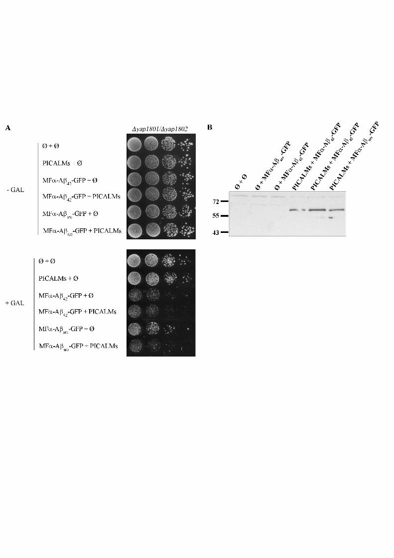
used. Expression of the S isoform of PICALM in YAP1801/YAP1802 double-deleted yeast
mutants partially restored the growth inhibition observed when MFα-Aβ42-GFP or MFα-
Aβarc-GFP were expressed in WT cells (Fig 5). This suggests that mouse PICALM protein is
able to complement YAP loss of function and that mammal and yeast proteins share a critical
role in Aβ toxicity.
In mammalian cells, CALM interacts with the clathrin-associated adaptor protein complex-2
AP-2 (Owen et al., 2000), and depletion of PICALM triggers delocalization of the AP-1
adaptor complex (Meyerholz et al., 2005). The toxicity induced by MFα-Aβ42-GFP and
Dise
ase
Mod
els &
Mec
hani
sms
D
MM
Acce
pted
man
uscr
ipt
MFα-Aβarc-GFP was partially suppressed when different subunits of the AP-1 and AP-2
complex were deleted. Two other classes of adaptors participate with AP-1 in clathrin-
mediated transport between the trans-Golgi network (TGN) and endosomes (Fig 4): Gga2p
protein and epsin-like protein Ent5p (Costaguta et al., 2006). The absence of these two
proteins also restored growth (table 3). The only identified cargo that is internalized by
Yap1801 and Yap1802 is the vSNARE Snc1p (Burston et al., 2009). Snc1p recycles to the
plasma membrane and this recycling requires Rcy1p (Galan et al., 2001). We observed that
the toxicity induced by MFα-Aβ42-GFP and MFα-Aβarc-GFP was strongly suppressed when
RCY1 was deleted (table 3 and Fig S2). Our findings indicate that defects in the endocytic or
recycling pathway reduce the toxicity triggered by the Aβ-GFP chimeric proteins. It is also
quite clear that MFα-Aβ42-GFP and MFα-Aβarc-GFP are still toxic in such background.
Cellular localization of Aβ-GFP
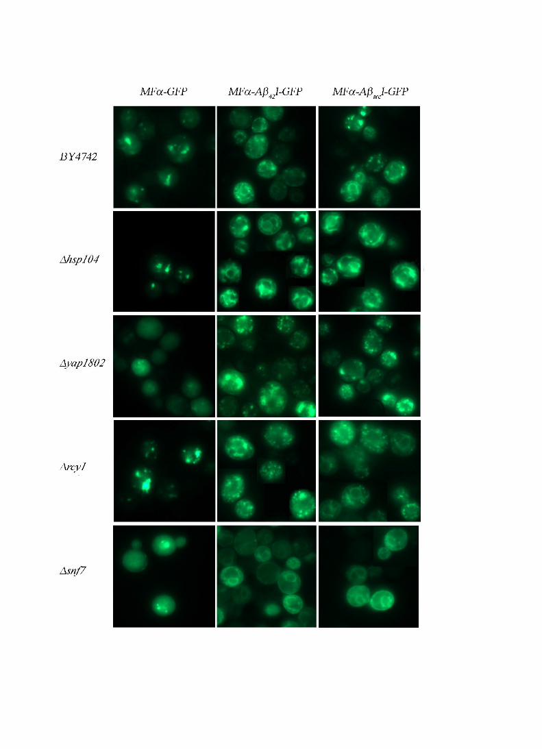
After induction of MFα-Aβ42-GFP and MFα-Aβarc-GFP in WT cells, proteins present a dual
pattern of punctuate and filamentous fluorescence structures (Fig 1). These two patterns are
not found systematically because filamentous organization is observed in most of the
fluorescent cells whereas punctuate foci are only present in some of them. In mutants which
are lower in Aβ toxicity and defective for traffic pathways, the ratio of the two fluorescence
patterns is inversed. Most of the cells present small foci and we occasionally observed the
filamentous pattern (Fig 6). This correlates the presence of filamentous aggregates with Aβ
toxicity and suggests that the punctuate structures correspond to membrane vesicles.
In mutant strains defective in the early stages of endocytosis such as the ∆snf7 mutant, part of
GFP fluorescence is associated with the plasma membrane (Fig 6, panel ∆snf7). In such
mutants, membrane invagination into multi vesicular bodies (MVB) is impaired (Babst et al.,
2002), and this decreases the rate of endocytosis. This result shows that the chimeric proteins
Dise
ase
Mod
els &
Mec
hani
sms
D
MM
Acce
pted
man
uscr
ipt
are able to reach the plasma membrane. Since none of chimeric proteins were detected in the
extracellular medium, we speculate that the proteins could be internalized once they had
reached the plasma membrane.
The use of yeast cells expressing red fluorescent protein fused to proteins whose localization
has been characterized previously (Huh et al., 2003) identified the ER as the sole
compartment clearly labeled by Aβ-GFP species (Fig S3 “RFP”). This finding does not rule
out the localization of Aβ-GFP on other membrane compartments since the speed of the
vesicles makes it unlikely to detect both fluorescent signals when they are not observed
simultaneously.
These findings show that disturbances of the endocytic or recycling pathways modify the
aggregation pattern of MFα-Aβ42-GFP and MFα-Aβarc-GFP as well as their cellular toxicity.
Hsp104p plays a role in Aβ chimeric protein toxicity
Hsp104p is critical for the toxicity and aggregation of the poly-glutamine Huntingtin protein
in yeast (Meriin et al., 2002). As Hsp104p plays a pivotal role for the aggregates formed in
yeast, we tested whether this chaperone protein is necessary for the toxicity of Aβ-GFP
chimeric proteins. As indicated in table 3 and FigS2, deletion of HSP104 partially restored the
viability of cells expressing MFα-Aβ42-GFP and MFα-Aβarc-GFP. This result was unexpected
because Hsp104p is a cytosolic protein and MFα-Aβ42-GFP and MFα-Aβarc-GFP were
associated with the secretory pathway. We therefore conclude that some of the toxic species
formed by Aβ-GFP chimeric proteins become cytosolic. This is consistent with the
observation that the mature form (Aβ-GFP; Fig 3C) detected by western-blot analysis is
sensitive to proteinase K.
Dise
ase
Mod
els &
Mec
hani
sms
D
MM
Acce
pted
man
uscr
ipt
In the ∆hsp104 strain, the fluorescent profile of Aβ-GFP chimeric proteins was filamentous
and no foci were observed (Fig 6). These results suggest that Hsp104p plays a role in the
toxicity of MFα-Aβarc-link-GFP, probably by favoring the conversion of big aggregates into
smaller and toxic ones.
Respiratory rate of cells expressing MFα-Aβarc-link-GFP is affected
Mitochondrial dysfunction plays a key role in AD. We therefore tested whether the toxicity
triggered by the expression of Aβ-GFP chimeric proteins in yeast was associated with a
mitochondrial disorder. To this end, we monitored the growth and consumption of oxygen in
aerobic conditions at different time points after Aβ induction. Yeast cells transformed with
the chimeric constructions were grown with lactate as a carbon source and 0.2% galactose
was added at the mid-log phase to induce expression of the chimeric protein. Under these
conditions, the growth of cells expressing MFα-GFP or MFα-Aβarc-link-GFP was the same as
that for cells carrying an empty vector during the first 4 h of the induction. After this time, the
growth rate of cells expressing the two chimeric proteins slowed, and this slowdown was
stronger for cells expressing MFα-Aβarc-link-GFP (Fig 7A). We then evaluated the
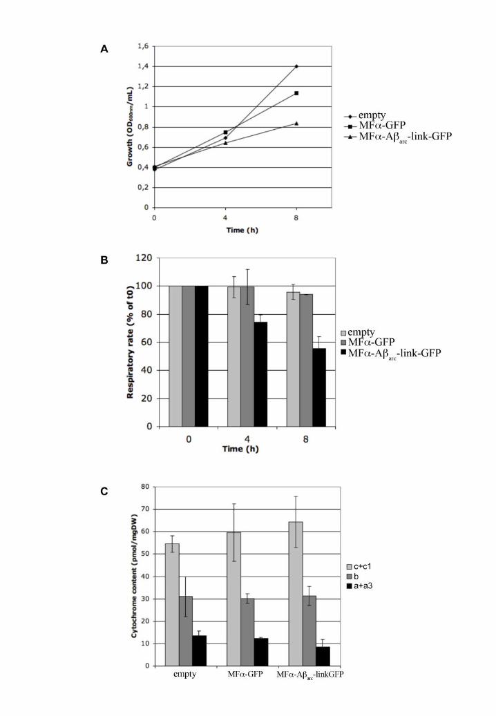
respiratory rate of the cells at different times during Aβ induction (Fig 7B). The consumption
of oxygen by cells with an empty vector or expressing MFα-GFP was unchanged 4 h after
induction (addition of galactose). In contrast, the respiratory rate decreased in the cells
expressing MFα-Aβarc-link-GFP. This reduction in oxygen consumption was higher after 8 h
of induction, whereas the respiratory rates of the two controls (empty vector and MFα-GFP)
decreased only by a small amount in both cases. This difference was not due to cell lethality
since the viability (measured by the number of colonies formed after plating) was the same
after 8 h of induction (data not shown). Such decline of oxygen consumption can be due
Dise
ase
Mod
els &
Mec
hani
sms
D
MM
Acce
pted
man
uscr
ipt
either to a decrease in mitochondria contents or to a defect in electron transport chain. We
measured the mitochondrial content by recording the optical absorption spectra of
mitochondrial cytochromes. The concentration of these different cytochromes is not
specifically changed during Aß expression (Fig 7C), as expected if the deleterious effect
depends on the inhibition of the respiratory chain.
Dise
ase
Mod
els &
Mec
hani
sms
D
MM
Acce
pted
man
uscr
ipt
DISCUSSION
Aβ must enter the secretory pathway to become toxic
In well-established model organisms such as Caenorhabditis elegans (Link, 1995), or
D. melanogaster (Crowther et al., 2005; Iijima et al., 2004), Aβ is always expressed in frame
with a signal peptide. This expression is thought to represent the mammalian situation and is
expected to yield extracellular Aβ. These models have shown that physiological impairments
can happen before the occurrence of large extracellular deposits and are instead correlated
with the intracellular accumulation of Aβ in worm (Fay et al., 1998) and fly (Crowther et al.,
2005). Consistent with these findings, the transgenic mouse AD model (Tg2576) displayed
defects in morphology, behavior, and memory months before Aβ42 plaque deposition was
apparent (Jacobsen et al., 2006). In addition, plaque prevalence does not strictly correlate with
dementia in AD (Schmitt et al., 2000; Davies et al., 1988). All of these results imply a pivotal
role for intracellular Aβ, but its production is always puzzling because the protein is directed
towards the extra-cellular space. Despite the usefulness of model organisms, there are no
published reports that specifically address the role of the secretory pathway in generating
toxic amyloid species. In the present study, we confirm that Aβ (expressed alone or stabilized
by the GFP tag) produced in the cytoplasm does not significantly impair yeast growth. In our
hands and even under harsher conditions (37°C), yeast growth was comparable regardless of
whether Aβ1-42, Aβarc, or GFP alone was expressed. In contrast, translocation of the same
species into the secretory pathway could dramatically altered their toxicity. Our results are in
complete agreement with those published recently in a yeast system (Treusch et al., 2011).
Dise
ase
Mod
els &
Mec
hani
sms
D
MM
Acce
pted
man
uscr
ipt
Chimeric Aß-GFP are relevant for toxicity study
Our biological system is based on translational fusion between Aß and GFP. With this
system, the results are at the opposite of a study recently published in which PICALM
orthologous genes protect from Aß toxicity (Treusch et al., 2011). In this model, Aβ is
supposed to transit through the secretory pathway to the plasma membrane. Aß toxicity is
observed only when a huge amount of this peptide is produced. This high production results
both from multiple tandem copies insertion and codon optimization for expression in yeast.
At a lower level of expression (one integrated copy), Aß toxicity can not be detected anymore
(Fig S1 (Treusch et al., 2011)). Interestingly, the results found with this system were validated
in C. elegans and rat hippocampal neurons. In these three models, cells are also exposed to
huge amount of Aß produced into the ER or added in the medium as pre-formed oligomeric
Aβ species. In contrast, when we expressed Aß without GFP, we get a modest toxic effect.
These results indicate that PICALM may protect cells in presence of large amounts of Aß, but
it does not rule out the possibility that the same properties (increasing endocytosis) may lead
to an opposite effect when Aß concentration is below some threshold value. In our system, the
MFα pre sequence warrants us to follow the efficient entry and processing into the secretory
pathway since this sequence is glycosylated and cleaved in the trans Golgi. It might also
change the aggregative properties of Aß that behaves, during this traffic, as part of the cargo
protein and mimics its properties during APP trafficking. We think that this difference with
the other yeast system may also explain some discrepancies. The presence of a folded protein,
such as the functional domain of the yeast translation termination factor Sup35p, downstream
Aß does not change the ability of the natural peptide to aggregate (Bagriantsev and Liebman,
2006). We can not rule out that the presence of GFP may interfere with the aggregative
properties of Aß, but this presence does not change the relative toxic properties of Aß (wt vs
arctic). As Aβ-GFP enters the secretory pathway, such toxicity could be due to a saturation of
Dise
ase
Mod
els &
Mec
hani
sms
D
MM
Acce
pted
man
uscr
ipt
the ER-associated degradation (ERAD) system. By determining the expression of a reporter
gene, we found that the unfolded protein response (UPR) is higher when GFP is secreted
alone (data not shown), though it is less toxic. In addition, MFα-Aβarc-GFP is more toxic than
MFα-Aβ-GFP and both species are expressed at the same level. It is therefore clear that the
toxicity is due to a qualitative effect rather than induced by an overwhelming UPR. This
conclusion is consistent with our experiment based on mutants acting downstream to the ER
translocation; the toxicity in some of these mutants was clearly lower indicating that critical
steps for deleterious effects are required in addition to the translocation event.
The yeast model gives new insight for the role of PICALM in AD
Recent genome-wide association studies have identified new genetic risk factors for AD.
These studies have focused our attention on PICALM (phosphatidylinositol- binding clathrin
assembly protein, also known as CALM, clathrin assembly lymphoid-myeloid leukemia
gene), which was identified independently by different groups as a locus associated with AD
risk (Lambert et al., 2011; Harold et al., 2009). In contrast with other genes such as APOE,
CLU, and CR1 which are also risk factors for AD and which have many data available in the
context of AD, little is known about PICALM. The Yap180 proteins include Yap1801p and
Yap1802p and are the yeast homologues of PICALM (Wendland and Emr, 1998). Like
PICALM, these proteins are involved in the assembly of clathrin cages at sites of formation of
endocytic vesicles. Deletions of one or both genes encoding these proteins reduce the toxicity
of Aβ. This decrease in toxicity was clearly reversed when mouse PICALM was expressed in
yeast, demonstrating a direct role of the corresponding proteins in Aβ toxicity. In a different
yeast model (Treusch et al., 2011), PICALM has the opposite effect. Their finding led them
to conclude that Aß expression led to a deficiency in clathrin-mediated endocytosis that could
be reversed by YAP overexpression. However, this idea must be tempered by the fact that this
mechanism is not essential in yeast and that its failure does not lead to an altered growth
Dise
ase
Mod
els &
Mec
hani
sms
D
MM
Acce
pted
man
uscr
ipt
phenotype, at least in normal growth conditions. Finally, the functional characterization of
PICALM mutations associated with an increase in AD risk will allow to assert the exact role
of this protein in Aß toxicity.
Translocated and non-translocated Aβ forms distinct cytoplasmic aggregates
We established that almost all of the synthesized Prepro-Aβ-GFP enters the secretory
pathway. The 41kDa species (corresponding to the unglycosylated form of Prepro-Aβ-GFP)
appeared only as a faint band on western-blot analysis. This band was also barely detectable
in the ∆KEX2 strain whereas it is the predominant species following deglycosylation
treatment of the crude extract. These biochemical properties are consistent with the GFP
profile exhibiting a clear ER pattern. Prepro-Aβ-GFP is thus not diverted to the cytoplasm (or
only an extremely small amount is diverted). This putative traffic, if it exists, may be
antagonized by selective degradation of the species, but the net result would be the same i.e.
almost all of the detectable protein goes into the secretary pathway. Despite being correctly
processed into the secretory pathway, most of the Aβ peptide is not secreted into the liquid
medium, but is retained within the cell. Some mutants showed a clear accumulation of
chimeric GFP proteins at the plasma membrane. The protein is thus transported within
membrane vesicles to the periphery of the yeast cells. Secreted Aβ-GFP is processed by the
glycosylation machinery and is finally released into the lumen of the secretory vesicle
budding from the trans-Golgi network where Kex2p is localized (Redding et al., 1991).
Aggregation of the protein may occur when Aβ is still linked to its pre-sequence. However,
such aggregation could impair Kex2p processing and it therefore seems reasonable to
postulate that aggregation will occur only after cleavage of the pre-sequence. We found that
Aß toxicity was partially controlled by Hsp104. Hsp104 is a cytoplasmic chaperone that
Dise
ase
Mod
els &
Mec
hani
sms
D
MM
Acce
pted
man
uscr
ipt
solubilizes large aggregates into smaller species and has been reported to inhibit the
fibrillization of monomeric and protofibrillar forms of Aβ in vitro (Arimon et al., 2008). The
function of Hsp104 in Aβ toxicity is probably direct, leading to the production of smaller
entities that might be toxic to yeast cells. This result argues for a cytoplasmic localization of
the toxic species. This intracellular localization is somewhat surprising given that the Aβ42
constructs are based on a signal sequence that should have led to the extracellular release of
Aβ42. The same Aβ-GFP chimeric proteins behave differently when expressed directly in the
cytoplasm (formation of foci and not toxic) or in the secretory pathway (undetectable under
fluorescence microscope and toxic). The presence of a linker between Aβ and GFP restores
the fluorescence of the secreted protein and we therefore postulate that the lack of
fluorescence is due to a particular aggregation of the Aβ moiety when the protein is secreted.
It is already known that partners that aggregate into insoluble material interfere with the
ability of GFP to achieve its native fluorescent structure (Waldo et al., 1999). This property
has been used to search for Aβ variants with reduced tendencies to aggregate (Wurth et al.,
2002). Our results suggest that the cytoplasmic aggregates formed by the secreted Aβ are
different from the aggregates formed during cytoplasmic expression of Aβ (without the signal
sequence).
With our yeast model, we were able to expand our finding and to demonstrate that the
endocytosis and recycling pathways are involved in Aβ toxicity. In the yeast model, this
effect would change the rising of toxic species produced during vesicle transport between the
plasma membrane and the Golgi apparatus. These species would then escape from the
membrane compartment. It has been recently shown in a cellular model that bundles of Aβ
fibrils formed from soluble extracellular Aβ penetrate the vesicular membrane and extend into
the cytoplasm prior to cell death (Friedrich et al., 2010). The same mechanism in yeast could
Dise
ase
Mod
els &
Mec
hani
sms
D
MM
Acce
pted
man
uscr
ipt
yield cytoplasmic aggregates. Such aggregates could be then taken in charge by Hsp104. In
the absence of this disaggregase, the equilibrium would be in favor of large and non toxic
aggregates. In contrast, the presence of Hsp104 would increase the ratio of small and toxic
aggregates versus larger aggregates. These species would then interact with cellular
compartments such as mitochondria. In our model, the change of oxygen consumption was
observed very early after Aβ expression. As S. cerevisiae is a facultative anaerobe,
mitochondrial integrity may have been affected without any dramatic change in cell viability.
However, the same events could have more severe consequences in mammalian cells.
Our study provides convincing arguments to use yeast cells as a cellular model for scoring Aβ
toxicity. Our yeast system is now suitable for screening procedures and will enable many
different leads to search for chemical or genetic modifiers of Aβ toxicity. Our study also
offers a conceptual framework to highlight the role of PICALM and endocytosis in cellular
injury and confirms the capacity for Aβ to cross cellular membranes. This initial analysis
paves the way for further research that should be as powerful as that used to define molecular
mechanisms underlying other amyloid related diseases.
Dise
ase
Mod
els &
Mec
hani
sms
D
MM
Acce
pted
man
uscr
ipt
MATERIALS AND METHODS
Yeast strains and media
Yeast strains (listed in Table 1) were transformed with plasmids carrying the different
chimeric constructs under the GAL10 promoter and were grown overnight on dextrose
medium (0.67% yeast nitrogen base, 2% dextrose) supplemented with 0.67% casaminoacids
or with some of the following aminoacids: 20 mg/L histidine (H), 20 mg/L lysine (K), and 60
mg/L leucine (L). Upon reaching exponential phase (OD600=1), the cells were placed in
galactose medium supplemented with either 0.67% casaminoacids or HKL to induce the
expression of chimeric proteins. After 6 h, cells were either collected obtain total cell extracts
or were observed under epifluorescence.
The Δyap1801/1802 strain was constructed by mating the two simple deleted strains
Δyap1801 and Δyap1802. The mating type of the Δyap1801 strain was changed by
transforming the strain with a plasmid carrying the HO gene. The two strains were then
crossed by patching them mixed on a selective medium. After verifying mating by
microscopy, the diploid strain was patched onto a sporulation medium. After 3 days of
growth, tetrads were dissected, and the resistance of the spores to G418 was tested. The G418
resistant spores from the double recombined tetrads were kept as Δyap1801/1802 strains. To
lose the plasmid carrying the HO gene, Δyap1801/1802 was then plated onto rich medium
containing 5FOA.
Oligonucleotides are listed in Table 2. The Aβ1-42 sequence was amplified by PCR from
pSG5-APP (a kind gift from A. Hémar) using oligonucleotide 792, which introduces a BamH
I restriction site at the 5’-end of the fragment and an ATG codon at the beginning of the Aβ1-
Dise
ase
Mod
els &
Mec
hani
sms
D
MM
Acce
pted
man
uscr
ipt
42 sequence, and oligonucleotide 794. The PCR fragment was then inserted into the plasmid
pYecHetsYGFP (Couthouis et al., 2009) —which had been previously linearized by BamH
I— using gap repair method (Orr-Weaver and Szostak, 1983). The pYeαAβYGFP and
pYeαAβARCYGFP plasmids were constructed by cloning a synthetic sequence in a BamH
I/BstX I digested pYeAβYGFP plasmid. These synthetic sequences, made by GeneScript,
were composed of BamH I restriction site followed by αfactor prepro sequence, Aβ WT or
Arctic mutant coding sequence, the 5’-end of the GFP sequence, and a BstX I restriction site.
The pYeAβARCYGFP plasmid was created by overlapping PCR using pYeαAβARCYGFP as a
template (with oligonucleotides 705, 706, 859, and 860). This allowed the amplification of a
PGAL-AβARC-GFP fragment, which was introduced by gap repair method into a BamH I/BstX I
digested pYeαAβARCYGFP plasmid. Similarly, PGAL-αfactor prepro-GFP and PGAL-GFP
sequences were created by overlapping PCR using pYeαAβYGFP and pYeAβYGFP as
templates (with oligonucleotides 705, 706, 856, 857 and 858). The fragments were
respectively inserted into pYeαAβYGFP and pYeAβGFP BamHI/BstXI digested plasmids.
Each of these plasmids is a multicopy yeast-expression plasmid with the URA3 selectable
marker and a GAL10 promoter in a pYeHFN2U backbone (Cullin and Minvielle-Sebastia,
1994). CALM long splice variant (CALM-L; GenBank ID BC011470) and short splice
variant (CALM-S; GenBank ID BC021491) from the American Tissue Culture Collection
(ATCC) were amplified by PCR using oligonucleotide 951 which introduced a BamH I site at
the 5’ end of the cDNA and oligonucleotide 952 which introduced a Not I site at the 3’ end of
the cDNA. After digestion by BamH I and Not I, the PCR fragment was inserted into the
pYeHFN2L plasmid. This plasmid is a multicopy yeast-expression plasmid with an LEU2
selectable marker and a GAL10 promoter (Cullin and Minvielle-Sebastia, 1994).
Spotting assay
Dise
ase
Mod
els &
Mec
hani
sms
D
MM
Acce
pted
man
uscr
ipt
All spotting assays were performed under the same conditions. Tenfold serial dilutions
starting with an equal number of cells (1 OD; l = 600nm) were performed in sterile water.
Spotting assays were derived from a pool of three independent fresh transformants. Ten µl
drops were then plated onto the appropriate SD or SG medium.
Fluorescence microscopy
Cells were washed in water and resuspended in medium. An Axioskop 2 plus (Zeiss)
fluorescence microscope was coupled with an AxioCam (Zeiss) black and white camera. The
following filters were used: LP-GFP (GFP) and N3 (RFP).
Protein extraction, deglycosylation, and western blotting
The alkaline lysis method was used for protein extraction. Briefly, 5 OD (l= 600 nm) units of
yeast cells in exponential growth phase were permeabilized with 500 µL of 0.185 M NaOH,
and 0.2% β-mercaptoethanol. After 10 min of incubation on ice, Trichloroaceticacid (TCA)
was added to obtain a final concentration of 5%, and the samples were incubated for an
additional 10 min on ice. Precipitates were then collected by centrifugation at 13000 g for 5
min. Pellets were dissolved in 35 µL of dissociation buffer (4% sodium dodecyl sulfate, 0.1
M Tris-HCl pH 6.8, 4 mM EDTA, 20% glycerol, 2% 2-mercaptoethanol, and 0.02%
bromophenol blue) and 15 µl of 1 M Tris-base. For deglycosylation assays, pellets were
suspended in 20µl Glycoprotein Denaturing Buffer (Biolabs), incubated for 10 min at 100°C,
and transferred for few minutes at 4°C. We then added 5µl 10X G7 Reaction Buffer, 5µl 10%
NP40, and 5µl Deglycosylation Enzyme Cocktail (PNGase F, 500,000 u/ml; endo-α-N-
acetylgalactosaminidase 400,000,000 u/ml; neuraminidase 50,000 u/ml; β1-4 galactosidase
Dise
ase
Mod
els &
Mec
hani
sms
D
MM
Acce
pted
man
uscr
ipt
8,000 u/ml ; β-N-acetylglucosaminidase 4,000 u/ml) to obtain a volume of 50µl and the
samples were then incubated for 4 h at 37°C before adding 15µl of sample buffer.
Yeast proteins were incubated for 5 min at 100 °C and separated by SDS-PAGE in a 12 %
polyacrylamide gel. Proteins were electrically transferred onto nitrocellulose membranes
(Optitran BA-S83, Schleicher & Schuell) in the presence of transfer buffer (39 mM glycine,
48 mM Tris-base, 2% EtOH, and 0.037% SDS) and were probed with monoclonal anti-GFP
antibodies (Sigma) or anti Aβ (Tebu) antibodies. Peroxidase-conjugated anti-mouse
antibodies (Sigma) were used as secondary antibodies. Binding was detected with the
SuperSignal reagent (Pierce) and the VersaDoc Imaging system (BioRad).
Fractionation and proteinase-K treatment
250 OD yeast culture in exponential growth phase in SG medium supplemented with 0.67%
casaminoacids was collected by centrifugation (4000g), washed in 20 mL H2O, and
resuspended in 12 ml of spheroplasting buffer (1.4 M sorbitol, 50 mM Tris-Cl, pH 7.5, 40
mM 2-mercaptoethanol, and 0.4 mg/ml zymolyase 20T) and incubated for 20 min at 30 °C
without shaking. Spheroplasts were centrifuged for 3 min at 1500 g at 4°C. The pellet was
resuspended in 20 ml of cold lysis buffer (20 mM triethanolamine, 1 mM EDTA, pH 7.2, 0.8
M sorbitol, 2 mM phenylmethylsulfonyl fluoride, and 5 mg/ml each of leupeptin,
chymostatin, aprotinin, pepstatin A, and antipain). The spheroplasts were then lysed with a
Dounce homogenizer (20 strokes). Lysates were cleared by centrifugation at 500 g for 5 min
at 4°C and the supernatant was centrifuged at 13000 g for 10 min. The P13 fraction was then
submitted to protease protection assays. P13 fractions were resuspended in lysis buffer and
incubated with combinations of 0.3125 mg/ml proteinase K (Roche) and/or 5 % Triton X-100
for 30 min at 30°C with gentle shaking. The reactions were then stopped with 10%
Dise
ase
Mod
els &
Mec
hani
sms
D
MM
Acce
pted
man
uscr
ipt
trichloroacetic acid for 10 min at 4°C. Samples were then centrifuged at 13000g for 10
minutes at 4°C and the pellets were suspended in 10µl protein sample buffer and separated by
SDS-PAGE polyacrylamide gel electrophoresis.
Oxygen consumption assays
Cells were grown aerobically at 28 °C in the following medium: 0.175% yeast nitrogen base,
0.5% (NH4)2SO4, 0.1% KH2PO4, 0.2% DL-lactate (w/v), pH 5.5. Respiration assays of
growing cells were performed in the growth medium. Samples of cells were harvested
throughout the growth period, washed twice in distilled water and their dry-weight was
determined. Oxygen consumption was measured polarographically at 28 °C using a Clark
oxygen electrode in a 1-ml thermostatically controlled chamber. Respiratory rates (JO2) were
determined from the slope of a plot of O2 concentration versus time and were expressed as
natO/min/mg of dry weight.
natO/min/mg of dry weight.
For determination of cytochrome content, cells were harvested after 8 h , washed twice
with distilled water and concentrated to obtain 2 ml of a cell suspension of about 50
optical density units at 600 nm. They were placed in a dual spectrophotometer (Aminco
DW2000) and a differential spectrum (from 500 to 650 nm) was performed between 1
ml of cells in the presence of 1 μl of H2O2 70% (w/v) (oxidised state) and 1 ml of cells in
the presence of a few grains of dithionite (reduced state). Calculations of cytochrome
c+c1 and cytochrome b contents were performed using an extinction coefficient of
18�000 M−1 cm−1 for the wavelength pairs 550–540 nm and 561–575 nm,
respectively. The calculation of cytochrome a+a3 contents was performed using an
extinction coefficient of 12�000 M−1 cm−1 for the 603–630 nm interval.
Dise
ase
Mod
els &
Mec
hani
sms
D
MM
Acce
pted
man
uscr
ipt
ACKNOWLEDGMENTS
Axel Edelman & co have proofread the manuscript.
FUNDING
This research received no specific grant from any funding agency in the public,
commercial or not-for-profit sectors. FDA was supported by a grant from le Ministère de
la Recherche.
AUTHOR CONTRIBUTIONS
FDA and HV performed all of the experiments apart from the EM experiments which were
performed by BS. JDM set up the centrifugation assays and PK analysis and contributed to
scientific discussions. AD supervised the respiratory analysis. CM and CC have conceived
and partially performed the work and wrote the paper.
REFERENCES
Almeida, C.G., Takahashi, R.H., and Gouras, G.K. (2006). Beta-amyloid accumulation
impairs multivesicular body sorting by inhibiting the ubiquitin-proteasome system. J Neurosci 26, 4277-288.
Arimon, M., Grimminger, V., Sanz, F., and Lashuel, H.A. (2008). Hsp104 targets
multiple intermediates on the amyloid pathway and suppresses the seeding capacity of Abeta fibrils and protofibrils. J Mol Biol 384, 1157-173.
Babst, M., Katzmann, D.J., Estepa-Sabal, E.J., Meerloo, T., and Emr, S.D. (2002). Escrt-
III: an endosome-associated heterooligomeric protein complex required for mvb sorting.
Dev Cell 3, 271-282.
Bagriantsev, S., and Liebman, S. (2006). Modulation of Abeta42 low-n oligomerization using a novel yeast reporter system. BMC Biol 4, 32.
Dise
ase
Mod
els &
Mec
hani
sms
D
MM
Acce
pted
man
uscr
ipt
Burston, H.E., Maldonado-Báez, L., Davey, M., Montpetit, B., Schluter, C., Wendland,
B., and Conibear, E. (2009). Regulators of yeast endocytosis identified by systematic quantitative analysis. J Cell Biol 185, 1097-1110.
Caine, J., Sankovich, S., Antony, H., Waddington, L., Macreadie, P., Varghese, J., and
Macreadie, I. (2007). Alzheimer's Abeta fused to green fluorescent protein induces growth stress and a heat shock response. FEMS Yeast Res 7, 1230-36.
Cooper, A.A., Gitler, A.D., Cashikar, A., Haynes, C.M., Hill, K.J., Bhullar, B., Liu, K., Xu,
K., Strathearn, K.E., et al. (2006). Alpha-synuclein blocks ER-Golgi traffic and Rab1
rescues neuron loss in Parkinson's models. Science 313, 324-28.
Costaguta, G., Duncan, M.C., Fernández, G.E., Huang, G.H., and Payne, G.S. (2006).
Distinct roles for TGN/endosome epsin-like adaptors Ent3p and Ent5p. Mol Biol Cell 17, 3907-920.
Couthouis, J., Rébora, K., Immel, F., Berthelot, K., Castroviejo, M., and Cullin, C.
(2009). Screening for toxic amyloid in yeast exemplifies the role of alternative pathway
responsible for cytotoxicity. PLoS ONE 4, e4539.
Crowther, D.C., Kinghorn, K.J., Miranda, E., Page, R., Curry, J.A., Duthie, F.A., Gubb,
D.C., and Lomas, D.A. (2005). Intraneuronal Abeta, non-amyloid aggregates and
neurodegeneration in a Drosophila model of Alzheimer's disease. Neuroscience 132,
123-135.
Cullin, C., and Minvielle-Sebastia, L. (1994). Multipurpose vectors designed for the fast generation of N- or C-terminal epitope-tagged proteins. Yeast 10, 105-112.
Davies, L., Wolska, B., Hilbich, C., Multhaup, G., Martins, R., Simms, G., Beyreuther,
K., and Masters, C.L. (1988). A4 amyloid protein deposition and the diagnosis of
Alzheimer's disease: prevalence in aged brains determined by immunocytochemistry
compared with conventional neuropathologic techniques. Neurology 38, 1688-693.
Echeverria, V., Ducatenzeiler, A., Alhonen, L., Janne, J., Grant, S.M., Wandosell, F.,
Muro, A., Baralle, F., Li, H., et al. (2004). Rat transgenic models with a phenotype of
intracellular Abeta accumulation in hippocampus and cortex. J Alzheimers Dis 6, 209-
219.
Fay, D.S., Fluet, A., Johnson, C.J., and Link, C.D. (1998). In vivo aggregation of beta-
amyloid peptide variants. J Neurochem 71, 1616-625.
Finelli, A., Kelkar, A., Song, H.-J., Yang, H., and Konsolaki, M. (2004). A model for
studying Alzheimer's A[beta]42-induced toxicity in Drosophila melanogaster. Mol Cell
Neurosci 26, 365 - 375.
Fonte, V., Kapulkin, V., Taft, A., Fluet, A., Friedman, D., and Link, C.D. (2002).
Interaction of intracellular beta amyloid peptide with chaperone proteins. Proc Natl
Acad Sci U S A 99, 9439-444.
Franssens, V., Boelen, E., Anandhakumar, J., Vanhelmont, T., Büttner, S., and
Winderickx, J. (2010). Yeast unfolds the road map toward alpha-synuclein-induced cell
death. Cell Death Differ 17, 746-753.
Dise
ase
Mod
els &
Mec
hani
sms
D
MM
Acce
pted
man
uscr
ipt
Friedrich, R.P., Tepper, K., Rönicke, R., Soom, M., Westermann, M., Reymann, K.,
Kaether, C., and Fändrich, M. (2010). Mechanism of amyloid plaque formation suggests an intracellular basis of A{beta} pathogenicity. Proc Natl Acad Sci U S A 107, 1942-47.
Fushimi, K., Long, C., Jayaram, N., Chen, X., Li, L., and Wu, J.Y. (2011). Expression of
human FUS/TLS in yeast leads to protein aggregation and cytotoxicity, recapitulating key features of FUS proteinopathy. Protein Cell 2, 141-49.
Galan, J.M., Wiederkehr, A., Seol, J.H., Haguenauer-Tsapis, R., Deshaies, R.J.,
Riezman, H., and Peter, M. (2001). Skp1p and the F-box protein Rcy1p form a non-SCF
complex involved in recycling of the SNARE Snc1p in yeast. Mol Cell Biol 21, 3105-117.
Hansson Petersen, C.A., Alikhani, N., Behbahani, H., Wiehager, B., Pavlov, P.F.,
Alafuzoff, I., Leinonen, V., Ito, A., Winblad, B., et al. (2008). The amyloid beta-peptide
is imported into mitochondria via the TOM import machinery and localized to mitochondrial cristae. Proc Natl Acad Sci U S A 105, 13145-150.
Harold, D., Abraham, R., Hollingworth, P., Sims, R., Gerrish, A., Hamshere, M.L.,
Pahwa, J.S., Moskvina, V., Dowzell, K., et al. (2009). Genome-wide association study
identifies variants at CLU and PICALM associated with Alzheimer's disease. Nat Genet
41, 1088-093.
Hoshino, T., Murao, N., Namba, T., Takehara, M., Adachi, H., Katsuno, M., Sobue, G.,
Matsushima, T., Suzuki, T., and Mizushima, T. (2011). Suppression of Alzheimer's
disease-related phenotypes by expression of heat shock protein 70 in mice. J Neurosci
31, 5225-234.
Hu, X., Crick, S.L., Bu, G., Frieden, C., Pappu, R.V., and Lee, J.M. (2009). Amyloid seeds
formed by cellular uptake, concentration, and aggregation of the amyloid-beta peptide.
Proc Natl Acad Sci U S A 106, 20324-29.
Huh, W.K., Falvo, J.V., Gerke, L.C., Carroll, A.S., Howson, R.W., Weissman, J.S., and
O'Shea, E.K. (2003). Global analysis of protein localization in budding yeast. Nature 425, 686-691.
Iijima, K., Liu, H.P., Chiang, A.S., Hearn, S.A., Konsolaki, M., and Zhong, Y. (2004).
Dissecting the pathological effects of human Abeta40 and Abeta42 in Drosophila: a potential model for Alzheimer's disease. Proc Natl Acad Sci U S A 101, 6623-28.
Iijima-Ando, K., Hearn, S.A., Shenton, C., Gatt, A., Zhao, L., and Iijima, K. (2009).
Mitochondrial mislocalization underlies Abeta42-induced neuronal dysfunction in a
Drosophila model of Alzheimer's disease. PLoS ONE 4, e8310.
Jacobsen, J.S., Wu, C.C., Redwine, J.M., Comery, T.A., Arias, R., Bowlby, M., Martone,
R., Morrison, J.H., Pangalos, M.N., et al. (2006). Early-onset behavioral and synaptic deficits in a mouse model of Alzheimer's disease. Proc Natl Acad Sci U S A 103, 5161-66.
Ju, S., Tardiff, D.F., Han, H., Divya, K., Zhong, Q., Maquat, L.E., Bosco, D.A., Hayward,
L.J., Brown, R.H., et al. (2011). A yeast model of FUS/TLS-dependent cytotoxicity. PLoS
Biol 9, e1001052.
Dise
ase
Mod
els &
Mec
hani
sms
D
MM
Acce
pted
man
uscr
ipt
Julius, D., Brake, A., Blair, L., Kunisawa, R., and Thorner, J. (1984). Isolation of the
putative structural gene for the lysine-arginine-cleaving endopeptidase required for processing of yeast prepro-alpha-factor. Cell 37, 1075-089.
Kandimalla, K.K., Scott, O.G., Fulzele, S., Davidson, M.W., and Poduslo, J.F. (2009).
Mechanism of Neuronal versus Endothelial Cell Uptake of Alzheimer's Disease Amyloid beta Protein. PLoS ONE 4, e4627.
Kurjan, J., and Herskowitz, I. (1982). Structure of a yeast pheromone gene (MF alpha):
a putative alpha-factor precursor contains four tandem copies of mature alpha-factor.
Cell 30, 933-943.
Lambert, J.C., Zelenika, D., Hiltunen, M., Chouraki, V., Combarros, O., Bullido, M.J.,
Tognoni, G., Fiévet, N., Boland, A., et al. (2011). Evidence of the association of BIN1
and PICALM with the AD risk in contrasting European populations. Neurobiol Aging 32, 756.e11-15.
Langui, D., Girardot, N., El Hachimi, K.H., Allinquant, B., Blanchard, V., Pradier, L.,
and Duyckaerts, C. (2004). Subcellular topography of neuronal Abeta peptide in APPxPS1 transgenic mice. Am J Pathol 165, 1465-477.
Li, J., Xu, H., Bentley, W.E., and Rao, G. (2002). Impediments to secretion of green
fluorescent protein and its fusion from Saccharomyces cerevisiae. Biotechnol Prog 18,
831-38.
Link, C.D. (1995). Expression of human beta-amyloid peptide in transgenic Caenorhabditis elegans. Proc Natl Acad Sci U S A 92, 9368-372.
Link, C.D. (1995). Expression of human beta-amyloid peptide in transgenic Caenorhabditis elegans. Proc Natl Acad Sci U S A 92, 9368-372.
Magrané, J., Smith, R.C., Walsh, K., and Querfurth, H.W. (2004). Heat shock protein 70
participates in the neuroprotective response to intracellularly expressed beta-amyloid
in neurons. J Neurosci 24, 1700-06.
Meriin, A.B., Zhang, X., He, X., Newnam, G.P., Chernoff, Y.O., and Sherman, M.Y.
(2002). Huntington toxicity in yeast model depends on polyglutamine aggregation mediated by a prion-like protein Rnq1. J Cell Biol 157, 997-1004.
Meyerholz, A., Hinrichsen, L., Groos, S., Esk, P.C., Brandes, G., and Ungewickell, E.J.
(2005). Effect of clathrin assembly lymphoid myeloid leukemia protein depletion on clathrin coat formation. Traffic 6, 1225-234.
Nixon, R.A. (2007). Autophagy, amyloidogenesis and Alzheimer disease. J Cell Sci 120, 4081-091.
Orr-Weaver, T.L., and Szostak, J.W. (1983). Yeast recombination: the association
between double-strand gap repair and crossing-over. Proc Natl Acad Sci U S A 80, 4417-
421.
Owen, D.J., Vallis, Y., Pearse, B.M., McMahon, H.T., and Evans, P.R. (2000). The structure and function of the beta 2-adaptin appendage domain. EMBO J 19, 4216-227.
Dise
ase
Mod
els &
Mec
hani
sms
D
MM
Acce
pted
man
uscr
ipt
Rebeck, G.W., Hoe, H.S., and Moussa, C.E. (2010). [beta]-Amyloid1-42 gene transfer
model exhibits intraneuronal amyloid, gliosis, tau phosphorylation, and neuronal loss. J Biol Chem
Redding, K., Holcomb, C., and Fuller, R.S. (1991). Immunolocalization of Kex2 protease
identifies a putative late Golgi compartment in the yeast Saccharomyces cerevisiae. J Cell
Biol 113, 527-538.
Schmitt, F.A., Davis, D.G., Wekstein, D.R., Smith, C.D., Ashford, J.W., and
Markesbery, W.R. (2000). "Preclinical" AD revisited: neuropathology of cognitively
normal older adults. Neurology 55, 370-76.
Selkoe, D.J., and Podlisny, M.B. (2002). Deciphering the genetic basis of Alzheimer's
disease. Annu Rev Genomics Hum Genet 3, 67-99.
Shie, F.S., LeBoeuf, R.C., Jin, L.W., and LeBoeur, R.C. (2003). Early intraneuronal Abeta deposition in the hippocampus of APP transgenic mice. Neuroreport 14, 123-29.
Sun, Z., Diaz, Z., Fang, X., Hart, M.P., Chesi, A., Shorter, J., and Gitler, A.D. (2011).
Molecular determinants and genetic modifiers of aggregation and toxicity for the ALS
disease protein FUS/TLS. PLoS Biol 9, e1000614.
Takahashi, R.H., Milner, T.A., Li, F., Nam, E.E., Edgar, M.A., Yamaguchi, H., Beal, M.F.,
Xu, H., Greengard, P., and Gouras, G.K. (2002). Intraneuronal Alzheimer abeta42
accumulates in multivesicular bodies and is associated with synaptic pathology. Am J
Pathol 161, 1869-879.
Treusch, S., Hamamichi, S., Goodman, J.L., Matlack, K.E., Chung, C.Y., Baru, V.,
Shulman, J.M., Parrado, A., Bevis, B.J., et al. (2011). Functional Links Between Aβ Toxicity, Endocytic Trafficking, and Alzheimer's Disease Risk Factors in Yeast. Science
von der Haar, T., Jossé, L., Wright, P., Zenthon, J., and Tuite, M.F. (2007).
Development of a novel yeast cell-based system for studying the aggregation of
Alzheimer's disease-associated Abeta peptides in vivo. Neurodegener Dis 4, 136-147.
von der Haar, T., Jossé, L., Wright, P., Zenthon, J., and Tuite, M.F. (2007).
Development of a novel yeast cell-based system for studying the aggregation of Alzheimer's disease-associated Abeta peptides in vivo. Neurodegener Dis 4, 136-147.
Waldo, G.S., Standish, B.M., Berendzen, J., and Terwilliger, T.C. (1999). Rapid
protein-folding assay using green fluorescent protein. Nat Biotechnol 17, 691-95.
Wendland, B., and Emr, S.D. (1998). Pan1p, yeast eps15, functions as a multivalent
adaptor that coordinates protein-protein interactions essential for endocytosis. J Cell
Biol 141, 71-84.
Willingham, S., Outeiro, T.F., DeVit, M.J., Lindquist, S.L., and Muchowski, P.J. (2003).
Yeast genes that enhance the toxicity of a mutant huntingtin fragment or alpha-
synuclein. Science 302, 1769-772.
Wurth, C., Guimard, N.K., and Hecht, M.H. (2002). Mutations that reduce aggregation
of the Alzheimer's Abeta42 peptide: an unbiased search for the sequence determinants of Abeta amyloidogenesis. J Mol Biol 319, 1279-290.
Dise
ase
Mod
els &
Mec
hani
sms
D
MM
Acce
pted
man
uscr
ipt
Zhao, X.L., Wang, W.A., Tan, J.X., Huang, J.K., Zhang, X., Zhang, B.Z., Wang, Y.H.,
YangCheng, H.Y., Zhu, H.L., et al. (2010). Expression of beta-amyloid induced age-dependent presynaptic and axonal changes in Drosophila. J Neurosci 30, 1512-522.
Dise
ase
Mod
els &
Mec
hani
sms
D
MM
Acce
pted
man
uscr
ipt
FIGURE LEGENDS
Table 1: Yeast strains used in this study
Table 2: Oligonucleotides used in this study
Table 3: Growth capacity of different strains expressing MFα-Aβ42-GFP or MFα-Aβarc-GFP
The growth capacity was evaluated by spotting assays and was compared to WT strains
expressing the same chimeric proteins or carrying an empty vector.
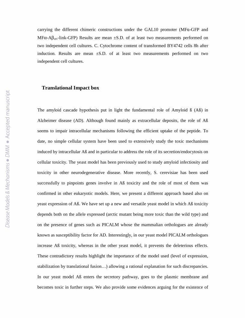
Figure 1: Aß aggregation and yeast growth
Ten-fold dilutions of exponentially growing cultures of BY4742 cells transformed with
plasmids carrying the different chimeric constructions under the GAL10 promoter were
spotted onto SD (-) or SG (+) agar supplemented with 20 mg/L histidine, 20 mg/L lysine, and
60 mg/L leucine. The cells were incubated at 30°C for 3 days. The cells were also grown for 6
h in SG liquid medium supplemented with 0.67% casaminoacids to induce the expression of
the chimeric proteins and were examined by epifluorescence microscopy. *: position of arc
mutation.
Dise
ase
Mod
els &
Mec
hani
sms
D
MM
Acce
pted
man
uscr
ipt
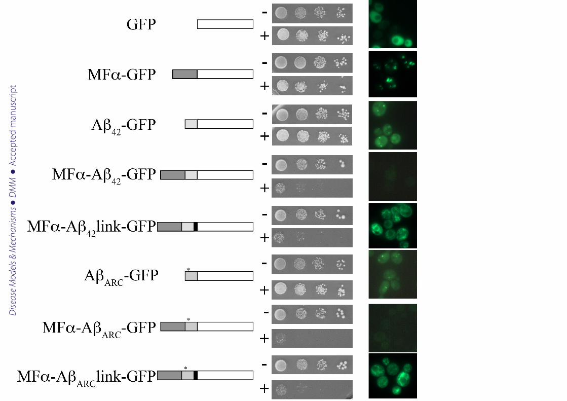
Figure 2: Maturation of secreted forms of Aß.
(A) BY4742 cells expressing the different chimeric proteins (6 h of expression) were
collected to perform total-protein extracts. Equal quantities of proteins were separated by
SDS-PAGE on a 12 % polyacrylamide gel and were then transferred onto a nitrocellulose
membrane and exposed to monoclonal anti-GFP antibodies (Sigma). (B) At different time
points of the induction, cells were collected to perform total-protein extracts. Equal quantities
of proteins were separated by SDS-PAGE on a 12 % polyacrylamide gel and were then
transferred onto a nitrocellulose membrane and exposed to monoclonal anti-GFP antibodies
(Sigma).
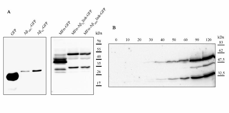
Figure 3: The secreted forms are glycosylated and processed by Kex2
(A) BY4742 WT or ∆kex2 cells expressing the different chimeric proteins (6 h of expression)
were collected for total protein extracts. Equal quantities of proteins were separated by SDS-
PAGE on a 12 % polyacrylamide gel, transferred onto a nitrocellulose membrane, and then
exposed to monoclonal anti-GFP antibodies (Sigma). (B) BY4742 expressing the different
chimeric proteins (6 h of expression) were collected for total-protein extracts and submitted or
not to a deglycosylation enzyme mix. Equal quantities of proteins were separated by SDS-
PAGE on a 12 % polyacrylamide gel, transferred onto a nitrocellulose membrane, and
exposed to monoclonal anti-GFP antibodies (Sigma). (C) After spheroplast fractionation of
BY4742 cells expressing MFα-Aβarc-GFP, P13 fractions were treated with proteinase K
(0.3125mg/ml) in the presence or absence of 5 % Triton X-100, resolved by SDS-PAGE, and
then analyzed by immunoblotting.
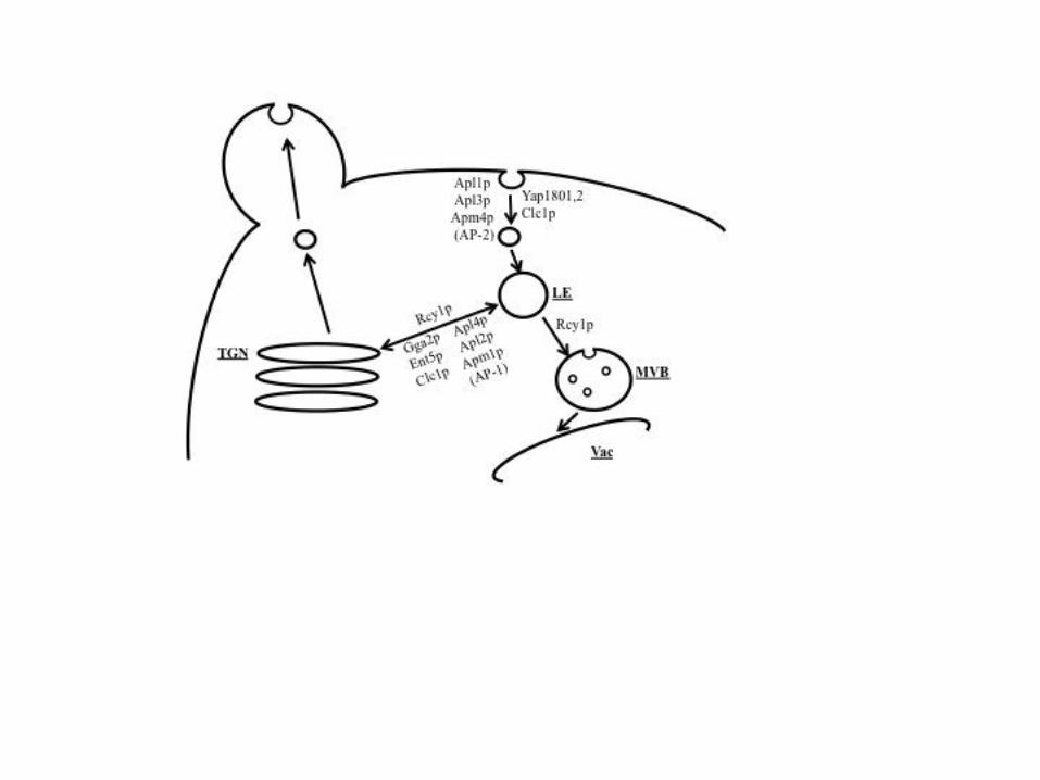
Figure 4: Schematic view of endocytosis and recycling in yeast
Cellular compartments are indicated: late endosome (LE), multivesicular body (MVB) shown
with intralumenal vesicles, vacuole (Vac), and trans-Golgi network (TGN). The proteins
Dise
ase
Mod
els &
Mec
hani
sms
D
MM
Acce
pted
man
uscr
ipt
involved in some of the steps of endocytosis and recycling are indicated next to the arrows
(note that not all proteins are included, only those tested in this study).
Figure 5: Mammalian PICALM enhances the toxicity of Aß in yeast
A Ten-fold dilutions of exponentially growing cultures of BY4742 cells transformed with
plasmids carrying the different chimeric constructions under the GAL10 promoter and, as
indicated, a plasmid containing or not the short form of the PICALM cDNA under the
GAL10 promoter were spotted on the same plate onto SD (-) or SG (+) agar supplemented
with 20 mg/L histidine, 20 mg/L lysine.
B Deleted strains expressing the different chimeric proteins (6 h of expression) with or
without PICALM protein were collected for total protein extracts. Proteins were separated by
SDS-PAGE on a 12 % polyacrylamide gel, transferred onto a nitrocellulose membrane, and
then exposed to polyclonal anti-PICALM antibodies (CALM H-134, Santa Cruz).
Figure 6: GFP pattern of aggregation in yeast mutants
WT or strains deleted for YAP1802, HSP104, RCY1, or SNF7 were grown for 6 h in SG liquid
medium supplemented with 0.67% casaminoacids to induce expression of the chimeric
proteins and were then examined by epifluorescence microscopy.
Figure 7: Aß expression leads to a decrease in O2 consumption without changing cytochrome
content
A. BY4742 cells transformed with an empty vector (empty) or plasmids carrying the different
chimeric constructions under the GAL10 promoter (MFα-GFP and MFα-Aβarc-link-GFP)
were grown aerobically in medium containing 2% DL-lactate and 0.2% galactose. B.
Respiratory rates of BY4742 cells transformed with an empty vector (empty) or plasmids
Dise
ase
Mod
els &
Mec
hani
sms
D
MM
Acce
pted
man
uscr
ipt
carrying the different chimeric constructions under the GAL10 promoter (MFα-GFP and
MFα-Aβarc-link-GFP) Results are mean ±S.D. of at least two measurements performed on
two independent cell cultures. C. Cytochrome content of transformed BY4742 cells 8h after
induction. Results are mean ±S.D. of at least two measurements performed on two
independent cell cultures.
Translational Impact box
The amyloid cascade hypothesis put in light the fundamental role of Amyloid ß (Aß) in
Alzheimer disease (AD). Although found mainly as extracellular deposits, the role of Aß
seems to impair intracellular mechanisms following the efficient uptake of the peptide. To
date, no simple cellular system have been used to extensively study the toxic mechanisms
induced by intracellular Aß and in particular to address the role of its secretion/endocytosis on
cellular toxicity. The yeast model has been previously used to study amyloid infectiosity and
toxicity in other neurodegenerative disease. More recently, S. cerevisiae has been used
successfully to pinpoints genes involve in Aß toxicity and the role of most of them was
confirmed in other eukaryotic models. Here, we present a different approach based also on
yeast expression of Aß. We have set up a new and versatile yeast model in which Aß toxicity
depends both on the allele expressed (arctic mutant being more toxic than the wild type) and
on the presence of genes such as PICALM whose the mammalian orthologues are already
known as susceptibility factor for AD. Interestingly, in our yeast model PICALM orthologues
increase Aß toxicity, whereas in the other yeast model, it prevents the deleterious effects.
These contradictory results highlight the importance of the model used (level of expression,
stabilization by translational fusion…) allowing a rational explanation for such discrepancies.
In our yeast model Aß enters the secretory pathway, goes to the plasmic membrane and
becomes toxic in further steps. We also provide some evidences arguing for the existence of
Dise
ase
Mod
els &
Mec
hani
sms
D
MM
Acce
pted
man
uscr
ipt

different aggregates (toxic and harmless) that depend on the capacity of the protein to be
translocated into the secretory pathway or not. Finally, we also demonstrate that Aß is able to
cross the membranes and target mitochondria.
Dise
ase
Mod
els &
Mec
hani
sms
D
MM
Acce
pted
man
uscr
ipt
Dise
ase
Mod
els &
Mec
hani
sms
D
MM
Acce
pted
man
uscr
ipt
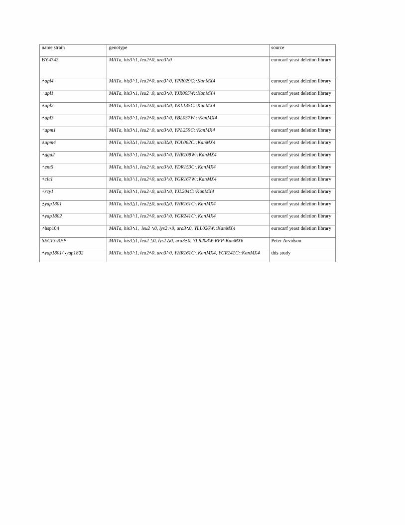
name strain genotype source
BY4742 MATa, his3∆1, leu2∆0, ura3∆0 eurocarf yeast deletion library
∆apl4 MATa, his3∆1, leu2∆0, ura3∆0, YPR029C::KanMX4 eurocarf yeast deletion library
∆apl1 MATa, his3∆1, leu2∆0, ura3∆0, YJR005W::KanMX4 eurocarf yeast deletion library
∆apl2 MATa, his3∆1, leu2∆0, ura3∆0, YKL135C::KanMX4 eurocarf yeast deletion library
∆apl3 MATa, his3∆1, leu2∆0, ura3∆0, YBL037W ::KanMX4 eurocarf yeast deletion library
∆apm1 MATa, his3∆1, leu2∆0, ura3∆0, YPL259C::KanMX4 eurocarf yeast deletion library
∆apm4 MATa, his3∆1, leu2∆0, ura3∆0, YOL062C::KanMX4 eurocarf yeast deletion library
∆gga2 MATa, his3∆1, leu2∆0, ura3∆0, YHR108W::KanMX4 eurocarf yeast deletion library
∆ent5 MATa, his3∆1, leu2∆0, ura3∆0, YDR153C::KanMX4 eurocarf yeast deletion library
∆clc1 MATa, his3∆1, leu2∆0, ura3∆0, YGR167W::KanMX4 eurocarf yeast deletion library
∆rcy1 MATa, his3∆1, leu2∆0, ura3∆0, YJL204C::KanMX4 eurocarf yeast deletion library
∆yap1801 MATa, his3∆1, leu2∆0, ura3∆0, YHR161C::KanMX4 eurocarf yeast deletion library
∆yap1802 MATa, his3∆1, leu2∆0, ura3∆0, YGR241C::KanMX4 eurocarf yeast deletion library
∆hsp104 MATa, his3∆1, leu2 ∆0, lys2 ∆0, ura3∆0, YLL026W::KanMX4 eurocarf yeast deletion library
SEC13-RFP MATa, his3∆1, leu2 ∆0, lys2 ∆0, ura3∆0, YLR208W-RFP-KanMX6 Peter Arvidson
∆yap1801/∆yap1802 MATa, his3∆1, leu2∆0, ura3∆0, YHR161C::KanMX4, YGR241C::KanMX4 this study
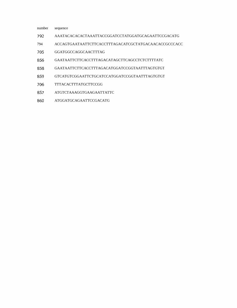
number sequence
792 AAATACACACACTAAATTACCGGATCCTATGGATGCAGAATTCCGACATG
794 ACCAGTGAATAATTCTTCACCTTTAGACATCGCTATGACAACACCGCCCACC
705 GGATGGCCAGGCAACTTTAG
856 GAATAATTCTTCACCTTTAGACATAGCTTCAGCCTCTCTTTTATC
858 GAATAATTCTTCACCTTTAGACATGGATCCGGTAATTTAGTGTGT
859 GTCATGTCGGAATTCTGCATCCATGGATCCGGTAATTTAGTGTGT
706 TTTACACTTTATGCTTCCGG
857 ATGTCTAAAGGTGAAGAATTATTC
860 ATGGATGCAGAATTCCGACATG
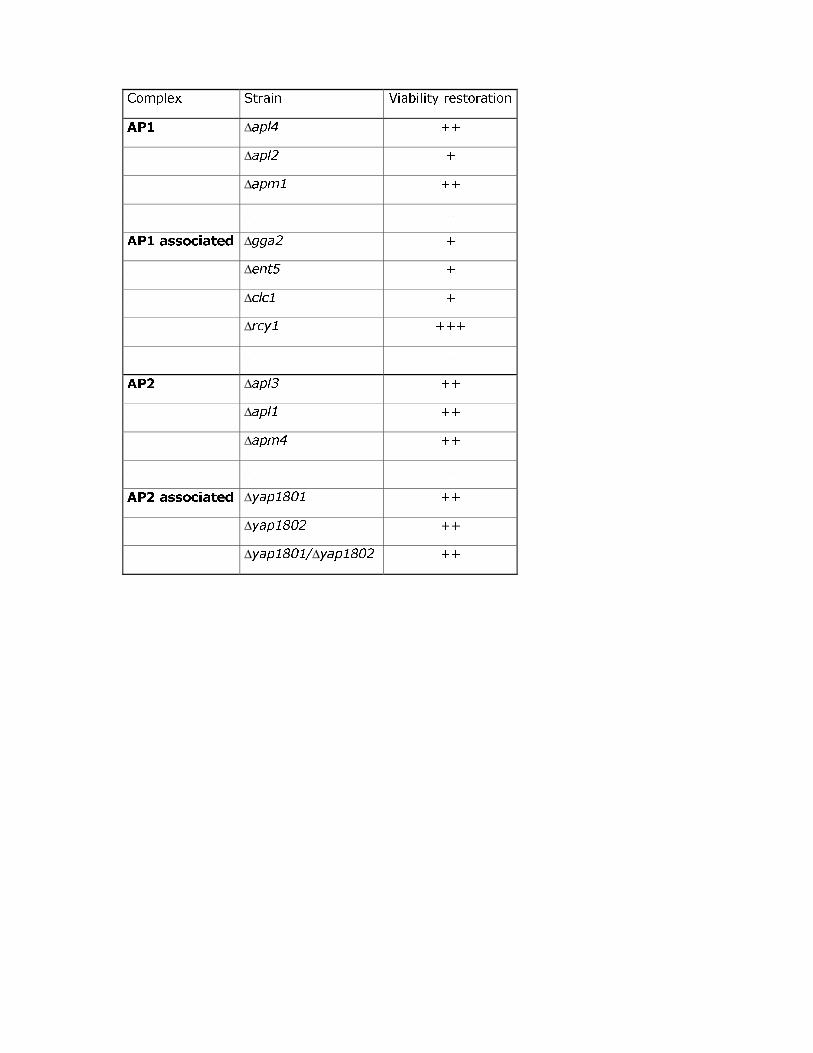
Complex Strain Viability restoration
AP1 Δapl4 ++
Δapl2 +
Δapm1 ++
AP1 associated Δgga2 +
Δent5 +
Δclc1 +
Δrcy1 +++
AP2 Δapl3 ++
Δapl1 ++
Δapm4 ++
AP2 associated Δyap1801 ++
Δyap1802 ++
Δyap1801/Δyap1802 ++
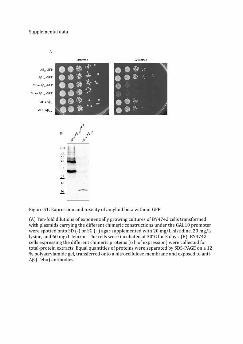
Supplemental data
Figure S1: Expression and toxicity of amyloid beta without GFP.
(A) Ten-‐fold dilutions of exponentially growing cultures of BY4742 cells transformed with plasmids carrying the different chimeric constructions under the GAL10 promoter were spotted onto SD (-‐) or SG (+) agar supplemented with 20 mg/L histidine, 20 mg/L lysine, and 60 mg/L leucine. The cells were incubated at 30°C for 3 days. (B): BY4742 cells expressing the different chimeric proteins (6 h of expression) were collected for total-‐protein extracts. Equal quantities of proteins were separated by SDS-‐PAGE on a 12 % polyacrylamide gel, transferred onto a nitrocellulose membrane and exposed to anti-‐Aβ (Tebu) antibodies.
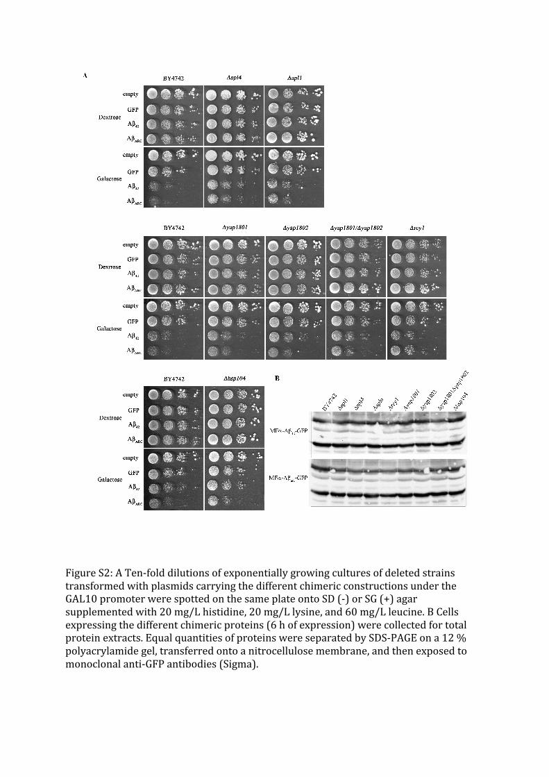
Figure S2: A Ten-‐fold dilutions of exponentially growing cultures of deleted strains transformed with plasmids carrying the different chimeric constructions under the GAL10 promoter were spotted on the same plate onto SD (-‐) or SG (+) agar supplemented with 20 mg/L histidine, 20 mg/L lysine, and 60 mg/L leucine. B Cells expressing the different chimeric proteins (6 h of expression) were collected for total protein extracts. Equal quantities of proteins were separated by SDS-‐PAGE on a 12 % polyacrylamide gel, transferred onto a nitrocellulose membrane, and then exposed to monoclonal anti-‐GFP antibodies (Sigma).
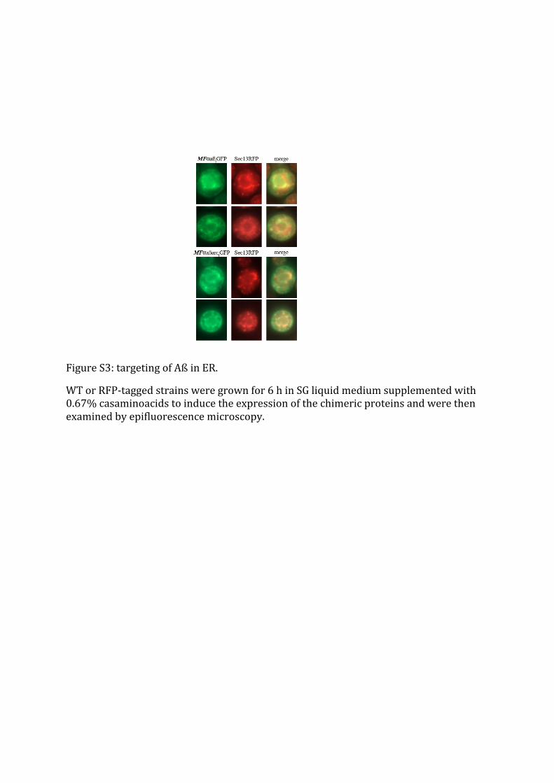
Figure S3: targeting of Aß in ER.
WT or RFP-‐tagged strains were grown for 6 h in SG liquid medium supplemented with 0.67% casaminoacids to induce the expression of the chimeric proteins and were then examined by epifluorescence microscopy.


