Embed Size (px)
Citation preview

ELSEVIER
THEO CHEM
Journal of Molecular Structure (Theochem) 314 (1994) 247-260
Ab initio HF-SCF studies of the equilibrium structures and vibrational spectra of the Be(NO&, Mg(NO& and Ca(NO&
molecules
V. Rossi, C. Sadun, L. Bencivenni, R. Caminiti*
Dipartimento di Chirnica, Universitb degli Studi di Roma, P. le A. Moro 5, 00185 Roma. Italy
Received 3 February 1994; accepted 20 February 1994
Abstract
HF-SCF calculations have been performed for the Mg(NO&, Ca(NO& and Be(NO,), molecules to determine their stable equilibrium structures and vibrational spectra. Bidentate and monodentate structures have been taken into account and the calculations suggest that non-planar Dzd is the most stable equilibrium structure for all the species considered in the work. The infrared spectra of the DZd and D2h structures of Mg(NO& have been simulated on the basis of the HF/6-31G* frequency and intensity calculations, and the “0 isotope shifts have been predicted in the stretching region of the NO; modes. The conclusions of the calculations have been used to assign the FT-IR spectra of molecular Cu(NO&. The paper includes calculations for the [BeNOj]+H20, [MgNOJ+H20, [CaN03]+H20, [A1N03]*+H20, [BeN02]+H20, [MgN02]+H20, [CaN02]+H20, and [A1N02]*+H20 hydrated species.
1. Introduction
Our research group has been quite active in the
structural [ 1,2] and spectroscopic [3-51 investiga- tion of inorganic molecules having appreciable ionic character due to the interaction of a metal
cation with an inorganic anionic group. More recently, this topic has been developed by means of HF-SCF and MP2 calculations [6-91. So far, much attention has been devoted to the associa-
tion of group la metal cations M+ with NO, and PO;. It has been definitively established that the [M+][NO;] and [Mf] [PO;] ion couples prefer bidentate coordination structures in the gas phase. A valuable contribution to the study of the
*Corresponding author.
coordination models of these ion couples is provided by the theoretical investigation on the interaction of [L?] and [Na+] with [NOT] and
[PO,] [7,10]. In fact, ab initio calculations have a great capability for describing the geometrical, energetic and spectroscopic features of these mol-
ecules. It has to be borne in mind that it is always desirable for a better understanding of the chemical problem to link theory with spectroscopy because unequivocal conclusions often cannot be reached
from experiment alone. Computational chemistry, therefore, plays an important role in clarifying experimental results and predicting helpful infor- mation for further investigations by means of more sophisticated experimental techniques. This is, for instance, the case with modern FT-IR spectroscopy coupled with low-temperature matrix isolation
0166-1280/94/$07.00 0 1994 Elsevier Science B.V. All rights reserved SSDI 0166-1280(94)03741-3

248 V. Rossi et al./J. Mol. Strut. (Theochem) 314 (1994) 247-260
spectroscopy which is particularly suitable for the investigation of the vibrational pattern of ion couples and their ‘80-isotope spectra.
In this paper we present, as a development of our former ab initio studies, novel results of a theo- retical investigation on the interaction of the metal cations Be*+, Mg*+ and Ca2+ with NOi. A further aim of this investigation is the assignment of the FT-IR matrix isolation spectrum of the stable molecular species Cu(NO&; the theoretical predictions obtained for the probe system Mg(N03)* have been employed for this purpose.
2. Computational methods
The calculations were accomplished by the IBM version of the GAUSSIAN 90 package [ 1 l] on an IBM- 3090 computer of the University of Rome. The calculations were performed at the HF-SCF level of theory employing the split-valence basis sets 6-31G and 6-31G* for studying the Mg(NO& and Be(N03)2 systems. The calculations for the Ca(N03)2 molecule are reported at the HF/ STO-3G* and HF-SCF level with the 4-31G* basis set for the N and 0 atoms, and the 14~9~ contracted to 8s4p basis set for Ca [12]. The 6-31G, 4-31G*, 6-31G* and STO-3G* basis sets are stored in the GAUSSIAN 90 program. The vibra- tional frequencies were computed analytically at each optimized HF-SCF structure. Single-point energy calculations have been carried out on the optimized lowest energy structures of Mg(N03)2 and Ca(N03)2 at the HF-SCF level employing the DZP basis [12] for the nitrate group and the 6-31G* basis for the Be and Mg atoms, and the 14~9~ contracted to 8s4p basis for the Ca atom [12].
3. Experimental methods
The FT-IR spectra were measured both in a solid argon matrix at approx. 10 K and in very dilute CC& solution at approx. 300K in the frequency range 2000-400 cm-’ by using a Bruker IFS-1 13V interferometer maintained under mechanical pump vacuum. A variable optical path liquid cell equipped with CsI windows was used for recording the Fl-IR
solution spectra. Measured frequencies are accurate within *l cm-’ and the highest resolution of the FT-IR spectra is 1 cm-’ . The FT-IR matrix spectra were measured after depositing molecular CU(NO~)~ and argon (Matheson, 99.998%) onto the optical surface of a reflecting copper block frozen up to approx. 10K by means of a He closed-cycle cryo- stat (Displex CSA). A commercial sample of Cu(N03)* (Aldrich, 99.99+%) was heated under a suitable high vacuum device inside a platinum evaporator at approx. 385 K.
4. Results and discussion
4.1. The M(NO,), molecules
The theoretical study was accomplished for the Be, Mg and Ca nitrates, M(N03)*, owing to the similarity of these molecules with CU(NO~)~ which is not at present suitable for a reliable ab initio investigation. Most of the computational effort was addressed to Mg(N03)2 to achieve a general picture for the structural models and vibra- tional spectra expected for the M(N03)2 molecules. We have therefore optimized at the HF-6-3 1 G and HF-6-31G* levels the structures shown in Fig. 1; these structures are the bidentate models la and lb and the monodentate models lc, Id and le. Among these models, the structures la, lc and le were considered for the CU(NO~)~ molecule in earlier electron diffraction studies [13,14], with model la being preferred, while the non-planar structure lb was suggested from a later electron diffraction investigation [ 151 linked with molecular orbital calculations.
The main results obtained from our computa- tions indicate that bidentate binding is preferred over the other coordinations and, moreover, the non-planar bidentate model of DZd symmetry shown in Fig. l(b) is the lowest energy configura- tion of the Mg(N03)*, Ca(N03)* and Be(N03)2 molecules. The planar monodentate model lc (D2h symmetry), the non-planar monodentate model Id (D2,, symmetry) and the staggered model le (DJd symmetry) are unstable stationary points and do not correspond to any minimum of the total SCF energy. Thus, according to the HF-SCF

V. Rossi et al./J. Mol. Struct. (Theochem) 314 (1994) 247-260 249
a
d -
e
Fig. 1. Mg(N03)2 molecule. Bidentate structures: (a) I&h sym- metry and (b) DZd symmetry; monodentate structures: (c) Dzh
symmetry structure and (d) DZd symmetry; (e) staggered D3d
symmetry.
single-point energy calculations carried out on the optimized HF-SCF geometries, the lowest energy coordination models are la and lb which are approx. 9 kJ mol-’ more stable than the monodentate models lc and Id, while the Dsd symmetry model le is the highest energy structure. In addition, the HF/6-3 lG*
vibrational frequencies, calculated in the harmonic approximation, indicate that the DM symmetry bidentate structure lb is the absolute minimum and that the Dz~ symmetry bidentate structure la is prob- ably the transition state connecting two equivalent D2d symmetry bidentate structures. Regarding the remaining high-energy structures, the results of the HF/6-3 1 G* harmonic frequencies indicate that all the monodentate models are high-order saddle points. The subsequent investigation on Ca and Be nitrates has been restricted to the biden- tate coordination models of DZd and DZh symmetry and also in this case we have reached the same conclusions as for Mg(NO&. In fact, the DZd bidentate structure is the lowest energy minimum and the DZh bidentate structure is the transition state, being a first-order saddle point with one calculated imaginary A, mode. The HF-SCF single-point energy difference separating the tran- sition state from the minimum is estimated to be approx. 4kJ mall’ for Ca(NO& and approx. 53 kJ mall’ for Be(NO&. These data, although they do not take into account electron correla- tion, indicate that such a barrier decreases remark- ably on going from Be to Mg and Ca nitrate and that tunnelling from two equivalent DZd minima through the DZh first-order saddle point is not hindered for the heavier metal nitrates. There is obviously consistency between the SCF barrier height and the value of the imaginary A, mode frequency of the Dzh bidentate structures, that is 131 icm-’ for Be(NO&, 61 icm-’ for Mg(NO& and 54 i cm-’ for Ca(NO&. A summary of the cal- culations is given in Table 1.
Let us now examine the most interesting struc- tural differences between the group 2a metal nitrates and the group la nitrates of Li and Na, previously studied at the HF/6-31G* and MP2/6- 3 1 G* levels [ 10,161. It is known that the interaction of the metal cation with NO, induces a perturba- tion of the anion and causes the lowering of the symmetry from Djh, that of free NOj, to the local C,, symmetry of the bidentate [M2’][NOf] moiety and it causes a general rearrangement of the geometry of the anion. The HF-SCF level predicts a difference of about 0.1 A between the ring and terminal bonds of coordinated NO; for the Be, Mg and Ca nitrates and also a large devia-

250 V. Rossi et al./J. Mol. Srruct. (Theochem) 314 (1994) 247-260
Table 1.
Equilibrium geometriesa and unscaled vibrational frequencies
of the LIZ,, and & symmetry bidentate structures of Be(NO&
Mg(NO& and Ca(NO&; bond lengths in A, angles in degrees,
and frequencies in cm’
DSd Geometries
Be(NO& Mg(NO& Ca(NO&
NO 1.155 1.163 1.171
NO’. 1.268 1.264 1.252
MeO’ 1.648 2.007 2.336
O’NO’ 109.1 113.5 114.8
ONO’ 125.4 123.3 122.6
HF-SCF vibrational frequencies Description
At 2010 1947 1900 NO str. sym.
1178 1175 1207 NO’ str. sym.
930 875 853 NO; def. sym.
306 254 229 MeO’ str. sym.
B2 1996 1933 1889 NO str. asym.
1178 1171 1204 NO’ str. asym.
1035 881 851 NO; def. asym.
908 598 440 MeO’ str. asym.
BI 135 51 35 Torsion
E 1499 1475 1530 NO’ str. asym.
930 942 976 NO; def. asym.
770 788 805 NO’, def. asym.
560 360 295 MeO’ str. asym.
280 187 134 o.p. def.
92 64 43 o.p. def.
Dth Geometries
Be(NO3)2 Mg(NO& Ca(NOA
NO 1.157 1.164 1.171
NO’ 1.265 1.263 1.251
MeO’ 1.685 2.013 2.339
O’NO’ 108.8 113.3 114.7
ONO’ 125.6 123.4 122.6s
HF-SCF vibrational frequencies Description
As 2004 1945 1899 NO str. sym.
1196 1178 1208 NO’ str. sym.
931 877 853 NO; def. sym.
318 255 229 MeO’ str. sym.
B3, 1469 1460 1524 NO’ str. sym
750 784 803 NO; def. sym.
208 283 264 MeO’ str. sym.
Bk 933 942 975 NO; def. sym.
183 149 119 def. sym.
Table 1. (Contd).
HF-SCF vibrational frequencies Description
1991 1931 1887
1188 1173 1205
934 881 851
910 591 436
1549 1498 1539
771 789 805
454 393 312
180 86 54
942 944 977
504 225 149
88 59 39
131 i 61i 34i
NO str. asym.
NO’ str. asym.
NO: def. asym.
MeO’ str. asym.
NO’ str. asym.
NO: def. asym.
MeO’ str. asym.
Def. asym.
NO; def. asym.
def. asym.
def. asym.
a 0’ indicates the oxygen atom of the cyclic frame of the mol-
ecule.
tion of the ring angle of the anion, decreasing from 120” by about 5” in the Mg and Ca nitrates and 11” in Be(N03)2. Therefore the effect of ion-pairing on NOj is more marked in Be(NO& than in Ca(N03)2 and Mg(NOs)*. The calculations also suggest that the consequences of ion-pairing exerted by Mg and Ca atoms are nearly of the same extent as those determined for the LiNOs and NaNOs molecules [ 10,161, while the inter- action of the Be atom induces a huge modification of the geometry of the anion. The extent of such effects is consistent with the M . . . N bond length, which increases passing from the Be to the Ca atom according to the ionic polarizability following the order Ca2+ > Mg*+ > Be*’ [ 171.
The conclusions reported for the Be, Mg and Ca nitrates might be useful for reinvestigating the IR spectrum of CU(NO~)~, a typical high-temperature molecule having noticeable stability in the gas phase. This molecule vaporizes as a monomer without appreciable decomposition [ 181 and there- fore is suitable for matrix-isolation studies. The FT-IR spectrum of gaseous CU(NO~)~ condensed onto an argon matrix at approx. 10 K and that of a highly diluted solution of Cu(NO& in CC14 closely mirror each other. In particular, the matrix spectrum shows sharp bands at 16155cm-‘, 1204.7cm-‘, 975.6cm-’ and 785.4cm-’ and these IR-active vibrations of coordinated NO; compare well with those reported in an earlier work on

V. Rossi et a/./J. Mol. Struct. (Theochem) 314 (1994) 247-260
3 1000 6 E
‘i S5 3) c I
600 a, t;
251
3000 50 -1
cm
Fig. 2. HF-SCF infrared spectra of Mg(N03)2 calculated for the bidentate DZh and bidentate D,, symmetry structures.
Cu(NO& [19]. It is interesting to apply the theo- retical predictions to the FT-IR spectrum of CU(NO~)~ in the light of the available IR measure- ments and of the electron diffraction studies [13-151. First of all, it is necessary to scale the HF/ 6-31G* frequencies for improving the agreement with the observed ones and this procedure is parti- cularly easy in this case because the scaling factors for the SCF force constants of the nitrate anion vibrations are known from previous work [7,10].
The fact that the FT-IR spectrum shows three bands in the stretching region of the NO, anion is a typical feature of both the Dzh and DZd structures because the IR-active stretching vibrations of coor- dinated NOT are the two Bt, and the BZu modes, in the case of DZh symmetry, and the B2 + E modes, in the case of DZd symmetry. However, the calculated relative intensity IR patterns are different for the two symmetries (see Fig. 2). The IR pattern of the DZh symmetry is 10:6: 1 for the B,, (1617cmP1), B2,, (1258cm-‘) and Bt, (965cm-‘) bands, and that of the Dzd symmetry is 10 : 3 : 2 for the B2 (1621cm-‘), E (1229cm-‘) and B2 (976cm-‘) bands. It is therefore clear that the FT-IR spectrum of Cu(NO& fits the IR features of Dzh symmetry better than those of DZd symmetry, since the experi- mentally measured relative intensity pattern of the 1616, 1205 and 976 cm-’ bands is 10 : 5 : 4. Our con- clusions would not support the most recent electron
diffraction study [ 151 according to which the NO, groups of the molecule do not lie in the same plane and form an average dihedral angle of 84.5” [15]. Further support of the planarity of the molecules comes from single-point energy calcula- tions at the ROHF-SCF level employing the 14s9p6d basis, contracted to 8s4p3d, for the Cu atom and 4-31G* basis sets for N and 0 atoms, carried out for the planar (D,, symmetry, 2B3, electronic ground state) and the non-planar (D2,j symmetry, 2B2 electronic ground state) structures of CU(NO~)~ based on the electron diffraction geometry data. This calculation in fact predicts that the planar structure is 137 kJmol_’ more stable than the non-planar one.
From the theoretical point of view, it is impor- tant to consider the expected IR spectra of DZh and
D2d '*O isotopically enriched divalent metal nitrate. It has to be remarked that SCF IgO isoto- pic frequency shifts have been employed success- fully for this purpose to explain the spectroscopic features of random “0 labelling of group la metal nitrates [7,10], metaphosphates [lo], chlorates [20] and D2d-symmetry Li2S04 and C2,-symmetry LiC104 molecules [9]. The “0 isotope pattern of the nitrate group vibrations of a D2t, and D2d sym- metry M(N03)2 molecule shows some characteris- tic differences in the region of the NO; stretching vibrations. The isotopic bands of the DZh structure
252
Table 2
V. Rossi et al./J. Mol. Struct. (Theochem) 314 (1994) 247-260
HF-SCF scaled frequencies (cm-‘) of the DZh and Dz~ models of the “0 isotopomers of Mg(N03)z
O.uygen atom numbering Ol-N(02)(03) Mg (04)(05)N-06
Isotopomers
1 016-N(016)(016) Mg (016)(016)N-016
D1 A, (1628) Bjg (1226) A, (965)
B,, (1617) BZu (1258) B,, (965)
2 016-N(016)(016) Mg (016)(018)N-016
CS A’ (1628) A’ (1616) A” (1254)
A” (1221) A’ (968) A’ (948)
3 016-N(016)(016) Mg (016)(016)N-018
C,, Al (1624) A, (1591) B2 (1258)
B2 (1226) A, (967) AI (955)
4 016-N(018)(016) Mg (016)(018)N-016
CS A’ (1628) A’ (1616) A’ (1249)
A’ (1218) A’ (950) A’ (946)
5 016-N(018)(016) Mg (016)(016)N-018
CS A’ (1623) A’ (1591) A’ (1254)
A’ (1221) A’ (955) A’ (948)
6 016-N(016)(018) Mg (016)(018)N-016
C,” A, (1628) A, (1616) Bz (1249)
Bz (1218) A, (950) AI (946)
7 016-N(016)(016) Mg (016)(018)N-018
C, A’ (1624) A’ (1590) A” (1254)
A” (1221) A’ (968) A’ (935)
8 016-N(016)(016) Mg (018)(018)N-016
CZ” A, (1628) B2 (1252) A, (968)
A, (1616) B2 (1215) A, (929)
9 018-N(016)(016) Mg (016)(016)N-018
&I, BI, (1586) B2u (1257) BI, (953) A, (1598) Bjg (1226) A, (956)
10 016-N(016)(016) Mg (018)(018)N-018
C,, A, (1624) Bz (1251) A, (968) A, (1589) B2 (1213) Al (916)
11 018-N(016)(016) Mg (018)(018)N-016
C,, A, (1622) B2 (1251) A, (955) A, (1591) B2 (1214) Al (929)
12 016-N(018)(016) Mg (018)(018)N-016
CS A’ (1627) A’ (1615) A’ (1245)
A’ (1218) A’ (948) A’ (929)
13 016-N(016)(018) Mg (018)(016)N-018
CS A’ (1623) A’ (1590) A’ (1248)
Dzd A, (1623) B2 (1611) E (1229)
A, (979) B2 (976)
G A’ (1623) A’ (1611) A’ (1229)
A” (1221) A’ (978) A’ (958)
C,V A, (1618) Bz (1229) A, (978)
B2 (1586) A, (1228) B2 (964)
C, A(1623) A(1611) B (1221)
B (1221) A (960) A (957)
CS A’ (1618) A’ (1586) A’ (1228)
A” (1221) A’ (965) A’ (958)
CS ,4’ (1618) A’ (1585) A’ (1229)
A” (1219) A’ (978) A’ (944)
C,, A, (1623) B2 (1229) A, (978)
A, (1611) B2 (1211) A, (939)
Dzd B2 (1580) E (1228) B2 (963)
A, (1592) A, (965)
C,, A, (1618) B2 (1229) A, (978)
A, (1584) Bz (1209) A, (925)
Cl” A, (1618) B2 (1228) A, (964)
A, (1589) Bz (1211) Al (939)
C, A’ (1622) A’ (1611) A’ (1221)
A” (1211) A’ (958) A’ (939)
c, (1618) (1585) (1221) A’ (1217) A’ (948) A’ (935) (1219) (958) (944)

Table 2. (Contd)
V. Rossi et al./J. Mol. Struct. (Theochem) 314 (1994) 247-260 253
14 018-N(016)(016) Mg (016)(018)N-018
CS A’ (1598) A’ (1586) A’ (1253)
A’ (1221) A’ (955) A’ (935)
15 018%N(018)(016) Mg (016)(018)N-018
CS A’ (1597) A’ (1585) A’ (1248)
A’ (1216) A’ (937) A’ (933)
16 018-N(018)(016) Mg (018)(018)N-016
Cs A’ (1622) A’ (1590) A’ (1244)
A’ (1211) A’ (935) A’ (928)
17 018-N(018)(016) Mg (018)(016)N-016
CS A’ (1623) A’ (1590) A’ (1248)
A’ (1217) A’ (948) A’(935)
18 016-N(018)(018) Mg (018)(018)N-016
&h BI, (1615) Bzu (1258) B,, (928) A, (1627) B1, (1208) A, (931)
19 018-N(016)(016) Mg (018)(018)N-018
C,, A, (1598) Bz (1250) A, (955)
A, (1585) BZ (1213) A, (915)
20 018-N(018)(018) Mg (018)(016)N~016
CS A’ (1623) A’ (1589) A” (1244)
A” (1211) A’ (948) A’ (915)
21 018~N(016)(018) Mg (016)(018)N-018
Cl, A, (1597) B, (1248) A, (937)
A, (1585) B2 (1216) A, (933)
22 018%N(016)(018) Mg (018)(018)N-018
CS A’ (1597) A’ (1585) A’ (1244)
A’ (1210) A’ (935) A’ (915)
23 018-N(018)(018) Mg (018)(018)N-016
C,, A, (1622) B2 (1238) A, (929)
A, (1589) Bz (1207) A, (915)
24 018~N(018)@18) Mg (018)(018)N-018
Dzi, BI, (1585) B2u (1237) BI, (914) A, (1597) B,, (1206) %(917)
15N molecule”
D, h (1581) B2u (1229) BI, (964) A, (1593) B3, (1197) AZ (967)
c2 A (1592) A (1580)
B (1219) A (965)
B (1219)
A (943)
A’ (1617) A’ (1585)
A” (1211) A’ (945)
A’ (1219)
A’ (938)
Cl (1618) (1585)
(1219) (958)
(1221)
(944)
D2d B2 (1610) E (1211)
A, (1622) A, (941) B2 (938)
Cl” A, (1593) B2 (1228)
A, (1580) B, (1209) 4 (964) A, (925)
A’ (1618) A’ (1584)
A” (1209) A’ (958)
A’ (1221)
A’ (925)
A (1592) B (1219)
A (1580) B (1219)
A (946)
A (943)
A’ (1592) A’ (1580)
A” (1209) A’ (945)
A’ (1219)
A’ (925)
A, (1617) B2 (1211)
A, (1584) B2 (1209) c2, AI (939)
AI (925)
&d B2 (1580) E (1209)
A, (1592) A, (927) J32 (923)
Dzd B2 (1584) E (1210)
A, (1594) A, (965) B2 (961)
a Stretching vibrations of the anion.
are in fact due to the 2A,, 2Bi,, BZu and B3s modes, while those of the Dzd model are due to the 2Ai, 2B2 and E vibrations. Restricting the discussion to the IR-active vibrations of coordinated NOT, the main difference between the 180 pattern of the 2Bi,
and BZu stretching modes of the Dzh symmetry and that of the 2B2 and E stretching frequencies of the Dz~ symmetry arises from the breakdown of degen- eracy of the E modes occurring in the isotopomers generated by the DZd structure. The contribution of

254 V. Rossi et al./J. Mol. Struct. (Theochem) 314 (1994) 247-260
Table 3
Equilibrium geometries’ and unscaled vibrational frequencies of the [M(NO,)]‘, [M(NOJ)]+H~O, [M(NOl)]+ and [M(N02)]+HzO
species; bond lengths in A, angles in degrees and frequencies in cm-’
NO 1.130 1.147 1.160 1.118
(1.181 1.201 1.213 1.163)
NO’ 1.305 1.283 1.264 1.335
(1.347 1.323 1.3005 1.393)
MeO’ 1.529 1.934 2.265 1.753
(1.551 1.963 2.281 1.794)
O’NO’ 106.0 118.0s 113.6 107.0
(107.0 113..3 114.3 108.2)
ONO’ 127.0 123.9 123.2 126.5
(126.5 123.3 122.8 125.9)
HF-SCF vibrational frequencies Description
Al 2149 2035 1950 2194
1120 1118 1174 983
1035 894 859 994
850 528 403 644
B2 1392
868
660
RI 881
318
1398 1481 1247
784 801 789
438 343 563
914 942 828
191 140 240
NO str.
NO’ str. asym.
NO’, scis.
O’Me str. sym.
NO’ str. asym.
NO; rock
O’Me str. asym.
NO; wag
Ring o.p. def.
Geometries
NO 1.137 1.151 1.163 1.122
NO’ 1.291 1.277, 1.262 1.321
MeO’ 1.566 1.953 2.284 1.770
O’NO’ 106.9 112.4 113.9 107.2
ONO’ 126.5, 123.8 123.0, 126.4
M OH2 1.568 1.982 2.383 1.791
OH 0.961 0.959 0.956 0.978
HOH 110.3 106.9 104.5 107.7
HF-SCF vibrational frequencies Description
Al 3916 3957 3993
2112 2014 1935
1817 1840 1875 1097 1134 1180
931 891 855
1204 621 440 442 342 282
B2 3989
1434
877
1422 1492
657 560
3681 OH str. sym.
2183 NO str. sym.
1819 H20 bending 1026 NO’ str. sym. 1008 NO; scis.
807 M. OH2 str. sym.
470 O’Me str. sym.
3718 OH str. asym.
1311 NO’ str. asym.
850 M OH2 str. asym.

V. Rossi et al./J. Mol. Strut. (Theochem) 314 (1994) 247-260 255
Table 3. (Contd)
HF-SCF vibrational frequencies Description
761 786
615 415
145 51
Bl 900
435
341
109
A2 219
923
484
189
52
109 52 140 Torsion
801
330
8
946
529
136
13
787
547
76
8.51
627
253
90
NO; rock
O’Me str. asym.
M OH2 i.p. bending
NO; wag
M OH* o.p. bending
Ring o.p. def.
o.p. def.
Geometries
Pe(NOdl’ bkWW+ KIWI’ WWd12+
NO’ I .253 1.237 1.234 1 .2435
Me0 1.546 1.955 2.274 1.817
O’NO’ 107.8 112.4 113.3 110.8
HF-SCF vibrational frequencies Description
Al
B2
BI
1516
1133
868
1386
692
435
Geometries
PeWdlfH20
1531 1570 1416
1024 981 1079
536 427 590
1434 1498 1336
439 385 503
396 329 519
PkWWl+H20 [C4NWl+H20 [A1(N02)]2+H20
NO str. sym.
NO* scis.
OMe str. sym.
NO str. asym.
OMe str. asym.
Ring o.p. def.
NO’ 1.247 1.237 1.234 1.250
MeO’ 1.584 1.971 2.288 1.808
O’NO’ 107.8 112.4 113.4 109.2
M OH2 1.564 1.986 2.386 1.792
OH 0.962 0.959 0.956 0.979
HOH 110.3 106.9 104.6 107.6
HF-SCF vibrational frequencies Description
Al 3903 3957 3994 3667
1816 1840 1874 1818
1554 1547 1577 1472
1049 1017 976 1105
1177 624 455 827
451 351 293 471
OH str. sym.
Hz0 bending
NO str. sym.
NOa scis.
M Hz0 str. sym.
OMe str. sym.
B2 3975 4025 4063 3703 OH str. asym.
1424 1446 1501 1356 NO str. asym.
817 653 557 843 M. Hz0 str. asym.
603 422 376 522 OMe str. asym.
159 56 4 83 M.. H20 i.p. bending
J% 495 487 523 636 M. OH2 o.p. bending
404 316 314 478 Ring o.p. def.
256 V. Rossi et al./J. Mol. Struct. (Theochem) 314 (1994) 247-260
Table 3. (Contd)
HF-SCF vibrational frequencies Description
157 64 6 108 o.p. def.
A2 227 106 70 140 Torsion
a 0’ indicates the oxygen atom of the cyclic frame of the molecule. The MP2 geometries are reported in parentheses. The data calculated for free Hz0 are: HF/6-31G* (OH = 0.947& HOH = 105.5”; A, 4069, 1826; Bz 4188cm-‘); HF/4-3lG* (OH = 0.948& HOH = 105.3”; A, 4053, 1830; Bz 4169cm-‘).
the Raman-active vibrations of the DZt, and Dzd structures to the respective IR spectra is evidently lacking because the dipole derivatives &/aQ asso- ciated with the vibrations of the C,, C,, and C, isotopomers are actually zero. The number of iso- topic combinations is very high in both cases (see Table 2) and it is the same for the DZh and DZd symmetry structures. However, the isotopic patterns mainly differ from each other for the Ct symmetry isotopomers of the Dti model which produce a char- acteristic pattern due to the expected band intensity ratios and frequencies. This distinctive pattern arises from the partial or complete overlap of some stretch- ing vibrations, as can be seen from Table 2.
This also simplifies the IR spectrum because bands of distinct isotopomers are predicted to lie in the same frequency region. This evidently occurs for a series of isotopic species of both models and ultimately makes possible the distinction between the two structures.
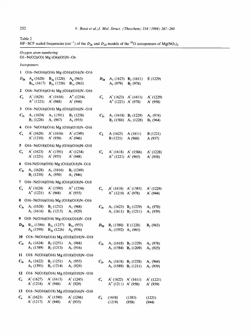
4.2. The [MeNOj]’ and [MeN03]‘H20 species
The equilibrium geometries and vibrational spectra of [MNOs]+ and [MN0s]+H20 species (M is Be, Mg, Ca) were computed to determine the stable minimum structures and the vibrational spectra for investigating the effects of the coordina- tion of the anion and for comparing with those of the neutral nitrates. The results of these calculations are summarized in Table 3. The models considered for the optimizations of the [MNOs]+ species are shown in Fig. 3 and the structures taken into account for the hydrated species are displayed in Fig. 4.
The stable minimum structures of both the [MeNOs]+ and [MeNO$H20 species (see Figs. 3(a) and 4(a)) have planar and bidentate shapes,
the same as the Li and Na nitrates [7,10]. The HF/6-31G* calculations, restricted to the mono- dentate model of [MgNOs]‘, suggest that monoden- tate binding (see Fig. 3(b)) is approx. 164 kJ mol-’ , unstable with respect to the bidentate interaction. The monodentate model is, furthermore, a second- order saddle point with imaginary B2 and B, frequen- cies. The CsV symmetry model of [MeNO$ (see Fig. 3(c)) lies very high in energy at the SCF level and therefore any further discussion of it is useless.
It is possible to examine in detail the effect of ion- pairing in the structures and vibrational spectra of the bidentate [MeNOs]+ and [A1N0s12+ complexes. As it concerns the structural effects, such an inter-
a
0
+
Mg 0
0
b
C
Fig. 3. Structures of [MN03]+: (a) bidentate Cl, symmetry, (b) monodentate C,, symmetry and (c) C,, symmetry.

V. Rossi et al./J. Mol. Struct. (Theochem) 314 (1994) 247-260 257
H
3 0 H
H
Ib 0
H
a
H
<
0
H
b
C
Fig. 4. Structures of [MN03]+H20: (a) bidentate planar C,, symmetry and(b) bidentate non-planar Cz, symmetry; (c) bidentate planar C,, symmetry, structure of [MNO:]H20.
action causes a large rearrangement of the geometry of the anion, as can be seen in the large difference between the lengths of the ring and terminal NOT bonds of NOT, indicated by AZ, and in the magni- tude of the deviation of the ring angle of the anion from 120” (the free anion bond angle), indicated by Aq5. These variations have the same sign and value at both the HF/6-31G* and MP2/6-31G* levels and therefore the discussion is based on the results of the highest computational level. Regarding the
bond distances, Al shows the highest values in [AlNOs]*+ (+0.23OA) and [BeNOs]+ (+O. 166 A), and decreases progressively to +O. 122 A in [MgNOs]+ and +O.O88A in [CaN03]+. In the same way, A4 follows the same trend, i.e. -12” and - 13” for [AlNOs]*+ and [BeNOs]+ respec- tively, and -7” and -6” for [MgNO$ and [CaNOs]+. Evidently, these effects are particularly enhanced with respect to those of LiNOs (Al = +0.070 A and A# = -4”) and of the neutral

258 V. Rossi et al./J. Mol. Struct. (Theochem) 314 (1994) 247-260
M(NO& species. The vibrational effects expected upon ion-pairing are quite strong in the vibrational pattern of NO; owing to the splitting of the E’ stretching and bending modes and the IR activity of the A’, stretching mode, occurring when the Dst, symmetry of the anion lowers to CzV. These results, and the comparison with the vibrational features of bidentate LiNOs where NO, is weakly perturbed by Lif, are very helpful for this purpose. We have in fact observed that the splitting of the E’ stretch- ing of NOJ increases as the perturbation of the metal cation M+ increases, being 360cmP’ for Li+, 469cmP’ for Ca*+, 637cmP’ for Mg*+, 757 cm-’ for Be*+ and 947 cm-’ for A13+. The IR intensity of the totally symmetric stretching mode of coordinated NOT, i.e. the Raman-active A’, vibration of free NOT, gains intensity appreciably as the distortion of the anion becomes larger. It is quite interesting to see that the relative intensity pattern of the three stretching vibrations of the C2, symmetry anion undergoes significant changes, depending on the extent of the perturba- tion induced by the cation. In fact, the relative intensity pattern of the AI/B2/AI stretching modes is 10/9/l for LiNOs, 10/9/2 for [CaNO$ and 10/S/ 3 for [MgN03]+ and presents intensity inversion between the A, and B2 modes for the remaining species, i.e. g/10/2 for [BeNOs]+ and 8/10/2 for [AlN0312+. As it concerns the bending vibrations of coordinated NO,, related to the E’ and A: modes of free NO: calculated at 797cm-’ and 984cmP’ respectively, these vibrations correlate the B2 (NO2 rocking) and Ai (NO2 scissoring), and the B1 (NO2 wagging) modes in the C2,-symmetry molecules. The bending frequencies follow the order B, > Ai > B2 in the case of bidentate LiN03 and the relative inten- sity pattern of the Bi and A, vibrations is 10/6/O, the intensity of the B2 component being negligible. The perturbation in the [CaN03]+ and [MgN03]’ species is manifested in the reinforcement of the intensity of the Ai component and the calculated pattern of the Bi and Ai vibrations is in fact 10/4 and 10/6 in the case of [CaNOs]+ and [MgNOs]+. The effects are predicted to be extremely large for the [BeN03]+ and [A1N0s12+ species where the perturbation is quite strong. Here, the three com- ponents follow the frequency order A, > Bi > B2 and the intensity distribution of the triplet is
thoroughly altered with respect to that of [MgN03]+ and [CaNOs]*+ and, moreover, the intensity of the B2 component is still weak but not completely negligible. Thus, the predicted intensity pattern is 10/2/l for [BeN03]+ and 10/2/ 0 for [A1N0312+. It has to be observed that the coupling between the totally symmetric bending mode of the anion and the stretching mode of the cation is stronger for the [BeN03]+ and [AlNOs]*+ species than for the others, and this determines the increase of the Al bending vibration of the anion. The extent of the splitting of the E’ bending mode is also related to the distortion of the anion. The separation of the Ai and B2 com- ponents of this bending vibration is, however, smaller than that occurring for the stretching modes, just 1 10cmP1 for [MgN03]+ and 58 cm-’ for [CaN03]+, where the perturbation due to Mg*+ and Ca2+ IS small. However, such a splitting reaches its highest extent when the distortion of the anion is large: 167cmP’ in [BeN03]+ and 205 cm-’ in [AlNOs]*+.
The final comment about the calculated frequen- cies of these charged species regards the B, out-of- plane ring deformation mode which has a large displacement of the cation with respect to the plane of the NO,. This low frequency is calculated at 182 cm-’ for the LiNOs molecule, and it is pre- dicted to fall in the same range for the Mg (191 cm-‘) and Ca (140cmPr) species. However, this bending frequency would occur higher for the Al (240 cm-‘) and Be (318 cm-‘) complexes, in agreement with the strong interaction of the Be2+ and A13+ ions with NOT.
Let us now consider the hydrated species [MeN03]+H20 and [MeN02]+H20, where the H20 molecule interacts with the metal. For the [MeN03]+H20 complexes, two bidentate C2V- symmetry structures were considered. These are the models shown in Fig. 4(a) where Hz0 is placed in the plane of [MeN03]+; an alternative model is that where H20 is placed in the plane perpen- dicular to [MeN03]+. The minimum energy con- figuration of [MeN0s]+H20 corresponds to the C2” symmetry planar structure of Fig. 4(a), as also evidenced by frequency calculations. The C2, symmetry non-planar structure (see Fig. 4(b)) is, however, a first-order saddle point, due to the

V. Rossi et al./J. Mol. Struct. (Theochem) 314 (1994) 247-260 259
100
0
. Wql’
q I~,l+Hzo
4000 50 cm -1
Fig. 5. HF-SCF infrared spectra of [BeN03]+ and [BeN03]+H20 calculated for the lowest energy minima structures
presence of one A2 imaginary frequency. At the SCF level, the two structures are separated by small energy differences ranging from 1 kJ mall’ for [CaN03]+H20 to 2 kJmol_’ for [MgN03]+H20, 4 kJmol_’ for [A1N03]2fH20, and 10 kJmol_’ for the [BeN0,]+H20 complex. The optimized geometries and vibrational frequen- cies of the hydrated complexes are summarized in Table 3. The typical structural feature obtained from the calculations for all the hydrated species is that the MO’ and M. . . OHz bond distances assume very close values, indicating in this way that the interactions of the metal cation with the anion and with Hz0 are comparable. The hydration is mainly responsible for frequency shifts of the totally sym- metric stretching mode MO’ and the effects are par- ticularly strong in the [BeNO$ and [A1N0312+ complexes (see Table 3). The frequency of this vibration strongly decreases from 850 to 442 cm-i on passing from [BeNOX]+ to [BeNO,]+H,O; this is the largest calculated red shift, the others being less appreciable. In fact, the corresponding red shifts of the other hydrated species are - 186cm-’ for [MgN03]+H20, - 174 cm’ for [AlN03]*+H20 and -121 cm-’ for [CaN0$H20. In contrast, the red shifts expected for the B2-type MeO’ stretching vibrations are small, ranging from -16cn-’ to -45 cm-‘. The calculations are useful for estimating
the M . . OH2 stretching vibrations occurring in these complexes. The values of this totally symmetric mode for [BeN0$H20, [MgN0s]+H20, [CaN03]‘H20 and [A1N03]2+H20 compare favourably with those of the simplest hydration models M”+ . . OH2 (X = 2, 3) which, as expected, have nearly identical MX+ . . . OH2 bond distances [21]. In fact, the calcu- lated Ml+ . I . OH, A, stretching frequencies of [BeN03]+H20, [MgNO$H20, [CaN03]+H20 and [A1N03]2+H20 are 1204, 621, 440 and 807cm-‘, respectively, whereas the values of this mode are 1057cm-’ for the [Be2+]H20 complex, 562cm-’ for [Mg2+]H20, 399cmP’ for [CaZf]H20 and 790cm-’ for [Al+]H20. Fig. 5 shows the calcu- lated IR spectra of [BeNOs]+ and [BeN03]+H20, revealing the most evident vibrational changes occurring upon hydration.
Finally, to complete the study of the interaction of divalent cations with NO;, it is worthwhile considering the ion pairs formed by M2+ and NOT, where M is Be, Mg, Ca, and that formed by A13+ and NO;. Also in this case, the formation of the hydrated complexes [MN02]+H20 (see Fig. 4(c)) exerts appreciable effects on the vibrational spectra of [BeN02]+ and [AlN02J2+, while effects are negli- gible for the other species. There are obviously close analogies between the vibrational and structural features of the hydrated [MeN02]+H20 and

260 V. Rossi et al./J. Mol. Struct. (Theochem) 314 (1994) 247-260
[MN03]+H20 species and these may be readily observed by inspection of Table 3.
Acknowledgements
This work has been supported from the CASPUR project through a CPU time grant. The authors thank Dr. Fabio Ramondo for valuable discussions.
References
HI
121
131
[41
[51
161
[71
R. Caminiti and T. Radnai, Z. Naturforsch., Teil A, 35 (1980) 1368. R. Caminiti, A. Mu&u, G. Paschina, G. Piccaluga and G. Pinna, Z. Naturforsch., Teil A, 36 (1981) 83 1. L. Bencivenni, F. Ramondo, R. Teghil and M. Pelino, Inorg. Chim. Acta, 121 (1986) 207. L. Bencivenni, F. Ramondo, L. D’Alessio and M. Pelino, Inorg. Chim. Acta, 121 (1986) 161. M. Barbeschi, L. Bencivenni and F. Ramondo, Chem. Phys., 112 (1987) 387. J.S. Francisco and I.H. Williams, Chem. Phys., 114 (1987) 339. J.S. Francisco and I.H. Williams, Chem. Phys., 120 (1988) 389.
[8] F. Ramondo, L. Bencivenni and V. Di Martino, Chem. Phys., 158 (1991) 41.
[9] F. Ramondo, L. Bencivenni, R. Caminiti and C. Sadun,
UOI
1111
WI
1131
[141
[151
S61
1171 1181
H91
1201
[211
Chem. Phys., 151 (1991) 179. F. Ramondo, L. Bencivenni, R. Caminiti and F. Grandinetti, Chem. Phys., 145 (1990) 27. M.J. Frisch et al., GAUSSIAN 90, Gaussian, Pittsburgh, PA, 1990. R. Poirier, R. Kari and I.G. Csizmadia, Handbook of Gaussian basis sets. A compendium for ab initio molecular orbital calculations, Elsevier, Amsterdam, 1985. R.E. La Villa and S.H. Bauer, J. Am. Chem. Sot., 85 (1963) 3597. A.N. Khodchenkov, V.P. Spiridonov and P.A. Akishin, Zh. Strukt. Khim., 6 (1965) 765. A.A. Ishchenko, E.Z. Zasorin, V.P. Spiridonov, I.B. Bersuker and S.S. Budnikov, Fifth All-Union Conference on Fizicheskie i Matematicheskie Metody Koordi- natsionnoi Khimii, Kishinev, 1974. F. Ramondo, L. Bencivenni, N. Sanna and S. Nunziante Cesaro, J. Mol. Struct. (Theochem), 253 (1992) 121. S.H. Patil, J. Chem. Phys., 87 (1987) 5949. C.C. Addison and B.J. Hathaway, J. Chem. Sot., (1958) 3099. D. Smith, D.W. James and J.P. Devlin, J. Chem. Phys., 54 (1971) 4437. F. Ramondo, L. Bencivenni and F. Grandinetti, Chem. Phys. Lett., 176 (1990) 562. F. Ramondo, L. Bencivenni, V. Rossi and R. Caminiti, J. Mol. Struct. (Theochem), 277 (1992) 185.


![Effects of solvation and core switching on the photoelectron angular distributions from (CO[sub 2])[sub n]−] and (CO[sub 2])[sub n]−]⋅H[sub 2]O](https://img.pdfslide.net/doc/110x75/635e8106a0f1eac29f0cb7f1/effects-of-solvation-and-core-switching-on-the-photoelectron-angular-distributions.jpg)


