Embed Size (px)
Citation preview
BRIEF ARTICLE
Aberrant promoter methylation of p16 in colorectal adenocarcinoma in North Indian patients
Pooja Malhotra, Rakesh Kochhar, Kim Vaiphei, Jai Dev Wig, Safrun Mahmood
Pooja Malhotra, Rakesh Kochhar, Department of Gastroenterology, Postgraduate Institute of Medical Education and Research, Chandigarh, 160012, IndiaKim Vaiphei, Department of Histopathology, Postgraduate Institute of Medical Education and Research, Chandigarh, 160012, IndiaJai Dev Wig, Department of General Surgery, Postgraduate Institute of Medical Education and Research, Chandigarh, 160012, IndiaSafrun Mahmood, Department of Experimental Medicine and Biotechnology, Postgraduate Institute of Medical Education and Research, Chandigarh, 160012, IndiaAuthor contributions: Malhotra P performed the research, analyzed the data and drafted the manuscript; Kochhar R and Mahmood S participated in its design, coordination and helped to draft the manuscript; Vaiphei K analyzed the immunohistochemical part of the study and Wig JD provided the tissue specimens and all the clinical information regarding the patient; all authors have read and approved the final manuscript. Supported by A Senior Research Fellowship of Postgraduate Institute of Medical Education and Research, Chandigarh, IndiaCorrespondence to: Safrun Mahmood, PhD, Geneticist, Department of Experimental Medicine and Biotechnology, Postgraduate Institute of Medical Education and Research, Chandigarh, 160012, India. [email protected]: +911722755237 Fax: +911722744401Received: October 28, 2009 Revised: January 19, 2010Accepted: January 26, 2010Published online: July 15, 2010
AbstractAIM: To investigate p16 gene methylation and its expression in 30 patients with sporadic colorectal adenocarcinoma in a North Indian population.
METHODS: Methylation specific polymerase chain reaction was used to detect p16 gene methylation and immunohistochemistry was used to study the p16 expression in 30 sporadic colorectal tumors as well as adjoining and normal tissue specimens.
RESULTS: Aberrant promoter methylation of p16 gene was detected in 12 (40%) tumor specimens, whereas no promoter methylation was observed in adjoining and normal tissue. Immunohistochemistry showed expression of p16 protein in 26 (86.6%) colorectal tumors whereas complete loss of expression was seen in 4 (13.3%) and reduced expression was observed in 12 (40%) tumors. In the adjoining mucosa, expression of p16 was in 11 (36.6%) whereas no clear positivity for p16 protein was seen in normal tissue. There was a significant difference in the expression of p16 protein in tumor tissue and adjoining mucosa (P < 0.001). The methylation of the p16 gene had a significant effect on the expression of p16 protein (P = 0.021). There was a significant association of methylation of p16 gene with the tumor size (P = 0.015) and of the loss/reduced expression of p16 protein with the proximal site of the tumor (P = 0.047). Promoter methylation and expression of p16 had no relation with the survival of the patients (P > 0.05).
CONCLUSION: Our study demonstrated that promoter hypermethylation of the p16 gene results in loss/reduced expression of p16 protein and this loss/reduced expression may contribute to tumor enlargement.
© 2010 Baishideng. All rights reserved.
Key words: Methylation specific polymerase chain reaction; p16; Methylation; Immunohistochemistry; Colorectal cancer
Peer reviewer: John M Mariadason, PhD, Assistant Professor, Department of Oncology, Albert Einstein College of Medicine, Montefiore Medical Center, Hofheimer Bldg. 413, 111 East 210th Street, Bronx, NY 10467, United States.
Malhotra P, Kochhar R, Vaiphei K, Wig JD, Mahmood S. Aberrant promoter methylation of p16 in colorectal adenocarcinoma in North Indian patients. World J Gastrointest Oncol 2010; 2(7): 295303 Available from: URL: http://www.wjgnet.com/1948-5204/full/v2/i7/295.htm DOI: http://dx.doi.org/10.4251/wjgo.v2.i7.295
295WJGO|www.wjgnet.com
Online Submissions: http://www.wjgnet.com/[email protected]:10.4251/wjgo.v2.i7.295
World J Gastrointest Oncol 2010 July 15; 2(7): 295-303ISSN 1948-5204 (online)
© 2010 Baishideng. All rights reserved.
July 15, 2010|Volume 2|Issue 7|
Malhotra P et al . p16 methylation in colorectal adenocarcinoma
INTRODUCTIONColorectal cancer (CRC) is the third most commonly di-agnosed cancer in the Western world[1]. The incidence of CRC is very dynamic worldwide and many Asian countries have experienced a two- to four-fold increase in the inci-dence of CRC during the past few decades[2]. Adopting a Western-style diet high in fat and meat protein and low in fiber, vegetables, and fruit and a more sedentary lifestyle are believed to be the reasons underlying the increase. How-ever, the interaction between these factors and the genetic characteristics of Asian populations might also have a piv-otal role. CRC is one of the best-studied systems of multi-stage human carcinogenesis. The well-described model of colorectal carcinogenesis envisions a stepwise accumulation of early and late genetic alterations in the progression of adenoma to carcinoma[3]. It is becoming increasingly appar-ent that cancer arises through a series of not only genetic but also epigenetic alterations. The epigenetic mechanisms play a prominent role in CRC. Aberrant DNA methylation has long been noted in colorectal carcinogenesis; imbalanc-es in methylation are thought to occur early in the process and are characterized by genome-wide hypomethylation and regional and locus-specific hypermethylation[4]. Among the common targets for aberrant DNA methylation is the p16 gene[5,6]. p16 is a gene which seems to play a major role in colorectal carcinogenesis. It is a tumor suppressor gene which is also known as MTS-1, INK4a, CDKN2A, and is composed of three exons, which encode 156 amino acids[7]. This gene is located on chromosome 9p21 and encodes a G1 cyclin-dependent kinase (CDK) inhibitor that can inter-rupt the tumor cycle by acting as a tumor suppressor gene and induces G1 cell cycle arrest[8,9]. Inactivation of the p16 gene is now recognized as the second most common mo-lecular defect in human cancer preferentially through de novo methylation of its 5’-promoter associated CpG Island[10,11].
In the present study, we looked the occurrence of p16 gene promoter methylation and expression. We also ex-plored the effect of p16 gene methylation on protein ex-pression in colorectal adenocarcinoma patients from India, in the cancer tissue, adjoining tissue and normal mucosa. The relationship of p16 gene methylation and expression with clinicopathological parameters was also studied. To the best of our knowledge this is the first study in Indian population. The prevalence of adenoma is quite low in Indians and this raises a question whether the Indian po-pulation follows the same pattern of genetic alterations as in the west. Epigenetic alterations that suppress gene expression are a reversible phenomenon. So a detailed understanding of these alterations will provide insight into possible preventive strategies for colorectal carcinogenesis.
MATERIALS AND METHODSSample collectionBetween July 2004 and July 2006, 30 patients meeting the selection criteria and diagnosed with colorectal adenocarci-noma undergoing surgery at the Postgraduate Institute of Medical Education and Research, India were prospectively
included in this study designed to examine the promoter methylation and expression of the p16 gene in normal co-lonic mucosa, adjoining and tumor tissues. Only patients undergoing resectional surgery were included. Patients who had history of prior chemotherapy or radiotherapy, with inoperable tumors, family history of colorectal ad-enocarcinoma, or those with mucinous/signet cell carci-noma were excluded from the study. Follow-up end point was September, 2007. A written informed consent was ob-tained from each patient for inclusion in this study, which was carried out after obtaining a formal approval from the Institute Ethics Committee. Apart from the description of the gross features of the tumor at the time of surgery, the rest of the colon was examined for any synchronous pol-yp or tumor. Fresh samples from tumor, adjoining (2-5 cm from the tumor) tissue and distant mucosa (5-10 cm from the main tumor mass) were taken from the resected color-ectal specimen.
For histopathological analysis, freshly removed tissue samples were immediately fixed in 10% buffered formalin for 24 h, embedded in paraffin and histopathological as-sessment was carried out to determine the tumor grade and invasion. Fresh tissues were snap frozen within 10-15 min of surgical removal and stored at -80℃ until further use. Each tissue for the molecular analysis was also assessed histologically by making a crushed smear to verify the presence of tumors and only those samples, which con-tained > 90% of tumor cells were included for the final analysis. Similarly, the presence of adjoining and normal colorectal mucosa was also confirmed histologically be-fore subjecting the tissue for further analysis. The normal mucosa was used as a control in each case.
Methylation analysis of the p16 gene promoterAll the specimens obtained during surgery procedure were treated with proteinase K and RNase. Each specimen was then subjected to DNA extraction using standard phenol-chloroform procedures[12]. The methylation status of 5’ CpG islands of p16 gene was assessed by bisulfite modifi-cation of DNA and methylation specific polymerase chain reaction (MSP) according to the method of Herman et al[13].
Bisulfite modificationBriefly, DNA (1 μg) in a volume of 50 μL was denatured by 0.2 mol/L NaOH for 10’ at 37°C, then 30 μL of 10 mmol/L hydroquinone (Sigma, St. Louis, USA) & 520 μL of 3 mol/L Na bisulfite, pH 5.0 (Sigma, St. Louis, USA), both freshly prepared, were added to each sample. These were then incubated at 50℃ for 16 h. Modified DNA was purified using the Wizard DNA Purification Kit (Prom-ega, Madison, WI, USA). Modification was completed by 0.3 mol/L NaOH treatment for 5 min at room tempera-ture, followed by ethanol precipitation. DNA was resus-pended in distilled water and stored at -20℃.
Methylation-specific polymerase chain reactionThe bisulfite modified DNA was polymerase chain reaction (PCR) amplified by using primers specific for methylated
296WJGO|www.wjgnet.com July 15, 2010|Volume 2|Issue 7|
CpG and unmethylated regions of the p16 gene promoter. The PCR mixture contained 1 × PCR buffer (16.6 mmol/L ammonium sulphate / 67 mmol/L Tris, pH 8.8 / 6.7 mmol/L MgCl2 / 10 mmol/L 2-mercaptoethanol), dNTPs (each at 2 mmol/L ), 10 pmol of each primer (p16 unmethyl-ated sense: 5’GGTAGTTAGGAAGGTTGTATTGT3’, p16 unmethylated antisense: 5’TCCCTACTCCCAAC-CACA3’, p16 methylated sense: 5’TTGGTAGTTAG-GAAGGTTGTATCGC3’, p16 methylated antisense: 5’ TCCCTACTCCCAACCGCG3’) and bisulfite treated DNA (approximately 50 ng) or unmodified DNA (50-100 ng) in a final volume of 25 μL. PCR specific for unmodified DNA also included 5% DMSO. Reactions were hot started at 95℃ for 5 min before the addition of 1.5 units of Taq DNA polymerase (Roche, GmbH, Germany). PCR am-plification of the modified DNA samples consisted of 1 cycle of 95℃ for 5 min; 40 cycles of 95℃ for 30 s, 64℃ for 1 min, and 72℃ for 1 min; and 1 cycle of 72℃ for 5 min. PCR products amplified by unmethylated and methylated primers were 124 bp and 126 bp respectively.
Each PCR product was loaded onto a 2% Agarose gel, stained with ethidium bromide and visualized under UV illumination. DNA treated in vitro with CpG methylase MSssI (Sss1 methyltransferase, New England Biolabs, Ipswich, MA, USA) was used as a positive control for meth-ylated alleles of this gene. Controls without DNA were performed for each set of PCR. Each MSP was repeated at least three times.
Immunohistochemistry Immunohistochemical analysis was performed according to the avidin/biotin complex method using the Novocas-tra concentrated peroxidase detection system (Novocastra, Newcastle, UK). The specimens were fixed with 10% formalin, embedded in paraffin, cut into sections 5-μm in thickness and mounted on slides coated with poly-L-lysine. The sections were deparaffinized by heating at 60℃ for 30 min, treated with xylenes, and dehydrated in alcohol. Endogenous peroxidase was blocked with 0.03% H2O2 in methanol for 20 min. The antigenic sites were unmas-ked by means of pressure cooker treatment for 15 min in 10 mmol/L Citrate buffer (pH 6.0). The sections were then incubated with p16 primary antibody (BD Biosciences, CA, USA) at 1:50 dilution for 3 h at room temperature. Sections were further incubated with biotinylated universal secondary antibody (1:40 dilution) for 20 min followed by incubation with strept avidin horseradish peroxidase (1:40 dilution) for 20 min at room temperature. The slides were developed using 3-3’ diaminobenzidine as the chromogen and counterstained with hematoxylin followed by mount-ing with DPX. The negative control in each case was served by omission of primary antibody. Inflammatory cells and reactive stromal cells served as positive internal controls for p16 staining
The immunohistochemistry results were scored by tak-ing percentage positivity and intensity of staining into acc-ount. An intensity score of 0: no staining, 1: weak positivity, 2: moderate positivity and 3: strong positivity was given.
Statistical analysisData were analyzed using statistical analysis software. Pea-rson’s χ2 was performed to analyze the relationship bet-ween p16 methylation status and expression and with each of the clinicopathological parameters. Mann-Whitney U test was performed to determine the relationship between the expression of p16 and each of the clinicopathological parameters. The overall survival (OS) and disease-free survival (DFS) were estimated by the Kaplan-Meier method and the Log rank test was used to evaluate the difference between survival of the patients with and without methyl-ation and expression of the p16 gene.
RESULTSClinicopathological featuresThere were 30 patients (20 males) with age range from 24-90 years (median age 56 years). All the patients were symptom-atic at the time of diagnosis. Presentation include abdomi-nal pain (n = 18), change in bowel habits (n = 17), rectal bleeding (n = 15), and loss of appetite (n = 12). The other signs and symptoms were subjective weight loss (n = 14), abdominal mass (n = 6), vomiting or abdominal distention (n = 4), anemia (n = 5).
The distribution of tumor was 10% (n = 3) in cecum, 16.6% (n = 5) in ascending colon, 3.3% (n = 1) in trans-verse colon, 3.3% (n = 1) in hepatic flexure, 6.6% (n = 2) in splenic flexure, 10% (n = 3) in descending colon, 33.3% (n = 10) in sigmoid colon, and 16.6% (n = 5) in the rectum. Thus 18 (60%) patients had the tumor in the distal colon, and 12 (40%) in the proximal colon. None of the patients had a synchronous adenoma or carcinoma. The median length of the tumors was 5 cm (range 2-10 cm). Accord-ing to the classification of International Union Against Cancer[14], there were 4 patients (13.3%) in stage Ⅰ, 18 (60%) in stage Ⅱ, 6 (20%) in stage Ⅲ and 2 (6.6%) in stage Ⅳ disease. Metastasis was found in 8 patients with distant metastasis in liver (n = 2) and lymph nodes (n = 6). None of the patients had a family history of CRC or any other kind of malignancy. All tumors were adenocarcinomas and on histological examination 6 were well differentiated, 21 moderately differentiated and 3 poorly differentiated adeno-carcinomas.
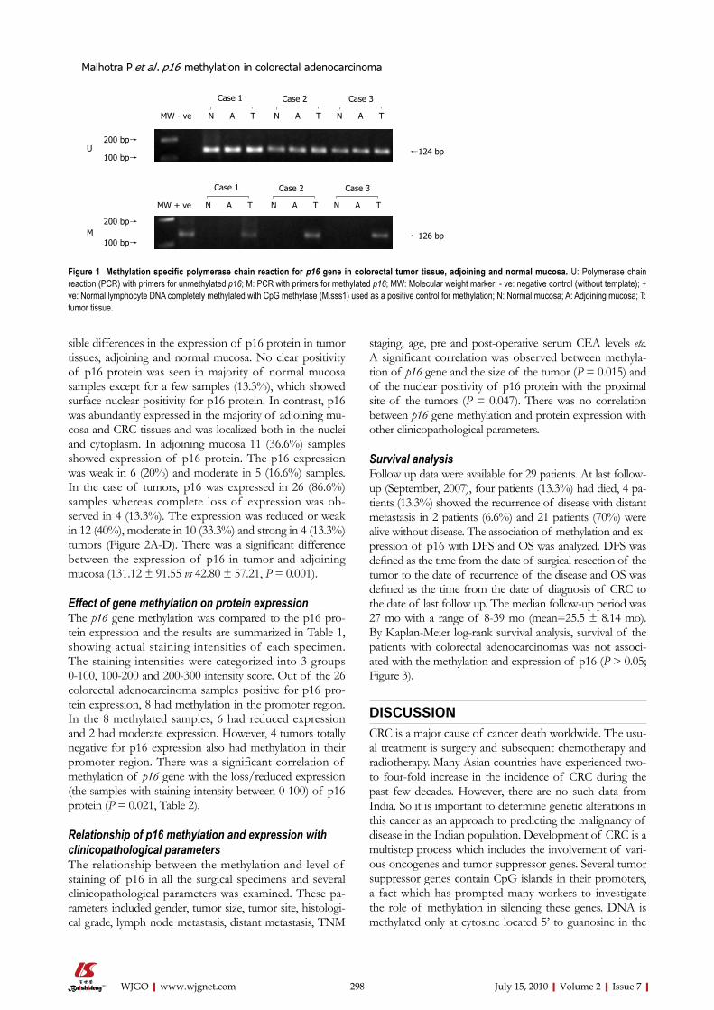
Methylation analysis of p16 promoter in surgical specimensMethylation status of the p16 gene promoter was evaluated in colorectal tumors, adjoining and normal mucosa using MSP. All the surgical specimens showed the presence of a band of unmethylated DNA that could be derived from unmethylated DNA of normal, adjoining mucosal cells and tumor cells as well as normal constituents in the stroma such as vascular endothelial cells, smooth muscles, fibrob-lasts and inflammatory cells. A band of methylated DNA was found in 12 (40%) tumors whereas no methylation was observed in adjoining and normal mucosa (Figure 1).
Expression of p16 protein in surgical specimensImmunohistochemistry was performed to evaluate pos-
297WJGO|www.wjgnet.com July 15, 2010|Volume 2|Issue 7|
Malhotra P et al . p16 methylation in colorectal adenocarcinoma
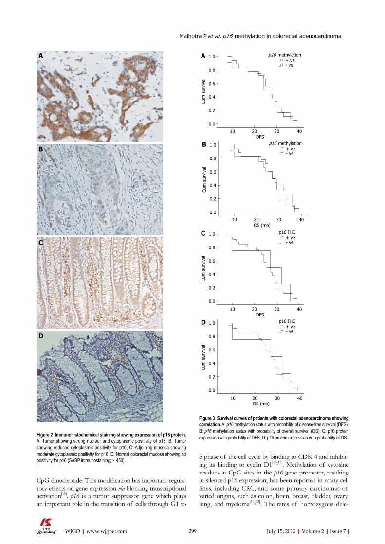
sible differences in the expression of p16 protein in tumor tissues, adjoining and normal mucosa. No clear positivity of p16 protein was seen in majority of normal mucosa samples except for a few samples (13.3%), which showed surface nuclear positivity for p16 protein. In contrast, p16 was abundantly expressed in the majority of adjoining mu-cosa and CRC tissues and was localized both in the nuclei and cytoplasm. In adjoining mucosa 11 (36.6%) samples showed expression of p16 protein. The p16 expression was weak in 6 (20%) and moderate in 5 (16.6%) samples. In the case of tumors, p16 was expressed in 26 (86.6%) samples whereas complete loss of expression was ob-served in 4 (13.3%). The expression was reduced or weak in 12 (40%), moderate in 10 (33.3%) and strong in 4 (13.3%) tumors (Figure 2A-D). There was a significant difference between the expression of p16 in tumor and adjoining mucosa (131.12 ± 91.55 vs 42.80 ± 57.21, P = 0.001).
Effect of gene methylation on protein expression The p16 gene methylation was compared to the p16 pro-tein expression and the results are summarized in Table 1, showing actual staining intensities of each specimen. The staining intensities were categorized into 3 groups 0-100, 100-200 and 200-300 intensity score. Out of the 26 colorectal adenocarcinoma samples positive for p16 pro-tein expression, 8 had methylation in the promoter region. In the 8 methylated samples, 6 had reduced expression and 2 had moderate expression. However, 4 tumors totally negative for p16 expression also had methylation in their promoter region. There was a significant correlation of methylation of p16 gene with the loss/reduced expression (the samples with staining intensity between 0-100) of p16 protein (P = 0.021, Table 2).
Relationship of p16 methylation and expression with clinicopathological parametersThe relationship between the methylation and level of staining of p16 in all the surgical specimens and several clinicopathological parameters was examined. These pa-rameters included gender, tumor size, tumor site, histologi-cal grade, lymph node metastasis, distant metastasis, TNM
staging, age, pre and post-operative serum CEA levels etc. A significant correlation was observed between methyla-tion of p16 gene and the size of the tumor (P = 0.015) and of the nuclear positivity of p16 protein with the proximal site of the tumors (P = 0.047). There was no correlation between p16 gene methylation and protein expression with other clinicopathological parameters.
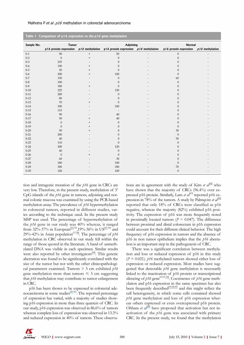
Survival analysisFollow up data were available for 29 patients. At last follow-up (September, 2007), four patients (13.3%) had died, 4 pa-tients (13.3%) showed the recurrence of disease with distant metastasis in 2 patients (6.6%) and 21 patients (70%) were alive without disease. The association of methylation and ex-pression of p16 with DFS and OS was analyzed. DFS was defined as the time from the date of surgical resection of the tumor to the date of recurrence of the disease and OS was defined as the time from the date of diagnosis of CRC to the date of last follow up. The median follow-up period was 27 mo with a range of 8-39 mo (mean=25.5 ± 8.14 mo). By Kaplan-Meier log-rank survival analysis, survival of the patients with colorectal adenocarcinomas was not associ-ated with the methylation and expression of p16 (P > 0.05; Figure 3).
DISCUSSIONCRC is a major cause of cancer death worldwide. The usu-al treatment is surgery and subsequent chemotherapy and radiotherapy. Many Asian countries have experienced two- to four-fold increase in the incidence of CRC during the past few decades. However, there are no such data from India. So it is important to determine genetic alterations in this cancer as an approach to predicting the malignancy of disease in the Indian population. Development of CRC is a multistep process which includes the involvement of vari-ous oncogenes and tumor suppressor genes. Several tumor suppressor genes contain CpG islands in their promoters, a fact which has prompted many workers to investigate the role of methylation in silencing these genes. DNA is methylated only at cytosine located 5’ to guanosine in the
298WJGO|www.wjgnet.com July 15, 2010|Volume 2|Issue 7|
Malhotra P et al . p16 methylation in colorectal adenocarcinoma
U200 bp→
100 bp→←124 bp
MW ve N A T N A T N A T
Case 1 Case 2 Case 3
200 bp→
100 bp→M ←126 bp
MW + ve N A T N A T N A T
Case 1 Case 2 Case 3
Figure 1 Methylation specific polymerase chain reaction for p16 gene in colorectal tumor tissue, adjoining and normal mucosa. U: Polymerase chain reaction (PCR) with primers for unmethylated p16; M: PCR with primers for methylated p16; MW: Molecular weight marker; - ve: negative control (without template); + ve: Normal lymphocyte DNA completely methylated with CpG methylase (M.sss1) used as a positive control for methylation; N: Normal mucosa; A: Adjoining mucosa; T: tumor tissue.
CpG dinucleotide. This modification has important regula-tory effects on gene expression via blocking transcriptional activation[15]. p16 is a tumor suppressor gene which plays an important role in the transition of cells through G1 to
S phase of the cell cycle by binding to CDK 4 and inhibit-ing its binding to cyclin D1[16-18]. Methylation of cytosine residues at CpG sites in the p16 gene promoter, resulting in silenced p16 expression, has been reported in many cell lines, including CRC, and some primary carcinomas of varied origins, such as colon, brain, breast, bladder, ovary, lung, and myeloma[19-32]. The rates of homozygous dele-
299WJGO|www.wjgnet.com July 15, 2010|Volume 2|Issue 7|
Malhotra P et al . p16 methylation in colorectal adenocarcinoma
Figure 2 Immunohistochemical staining showing expression of p16 protein. A: Tumor showing strong nuclear and cytoplasmic positivity of p16; B: Tumor showing reduced cytoplasmic positivity for p16; C: Adjoining mucosa showing moderate cytoplasmic positivity for p16; D: Normal colorectal mucosa showing no positivity for p16 (SABP immunostaining; × 450).
A
B
C
D
1.0
0.8
0.6
0.4
0.2
0.0
10 20 30 40DFS
1.0
0.8
0.6
0.4
0.2
0.0
Cum
sur
viva
l
C p16 IHC+ ve ve
10 20 30 40
1.0
0.8
0.6
0.4
0.2
0.0
Cum
sur
viva
l
OS (mo)
B p16 methylation+ ve ve
Cum
sur
viva
l
10 20 30 40DFS
p16 methylation+ ve ve
A
10 20 30 40OS (mo)
1.0
0.8
0.6
0.4
0.2
0.0
Cum
sur
viva
l
p16 IHC+ ve ve
D
Figure 3 Survival curves of patients with colorectal adenocarcinoma showing correlation. A: p16 methylation status with probability of disease-free survival (DFS); B: p16 methylation status with probability of overall survival (OS); C: p16 protein expression with probability of DFS; D: p16 protein expression with probability of OS.
tion and intragenic mutation of the p16 gene in CRCs are very low. Therefore, in the present study, methylation of 5’ CpG islands of the p16 gene in tumors, adjoining and nor-mal colonic mucosa was examined by using the PCR-based methylation assay. The prevalence of p16 hypermethylation in colorectal tumors, reported in different studies, var-ies according to the technique used. In the present study MSP was used. The percentage of hypermethylation of the p16 gene in our study was 40% whereas, it ranged from 32%-37% in European[23,33],19%-36% in US[34-36] and 29%-42% in Asian population[37,38]. The percentage of p16 methylation in CRC observed in our study fell within the range of those quoted in the literature. A band of unmeth-ylated DNA was visible in each specimen. Similar results were also reported by other investigators[34]. This genetic aberration was found to be significantly correlated with the size of the tumor but not with the other clinicopathologi-cal parameters examined. Tumors > 5 cm exhibited p16 gene methylation more than tumors ≤ 5 cm suggesting that p16 methylation may contribute to tumor enlargement in CRC.
p16 has been shown to be expressed in colorectal ade-nocarcinoma in some studies[39-41]. The reported percentage of expression has varied, with a majority of studies show-ing p16 expression in more than three quarters of CRC. In our study, p16 expression was observed in 86.6% of tumors whereas complete loss of expression was observed in 13.3% and reduced expression in 40% of tumors. These observa-
tions are in agreement with the study of Kim et al[42] who have shown that the majority of CRCs (96.4%) over ex-pressed p16 protein. Similarly, Lam et al[43] reported p16 ex-pression in 78% of the tumors. A study by Palmqvist et al[44] reported that only 18% of CRCs were classified as p16 negative, whereas the majority (82%) exhibited p16 posi-tivity. The expression of p16 was more frequently noted in proximally located tumors (P = 0.047). The difference between proximal and distal colorectum in p16 expression could account for their different clinical behavior. The high frequency of p16 expression in tumors and the absence of p16 in non tumor epithelium implies that the p16 aberra-tion is an important step in the pathogenesis of CRC.
There was a significant correlation between methyla-tion and loss or reduced expression of p16 in this study (P = 0.021). p16 methylated tumors showed either loss of expression or reduced expression. Most studies have sug-gested that detectable p16 gene methylation is necessarily linked to the inactivation of p16 protein or transcriptional silencing of p16 gene[10,45-48]. Coexistence of p16 gene meth-ylation and p16 expression in the same specimen has also been frequently described[39,48,49] and this might reflect the cell heterogeneity, in which some cells contained showed p16 gene methylation and loss of p16 expression wher-eas others expressed or even overexpressed p16 protein. Ohhara et al[50] have proposed that activation but not in-activation of the p16 gene was associated with primary CRC. In the present study, we found that the methylation
300WJGO|www.wjgnet.com July 15, 2010|Volume 2|Issue 7|
Malhotra P et al . p16 methylation in colorectal adenocarcinoma
Table 1 Comparison of p16 expression to the p16 gene methylation
Sample No. Tumor Adjoining Normalp16 protein expression p16 methylation p16 protein expression p16 methylation p16 protein expression p16 methylation
S-1 90 + 50 - 0 -S-2 0 + 0 - 0 -S-3 235 - 0 - 0 -S-4 100 + 0 - 0 -S-5 50 + 0 - 0 -S-6 200 + 100 - 0 -S-7 150 - 0 - 60 -S-8 100 - 0 - 0 -S-9 180 + 0 - 0 -S-10 225 - 120 - 0 -S-11 200 - 0 - 0 -S-12 80 - 0 - 0 -S-13 70 + 0 - 0 -S-14 300 - 100 - 0 -S-15 115 - - - 0 -S-16 90 - 40 - 0 -S-17 50 + 40 - 0 -S-18 0 + 0 - 0 -S-19 0 + 0 - 0 -S-20 50 + 0 - 30 -S-21 200 - 0 - 0 -S-22 40 - 0 - 50 -S-23 110 - 0 - 0 -S-24 200 - 120 - 0 -S-25 40 - 0 - 0 -S-26 0 + 0 - 0 -S-27 60 - 30 - 0 -S-28 180 - 100 - 0 -S-29 300 - 180 - 70 -S-30 120 - 100 - 0 -
301WJGO|www.wjgnet.com July 15, 2010|Volume 2|Issue 7|
Malhotra P et al . p16 methylation in colorectal adenocarcinoma
of the p16 gene results in loss or reduced expression of p16 protein, but the overall percentage of expression of p16 was high, whereas majority of the samples showed reduced p16 expression. The reduced expression of p16 in some tumors lacking methylation suggested that not only the methylation but some other genetic alterations are responsible. This other genetic alteration could possibil-ity be mutation in the coding region of p16 gene which could also reduce the expression of p16 protein in tumors lacking methylation. Preliminary observation on the same CRC specimens indicate that 20% of the samples of CRC showed mutation in exons 2 and 3 of the coding region of p16 gene, although more comprehensive analysis of the site and nature of mutation is required. The overexpres-sion of p16 protein in some of the tumors lacking methyl-ation indicated that the activation, but not the inactivation, of the gene was associated with the overexpression and tumor progression. Taken together these results suggest that p16 hypermethylation may, at least in part, contribute to reduced expression of p16. The elucidation of the re-lationship between p16 expression and p16 gene methyla-tion in primary tumors requires further studies on a large number of samples and this may certainly help us to better understand the role of methylation of tumor suppressor genes in carcinogenesis.
There are not many reports in the literature evaluating the prognostic role of p16 hypermethylation and expres-sion. Some investigators have shown that hypermethylation of the p16 gene was associated with advanced tumor stage and shorter survival[37,38]. In our patients no correlation could be established between the methylation and expres-sion of p16 and survival.
All reported studies have assumed the adenoma→carcinoma sequence and reported the presence of genetic alterations in adenomas, a precursor lesion that finally deve-lops to carcinoma. However, only a very small proportion of the Asian patient population has develop adenomas. The present study, therefore included adjoining mucosa in order to determine the initial changes in CRC. No methylation was observed in the adjoining mucosa, suggesting that there were no changes near the tumor region and that the process of tumorigenesis is restricted to a limited area. This is the first study in which we analyzed both the adjoining and normal mucosa to determine the initial changes in CRC.
This is the first study in the Indian population in which p16 gene has been analyzed at both genetic and expression
level in CRC in relation to clinicopathological features and prognosis. The frequency of alterations of this gene in this cohort is similar to others. This shows that despite the rare occurrence of synchronous adenomas in the population studied, the frequency of alterations in this gene is almost same as observed in other studies, suggesting that the pro-cess of tumorigenesis is similar overall although there is a difference at the initiation stage. This study is the first one in an Indian population. It was limited to by the size of the cohort and confined to North India. We need more data from the other parts of the country to validate our findings
In conclusion, our study demonstrated that majority of CRC tissues expressed p16 protein and that the low or reduced expression of p16 among CRC tissues, probably caused by hypermethylation, may contribute to tumor enlargement.
ACKNOWLEDGMENTSWe wish to thank Mrs. Alka the staff member of Depart-ment of Pathology for their help in immunohistochemistry, Mr. Mahinder Singh for the final layout of this paper and Mrs. Kusum for helping us with the statistical analysis.
COMMENTSBackgroundColorectal cancer (CRC) is a worldwide health problem. The incidence of CRC is very dynamic worldwide and during the past few decades there has been a dramatic increase in the incidence of CRC in Asian countries although the exact cause of this increase is not known. Therefore, this study was planned to inves-tigate the molecular genetic alterations responsible for the development of CRC in the Indian population. p16 is a cell cycle regulator and inactivation of this gene leads to uncontrolled cell proliferation and growth. The inactivation of this gene is mainly associated with aberrant promoter methylation. Therefore in the present study the authors analyzed the promoter methylation and expression of the p16 gene in colorectal adenocarcinoma in Indian patients.Research frontiersp16 is a major genetic marker in the development of colorectal carcinogenesis. It is well studied in various populations but there are no previous data from Indian population.Innovations and breakthroughsp16 gene hypermethylation may at least in part contribute to reduced/loss of ex-pression of p16 protein. This is the first study in an Indian population and for the first time p16 has been analyzed at both genetic and expression levels in CRC in relation to clinicopathological features and prognosis.Applicationsp16 plays a very important role in the development of colorectal carcinogenesis. Inactivation of the gene due to promoter methylation leads to lost or reduced
Table 2 Comparison of p16 protein expression with p16 gene methylation
p16 expression (IHC score) Total0-100 100-200 200-300
p16 Methylation - ve Count 6 8 4 18% within p16 expression 33.3% 44.4% 22.2% 100.0%Count 10 2 0 12
Total + ve1 % within p16 expression 83.3% 16.6% 0.0% 100.0%Count 16 10 4 30% within p16 expression 53.3% 33.3% 13.3% 100.0%
1P = 0.021, Pearson's χ2 test.
COMMENTS
302WJGO|www.wjgnet.com July 15, 2010|Volume 2|Issue 7|
Malhotra P et al . p16 methylation in colorectal adenocarcinoma
gene function. Hypermethylation is a reversible phenomenon. The identification of these genetic markers are implicated in colorectal carcinogenesis at its early stages can be very helpful for treating the patients with CRC.Peer reviewThis study examines p16 promoter methylation using methylation-specific PCR in resected tumors from a cohort of 30 colon cancer patients in North India. p16 meth-ylation in matched adjacent tumor and normal tissue is also examined. p16 expres-sion was determined by IHC score. This paper is appropriate and well written.
REFERENCES1 American Cancer Society. Colorectal Cancer Facts & Figures
2008-2010. Atlanta: American Cancer Society, 20082 Sung JJ, Lau JY, Goh KL, Leung WK. Increasing incidence of
colorectal cancer in Asia: implications for screening. Lancet Oncol 2005; 6: 871-876
3 Fearon ER, Vogelstein B. A genetic model for colorectal tumor-igenesis. Cell 1990; 61: 759-767
4 Zingg JM, Jones PA. Genetic and epigenetic aspects of DNA methylation on genome expression, evolution, mutation and carcinogenesis. Carcinogenesis 1997; 18: 869-882
5 Herman JG, Merlo A, Mao L, Lapidus RG, Issa JP, Davidson NE, Sidransky D, Baylin SB. Inactivation of the CDKN2/p16/MTS1 gene is frequently associated with aberrant DNA methylation in all common human cancers. Cancer Res 1995; 55: 4525-4530
6 Ahuja N, Mohan AL, Li Q, Stolker JM, Herman JG, Hamilton SR, Baylin SB, Issa JP. Association between CpG island methyl-ation and microsatellite instability in colorectal cancer. Cancer Res 1997; 57: 3370-3374
7 Kamb A, Gruis NA, Weaver-Feldhaus J, Liu Q, Harshman K, Tavtigian SV, Stockert E, Day RS 3rd, Johnson BE, Skolnick MH. A cell cycle regulator potentially involved in genesis of many tumor types. Science 1994; 264: 436-440
8 Serrano M, Hannon GJ, Beach D. A new regulatory motif in cell-cycle control causing specific inhibition of cyclin D/CDK4. Nature 1993; 366: 704-707
9 Sherr CJ, Roberts JM. CDK inhibitors: positive and negative regulators of G1-phase progression. Genes Dev 1999; 13: 1501- 1512
10 Gonzalez-Zulueta M, Bender CM, Yang AS, Nguyen T, Beart RW, Van Tornout JM, Jones PA. Methylation of the 5' CpG island of the p16/CDKN2 tumor suppressor gene in normal and transformed human tissues correlates with gene silencing. Cancer Res 1995; 55: 4531-4535
11 Lo KW, Cheung ST, Leung SF, van Hasselt A, Tsang YS, Mak KF, Chung YF, Woo JK, Lee JC, Huang DP. Hypermethylation of the p16 gene in nasopharyngeal carcinoma. Cancer Res 1996; 56: 2721-2725
12 Sambrook J, Russell DW. Preparation and analysis of euka-ryotic genomic DNA. In: Sambrook J, Russell DW, editors. Mol-ecular cloning: a laboratory manual. 3rd ed. New york: cold spring harbor laboratory press, 2001: 6.4-6.12
13 Herman JG, Graff JR, Myöhänen S, Nelkin BD, Baylin SB. Methylation-specific PCR: a novel PCR assay for methylation status of CpG islands. Proc Natl Acad Sci USA 1996; 93: 9821- 9826
14 Sobin LH, Wittekind CH. In: Sobin LH, Wittekind CH, editors. International union against cancer (UICC): TNM classification of malignant tumors. 5th ed. New york: Wiley liss, 1997
15 Singal R, Ginder GD. DNA methylation. Blood 1999; 93: 4059- 4070
16 Wieser RJ, Faust D, Dietrich C, Oesch F. p16INK4 mediates contact-inhibition of growth. Oncogene 1999; 18: 277-281
17 Merlo A, Herman JG, Mao L, Lee DJ, Gabrielson E, Burger PC, Baylin SB, Sidransky D. 5' CpG island methylation is associated with transcriptional silencing of the tumour suppressor p16/CDKN2/MTS1 in human cancers. Nat Med 1995; 1: 686-692
18 Yu WL, Huang ZH. P16 gene and digestive tract neoplasms.
Shijie Huaren Xiaohua Zazhi 1999; 7: 1061-1062 19 Esteller M, Sanchez-Cespedes M, Rosell R, Sidransky D,
Baylin SB, Herman JG. Detection of aberrant promoter hyperm-ethylation of tumor suppressor genes in serum DNA from non-small cell lung cancer patients. Cancer Res 1999; 59: 67-70
20 Nakamura M, Sugita K, Inukai T, Goi K, Iijima K, Tezuka T, Kojika S, Shiraishi K, Miyamoto N, Karakida N, Kagami K, O-Koyama T, Mori T, Nakazawa S. p16/MTS1/INK4A gene is frequently inactivated by hypermethylation in childhood acute lymphoblastic leukemia with 11q23 translocation. Leukemia 1999; 13: 884-890
21 Matsuda Y, Ichida T, Matsuzawa J, Sugimura K, Asakura H. p16(INK4) is inactivated by extensive CpG methylation in human hepatocellular carcinoma. Gastroenterology 1999; 116: 394-400
22 Zöchbauer-Müller S, Fong KM, Virmani AK, Geradts J, Gazdar AF, Minna JD. Aberrant promoter methylation of multiple genes in non-small cell lung cancers. Cancer Res 2001; 61: 249- 255
23 Esteller M, González S, Risques RA, Marcuello E, Mangues R, Germà JR, Herman JG, Capellà G, Peinado MA. K-ras and p16 aberrations confer poor prognosis in human colorectal cancer. J Clin Oncol 2001; 19: 299-304
24 Nakahara Y, Shintani S, Mihara M, Ueyama Y, Matsumura T. High frequency of homozygous deletion and methylation of p16(INK4A) gene in oral squamous cell carcinomas. Cancer Lett 2001; 163: 221-228
25 Nielsen NH, Roos G, Emdin SO, Landberg G. Methylation of the p16(Ink4a) tumor suppressor gene 5'-CpG island in breast cancer. Cancer Lett 2001; 163: 59-69
26 Tannapfel A, Benicke M, Katalinic A, Uhlmann D, Köckerling F, Hauss J, Wittekind C. Frequency of p16(INK4A) alterations and K-ras mutations in intrahepatic cholangiocarcinoma of the liver. Gut 2000; 47: 721-727
27 Trzeciak L, Hennig E, Kolodziejski J, Nowacki M, Ostrowski J. Mutations, methylation and expression of CDKN2a/p16 gene in colorectal cancer and normal colonic mucosa. Cancer Lett 2001; 163: 17-23
28 Muto S, Horie S, Takahashi S, Tomita K, Kitamura T. Genetic and epigenetic alterations in normal bladder epithelium in patients with metachronous bladder cancer. Cancer Res 2000; 60: 4021-4025
29 Sanchez-Cespedes M, Esteller M, Wu L, Nawroz-Danish H, Yoo GH, Koch WM, Jen J, Herman JG, Sidransky D. Gene pro-moter hypermethylation in tumors and serum of head and neck cancer patients. Cancer Res 2000; 60: 892-895
30 Guo SX, Taki T, Ohnishi H, Piao HY, Tabuchi K, Bessho F, Hanada R, Yanagisawa M, Hayashi Y. Hypermethylation of p16 and p15 genes and RB protein expression in acute leuke-mia. Leuk Res 2000; 24: 39-46
31 Zhu ZY, Tian X, Wang X, Yang YL. P16 gene and APC gene mutation in gastrinomas. Shijie Huaren Xiaohua Zazhi, 2000; 8: 1418-1419
32 Yakoob J, Fan XG, Hu GL, Zhang Z. DNA methylation and carcinogenesis in digestive neoplasms. World J Gastroenterol 1998; 4: 174-177
33 Burri N, Shaw P, Bouzourene H, Sordat I, Sordat B, Gillet M, Schorderet D, Bosman FT, Chaubert P. Methylation silencing and mutations of the p14ARF and p16INK4a genes in colon cancer. Lab Invest 2001; 81: 217-229
34 Wiencke JK, Zheng S, Lafuente A, Lafuente MJ, Grudzen C, Wrensch MR, Miike R, Ballesta A, Trias M. Aberrant methyl-ation of p16INK4a in anatomic and gender-specific subtypes of sporadic colorectal cancer. Cancer Epidemiol Biomarkers Prev 1999; 8: 501-506
35 Ashktorab H, Smoot DT, Carethers JM, Rahmanian M, Kittles R, Vosganian G, Doura M, Nidhiry E, Naab T, Momen B, Shakhani S, Giardiello FM. High incidence of microsatellite instability in colorectal cancer from African Americans. Clin Cancer Res 2003; 9: 1112-1117
303WJGO|www.wjgnet.com July 15, 2010|Volume 2|Issue 7|
Malhotra P et al . p16 methylation in colorectal adenocarcinoma
36 Van Rijnsoever M, Elsaleh H, Joseph D, McCaul K, Iacopetta B. CpG island methylator phenotype is an independent predictor of survival benefit from 5-fluorouracil in stage III colorectal cancer. Clin Cancer Res 2003; 9: 2898-2903
37 Liang JT, Chang KJ, Chen JC, Lee CC, Cheng YM, Hsu HC, Wu MS, Wang SM, Lin JT, Cheng AL. Hypermethylation of the p16 gene in sporadic T3N0M0 stage colorectal cancers: association with DNA replication error and shorter survival. Oncology 1999; 57: 149-156
38 Yi J, Wang ZW, Cang H, Chen YY, Zhao R, Yu BM, Tang XM. p16 gene methylation in colorectal cancers associated with Duke's staging. World J Gastroenterol 2001; 7: 722-725
39 Tada T, Watanabe T, Kazama S, Kanazawa T, Hata K, Komuro Y, Nagawa H. Reduced p16 expression correlates with lymp-hatic invasion in colorectal cancers. Hepatogastroenterology 2003; 50: 1756-1760
40 Zhao P, Hu YC, Talbot IC. Expressing patterns of p16 and CDK4 correlated to prognosis in colorectal carcinoma. World J Gastroenterol 2003; 9: 2202-2206
41 Cui X, Shirai Y, Wakai T, Yokoyama N, Hirano S, Hatakeyama K. Aberrant expression of pRb and p16(INK4), alone or in combination, indicates poor outcome after resection in patients with colorectal carcinoma. Hum Pathol 2004; 35: 1189-1195
42 Kim BN, Yamamoto H, Ikeda K, Damdinsuren B, Sugita Y, Ngan CY, Fujie Y, Ogawa M, Hata T, Ikeda M, Ohue M, Sekimoto M, Monden T, Matsuura N, Monden M. Methylation and expression of p16INK4 tumor suppressor gene in primary colorectal cancer tissues. Int J Oncol 2005; 26: 1217-1226
43 Lam AKY, Ong K, Ho YH. Colorectal mucinous adenocar-cinoma: the clinicopathologic features and significance of p16 and p53 expression. Dis Colon Rectum 2006; 49: 1275-1283
44 Palmqvist R, Rutegârd JN, Bozoky B, Landberg G, Stenling R. Human colorectal cancers with an intact p16/cyclin D1/pRb pathway have up-regulated p16 expression and decreased proliferation in small invasive tumor clusters. Am J Pathol 2000; 157: 1947-1953
45 Baylin SB, Herman JG, Graff JR, Vertino PM, Issa JP. Alter-ations in DNA methylation: a fundamental aspect of neoplasia. Adv Cancer Res 1998; 72: 141-196
46 Schmutte C, Jones PA. Involvement of DNA methylation in human carcinogenesis. Biol Chem 1998; 379: 377-388
47 El-Naggar AK, Lai S, Clayman G, Lee JK, Luna MA, Goepfert H, Batsakis JG. Methylation, a major mechanism of p16/CDKN2 gene inactivation in head and neck squamous carcinoma. Am J Pathol 1997; 151: 1767-1774
48 Shim YH, Kang GH, Ro JY. Correlation of p16 hypermet-hylation with p16 protein loss in sporadic gastric carcinomas. Lab Invest 2000; 80: 689-695
49 Simpson DJ, Bicknell JE, McNicol AM, Clayton RN, Farrell WE. Hypermethylation of the p16/CDKN2A/MTSI gene and loss of protein expression is associated with nonfunctional pituitary adenomas but not somatotrophinomas. Genes Chromosomes Cancer 1999; 24: 328-336
50 Ohhara M, Esumi M, Kurosu Y. Activation but not inactivation of the MTS1 gene is associated with primary colorectal carcin-omas. Biochem Biophys Res Commun 1996; 226: 791-795
S- Editor Wang JL L- Editor Hughes D E- Editor Yang C


