Embed Size (px)
Citation preview
Journal ofNeurology, Neurosurgery, andPsychiatry, 1976, 39, 424-428
Abnormal dynamic scintigraphy in hydrocephalus:a proposed mechanism
M. M. RASKIN, A. N. SERAFINI, J. J. SHELDON, AND W. M. SMOAK'
From the Department ofRadiology and the Division ofNuclear Medicine,Mount Sinai Medical Center, Miami Beach, and the Department ofRadiology, University ofMiami
School ofMedicine, Miami, Florida, USA
SYNOPSIS Dynamic brain scintigraphy was performed on seven patients with documented hydro-cephalus of various aetiologies. The patients had a characteristic abnormality consisting of lateraldisplacement of the proximal middle cerebral activity associated with a paracentral lucent zone. Nofocal areas of increased activity were identified on the static brain images. A proposed mechanismfor these findings is discussed.
Hydrocephalus may cause displacement ofvascular structures, although these changesrarely have been appreciated by anterior dynamicbrain scintigraphy (Rosler and Huber, 1969;Meschan et al., 1971; Kinser and Rosler, 1974).Dynamic brain scintigraphy does not allowdelineation of individual intracranial arteries,although the branches of the middle cerebralartery can be seen as a distinct area of activitywithin the Sylvian fissure. These Sylvian vesselsare related to the insula and the ventricles.Normally, the ventricular space is not seen.However, as the ventricles enlarge, there islateral displacement of the middle cerebralgroup of vessels and the enlarging ventriclebecomes visualized as a paracentral void.We recognized abnormal scintigraphic findings,
suspicious for hydrocephalus, in seven patientsin whom the presence of hydrocephalus wassubsequently confirmed by pneumoencephalo-graphy or at necropsy. Three representative casesare reported in this paper and a mechanismto explain the scintigraphic abnormalities isproposed.
I Address for reprint requests: Dr W. M. Smoak, Division of NuclearMedicine, Mount Sinai Medical Center, 4300 Alton Road, MiamiBeach, Florida 33140, USA.(Accepted 22 December 1975.)
METHODS
Dynamic brain scintigraphy was performed in theanterior projection using an Anger scintillationcamera, immediately after an intravenous injectionof a 15 mCi bolus 99mTc-pertechnetate. Serialdynamic scintigrams were obtained on 35 mm film atintervals of two seconds. One to two hours afterinjection, anterior, posterior, vertex, and both lateralstatic images were obtained, accumulating 400 000counts.
CASE 1
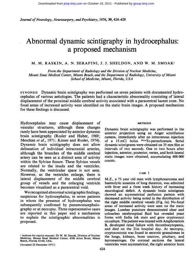
M.Z., a 73 year old man with lymphosarcoma andhaemolytic anaemia of long duration, was admittedwith fever and a three week history of increasingneurological deficit. A dynamic brain scintigramshowed an asymmetrical perfusion pattern withdecreased activity being noted in the distribution ofthe right middle cerebral vessels (Fig. la). No focalareas of increased activity were seen on the staticimages. Lumbar puncture demonstrated clear andcolourless cerebrospinal fluid but revealed yeastforms with India ink stain and grew cryptococcion culture. Thepatient was treated with AmphotericinB, developed renal failure with thrombocytopenia,and died on the 21st hospital day. At necropsy,cryptococcosis was found in necrotic granulomas inthe lungs, kidneys, bone marrow, pituitary, andleptomeninges. On coronal sections the lateralventricles were asymmetrical, the right anterior horn
424
group.bmj.com on October 19, 2011 - Published by jnnp.bmj.comDownloaded from
Abnormal dynamic scintigraphy in hydrocephalus: a proposed mechanism
FIG. la Case 1.Dynamic brainscintigram showsasymmetrical lateraldisplacement of theright middle cerebralartery in the firstframe (arrow). Aparacentral lucentzone is seen on rightin the secondframe(arrow).Asymmetrical venousclearance is noted inthe fifth frame(arrow).
FIG. lb Coronal section through thefrontal lobes shows asymmetrical dila-tation of the right anterior horn of thelateral ventricle (arrow).
425
i4 i a I 4 t !. 0 I- j... .... . .-. .
group.bmj.com on October 19, 2011 - Published by jnnp.bmj.comDownloaded from
M. M. Raskin, A. N. Serafini, J. J. Sheldon, and W. M. Smoak
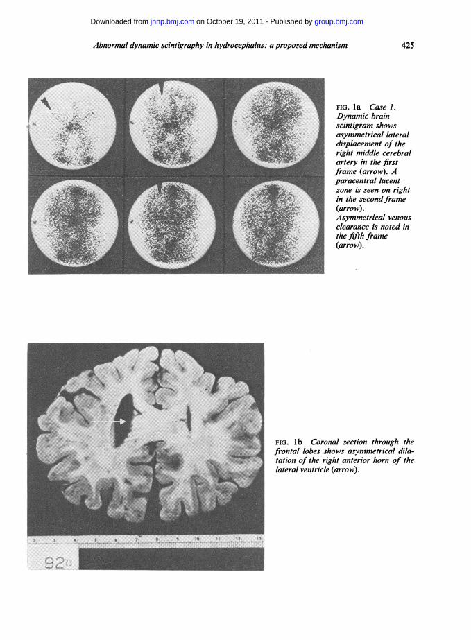
FIG. 2a Case 2. Dy-namic brain scinti-gram shows bilateralparacentral lucentareas (arrows) withsymmetrical venousclearance.
and body of the lateral ventricles being larger thantheir counterparts on the left (Fig. Ib). These findingswere consistent with unilateral obstructive hydro-cephalus.
CASE 2
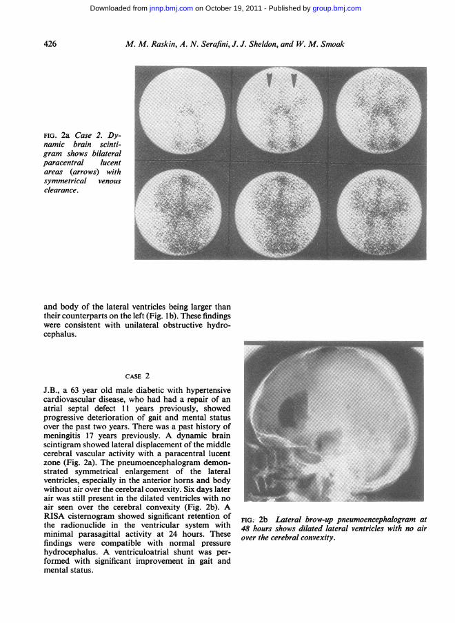
J.B., a 63 year old male diabetic with hypertensivecardiovascular disease, who had had a repair of anatrial septal defect 11 years previously, showedprogressive deterioration of gait and mental statusover the past two years. There was a past history ofmeningitis 17 years previously. A dynamic brainscintigram showed lateral displacement of the middlecerebral vascular activity with a paracentral lucentzone (Fig. 2a). The pneumoencephalogram demon-strated symmetrical enlargement of the lateralventricles, especially in the anterior horns and bodywithout air over the cerebral convexity. Six days laterair was still present in the dilated ventricles with noair seen over the cerebral convexity (Fig. 2b). ARISA cisternogram showed significant retention ofthe radionuclide in the ventricular system withminimal parasagittal activity at 24 hours. Thesefindings were compatible with normal pressurehydrocephalus. A ventriculoatrial shunt was per-formed with significant improvement in gait andmental status.
FIG. 2b Lateral brow-up pneumoencephalogram at48 hours shows dilated lateral ventricles with no airover the cerebral convexity.
A.276
group.bmj.com on October 19, 2011 - Published by jnnp.bmj.comDownloaded from
Abnormal dynamic scintigraphy in hydrocephalus: a proposed mechanism
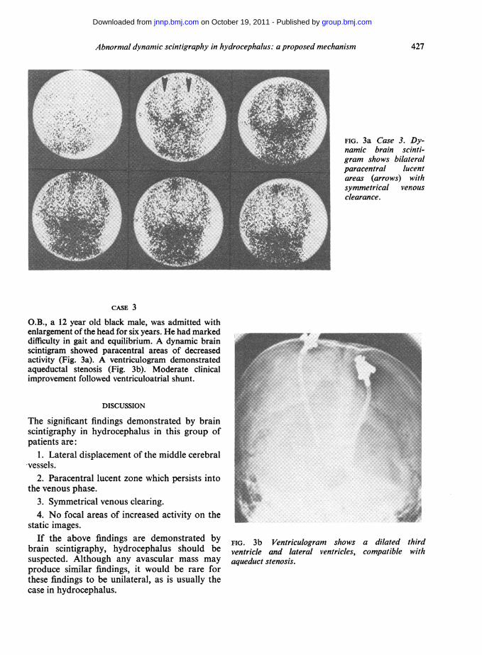
FIG. 3a Case 3. Dy-namic brain scinti-gram shows bilateralparacentral lucentareas (arrows) withsymmetrical venousclearance.
CASE 3
O.B., a 12 year old black male, was admitted withenlargement of the head for six years. He had markeddifficulty in gait and equilibrium. A dynamic brainscintigram showed paracentral areas of decreasedactivity (Fig. 3a). A ventriculogram demonstratedaqueductal stenosis (Fig. 3b). Moderate clinicalimprovement followed ventriculoatrial shunt.
DISCUSSION
The significant findings demonstrated by brainscintigraphy in hydrocephalus in this group ofpatients are:
1. Lateral displacement of the middle cerebral-vessels.
2. Paracentral lucent zone which persists intothe venous phase.
3. Symmetrical venous clearing.4. No focal areas of increased activity on the
static images.If the above findings are demonstrated by
brain scintigraphy, hydrocephalus should besuspected. Although any avascular mass mayproduce similar findings, it would be rare forthese findings to be unilateral, as is usually thecase in hydrocephalus.
-.
...... ......
*..
*. ..
...R::...
FIG. 3b Ventriculogram shows a dilated thirdventricle and lateral ventricles, compatible withaqueduct stenosis.
427
group.bmj.com on October 19, 2011 - Published by jnnp.bmj.comDownloaded from
M. M. Raskin, A. N. Serafini, J. J. Sheldon, and W. M. Smoak
ANATOMICAL CONSIDERATIONS The middle cere-bral artery is the main branch of the internalcarotid artery and supplies the largest area withthe most complex branching of the intracerebralvessels. The proximal portion of the middlecerebral artery is horizontal in its course and itextends laterally from the bifurcation of theinternal cerebral artery, and is situated betweenthe temporal lobe and the lower aspect of theinsula. Its branches extend upward around thelower aspect of the insula and continue upwardand backward in the deepest portion of theSylvian fissure, between the outer surface of theinsula and the medial surface of the temporallobe (Taveras and Wood, 1964).The outer surface of the insula is surrounded
by the opercular portions of the frontal, parietal,and temporal lobes. As the five to eight branchesof the middle cerebral artery reach the uppermostportion of the outer surface of the insula, theyreverse their course and are directed inferolateralto the lower margin of the frontoparietal oper-culum where they emerge from the Sylvianfissure (Ring, 1962; De Long, 1973). Immediatelyafter emerging, the majority of the branchescontinue superiorly in an anterior or posteriordirection on the outer surface of the cerebralhemisphere, usually within the cerebral sulci(Taveras and Wood, 1964).
Increased or decreased CSF pressure mayaccompany small, dilated, or normal sizedventricles. The force produced by the CSFsystem is the product of the mean CSF and themean ventricular surface area (Hakim, 1972). Asthe lateral ventricles enlarge, pressure is trans-mitted with decreasing force as the distancefrom the ventricles increases. This results in
greater compression of the vessels closest to theventricles. Widening of the sweep of the thala-mostriate veins is the most sensitive angiographicindicator of ventricular dilatation. Less sensitiveis widening of the sweep of the pericallosalbranch of the anterior cerebral artery (Petersonand Kieffer, 1972). Since branches of the middlecerebral artery lie between the temporal-parietaloperculum and the insula, they will be displacedlaterally if there is sufficient compression of theinsula by the dilated lateral ventricle. Therefore,the paracentral lucent zone is produced by bothcompression and displacement of branches of themiddle cerebral arteries.
REFERENCES
DeLong, W. B. (1973). Anatomy of the middle cerebralartery: the temporal branches. Stroke, 4, 412-418.
Hakim, S. (1972). Biomechanics of hydrocephalus. InCisternography and Hydrocephalus, pp 25-55. Editedby J. C. Harbert, Thomas: Springfield, Ill.
Kinser, J., and Rosler, H. (1974). Brain scintigraphy.Medical Progress in Technology, 2, 63-70.
Meschan, I., Lytle, W. P., Maynard, C. D., Cowan, R.J.,and Janeway, R. (1971). Statistical relationship ofbrain scans, cervicocranial dynamic studies, and cere-bral arteriograms. Radiology, 100, 623-629.
Peterson, H. O., and Kieffer, S. A. (1972). Introduction toNeuroradiology, p. 81. Harper and Row: Hagerstown,Md.
Ring, B. A. (1962). Middle cerebral artery: anatomicaland radiographic study. Acta Radiologica, 57, 289-300.
Rosler, V. H., and Huber, P. (1969). Die cerebrale serien-szintigraphie. Fortschritte aufdem Gebiete der Rdntgen-strahlen, 111, 467-480.
Taveras, J. M., and Wood, E. H. (1964). DiagnosticNeuroradiology, p. 1.507. Williams and Wilkins:Baltimore.
428
group.bmj.com on October 19, 2011 - Published by jnnp.bmj.comDownloaded from
doi: 10.1136/jnnp.39.5.424 1976 39: 424-428J Neurol Neurosurg Psychiatry
M M Raskin, A N Serafini, J J Sheldon, et al. mechanism.hydrocephalus: a proposedscintigraphy in Abnormal dynamic
http://jnnp.bmj.com/content/39/5/424at: Updated information and services can be found
These include:
serviceEmail alerting
corner of the online article.this article. Sign up in the box at the top right Receive free email alerts when new articles cite
Notes
http://group.bmj.com/group/rights-licensing/permissionsTo request permissions go to:
http://journals.bmj.com/cgi/reprintformTo order reprints go to:
http://group.bmj.com/subscribe/To subscribe to BMJ go to:
group.bmj.com on October 19, 2011 - Published by jnnp.bmj.comDownloaded from


