Embed Size (px)
Citation preview
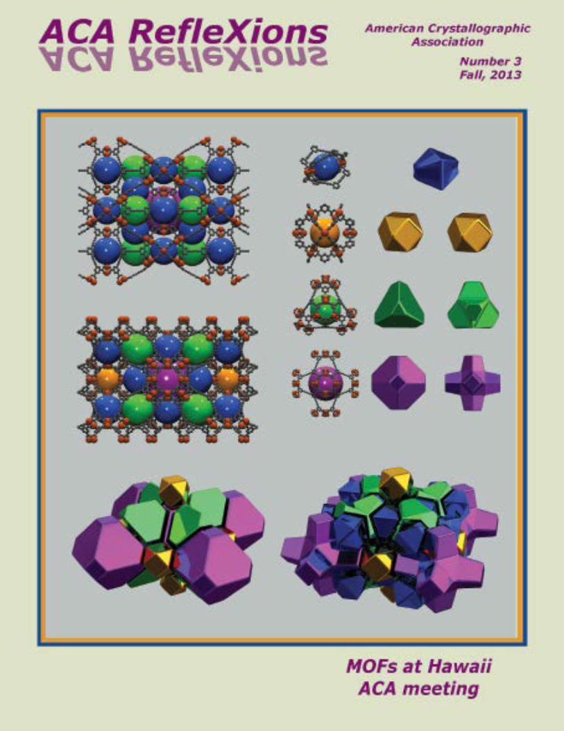

Table of Contents 3 President’s Column, On the Cover, Sad News about Dave Rognlie 4 Letter to the Editor, Council Highlights, ACARefleXionsCo-Editors&Staff,Errata6 News&Awards9 IntroducingnewstaffmemberChiaraPastore, 2013ClassofACAFellows 10 Contributors to this Issue13 NetRefleXions,IndexofAdvertisers15 JeromeKarle(1918-2013)18 RayDavis(1938-2013)20 CharlesNorrisCaughlan(1915-2013), Student Help in Hawaii 21 Poster Prizes in Hawaii 24 Accompanying Members 25 Annual ACA Meeting in Hawaii 64 High School Outreach in Hawaii 65 Books 66 Workshop on Dynamic Structural Photocrystallography 67 Puzzle Corner 68 46th Course at Erice 71 Bruker/MIT Symposium
72 2014 ACA Meeting in Albuquerque 75 Corporate Members77 CalendarofMeetings
Contributions to ACA RefleXions may be sent to either of the Editors:
Connie (Chidester) Rajnak Judith L. Flippen-Anderson 4210 Old Field Trail 3521 Launcelot Way Kalamazoo, MI 49008 Annandale, VA 22003 269-342-1600 703-346-2441 [email protected]@gmail.com
Please address matters pertaining to advertisements, membership inquiries, or use of the ACA mailing list to:
Marcia J. Colquhoun, Director of Administrative Services American Crystallographic Association P.O. Box 96, Ellicott Station Buffalo, NY 14205 tel: 716-898-8692; fax: 716-898-8695 [email protected]
Deadlinesforcontributionsare:February1(Spring),May1(Summer),August1(Fall)andNovember1(Winter)
American Crystallographic Association
ACA RefleXions
ACA RefleXions (ISSN 1058-9945) Number 3, 2013. Published four times per year in the spring, summer, fall and winter for the membership of the American Crystallographic Association, P.O. Box 96, Ellicott Station, Buffalo, NY 14205-0096. Membership in the ACA includes a non-deductible charge of $1.75 from membership dues to be applied to a subscription to ACA RefleXions. Periodicals postage paid at Buffalo, New York. POSTMAS-TER: Send address changes to ACA, P.O.Box 96, Ellicott Station, Buffalo, NY, 14205-0096.
Fall, 2013ACA HOME PAGE: www.AmerCrystalAssn.org
Cover: The cover image, from Omar Farha and Chris Wilmer, Northwestern
University, depicts the metal-organic framework (MOF), of NU-111.
See On the Cover, p 3.
The D8 QUEST ECO With the D8 QUEST ECO, making the right choice is easy. Not only is it affordable and economical with minimal maintenance, it is also environmentally friendly offering low power requirements andno external water cooling. Yet, the D8 QUEST ECO still delivers the high performance and quality data you have to come to expect from Bruker.
The New D8 QUEST ECOCrystallography with a Conscience
Contact us for a personal system demonstration www.bruker.com/d8questeco
Innovation with Integrity Crystal lography
C
M
Y
CM
MY
CY
CMY
K
SC-XRD_ACA_PrintFP_D8 Quest ECO_Sep 2013 Print.pdf 1 9/3/13 1:22 PM
3
Fall 2013
This research has been published in JACS: ‘Designing Higher Surface Area Metal-Organic Frameworks: Are Triple Bonds Better Than Phenyls?' by O.K. Farha, C.E. Wilmer, I. Ery-azici, B.G. Hauser, P.A. Parilla, K. O’Neill, A.A. Sarjeant, S.T. Nguyen, R.Q. Snurr & J.T. Hupp, J. Am. Chem. Soc. 2012, 134, 9860-9863. Amy Sarjeant gave a talk at the ACA meeting in Hawaii on NU-110 (work built on the knowledge from NU-111 project), which is how it came to my attention (Connie Rajnak). This work was also published in JACS: 'Metal-organic Framework Materials with Ultrahigh Surface Areas: Is the Sky the Limit?,' by O.K. Farha, I. Eryazici, N.C. Jeong, B.G. Hauser, C.E. Wilmer, A.A. Sarjeant, R.Q. Snurr, S.T. Nguyen, A.Ö. Yazaydın & J.T. Hupp, J. Am. Chem. Soc. 2012, 134, 15016-15021. Omar Farha and Chris Wilmer, Northwestern University
President's Column & On the Cover
The annual ACA meeting in Honolulu was both a scientific and social success with 767 attendees. Crystallography informs many diverse areas of science; more and more our meetings have become venues for educating members in the theory and applications of crystallography in disciplines other than their own. The four day meeting format made it challenging to fit in the three workshops, the 20 oral sessions (263 talks) and 3 evening poster sessions (291 presentations). However, despite the beautiful surroundings and the near perfect weather, all were well attended. A record 78 students were given travel grants to support their attendance. The first Bau Neutron Diffraction Award was presented to Tom Koetzle, the Fankuchen Award went to Richard Dickerson (as Dick was not able to attend, Alex McPherson presented a retrospective on Dickerson’s work). The Trueblood Award went to Tom Terwilliger, and Eric Ortlund won the Etter Early Career Award. On the journal front, we are very pleased to announce that our new journal, Structural Dynamics, co-published by the ACA and AIP Publishing started accepting papers at the beginning of Septem-ber. The editor of the journal is Majed Chergui from Lausanne, Switzerland. Two associate editors, Thomas Elsaesser and Franz Pfeiffer, are based in Germany and three associate editors, George Phillips, Jr., Gwyn P. Williams and Linda Young, are based in the U.S. Appropriate journal topics include: structural dynamics of molecular systems, biological systems, solid materials, liquids and solutions, and surfaces and interfaces; static structural determina-tion, static imaging techniques and studies using highly coherent sources; dynamical studies of systems both in and out of equilibrium, with a time resolution from femtoseconds to milliseconds; spatial resolutions from 1 Å to 1 mm; and electronic structure studies connected to molecular/lattice/protein structure. Further details are available on the journal website (sd.aip.org).
The strategic planning commit-tee met in Hawaii and estab-lished action items to help us move forward. Each committee member has an assignment and we plan to touch base in Sep-tember. Action items are related to organizational structure, by-laws, marketing, education, and expanding involvement of members. We are committed to continuing the process until we have developed a plan for the future of ACA. As we consider where we are going, I think it is important to remember where we have been and to remember the colleagues and friends we have lost since our summer issue went to press; Jerome Karle (past ACA and IUCr president), Ray Davis (past ACA president), Charles Caughlan (local chair for the Boze-man ACA meeting back in 1964) and Dave Rognlie (known to crystallographers as the smiling face of Blake Industries). Each of these individuals contributed to our community and each will be missed. Please remember that we are a volunteer organization and you get out as much as you put in. So do something note-worthy. Volunteer for a committee. Nominate your colleagues for awards. Run for a position on the Council. Organize a scientific session for the annual meeting. Serve as a poster judge. Organize a focus group. Get involved. You will be glad that you did. Thanks!
Cheryl Stevens
The cover image depicts the metal-organic framework (MOF), of NU-111. MOFs are a new class of multifunctional, crystalline, porous materials that have received tremen-dous interest due to their potential applications in gas and chemical storage, separa-tions, sensing, catalysis, ion exchange, light harvesting and drug delivery. MOFs are hybrid materials comprised of multitopic organic ligands (struts) linking metal-based
nodes. NU-111 exhibits many remarkable features including a high surface area (5,000 m2/g) and a high uptake of both hydrogen and methane. The images on the cover show the representa-tion of NU-111 using three polyhedral cages (cuboctahedral, truncated tetrahedron, and truncated cuboctahedron) that are derived from sketching straight lines between copper paddlewheel nodes and are glued in such a way that they form an uninterrupted pathway. Taking the curvature of the struts into account gives four types of polyhedral cages, which is another way of representing NU-111 and one that better matches experimental pore size distributions.
It is with great sadness that we announce the passing on August 5th ofDaveRognlie.Davewaswell-knownasthePresidentandownerofBlakeIndustriesandasalongtermsupporteroftheICDD.Thisissueisabouttogotopress,butweplantopublishafullappreciationofhislifeinthewinterRefleXions. Anyone who wishes to contribute 'I knew Dave when' or 'there was that time that Dave . .' memories should contactthewinterissueeditor,[email protected].
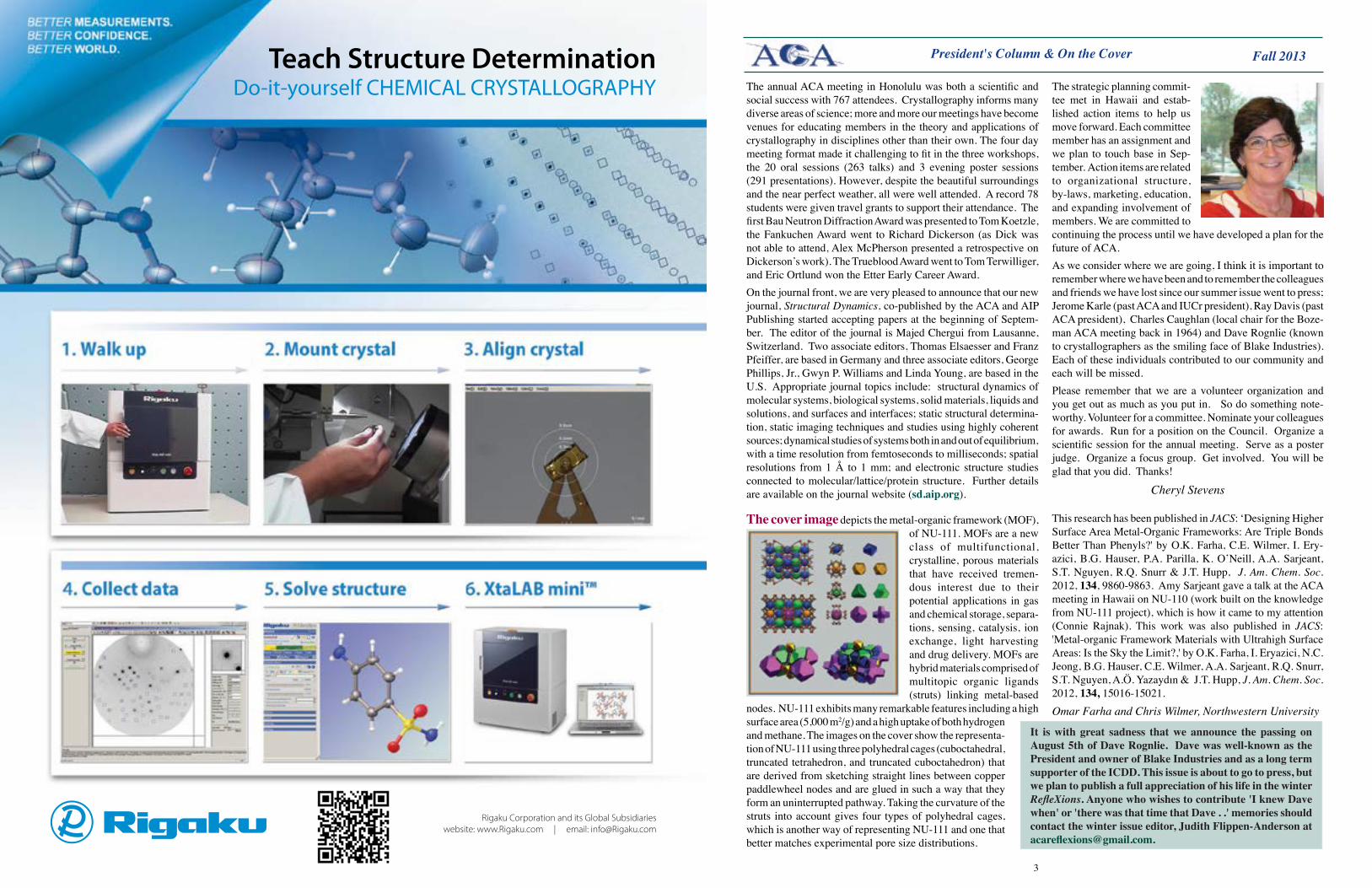
Rigaku Corporation and its Global Subsidiarieswebsite: www.Rigaku.com | email: [email protected]
Teach Structure DeterminationDo-it-yourself CHEMICAL CRYSTALLOGRAPHY
4
Fall 2013
PhotographerPeter Müller [email protected]
ACA RefleXionsSTAFF
Books Editor
JudithFlippen-Anderson [email protected]
FrankFronczek [email protected]
EdgarF.Meyer [email protected]
[email protected] http://molecular-sculpture.com
Connie Rajnak [email protected]
ACA RefleXionsCO-EDITORS
Puzzle Corner Editor
JoeFerrara [email protected]
Copy EditorJaneF.Griffin [email protected]
HistorianVirginia Pett [email protected]
Letter to the Editor, Errata, Council News
Dear Connie, Judy:While I was reading the Summer edition of RefleXions, I noticed a few minor typographical errors. The first is in the announcement of the 2014 Patterson Award to John Helliwell. John's award is indeed well deserved, and while I would love to say that I work at the same university where John studied as an undergraduate, that is not the case. John went to the University of York (in England), not York University (in Toronto, Canada). This was mixed up on line 7 of the first paragraph. The second error was again a “York” error, and is found within the announcement of the ACA Travel Awardees for the 2013 meeting in Honolulu. Agnesa Shala, one of my gradu-ate students, is of course at York University (in Canada), but was put under the UK.Now while these are not big issues, I suspect John may want his undergraduate university corrected.Many Thanks! Gerald Audette
Errata: the editors regret the omis-sion of LeemorJoshua-Tor, an HHMI Investigator, and Professor at Wm M. Keck Structural Biology Laboratory, Cold Spring Harbor, from the AAAS fellows listed in the spring issue News & Awards section, page 17. Leemor is an ACA member, and as such was supposed to be listed. Leemor and her team do structural and biochemical studies of key proteins in the gene silenc-ing process called RNA interference or RNAi. They are trying to get a true mechanistic understanding of the RNAi machinery; how the components fit together and how they function. Among other activities the RNAi pathway mediates the function of the endogenous, non-coding regulatory RNAs called miRNAs. We are so sorry, Leemor!
ACA Council Meeting Highlights, July 2013President Cheryl Stevens announced that the program chairs for the 2014 annual meeting in Albuquerque, NM will be Petrus Zwart and Christine Beavers. In an update on the new ACA journal, Structure and Dynamics, Judy Flippen-Anderson reported that an editorial board has been formed, and a temporary website set up. Policies are being set to encourage submissions from ACA members, notably a discounted publication fee for members.Considerable discussion was devoted to the International Year of Crystallography 2014 (IYCr14), and specifically to how the ACA could best support this initiative. Vice President Martha Teeter has assembled an InternationalYearofCrystallography2014TaskForce, which is considering a wide range of relevant topics, includ-ing schools outreach, media, web outreach, funding, and liasons with politicians and with other scientific organizations. Council decided that the ACA should commit some funds to activities related to IYCr14 in North America, and agreed on a sum of $20K. This is approximately equal to the organization’s dividend income for a year, which means that this amount can be committed without dip-ping into reserves. Funds will be allocated via a call for proposals, which will be coordinated by the IYCr14 Task Force. The 2013 class of ACA fellows was approved, and names were announced at the banquet during the Hawaii meeting. The 6 new fel-lows are: Sidney Abrahams, Wim Hol, Jim Ibers, AlexMcPherson, KeithMoffat, and AlexWlodawer.Past President George Phillips reported that many inconsistent and outdated items have accumulated in the ACA’s bylaws, owing to years of neglect. He has embarked on an effort to streamline and update this important document, but warned that this will be an ongoing and iterative process. Given the strategic planning process that was initiated earlier this year, this is an opportune time to address this problem.Council also decided to try a new policy of using videoconferencing for their spring and fall meetings, in order to save money. The first test of this approach will be at the Council meeting this fall.
Patrick Loll, ACA secretary
Art in Crystallography Editor
Net RefleXions Editor
Amy Sarjeant asarjeant@ northwestern.edu
News&Awards Editor
Chiara Pastore chiara.pastore @googlemail.com
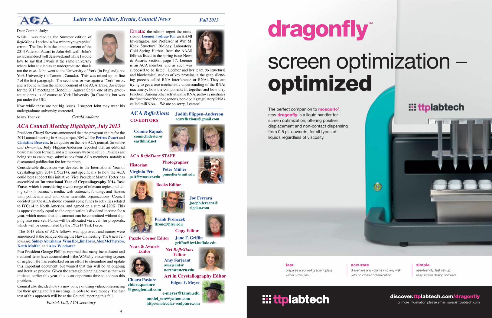
fastprepares a 96-well gradient plate
within 5 minutes
accuratedispenses any volume into any well
with no cross-contamination
simpleuser-friendly, fast set-up,
easy screen design software
discover.ttplabtech.com/dragonfly
The perfect companion to mosquito®, new dragonfly is a liquid handler for screen optimization, offering positive displacement and non-contact dispensing from 0.5 µL upwards, for all types of liquids regardless of viscosity.
For more information please email: [email protected]
screen optimization – optimized
TTP0007 A4 Advert US 8x11.indd 1 09/08/2013 10:51
6
Fall 2013
Priestley Medal To Stephen Lippard The American Chemical Society awarded the 2014 Priestley Medal - its highest recogni-tion - to Stephen J. Lippard ‘for mentoring legions of scientists in the course of furthering the basic science of inorganic chemistry and paving the way for improvements in human health.’ Stephen Lippard is the Arthur Amos Noyes Professor of Chemistry at MIT. During an exceptional five-decade career he has made major contributions to the field of bioinorganic chemistry and mentored more than 100 PhD students and 150 postdocs. He has published more than 830 scientific
papers plus a dozen or so patents. He also co-authored with Jeremy M. Berg the book Principles of Bioinorganic Chemistry which has been a leading text in the field for almost 20 years. Stephen was born in Pittsburgh in 1940. In 1962 he obtained a B.A. from Haverford College where he was a premed student with a keen interest in English literature, and a passion for chemistry. He pursued his graduate and post-doctoral training at MIT, and in 1966 joined Columbia University where he was rapidly promoted and became a full professor in 1972. In 1983 he returned to MIT, where he headed the chemistry department from 1995 to 2005. Lippard's research is at the interface between inorganic chemistry and biology and he has contributed seminal works to both fields. He is widely acknowledged for groundbreaking discoveries on metallo-intercalators, planar metallo-organic molecules that interact with the DNA double helix, unwinding it. In particular, he used a combination of structural, biochemical and functional studies to unveil the mechanism of action of the platinum-based anti-cancer drug cisplatin. The fundamental insights he gained from these studies fuelled further research worldwide and led to the development of new, more efficient platinum-based drugs.Because of his fascination with the role of metals in biology, Lip-pard carried out detailed structural and functional studies on bacterial methane monooxygenases, extraordinary enzymes containing two iron centers that convert methane gas and oxygen into methanol and water. Studies on these remarkable bacterial biomachines are fundamental to understanding the activity of the corresponding eukaryotic proteins involved in mitochondrial metabolism. Importantly, they have opened new avenues of investigation in the field of biomimetic chemistry, i.e. new chemistry based on natural reactions. In fact the mechanism of methane monooxygenase relates to one used by certain microorganisms to clean the environment, a process called bioremediation. Research in this field may well have a huge impact in the battle for a greener environment.His most recent interest, dubbed by him metalloneurochemistry, pushes the frontiers of inorganic chemistry even further into biology and medicine. His efforts to design fluorescent chelating agents that could trap and visualize free Zn in the brain have advanced research in neurobiology and earned him, together with his collaborator James McNamara, Duke University, the 2012ChristopherJ.FredericksonPrizeforResearchinNeurobiologyofZinc. Lippard has also been recognized with an impressive number of other awards. He will receive the Priestley Medal during the ACS fall national meeting.
News & Awards
This prestigious award is given to researchers who are ‘judged to have made the most valuable contribution to ceramic technical literature.’ The study was a collaborative effort between Drexel University and Linkoping University in Sweden. Michael Naguib, Olha Mashtalir, Joshua Carle, Volker Presser, Jun Lu, Lars Hultman, Yury Gogotsi, and Michel W. Barsoum authored the article, which describes a simple method to produce a new family of two-dimensional materials named MXenes. MXenes are transition-metal carbides and nitrides obtained by removing the aluminum element from an older generation of materials known as MAX phases. They share similar properties to graphene, but present a more complex and versatile chemistry. Through a process called lamination, MXenes can be thinned to be only a few atoms thick, and, through intercalation with different types of atoms or molecules, their properties can be tuned for specific applications. As an example, intercala-tion with lithium renders MXenes desirable materials for lithium-ion batteries and electrochemical supercapacitors. Research with these materials has only just begun so the range of possible applications will likely grow in the next few years. The authors of the article will receive the award during the MaterialsScienceandTechnologyConference in Montréal, Canada, in October.
American Chemical Society Ross CoffinPurdy Award goes to the article 'Two-Dimensional Transition Metal Carbides' published in 2012 by ACS Nano.
Five American High school students won three gold and two silver medals in the 44th International Physics Olympiad that took place in Copenhagen, Denmark, from July 7-15. Kevin Zhou, High Technology High School, Lincroft, NJ, JeffreyYan, Palo Alto High School, and Calvin Huang, Gunn High School, also in Palo Alto, CA won the gold medals, scoring respectively the 5th, 8th and 24th overall positions. Calvin Huang also achieved the highest score in the experimental portion of the competition.Jeffrey Cai, Ridge High School, Basking Ridge, NJ and Samuel Zbarsky, Montgomery Blair High School, Rockville, MD, earned the silver medals. The American Association of Physics Teachers selected these five outstanding students through a multi-step selec-tion process and intensely trained them for the competition. With three gold and two silver medals, the USA team placed third overall, together with Thailand and Taiwan. China and Korea tied for first place with five gold medals; Russia and Singapore both earned 4 gold medals and one silver medal.
9
Fall 2013
Wim Hol is Professor of Biochemistry and Biological Structure at the University of Wash-ington. For many years his research career has centered on the application of structure-based methods to the design of therapeutics that target globally important diseases such as malaria, trypanosomiasis, and schistosomia-sis. These diseases, while affecting hundreds of millions of people worldwide, preferentially affect poor nations, and thus tend to be neglected by pharmaceutical companies. Wim Hol and his team have made important strides toward closing this gap, and have thereby demonstrated that academic scientists can make meaningful contributions to drug discovery and development.
AlexWlodawer is Chief of the Macro-molecular Crystallography Laboratory at the National Cancer Institute. Alex was an early proponent of the use of both synchrotron radiation and neutron crystal-lography to study macromolecules, and
has forged a prolific career in structural biology. His work has vastly expanded our knowledge of many different biological macromolecules, including proteases such as those found in HIV and other viruses, cytokines, and viral integrases. He has also been an effective advocate for transparency in research, and has played a major role in convincing journals and fund-ing agencies to require deposition of coordinates and structure factors. Sidney Abrahams is a retired staff member of AT&T Bell Labs and is currently Adjunct Professor of Physics at Southern Oregon University. Sidney has made major contribu-tions to our understanding of the dielectric properties of condensed matter, most notably advancing the structural and functional analyses of ferroelectric, pyroelectric, and piezoelectric materials. He has also been a pioneer in the development of automated neutron and x-ray dif-fractometry, and the application of normal probability analysis to crystallographic problems. Among many contributions to the crystallographic community, he is a Past President of the ACA and has served as Editor-in-Chief of Acta Crystallographica.
News & Awards, cont'd
K e i t h Moffat is Louis Block Professor of Biochem-istry and Molecular Biology at the University of Chicago and the recipient of the ACA’s 2011 Patterson Award. He has been a leader in the application of ultra-fast time-resolved scattering methods to the study of biological macromolecules. Related to this effort, he and his team have made important contributions towards our understanding of protein dynamics, particularly in systems where conformational changes are triggered by light. His role in developing and operating a world-class synchrotron user facility for structural biology has been widely heralded by
the crystallographic community. James Ibers is Charles and Emma Morrison Professor of Chemistry at Northwestern Uni-versity, and the ACA’s 2003 Buerger Awardee. His research career has focused on the role of structure in inorganic chemistry, and along the
way he has made countless contributions to the crystallographic science that supports these structural investigations. He introduced a new treatment of structure factors that became the standard in the field, and made important contributions to many other practical components of crystallographic analyses, including group refine-ment and the treatment of anomalous dispersion. He has long been one of the most prominent champions of rigor and accuracy in the crystallographic community.AlexMcPhersonis Professor of Molecular Biology and Biochemistry at the University of California at Irvine, and received the ACA’s 2003 Fankuchen Award. Alex has made remarkable contributions to structural biol-ogy in such diverse areas as virus and antibody structure and the mechanisms of glycosidase enzymes. He also pioneered the study of the physical processes controlling the crystallization of biological macromolecules. He has been an exceptional ambassador for crystallography; among many other outreach efforts, he was the key driving force for the Cold Spring Harbor Macromolecular Crystallography Course that has educated generations of crystallographers. He also wrote the definitive book on protein crystallization, Crystallization of Biological Macromolecules.
2013 Class of ACA Fellows
The prestigious KołosMedal is awarded every 2 years by the University of Warsaw and the Polish Chemical Society for distinction in theoretical or experimental physical chemistry. It was established in 1998 to commemorate the life and career of WłodzimierzKołos, one of the founding fathers of modern quantum chemistry. Previous winners of the award were: Roald Hoffmann (1998); Richard Bader (2000); Paul von Ragué Schleyer (2002); Jan Peter Toennies (2005); Jeremy M Hutson (2007); Joachim Sauer (2009); and Y. T. Lee (2011). Philip Coppens, SUNY Distinguished Emeritus Professor, received the 2013 Kolos Medal and Lecture Award in recognition of his pioneering achievements in crystallography at a ceremony that took place Sept.16, 2013 during the LVI Conference of the Polish Chemical Society in Siedlce. Philip will deliver the Kolos Lecture and participate in a meeting with the
2013 Kolos Medal and Lecture Award to Philip Coppens
gradudate students at the University of Warsaw, Department of Chemistry. Lucjan Piela, a Professor of Theoretical Chemistry at the University of Warsaw said in his letter to Philip ‘It is my pleasure to congratulate you cordially. As a former student of Profes-sor Wlodzimierz Kolos I am also personally pleased that the Kolos Award goes to such a distinguished scientist of international reputation, with such tight connections to Polish Science.’
Confidence means a detector that automatically optimizes its sensitivity for every sample you investigate. Agilent’s new Eos S2, Atlas S2, and Titan S2 CCD detectors employ groundbreaking Smart Sensitivity Control, which maximizes data quality by intelligently tuning detector sensitivity to match the strength of the data observed.
Combined with up to 2x faster readout times and instantly switching hardware binning, the S2 range redefines expectations for X-ray diffraction detector performance.
Learn how to apply Agilent S2 detectors and Smart Sensitivity Control to your research at www.agilent.com/chem/S2CCD.
Apply SmArT SEnSiTiviTy ConTroL
© Agilent Technologies, inc. 2013
ACADEMIC & INSTITUTIONAL RESEARCH
10
Fall 2013News & Announcements, Contributors to this Issue
Plans are moving forward for the construction of the International Linear Collider (ILC), the new particle accelerator that, together with the existing Large Hadron Collider (LHC), will help sci-entists shed light on both dark matter and the Higgs boson. On June 12, 2013, the global design team presented the technical design report (TDR), a five-volume document that contains
everything necessary to justify the ILC to collaborating governments: summaries of years of globally coordinated research; technical designs for state-of-the-art, ultra-precise instrumentation; implementation plans; risk, cost and performance assessments; and geological and civil engineering studies aimed at guiding the best location choice for the new collider. The location for the ILC has not yet been determined, but according to Barry Barish, director of the effort, there are “strong signs from Japan that a bid will be submitted to host the project”.
International Linear Collider: time to buildThe ILC project involves more than 1000 scientists and engineers from more than 100 universities in more than 24 countries. When completed, the ILC will consist of two facing linear accelerators that will accelerate and collide electrons against their anti-particles, positrons. Superconducting cavities operating at near-zero temperature will accelerate the point-like particles giving them increasing energy until they collide, at the astonishing rate of 14000 times per second, releasing a total energy of 500 billion GeV. The impacts will occur at the centre of the 31 km instru-ment and should produce a myriad of new particles that will be tracked and registered by ILC detectors. Scientists expect that the data will provide a wealth of new informa-tion that could answer fundamental questions about matter and the universe.
Judy and Connie are pleased to announce that Chiara Pastore has joined ACA RefleXions as News &AwardsEditor. Chiara has a PhD in chemistry from the Scuola Normale Superiore in Pisa, Italy. She did post-doctoral research at the National Institute for Medical Research in London, and has spent the last 6 years as an Associate Research Scientist at Columbia. In addition to her native Italian (she assured us that she is 100% Italian), she is proficient in English and Spanish.Chiara wrote an article for the summer issue about John Helliwell, who is to receive the 2014 Pat-terson Award at our Albuquerque ACA meeting. We are delighted that her article was picked up by the Journal of Applied Crystallography.
Contributorstothisissue: HidekiAihara,GeraldAudette,RebeccaBeadling,ChristineBeavers,MichaelBecker,OlafBorkie-wicz, Richard Bromund, Sue Byram, Shane Caldwell, Barbara Campana, Chuck Campana, Ivan Campeotto, Paul Carey, Twinkle Christian,AndreiChruakov,ChrisColbert,KierstenColey,EdCollins,MarciaColquhoun,GraemeConn,PhilipCoppens,RayDavisFamily,LouiseDawe,ChampionDeivanayagam,ZygmuntDerewenda,GracielaDiazdeDelgado,AntoniodosSantos,ChristineDunham,OmarFarha,JeanetteFerrara,JoeFerrara,JimFettinger,OrianaFisher,ZoeFisher,FrankFronczek,MartinFuchs,SandraGabelli,ChristineGee,JuanManuelGerman-Acacio,HarryGill,RichardGillilan,StephenGinell,JaneGriffin,ShuaiqiGuo,IliaGuzei,MarvHackert,JaneHahn,TheoHahn,MichalHammel,ByungWooHan,JonHanson,WayneHendrickson,TravisHolman,JudithHoward,GregHura,EvgheniJacov,KatarzynaJarzembska,ZhangJiang,KyraJones,LeemorJoshua-Tor,CatherineKaduk,Louise Karle Hanson, Jean Karle, Kyung Rok Kim, Cheryl Klein, Tom Koetzle, Jeanette Krause, Andrew Kruse, Nicole LaRonde, EdLattman,HongLing,PatrickLoll,GeorgeLountos,MichaelLufaso,VincentLynch,AngelineLyon,GarryMcIntyre,RobertMcKenna,AlexMcPherson,DuncanMcRee,JasonMercer,EricMontemayor,PeterMüller,MichaelMurphy,PaulMusille,KiyoshiNagai, Soshichiro Nagano, Julien Nomme, Craig Ogata, Bill Ojala, Allen Oliver, Eric Ortlund, Katharine Page, Rebecca Page, Chiara Pastore,ArwenPearson,KayPerry,GregPetsko,VirginiaPett,AnnaPlonka,PattiPotter,MariannePusztai-Carey,NigamRath,AlbertReger,SusanReutzel-Edens,DaveRichardson,JaneRichardson,AicardoRoa-Espinosa,JohnRose,GerdRosenbaum,RogerRowlett, Evgeniya Rubin, Bhupinder Sandu, Amy Sarjeant, Yulia Sevryugina, Brian Shilton, Sanjita Sinha, Namthip Sittachitta, Carla Slebodnick, Clyde Smith, Emmanuel Smith, Ward Smith, Eddie Snell, Hazel Sparkes, Richard Staples, Daniela Stock, Emina Stojkovic,Xiao-DongSu,QiSun,MarianSzebenyi,SimonTeat,MarthaTeeter,TomTerwilliger,AndrewTorelli,CrystalTowns,JillTrewhella,ElzbietaTrzop,KristinaVitali,XiaopingWang,Yun-XingWang,MaxWatson,KraigWheeler,JeneyWierman,ChrisWilmer,CarrieWilmot,ZacharyWood,JoanneYeh,VictorYoung,ZhenjieZhang,Hong-CaiZhou,ChristianZimanyi,PeterZwart.
Note:seeAddendumonpage49
Remember to Vote!Members will be mailed postcards with instructions on how to cast an
online ballot. The deadline is November 15th.
Judith Flippen-Anderson has been selected as corporate secretary designate forAIP. She will serve in this capacity until the Governing Board can consider a formal appointment when it convenes on Nov. 12, 2013. Her email address at AIP is
13
Fall 2013
you’ve left your tablet at home, never fear! The same rendering engine is available on-line under the name GLmol (http://webglmol.sourceforge.jp/glmol/viewer.html) which has even more features for view customization. All of these apps have portals to the PDB and PubMed to download structures in the public domain. Additionally, you can easily access your own structures from a Dropbox folder or other storage locations on your devices. With these handy applications, you’ll never be without some proteins in your pocket.Next up: A review of apps for the small molecule set!
Amy Sarjeant
Is that a protein in your pocket?Imagine for a minute you were one of the lucky folks fortunate enough to attend the 2013 ACA Meeting in Hawaii this past July. Now, imagine how you would have spent your time in Waikiki… Taking surfing lessons? Hiking up Diamond Head? Not you! You’d be sharing all your fabulous research with colleagues you hadn’t seen since Boston. But you face a dilemma any time you travel to a tropical paradise. There’s
only enough room in your carryon for a pair of shorts, a few T-shirts and your trusty tablet (iPad or Android, your choice). How can you show off your latest crystal structures without hauling your 5 pound laptop halfway across the globe? If only there were a way to keep all your protein structures right in your pocket.Lucky for you, there are several apps out there up to the task. If protein crystallog-raphy is your thing, chances are you’re familiar with PyMol. But did you know that PyMol has been ported to iOS and will work on any iPad? (appfinder.lisisoft.com/ipad-iphone-apps/pymol.html) Indeed, the great rendering features you know from desktop PyMol are available in the palm of your hand. Users can display 3D protein structures in a variety of formats, and PyMol will also generate surfaces for your pro-tein, ligand, or ligand binding sites. Once you have the view you like, choose the Ray-trace option to create a high-quality figure ready to insert in that presentation you’re writing by the poolside bar. If you’re the sort who prefers 3D glasses to sunglasses, PyMol has you covered with an anaglyph 3D view setting.iMolview (www.molsoft.com/iMolview.html) provides another venue for displaying and
manipulating protein structures. Available as a free download for iOS or Android, iMolview allows the user to select indi-vidual residues to calculate surfaces and will display metal binding sites with appropriate labels. For a small fee, users on iOS can upgrade to an enhanced ver-sion which provides support for distance and angle measurements, 3D displays, multiple structure superpositioning and VGA output. Finally, NDKmol, for Android users (www.appbrain.com/app/ndkmol-molecular-viewer/jp.sfjp.webglmol.NDKmol) only, has a snappy interface for showing off protein structures as well. While NDKmol won’t calculate distances or surfaces, it does show the protein as both the biological unit and as it packs in the unit cell. Users can quickly bring up the actual PDB record inside the app, without
having to launch a separate web browser. If
Close up of a segment of the protein, in cartoon view, PyMol.
2 iMolview display showing a surface calculation and two metal binding sites.
GLmol lets you view structures from the PDB or your own desktop through the easy-to-use web
application.
Net RefleXions
ConIndexofAdvertisers Agilent 8 Anton Paar 74 Art Robbins 14 ATPS, Inc. 49BrukerAXSInsideFront Labcyte 65MiTeGen,LLCOutsideCover Molecular Dimensions 76 OxfordCryosystems70RayonixLLC71 Rigaku Americas, Inc. InsideBackRigakuGlobal2StructuralDynamicsflyer9 TA Instruments 12 TTP Laptech 5 Wyatt Technology Corporation 7
Discover MoreMolecular Structures and Interactions
www.tainstruments.com
Nano ITCProtein – Protein Interactions•PrioritizeDrugCandidateTarget
Interactions•ValidateLigandBindingto NucleicAcid
•QuantifybothEnthalpyandEntropy inOneTitration
•Nolabelingorimmobilizationrequired
Nano DSCProtein Structural Domains and Stability•ExcipientInfluenceonMolecularStability•StabilityofBiopharmaceuticals•DirectMeasureofMolecularThermodynamics
ACA Nano Ad 2013 letter.indd 1 3/5/13 3:08 PM
15
Fall 2013
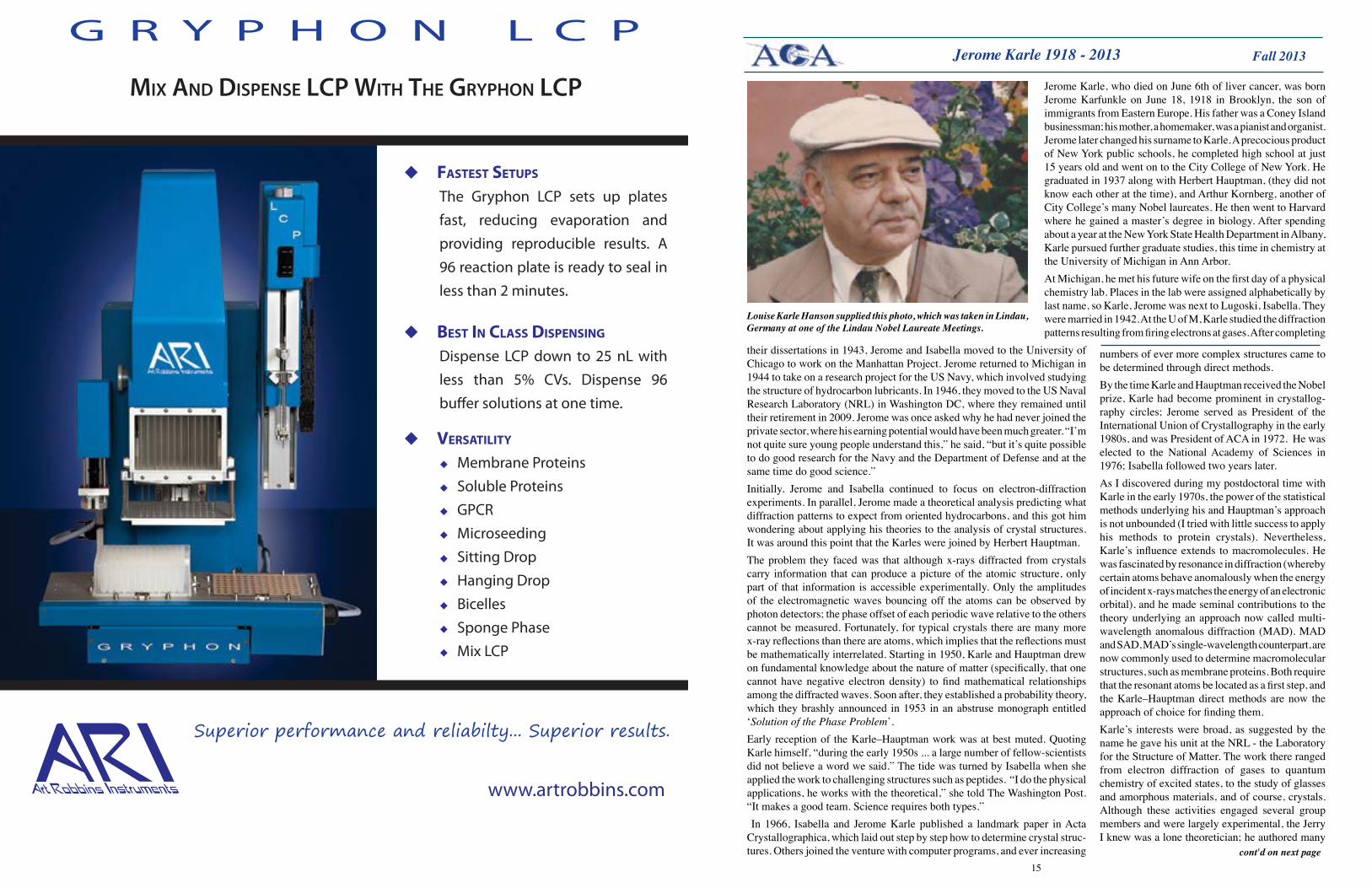
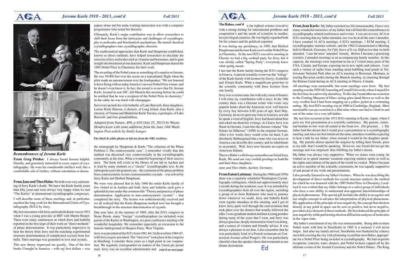
Louise Karle Hanson supplied this photo, which was taken in Lindau, Germany at one of the Lindau Nobel Laureate Meetings.
Jerome Karle, who died on June 6th of liver cancer, was born Jerome Karfunkle on June 18, 1918 in Brooklyn, the son of immigrants from Eastern Europe. His father was a Coney Island businessman; his mother, a homemaker, was a pianist and organist. Jerome later changed his surname to Karle. A precocious product of New York public schools, he completed high school at just 15 years old and went on to the City College of New York. He graduated in 1937 along with Herbert Hauptman, (they did not know each other at the time), and Arthur Kornberg, another of City College’s many Nobel laureates. He then went to Harvard where he gained a master’s degree in biology. After spending about a year at the New York State Health Department in Albany, Karle pursued further graduate studies, this time in chemistry at the University of Michigan in Ann Arbor. At Michigan, he met his future wife on the first day of a physical chemistry lab. Places in the lab were assigned alphabetically by last name, so Karle, Jerome was next to Lugoski, Isabella. They were married in 1942. At the U of M, Karle studied the diffraction patterns resulting from firing electrons at gases. After completing
their dissertations in 1943, Jerome and Isabella moved to the University of Chicago to work on the Manhattan Project. Jerome returned to Michigan in 1944 to take on a research project for the US Navy, which involved studying the structure of hydrocarbon lubricants. In 1946, they moved to the US Naval Research Laboratory (NRL) in Washington DC, where they remained until their retirement in 2009. Jerome was once asked why he had never joined the private sector, where his earning potential would have been much greater. “I’m not quite sure young people understand this,” he said, “but it’s quite possible to do good research for the Navy and the Department of Defense and at the same time do good science.”Initially, Jerome and Isabella continued to focus on electron-diffraction experiments. In parallel, Jerome made a theoretical analysis predicting what diffraction patterns to expect from oriented hydrocarbons, and this got him wondering about applying his theories to the analysis of crystal structures. It was around this point that the Karles were joined by Herbert Hauptman.The problem they faced was that although x-rays diffracted from crystals carry information that can produce a picture of the atomic structure, only part of that information is accessible experimentally. Only the amplitudes of the electromagnetic waves bouncing off the atoms can be observed by photon detectors; the phase offset of each periodic wave relative to the others cannot be measured. Fortunately, for typical crystals there are many more x-ray reflections than there are atoms, which implies that the reflections must be mathematically interrelated. Starting in 1950, Karle and Hauptman drew on fundamental knowledge about the nature of matter (specifically, that one cannot have negative electron density) to find mathematical relationships among the diffracted waves. Soon after, they established a probability theory, which they brashly announced in 1953 in an abstruse monograph entitled ‘Solution of the Phase Problem’.Early reception of the Karle–Hauptman work was at best muted. Quoting Karle himself, “during the early 1950s ... a large number of fellow-scientists did not believe a word we said.” The tide was turned by Isabella when she applied the work to challenging structures such as peptides. “I do the physical applications, he works with the theoretical,” she told The Washington Post. “It makes a good team. Science requires both types.” In 1966, Isabella and Jerome Karle published a landmark paper in Acta Crystallographica, which laid out step by step how to determine crystal struc-tures. Others joined the venture with computer programs, and ever increasing
numbers of ever more complex structures came to be determined through direct methods.By the time Karle and Hauptman received the Nobel prize, Karle had become prominent in crystallog-raphy circles; Jerome served as President of the International Union of Crystallography in the early 1980s, and was President of ACA in 1972. He was elected to the National Academy of Sciences in 1976; Isabella followed two years later.As I discovered during my postdoctoral time with Karle in the early 1970s, the power of the statistical methods underlying his and Hauptman’s approach is not unbounded (I tried with little success to apply his methods to protein crystals). Nevertheless, Karle’s influence extends to macromolecules. He was fascinated by resonance in diffraction (whereby certain atoms behave anomalously when the energy of incident x-rays matches the energy of an electronic orbital), and he made seminal contributions to the theory underlying an approach now called multi-wavelength anomalous diffraction (MAD). MAD and SAD, MAD’s single-wavelength counterpart, are now commonly used to determine macromolecular structures, such as membrane proteins. Both require that the resonant atoms be located as a first step, and the Karle–Hauptman direct methods are now the approach of choice for finding them.Karle’s interests were broad, as suggested by the name he gave his unit at the NRL - the Laboratory for the Structure of Matter. The work there ranged from electron diffraction of gases to quantum chemistry of excited states, to the study of glasses and amorphous materials, and of course, crystals. Although these activities engaged several group members and were largely experimental, the Jerry I knew was a lone theoretician; he authored many
Jerome Karle 1918 - 2013
cont'd on next page
Superior performance and reliabilty... Superior results.
www.artrobbins.com
VERSATILITY
Membrane Proteins Soluble Proteins GPCR Microseeding Sitting Drop Hanging Drop Bicelles Sponge Phase Mix LCP
BEST IN CLASS DISPENSING
Dispense LCP down to 25 nL with less than 5% CVs. Dispense 96 bu�er solutions at one time.
FASTEST SETUPS
The Gryphon LCP sets up plates fast, reducing evaporation and providing reproducible results. A 96 reaction plate is ready to seal in less than 2 minutes.
G R Y P H O N L C P
MIX AND DISPENSE LCP WITH THE GRYPHON LCP
1716
Fall 2013 Fall 2013
The black & white photos at left are from the NRL Archives.
papers alone and his main working interaction was with a computer programmer who tested his theories.Ultimately, Karle’s major contribution was to allow researchers to shift their focus from the intricacies and challenges of crystallogra-phy to molecules and biochemical mechanisms. He turned chemical crystallographers into crystallographic chemists. The mathematical approaches that Karle and Hauptman established, known as direct methods, have helped researchers to elucidate the structure of key molecules such as vitamins and hormones, and to gain insight into biochemical mechanisms. Karle and Hauptman shared the 1985 Nobel Prize in Chemistry for their work.The awarding of the Nobel came as something of a surprise to Jerome. He was 39,000 feet over the ocean on a transatlantic flight when the pilot made an announcement over the loudspeaker. “We are honored to have flying with us today America’s newest Nobel Prize winner, and he doesn’t even know it. In fact, the award is so new that Dr. Jerome Karle, located in seat 29C, left Munich this morning before he could be notified that he was a recipient of the Nobel Prize in chemistry.” In the cabin, he was feted with champagne. Survivors include his wife Isabella, of Lake Barcroft; three daughters, Louise Karle Hanson, a chemist, of Long Island, Jean Karle, also a chemist, of Vienna and Madeleine Karle Tawney, a geologist, of Lake Barcroft; and four grandchildren.
Adapted from Nature, 499, p 410 (July 25, 2013) by Wayne Hendrickson with some additions from the June 14th Wash-ington Post article by Emily Langer.
Remembrances of Jerome KarleFromGreg Petsko: I always found Jerome helpful, friendly, and genuinely interested in every aspect of crys-tallography. He wore his considerable intellect well, like a comfortable suit of clothes, without ostentation or drama.
FromJaneandTheoHahn:We both were very sad learn-ing of Jerry Karle’s death. We knew the Karle family more than fifty years and were always very happy when we met “The Karles” at international crystallographic congresses.I will describe some of these meetings and, in particular, mention the long work for the International Union of Crys-tallography (IUCr) by Jerry.My first encounter with Jerry and Isabella Karle was in 1953 when I was a young post-doc at MIT with Martin Burger. There were many conferences in which Jerry and Isabella reported on the first steps of their work on “direct methods” of phase determination. It was particularly impressive to hear the theory from Jerry and the matching experimental structure determinations of complicated crystals from Isa-bella. Their marriage was grounded in love and crystals. This new theory impressed me greatly. One of the first books I bought in America – with my first dollars – was
the monograph by Hauptman & Karle “The solutions of the Phase Problem I. The centrosymetric case”. I remember vividly that this method was discussed very controversially in the crystallographic community at the time. What a wonderful beginning of their success story. The book still exists in the library of our lab in Aachen and is read by many students as a “classic” crystallographic text. In the subsequent years the general case – the extension of the phase problem from centrosymetric to non-centrosymentric crystals – was solved by Jerry Karle and Herbert Hauptman. We were very pleased that in July 1968 the entire Karle family of five visited us in Aachen and both, Jerry and Isabella, each gave a splendid lecture under the common title “Theory and practice of phase determination. Application to non-centrosymetric crystals” – that completed the story. The lecture was enthusiastically received and we all realized that the Karle-Hauptman method now has brought a breakthrough in the structure determination of crystals.One year later, in the summer of 1969, after the IUCr congress in Stony Brook, many “foreign” crystallographers (us included) were guests of the Karles in Washington, at a post-conference meeting with wonderful hospitality. We remember especially an excursion to the historic battleground of Harpers Ferry, West Virginia. Jerry was president of the IUCr from 1981-84. I followed him 1984-87, with Jerry as past-president. The “turn over” took place at the congress in Hamburg. I consider these years as a high point in our coopera-tion. We regularly corresponded on matters of the Union per postal mail. Jerry was an excellent administrator and
Jerome Karle 1918 - 2013, cont'd
cont'd on next page
FromJeanKarle: My father enriched my life immeasurably. I have very many wonderful memories, of my father, but will limit this remembrance to crystallography related conferences and events. I was not at every ACA or IUCr meeting that my father attended, nor was he at all the ones I attended. I have counted 16 ACA meetings, 6 IUCr meetings, 1 ECM meeting, 2 crystallographic summer schools, and the 1962 Commemorative Meeting held in Munich, Germany, for Fifty Years of X-ray Diffraction that we both attended. I use the term ‘attended’ loosely. Before I became a practicing scientist, I attended meetings as an accompanying family member. In this capacity, the meetings were important to me as I visited many parts of the USA, Canada, and Europe, exposing me to new sights and cultures. I saw such a variety of sights from standing amid bubbling mud ponds in Yel-lowstone National Park after an ACA meeting in Bozeman, Montana, to touring Bavarian castles during the Munich meeting, to canoeing through the Rideau Canal during an ACA meeting in Ottawa, Canada.All meetings were memorable, but some meetings were firsts. My first meeting was the 1959 ACA meeting at Cornell University where I stayed for the first time in a university dormitory. To this day I remember an excursion to the Corning Museum of Glass seeing glass hand blown as well as the very swollen foot I had from stepping on a yellow jacket at a swimming outing. My first IUCr meeting was in 1960 in Cambridge, England. Most memorable was an excursion to a flint mine where one had to climb in and out of the mine via a very tall ladder. My last first occurred at the 1972 IUCr meeting in Kyoto, Japan, where I gave my first presentation at a scientific conference. My parents, sisters, and brother-in-law were all seated in the front row. (No pressure. . .) My father had this dream that I would give a presentation at a crystallography meeting, and since our first initials are the same, attendees would be expecting to hear a talk by my father, when instead it would be his daughter speak-ing. My parents almost spoiled the surprise by telling their friends, prior to my talk, that I would be speaking. However, one friend did not get the message and was surprised, thus fulfilling my father's dream.My father was always very supportive. When we sisters were young, he wanted us to spend summer vacations enjoying outdoor sports as well as the sights and cultures of the parts of the world we visited. When I became an active member of the scientific community, he was equally supportive of and proud of my work and presentations.I also proudly listened to my father's lectures. When he was describing the development of direct methods for crystal structure analysis, the method for which he was honored with the 1985 Nobel Prize in Chemistry, I real-ized it was evident that my father belongs to a select group of individuals who have a rare ability to understand non-apparent interrelationships of physical phenomena. This special gift includes the ability to recognize and use simple concepts to advance the interpretation of physical phenomena. His application of the principle of non-negativity, the concept that electron density at any point in space can be zero or positive, but never negative, provided a key element of direct methods. He first deduced the principle of non-negativity while performing electron diffraction analyses of molecules in the vapor state. My father's enrichment of my life was immeasurable. Being able to share Nobel week with him in Stockholm in 1985 is a memory I will never forget. Just after my family arrived, Stockholm was blanketed by a heavy snowfall covering the city with glistening crystalline snowflakes, appropri-ate for a Nobel Prize being awarded in crystallography. We had exquisite receptions, concerts, tours, dinners, and Nobel lectures capped off by the ultimate events of the Awards Ceremony and the Nobel Dinner. The King
FromEatonLattman: During the 1960s and 1970s there was a regularly scheduled Washington Crystal-lography colloquium, which met approximately once a month during the academic year. It was attended by crystallographers from all over the region, including a group of us from Baltimore who used to journey down whenever we could. Jerry and Isabella Karle were regular attendees at this meeting, and I got to know Jerry quite well through the conversations that took place over the dinners that usually followed the talks. I was a graduate student and then a young postdoc during many of the years that I went, and Jerry was always gracious, deeply interested in what I was doing, and a source of wisdom and friendly advice. It was always a pleasure to see him. I also remember that he was particularly fond of a French restaurant on Con-necticut Avenue called Pouget's. He was particularly cheerful when the speaker chose that restaurant as our dinner destination.
a far-sighted science-executive with a strong feeling for international problems and cooperation’s and the needs of scientists in smaller, less privileged countries. He was highly regarded both for his science and his political expertise.It was during my presidency, in 1985, that Herbert Hauptmann and Jerome Karle received the Nobel Prize in Chemistry. At the next IUCr executive meeting in Chester we had a big cocktail party for Jerry, but it was strictly called “Spring Party”, everybody knew what spring meant.I last met the Karle family during the IUCr congress in Geneva. A special scientific event was the “trilogy” of the Karle family with lectures by J(erry), I(sabella) and J(Jean) Karle. What a magnificent good-bye to the scientific community with three lectures from one family.Jerry was a serious man, but with a dry sense of humor. I will close my contribution with a story: In the 19th century there was a German writer who wrote very popular books about the American west, well known by every boy between 8-80 years of age. Karl May. Curiously, he never spent any time in America, nor did he speak a word of English. Jerry had heard about him and asked me about his writings. As I knew Jerry was fluent in German, I sent him the famous volume “Der Schatz im Silbersee” (1890) in the original German. After a few weeks Jerry would write me back: I am absolutely flabbergasted that a man who was never in America can describe this country and its inhabitants so accurately. Well, Jerry now became an expert on American Indians.The two Hahn’s will always remember our friend Jerry Karle. We send our very cordial greetings to Isabella and their three daughters.Jane and Theo Hahn, Aachen, Germany.
Jerome Karle 1918 - 2013, cont'dThe Hahns, cont'd
1918
Fall 2013 Fall 2013
and Queen were most gracious hosts, not appearing bothered by my photography at the Nobel Dinner. When a daughter of another 1985 Nobel Prize recipient was asked what her Stockholm experience was like, she responded that it was like a fairy tale. I absolutely agree.The saying goes that we do not get to choose our parents. I will be forever grateful that my father chose to have me.
Jerome Karle, cont'd, Ray Davis 1938 - 2013
Jean Karle, cont'd
Professor Raymond Edward Davis was born November 7,
1938 in Hobbs, New Mexico, to proud parents, Edward and Louise Davis. When he was 2 years old, the family moved to Neodesha, Kansas, where Ray spent his childhood and school years, and where he also met his high school sweetheart and the love of his life, Sharon Klingenberg. Ray attended the University of Kansas at Lawrence, was honored with a membership in Phi Beta Kappa and, in 1960, received a Bachelor of Science Degree with Honors in Chemistry. Ray and Sharon were married that summer in the First United Methodist Church in Neodesha. Ray studied at Yale University under the direction of Al Tulinsky and received his PhD in 1964. He then spent two years working as a post-doc with David Harker at the Roswell Park Memorial Institute in Buffalo, New York. The train trip to Buffalo was a memorable one for Ray and Sharon because Sharon was pregnant with Laura at the time and Ray had just had an appendectomy. Their youngest daughter, Angela, was excited about the train ride and wanted to sit on daddy’s lap and look out the window for most of the trip. Ray said he could remember each and every bump in the tracks. Ray accepted a position as an assistant professor in the Chemistry Department of The University of Texas at Austin in the fall of 1966. Ray had ordered the equipment he needed for his research before he arrived on campus. He walked into his lab to a big pile of boxes. Shortly after, in walked Stan Simonsen with his overalls and a tool box ready to help Ray put his lab in order. Ray was surprised that a full professor would take all that time out of his busy week to help a junior faculty member. It was the start of a lifelong friendship between the two and a lesson that throughout his tenure Ray would apply to other faculty in the department. There are no words to express Ray’s love of teaching and learning; teaching was a passion for him and was a gift to be shared with every student he came in contact with. Ray received numerous teaching awards including the MinnieStevensPiperProfessorshipin 1992, the JeanHollowayAwardforExcellenceinTeaching in
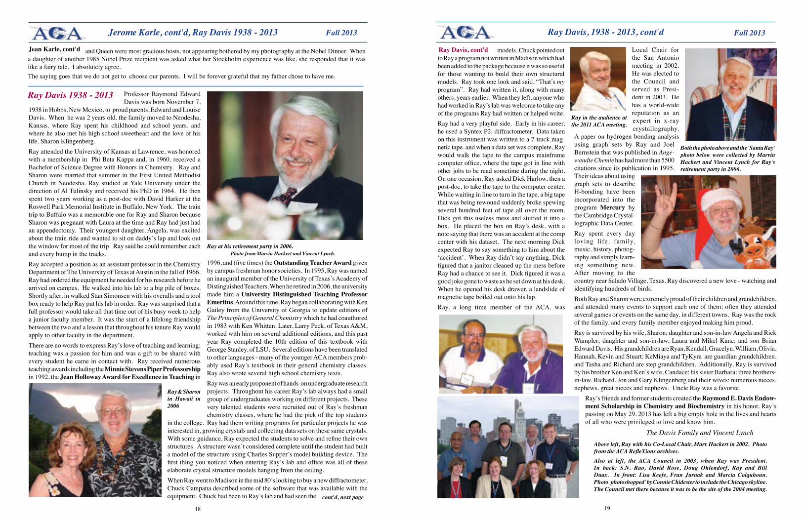
Ray Davis 1938 - 2013
Ray at his retirement party in 2006. Photo from Marvin Hackert and Vincent Lynch.
1996, and (five times) the Outstanding Teacher Award given by campus freshman honor societies. In 1995, Ray was named an inaugural member of the University of Texas’s Academy of Distinguished Teachers. When he retired in 2006, the university made him a UniversityDistinguishedTeachingProfessorEmeritus. Around this time, Ray began collaborating with Ken Gailey from the University of Georgia to update editions of The Principles of General Chemistry which he had coauthored in 1983 with Ken Whitten. Later, Larry Peck, of Texas A&M, worked with him on several additional editions, and this past year Ray completed the 10th edition of this textbook with George Stanley, of LSU. Several editions have been translated to other languages - many of the younger ACA members prob-ably used Ray’s textbook in their general chemistry classes. Ray also wrote several high school chemistry texts. Ray was an early proponent of hands-on undergraduate research projects. Throughout his career Ray’s lab always had a small group of undergraduates working on different projects. These very talented students were recruited out of Ray’s freshman chemistry classes, where he had the pick of the top students
Ray & Sharon in Hawaii in 2006
in the college. Ray had them writing programs for particular projects he was interested in, growing crystals and collecting data sets on these same crystals. With some guidance, Ray expected the students to solve and refine their own structures. A structure wasn’t considered complete until the student had built a model of the structure using Charles Supper’s model building device. The first thing you noticed when entering Ray’s lab and office was all of these elaborate crystal structure models hanging from the ceiling. When Ray went to Madison in the mid 80’s looking to buy a new diffractometer, Chuck Campana described some of the software that was available with the equipment. Chuck had been to Ray’s lab and had seen the cont'd, next page
models. Chuck pointed out to Ray a program not written in Madison which had been added to the package because it was so useful for those wanting to build their own structural models. Ray took one look and said, “That’s my program”. Ray had written it, along with many others, years earlier. When they left, anyone who had worked in Ray’s lab was welcome to take any of the programs Ray had written or helped write. Ray had a very playful side. Early in his career, he used a Syntex P21 diffractometer. Data taken on this instrument was written to a 7-track mag-netic tape, and when a data set was complete, Ray would walk the tape to the campus mainframe computer office, where the tape got in line with other jobs to be read sometime during the night. On one occasion, Ray asked Dick Harlow, then a post-doc, to take the tape to the computer center. While waiting in line to turn in the tape, a big tape that was being rewound suddenly broke spewing several hundred feet of tape all over the room. Dick got this useless mess and stuffed it into a box. He placed the box on Ray’s desk, with a note saying that there was an accident at the comp center with his dataset. The next morning Dick expected Ray to say something to him about the ‘accident’. When Ray didn’t say anything, Dick figured that a janitor cleaned up the mess before Ray had a chance to see it. Dick figured it was a good joke gone to waste as he set down at his desk. When he opened his desk drawer, a landslide of magnetic tape boiled out onto his lap.Ray, a long time member of the ACA, was
Ray Davis, 1938 - 2013, cont'd
Ray Davis, cont'd Local Chair for the San Antonio meeting in 2002. He was elected to the Council and served as Presi-dent in 2003. He has a world-wide reputation as an expert in x-ray crystallography.
A paper on hydrogen bonding analysis using graph sets by Ray and Joel Bernstein that was published in Ange-wandte Chemie has had more than 5500 citations since its publication in 1995. Their ideas about using graph sets to describe H-bonding have been incorporated into the program Mercury by the Cambridge Crystal-lographic Data Center.Ray spent every day loving life, family, music, history, photog-raphy and simply learn-ing something new. After moving to the country near Salado Village, Texas, Ray discovered a new love - watching and identifying hundreds of birds. Both Ray and Sharon were extremely proud of their children and grandchildren, and attended many events to support each one of them; often they attended several games or events on the same day, in different towns. Ray was the rock of the family, and every family member enjoyed making him proud. Ray is survived by his wife, Sharon; daughter and son-in-law Angela and Rick Wampler; daughter and son-in-law, Laura and Mikel Kane; and son Brian Edward Davis. His grandchildren are Ryan, Kendall, Gracelyn, William, Olivia, Hannah, Kevin and Stuart; KeMiaya and TyKyra are guardian grandchildren, and Tasha and Richard are step grandchildren. Additionally, Ray is survived by his brother Ken and Ken’s wife, Candace; his sister Barbara; three brothers-in-law, Richard, Jon and Gary Klingenberg and their wives; numerous nieces, nephews, great nieces and nephews. Uncle Ray was a favorite.
Ray’s friends and former students created the Raymond E. Davis Endow-ment Scholarship in Chemistry and Biochemistry in his honor. Ray’s passing on May 29, 2013 has left a big empty hole in the lives and hearts of all who were privileged to love and know him.
The Davis Family and Vincent Lynch
Both the photo above and the 'Santa Ray' photo below were collected by Marvin Hackert and Vincent Lynch for Ray's retirement party in 2006.
Above left, Ray with his Co-Local Chair, Marv Hackert in 2002. Photo from the ACA RefleXions archives.Also at left, the ACA Council in 2003, when Ray was President. In back: S.N. Rao, David Rose, Doug Ohlendorf, Ray and Bill Duax. In front: Lisa Keefe, Fran Jurnak and Marcia Colquhoun. Photo 'photoshopped' by Connie Chidester to include the Chicago skyline. The Council met there because it was to be the site of the 2004 meeting.
Ray in the audience at the 2011 ACA meeting.
2120
Fall 2013 Fall 2013
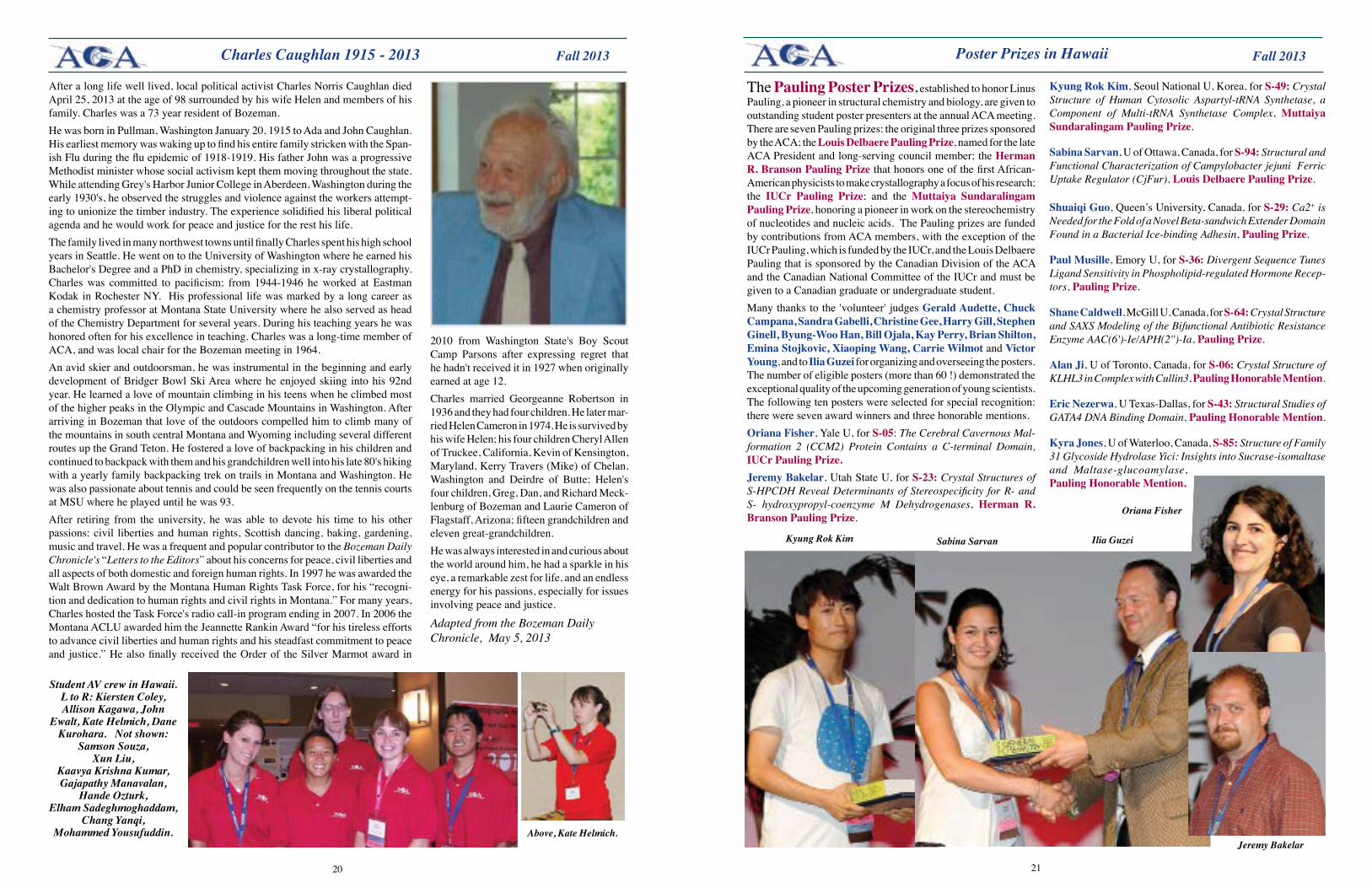
After a long life well lived, local political activist Charles Norris Caughlan died April 25, 2013 at the age of 98 surrounded by his wife Helen and members of his family. Charles was a 73 year resident of Bozeman. He was born in Pullman, Washington January 20, 1915 to Ada and John Caughlan. His earliest memory was waking up to find his entire family stricken with the Span-ish Flu during the flu epidemic of 1918-1919. His father John was a progressive Methodist minister whose social activism kept them moving throughout the state. While attending Grey's Harbor Junior College in Aberdeen, Washington during the early 1930's, he observed the struggles and violence against the workers attempt-ing to unionize the timber industry. The experience solidified his liberal political agenda and he would work for peace and justice for the rest his life. The family lived in many northwest towns until finally Charles spent his high school years in Seattle. He went on to the University of Washington where he earned his Bachelor's Degree and a PhD in chemistry, specializing in x-ray crystallography. Charles was committed to pacificism; from 1944-1946 he worked at Eastman Kodak in Rochester NY. His professional life was marked by a long career as a chemistry professor at Montana State University where he also served as head of the Chemistry Department for several years. During his teaching years he was honored often for his excellence in teaching. Charles was a long-time member of ACA, and was local chair for the Bozeman meeting in 1964.An avid skier and outdoorsman, he was instrumental in the beginning and early development of Bridger Bowl Ski Area where he enjoyed skiing into his 92nd year. He learned a love of mountain climbing in his teens when he climbed most of the higher peaks in the Olympic and Cascade Mountains in Washington. After arriving in Bozeman that love of the outdoors compelled him to climb many of the mountains in south central Montana and Wyoming including several different routes up the Grand Teton. He fostered a love of backpacking in his children and continued to backpack with them and his grandchildren well into his late 80's hiking with a yearly family backpacking trek on trails in Montana and Washington. He was also passionate about tennis and could be seen frequently on the tennis courts at MSU where he played until he was 93.After retiring from the university, he was able to devote his time to his other passions: civil liberties and human rights, Scottish dancing, baking, gardening, music and travel. He was a frequent and popular contributor to the Bozeman Daily Chronicle's “Letters to the Editors” about his concerns for peace, civil liberties and all aspects of both domestic and foreign human rights. In 1997 he was awarded the Walt Brown Award by the Montana Human Rights Task Force, for his “recogni-tion and dedication to human rights and civil rights in Montana.” For many years, Charles hosted the Task Force's radio call-in program ending in 2007. In 2006 the Montana ACLU awarded him the Jeannette Rankin Award “for his tireless efforts to advance civil liberties and human rights and his steadfast commitment to peace and justice.” He also finally received the Order of the Silver Marmot award in
2010 from Washington State's Boy Scout Camp Parsons after expressing regret that he hadn't received it in 1927 when originally earned at age 12.Charles married Georgeanne Robertson in 1936 and they had four children. He later mar-ried Helen Cameron in 1974. He is survived by his wife Helen; his four children Cheryl Allen of Truckee, California, Kevin of Kensington, Maryland, Kerry Travers (Mike) of Chelan, Washington and Deirdre of Butte; Helen's four children, Greg, Dan, and Richard Meck-lenburg of Bozeman and Laurie Cameron of Flagstaff, Arizona; fifteen grandchildren and eleven great-grandchildren. He was always interested in and curious about the world around him, he had a sparkle in his eye, a remarkable zest for life, and an endless energy for his passions, especially for issues involving peace and justice.
Adapted from the Bozeman Daily Chronicle, May 5, 2013
Charles Caughlan 1915 - 2013 Poster Prizes in Hawaii
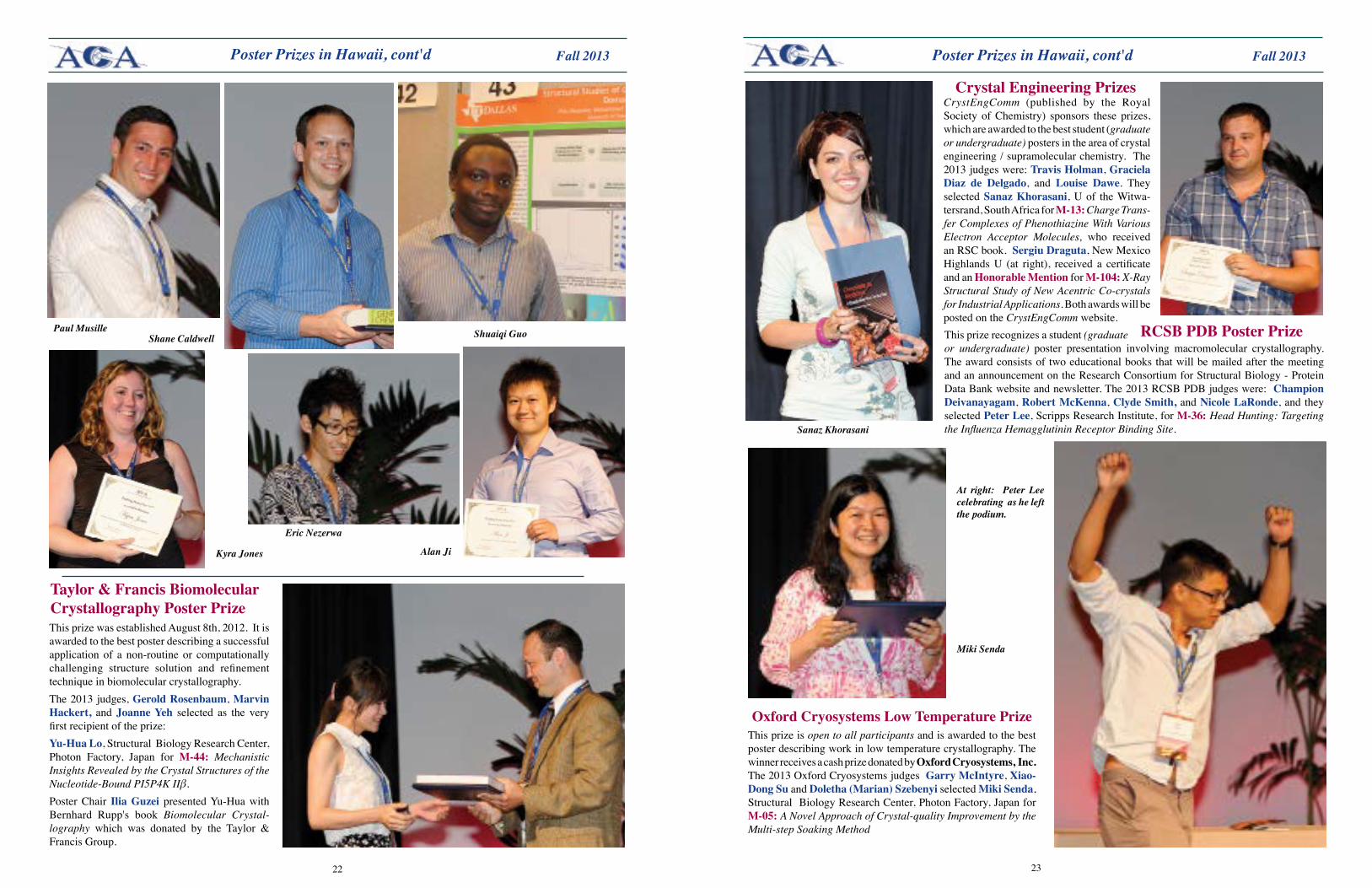
The Pauling Poster Prizes, established to honor Linus Pauling, a pioneer in structural chemistry and biology, are given to outstanding student poster presenters at the annual ACA meeting. There are seven Pauling prizes: the original three prizes sponsored by the ACA; the Louis Delbaere Pauling Prize, named for the late ACA President and long-serving council member; the Herman R. Branson Pauling Prize that honors one of the first African-American physicists to make crystallography a focus of his research; the IUCr Pauling Prize; and the Muttaiya Sundaralingam Pauling Prize, honoring a pioneer in work on the stereochemistry of nucleotides and nucleic acids. The Pauling prizes are funded by contributions from ACA members, with the exception of the IUCr Pauling, which is funded by the IUCr, and the Louis Delbaere Pauling that is sponsored by the Canadian Division of the ACA and the Canadian National Committee of the IUCr and must be given to a Canadian graduate or undergraduate student.Many thanks to the 'volunteer' judges GeraldAudette,ChuckCampana,SandraGabelli,ChristineGee,HarryGill,StephenGinell,Byung-WooHan,BillOjala,KayPerry,BrianShilton,EminaStojkovic,XiaopingWang,CarrieWilmotand Victor Young, and to IliaGuzei for organizing and overseeing the posters. The number of eligible posters (more than 60 !) demonstrated the exceptional quality of the upcoming generation of young scientists. The following ten posters were selected for special recognition: there were seven award winners and three honorable mentions. OrianaFisher, Yale U, for S-05: The Cerebral Cavernous Mal-formation 2 (CCM2) Protein Contains a C-terminal Domain, IUCr Pauling Prize.Jeremy Bakelar, Utah State U, for S-23: Crystal Structures of S-HPCDH Reveal Determinants of Stereospecificity for R- and S- hydroxypropyl-coenzyme M Dehydrogenases, Herman R. Branson Pauling Prize.
Kyung Rok Kim, Seoul National U, Korea, for S-49: Crystal Structure of Human Cytosolic Aspartyl-tRNA Synthetase, a Component of Multi-tRNA Synthetase Complex, Muttaiya Sundaralingam Pauling Prize.
Sabina Sarvan, U of Ottawa, Canada, for S-94: Structural and Functional Characterization of Campylobacter jejuni Ferric Uptake Regulator (CjFur), Louis Delbaere Pauling Prize.
ShuaiqiGuo, Queen’s University, Canada, for S-29: Ca2+ is Needed for the Fold of a Novel Beta-sandwich Extender Domain Found in a Bacterial Ice-binding Adhesin, Pauling Prize.
Paul Musille, Emory U, for S-36: Divergent Sequence Tunes Ligand Sensitivity in Phospholipid-regulated Hormone Recep-tors, Pauling Prize.
Shane Caldwell, McGill U, Canada, for S-64: Crystal Structure and SAXS Modeling of the Bifunctional Antibiotic Resistance Enzyme AAC(6')-Ie/APH(2'')-Ia, Pauling Prize.
Alan Ji, U of Toronto, Canada, for S-06: Crystal Structure of KLHL3 in Complex with Cullin3, Pauling Honorable Mention.
Eric Nezerwa, U Texas-Dallas, for S-43: Structural Studies of GATA4 DNA Binding Domain, Pauling Honorable Mention.
Kyra Jones, U of Waterloo, Canada, S-85: Structure of Family 31 Glycoside Hydrolase Yici: Insights into Sucrase-isomaltase and Maltase-glucoamylase, Pauling Honorable Mention.
Jeremy Bakelar
Sabina Sarvan
Oriana Fisher
Kyung Rok Kim
Student AV crew in Hawaii. L to R: Kiersten Coley, Allison Kagawa, John
Ewalt, Kate Helmich, Dane Kurohara. Not shown:
Samson Souza, Xun Liu,
Kaavya Krishna Kumar, Gajapathy Manavalan,
Hande Ozturk, Elham Sadeghmoghaddam,
Chang Yanqi, Mohammed Yousufuddin.
Ilia Guzei
Above, Kate Helmich.
2322
Fall 2013 Fall 2013Poster Prizes in Hawaii, cont'd
Alan Ji
Eric Nezerwa
Kyra Jones
Paul MusilleShane Caldwell Shuaiqi Guo
Taylor&FrancisBiomolecularCrystallography Poster PrizeThis prize was established August 8th, 2012. It is awarded to the best poster describing a successful application of a non-routine or computationally challenging structure solution and refinement technique in biomolecular crystallography. The 2013 judges, GeroldRosenbaum, Marvin Hackert, and Joanne Yeh selected as the very first recipient of the prize:Yu-HuaLo, Structural Biology Research Center, Photon Factory, Japan for M-44: Mechanistic Insights Revealed by the Crystal Structures of the Nucleotide-Bound PI5P4K IIβ.Poster Chair IliaGuzei presented Yu-Hua with Bernhard Rupp's book Biomolecular Crystal-lography which was donated by the Taylor & Francis Group.
Poster Prizes in Hawaii, cont'd
Miki Senda
Sanaz Khorasani
CrystEngComm (published by the Royal Society of Chemistry) sponsors these prizes, which are awarded to the best student (graduate or undergraduate) posters in the area of crystal engineering / supramolecular chemistry. The 2013 judges were: Travis Holman, GracielaDiaz de Delgado, and Louise Dawe. They selected Sanaz Khorasani, U of the Witwa-tersrand, South Africa for M-13: Charge Trans-fer Complexes of Phenothiazine With Various Electron Acceptor Molecules, who received an RSC book. Sergiu Draguta, New Mexico Highlands U (at right), received a certificate and an Honorable Mention for M-104: X-Ray Structural Study of New Acentric Co-crystals for Industrial Applications. Both awards will be posted on the CrystEngComm website. This prize recognizes a student (graduate or undergraduate) poster presentation involving macromolecular crystallography. The award consists of two educational books that will be mailed after the meeting and an announcement on the Research Consortium for Structural Biology - Protein Data Bank website and newsletter. The 2013 RCSB PDB judges were: Champion Deivanayagam, Robert McKenna, Clyde Smith, and Nicole LaRonde, and they selected Peter Lee, Scripps Research Institute, for M-36: Head Hunting: Targeting the Influenza Hemagglutinin Receptor Binding Site.
Crystal Engineering Prizes
RCSB PDB Poster Prize
OxfordCryosystemsLowTemperaturePrizeThis prize is open to all participants and is awarded to the best poster describing work in low temperature crystallography. The winner receives a cash prize donated by OxfordCryosystems,Inc. The 2013 Oxford Cryosystems judges GarryMcIntyre, Xiao-Dong Su and Doletha(Marian)Szebenyiselected Miki Senda, Structural Biology Research Center, Photon Factory, Japan for M-05: A Novel Approach of Crystal-quality Improvement by the Multi-step Soaking Method
At right: Peter Lee celebrating as he left the podium.
2524
Fall 2013 Fall 2013
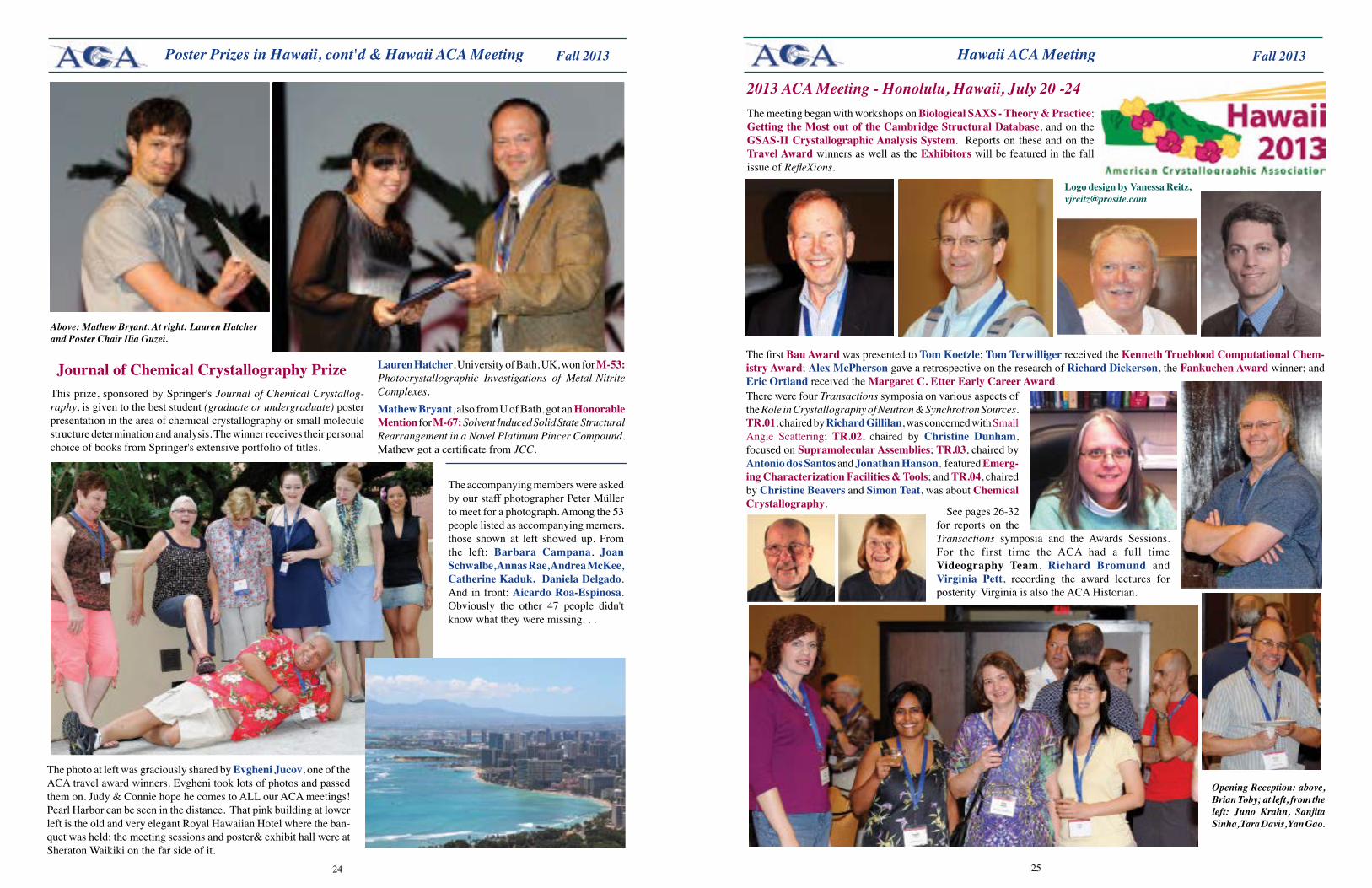
Above: Mathew Bryant. At right: Lauren Hatcher and Poster Chair Ilia Guzei.
Poster Prizes in Hawaii, cont'd & Hawaii ACA Meeting
JournalofChemicalCrystallographyPrizeThis prize, sponsored by Springer's Journal of Chemical Crystallog-raphy, is given to the best student (graduate or undergraduate) poster presentation in the area of chemical crystallography or small molecule structure determination and analysis. The winner receives their personal choice of books from Springer's extensive portfolio of titles.
Lauren Hatcher, University of Bath, UK, won for M-53:Photocrystallographic Investigations of Metal-Nitrite Complexes.Mathew Bryant, also from U of Bath, got an Honorable Mention for M-67: Solvent Induced Solid State Structural Rearrangement in a Novel Platinum Pincer Compound.Mathew got a certificate from JCC.
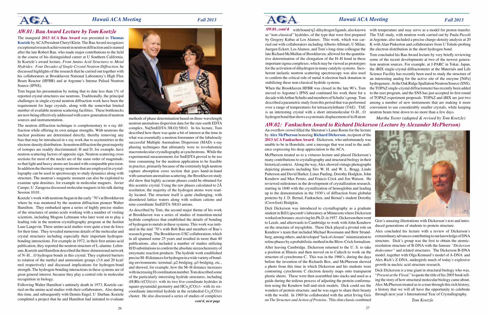
The accompanying members were asked by our staff photographer Peter Müller to meet for a photograph. Among the 53 people listed as accompanying memers, those shown at left showed up. From the left: Barbara Campana, Joan Schwalbe, Annas Rae, Andrea McKee, Catherine Kaduk, Daniela Delgado. And in front: AicardoRoa-Espinosa. Obviously the other 47 people didn't know what they were missing. . .
The photo at left was graciously shared by Evgheni Jucov, one of the ACA travel award winners. Evgheni took lots of photos and passed them on. Judy & Connie hope he comes to ALL our ACA meetings! Pearl Harbor can be seen in the distance. That pink building at lower left is the old and very elegant Royal Hawaiian Hotel where the ban-quet was held; the meeting sessions and poster& exhibit hall were at Sheraton Waikiki on the far side of it.
Hawaii ACA Meeting
Logo design by Vanessa Reitz, [email protected]
2013 ACA Meeting - Honolulu, Hawaii, July 20 -24The meeting began with workshops on BiologicalSAXS-Theory&Practice; GettingtheMostoutoftheCambridgeStructuralDatabase, and on the GSAS-IICrystallographicAnalysisSystem. Reports on these and on the Travel Award winners as well as the Exhibitors will be featured in the fall issue of RefleXions.
The first Bau Award was presented to Tom Koetzle; Tom Terwilliger received the Kenneth Trueblood Computational Chem-istry Award; AlexMcPherson gave a retrospective on the research of Richard Dickerson, the FankuchenAwardwinner; and Eric Ortland received the Margaret C. Etter Early Career Award.There were four Transactions symposia on various aspects of the Role in Crystallography of Neutron & Synchrotron Sources. TR.01, chaired by RichardGillilan, was concerned with Small Angle Scattering; TR.02, chaired by Christine Dunham, focused on Supramolecular Assemblies; TR.03, chaired by Antonio dos Santos and Jonathan Hanson, featured Emerg-ingCharacterizationFacilities&Tools; and TR.04, chaired by Christine Beavers and Simon Teat, was about Chemical Crystallography.
See pages 26-32 for reports on the Transactions symposia and the Awards Sessions. For the first time the ACA had a full time Videography Team, Richard Bromund and Virginia Pett, recording the award lectures for posterity. Virginia is also the ACA Historian.
Opening Reception: above, Brian Toby; at left, from the left: Juno Krahn, Sanjita Sinha, Tara Davis, Yan Gao.
2726
Fall 2013 Fall 2013 Hawaii ACA Meeting
AW.01: Bau Award Lecture by Tom KoetzleThe inaugural 2013 ACA Bau Award was presented to Thomas Koetzle by ACA President Cheryl Klein. The Bau Award recognizes exceptional research achievement in neutron diffraction and is named after the late Robert Bau, who made major contributions to the field in the course of his distinguished career at U Southern California. In Koetzle’s award lecture, From Amino Acid Structures to Metal Hydrides: Four Decades of Single-Crystal Neutron Diffraction, he discussed highlights of the research that he carried out together with his collaborators at Brookhaven National Laboratory’s High Flux Beam Reactor (HFBR) and at Argonne’s Intense Pulsed Neutron Source (IPNS). Tom began his presentation by noting that to date less than 1% of reported crystal structures use neutrons. Traditionally, the principal challenges in single-crystal neutron diffraction work have been the requirement for large crystals, along with the somewhat limited number of available neutron scattering facilities. These bottlenecks are now being effectively addressed with a new generation of neutron sources and instrumentation. The neutron diffraction technique is complementary to x-ray dif-fraction while offering its own unique strengths. With neutrons the nuclear positions are determined directly, thereby removing any bias that may be introduced in x-ray work due to asymmetries in the electron-density distribution. In neutron diffraction the great majority of isotopes are readily discriminated; H and D, for example, have neutron scattering factors of opposite sign. Neutron scattering cross sections for most of the nuclei are of the same order of magnitude, so that light and heavy atoms are located with comparable precision. In addition the thermal-energy neutrons that are employed in crystal-lography can be used in spectroscopy to study dynamics along with structure. The neutron’s magnetic moment can also be exploited to examine spin densities, for example in molecular magnets. Javier Campo, U Zaragoza discussed molecular magnets in his talk during Session 10.01.Koetzle’s work with neutrons began in the early ‘70’s at Brookhaven where he was mentored by the neutron diffraction pioneer Walter Hamilton. They embarked upon a series of highly precise studies of the structures of amino acids working with a number of visiting scientists, including Mogens Lehmann who later went on to play a leading role in the neutron crystallography program at the Institut Laue Langevin. These amino acid studies were quite a tour de force for their time. They revealed numerous details of the molecular and crystal structures including especially their important hydrogen-bonding interactions. For example in 1972, in their first amino-acid publication, they reported the neutron structure of L-alanine. Lehm-ann, Koetzle and Hamilton described the three-dimensional network of N–H…O hydrogen bonds in this crystal. They explored barriers to rotation of the methyl and ammonium groups (3.6 and 20 kcal/mol respectively) and discussed implications for hydrogen-bond strength. The hydrogen-bonding interactions in these systems are of great general interest, because they play a central role in molecular recognition in biology.Following Walter Hamilton’s untimely death in 1973, Koetzle car-ried on the amino acid studies with their collaborators. Also during this time, and subsequently with Dennis Engel, U Durban, Koetzle completed a project that he and Hamilton had initiated to evaluate
methods of phase determination based on three-wavelength neutron anomalous dispersion data for the rare-earth EDTA complex, Na[SmEDTA·3H2O]·5H2O. In his lecture, Tom described how there was quite a bit of interest at the time in what was essentially a neutron forerunner of the fabulously successful Multiple Anomalous Dispersion (MAD) x-ray phasing techniques that ultimately were to revolutionize macromolecular crystallography at synchrotrons. While the experimental measurements for SmEDTA proved to be too time consuming for the neutron application to be feasible in practice, primarily because of the extremely high neutron capture absorption cross section that goes hand-in-hand with samarium anomalous scattering, the Brookhaven study did show that highly accurate phases could be obtained for this acentric crystal. Using the raw phases calculated to 2Å resolution, the majority of the hydrogen atoms were read-ily located. The structure itself is quite challenging, with disordered lattice waters along with sodium cations and nine-coordinate SmEDTA·3H2O anions.As described by Tom, the second major theme of his work at Brookhaven was a series of studies of transition-metal hydride complexes that established the details of bonding of hydrogen to metals in these systems. This work was initi-ated in the mid ‘70’s with Bob Bau and members of Bau’s research group. The Brookhaven-USC collaboration, which in all spanned some 25 years and resulted in more than 40 publications, also included a number of studies utilizing H/D substitutions to confirm the absolute stereochemistry of enzymatic reaction products. The hydride work established precise M–H distances for hydrogen in a wide variety of bond-ing environments: terminal, μ2-bridging, μ3-bridging, etc., and showed, for example, how the M–H distance increases with increasing H coordination number. Tom described some of the particularly interesting hydride structures, including (H2Rh13(CO)24)3- with its two five-coordinate hydrides in square-pyramidal geometry and HCo6(CO)15− with its six-coordinate interstitial hydride in the octahedral Co6(CO)15 cluster. He also discussed a series of studies of complexes
cont'd, next page
An overflow crowd filled the Sheraton’s Lanai Room for the lecture by AlexMcPherson honoring Richard Dickerson, recipient of the 2013ACAFankuchenAward. Dickerson, who unfortunately was unable to be in Honolulu, sent a message that was read to the audi-ence expressing his deep appreciation to the ACA.McPherson treated us to a virtuoso lecture and placed Dickerson’s many contributions to crystallography and structural biology in their historical context. Along the way, Alex showed vintage photographs depicting pioneers including Sirs W. H. and W. L. Bragg, Lindo Patterson and David Harker, Linus Pauling, Dorothy Hodgkin, John Kendrew and Max Perutz, and Francis Crick and Jim Watson. He reviewed milestones in the development of crystallization research, starting in 1840 with the crystallization of hemoglobin and leading up to the demonstration in the 1930’s of diffraction from globular proteins by J. D. Bernal, Fankuchen, and Bernal’s student Dorothy (Crowfoot) Hodgkin.Dick Dickerson was introduced to crystallography as a graduate student in Bill Lipscomb’s laboratory at Minnesota where Dickerson worked on boranes, receiving his Ph.D. in 1957. Dickerson then went to Leeds, and afterwards to Cambridge to work with John Kendrew on the structure of myoglobin. There Dick played a pivotal role on Kendrew’s team that included Michael Rossmann and Brör Strand-berg, among others, and developed “lack of closure” minimization to refine phases by a probabilistic method in the Blow-Crick formalism.After leaving Cambridge, Dickerson returned to the U. S. to take a position at Illinois and then moved on to Caltech to work on the structure of cytochrome C. This was in the 1960’s, during the days before the invention of the Richards Box, and McPherson showed a photo from this time in which Dickerson and his students were contouring cytochrome C electron density maps onto transparent plastic sheets. These were then assembled into stacks and used as a guide during the tedious process of adjusting the protein conforma-tion using the Kendrew ball-and-stick models. Dick could see the wonders of protein structure, and he was eager to share their beauty with the world. In 1969 he collaborated with the artist Irving Geis on The Structure and Action of Proteins. This slim classic combined
Hawaii ACA Meeting
AW.02: Fankuchen Award to Richard Dickerson (Lecture by Alexander McPherson)
Geis’s amazing illustrations with Dickerson’s text and intro-duced generations of students to protein structure.Alex concluded his lecture with a review of Dickerson’s extraordinary advances contributing to our knowledge of DNA structure. Dick’s group was the first to obtain the atomic-resolution structure of B-DNA with the famous “Dickerson dodecamer” and related structures. The Dickerson B-DNA model, together with Olga Kennard’s model of A-DNA and Alex Rich’s Z-DNA, undergirds much of today’s explosive growth in nucleic acid structure research.Dick Dickerson is a true giant in structural biology who was, “Present at the Flood,” to quote the title of his 2005 book tell-ing the story of how structural molecular biology came about. Alex McPherson treated us to a tour through this rich history, a history that we will all have the opportunity to celebrate through next year’s International Year of Crystallography.
Tom Koetzle
with bound η2-dihydrogen ligands, also known as “non-classical” hydrides, of the type that were first prepared by Gregory Kubas at Los Alamos. This work, which was car-ried out with collaborators including Alberto Albinati, U Milan, Juergen Eckert, Los Alamos, and Tom’s long-time colleague the late Richard McMullan of Brookhaven, allowed for the quantita-tive determination of the elongation of the H–H bond in these important sigma complexes, which may be viewed as prototypes for the activation of dihydrogen in many catalytic systems. Inco-herent inelastic neutron scattering spectroscopy was also used to confirm the critical role of metal π-electron back donation in stabilizing these non-classical hydride systems.When the Brookhaven HFBR was closed in the late 90’s, Tom moved to Argonne’s IPNS and continued his work there for a decade with Arthur Schultz and members of Schultz’s group. Tom described a parametric study from this period that was performed over a range of temperatures for tetraacetylethane (TAE). TAE is an interesting crystal with a short intramolecular O–H…O hydrogen bond that shows a systematic displacement of its H atom
with temperature and may serve as a model for proton transfer. The TAE study, with neutron work carried out by Paula Piccoli at Argonne, also included a precise charge-density analysis at 20 K with Alan Pinkerton and collaborators from U Toledo probing the electron distribution in the short hydrogen bond.Tom concluded his Bau Award lecture by very briefly reviewing some of the recent developments at two of the newest genera-tion neutron sources. For example, at J-PARC in Tokai, Japan, the iBIX single-crystal diffractometer at the Materials and Life Science Facility has recently been used to study the structure of an interesting analog for the active site of the enzyme [NiFe]hydrogenase. At the Oak Ridge Spallation Neutron Source (SNS), the TOPAZ single-crystal diffractometer has recently been added to the user program, and the SNS has just accepted its first round of TOPAZ experiment proposals. TOPAZ and iBIX are just two among a number of new instruments that are making it more convenient to use considerably smaller crystals, while keeping neutron beam time down to no more than a few days.
Martha Teeter (adapted & revised by Tom Koetzle)
AW.01, cont'd
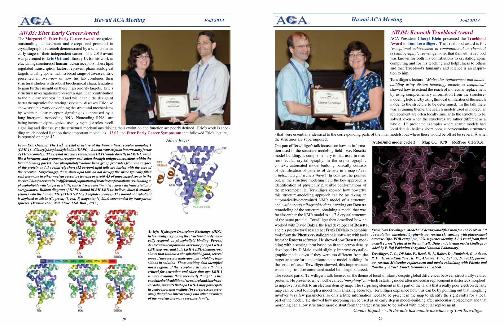
2928
Fall 2013 Fall 2013 Hawaii ACA Meeting
The Margaret C. Etter Early Career Award recognizes outstanding achievement and exceptional potential in crystallographic research demonstrated by a scientist at an early stage of their independent career. The 2013 award was presented to Eric Ortlund, Emory U, for his work in elucidating structures of human nuclear receptors. These lipid regulated transcription factors represent pharmacological targets with high potential in a broad range of diseases. Eric presented an overview of how his lab combines their structural studies with robust biochemical characterization to gain further insight on these high priority targets. Eric’s structural investigations represent a significant contribution to the nuclear receptor field and will enable the design of better therapeutics for treating associated diseases. Eric also showcased his work on defining the structural mechanisms by which nuclear receptor signaling is suppressed by a long intergenic noncoding RNA. Noncoding RNAs are being increasingly recognized as playing major roles in cell
AW.03: Etter Early Career Award
Hawaii ACA Meeting
signaling and disease, yet the structural mechanisms driving their evolution and function are poorly defined. Eric’s work is shed-ding much needed light on these important molecules. 12.02, the Etter Early Career Symposium that followed Eric's lecture,
Albert RegerFrom Eric Ortlund: The 1.8Å crystal structure of the human liver receptor homolog 1 (LRH-1) – dilaurylphosphatidylcholine (DLPC) – human transcription intermediary factor 2 (TIF2) complex. The crystal structure reveals that DLPC binds directly to LRH-1, much like a hormone, and promotes receptor activation through unique interactions within the ligand binding pocket. The phosphatidylcholine head group protrudes from the surface of the protein and the relatively short (12 carbon) lipid tails are buried with the core of the receptor. Surprisingly, these short lipid tails do not occupy the space typically filled with hormone in other nuclear receptors leaving over 800 Å3 of unoccupied space in the pocket. This space results in differential population of protein conformations (vs. binding to phospholipids with longer acyl tails) which drives selective interaction with transcriptional coregulators. Ribbon diagram of DLPC bound hLRH-LBD (α-helices, blue; β-strands, yellow) with the human TIF (hTIF) NR box 3 peptide (orange). The bound phospholipid is depicted as sticks (C, green; O, red; P, magenta; N, blue) surrounded by transparent spheres. (Musille et al., Nat. Struc. Mol. Biol., 2012.)
At left: Hydrogen-Deuterium Exchange (HDX) helps identify regions of the structure that dynami-cally respond to phospholipid binding. Percent deuterium incorporation over time for apo LRH-1 LBD (top row) and holo LRH-1 LBD (bottom row) shows that without a phospholipid ligand, several areas of the receptor undergo rapid unfolding tran-sitions in solution. These exciting data identified novel regions of the receptor’s structure that are critical for activation and show that apo LRH-1 is more dynamic than previously thought. This, combined with additional structural and biochemi-cal data, suggests that apo LRH-1 may participate in gene repression mediated by corepressors previ-ously thought to interact only with other members of the nuclear hormone receptor family.
is reported on page 42.
ACA President Cheryl Klein presented the Trueblood Award to Tom Terwilliger. The Trueblood award is for, "exceptional achievement in computational or chemical crystallography". Terwilliger noted that Kenneth Trueblood was known for both his contributions to crystallographic computing and for his teaching and helpfulness to others and that Trueblood's humanity and science is an inspira-tion to him.Terwilliger's lecture, "Molecular replacement and model-building using distant homology models as templates," showed how to extend the reach of molecular replacement by using complementary information from the structure-modeling field and by using the local similarities of the search model to the structure to be determined. In the talk there was a running theme: the search models used in molecular replacement are often locally similar to the structure to be solved, even when the structures are rather different as a whole. He presented examples where search models had local details - helices, short loops, supersecondary structures
AW.04: Kenneth Trueblood Award
- that were essentially identical to the corresponding parts of the final models, but where these would be offset be several Å when the structures are superimposed.One part of Terwilliger's talk focused on how the informa-tion used in the structure-modeling field, e.g. Rosetta model-building, is complementary to that used in mac-romolecular crystallography. In the crystallographic context, automated model-building basically consists of identification of patterns of density in a map ('I see a helix, let's put a helix there'). In contrast, he pointed out, in the structure modeling field the key approach is identification of physically plausible conformations of the macromolecule. Terwilliger showed how powerful this structure-modeling approach can be by taking an automatically-determined NMR model of a structure, and, without crystallographic data, carrying out Rosetta remodeling of the structure, obtaining a model that was far closer than the NMR model to a 1.7 Å crystal structure of the same protein. Terwilliger then described how he worked with David Baker, the lead developer of Rosetta, and his postdoctoral researcher Frank DiMaio to combine tools from the Phenix crystallographic software with tools from the Rosetta software. He showed how Rosetta mod-eling with a scoring term based on fit to electron density developed by DiMaio could slightly improve crystallo-graphic models even if they were too different from the target structure for standard automated model-building. In the series of cases Terwilliger showed, this improvement was enough to allow automated model-building to succeed.The second part of Terwilliger's talk focused on the theme of local similarity despite global differences between structurally-related proteins. He presented a method he called, "morphing", in which a starting model after molecular replacement is distorted (morphed) to improve its match to an electron density map. The surprising element in this part of the talk is that a really poor electron density map can be used to morph a model with amazing accuracy. Terwilliger explained how this can be by pointing out that morphing involves very few parameters, so only a little information needs to be present in the map to identify the right shifts for a local part of the model. He showed how morphing can be used as an early step in model-building after molecular replacement and that morphing can allow structures more distant from the target structure to be solved with molecular replacement. Connie Rajnak - with the able last minute assistance of Tom Terwilliger
From Tom Terwilliger: Model and density-modified map for cab55348 at 1.9 Å resolution calculated by phenix.mr_rosetta (1) starting with glucuronoyl esterase Cip2 (PDB entry 3pic, 32% sequence identity, 2.1 Å rmsd from final model) correctly placed in the unit cell. Data and starting model kindly pro-vided by P. Raj Pokkuluri (Argonne National Laboratory).Terwilliger, T. C., DiMaio, F., Read, R. J., Baker, D., Bunkóczi, G., Adams, P. D., Grosse-Kunstleve, R. W., Afonine, P. V., Echols, N. (2012).phenix.mr_rosetta: Molecular replacement and model rebuilding with Phenix and Rosetta. J. Struct. Funct. Genomics 13, 81-90.
AutoBuildmodelcycle2 MapCC:0.78R/Rfree=0.26/0.31
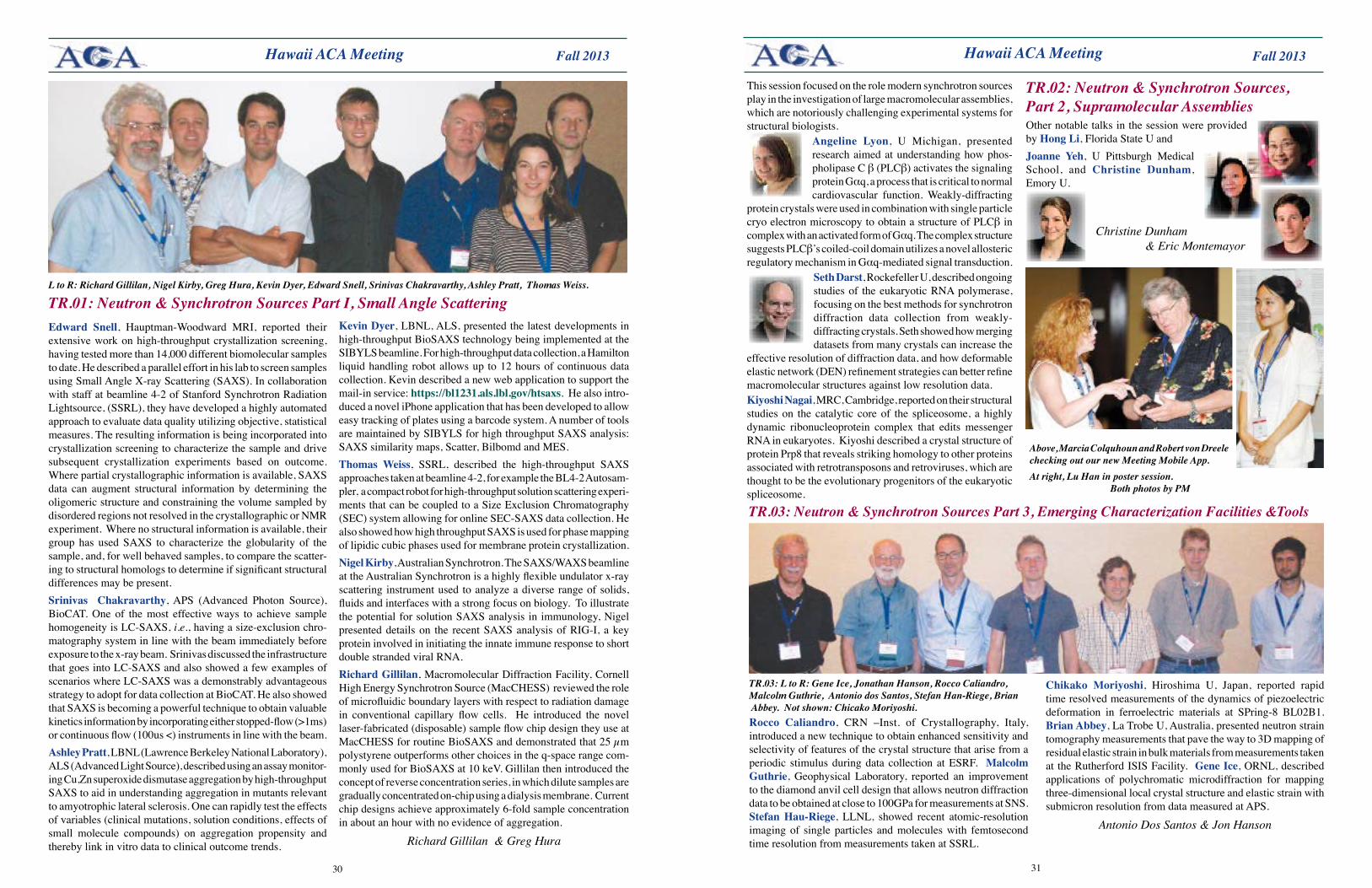
3130
Fall 2013 Fall 2013
Other notable talks in the session were provided by Hong Li, Florida State U and Joanne Yeh, U Pittsburgh Medical School, and Christine Dunham, Emory U.
Christine Dunham & Eric Montemayor
Hawaii ACA Meeting Hawaii ACA Meeting
Edward Snell, Hauptman-Woodward MRI, reported their extensive work on high-throughput crystallization screening, having tested more than 14,000 different biomolecular samples to date. He described a parallel effort in his lab to screen samples using Small Angle X-ray Scattering (SAXS). In collaboration with staff at beamline 4-2 of Stanford Synchrotron Radiation Lightsource, (SSRL), they have developed a highly automated approach to evaluate data quality utilizing objective, statistical measures. The resulting information is being incorporated into crystallization screening to characterize the sample and drive subsequent crystallization experiments based on outcome. Where partial crystallographic information is available, SAXS data can augment structural information by determining the oligomeric structure and constraining the volume sampled by disordered regions not resolved in the crystallographic or NMR experiment. Where no structural information is available, their group has used SAXS to characterize the globularity of the sample, and, for well behaved samples, to compare the scatter-ing to structural homologs to determine if significant structural differences may be present.Srinivas Chakravarthy, APS (Advanced Photon Source), BioCAT. One of the most effective ways to achieve sample homogeneity is LC-SAXS, i.e., having a size-exclusion chro-matography system in line with the beam immediately before exposure to the x-ray beam. Srinivas discussed the infrastructure that goes into LC-SAXS and also showed a few examples of scenarios where LC-SAXS was a demonstrably advantageous strategy to adopt for data collection at BioCAT. He also showed that SAXS is becoming a powerful technique to obtain valuable kinetics information by incorporating either stopped-flow (>1ms) or continuous flow (100us <) instruments in line with the beam. Ashley Pratt, LBNL (Lawrence Berkeley National Laboratory), ALS (Advanced Light Source), described using an assay monitor-ing Cu,Zn superoxide dismutase aggregation by high-throughput SAXS to aid in understanding aggregation in mutants relevant to amyotrophic lateral sclerosis. One can rapidly test the effects of variables (clinical mutations, solution conditions, effects of small molecule compounds) on aggregation propensity and thereby link in vitro data to clinical outcome trends.
TR.01: Neutron & Synchrotron Sources Part I, Small Angle ScatteringKevin Dyer, LBNL, ALS, presented the latest developments in high-throughput BioSAXS technology being implemented at the SIBYLS beamline. For high-throughput data collection, a Hamilton liquid handling robot allows up to 12 hours of continuous data collection. Kevin described a new web application to support the mail-in service: https://bl1231.als.lbl.gov/htsaxs. He also intro-duced a novel iPhone application that has been developed to allow easy tracking of plates using a barcode system. A number of tools are maintained by SIBYLS for high throughput SAXS analysis: SAXS similarity maps, Scatter, Bilbomd and MES. Thomas Weiss, SSRL, described the high-throughput SAXS approaches taken at beamline 4-2, for example the BL4-2 Autosam-pler, a compact robot for high-throughput solution scattering experi-ments that can be coupled to a Size Exclusion Chromatography (SEC) system allowing for online SEC-SAXS data collection. He also showed how high throughput SAXS is used for phase mapping of lipidic cubic phases used for membrane protein crystallization. Nigel Kirby, Australian Synchrotron. The SAXS/WAXS beamline at the Australian Synchrotron is a highly flexible undulator x-ray scattering instrument used to analyze a diverse range of solids, fluids and interfaces with a strong focus on biology. To illustrate the potential for solution SAXS analysis in immunology, Nigel presented details on the recent SAXS analysis of RIG-I, a key protein involved in initiating the innate immune response to short double stranded viral RNA. RichardGillilan, Macromolecular Diffraction Facility, Cornell High Energy Synchrotron Source (MacCHESS) reviewed the role of microfluidic boundary layers with respect to radiation damage in conventional capillary flow cells. He introduced the novel laser-fabricated (disposable) sample flow chip design they use at MacCHESS for routine BioSAXS and demonstrated that 25 µm polystyrene outperforms other choices in the q-space range com-monly used for BioSAXS at 10 keV. Gillilan then introduced the concept of reverse concentration series, in which dilute samples are gradually concentrated on-chip using a dialysis membrane. Current chip designs achieve approximately 6-fold sample concentration in about an hour with no evidence of aggregation.
Richard Gillilan & Greg Hura
L to R: Richard Gillilan, Nigel Kirby, Greg Hura, Kevin Dyer, Edward Snell, Srinivas Chakravarthy, Ashley Pratt, Thomas Weiss.
Rocco Caliandro, CRN –Inst. of Crystallography, Italy, introduced a new technique to obtain enhanced sensitivity and selectivity of features of the crystal structure that arise from a periodic stimulus during data collection at ESRF. Malcolm Guthrie, Geophysical Laboratory, reported an improvement to the diamond anvil cell design that allows neutron diffraction data to be obtained at close to 100GPa for measurements at SNS. StefanHau-Riege, LLNL, showed recent atomic-resolution imaging of single particles and molecules with femtosecond time resolution from measurements taken at SSRL.
TR.03: Neutron & Synchrotron Sources Part 3, Emerging Characterization Facilities &Tools
TR.03: L to R: Gene Ice, Jonathan Hanson, Rocco Caliandro, Malcolm Guthrie, Antonio dos Santos, Stefan Han-Riege, Brian Abbey. Not shown: Chicako Moriyoshi.
Chikako Moriyoshi, Hiroshima U, Japan, reported rapid time resolved measurements of the dynamics of piezoelectric deformation in ferroelectric materials at SPring-8 BL02B1. Brian Abbey, La Trobe U, Australia, presented neutron strain tomography measurements that pave the way to 3D mapping of residual elastic strain in bulk materials from measurements taken at the Rutherford ISIS Facility. GeneIce, ORNL, described applications of polychromatic microdiffraction for mapping three-dimensional local crystal structure and elastic strain with submicron resolution from data measured at APS.
Antonio Dos Santos & Jon Hanson
TR.02: Neutron & Synchrotron Sources, Part 2, Supramolecular Assemblies
This session focused on the role modern synchrotron sources play in the investigation of large macromolecular assemblies, which are notoriously challenging experimental systems for structural biologists.
Angeline Lyon, U Michigan, presented research aimed at understanding how phos-pholipase C β (PLCβ) activates the signaling protein Gαq, a process that is critical to normal cardiovascular function. Weakly-diffracting
protein crystals were used in combination with single particle cryo electron microscopy to obtain a structure of PLCβ in complex with an activated form of Gαq. The complex structure suggests PLCβ’s coiled-coil domain utilizes a novel allosteric regulatory mechanism in Gαq-mediated signal transduction.
Seth Darst, Rockefeller U, described ongoing studies of the eukaryotic RNA polymerase, focusing on the best methods for synchrotron diffraction data collection from weakly-diffracting crystals. Seth showed how merging datasets from many crystals can increase the
effective resolution of diffraction data, and how deformable elastic network (DEN) refinement strategies can better refine macromolecular structures against low resolution data. Kiyoshi Nagai, MRC, Cambridge, reported on their structural studies on the catalytic core of the spliceosome, a highly dynamic ribonucleoprotein complex that edits messenger RNA in eukaryotes. Kiyoshi described a crystal structure of protein Prp8 that reveals striking homology to other proteins associated with retrotransposons and retroviruses, which are thought to be the evolutionary progenitors of the eukaryotic spliceosome.
Above, Marcia Colquhoun and Robert von Dreele checking out our new Meeting Mobile App. At right, Lu Han in poster session. Both photos by PM
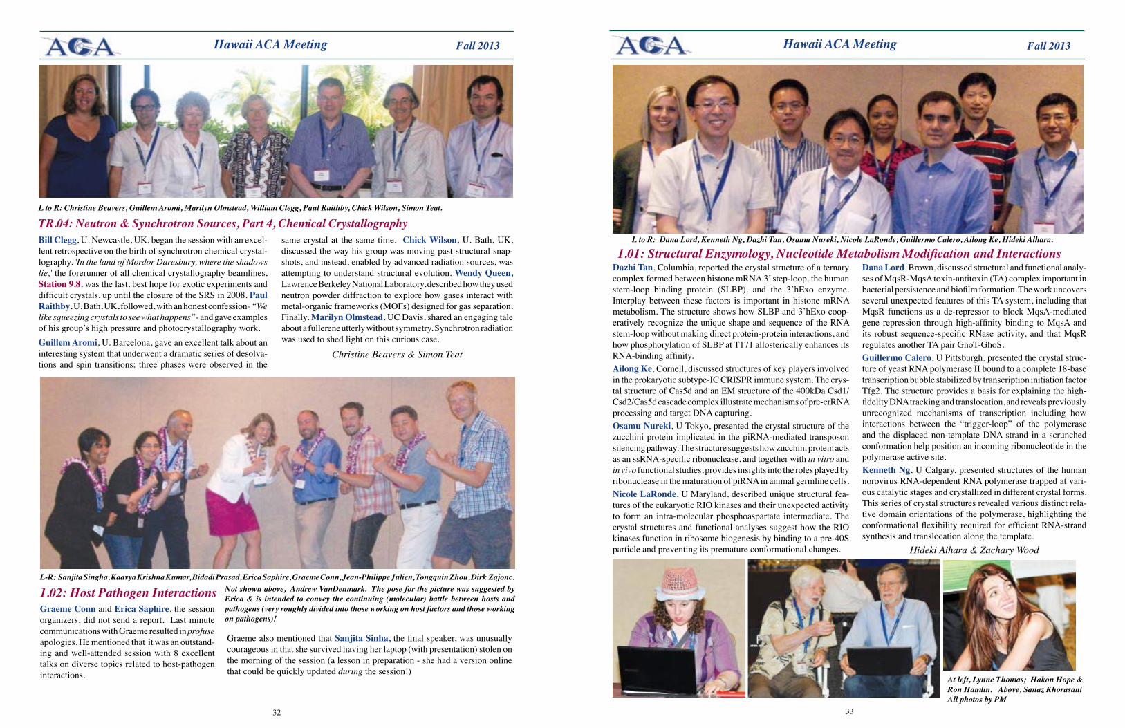
3332
Fall 2013 Fall 2013 Hawaii ACA Meeting Hawaii ACA Meeting
TR.04: Neutron & Synchrotron Sources, Part 4, Chemical CrystallographyBill Clegg, U. Newcastle, UK, began the session with an excel-lent retrospective on the birth of synchrotron chemical crystal-lography. 'In the land of Mordor Daresbury, where the shadows lie,' the forerunner of all chemical crystallography beamlines, Station 9.8, was the last, best hope for exotic experiments and difficult crystals, up until the closure of the SRS in 2008. Paul Raithby, U. Bath, UK, followed, with an honest confession- “We like squeezing crystals to see what happens”- and gave examples of his group’s high pressure and photocrystallography work. GuillemAromi, U. Barcelona, gave an excellent talk about an interesting system that underwent a dramatic series of desolva-tions and spin transitions; three phases were observed in the
same crystal at the same time. Chick Wilson, U. Bath, UK, discussed the way his group was moving past structural snap-shots, and instead, enabled by advanced radiation sources, was attempting to understand structural evolution. WendyQueen,Lawrence Berkeley National Laboratory, described how they used neutron powder diffraction to explore how gases interact with metal-organic frameworks (MOFs) designed for gas separation. Finally, Marilyn Olmstead, UC Davis, shared an engaging tale about a fullerene utterly without symmetry. Synchrotron radiation was used to shed light on this curious case.
Christine Beavers & Simon Teat
L to R: Christine Beavers, Guillem Aromi, Marilyn Olmstead, William Clegg, Paul Raithby, Chick Wilson, Simon Teat.
Dazhi Tan, Columbia, reported the crystal structure of a ternary complex formed between histone mRNA 3’ step-loop, the human stem-loop binding protein (SLBP), and the 3’hExo enzyme. Interplay between these factors is important in histone mRNA metabolism. The structure shows how SLBP and 3’hExo coop-eratively recognize the unique shape and sequence of the RNA stem-loop without making direct protein-protein interactions, and how phosphorylation of SLBP at T171 allosterically enhances its RNA-binding affinity.Ailong Ke, Cornell, discussed structures of key players involved in the prokaryotic subtype-IC CRISPR immune system. The crys-tal structure of Cas5d and an EM structure of the 400kDa Csd1/Csd2/Cas5d cascade complex illustrate mechanisms of pre-crRNA processing and target DNA capturing.Osamu Nureki, U Tokyo, presented the crystal structure of the zucchini protein implicated in the piRNA-mediated transposon silencing pathway. The structure suggests how zucchini protein acts as an ssRNA-specific ribonuclease, and together with in vitro and in vivo functional studies, provides insights into the roles played by ribonuclease in the maturation of piRNA in animal germline cells.Nicole LaRonde, U Maryland, described unique structural fea-tures of the eukaryotic RIO kinases and their unexpected activity to form an intra-molecular phosphoaspartate intermediate. The crystal structures and functional analyses suggest how the RIO kinases function in ribosome biogenesis by binding to a pre-40S particle and preventing its premature conformational changes.
1.01: Structural Enzymology, Nucleotide Metabolism Modification and InteractionsDana Lord, Brown, discussed structural and functional analy-ses of MqsR-MqsA toxin-antitoxin (TA) complex important in bacterial persistence and biofilm formation. The work uncovers several unexpected features of this TA system, including that MqsR functions as a de-repressor to block MqsA-mediated gene repression through high-affinity binding to MqsA and its robust sequence-specific RNase activity, and that MqsR regulates another TA pair GhoT-GhoS.GuillermoCalero, U Pittsburgh, presented the crystal struc-ture of yeast RNA polymerase II bound to a complete 18-base transcription bubble stabilized by transcription initiation factor Tfg2. The structure provides a basis for explaining the high-fidelity DNA tracking and translocation, and reveals previously unrecognized mechanisms of transcription including how interactions between the “trigger-loop” of the polymerase and the displaced non-template DNA strand in a scrunched conformation help position an incoming ribonucleotide in the polymerase active site.Kenneth Ng, U Calgary, presented structures of the human norovirus RNA-dependent RNA polymerase trapped at vari-ous catalytic stages and crystallized in different crystal forms. This series of crystal structures revealed various distinct rela-tive domain orientations of the polymerase, highlighting the conformational flexibility required for efficient RNA-strand synthesis and translocation along the template.
Hideki Aihara & Zachary Wood
L to R: Dana Lord, Kenneth Ng, Dazhi Tan, Osamu Nureki, Nicole LaRonde, Guillermo Calero, Ailong Ke, Hideki Alhara.
1.02: Host Pathogen InteractionsL-R: Sanjita Singha, Kaavya Krishna Kumar, Bidadi Prasad, Erica Saphire, Graeme Conn, Jean-Philippe Julien, Tongquin Zhou, Dirk Zajonc.
Not shown above, Andrew VanDenmark. The pose for the picture was suggested by Erica & is intended to convey the continuing (molecular) battle between hosts and pathogens (very roughly divided into those working on host factors and those working on pathogens)!
GraemeConn and Erica Saphire, the session organizers, did not send a report. Last minute communications with Graeme resulted in profuse apologies. He mentioned that it was an outstand-ing and well-attended session with 8 excellent talks on diverse topics related to host-pathogen interactions.
Graeme also mentioned that Sanjita Sinha, the final speaker, was unusually courageous in that she survived having her laptop (with presentation) stolen on the morning of the session (a lesson in preparation - she had a version online that could be quickly updated during the session!)
At left, Lynne Thomas; Hakon Hope & Ron Hamlin. Above, Sanaz Khorasani All photos by PM
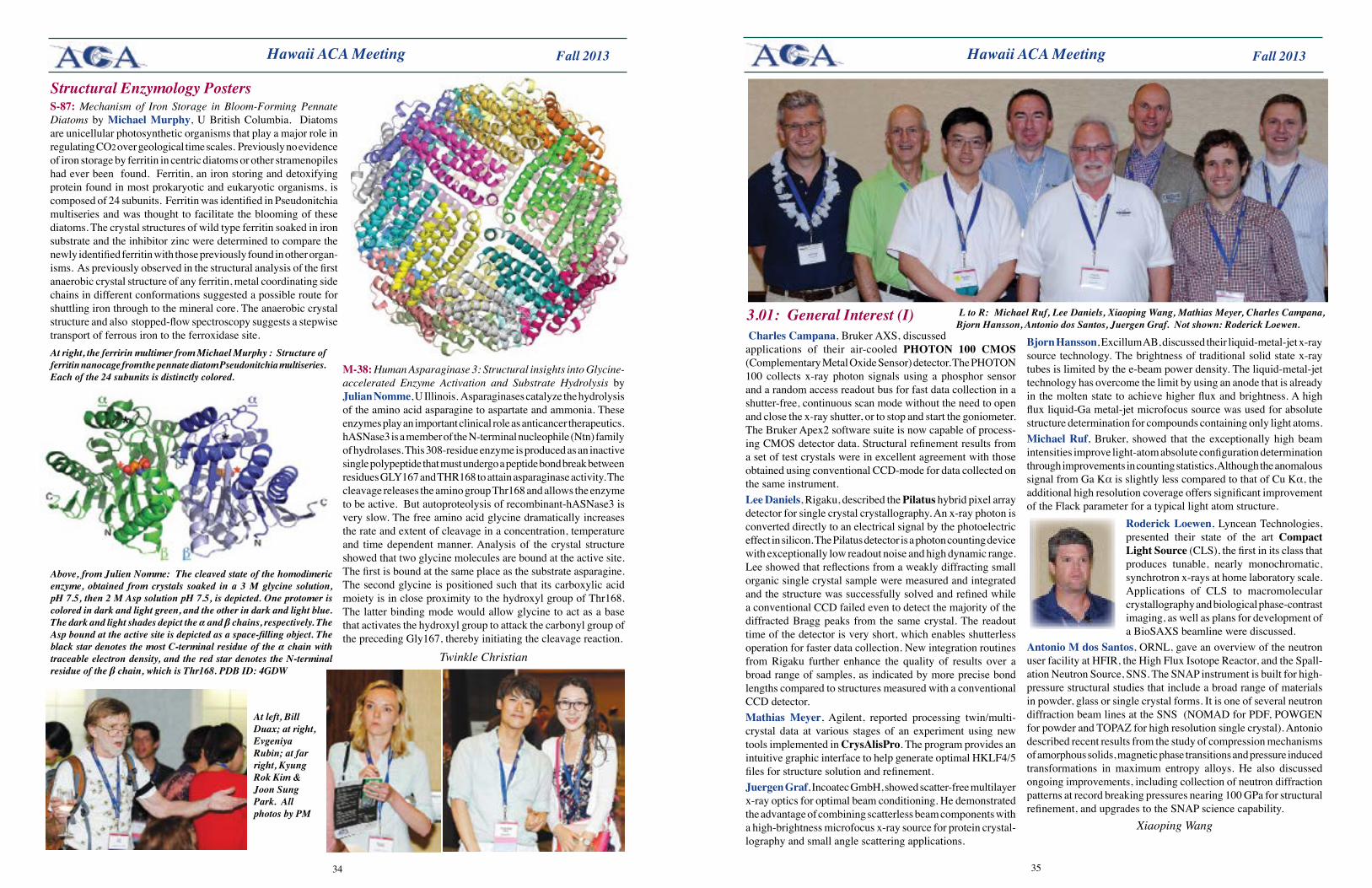
3534
Fall 2013 Fall 2013 Hawaii ACA Meeting
Above, from Julien Nomme: The cleaved state of the homodimeric enzyme, obtained from crystals soaked in a 3 M glycine solution, pH 7.5, then 2 M Asp solution pH 7.5, is depicted. One protomer is colored in dark and light green, and the other in dark and light blue. The dark and light shades depict the α and β chains, respectively. The Asp bound at the active site is depicted as a space-filling object. The black star denotes the most C-terminal residue of the α chain with traceable electron density, and the red star denotes the N-terminal residue of the β chain, which is Thr168. PDB ID: 4GDW
At right, the ferririn multimer from Michael Murphy : Structure of ferritin nanocage from the pennate diatom Pseudonitchia multiseries. Each of the 24 subunits is distinctly colored.
Hawaii ACA Meeting
Charles Campana, Bruker AXS, discussed applications of their air-cooled PHOTON 100 CMOS (Complementary Metal Oxide Sensor) detector. The PHOTON 100 collects x-ray photon signals using a phosphor sensor and a random access readout bus for fast data collection in a shutter-free, continuous scan mode without the need to open and close the x-ray shutter, or to stop and start the goniometer. The Bruker Apex2 software suite is now capable of process-ing CMOS detector data. Structural refinement results from a set of test crystals were in excellent agreement with those obtained using conventional CCD-mode for data collected on the same instrument.Lee Daniels, Rigaku, described the Pilatus hybrid pixel array detector for single crystal crystallography. An x-ray photon is converted directly to an electrical signal by the photoelectric effect in silicon. The Pilatus detector is a photon counting device with exceptionally low readout noise and high dynamic range. Lee showed that reflections from a weakly diffracting small organic single crystal sample were measured and integrated and the structure was successfully solved and refined while a conventional CCD failed even to detect the majority of the diffracted Bragg peaks from the same crystal. The readout time of the detector is very short, which enables shutterless operation for faster data collection. New integration routines from Rigaku further enhance the quality of results over a broad range of samples, as indicated by more precise bond lengths compared to structures measured with a conventional CCD detector. Mathias Meyer, Agilent, reported processing twin/multi-crystal data at various stages of an experiment using new tools implemented in CrysAlisPro. The program provides an intuitive graphic interface to help generate optimal HKLF4/5 files for structure solution and refinement.JuergenGraf, Incoatec GmbH, showed scatter-free multilayer x-ray optics for optimal beam conditioning. He demonstrated the advantage of combining scatterless beam components with a high-brightness microfocus x-ray source for protein crystal-lography and small angle scattering applications.
3.01: General Interest (I) Bjorn Hansson, Excillum AB, discussed their liquid-metal-jet x-ray source technology. The brightness of traditional solid state x-ray tubes is limited by the e-beam power density. The liquid-metal-jet technology has overcome the limit by using an anode that is already in the molten state to achieve higher flux and brightness. A high flux liquid-Ga metal-jet microfocus source was used for absolute structure determination for compounds containing only light atoms. MichaelRuf, Bruker, showed that the exceptionally high beam intensities improve light-atom absolute configuration determination through improvements in counting statistics. Although the anomalous signal from Ga Kα is slightly less compared to that of Cu Kα, the additional high resolution coverage offers significant improvement of the Flack parameter for a typical light atom structure.
Roderick Loewen, Lyncean Technologies, presented their state of the art Compact Light Source (CLS), the first in its class that produces tunable, nearly monochromatic, synchrotron x-rays at home laboratory scale. Applications of CLS to macromolecular crystallography and biological phase-contrast imaging, as well as plans for development of a BioSAXS beamline were discussed.
Antonio M dos Santos, ORNL, gave an overview of the neutron user facility at HFIR, the High Flux Isotope Reactor, and the Spall-ation Neutron Source, SNS. The SNAP instrument is built for high-pressure structural studies that include a broad range of materials in powder, glass or single crystal forms. It is one of several neutron diffraction beam lines at the SNS (NOMAD for PDF, POWGEN for powder and TOPAZ for high resolution single crystal). Antonio described recent results from the study of compression mechanisms of amorphous solids, magnetic phase transitions and pressure induced transformations in maximum entropy alloys. He also discussed ongoing improvements, including collection of neutron diffraction patterns at record breaking pressures nearing 100 GPa for structural refinement, and upgrades to the SNAP science capability.
Xiaoping Wang
L to R: Michael Ruf, Lee Daniels, Xiaoping Wang, Mathias Meyer, Charles Campana, Bjorn Hansson, Antonio dos Santos, Juergen Graf. Not shown: Roderick Loewen.
S-87: Mechanism of Iron Storage in Bloom-Forming Pennate Diatoms by Michael Murphy, U British Columbia. Diatoms are unicellular photosynthetic organisms that play a major role in regulating CO2 over geological time scales. Previously no evidence of iron storage by ferritin in centric diatoms or other stramenopiles had ever been found. Ferritin, an iron storing and detoxifying protein found in most prokaryotic and eukaryotic organisms, is composed of 24 subunits. Ferritin was identified in Pseudonitchia multiseries and was thought to facilitate the blooming of these diatoms. The crystal structures of wild type ferritin soaked in iron substrate and the inhibitor zinc were determined to compare the newly identified ferritin with those previously found in other organ-isms. As previously observed in the structural analysis of the first anaerobic crystal structure of any ferritin, metal coordinating side chains in different conformations suggested a possible route for shuttling iron through to the mineral core. The anaerobic crystal structure and also stopped-flow spectroscopy suggests a stepwise transport of ferrous iron to the ferroxidase site.
M-38: Human Asparaginase 3: Structural insights into Glycine-accelerated Enzyme Activation and Substrate Hydrolysis by Julian Nomme, U Illinois. Asparaginases catalyze the hydrolysis of the amino acid asparagine to aspartate and ammonia. These enzymes play an important clinical role as anticancer therapeutics. hASNase3 is a member of the N-terminal nucleophile (Ntn) family of hydrolases. This 308-residue enzyme is produced as an inactive single polypeptide that must undergo a peptide bond break between residues GLY167 and THR168 to attain asparaginase activity. The cleavage releases the amino group Thr168 and allows the enzyme to be active. But autoproteolysis of recombinant-hASNase3 is very slow. The free amino acid glycine dramatically increases the rate and extent of cleavage in a concentration, temperature and time dependent manner. Analysis of the crystal structure showed that two glycine molecules are bound at the active site. The first is bound at the same place as the substrate asparagine. The second glycine is positioned such that its carboxylic acid moiety is in close proximity to the hydroxyl group of Thr168. The latter binding mode would allow glycine to act as a base that activates the hydroxyl group to attack the carbonyl group of the preceding Gly167, thereby initiating the cleavage reaction.
Twinkle Christian
Structural Enzymology Posters
At left, Bill Duax; at right, Evgeniya Rubin; at far right, Kyung Rok Kim & Joon Sung Park. All photos by PM
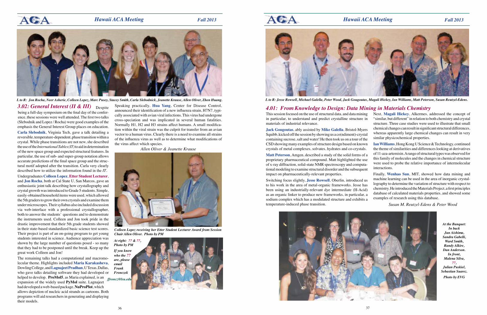
3736
Fall 2013 Fall 2013 Hawaii ACA Meeting
L to R: Jon Rocha, Neer Asherie, Colleen Lopez, Marc Pusey, Stacey Smith, Carla Slebodnick, Jeanette Krause, Allen Oliver, Zhen Huang.
Despite being a full-day symposium on the final day of the confer-ence, these sessions were well attended. The first two talks (Slebodnik and Lopez / Rocha) were good examples of the emphasis the General Interest Group places on education. Carla Slebodnik, Virginia Tech, gave a talk detailing a reversible, temperature-dependent, phase transition within a crystal. While phase transitions are not new, she described the use of the International Tables (IT) to aid in determination of the new space group and expected structural features. In particular, the use of sub- and super-group notation allows accurate predictions of the final space group and the struc-tural motif adopted after the transition. Carla very clearly described how to utilize the information found in the IT.Undergraduates Colleen Lopez, Etter Student Lecturer, and Jon Rocha, both at Cal State U, San Marcos, gave an enthusiastic joint talk describing how crystallography and crystal growth was introduced to Grade 5 students. Simple, easily-obtained household items were used, which allowed the 5th graders to grow their own crystals and examine them under microscopes. Their syllabus also included discussion via web-interface with a professional crystallographer, both to answer the students’ questions and to demonstrate the instruments used. Colleen and Jon took pride in the drastic improvement that their 5th grade students showed in their state-based standardized basic science test scores. Their project is part of an on-going program to get young students interested in science. Audience appreciation was shown by the large number of questions posed - so many that they had to be postponed until the break. Keep up the great work Colleen and Jon!The remaining talks had a computational and macromo-lecular theme. Highlights included Maria Karakasheva, Dowling College, and Lagnajeet Pradhan, U Texas, Dallas, who gave talks detailing software they had developed or helped to develop. ProMol5, as Maria explained, is an expansion of the widely used PyMol suite. Lagnajeet had developed a web-based package, NuProPlot, which allows depiction of nucleic acid strands as cartoons. Both programs will aid researchers in generating and displaying their models.
3.02: General Interest (II & III) Speaking practically, Hua Yang, Center for Disease Control, announced their identification of a new influenza strain, H7N7, typi-cally associated with avian viral infections. This virus had undergone cross-speciation and was implicated in several human fatalities. Normally H1, H2 and H3 strains affect humans. A small modifica-tion within the viral strain was the culprit for transfer from an avian vector to a human virus. Clearly there is a need to examine all strains of the influenza virus as well as to determine what modifications of the virus affect which species.
Allen Oliver & Jeanette Krause
Colleen Lopez receiving her Etter Student Lecturer Award from Session Chair Allen Oliver. Photo by PM
Hawaii ACA Meeting
L to R: Jesse Rowsell, Michael Galella, Peter Wood, Jack Gougoutas, Magali Hickey, Ian Williams, Matt Peterson, Susan Reutzel-Edens.
4.01: From Knowledge to Design: Data Mining in Materials ChemistryThis session focused on the use of structural data, and data mining in particular, to understand and predict crystalline structure in materials of industrial relevance.JackGougoutas, ably assisted by MikeGalella, Bristol-Myers Squibb, kicked off the session by showing us a co(ndiment)-crystal containing sucrose, salt and water! He then took us on a tour of the CSD showing many examples of structure design based on known crystals of metal complexes, solvates, hydrates and co-crystals. Matt Peterson, Amgen, described a study of the solid forms of a proprietary pharmaceutical compound. Matt highlighted the use of x-ray diffraction, solid-state NMR spectroscopy and computa-tional modeling to examine structural disorder and the subsequent impact on pharmaceutically-relevant properties. Switching focus slightly, Jesse Rowsell, Oberlin, introduced us to his work in the area of metal-organic frameworks. Jesse has been using an industrially-relevant dye intermediate (H-Acid) as an organic linker to produce new frameworks, in particular, a sodium complex which has a modulated structure and exhibits a temperature-induced phase transition.
Next, Magali Hickey, Alkermes, addressed the concept of “similar, but different” in relation to both chemistry and crystal structure. Three case studies were used to illustrate that small chemical changes can result in significant structural differences, whereas apparently large chemical changes can result in very similar physicochemical properties. Ian Williams, Hong Kong U Science & Technology, continued the theme of similarities and differences looking at derivatives of 11-aza-artemisin. A range of structural types was observed for this family of molecules and the changes in chemical structure were used to probe the relative importance of intermolecular interactions.Finally, Wenhao Sun, MIT, showed how data mining and machine learning can be used in the area of inorganic crystal-lography to determine the variation of structure with respect to chemistry. He introduced the Materials Project, a first principles database of calculated materials properties, and showed some examples of research using this database.
Susan M. Reutzel-Edens & Peter Wood
At the Banquet: In back
Jun Aishima, Sandra Gabelli,
Ward Smith, Randy Alkire,
Dan Anderson. In front,
Malena Silva, ??,
Julian Puskiel, Sebastian Suarez.
Photo by EVG
At right: ?? & ??, Photo by PM If you know who the ?? are, please email Frank Fronczek
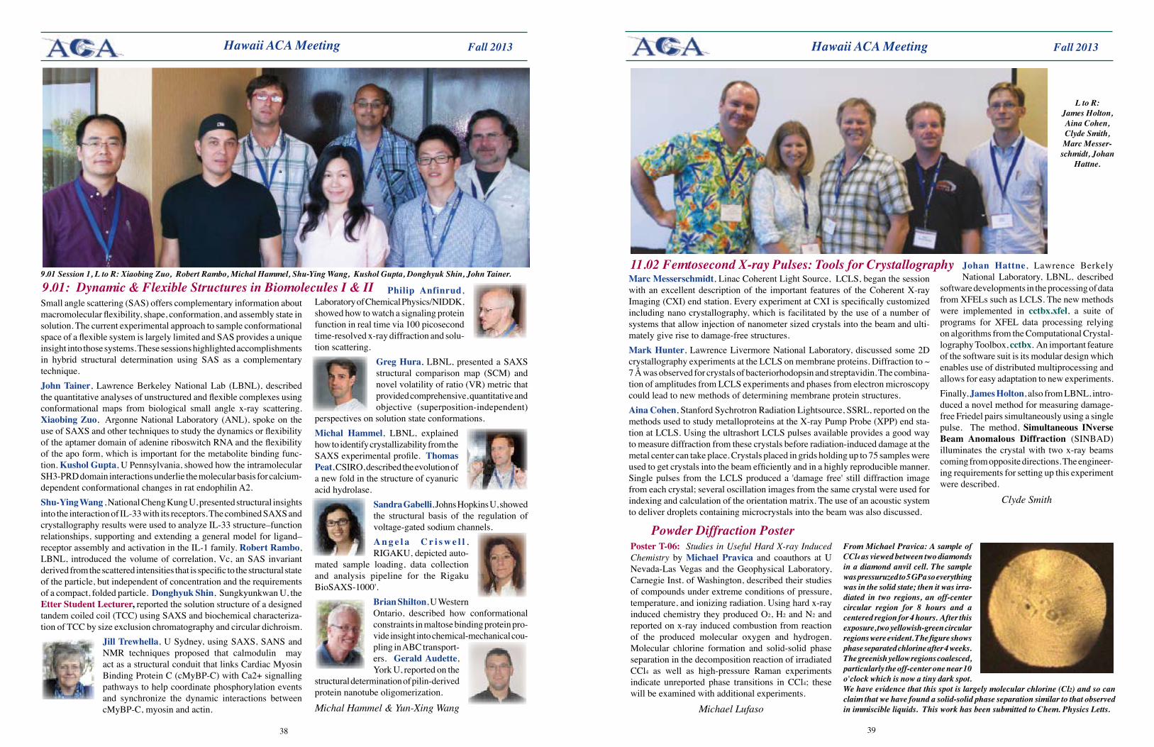
3938
Fall 2013 Fall 2013 Hawaii ACA Meeting Hawaii ACA Meeting
Small angle scattering (SAS) offers complementary information about macromolecular flexibility, shape, conformation, and assembly state in solution. The current experimental approach to sample conformational space of a flexible system is largely limited and SAS provides a unique insight into those systems. These sessions highlighted accomplishments in hybrid structural determination using SAS as a complementary technique.John Tainer, Lawrence Berkeley National Lab (LBNL), described the quantitative analyses of unstructured and flexible complexes using conformational maps from biological small angle x-ray scattering. XiaobingZuo, Argonne National Laboratory (ANL), spoke on the use of SAXS and other techniques to study the dynamics or flexibility of the aptamer domain of adenine riboswitch RNA and the flexibility of the apo form, which is important for the metabolite binding func-tion. KusholGupta, U Pennsylvania, showed how the intramolecular SH3-PRD domain interactions underlie the molecular basis for calcium-dependent conformational changes in rat endophilin A2. Shu-YingWang , National Cheng Kung U, presented structural insights into the interaction of IL-33 with its receptors. The combined SAXS and crystallography results were used to analyze IL-33 structure–function relationships, supporting and extending a general model for ligand–receptor assembly and activation in the IL-1 family. Robert Rambo, LBNL, introduced the volume of correlation, Vc, an SAS invariant derived from the scattered intensities that is specific to the structural state of the particle, but independent of concentration and the requirements of a compact, folded particle. Donghyuk Shin, Sungkyunkwan U, the Etter Student Lecturer, reported the solution structure of a designed tandem coiled coil (TCC) using SAXS and biochemical characteriza-tion of TCC by size exclusion chromatography and circular dichroism.
Jill Trewhella, U Sydney, using SAXS, SANS and NMR techniques proposed that calmodulin may act as a structural conduit that links Cardiac Myosin Binding Protein C (cMyBP-C) with Ca2+ signalling pathways to help coordinate phosphorylation events and synchronize the dynamic interactions between cMyBP-C, myosin and actin.
9.01: Dynamic & Flexible Structures in Biomolecules I & II9.01 Session 1, L to R: Xiaobing Zuo, Robert Rambo, Michal Hammel, Shu-Ying Wang, Kushol Gupta, Donghyuk Shin, John Tainer.
Philip Anfinrud, Laboratory of Chemical Physics/NIDDK, showed how to watch a signaling protein function in real time via 100 picosecond time-resolved x-ray diffraction and solu-tion scattering.
GregHura, LBNL, presented a SAXS structural comparison map (SCM) and novel volatility of ratio (VR) metric that provided comprehensive, quantitative and objective (superposition-independent)
perspectives on solution state conformations. Michal Hammel, LBNL, explained how to identify crystallizability from the SAXS experimental profile. Thomas Peat, CSIRO, described the evolution of a new fold in the structure of cyanuric acid hydrolase.
SandraGabelli, Johns Hopkins U, showed the structural basis of the regulation of voltage-gated sodium channels. A n g e l a C r i s w e l l , RIGAKU, depicted auto-
mated sample loading, data collection and analysis pipeline for the Rigaku BioSAXS-1000'.
Brian Shilton, U Western Ontario, described how conformational constraints in maltose binding protein pro-vide insight into chemical-mechanical cou-pling in ABC transport-ers. GeraldAudette, York U, reported on the
structural determination of pilin-derived protein nanotube oligomerization.
Michal Hammel & Yun-Xing Wang
L to R: James Holton, Aina Cohen, Clyde Smith, Marc Messer-
schmidt, Johan Hattne.
PosterT-06: Studies in Useful Hard X-ray Induced Chemistry by Michael Pravica and coauthors at U Nevada-Las Vegas and the Geophysical Laboratory, Carnegie Inst. of Washington, described their studies of compounds under extreme conditions of pressure, temperature, and ionizing radiation. Using hard x-ray induced chemistry they produced O2, H2 and N2 and reported on x-ray induced combustion from reaction of the produced molecular oxygen and hydrogen. Molecular chlorine formation and solid-solid phase separation in the decomposition reaction of irradiated CCl4 as well as high-pressure Raman experiments indicate unreported phase transitions in CCl4; these will be examined with additional experiments.
Michael Lufaso
From Michael Pravica: A sample of CCl4 as viewed between two diamonds in a diamond anvil cell. The sample was pressuruzed to 5 GPa so everything was in the solid state; then it was irra-diated in two regions, an off-center circular region for 8 hours and a centered region for 4 hours. After this exposure, two yellowish-green circular regions were evident. The figure shows phase separated chlorine after 4 weeks. The greenish yellow regions coalesced, particularly the off-center one near 10 o'clock which is now a tiny dark spot. We have evidence that this spot is largely molecular chlorine (Cl2) and so can claim that we have found a solid-solid phase separation similar to that observed in immiscible liquids. This work has been submitted to Chem. Physics Letts.
Marc Messerschmidt, Linac Coherent Light Source, LCLS, began the session with an excellent description of the important features of the Coherent X-ray Imaging (CXI) end station. Every experiment at CXI is specifically customized including nano crystallography, which is facilitated by the use of a number of systems that allow injection of nanometer sized crystals into the beam and ulti-mately give rise to damage-free structures. Mark Hunter, Lawrence Livermore National Laboratory, discussed some 2D crystallography experiments at the LCLS on membrane proteins. Diffraction to ~ 7 Å was observed for crystals of bacteriorhodopsin and streptavidin. The combina-tion of amplitudes from LCLS experiments and phases from electron microscopy could lead to new methods of determining membrane protein structures. Aina Cohen, Stanford Sychrotron Radiation Lightsource, SSRL, reported on the methods used to study metalloproteins at the X-ray Pump Probe (XPP) end sta-tion at LCLS. Using the ultrashort LCLS pulses available provides a good way to measure diffraction from these crystals before radiation-induced damage at the metal center can take place. Crystals placed in grids holding up to 75 samples were used to get crystals into the beam efficiently and in a highly reproducible manner. Single pulses from the LCLS produced a 'damage free' still diffraction image from each crystal; several oscillation images from the same crystal were used for indexing and calculation of the orientation matrix. The use of an acoustic system to deliver droplets containing microcrystals into the beam was also discussed.
Johan Hattne, Lawrence Berkely National Laboratory, LBNL, described
software developments in the processing of data from XFELs such as LCLS. The new methods were implemented in cctbx.xfel, a suite of programs for XFEL data processing relying on algorithms from the Computational Crystal-lography Toolbox, cctbx. An important feature of the software suit is its modular design which enables use of distributed multiprocessing and allows for easy adaptation to new experiments. Finally, James Holton, also from LBNL, intro-duced a novel method for measuring damage-free Friedel pairs simultaneously using a single pulse. The method, Simultaneous INverse Beam Anomalous Diffraction (SINBAD) illuminates the crystal with two x-ray beams coming from opposite directions. The engineer-ing requirements for setting up this experiment were described.
Clyde Smith
11.02 Femtosecond X-ray Pulses: Tools for Crystallography
Powder Diffraction Poster
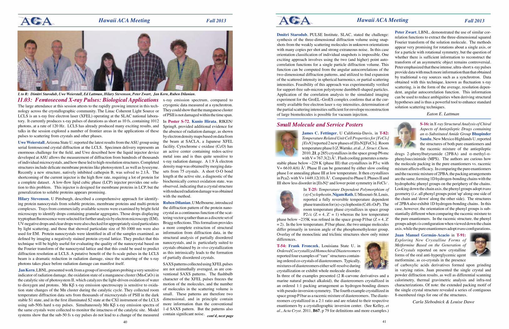
4140
Fall 2013 Fall 2013 Hawaii ACA Meeting Hawaii ACA Meeting
The large attendance at this session attests to the rapidly growing interest in this tech-nology across the crystallographic community. The Linac Coherent Light Source or LCLS is an x-ray free electron laser (XFEL) operating at the SLAC national labora-tory. It currently produces x-ray pulses of durations as short as 10 fs, containing 1012 photons, at a rate of 120 Hz. LCLS has already produced many exciting results, and talks in the session explored a number of frontier areas in the applications of these pulses to scattering from crystals and other phases.Uwe Weierstall, Arizona State U, reported the latest results from the ASU group using serial femtosecond crystal diffraction at the LCLS. Specimen delivery represents an enormous challenge for the field, and Uwe described how the liquid injector device developed at ASU allows the measurement of diffraction from hundreds of thousands of individual microcrystals, and how these led to high-resolution structures. Completed structures include delicate membrane proteins like photosystem I, as well as lysozyme. Recently a new structure, natively inhibited cathepsin B, was solved to 2.1Å. The shortcoming of the current injector is the high flow rate, requiring a lot of protein for a complete dataset. A low-flow lipid cubic phase (LCP) injector provides one solu-tion to this problem. This injector is designed for membrane proteins in LCP, but the generalization to soluble proteins appears promising.Hilary Stevenson, U Pittsburgh, described a comprehensive approach for identify-ing protein nanocrystals from soluble proteins, membrane proteins and multi-protein complexes. Trays from commercially available screens were examined by bright field microscopy to identify drops containing granular aggregates. Those drops displaying tryptophan fluorescence were selected for further analysis by electron microscopy (EM). UV-negative drops and clear drops were also checked for appropriately sized particulates by light scattering, and those that showed particulate size of 50-1000 nm were also used for EM. Protein nanocrystals were identified in all of the samples examined, as defined by imaging a negatively stained protein crystal lattice. They predict that this technique will be highly useful for evaluating the quality of the nanocrystal based on the Fourier transform of the nanocrystal lattice and that this could be used to predict diffraction resolution at LCLS. A putative benefit of the fs-scale pulses in the LCLS beam is a dramatic reduction in radiation damage, since the scattering of the x-ray photons takes place before radiation damage can manifest itself. Jan Kern, LBNL, presented work from a group of investigators probing a very sensitive indicator of radiation damage, the oxidation state of a manganese cluster (Mn4CaO5) in the catalytic site of photosystem II, which catalyzes the light-driven oxidation of water to dioxygen and protons. Mn Kβ x-ray emission spectroscopy is sensitive to oxida-tion state changes of the Mn cluster during the catalytic cycle. They collected room temperature diffraction data sets from thousands of microcrystals of PSII in the dark stable S1 state, and in the first illuminated S2 state at the CXI instrument of the LCLS using sub-50fs hard x-ray pulses. Simultaneously Mn Kβ x-ray emission spectra of the same crystals were collected to monitor the intactness of the catalytic site. Μodel systems show that the sub-50 fs x-ray pulses do not lead to a change of the measured
11.03: Femtosecond X-ray Pulses: Biological ApplicationsL to R: Dimitri Starodub, Uwe Weierstall, Ed Lattman, Hilary Stevenson, Peter Zwart, Jan Kern, Ruben Dilanian.
x-ray emission spectrum, compared to cryogenic data measured at a synchrotron. They could show that the manganese cluster of PSII is not damaged within the time span. In Poster S-72, Kunio Hirata, RIKEN/Spring8, provided additional evidence for the absence of radiation damage, as shown by electron density maps based on data from the beam at SACLA, a Japanese XFEL facility. Cytochrome c oxidase (CcO) has an active center containing redox-active metal ions and is thus quite sensitive to x-ray radiation damage. A 1.9 Å electron density map was obtained by merging data-sets from 75 crystals. A short O-O bond length at the active site, a diagnostic of the biochemically correct oxidation state, was observed, indicating that a crystal structure with reduced radiation damage was obtained with the method.Ruben Dilanian, U Melbourne, introduced the diffraction pattern of the protein nano-crystal as a continuous function of the scat-tering vector q rather than as a discrete set of Bragg reflections. This formalism provides a more complete extraction of structural information from diffraction data, in the structural analysis of partially disordered nanocrystals, and is particularly suited to crystals obtained by in vivo crystallization as this intrinsically leads to the formation of partially disordered crystals.SAXS patterns collected using XFEL pulses are not azimuthally averaged, as are con-ventional SAXS patterns. The flashbulb character of the XFEL pulses freezes the motion of the molecules, and the number of molecules in the scattering volume is small. These patterns are therefore two dimensional, and in principle contain more information than the conventional 1-d SAXS pattern. But the patterns also contain significant noise. cont'd, next page
Dmitri Starodub, PULSE Institute, SLAC, stated the challenge: synthesis of the three-dimensional diffraction volume using snap-shots from the weakly scattering molecules in unknown orientations with many copies per shot and strong extraneous noise. In this case orientation classification of individual snapshots is impossible. One exciting approach involves using the two (and higher) point auto-correlation functions for a single particle diffraction volume. This function can be computed from the angular autocorrelations of the two-dimensional diffraction patterns, and utilized to find expansion of the scattered intensity in spherical harmonics, or partial scattering intensities. Feasibility of this approach was experimentally verified for support-free sub-micron polystyrene dumbbell-shaped particles. Application of the correlation analysis to the simulated imaging experiment for the GroEL–GroES complex confirms that at the cur-rently available free electron laser x-ray intensities, determination of the partial scattering intensities sufficient for envelope reconstruction of large biomolecules is possible for vacuum injection.
Peter Zwart, LBNL, demonstrated the use of similar cor-relation functions to extract the three-dimensional squared Fourier transform of the solution molecule. The methods appear very promising for rotations about a single axis, or for a particle with rotational symmetry, but the question of whether there is sufficient information to reconstruct the transform of an asymmetric object remains controversial. Peter emphasized that these intense, ultra-short x-ray pulses provide data with much more information than that obtained by traditional x-ray sources such as a synchrotron. Data obtained with this technique, known as fluctuation x-ray scattering, is in the form of the average, resolution depen-dent, angular autocorrelation function. This information can be used to reduce ambiguities when deriving structural hypotheses and is thus a powerful tool to enhance standard solution scattering techniques.
Eaton E. Lattman
Small Molecule and Service PostersJamesC.Fettinger, U California-Davis, in T-82: Temperature Related Unit Cell Properties for [FeCl4][Et4N] reported 2 new phases of [Et4N][FeCl4]. Room temperature phase I (Z.Warnke, et al., J. Struct. Chem. 2010, 21, p 285) crystallizes in the space group P63mc with V = 767.3(2) Å3. Flash cooling generates a meta-
stable phase below ~229 K (phase III) that crystallizes in P31c with V= 6610.4(6) Å3. Phase II can be generated by either slow cooling phase I or annealing phase III at low temperature. It then crystallizes in Pca21 with V= 1449.12(10) Å3. Compared to Phase I, Phases II and III show less disorder in [Et4N]+ and lower point symmetry in FeCl4−.
In T-25: Temperature Dependent Polymorphism of (±)-Cyclophostin, Nigam Rath, U Missouri-St. Louis, reported a fully reversible temperature dependent phase transition for (±)-cyclophostin (C8H11O6P). The room temperature phase crystallizes in space group P21/c (Z = 4, Z’ = 1) whereas the low temperature
phase below ~235K was refined in the space group P1bar (Z = 4, Z' = 2). In the low temperature, P1bar phase, the two unique molecules differ primarily in torsion angle of the phosphomethylester group. Overlay of the monoclinic and triclinic structures show only minor differences.T-54: Frank Fronczek, Louisiana State U, in Ordered Cocrystallized Homochiral Diastereomers reported four examples of “rare” structures contain-ing ordered co-crystals of diastereomers. Typically, mixtures of diastereomers either self-resolve during crystallization or exhibit whole molecule disorder. In three of the examples presented (2 R-carvone derivatives and a marine natural product alkaloid), the diastereomers crystallized in an ordered 1:1 packing arrangement as hydrogen-bonding dimers with pseudo-inversion symmetry. The fourth example crystallized in space group P1bar as a racemic mixture of diastereomers. The diaste-reomers crystallized in a 2:1 ratio and are related to their respective enantiomers by a crystallographic inversion center. (See Kelley, et al., Acta Cryst. 2011, B67, p 79 for definitions and more examples.)
S-16: in X-ray Structural Analysis of Chiral Aspects of Antiepileptic Drugs containing an α-Substituted Amide Group Bhupinder Sandu, New Mexico Highlands U, reported the structures of both pure enantiomers and the racemic mixture of the antiepileptic
drugs 2-phenylbutyramide (2PBA) and α-methyl-α-phenylsuccinimide (MPS). The authors are curious how the molecule packing in the pure enantiomers vs. racemic mixture affects efficacy. In comparing the pure enantiomers and the racemic mixture of 2PBA, the packing arrangements are the same, forming 1D hydrogen-bonding chains with the hydrophobic phenyl groups on the periphery of the chains. Looking down the chain axis, the phenyl groups adopt trans geometry (i.e. all phenyl groups point 'up' along one side of the chain and 'down' along the other side). The structures of 2PBA also exhibit 1D hydrogen-bonding chains. In this case, however, the orientation of the phenyl groups is sub-stantially different when comparing the racemic mixture to the pure enantiomers. In the racemic structure, the phenyl groups adopt cis configuration when viewed down the chain axis, while the pure enantiomers adopt trans configurations.Juan Manuel Germán-Acacio in T-91: Exploring New Crystalline Forms of Metformine Based on the Generation of Co-Crystals reported on new crystalline forms of the oral anti-hyperglycemic agent metformine, as co-crystals in the presence of carboxylic acids derivatives formed upon grinding in varying ratios. Juan presented the single crystal and powder diffraction results, as well as differential scanning calorimetry, thermal gravimetric analysis and infra-red characterizations. Of note: the extended packing motif of the single crystal structure revealed a series of contiguous 8-membered rings for one of the structures.
Carla Slebodnick & Louise Dawe
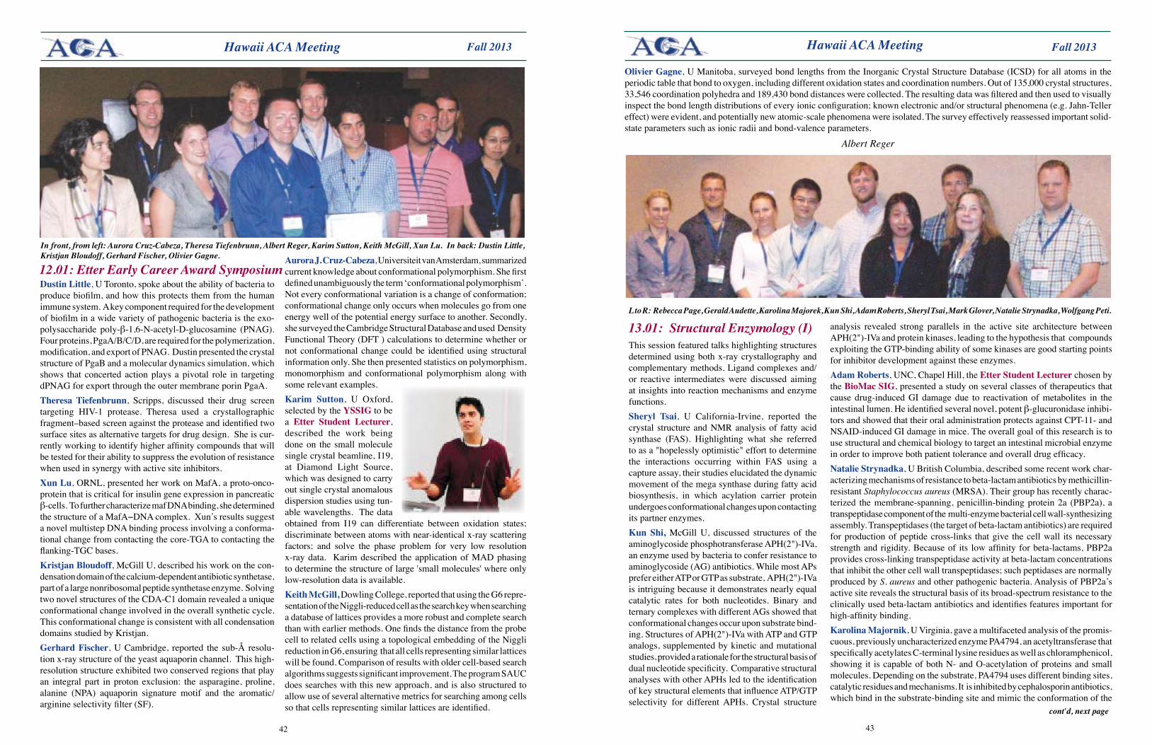
4342
Fall 2013 Fall 2013 Hawaii ACA Meeting Hawaii ACA Meeting
Dustin Little, U Toronto, spoke about the ability of bacteria to produce biofilm, and how this protects them from the human immune system. A key component required for the development of biofilm in a wide variety of pathogenic bacteria is the exo-polysaccharide poly-β-1,6-N-acetyl-D-glucosamine (PNAG). Four proteins, PgaA/B/C/D, are required for the polymerization, modification, and export of PNAG. Dustin presented the crystal structure of PgaB and a molecular dynamics simulation, which shows that concerted action plays a pivotal role in targeting dPNAG for export through the outer membrane porin PgaA.Theresa Tiefenbrunn, Scripps, discussed their drug screen targeting HIV-1 protease. Theresa used a crystallographic fragment–based screen against the protease and identified two surface sites as alternative targets for drug design. She is cur-rently working to identify higher affinity compounds that will be tested for their ability to suppress the evolution of resistance when used in synergy with active site inhibitors.XunLu, ORNL, presented her work on MafA, a proto-onco-protein that is critical for insulin gene expression in pancreatic β-cells. To further characterize maf DNA binding, she determined the structure of a MafA−DNA complex. Xun’s results suggest a novel multistep DNA binding process involving a conforma-tional change from contacting the core-TGA to contacting the flanking-TGC bases.KristjanBloudoff, McGill U, described his work on the con-densation domain of the calcium-dependent antibiotic synthetase, part of a large nonribosomal peptide synthetase enzyme. Solving two novel structures of the CDA-C1 domain revealed a unique conformational change involved in the overall synthetic cycle. This conformational change is consistent with all condensation domains studied by Kristjan.GerhardFischer, U Cambridge, reported the sub-Å resolu-tion x-ray structure of the yeast aquaporin channel. This high-resolution structure exhibited two conserved regions that play an integral part in proton exclusion: the asparagine, proline, alanine (NPA) aquaporin signature motif and the aromatic/arginine selectivity filter (SF).
12.01: Etter Early Career Award Symposium
In front, from left: Aurora Cruz-Cabeza, Theresa Tiefenbrunn, Albert Reger, Karim Sutton, Keith McGill, Xun Lu. In back: Dustin Little, Kristjan Bloudoff, Gerhard Fischer, Olivier Gagne. AuroraJ.Cruz-Cabeza, Universiteit van Amsterdam, summarized
current knowledge about conformational polymorphism. She first defined unambiguously the term ‘conformational polymorphism’. Not every conformational variation is a change of conformation; conformational change only occurs when molecules go from one energy well of the potential energy surface to another. Secondly, she surveyed the Cambridge Structural Database and used Density Functional Theory (DFT ) calculations to determine whether or not conformational change could be identified using structural information only. She then presented statistics on polymorphism, monomorphism and conformational polymorphism along with some relevant examples.Karim Sutton, U Oxford, selected by the YSSIG to be a Etter Student Lecturer, described the work being done on the small molecule single crystal beamline, I19, at Diamond Light Source, which was designed to carry out single crystal anomalous dispersion studies using tun-able wavelengths. The data obtained from I19 can differentiate between oxidation states; discriminate between atoms with near-identical x-ray scattering factors; and solve the phase problem for very low resolution x-ray data. Karim described the application of MAD phasing to determine the structure of large 'small molecules' where only low-resolution data is available.KeithMcGill, Dowling College, reported that using the G6 repre-sentation of the Niggli-reduced cell as the search key when searching a database of lattices provides a more robust and complete search than with earlier methods. One finds the distance from the probe cell to related cells using a topological embedding of the Niggli reduction in G6, ensuring that all cells representing similar lattices will be found. Comparison of results with older cell-based search algorithms suggests significant improvement. The program SAUC does searches with this new approach, and is also structured to allow use of several alternative metrics for searching among cells so that cells representing similar lattices are identified.
OlivierGagne, U Manitoba, surveyed bond lengths from the Inorganic Crystal Structure Database (ICSD) for all atoms in the periodic table that bond to oxygen, including different oxidation states and coordination numbers. Out of 135,000 crystal structures, 33,546 coordination polyhedra and 189,430 bond distances were collected. The resulting data was filtered and then used to visually inspect the bond length distributions of every ionic configuration; known electronic and/or structural phenomena (e.g. Jahn-Teller effect) were evident, and potentially new atomic-scale phenomena were isolated. The survey effectively reassessed important solid-state parameters such as ionic radii and bond-valence parameters.
Albert Reger
This session featured talks highlighting structures determined using both x-ray crystallography and complementary methods. Ligand complexes and/or reactive intermediates were discussed aiming at insights into reaction mechanisms and enzyme functions. Sheryl Tsai, U California-Irvine, reported the crystal structure and NMR analysis of fatty acid synthase (FAS). Highlighting what she referred to as a "hopelessly optimistic" effort to determine the interactions occurring within FAS using a capture assay, their studies elucidated the dynamic movement of the mega synthase during fatty acid biosynthesis, in which acylation carrier protein undergoes conformational changes upon contacting its partner enzymes.Kun Shi, McGill U, discussed structures of the aminoglycoside phosphotransferase APH(2")-IVa, an enzyme used by bacteria to confer resistance to aminoglycoside (AG) antibiotics. While most APs prefer either ATP or GTP as substrate, APH(2")-IVa is intriguing because it demonstrates nearly equal catalytic rates for both nucleotides. Binary and ternary complexes with different AGs showed that conformational changes occur upon substrate bind-ing. Structures of APH(2")-IVa with ATP and GTP analogs, supplemented by kinetic and mutational studies, provided a rationale for the structural basis of dual nucleotide specificity. Comparative structural analyses with other APHs led to the identification of key structural elements that influence ATP/GTP selectivity for different APHs. Crystal structure
13.01: Structural Enzymology (I)L to R: Rebecca Page, Gerald Audette, Karolina Majorek, Kun Shi, Adam Roberts, Sheryl Tsai, Mark Glover, Natalie Strynadka, Wolfgang Peti.
analysis revealed strong parallels in the active site architecture between APH(2")-IVa and protein kinases, leading to the hypothesis that compounds exploiting the GTP-binding ability of some kinases are good starting points for inhibitor development against these enzymes. Adam Roberts, UNC, Chapel Hill, the Etter Student Lecturer chosen by the BioMacSIG, presented a study on several classes of therapeutics that cause drug-induced GI damage due to reactivation of metabolites in the intestinal lumen. He identified several novel, potent β-glucuronidase inhibi-tors and showed that their oral administration protects against CPT-11- and NSAID-induced GI damage in mice. The overall goal of this research is to use structural and chemical biology to target an intestinal microbial enzyme in order to improve both patient tolerance and overall drug efficacy. Natalie Strynadka, U British Columbia, described some recent work char-acterizing mechanisms of resistance to beta-lactam antibiotics by methicillin-resistant Staphylococcus aureus (MRSA). Their group has recently charac-terized the membrane-spanning, penicillin-binding protein 2a (PBP2a), a transpeptidase component of the multi-enzyme bacterial cell wall-synthesizing assembly. Transpeptidases (the target of beta-lactam antibiotics) are required for production of peptide cross-links that give the cell wall its necessary strength and rigidity. Because of its low affinity for beta-lactams, PBP2a provides cross-linking transpeptidase activity at beta-lactam concentrations that inhibit the other cell wall transpeptidases; such peptidases are normally produced by S. aureus and other pathogenic bacteria. Analysis of PBP2a’s active site reveals the structural basis of its broad-spectrum resistance to the clinically used beta-lactam antibiotics and identifies features important for high-affinity binding. Karolina Majornik, U Virginia, gave a multifaceted analysis of the promis-cuous, previously uncharacterized enzyme PA4794, an acetyltransferase that specifically acetylates C-terminal lysine residues as well as chloramphenicol, showing it is capable of both N- and O-acetylation of proteins and small molecules. Depending on the substrate, PA4794 uses different binding sites, catalytic residues and mechanisms. It is inhibited by cephalosporin antibiotics, which bind in the substrate-binding site and mimic the conformation of the
cont'd, next page
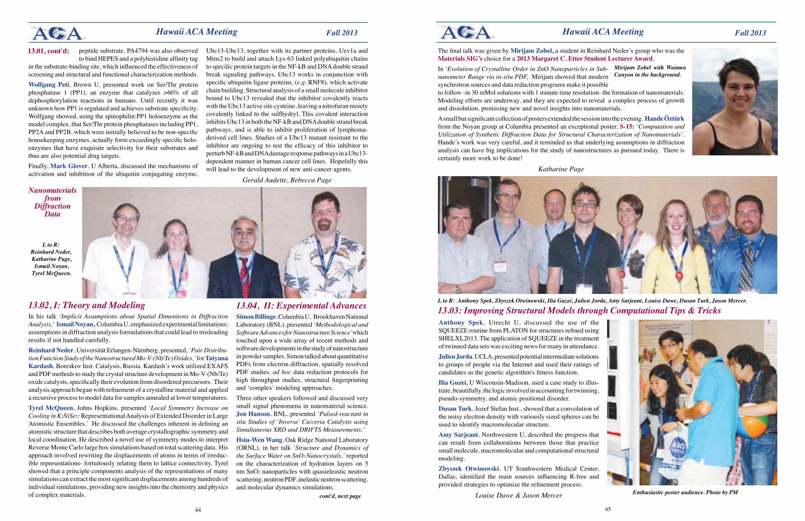
4544
Fall 2013 Fall 2013 Hawaii ACA Meeting
peptide substrate. PA4794 was also observed to bind HEPES and a polyhistidine affinity tag
in the substrate-binding site, which influenced the effectiveness of screening and structural and functional characterization methods.Wolfgang Peti, Brown U, presented work on Ser/Thr protein phosphatase 1 (PP1), an enzyme that catalyzes >60% of all dephosphorylation reactions in humans. Until recently it was unknown how PP1 is regulated and achieves substrate specificity. Wolfgang showed, using the spinophilin:PP1 holoenzyme as the model complex, that Ser/Thr protein phosphatases including PP1, PP2A and PP2B, which were initially believed to be non-specific housekeeping enzymes, actually form exceedingly specific holo-enzymes that have exquisite selectivity for their substrates and thus are also potential drug targets.Finally, MarkGlover, U Alberta, discussed the mechanisms of activation and inhibition of the ubiquitin conjugating enzyme,
Ubc13-Ubc13, together with its partner proteins, Uev1a and Mms2 to build and attach Lys-63 linked polyubiquitin chains to specific protein targets in the NF-kB and DNA double strand break signaling pathways. Ubc13 works in conjunction with specific ubiquitin ligase proteins, (e.g. RNF8), which activate chain building. Structural analysis of a small molecule inhibitor bound to Ubc13 revealed that the inhibitor covalently reacts with the Ubc13 active site cysteine, leaving a nitrofuran moiety covalently linked to the sulfhydryl. This covalent interaction inhibits Ubc13 in both the NF-kB and DNA double strand break pathways, and is able to inhibit proliferation of lymphoma-derived cell lines. Studies of a Ubc13 mutant resistant to the inhibitor are ongoing to test the efficacy of this inhibitor to perturb NF-kB and DNA damage response pathways in a Ubc13-dependent manner in human cancer cell lines. Hopefully this will lead to the development of new anti-cancer agents.
Gerald Audette, Rebecca Page
13.01,cont'd:
Hawaii ACA Meeting
Anthony Spek, Utrecht U, discussed the use of the SQUEEZE routine from PLATON for structures refined using SHELXL2013. The application of SQUEEZE in the treatment of twinned data sets was exciting news for many in attendance.Julien Jorda, UCLA, presented potential intermediate solutions to groups of people via the Internet and used their ratings of candidates as the genetic algorithm's fitness function.IliaGuzei, U Wisconsin-Madison, used a case study to illus-trate, beautifully, the logic involved in accounting for twinning, pseudo-symmetry, and atomic positional disorder. Dusan Turk, Jozef Stefan Inst., showed that a convolution of the noisy electron density with variously sized spheres can be used to identify macromolecular structure. Amy Sarjeant, Northwestern U, described the progress that can result from collaborations between those that practice small molecule, macromolecular and computational structural modeling. Zbyszek Otwinowski, UT Southwestern Medical Center, Dallas, identified the main sources influencing R-free and provided strategies to optimize the refinement process.
Louise Dawe & Jason Mercer
13.03: Improving Structural Models through Computational Tips & Tricks
L to R: Reinhard Neder, Katharine Page, Ismail Noyan,
Tyrel McQueen.
13.02, I: Theory and Modeling In his talk ‘Implicit Assumptions about Spatial Dimentions in Diffraction Analysis,’ Ismail Noyan, Columbia U, emphasized experimental limitations; assumptions in diffraction analysis formulations that could lead to misleading results if not handled carefully. Reinhard Neder, Universität Erlangen-Nürnberg, presented, ‘Pair Distribu-tion Function Study of the Nanostructured Mo-V-(Nb/Te) Oxides,’ for Tatyana Kardash, Boreskov Inst. Catalysis, Russia. Kardash’s work utilized EXAFS and PDF methods to study the crystal structure development in Mo-V-(Nb/Te) oxide catalysts, specifically their evolution from disordered precursors. Their analysis approach began with refinement of a crystalline material and applied a recursive process to model data for samples annealed at lower temperatures. TyrelMcQueen, Johns Hopkins, presented ‘Local Symmetry Increase on Cooling in K2NiSe2: Representational Analysis of Extended Disorder in Large Atomistic Ensembles.’ He discussed the challenges inherent in defining an atomistic structure that describes both average crystallographic symmetry and local coordination. He described a novel use of symmetry modes to interpret Reverse Monte Carlo large box simulations based on total scattering data. His approach involved rewriting the displacements of atoms in terms of irreduc-ible representations- fortuitously relating them to lattice connectivity. Tyrel showed that a principle components analysis of the representations of many simulations can extract the most significant displacements among hundreds of individual simulations, providing new insights into the chemistry and physics of complex materials.
13.04, II: Experimental Advances Simon Billinge, Columbia U, Brookhaven National Laboratory (BNL), presented ‘Methodological and Software Advances for Nanostructure Science’ which touched upon a wide array of recent methods and software developments in the study of nanostructure in powder samples. Simon talked about quantitative PDFs from electron diffraction, spatially resolved PDF studies, ad hoc data reduction protocols for high throughput studies, structural fingerprinting and ‘complex’ modeling approaches. Three other speakers followed and discussed very small signal phenomena in nanomaterial science. Jon Hanson, BNL, presented ‘Pulsed-reactant in situ Studies of ‘Inverse’ Cu/ceria Catalysts using Simultaneous XRD and DRIFTS Measurements.’ Hsiu-WenWang, Oak Ridge National Laboratory (ORNL), in her talk ‘Structure and Dynamics of the Surface Water on SnO2 Nanocrystals,’ reported on the characterization of hydration layers on 5 nm SnO2 nanoparticles with quasieleastic neutron scattering, neutron PDF, inelastic neutron scattering, and molecular dynamics simulations.
Nanomaterials from
Diffraction Data
cont'd, next page
The final talk was given by Mirijam Zobel, a student in Reinhard Neder’s group who was the MaterialsSIG’s choice for a 2013 Margaret C. Etter Student Lecturer Award. In ‘Evolution of Crystalline Order in ZnO Nanoparticles in Sub-nanometer Range via in-situ PDF, Mirijam showed that modern synchrotron sources and data reduction programs make it possible to follow -in 30 mMol solutions with 1 minute time resolution- the formation of nanomaterials. Modeling efforts are underway, and they are expected to reveal a complex process of growth and dissolution, promising new and novel insights into nanomaterials. A small but significant collection of posters extended the session into the evening. Hande Öztürk from the Noyan group at Columbia presented an exceptional poster, S-15: ‘Computation and Utilization of Synthetic Diffraction Data for Structural Characterization of Nanomaterials’. Hande’s work was very careful, and it reminded us that underlying assumptions in diffraction analysis can have big implications for the study of nanostructures as pursued today. There is certainly more work to be done!
Katharine Page
Mirijam Zobel with Waimea Canyon in the background.
L to R: Anthony Spek, Zbyszek Otwinowski, Ilia Guzei, Julien Jorda, Amy Sarjeant, Louise Dawe, Dusan Turk, Jason Mercer.
Enthusiastic poster audience. Photo by PM
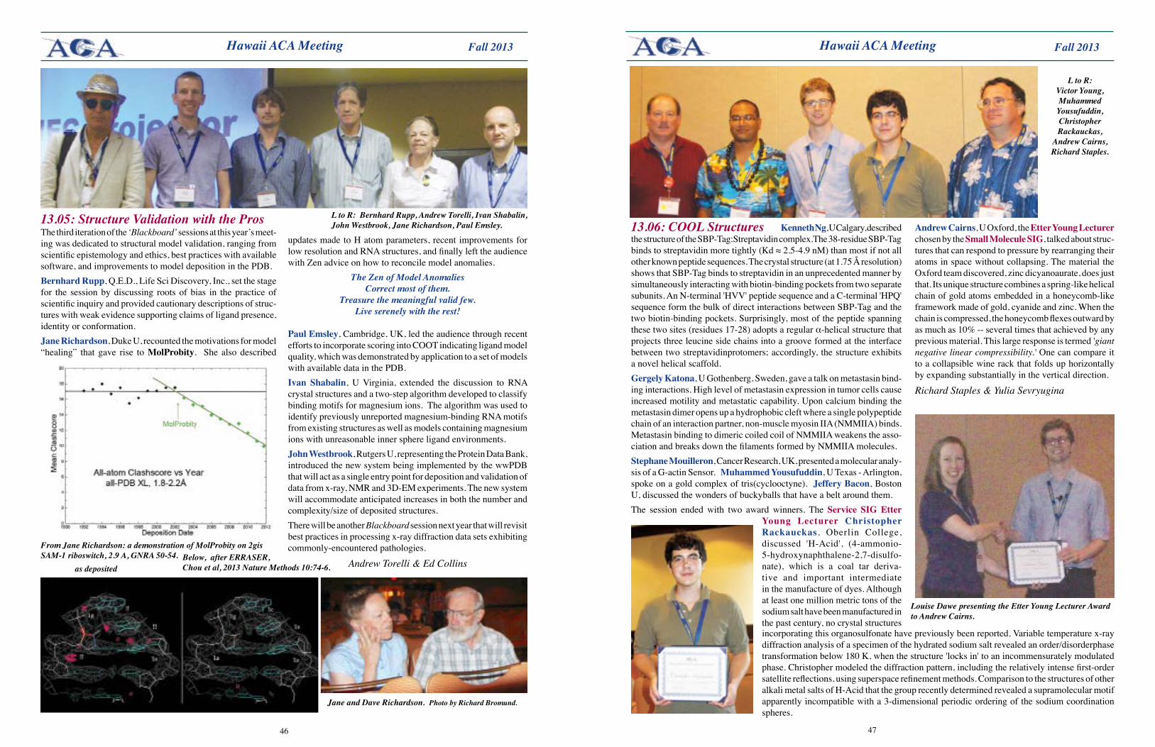
4746
Fall 2013 Fall 2013
From Jane Richardson: a demonstration of MolProbity on 2gis SAM-1 riboswitch, 2.9 A, GNRA 50-54.
Hawaii ACA Meeting Hawaii ACA Meeting
The third iteration of the ‘Blackboard’ sessions at this year’s meet-ing was dedicated to structural model validation, ranging from scientific epistemology and ethics, best practices with available software, and improvements to model deposition in the PDB.Bernhard Rupp, Q.E.D., Life Sci Discovery, Inc., set the stage for the session by discussing roots of bias in the practice of scientific inquiry and provided cautionary descriptions of struc-tures with weak evidence supporting claims of ligand presence, identity or conformation. Jane Richardson, Duke U, recounted the motivations for model “healing” that gave rise to MolProbity. She also described
13.05: Structure Validation with the Pros L to R: Bernhard Rupp, Andrew Torelli, Ivan Shabalin, John Westbrook, Jane Richardson, Paul Emsley.
as deposited
updates made to H atom parameters, recent improvements for low resolution and RNA structures, and finally left the audience with Zen advice on how to reconcile model anomalies.
The Zen of Model AnomaliesCorrect most of them.
Treasure the meaningful valid few.Live serenely with the rest!
Paul Emsley, Cambridge, UK, led the audience through recent efforts to incorporate scoring into COOT indicating ligand model quality, which was demonstrated by application to a set of models with available data in the PDB. Ivan Shabalin, U Virginia, extended the discussion to RNA crystal structures and a two-step algorithm developed to classify binding motifs for magnesium ions. The algorithm was used to identify previously unreported magnesium-binding RNA motifs from existing structures as well as models containing magnesium ions with unreasonable inner sphere ligand environments. John Westbrook, Rutgers U, representing the Protein Data Bank, introduced the new system being implemented by the wwPDB that will act as a single entry point for deposition and validation of data from x-ray, NMR and 3D-EM experiments. The new system will accommodate anticipated increases in both the number and complexity/size of deposited structures. There will be another Blackboard session next year that will revisit best practices in processing x-ray diffraction data sets exhibiting commonly-encountered pathologies.
Andrew Torelli & Ed Collins
Jane and Dave Richardson. Photo by Richard Bromund.
Kenneth Ng, U Calgary, described the structure of the SBP-Tag:Streptavidin complex.The 38-residue SBP-Tag binds to streptavidin more tightly (Kd ≈ 2.5-4.9 nM) than most if not all other known peptide sequences. The crystal structure (at 1.75 Å resolution) shows that SBP-Tag binds to streptavidin in an unprecedented manner by simultaneously interacting with biotin-binding pockets from two separate subunits. An N-terminal 'HVV' peptide sequence and a C-terminal 'HPQ' sequence form the bulk of direct interactions between SBP-Tag and the two biotin-binding pockets. Surprisingly, most of the peptide spanning these two sites (residues 17-28) adopts a regular α-helical structure that projects three leucine side chains into a groove formed at the interface between two streptavidinprotomers; accordingly, the structure exhibits a novel helical scaffold. GergelyKatona, U Gothenberg, Sweden, gave a talk on metastasin bind-ing interactions. High level of metastasin expression in tumor cells cause increased motility and metastatic capability. Upon calcium binding the metastasin dimer opens up a hydrophobic cleft where a single polypeptide chain of an interaction partner, non-muscle myosin IIA (NMMIIA) binds. Metastasin binding to dimeric coiled coil of NMMIIA weakens the asso-ciation and breaks down the filaments formed by NMMIIA molecules. Stephane Mouilleron, Cancer Research, UK, presented a molecular analy-sis of a G-actin Sensor. MuhammedYousufuddin, U Texas - Arlington, spoke on a gold complex of tris(cyclooctyne). JefferyBacon, Boston U, discussed the wonders of buckyballs that have a belt around them. The session ended with two award winners. The Service SIGEtter
Young Lecturer Christopher Rackauckas, Oberlin College, discussed 'H-Acid', (4-ammonio- 5-hydroxynaphthalene-2,7-disulfo-nate), which is a coal tar deriva-tive and important intermediate in the manufacture of dyes. Although at least one million metric tons of the sodium salt have been manufactured in the past century, no crystal structures
13.06: COOL Structures
L to R: Victor Young, Muhammed Yousufuddin, Christopher Rackauckas,
Andrew Cairns, Richard Staples.
Louise Dawe presenting the Etter Young Lecturer Award to Andrew Cairns.
incorporating this organosulfonate have previously been reported. Variable temperature x-ray diffraction analysis of a specimen of the hydrated sodium salt revealed an order/disorderphase transformation below 180 K, when the structure 'locks in' to an incommensurately modulated phase. Christopher modeled the diffraction pattern, including the relatively intense first-order satellite reflections, using superspace refinement methods. Comparison to the structures of other alkali metal salts of H-Acid that the group recently determined revealed a supramolecular motif apparently incompatible with a 3-dimensional periodic ordering of the sodium coordination spheres.
Andrew Cairns, U Oxford, the Etter Young Lecturer chosen by the SmallMoleculeSIG, talked about struc-tures that can respond to pressure by rearranging their atoms in space without collapsing. The material the Oxford team discovered, zinc dicyanoaurate, does just that. Its unique structure combines a spring-like helical chain of gold atoms embedded in a honeycomb-like framework made of gold, cyanide and zinc. When the chain is compressed, the honeycomb flexes outward by as much as 10% -- several times that achieved by any previous material. This large response is termed 'giant negative linear compressibility.' One can compare it to a collapsible wine rack that folds up horizontally by expanding substantially in the vertical direction.
Richard Staples & Yulia Sevryugina
Below, after ERRASER, Chou et al, 2013 Nature Methods 10:74-6.
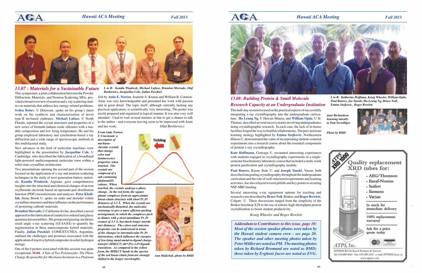
4948
Fall 2013 Fall 2013 Hawaii ACA Meeting Hawaii ACA Meeting
This symposium, a joint collaboration between the Powder Diffraction, Materials, and Neutron Scattering SIGs, pro-vided a broad overview of neutron and x-ray scattering stud-ies on materials that address key energy-related problems. Svilen Bobev, U Delaware, spoke on his group’s latest work on the synthesis and characterization of novel type-II tin-based clathrates. Michael Lufaso, U North Florida, reported the crystal structures and properties of a new series of bismuth indium oxide sillenites with a tun-able composition and low firing temperature. He and his group employed laboratory and synchrotron-based x-ray diffraction and a wide range of spectroscopic methods in this multifaceted study. New advances in the field of molecular machines were highlighted in the presentation by Jacqueline Cole, U Cambridge, who described the fabrication of a broadband light-powered multicomponent molecular rotor within a solid-state crystalline architecture. Two presentations opening the second part of the session focused on the application of x-ray and neutron scattering techniques in the study of next-generation battery materi-als. Kamila Wiaderek, Argonne, gave comprehensive insights into the structural and chemical changes of an iron oxyfluoride electrode based on operando pair distribution function (PDF) measurements and analyses. Peter Khali-fah, Stony Brook U, spoke on order and disorder within crystalline structures and their influence on the performance of promising cathode materials. Brandon Mercado, U California-Irvine, described a novel approach to the fabrication of conductive ordered and glassy quantum dot assemblies. His group used grazing-incidence small angle x-ray scattering (GI-SAXS) to quantify the regimentation in these nanocomposite hybrid materials. Finally, Julian Puszkiel, CONICET-CNEA, Argentina, outlined the challenges and promises associated with the application of reactive hybrid composites in solid-hydrogen storage. One of the 6 posters associated with this session was quite exceptional, M.04, A Tale of Two Polymorphs: The Phase Change Responsible for Mechanochromism in a Platinum
13.07 : Materials for a Sustainable Future
From Amie Norton, U Cincinnati: a description of mechano- chromic crystals that change color and luminescence properties when touched. The crystals are composed of a salt containing platinum(II) cations. When touched, the crystals undergo a phase change. In the red form, the square planar complexes form an approximate linear-chain structure with short Pt..Pt distances of 3.3 Å. When the crystals are mechanically disturbed, the molecules rearrange to give a more efficient packing arrangement, in which the complexes pack as dimers with a short intradimer Pt..Pt contact of 3.3 Å, but much longer interdi-mer distances. The colors and emission properties can be understood in terms of the changes in intermolecular Pt..Pt interactions, which influence the energies of low-lying metal-metal-to-ligand charge-transfer (MMLCT) dσ*(Pt)-->π*(ligand) transitions. As compared to the yellow form, the MMLCT bands in the spectra of the red linear-chain form are strongly shifted to the longer wavelengths.
L to R: Kamila Wiaderek, Michael Lufaso, Brandon Mercado, Olaf Borkiewicz, Jacqueline Cole, Julian Puszkiel.
Salt by Amie E. Norton, Jeanette A. Krause and William B. Connick. Amie was very knowledgeable and presented her work with passion and in great detail. The topic itself, although currently lacking any practical application, is scientifically very interesting. The poster was nicely prepared and organized in logical manner. It was also very well attended - I had to wait several minutes in line to get a chance to talk to the author - and everyone leaving seem to be impressed with Amie and her work. Olaf Borkiewicz
L to R: Katherine Hoffman, Kraig Wheeler, William Ojala, Paul Baures, Joe Tanski, Ho-Leung Ng, Bruce Noll, Emina Stojkovic, Roger Rowlett.
This half-day session focused on the practical aspects of successfully integrating x-ray crystallography into the undergraduate curricu-lum. Ho Leung Ng, U Hawaii-Manoa, and William Ojala, U St. Thomas, described several success stories involving undergraduates doing crystallographic research. In each case, the lack of in-house facilities forged the way to fruitful collaborations. The peer-led team learning strategy highlighted by Emina Stojkovic, Northeastern Illinois U, demonstrated the value of incorporating student-centered experiments into a research course about the essential components of protein x-ray crystallography. KateHoffmann, Gonzaga U, recounted interesting experiences with students engaged in crystallography experiments in a single-semester biochemistry laboratory course that included a multi-week protein purification and crystallography module. Paul Baures, Keene State U, and Joseph Tanski, Vassar, both described integrating crystallography throughout the undergraduate curriculum and the role of well-structured experiments and learning activities. Joe also relayed several pitfalls and key points to securing NSF-MRI funding. Several interesting x-ray equipment options for teaching and research were described by Bruce Noll, Bruker, and Roger Rowlett, Colgate U. These discussions ranged from the simplicity of the Bruker benchtop X2S to the use of robotic high-throughput protein crystallization to boost student productivity.
Kraig Wheeler and Roger Rowlett
13.08: Building Protein & Small Molecule Research Capacity at an Undergraduate Institution
Addendum to Contributors to this issue, page 10: Most of the session speaker photos were taken by the Hawaii student camera crew - see page 20. The speaker and other meeting photos taken by Peter Müller are noted as PM. The meeting photos taken by Richard Bromund are noted as BMD; those taken by Evgheni Jucov are noted as EVG.Ann Mulichak, photo by BMD
Jane Richardson listening intently to Tom Terwilliger.
Photo by BMD
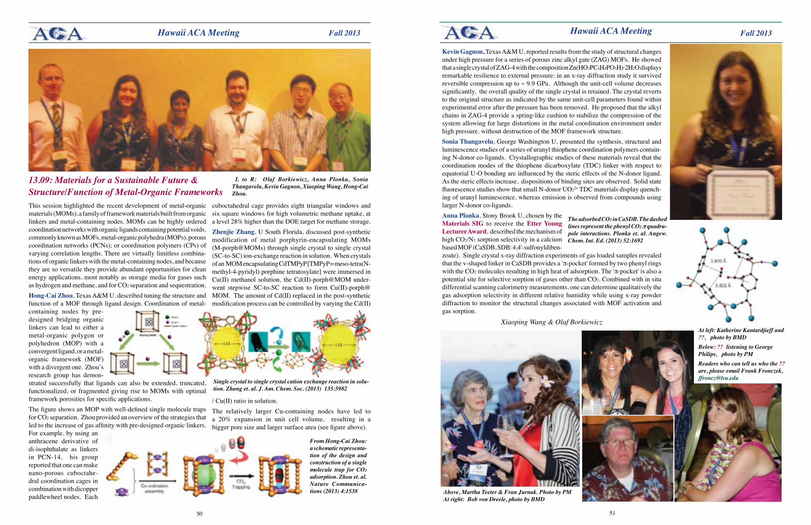
5150
Fall 2013 Fall 2013
cuboctahedral cage provides eight triangular windows and six square windows for high volumetric methane uptake, at a level 28% higher than the DOE target for methane storage.Zhenjie Zhang, U South Florida, discussed post-synthetic modification of metal porphyrin-encapsulating MOMs (M-porph@MOMs) through single crystal to single crystal (SC-to-SC) ion-exchange reaction in solution. When crystals of an MOM encapsulating CdTMPyP [TMPyP = meso-tetra(N-methyl-4-pyridyl) porphine tetratosylate] were immersed in Cu(II) methanol solution, the Cd(II)-porph@MOM under-went stepwise SC-to-SC reaction to form Cu(II)-porph@MOM. The amount of Cd(II) replaced in the post-synthetic modification process can be controlled by varying the Cd(II)
/ Cu(II) ratio in solution. The relatively larger Cu-containing nodes have led to a 20% expansion in unit cell volume, resulting in a bigger pore size and larger surface area (see figure above).
This session highlighted the recent development of metal-organic materials (MOMs), a family of framework materials built from organic linkers and metal-containing nodes. MOMs can be highly ordered coordination networks with organic ligands containing potential voids, commonly known as MOFs, metal-organic polyhedra (MOPs), porous coordination networks (PCNs); or coordination polymers (CPs) of varying correlation lengths. There are virtually limitless combina-tions of organic linkers with the metal-containing nodes, and because they are so versatile they provide abundant opportunities for clean energy applications, most notably as storage media for gases such as hydrogen and methane, and for CO2 separation and sequestration.Hong-CaiZhou, Texas A&M U, described tuning the structure and function of a MOF through ligand design. Coordination of metal-containing nodes by pre-designed bridging organic linkers can lead to either a metal-organic polygon or polyhedron (MOP) with a convergent ligand, or a metal-organic framework (MOF) with a divergent one. Zhou’s research group has demon-strated successfully that ligands can also be extended, truncated, functionalized, or fragmented giving rise to MOMs with optimal framework porosities for specific applications. The figure shows an MOP with well-defined single molecule traps for CO2 separation. Zhou provided an overview of the strategies that led to the increase of gas affinity with pre-designed organic linkers. For example, by using an anthracene derivative of di-isophthalate as linkers in PCN-14, his group reported that one can make nano-porous cuboctahe-dral coordination cages in combination with dicopper paddlewheel nodes. Each
Hawaii ACA Meeting Hawaii ACA Meeting
L to R: Olaf Borkiewicz, Anna Plonka, Sonia Thangavelu, Kevin Gagnon, Xiaoping Wang, Hong-Cai Zhou.
13.09: Materials for a Sustainable Future & Structure/Function of Metal-Organic Frameworks
Single crystal to single crystal cation exchange reaction in solu-tion. Zhang et. al. J. Am. Chem. Soc. (2013) 135:5982
The adsorbed CO2 in CaSDB. The dashed lines represent the phenyl CO2 π quadru-pole interactions. Plonka et. al. Angew. Chem. Int. Ed. (2013) 52:1692
From Hong-Cai Zhou: a schematic representa-tion of the design and construction of a single molecule trap for CO2 adsorption. Zhou et. al. Nature Communica-tions (2013) 4:1538
KevinGagnon, Texas A&M U, reported results from the study of structural changes under high pressure for a series of porous zinc alkyl gate (ZAG) MOFs. He showed that a single crystal of ZAG-4 with the composition Zn(HO3PC4H8PO3H)·2H2O displays remarkable resilience to external pressure; in an x-ray diffraction study it survived reversible compression up to ~ 9.9 GPa. Although the unit-cell volume decreases significantly, the overall quality of the single crystal is retained. The crystal reverts to the original structure as indicated by the same unit-cell parameters found within experimental error after the pressure has been removed. He proposed that the alkyl chains in ZAG-4 provide a spring-like cushion to stabilize the compression of the system allowing for large distortions in the metal coordination environment under high pressure, without destruction of the MOF framework structure.Sonia Thangavelu, George Washington U, presented the synthesis, structural and luminescence studies of a series of uranyl thiophene coordination polymers contain-ing N-donor co-ligands. Crystallographic studies of these materials reveal that the coordination modes of the thiophene dicarboxylate (TDC) linker with respect to equatorial U-O bonding are influenced by the steric effects of the N-donor ligand. As the steric effects increase, dispositions of binding sites are observed. Solid state fluorescence studies show that small N-donor UO22+ TDC materials display quench-ing of uranyl luminescence, whereas emission is observed from compounds using larger N-donor co-ligands. Anna Plonka, Stony Brook U, chosen by the Materials SIG to receive the Etter Young Lecturer Award, described the mechanism of high CO2/N2 sorption selectivity in a calcium based MOF (CaSDB, SDB: 4,4'-sulfonyldiben-zoate). Single crystal x-ray diffraction experiments of gas loaded samples revealed that the v-shaped linker in CaSDB provides a 'π-pocket' formed by two phenyl rings with the CO2 molecules resulting in high heat of adsorption. The 'π-pocket' is also a potential site for selective sorption of gases other than CO2. Combined with in situ differential scanning calorimetry measurements, one can determine qualitatively the gas adsorption selectivity in different relative humidity while using x-ray powder diffraction to monitor the structural changes associated with MOF activation and gas sorption.
Xiaoping Wang & Olaf Borkiewicz
Above, Martha Teeter & Fran Jurnak. Photo by PM At right: Bob von Dreele, photo by BMD
At left: Katherine Kantardjieff and ??, photo by BMDBelow: ?? listening to George Philips, photo by PMReaders who can tell us who the ?? are, please email Frank Fronczek, [email protected]
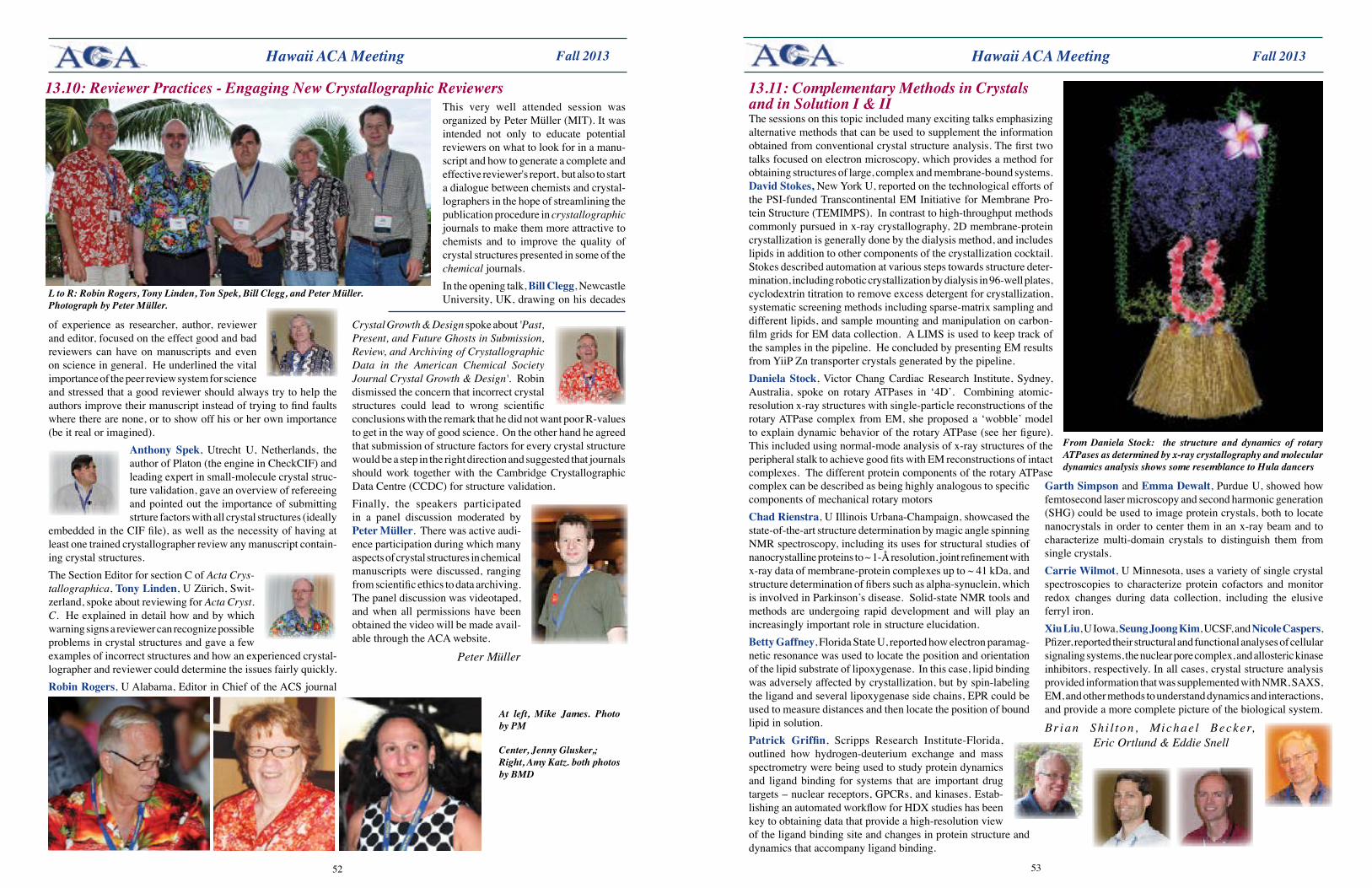
5352
Fall 2013 Fall 2013
Crystal Growth & Design spoke about 'Past, Present, and Future Ghosts in Submission, Review, and Archiving of Crystallographic Data in the American Chemical Society Journal Crystal Growth & Design'. Robin dismissed the concern that incorrect crystal structures could lead to wrong scientific conclusions with the remark that he did not want poor R-values to get in the way of good science. On the other hand he agreed that submission of structure factors for every crystal structure would be a step in the right direction and suggested that journals should work together with the Cambridge Crystallographic Data Centre (CCDC) for structure validation. Finally, the speakers participated in a panel discussion moderated by Peter Müller. There was active audi-ence participation during which many aspects of crystal structures in chemical manuscripts were discussed, ranging from scientific ethics to data archiving. The panel discussion was videotaped, and when all permissions have been obtained the video will be made avail-able through the ACA website.
Peter Müller
of experience as researcher, author, reviewer and editor, focused on the effect good and bad reviewers can have on manuscripts and even on science in general. He underlined the vital importance of the peer review system for science and stressed that a good reviewer should always try to help the authors improve their manuscript instead of trying to find faults where there are none, or to show off his or her own importance (be it real or imagined).
Anthony Spek, Utrecht U, Netherlands, the author of Platon (the engine in CheckCIF) and leading expert in small-molecule crystal struc-ture validation, gave an overview of refereeing and pointed out the importance of submitting strture factors with all crystal structures (ideally
embedded in the CIF file), as well as the necessity of having at least one trained crystallographer review any manuscript contain-ing crystal structures. The Section Editor for section C of Acta Crys-tallographica, Tony Linden, U Zürich, Swit-zerland, spoke about reviewing for Acta Cryst. C. He explained in detail how and by which warning signs a reviewer can recognize possible problems in crystal structures and gave a few examples of incorrect structures and how an experienced crystal-lographer and reviewer could determine the issues fairly quickly. Robin Rogers, U Alabama, Editor in Chief of the ACS journal
Hawaii ACA Meeting Hawaii ACA Meeting
L to R: Robin Rogers, Tony Linden, Ton Spek, Bill Clegg, and Peter Müller. Photograph by Peter Müller.
13.10: Reviewer Practices - Engaging New Crystallographic ReviewersThis very well attended session was organized by Peter Müller (MIT). It was intended not only to educate potential reviewers on what to look for in a manu-script and how to generate a complete and effective reviewer's report, but also to start a dialogue between chemists and crystal-lographers in the hope of streamlining the publication procedure in crystallographic journals to make them more attractive to chemists and to improve the quality of crystal structures presented in some of the chemical journals. In the opening talk, Bill Clegg, Newcastle University, UK, drawing on his decades
13.11: Complementary Methods in Crystals and in Solution I & IIThe sessions on this topic included many exciting talks emphasizing alternative methods that can be used to supplement the information obtained from conventional crystal structure analysis. The first two talks focused on electron microscopy, which provides a method for obtaining structures of large, complex and membrane-bound systems. David Stokes, New York U, reported on the technological efforts of the PSI-funded Transcontinental EM Initiative for Membrane Pro-tein Structure (TEMIMPS). In contrast to high-throughput methods commonly pursued in x-ray crystallography, 2D membrane-protein crystallization is generally done by the dialysis method, and includes lipids in addition to other components of the crystallization cocktail. Stokes described automation at various steps towards structure deter-mination, including robotic crystallization by dialysis in 96-well plates, cyclodextrin titration to remove excess detergent for crystallization, systematic screening methods including sparse-matrix sampling and different lipids, and sample mounting and manipulation on carbon-film grids for EM data collection. A LIMS is used to keep track of the samples in the pipeline. He concluded by presenting EM results from YiiP Zn transporter crystals generated by the pipeline.Daniela Stock, Victor Chang Cardiac Research Institute, Sydney, Australia, spoke on rotary ATPases in ‘4D’. Combining atomic-resolution x-ray structures with single-particle reconstructions of the rotary ATPase complex from EM, she proposed a ‘wobble’ model to explain dynamic behavior of the rotary ATPase (see her figure). This included using normal-mode analysis of x-ray structures of the peripheral stalk to achieve good fits with EM reconstructions of intact complexes. The different protein components of the rotary ATPase
GarthSimpson and Emma Dewalt, Purdue U, showed how femtosecond laser microscopy and second harmonic generation (SHG) could be used to image protein crystals, both to locate nanocrystals in order to center them in an x-ray beam and to characterize multi-domain crystals to distinguish them from single crystals. Carrie Wilmot, U Minnesota, uses a variety of single crystal spectroscopies to characterize protein cofactors and monitor redox changes during data collection, including the elusive ferryl iron. XiuLiu, U Iowa, Seung Joong Kim, UCSF, and Nicole Caspers, Pfizer, reported their structural and functional analyses of cellular signaling systems, the nuclear pore complex, and allosteric kinase inhibitors, respectively. In all cases, crystal structure analysis provided information that was supplemented with NMR, SAXS, EM, and other methods to understand dynamics and interactions, and provide a more complete picture of the biological system.
Brian Sh i l ton , Michae l Becker, Eric Ortlund & Eddie Snell
From Daniela Stock: the structure and dynamics of rotary ATPases as determined by x-ray crystallography and molecular dynamics analysis shows some resemblance to Hula dancers
complex can be described as being highly analogous to specific components of mechanical rotary motorsChad Rienstra, U Illinois Urbana-Champaign, showcased the state-of-the-art structure determination by magic angle spinning NMR spectroscopy, including its uses for structural studies of nanocrystalline proteins to ~ 1-Å resolution, joint refinement with x-ray data of membrane-protein complexes up to ~ 41 kDa, and structure determination of fibers such as alpha-synuclein, which is involved in Parkinson’s disease. Solid-state NMR tools and methods are undergoing rapid development and will play an increasingly important role in structure elucidation. BettyGaffney, Florida State U, reported how electron paramag-netic resonance was used to locate the position and orientation of the lipid substrate of lipoxygenase. In this case, lipid binding was adversely affected by crystallization, but by spin-labeling the ligand and several lipoxygenase side chains, EPR could be used to measure distances and then locate the position of bound lipid in solution.Patrick Griffin, Scripps Research Institute-Florida, outlined how hydrogen-deuterium exchange and mass spectrometry were being used to study protein dynamics and ligand binding for systems that are important drug targets – nuclear receptors, GPCRs, and kinases. Estab-lishing an automated workflow for HDX studies has been key to obtaining data that provide a high-resolution view of the ligand binding site and changes in protein structure and dynamics that accompany ligand binding.
At left, Mike James. Photo by PM
Center, Jenny Glusker,; Right, Amy Katz. both photos by BMD
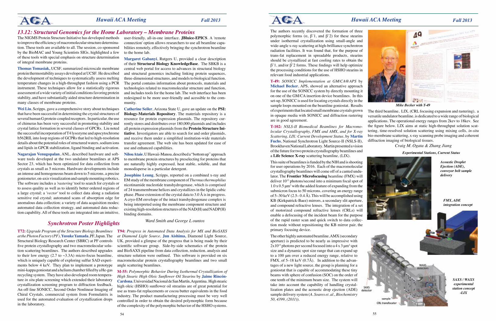
5554
Fall 2013 Fall 2013 Hawaii ACA Meeting Hawaii ACA Meeting
13.12: Structural Genomics for the Home Laboratory – Membrane ProteinsThe NIGMS Protein Structure Initiative has developed methods to improve the efficiency of macromolecular structure determina-tion. These tools are available to all. The session, co-sponsored by the BioMAC and Young Scientists SIGs, highlighted a few of these tools with special emphasis on structure determination of integral membrane proteins.Thomas Tomasiak, UCSF, summarized microscale membrane protein thermostability assays developed at UCSF. He described the development of techniques to systematically assess melting temperature changes in a high-throughput fashion using a PCR instrument. These techniques allow for a statistically rigorous assessment of a wide variety of initial conditions favoring protein stability, and have substantially aided structure determination in many classes of membrane proteins. Wei Liu, Scripps, gave a comprehensive story about techniques that have been successful in determining the crystal structures of several human G protein-coupled receptors. In particular, the use of chimeras has been very successful in promoting stability and crystal lattice formation in several classes of GPCRs. Liu noted the successful incorporation of T4 lysozyme and apocytochrome b562RIL into loop regions of GCPRs that led to high-resolution details about the potential roles of structured waters, sodium ions and lipids in GPCR stabilization, ligand binding and activation. Nagarajan Venugopalan, ANL, described hardware and soft-ware tools developed at the two undulator beamlines at APS Sector 23, which has been optimized for data collection from crystals as small as 5 microns. Hardware developments include an intense and homogeneous beam down to 5 microns, a precise goniometer, on-axis visualization and sample mounting robotics. The software includes a 'rastering' tool to search for crystals or to assess quality as well as to identify better ordered regions of a large crystal; a 'vector' tool to collect data along a radiation sensitive rod crystal; automated scans of absorption edge for anomalous data collection; a variety of data acquisition modes; automated data collection strategy, and automated data reduc-tion capability. All of these tools are integrated into an intuitive,
user-friendly, all-in-one interface, JBluice-EPICS. A 'remote connection' option allows researchers to use all beamline capa-bilities remotely, effectively bringing the synchrotron beamline to the home lab.MargaretGabanyi, Rutgers U, provided a clear description of their Structural Biology KnowledgeBase. The SBKB is a central web portal for access to advances in structural biology and structural genomics including linking protein sequences, three-dimensional structures, and models to biological function. The portal contains information about protocols, materials and technologies related to macromolecular structure and function, and includes tools for the home lab. The web interface has been redesigned to be more user-friendly and acessible to the com-munity. Catherine Seiler, Arizona State U, gave an update on thePSI:Biology-MaterialsRepository. The materials repository is a resource for protein expression plasmids. The repository cur-rently stores and distributes over 180,000 plasmids and includes all protein expression plasmids from the Protein Structure Ini-tiative. Investigators are able to search for and order plasmids, and receive them under a simplified institution-wide materials transfer agreement. The web site has been updated for ease of use and enhanced capabilities.Nilou Ataie, U Hawaii-Manoa, described a “bottom up” approach to membrane protein structures by preselecting for proteins that are naturally highly expressed, heat stable, soluble, and that monodisperse in a particular detergent. Josephine Leung, Scripps, reported on a combined x-ray and EM study of the membrane component of Thermus thermophilus nicotinamide nucleotide transhydrogenase, which is comprised of 24 transmembrane helices and crystallizes in the lipidic cubic phase. Phase extension of the crystal data to 3.0 Å is in progress. A cryo-EM envelope of the intact transhydrogenase complex is being interpreted using the membrane component structure and existing high-resolution structures of the NAD(H) and NADP(H) binding domains.
Ward Smith and George L ountosSynchrotron Poster Highlights T72: Upgrade Program of the Structure Biology Beamlines at the Photon Factory (PF), Yusuke Yamada, PF, Japan. The Structural Biology Research Center (SBRC) at PF controls five protein crystallography and two macromolecular solu-tion scattering beamlines. The authors described upgrades to their low energy (2.7 to ~3.3A) micro-focus beamline, which is uniquely capable of exploring sulfur SAD experi-ments below 4 keV. They plan to implement a prototype mini-kappa goniostat and a helium chamber filled by a He-gas recycling system. They have also developed room tempera-ture in situ plate screening which extended their laboratory crystallization screening program to diffraction feedback. An off-line SONICC, Second Order Nonlinear Imaging of Chiral Crystals, commercial system from Formulatrix is used for the automated evaluation of crystallization drops in the laboratory.
in opaque media with SONICC and diffraction rastering are in good agreement. T-102: NSLS-II Biomedical Beamlines for Macromo-lecular Crystallography, FMX and AMX, and for X-ray Scattering, LIX: Current Development Status, by Martin Fuchs, National Synchrotron Light Source-II (NSLS-II), Brookhaven National Laboratory. Martin presented a vision of the future for two protein crystallography beamlines and a LifeScienceX-ray scattering beamline, (LIX). This suite of beamlines is funded by the NIH and is shooting
T94:Progress in Automated Data Analysis for MX and BioSAXS at Diamond Light Source, Jun Aishima, Diamond Light Source, UK, provided a glimpse of the progress that is being made by their scientific software group. Side-by-side schematics of the protein and BioSAXS pipeline from data collection, reduction, analysis and structure solution were outlined. This software is provided on six macromolecular protein crystallography beamlines and two small angle scattering beamlines.M-55: Polymorphic Behavior During Isothermal Crystallization of High Stearic High Oleic Sunflower Oil Stearins byJaimeRincón-Cardona, Universidad Nacional de San Martín, Argentina. High stearic high oleic (HSHO) sunflower oil strearins are of great potential for use as trans-fat replacements or cocoa butter equivalents in the food industry. The product manufacturing processing must be very well controlled in order to obtain the desired polymorphic form because of the complexity of the polymorphic behavior of the HSHO systems.
The authors recently discovered the formation of three polymorphic forms (α, β’1, and β’2) for these stearins under isothermal crystallization using small-angle and wide-angle x-ray scattering at high-brilliance synchrotron radiation facilities. It was found that, for the purpose of trans-fat replacement in spreadable products, stearins should be crystallized at fast cooling rates to obtain the β’1, and/or β’2 forms. These findings will help optimize the processing conditions for the use of HSHO stearins in relevant food industrial applications. T-49: SONICC Implementation at GM/CA@APS by Michael Becker, APS, showed an alternative approach for the use of the SONICC system by directly mounting it on one of the GM/CA insertion device beamlines. In this set-up, SONICC is used for locating crystals directly in the sample loops mounted on the beamline goniostat. Results of experiments that located small membrane crystals grown
Mike Becker with T-49
for user operations by 2016. Each of the macromolecular crystallography beamlines will come off of a canted undu-lator. The FrontierMicrofocusing beamline (FMX) will deliver 1013 photons/second into a minimum focal spot of 1.0 x 0.5 μm2 with the added feature of expanding from the submicron focus to 50 microns, covering an energy range of 5–30 keV (2.5– 0.4 Å). This will be accomplished using KB (Kirkpatrick-Baez) mirrors, a secondary slit aperture, and compound refractive lenses. The integration of a set of motorized compound refractive lenses (CRLs) will enable a defocusing of the incident beam for the purpose of the rapid raster scan and quick switch to data collec-tion mode without repositioning the KB mirror pair, the primary focusing device.The other highly automated beamline, AMX (secondary aperture) is predicted to be nearly as impressive with 2x10¹³ photons per second focused into a 4 x 3 μm2 spot size and a dynamic spot size range that can expand up to a 100 μm over a reduced energy range, relative to FMX, of 5–18 keV (0.7Å). In addition to the advan-tages of a new light source, the group is planning for a goniostat that is capable of accommodating these tiny beams with sphere of confusion (SOC) on the order of one tenth of the minimum beam size. The system will take into account the capability of handling crystal-lization plates and the acoustic drop ejection (ADE) sample delivery system (A. Soares et. al., Biochemistry 50, 4399, (2011)).
Acoustic Droplet Ejection (ADE), conveyor belt sample delivery
SAXS / WAXS experimental
station concept -LIX
FMX, ADE integration concept
Experimental Stations, Current Status
The third beamline, LIX, (CRL focusing expansion and rastering), a versatile undulator beamline, is dedicated to a wide range of biological applications. The operational energy ranges from 2kev to 18kev. See the figure below. LIX aims at static high-throughput solution scat-tering, time-resolved solution scattering using mixing cells, in-situ bio-membrane scattering, x-ray scanning probe imaging and coherent diffraction imaging of biological tissues.
Craig M. Ogata & Zhang Jiang
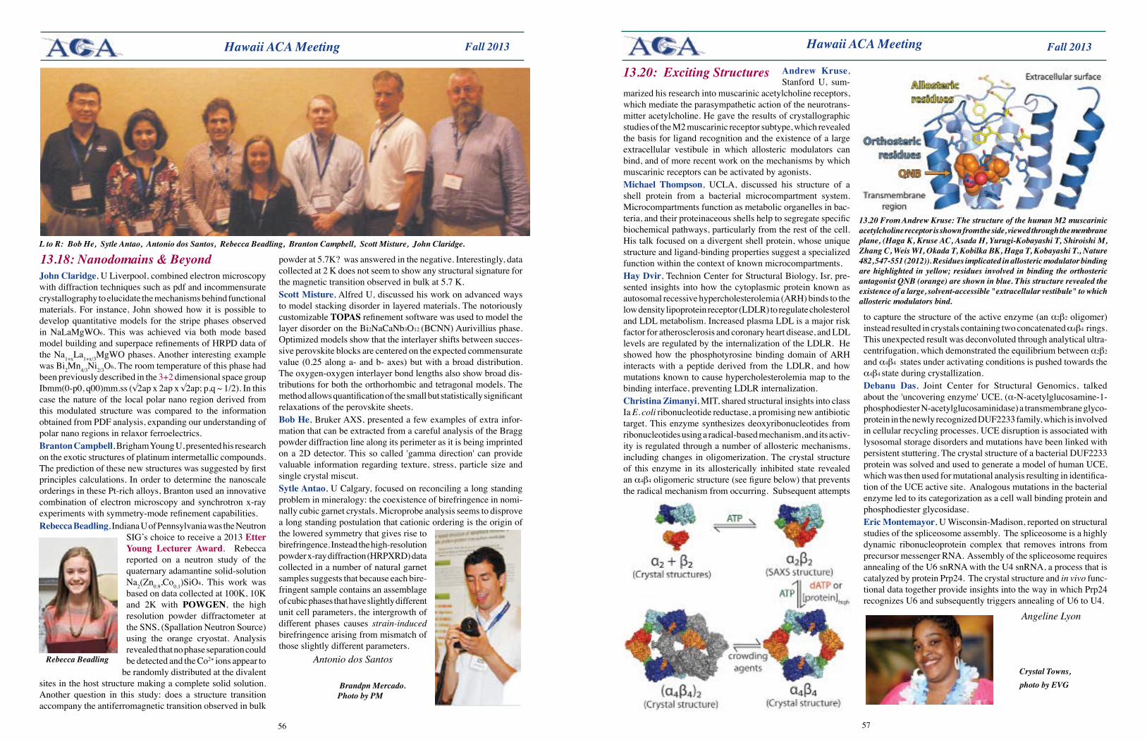
5756
Fall 2013 Fall 2013
John Claridge, U Liverpool, combined electron microscopy with diffraction techniques such as pdf and incommensurate crystallography to elucidate the mechanisms behind functional materials. For instance, John showed how it is possible to develop quantitative models for the stripe phases observed in NaLaMgWO6. This was achieved via both mode based model building and superpace refinements of HRPD data of the Na1+xLa1+x/3MgWO phases. Another interesting example was Bi2Mn4/3Ni2/3O6. The room temperature of this phase had been previously described in the 3+2 dimensional space group Ibmm(0-p0, q00)mm.ss (√2ap x 2ap x √2ap; p,q ~ 1/2). In this case the nature of the local polar nano region derived from this modulated structure was compared to the information obtained from PDF analysis, expanding our understanding of polar nano regions in relaxor ferroelectrics.Branton Campbell, Brigham Young U, presented his research on the exotic structures of platinum intermetallic compounds. The prediction of these new structures was suggested by first principles calculations. In order to determine the nanoscale orderings in these Pt-rich alloys, Branton used an innovative combination of electron microscopy and synchrotron x-ray experiments with symmetry-mode refinement capabilities.Rebecca Beadling, Indiana U of Pennsylvania was the Neutron
SIG’s choice to receive a 2013 Etter Young Lecturer Award. Rebecca reported on a neutron study of the quaternary adamantine solid-solution Na2(Zn0.9,Co0.1)SiO4. This work was based on data collected at 100K, 10K and 2K with POWGEN, the high resolution powder diffractometer at the SNS, (Spallation Neutron Source) using the orange cryostat. Analysis revealed that no phase separation could be detected and the Co2+ ions appear to
be randomly distributed at the divalent sites in the host structure making a complete solid solution. Another question in this study: does a structure transition accompany the antiferromagnetic transition observed in bulk
Hawaii ACA Meeting Hawaii ACA Meeting
L to R: Bob He, Sytle Antao, Antonio dos Santos, Rebecca Beadling, Branton Campbell, Scott Misture, John Claridge.
13.20 From Andrew Kruse: The structure of the human M2 muscarinic acetylcholine receptor is shown from the side, viewed through the membrane plane, (Haga K, Kruse AC, Asada H, Yurugi-Kobayashi T, Shiroishi M, Zhang C, Weis WI, Okada T, Kobilka BK, Haga T, Kobayashi T., Nature 482, 547-551 (2012)). Residues implicated in allosteric modulator binding are highlighted in yellow; residues involved in binding the orthosteric antagonist QNB (orange) are shown in blue. This structure revealed the existence of a large, solvent-accessible "extracellular vestibule" to which allosteric modulators bind.
Andrew Kruse, Stanford U, sum-
marized his research into muscarinic acetylcholine receptors, which mediate the parasympathetic action of the neurotrans-mitter acetylcholine. He gave the results of crystallographic studies of the M2 muscarinic receptor subtype, which revealed the basis for ligand recognition and the existence of a large extracellular vestibule in which allosteric modulators can bind, and of more recent work on the mechanisms by which muscarinic receptors can be activated by agonists. Michael Thompson, UCLA, discussed his structure of a shell protein from a bacterial microcompartment system. Microcompartments function as metabolic organelles in bac-teria, and their proteinaceous shells help to segregate specific biochemical pathways, particularly from the rest of the cell. His talk focused on a divergent shell protein, whose unique structure and ligand-binding properties suggest a specialized function within the context of known microcompartments.Hay Dvir, Technion Center for Structural Biology, Isr, pre-sented insights into how the cytoplasmic protein known as autosomal recessive hypercholesterolemia (ARH) binds to the low density lipoprotein receptor (LDLR) to regulate cholesterol and LDL metabolism. Increased plasma LDL is a major risk factor for atherosclerosis and coronary heart disease, and LDL levels are regulated by the internalization of the LDLR. He showed how the phosphotyrosine binding domain of ARH interacts with a peptide derived from the LDLR, and how mutations known to cause hypercholesterolemia map to the binding interface, preventing LDLR internalization.Christina Zimanyi, MIT, shared structural insights into class Ia E. coli ribonucleotide reductase, a promising new antibiotic target. This enzyme synthesizes deoxyribonucleotides from ribonucleotides using a radical-based mechanism, and its activ-ity is regulated through a number of allosteric mechanisms, including changes in oligomerization. The crystal structure of this enzyme in its allosterically inhibited state revealed an α4β4 oligomeric structure (see figure below) that prevents the radical mechanism from occurring. Subsequent attempts
13.20: Exciting Structures
to capture the structure of the active enzyme (an α2β2 oligomer) instead resulted in crystals containing two concatenated α4β4 rings. This unexpected result was deconvoluted through analytical ultra-centrifugation, which demonstrated the equilibrium between α2β2 and α4β4 states under activating conditions is pushed towards the α4β4 state during crystallization.Debanu Das, Joint Center for Structural Genomics, talked about the 'uncovering enzyme' UCE, (α-N-acetylglucosamine-1-phosphodiester N-acetylglucosaminidase) a transmembrane glyco-protein in the newly recognized DUF2233 family, which is involved in cellular recycling processes. UCE disruption is associated with lysosomal storage disorders and mutations have been linked with persistent stuttering. The crystal structure of a bacterial DUF2233 protein was solved and used to generate a model of human UCE, which was then used for mutational analysis resulting in identifica-tion of the UCE active site. Analogous mutations in the bacterial enzyme led to its categorization as a cell wall binding protein and phosphodiester glycosidase. Eric Montemayor, U Wisconsin-Madison, reported on structural studies of the spliceosome assembly. The spliceosome is a highly dynamic ribonucleoprotein complex that removes introns from precursor messenger RNA. Assembly of the spliceosome requires annealing of the U6 snRNA with the U4 snRNA, a process that is catalyzed by protein Prp24. The crystal structure and in vivo func-tional data together provide insights into the way in which Prp24 recognizes U6 and subsequently triggers annealing of U6 to U4.
Angeline Lyon
13.18: Nanodomains & Beyond
Rebecca Beadling
powder at 5.7K? was answered in the negative. Interestingly, data collected at 2 K does not seem to show any structural signature for the magnetic transition observed in bulk at 5.7 K. Scott Misture, Alfred U, discussed his work on advanced ways to model stacking disorder in layered materials. The notoriously customizable TOPAS refinement software was used to model the layer disorder on the Bi2NaCaNb3O12 (BCNN) Aurivillius phase. Optimized models show that the interlayer shifts between succes-sive perovskite blocks are centered on the expected commensurate value (0.25 along a- and b- axes) but with a broad distribution. The oxygen-oxygen interlayer bond lengths also show broad dis-tributions for both the orthorhombic and tetragonal models. The method allows quantification of the small but statistically significant relaxations of the perovskite sheets.Bob He, Bruker AXS, presented a few examples of extra infor-mation that can be extracted from a careful analysis of the Bragg powder diffraction line along its perimeter as it is being imprinted on a 2D detector. This so called 'gamma direction' can provide valuable information regarding texture, stress, particle size and single crystal miscut.Sytle Antao, U Calgary, focused on reconciling a long standing problem in mineralogy: the coexistence of birefringence in nomi-nally cubic garnet crystals. Microprobe analysis seems to disprove a long standing postulation that cationic ordering is the origin of the lowered symmetry that gives rise to birefringence. Instead the high-resolution powder x-ray diffraction (HRPXRD) data collected in a number of natural garnet samples suggests that because each bire-fringent sample contains an assemblage of cubic phases that have slightly different unit cell parameters, the intergrowth of different phases causes strain-induced birefringence arising from mismatch of those slightly different parameters.
Antonio dos SantosCrystal Towns, photo by EVG Brandpn Mercado.
Photo by PM
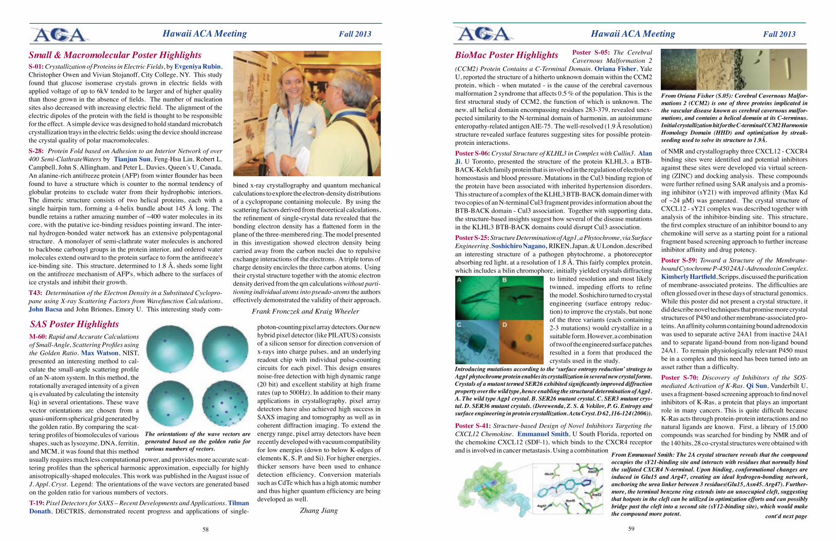
5958
Fall 2013 Fall 2013 Hawaii ACA Meeting
BioMac Poster Highlights Poster S-05: The Cerebral Cavernous Malformation 2
(CCM2) Protein Contains a C-Terminal Domain. OrianaFisher, Yale U, reported the structure of a hitherto unknown domain within the CCM2 protein, which - when mutated - is the cause of the cerebral cavernous malformation 2 syndrome that affects 0.5 % of the population. This is the first structural study of CCM2, the function of which is unknown. The new, all helical domain encompassing residues 283-379, revealed unex-pected similarity to the N-terminal domain of harmonin, an autoimmune enteropathy-related antigen AIE-75. The well-resolved (1.9 Å resolution) structure revealed surface features suggesting sites for possible protein-protein interactions.PosterS-06: Crystal Structure of KLHL3 in Complex with Cullin3. Alan Ji, U Toronto, presented the structure of the protein KLHL3, a BTB-BACK-Kelch family protein that is involved in the regulation of electrolyte homeostasis and blood pressure. Mutations in the Cul3 binding region of the protein have been associated with inherited hypertension disorders. This structure of a complex of the KLHL3 BTB-BACK domain dimer with two copies of an N-terminal Cul3 fragment provides information about the BTB-BACK domain - Cul3 association. Together with supporting data, the structure-based insights suggest how several of the disease mutations in the KLHL3 BTB-BACK domains could disrupt Cul3 association. PosterS-25:Structure Determination of Agp1, a Phytochrome, via Surface Engineering. Soshichiro Nagano, RIKEN, Japan, & U London, described an interesting structure of a pathogen phytochrome, a photoreceptor absorbing red light, at a resolution of 1.8 Å. This fairly complex protein, which includes a bilin chromophore, initially yielded crystals diffracting
to limited resolution and most likely twinned, impeding efforts to refine the model. Soshichiro turned to crystal engineering (surface entropy reduc-tion) to improve the crystals, but none of the three variants (each containing 2-3 mutations) would crystallize in a suitable form. However, a combination of two of the engineered surface patches resulted in a form that produced the crystals used in the study.
PosterS-41:Structure-based Design of Novel Inhibitors Targeting the CXCL12 Chemokine. Emmanuel Smith, U South Florida, reported on the chemokine CXCL12 (SDF-1), which binds to the CXCR4 receptor and is involved in cancer metastasis. Using a combination
of NMR and crystallography three CXCL12 - CXCR4 binding sites were identified and potential inhibitors against these sites were developed via virtual screen-ing (ZINC) and docking analysis. These compounds were further refined using SAR analysis and a promis-ing inhibitor (sY21) with improved affinity (Max Kd of ~24 μM) was generated. The crystal structure of CXCL12 - sY21 complex was described together with analysis of the inhibitor-binding site. This structure, the first complex structure of an inhibitor bound to any chemokine will serve as a starting point for a rational fragment based screening approach to further increase inhibitor affinity and drug potency. PosterS-59: Toward a Structure of the Membrane-bound Cytochrome P-450 24A1-Adrenodoxin Complex. KimberlyHartfield, Scripps, discussed the purification of membrane-associated proteins. The difficulties are often glossed over in these days of structural genomics. While this poster did not present a crystal structure, it did describe novel techniques that promise more crystal structures of P450 and other membrane-associated pro-teins. An affinity column containing bound adrenodoxin was used to separate active 24A1 from inactive 24A1 and to separate ligand-bound from non-ligand bound 24A1. To remain physiologically relevant P450 must be in a complex and this need has been turned into an asset rather than a difficulty.Poster S-70: Discovery of Inhibitors of the SOS-mediated Activation of K-Ras. QiSun, Vanderbilt U, uses a fragment-based screening approach to find novel inhibitors of K-Ras, a protein that plays an important role in many cancers. This is quite difficult because K-Ras acts through protein-protein interactions and no natural ligands are known. First, a library of 15,000 compounds was searched for binding by NMR and of the 140 hits, 28 co-crystal structures were obtained with
From Oriana Fisher (S.05): Cerebral Cavernous Malfor-mations 2 (CCM2) is one of three proteins implicated in the vascular disease known as cerebral cavernous malfor-mations, and contains a helical domain at its C-terminus. Initial crystallization hit for the C-terminal CCM2 Harmonin Homology Domain (HHD) and optimization by streak-seeding used to solve its structure to 1.9Å.
Introducing mutations according to the ‘surface entropy reduction’ strategy to Agp1 phytochrome protein enables its crystallization in several new crystal forms. Crystals of a mutant termed SER26 exhibited significantly improved diffraction property over the wild type, hence enabling the structural determination of Agp1. A. The wild type Agp1 crystal. B. SER26 mutant crystal. C. SER3 mutant crys-tal. D. SER36 mutant crystals. (Derewenda, Z. S. & Vekilov, P. G. Entropy and surface engineering in protein crystallization. Acta Cryst. D 62, 116-124 (2006)).
From Emmanuel Smith: The 2A crystal structure reveals that the compound occupies the sY21-binding site and interacts with residues that normally bind the sulfated CXCR4 N-terminal. Upon binding, conformational changes are induced in Glu15 and Arg47, creating an ideal hydrogen-bonding network, anchoring the urea linker between 3 residues(Glu15, Asn45. Arg47). Further-more, the terminal benzene ring extends into an unoccupied cleft, suggesting that hotpots in the cleft can be utilized in optimization efforts and can possibly bridge past the cleft into a second site (sY12-binding site), which would make the compound more potent.
Hawaii ACA Meeting
Small & Macromolecular Poster Highlights S-01: Crystallization of Proteins in Electric Fields, by Evgeniya Rubin, Christopher Owen and Vivian Stojanoff, City College, NY. This study found that glucose isomerase crystals grown in electric fields with applied voltage of up to 6kV tended to be larger and of higher quality than those grown in the absence of fields. The number of nucleation sites also decreased with increasing electric field. The alignment of the electric dipoles of the protein with the field is thought to be responsible for the effect. A simple device was designed to hold standard microbatch crystallization trays in the electric fields; using the device should increase the crystal quality of polar macromolecules.S-28: Protein Fold based on Adhesion to an Interior Network of over 400 Semi-ClathrateWaters by Tianjun Sun, Feng-Hsu Lin, Robert L. Campbell, John S. Allingham, and Peter L. Davies, Queen’s U, Canada. An alanine-rich antifreeze protein (AFP) from winter flounder has been found to have a structure which is counter to the normal tendency of globular proteins to exclude water from their hydrophobic interiors. The dimeric structure consists of two helical proteins, each with a single hairpin turn, forming a 4-helix bundle about 145 Å long. The bundle retains a rather amazing number of ~400 water molecules in its core, with the putative ice-binding residues pointing inward. The inter-nal hydrogen-bonded water network has an extensive polypentagonal structure. A monolayer of semi-clathrate water molecules is anchored to backbone carbonyl groups in the protein interior, and ordered water molecules extend outward to the protein surface to form the antifreeze's ice-binding site. This structure, determined to 1.8 Å, sheds some light on the antifreeze mechanism of AFP's, which adhere to the surfaces of ice crystals and inhibit their growth.T43: Determination of the Electron Density in a Substituted Cyclopro-pane using X-ray Scattering Factors from Wavefunction Calculations, John Bacsa and John Briones, Emory U. This interesting study com-
bined x-ray crystallography and quantum mechanical calculations to explore the electron-density distributions of a cyclopropane containing molecule. By using the scattering factors derived from theoretical calculations, the refinement of single-crystal data revealed that the bonding electron density has a flattened form in the plane of the three-membered ring. The model presented in this investigation showed electron density being carried away from the carbon nuclei due to repulsive exchange interactions of the electrons. A triple torus of charge density encircles the three carbon atoms. Using their crystal structure together with the atomic electron density derived from the qm calculations without parti-tioning individual atoms into pseudo-atoms the authors effectively demonstrated the validity of their approach.
Frank Fronczek and Kraig Wheeler
M-60: Rapid and Accurate Calculations of Small-Angle, Scattering Profiles using the Golden Ratio. MaxWatson, NIST, presented an interesting method to cal-culate the small-angle scattering profile of an N-atom system. In this method, the rotationally averaged intensity of a given q is evaluated by calculating the intensity I(q) in several orientations. These wave vector orientations are chosen from a quasi-uniform spherical grid generated by the golden ratio. By comparing the scat-tering profiles of biomolecules of various shapes, such as lysozyme, DNA, ferritin, and MCM, it was found that this method usually requires much less computational power, and provides more accurate scat-tering profiles than the spherical harmonic approximation, especially for highly anisotropically-shaped molecules. This work was published in the August issue of J. Appl. Cryst. Legend: The orientations of the wave vectors are generated based on the golden ratio for various numbers of vectors.T-19: Pixel Detectors for SAXS – Recent Developments and Applications. Tilman Donath, DECTRIS, demonstrated recent progress and applications of single-
SAS Poster Highlights
The orientations of the wave vectors are generated based on the golden ratio for various numbers of vectors.
photon-counting pixel array detectors. Our new hybrid pixel detector (like PILATUS) consists of a silicon sensor for direction conversion of x-rays into charge pulses, and an underlying readout chip with individual pulse-counting circuits for each pixel. This design ensures noise-free detection with high dynamic range (20 bit) and excellent stability at high frame rates (up to 500Hz). In addition to their many applications in crystallography, pixel array detectors have also achieved high success in SAXS imaging and tomography as well as in coherent diffraction imaging. To extend the energy range, pixel array detectors have been recently developed with vacuum compatibility for low energies (down to below K-edges of elements K, S, P, and Si). For higher energies, thicker sensors have been used to enhance detection efficiency. Conversion materials such as CdTe which has a high atomic number and thus higher quantum efficiency are being developed as well.
Zhang Jiangcont'd next page
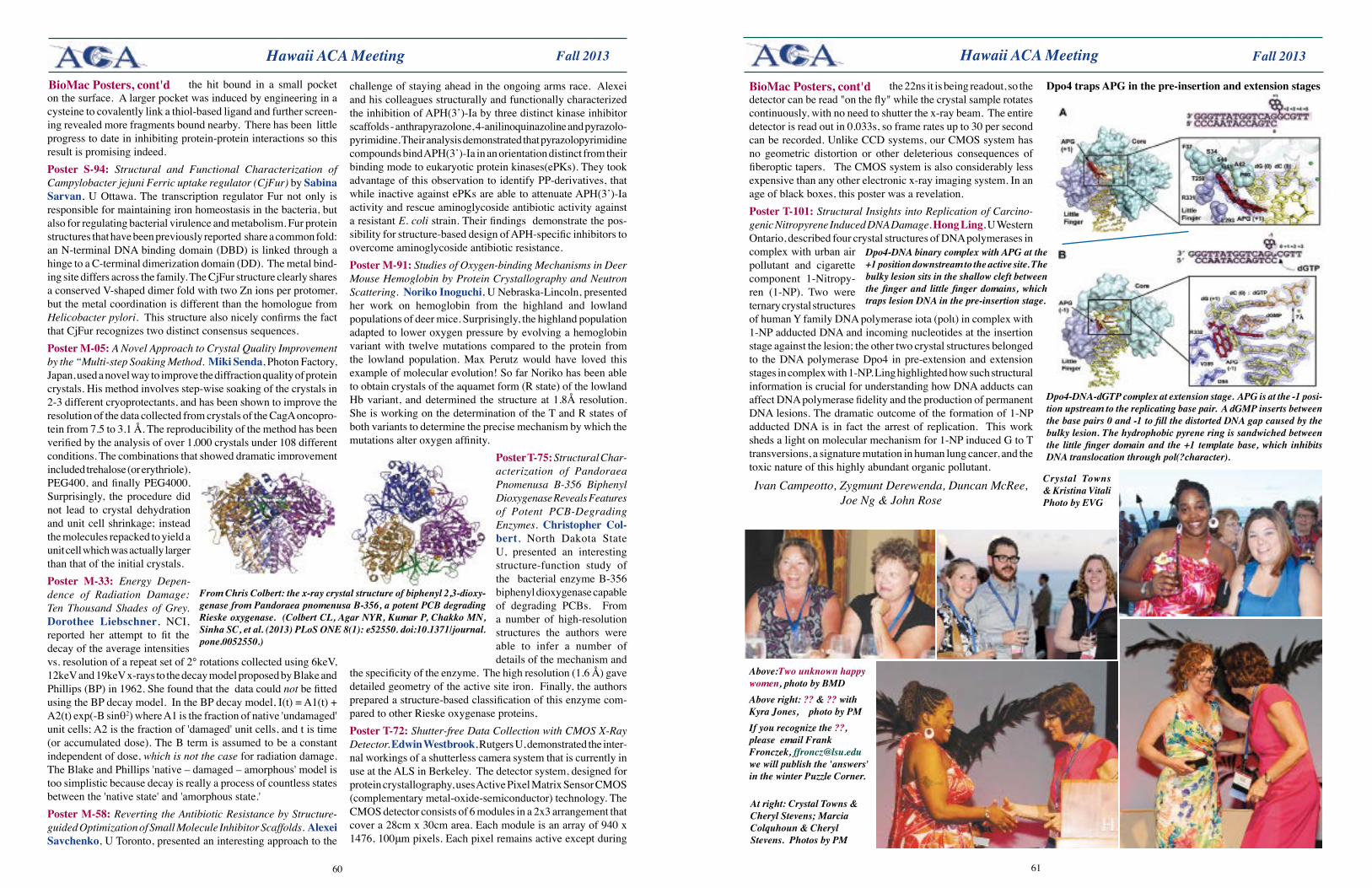
6160
Fall 2013 Fall 2013 Hawaii ACA Meeting
the hit bound in a small pocket on the surface. A larger pocket was induced by engineering in a cysteine to covalently link a thiol-based ligand and further screen-ing revealed more fragments bound nearby. There has been little progress to date in inhibiting protein-protein interactions so this result is promising indeed.Poster S-94: Structural and Functional Characterization of Campylobacter jejuni Ferric uptake regulator (CjFur) by Sabina Sarvan, U Ottawa. The transcription regulator Fur not only is responsible for maintaining iron homeostasis in the bacteria, but also for regulating bacterial virulence and metabolism. Fur protein structures that have been previously reported share a common fold: an N-terminal DNA binding domain (DBD) is linked through a hinge to a C-terminal dimerization domain (DD). The metal bind-ing site differs across the family. The CjFur structure clearly shares a conserved V-shaped dimer fold with two Zn ions per protomer, but the metal coordination is different than the homologue from Helicobacter pylori. This structure also nicely confirms the fact that CjFur recognizes two distinct consensus sequences.PosterM-05: A Novel Approach to Crystal Quality Improvement by the “Multi-step Soaking Method. Miki Senda, Photon Factory, Japan, used a novel way to improve the diffraction quality of protein crystals. His method involves step-wise soaking of the crystals in 2-3 different cryoprotectants, and has been shown to improve the resolution of the data collected from crystals of the CagA oncopro-tein from 7.5 to 3.1 Å. The reproducibility of the method has been verified by the analysis of over 1,000 crystals under 108 different conditions. The combinations that showed dramatic improvement included trehalose (or erythriole), PEG400, and finally PEG4000. Surprisingly, the procedure did not lead to crystal dehydration and unit cell shrinkage; instead the molecules repacked to yield a unit cell which was actually larger than that of the initial crystals.Poster M-33: Energy Depen-dence of Radiation Damage: Ten Thousand Shades of Grey. Dorothee Liebschner, NCI, reported her attempt to fit the decay of the average intensities vs. resolution of a repeat set of 2° rotations collected using 6keV, 12keV and 19keV x-rays to the decay model proposed by Blake and Phillips (BP) in 1962. She found that the data could not be fitted using the BP decay model. In the BP decay model, I(t) = A1(t) + A2(t) exp(-B sinθ2) where A1 is the fraction of native 'undamaged' unit cells; A2 is the fraction of 'damaged' unit cells, and t is time (or accumulated dose). The B term is assumed to be a constant independent of dose, which is not the case for radiation damage. The Blake and Phillips 'native – damaged – amorphous' model is too simplistic because decay is really a process of countless states between the 'native state' and 'amorphous state.'PosterM-58: Reverting the Antibiotic Resistance by Structure-guided Optimization of Small Molecule Inhibitor Scaffolds. AlexeiSavchenko, U Toronto, presented an interesting approach to the
challenge of staying ahead in the ongoing arms race. Alexei and his colleagues structurally and functionally characterized the inhibition of APH(3’)-Ia by three distinct kinase inhibitor scaffolds - anthrapyrazolone, 4-anilinoquinazoline and pyrazolo-pyrimidine. Their analysis demonstrated that pyrazolopyrimidine compounds bind APH(3’)-Ia in an orientation distinct from their binding mode to eukaryotic protein kinases(ePKs). They took advantage of this observation to identify PP-derivatives, that while inactive against ePKs are able to attenuate APH(3’)-Ia activity and rescue aminoglycoside antibiotic activity against a resistant E. coli strain. Their findings demonstrate the pos-sibility for structure-based design of APH-specific inhibitors to overcome aminoglycoside antibiotic resistance.PosterM-91: Studies of Oxygen-binding Mechanisms in Deer Mouse Hemoglobin by Protein Crystallography and Neutron Scattering. Noriko Inoguchi, U Nebraska-Lincoln, presented her work on hemoglobin from the highland and lowland populations of deer mice. Surprisingly, the highland population adapted to lower oxygen pressure by evolving a hemoglobin variant with twelve mutations compared to the protein from the lowland population. Max Perutz would have loved this example of molecular evolution! So far Noriko has been able to obtain crystals of the aquamet form (R state) of the lowland Hb variant, and determined the structure at 1.8Å resolution. She is working on the determination of the T and R states of both variants to determine the precise mechanism by which the mutations alter oxygen affinity.
PosterT-75: Structural Char-acterization of Pandoraea Pnomenusa B-356 Biphenyl Dioxygenase Reveals Features of Potent PCB-Degrading Enzymes. Christopher Col-bert, North Dakota State U, presented an interesting structure-function study of the bacterial enzyme B-356 biphenyl dioxygenase capable of degrading PCBs. From a number of high-resolution structures the authors were able to infer a number of details of the mechanism and
the specificity of the enzyme. The high resolution (1.6 Å) gave detailed geometry of the active site iron. Finally, the authors prepared a structure-based classification of this enzyme com-pared to other Rieske oxygenase proteins, PosterT-72: Shutter-free Data Collection with CMOS X-Ray Detector. Edwin Westbrook, Rutgers U, demonstrated the inter-nal workings of a shutterless camera system that is currently in use at the ALS in Berkeley. The detector system, designed for protein crystallography, uses Active Pixel Matrix Sensor CMOS (complementary metal-oxide-semiconductor) technology. The CMOS detector consists of 6 modules in a 2x3 arrangement that cover a 28cm x 30cm area. Each module is an array of 940 x 1476, 100μm pixels. Each pixel remains active except during
From Chris Colbert: the x-ray crystal structure of biphenyl 2,3-dioxy-genase from Pandoraea pnomenusa B-356, a potent PCB degrading Rieske oxygenase. (Colbert CL, Agar NYR, Kumar P, Chakko MN, Sinha SC, et al. (2013) PLoS ONE 8(1): e52550. doi:10.1371/journal.pone.0052550.)
BioMac Posters, cont'd
Dpo4-DNA-dGTP complex at extension stage. APG is at the -1 posi-tion upstream to the replicating base pair. A dGMP inserts between the base pairs 0 and -1 to fill the distorted DNA gap caused by the bulky lesion. The hydrophobic pyrene ring is sandwiched between the little finger domain and the +1 template base, which inhibits DNA translocation through pol(?character).
Dpo4trapsAPGinthepre-insertionandextensionstagesthe 22ns it is being readout, so the detector can be read "on the fly" while the crystal sample rotates continuously, with no need to shutter the x-ray beam. The entire detector is read out in 0.033s, so frame rates up to 30 per second can be recorded. Unlike CCD systems, our CMOS system has no geometric distortion or other deleterious consequences of fiberoptic tapers. The CMOS system is also considerably less expensive than any other electronic x-ray imaging system. In an age of black boxes, this poster was a revelation.PosterT-101:Structural Insights into Replication of Carcino-genic Nitropyrene Induced DNA Damage. Hong Ling, U Western Ontario, described four crystal structures of DNA polymerases in complex with urban air pollutant and cigarette component 1-Nitropy-ren (1-NP). Two were ternary crystal structures of human Y family DNA polymerase iota (polι) in complex with 1-NP adducted DNA and incoming nucleotides at the insertion stage against the lesion; the other two crystal structures belonged to the DNA polymerase Dpo4 in pre-extension and extension stages in complex with 1-NP. Ling highlighted how such structural information is crucial for understanding how DNA adducts can affect DNA polymerase fidelity and the production of permanent DNA lesions. The dramatic outcome of the formation of 1-NP adducted DNA is in fact the arrest of replication. This work sheds a light on molecular mechanism for 1-NP induced G to T transversions, a signature mutation in human lung cancer, and the toxic nature of this highly abundant organic pollutant.
Ivan Campeotto, Zygmunt Derewenda, Duncan McRee, Joe Ng & John Rose
Dpo4-DNA binary complex with APG at the +1 position downstream to the active site. The bulky lesion sits in the shallow cleft between the finger and little finger domains, which traps lesion DNA in the pre-insertion stage.
BioMac Posters, cont'd
Crystal Towns & Kristina Vitali Photo by EVG
Above:Two unknown happy women, photo by BMDAbove right: ?? & ?? with Kyra Jones, photo by PM If you recognize the ??, please email Frank Fronczek, [email protected] we will publish the 'answers' in the winter Puzzle Corner.
Hawaii ACA Meeting
At right: Crystal Towns & Cheryl Stevens; Marcia Colquhoun & Cheryl Stevens. Photos by PM
6362
Fall 2013 Fall 2013 Banquet in Hawaii
Banquet overview at top: photo by EVG. Immediately above, Jon Hanson, Louise Karle Hanson & ??. Photo by BMD (see preceding page about the ??s.)Below: Sitting, L to R, ??, Marvin Hackert, ??; standing in back: ??, ??, David Grossie. Photo by EVG
Banquet in Hawaii
Above: Marvin Hackert, photo by BMDAround table below: In back: Corrina Beavers, Daniela Morando, Christine Beavers, Simon Teat, Byung Woo Han. In front: Tracey Kniffin, Kevon Gagnon, Bill Clegg & Joon Sung Park.
At right: Sitting, L-R: Juergen Graf, Marilyn Olm-stead, Ashley Troumouliaris, Brandon Mercado. Standing, L-R: Neelam Chishty, Bruce Noll, Stacey Smith, Barbara Ramirez, Svilen Bobev. Photo by PM
Standing: Mirijam Zoebel, Jim Pflugrath, Jacok Park, Brahm Yachim; Sitting: Håkon Hope, Sally Springer, ?? Photos above and below by EVG
Opposite page: Virginia Pett, ordering. Photo by BMD
Photo on opposite page cont'd above. Standing, from left: Stan Rajnak, Shuaiqi Guo, Lagnajeet Pradham, Evgeniya Rubin, Lar-issa Romanello, Juliana Torini de Souza. Sitting, Kimberly Hartfield, Lu Han, Jeferson de Oliveira. Photo by EVG
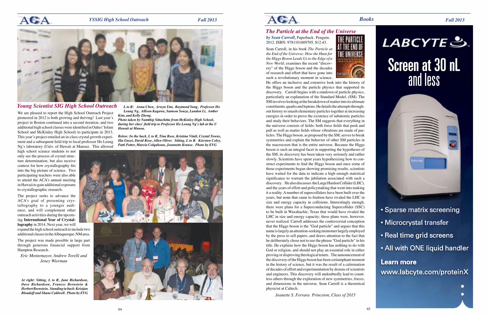
6564
Fall 2013 Fall 2013
We are pleased to report the High School Outreach Project pioneered in 2012 is both growing and thriving! Last year’s project in Boston continued into a second iteration, and two additional high school classes were identified in Oahu (‘Iolani School and McKinley High School) to participate in 2013. This year’s project entailed an in-class crystal growth experi-ment and a subsequent field trip to local professor Ho Leung Ng’s laboratory (Univ. of Hawaii at Manoa). This allowed high school science students to not only see the process of crystal struc-ture determination, but also receive context for how crystallography fits into the big picture of science. Two participating teachers were also able to attend the ACA’s annual meeting in Hawaii to gain additional exposure to crystallographic research.The project seeks to advance the ACA’s goal of presenting crys-tallography to a younger audi-ence, and will complement other outreach activities during the upcom-ingInternationalYearofCrystal-lography in 2014. Next year, we will expand the high school outreach to include two additional classes in the Albuquerque, NM area.The project was made possible in large part through generous financial support from Hampton Research.
Eric Montemayor, Andrew Torelli and Jeney Wierman
YSSIG High School Outreach
L to R: Anna Chen, Arwyn Yim, Raymond Yang, Professor Ho Leung Ng, Allison Kagawa, Samson Souza, Landon Li, Amber
Kim, and Kelly Zheng. Photo taken by Namthip Sittachitta from McKinley High School, during her class' field trip to Professor Ho Leung Ng's lab at the U Hawaii at Manoa.
Young Scientist SIG High School Outreach
At right: Sitting, L to R, Jane Richardson, Dave Richardson, Frances Bernstein & Herbert Bernstein. Standing in back: Kristjan Bloudoff and Shane Caldwell. Photo by EVG
Below: In the back, L to R, Tina Rose, Kristina Vitali, Crystal Towns, Ilia Guzei, David Rose, Allen Oliver. Sitting, L to R. Kiersten Coley, Patti Potter, Marcia Colquhoun, Jeannette Krause. Photo by EVG
Books
Sean Carroll, in his book The Particle at the End of the Universe: How the Hunt for the Higgs Boson Leads Us to the Edge of a New World, examines the recent "discov-ery" of the Higgs boson and the decades of research and effort that have gone into such a revolutionary moment in science. He offers an inclusive and extensive look into the history of the Higgs boson and the particle physics that supported its discovery. Carroll begins with a rundown of particle physics, particularly an explanation of the Standard Model, (SM). The SM involves looking at the breakdown of matter into its ultimate constituents, quarks and leptons. He details the attempts through-out history to smash elementary particles together at increasing energies in order to prove the existence of subatomic particles and study their behaviors. The SM suggests that everything in the universe consists of fields: both force fields that push and pull as well as matter fields whose vibrations are made of par-ticles. The Higgs boson, as proposed by the SM, serves to break symmetries and explain the behavior of other SM particles in the macrocosm that is the entire universe. Because the Higgs boson is such an integral facet in supporting the hypotheses of the SM, its discovery has been taken very seriously and rather slowly. Scientists have spent years hypothesizing how to con-struct experiments to find the Higgs boson and once some of those experiments began showing promising results, scientists have waited for the data to indicate a high enough statistical significance to warrant the jubilation associated with such a discovery. He also discusses the Large Hardon Collider (LHC), and the years of effort and policymaking that went into making it a reality. A number of supercolliders have been built over the years, but none that came to fruition have rivaled the LHC in size and energy capacity in collisions. Interestingly enough, there were plans for a Superconducting Supercollider (SSC) to be built in Waxahachie, Texas that would have rivaled the LHC in size and energy capacity; these plans were, however, never realized. Carroll addresses the controversial conception that the Higgs boson is the "God particle" and argues that this name is largely an attention-seeking misnomer largely employed by the press to sell papers, and draws attention to the fact that he deliberately chose not to use the phrase "God particle" in his title. He explains how the Higgs boson has nothing to do with God or religion, and should not play an essential role in either proving or disproving theological tenets. The announcement of the discovery of the Higgs boson has been a triumphant moment in the history of science, but it was the result of a culmination of decades of effort and experimentation by dozens of scientists and engineers. This discovery will undoubtedly lead to count-less others through the exploration of new symmetries, forces, and dimensions in the universe. Sean Carroll is a theoretical physicist at Caltech.
Jeanette S. Ferrara Princeton, Class of 2015
The Particle at the End of the Universe by Sean Carroll, Paperback, Penguin, 2012. ISBN: 9781101609705, $12.43.
6766
Fall 2013 Fall 2013
Workshop on Dynamic Structural Photocrystallography in Chemistry&MaterialsScience,SUNY-Buffalo,June16-20,2013Photocrystallography is an x-ray diffraction technique combined with external light excitation of the sample. It is utilized for studying out-of-equilibrium phenomena in crystals, i.e. relaxation processes, long-lived metastable states, short-lived excited states, and solid-state photochemical reactions. The rapid development of the experimental techniques, and the availability of pulsed synchrotron x-ray sources have made photocrystallography much more achievable, and it is currently at the very frontier of modern crystallography. It is now possible to conduct time-resolved studies of species with microsecond or even shorter lifetimes. Recent advances in the field show not only its potential and importance, but also the need to train students and young researchers in the subject. This workshop was initiated and organized by Philip Coppens, and supported by members of his research group, (Lisa Zimmerman - organization and facilities; ElżbietaTrzop & BertrandFournier - software exercises; Katarzyna Jarzembska, RadoslawKamiński,AnnaMakal (former group member) and Jesse Sokolow - tutorial preparation and general help). The event benefited from the advice of the InternationalAdvisoryCommittee: Eric Collet (U of Rennes, France) and Masaki Takata (U of Tokyo, Japan). Fifty-five scientists, graduate and undergraduate stu-dents, young researchers, and invited speakers from American, European and Asian universities and research centers participated in the workshop.Light-induced processes such as photochemical reactions, linkage photoisomers, and photoinduced molecular excited states in the crystalline solids - on timescales of hours to femtoseconds - were studied during the workshop. Development of experimental laser pump / x-ray probe techniques used either for in-house experiments at high intensity x-ray sources, or for experiments performed at synchrotron beamlines and/or at Free-Electron Laser sources were a major focus. New computational techniques for time-resolved data processing and interpretation of results were also considered.
Dynamic Structural Photocrystallography
The workshop program was divided into two parts: an opening lecture by Philip Coppens followed by plenary lectures given by the invited speakers: Panche Naumov, NYU Abu Dhabi & U of Kyoto; Jason Benedict, U at Buffalo, SUNY; Eric Collet, U of Rennes, France; Anna Makal, U of Warsaw, Poland; Marc Messerschmidt, SLAC National Accelerator Laboratory; John Spence, ASU (Arizona State University); and FriedrichSchotte, NIH, NIDDK. Software exercises followed. The second part was carried out in three separate tracks: (1) Monitoring Chemical Reactions; (2) Pump-Probe Experiments of Excited State Structure - supervised by E. Trzop, A. Makal and B.Fournier; and (3) Treatment of Liquid-Jet Injection LCLS data - supervised and supported with examples by Nadia Zatsepin, ASU. The workshop week was interspersed with manufacturer presentations, given by Chris Ceccarelli, Agilent, Lee Daniels, Rigaku, and Charles Campana, Bruker AXS, on potential applications of the commercially available x-ray diffraction systems for study-ing light-induced phenomena.
Left corner, the in-house pump-probe photo-crystallography instrument. Below, software exercise class.
Participants could visit the laboratory of P. Coppens and J. Benedict to view the in-house pump-probe photocrystallography-dedicated experi-mental setup. Additionally, students and young scientists presented and discussed their research work during a poster session.Despite the tight schedule of the workshop, time was found for leisure and social events, and an excursion to the famous Niagara Falls was a must. The trip, guided by L. Zimmerman and E. Trzop, included the "Maid of the Mist" boat tour, and ended up with the official workshop dinner. Thanks to generous support for travel by the IUCr, travel grants were awarded to the following six young scientists: Shibom Basu, ASU; GastonCorthey, Center for Free Electron Laser Science, U of Hamburg, Germany; Raghavender Medishetty, National U of Singapore; Philip Roedig, Deutsches Elektronen-Synchrotron, Germany; Sounak Sarkar, Indian Inst. of Science, India; and Tobias Tyborski, Max-Born-Institut für Nichtlineare Optik und Kurzzeitspektroskopie im Forschungsverbund Berlin, Germany.Because of the enthusiastic reception by participants, other workshops may well be organized in the future, or even could become periodic events. The field is certainly developing rapidly, and the exchange of ideas and new methods remains crucial.
Elżbieta Trzop & Katarzyna Jarzembska
The organizers are grateful to the sponsors: Agilent Technologies Inc., Bruker AXS Inc., Rigaku Americas Corp., IUCr, and ChemMatCARS at U Chicago for their financial support. Thanks also to The University at Buffalo, SUNY because access to the university facilities: lecture halls, computer laboratories and residences, kept the cost for participants low. In addition, gratitude is extended to Dave Yearke and Leonid Danilovich of UB Science and Engineering Node Services for
Final workshop dinner after the boat trip.
Structural Photocrystallography Workshop, cont'd
Puzzle Corner Editor Frank Fronczek [email protected]
Puzzle Corner
In the summer issue of ACA RefleXions we posed the DISORDERED puzzle at below right. The solution is shown. A new DISORDERED puzzle is below left. Please send ideas for new puzzles / problems to Frank Fronczek.
4D Numerical Puzzle, The Snake with Diamonds & The Triangular Grid Puzzle: © 2004, 2004, 2008. All rights reserved by Peter Grabarchuk; and Quartzic by Zarang: Content © 2013 Zarang.
Puzzles Found on the Web
their indispensible informatics support. Special thanks to Crystal Towns and Marcia Colquhoun of the ACA for expert help with the financial arrangements, to Jordan Cox and Jarrett Coppin (members of the Benedict group) and all other people who helped.
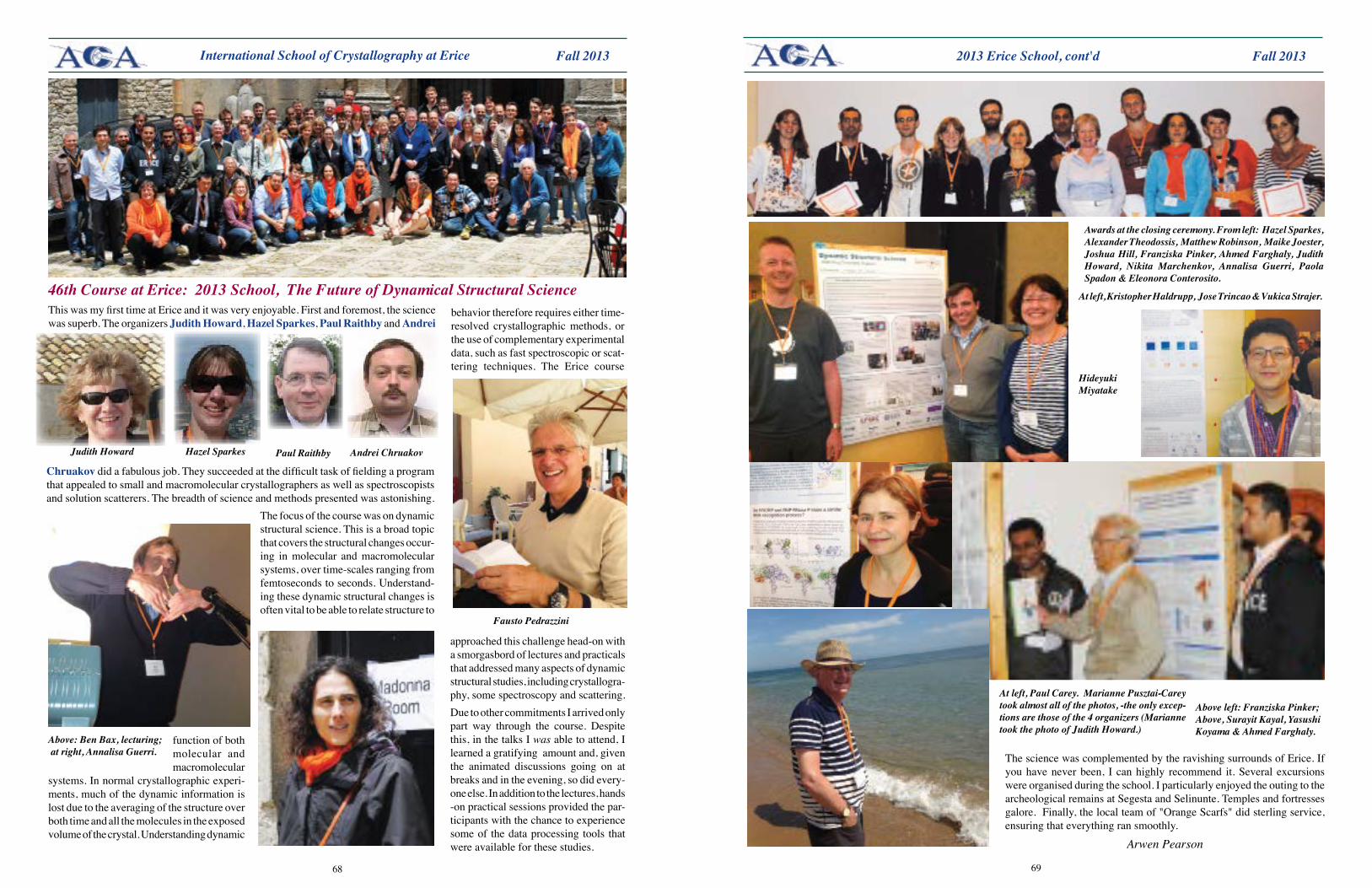
6968
Fall 2013 Fall 2013
function of both molecular and macromolecular
systems. In normal crystallographic experi-ments, much of the dynamic information is lost due to the averaging of the structure over both time and all the molecules in the exposed volume of the crystal. Understanding dynamic
Chruakov did a fabulous job. They succeeded at the difficult task of fielding a program that appealed to small and macromolecular crystallographers as well as spectroscopists and solution scatterers. The breadth of science and methods presented was astonishing.
The focus of the course was on dynamic structural science. This is a broad topic that covers the structural changes occur-ing in molecular and macromolecular systems, over time-scales ranging from femtoseconds to seconds. Understand-ing these dynamic structural changes is often vital to be able to relate structure to
International School of Crystallography at Erice
This was my first time at Erice and it was very enjoyable. First and foremost, the science was superb. The organizers Judith Howard, Hazel Sparkes, Paul Raithby and Andrei
46th Course at Erice: 2013 School, The Future of Dynamical Structural Science
Judith Howard
Above: Ben Bax, lecturing; at right, Annalisa Guerri.
Hazel Sparkes Paul Raithby Andrei Chruakov
Fausto Pedrazzini
behavior therefore requires either time-resolved crystallographic methods, or the use of complementary experimental data, such as fast spectroscopic or scat-tering techniques. The Erice course
approached this challenge head-on with a smorgasbord of lectures and practicals that addressed many aspects of dynamic structural studies, including crystallogra-phy, some spectroscopy and scattering. Due to other commitments I arrived only part way through the course. Despite this, in the talks I was able to attend, I learned a gratifying amount and, given the animated discussions going on at breaks and in the evening, so did every-one else. In addition to the lectures, hands -on practical sessions provided the par-ticipants with the chance to experience some of the data processing tools that were available for these studies.
Awards at the closing ceremony. From left: Hazel Sparkes, Alexander Theodossis, Matthew Robinson, Maike Joester, Joshua Hill, Franziska Pinker, Ahmed Farghaly, Judith Howard, Nikita Marchenkov, Annalisa Guerri, Paola Spadon & Eleonora Conterosito.
At left, Paul Carey. Marianne Pusztai-Carey took almost all of the photos, -the only excep-tions are those of the 4 organizers (Marianne took the photo of Judith Howard.)
2013 Erice School, cont'd
Hideyuki Miyatake
At left, Kristopher Haldrupp, Jose Trincao & Vukica Strajer.
Above left: Franziska Pinker; Above, Surayit Kayal, Yasushi Koyama & Ahmed Farghaly.
The science was complemented by the ravishing surrounds of Erice. If you have never been, I can highly recommend it. Several excursions were organised during the school. I particularly enjoyed the outing to the archeological remains at Segesta and Selinunte. Temples and fortresses galore. Finally, the local team of "Orange Scarfs" did sterling service, ensuring that everything ran smoothly.
Arwen Pearson
71
Fall 2013Future Meetings
The Bruker/MIT Symposium is a small but high-quality event hosted by MIT's Department of Chemistry and funded by Bruker-AXS. This symposium has been compared to a Gordon Conference more than once, which attests to the high level of excellence of the scientific presentations. The 2014 symposium will be held from Friday, February 21st to Sunday February 23rd at MIT in Cambridge, MA. The theme will be 'Modern Approaches to Structure Solution.' CarmeloGiacovazzo, Inst. of Crystallography, Bari, Italy (SIR), LukášPalatinus, Inst Physics, Prague, Czeck Republic, (Super-flip) and GeorgeSheldrick, U Göttingen, (SHELX) have agreed to give lectures and hold workshops on the most popular structure solution programs used for single-crystal structures. Giaccovazzo, Palatinus and Sheldrick are among the world's best known experts on structure solution, so no active crystallographer should miss this wonderful opportunity. Participation is free for everyone. For more details see the symposium homepage: http://web.mit.edu/x-ray/bmit14.html.
From left:
George Sheldrick Lukáš Palatinus
Carmelo Giacovazzo
expertise development range quality support
it’s what makes us cooler
Fine detail produces fine results
Every low temperature device we produce is designed,
built and tested by us in our Oxfordshire workshops under
compromise-free manufacturing processes.
Our highly skilled development engineers and technicians are
dedicated to giving you the best cooling systems possible, ensuring
that best practice and quality control are observed and implemented
through every step.
This level of quality and total focus in the field of sample cooling
allows us to manufacture low temperature devices that always out-
perform the rest, making them the ‘cooler of choice’ throughout the
crystallography community.
For full details of our quality cooling product range visit
www.oxcryo.com or call us on +44 1993 883 488 to discuss
your specific requirements.
quality
OxCryo-ACA-01-12-12.indd 1 29/11/2012 18:08
Toll free (North America): 877 627 9729
International: +1 847 869 1548
Rayonix frame-shift technologyOnly one millisecond dead time
No gaps in imaging area
High speed & high resolutionRayonix HS X-ray detectors
Square X-ray area detectors
MX170-HSMX225-HSMX300-HSMX340-HSMX425-HS
The only X-ray detectors designed with cutout for simultaneous SAXS/WAXS
LX170-HSLX255-HS
www.rayonix.com

7372
Fall 2013 Fall 20132014 Annual Meeting in Albuquerque 2014 Annual Meeting in Albuquerque

Your way
Brilliance your way:Nanostructure analysiswith SAXSpace
4 From fast and easy operations to
full experimental flexibility …
4 From SAXS to WAXS to GI-SAXS analysis …
4 From one beam line to multiple beam lines …
4 From isotropic samples to oriented materials …
www.anton-paar.com… whatever your application, SAXSpace is ready at once.
ACA_Reflexions_8,5x11.indd 1 18.02.13 08:16
76
Fall 2013
MAY 201418-21 MolecularMachines:lessonsfrom
integrating structure, biophysics and chemistry. EMBO EMBL Symposium, Heidelberg, Germany. www.embo-embl-symposia.org/symposia/2014/EES14-03/index.html
20-24 ACA 2014 Annual Meeting, Albu-querque, NM, Albuquerque Convention Center&HyattRegencyHotel.ProgramChairs: Christine Beavers, & Petrus Zwart.LocalChairs:ZoeFisher&Kate Page. www.AmerCrystalAssn.org
30-8 Erice 2014: StructuralBasisofPharma-cology, Erice, Italy. www.crystalerice.org/Erice2014/2014.htm
AUGUST 20145-12 XXIII Congress and General Assembly
of the IUCr, Montreal, Quebec, Canada. www.iucr2014.org/
Calendar of Future Meetings
OCTOBER 201328 - 2 PhysicsofCrystals2013. www.misis.ru/crystalxxi
NOVEMBER 20133-6 PSDI2013:Protein Structure Determination in Industry,
Lucerne, Switzerland.http://indico.psi.ch/conferenceDisplay.py?confId=2235
4-6 Workshop:ModelingthePhysicalPropertiesofClus-tering Crystal. Lausanne, Switzerland. www.cecam.org/workshop-928.html
12-13 TogetherWeStandDividedWeFall,CrystallographyWithin Material Science, Diamond, Didcot. https://sites.google.com/site/bcaindgrp/meetings/autumn-meeting-2013
18-22 6th ILLAnnualSchool onAdvancedNeutronDif-fractionDataTreatmentusingtheFullProfSuite, ILL Grenoble, France.www.ill.eu/FPSchool2013/
DECEMBER 20131-6 2013MRSFallMeetingandExhibit, Boston, MA, USA.
www.mrs.org/fall2013/2-6 Thermec2013: Neutron Scattering & X-Ray Studies for the
Advancement of Materials, Las Vegas, NV www.thermec.org/template3s/
FEBRUARY 201419-21 NIBB 2014. Neutrons in Biology and Biotechnology,
Grenoble, France. www.ill.eu/html/news-events/events/nibb-2014-neutrons-in-biology-and-biotechnology/
21-23 Bruker/MIT Symposium: ModernApproaches to Structure Solution. MIT, Cambridge, MA. http://web.mit.edu/x-ray/bmit14.html.
APRIl 20147-10 BCA Annual Spring Meeting, U Loughborough. http://
crystallography.org.uk/spring-meeting-2014/
moleculardimensions.com
2-drop MRC Plates3-drop MRC Plates
MRC Maxi PlateUV Plates
Hanging-dropCapillary
24-well platesLCP 96-well plates
LCP
www.m
oleculardimen
sion
s.com
Which Appswill YOUChoose?
intelligent solutionsfor protein crystallization plates
MolecularDimensions849 Sunshine Lane,Altamonte Springs,Florida, 32714USA
T 1 877 479 4339 or 1 407 886 6901F 1 321 972-8896E [email protected]
www.moleculardimensions.com
Smart App Adverts US Update_Layout 1 1/22/13 10:19 AM Page 3
Rigaku Corporation and its Global Subsidiarieswebsite: www.Rigaku.com | email: [email protected]
Teach Structure DeterminationDo-it-yourself CHEMICAL CRYSTALLOGRAPHY
Impact Factor [im●pakt fak●ter]
n : a measure of the number of citations of published work. True Impact Factor [troo im●pakt fak●ter]
n : the difference you make in the lives or work of others.
www.mitegen.com/citations
Since 2004, MiTeGen has been making a difference by providing the most innovative and enabling tools for crystallographers. In the last two years, our customers have cited our tools in over 300 published journal articles. Are you one of them?


