Embed Size (px)
Citation preview

aAnn & RobertbChildren’s HoscUniversity of MdUniversity of MeRiley HospitalfTexas ScottishgC. S. Mott ChhChildren’s HosiDivision of GenjAmerican AcadkChildren’s HealSeattle ChildrenmNew Jersey PenAmerican AssoCongress of NeoSt. Paul RadiolpDuke UniversiqAmerican Pedi
ª 2014 America
1546-1440/14/$
ACR Appropriateness Criteria HeadTrauma—Child
Maura E. Ryan, MDa, Susan Palasis, MDb, Gaurav Saigal, MDc, Adam D. Singer, MDd,
Boaz Karmazyn, MDe, Molly E. Dempsey, MDf, Jonathan R. Dillman, MDg,
Christopher E. Dory, MDh,MatthewGarber, MDi,j, Laura L. Hayes,MDk, Ramesh S. Iyer, MDl,
Catherine A. Mazzola, MDm,n, Molly E. Raske, MDo, Henry E. Rice, MDp,q,
Cynthia K. Rigsby, MDr, Paul R. Sierzenski, MDs, Peter J. Strouse, MDg, Sjirk J. Westra, MDt,
Sandra L. Wootton-Gorges, MDu, Brian D. Coley, MDv
Head trauma is a frequent indication for cranial imaging in children. CT is considered the first line of studyfor suspected intracranial injury because of its wide availability and rapid detection of acute hemorrhage.However, the majority of childhood head injuries occur without neurologic complications, and particularconsideration should be given to the greater risks of ionizing radiation in young patients in the decision to useCT for those with mild head trauma. MRI can detect traumatic complications without radiation, but oftenrequires sedation in children, owing to the examination length and motion sensitivity, which limits rapidassessment and exposes the patient to potential anesthesia risks. MRI may be helpful in patients with sus-pected nonaccidental trauma, with which axonal shear injury and ischemia are more common and docu-mentation is critical, as well as in those whose clinical status is discordant with CT findings. Advancedtechniques, such as diffusion tensor imaging, may identify changes occult by standard imaging, but data arecurrently insufficient to support routine clinical use.
The ACR Appropriateness Criteria are evidence-based guidelines for specific clinical conditions that arereviewed every 3 years by a multidisciplinary expert panel. The guideline development and review include anextensive analysis of current medical literature from peer-reviewed journals and the application of a well-established consensus methodology (modified Delphi) to rate the appropriateness of imaging and treat-ment procedures by the panel. In those instances in which evidence is lacking or not definitive, expert opinionmay be used to recommend imaging or treatment.
Key Words: Appropriateness criteria, head trauma, nonaccidental injury, pediatric, CT, MRI
J Am Coll Radiol 2014;11:939-947. Copyright © 2014 American College of Radiology
H. Lurie Children’s Hospital of Chicago, Chicago, Illinois.
pital of Atlanta, Atlanta, Georgia.
iami Health System, Miami, Florida.
iami Jackson Memorial Hospital, Miami, Florida.
for Children, Indiana University, Indianapolis, Indiana.
Rite Hospital, Dallas, Texas.
ildren’s Hospital, Ann Arbor, Michigan.
pitals, San Diego, California.
eral and Hospital Pediatrics, Columbia, South Carolina.
emy of Pediatrics, Elk Grove Village, Illinois.
lthcare of Atlanta, Atlanta, Georgia.
’s Hospital, Seattle, Washington.
diatric Neuroscience Institute, Morristown, New Jersey.
ciation of Neurological Surgeons, Rolling Meadows, Illinois/urological Surgeons, Schaumburg, Illinois.
ogy PA, St. Paul, Minnesota.
ty Medical Center, Durham, North Carolina.
atric Surgical Association, Deerfield, Illinois.
rChildren’s Memorial Hospital, Chicago, Illinois.sChristiana Care Health System, Newark, Delaware; American College ofEmergency Physicians, Irving, Texas.tMassachusetts General Hospital, Boston, Massachusetts.uUniversity of California Davis, Sacramento, California.vCincinnati Children’s Hospital Medical Center, Cincinnati, Ohio.
Corresponding Author: Maura E. Ryan, MD, Ann & Robert H. LurieChildren’s Hospital of Chicago, 225 E. Chicago Ave. Chicago, Illinois60611; e-mail: [email protected].
Reprint requests to: [email protected].
The ACR seeks and encourages collaboration with other organizations onthe development of the ACR Appropriateness Criteria� through societyrepresentation on expert panels. Participation by representatives fromcollaborating societies on the expert panel does not necessarily imply indi-vidual or society endorsement of the final document.
Boaz K Karmazyn, MD, receives grant funding for research into CTdose and dose reduction techniques from Siemens Medical, but does nothave any ownership in or receive any direct compensation from SiemensMedical.
n College of Radiology 93936.00 � http://dx.doi.org/10.1016/j.jacr.2014.07.017

940 Journal of the American College of Radiology/Vol. 11 No. 10 October 2014
SUMMARY OF LITERATURE REVIEW
Introduction/BackgroundHead trauma is a common indication for cranial imag-ing in children. Although the vast majority of head in-juries are mild and do not require intervention [1],traumatic brain injury (TBI) remains a leading cause ofdeath and disability in children [2]. As in adults, thenecessity of identifying significant, potentially treatableinjury must be weighed against appropriate resourceutilization and the risks of performing imaging studies.However, several aspects of head trauma in the pediatricpopulation deserve special attention.Children are more sensitive to ionizing radiation
than adults, heightening concern for the effects of CT,which traditionally has been the primary imaging studyfor suspected TBI. MRI can detect traumatic lesionswithout radiation but often requires sedation in chil-dren, owing to the examination length and motionsensitivity. Clinical evaluation can be more difficult,particularly in preverbal children, and some indicatorsof adult injury, such as emesis, may not be as reliable inchildren [3,4]. Imaging assessment may also be morechallenging in very young children because of thehigher water content of incompletely myelinated whitematter.Patterns of injury are different in this population as
well. Children are more likely to sustain calvarial frac-tures due to a larger craniofacial ratio and thinner skull.Abused children may present with trauma from mech-anisms not typically encountered in adults, such asrepeated rotational forces. Furthermore, radiologicdocumentation, in addition to identification of injuries,presents a uniquely important challenge in evaluatingchildren with suspected nonaccidental trauma.
Minor Head InjuryThe precise criteria for minor head injury are notconsistent in the literature, but this usually refers to apatient with normal or near-normal postevent mentalstatus; in pediatric studies, minor is often defined by aGlasgow Coma Score (GCS) of >13 [5]. Approximately3%-5% of children with minor head trauma have ab-normalities identifiable by imaging, and typically <1%require neurosurgical intervention [6-9]. NoncontrastCT plays a central role in screening for intracranialtraumatic injury, owing to its wide availability, speed,and ability to detect significant hemorrhage, herniation,hydrocephalus, and fractures. Small hemorrhages maybe missed by CT, although the sensitivity can beimproved with multiplanar reformations [10].Three-dimensional reconstructed images can be
particularly helpful in young patients to help distinguishfractures from normal or variant sutures. In the absenceof suspected vascular injury, contrast-enhanced CT haslittle use in trauma evaluation because contrast canobscure underlying high-density blood products. Themost significant disadvantage of CT is the necessary
exposure to ionizing radiation, which is of particularconcern in pediatric patients because of the increasedradiosensitivity of young tissues, longer lifespan, andoverall greater risk of subsequent iatrogenic malignancy[11,12]. The absolute incidence of induced lethal ma-lignancy has not been definitively proven, but the esti-mated risk in children is 1 in 1,000 to 1 in 5,000 percranial CT [12]. Dedicated pediatric CT parameterswith protocols tailored by patient size should always beused to avoid unnecessarily high radiation doses [13].See the Image Gently� [14] website for additionalinformation.
MRI has an equal or higher sensitivity than CT formost intracranial hemorrhage and often reveals moretraumatic parenchymal lesions [15,16]. The identifica-tion of small bleeds, particularly in the posterior fossa orbrainstem, is further increased with newer heme-sensitive techniques such as susceptibility-weighted im-aging (SWI) [1,17-19]. Diffusion-weighted imaging(DWI) can be helpful in identifying nonhemorrhagicinjuries and associated ischemia as well [16]. However,the use of MRI in the acute traumatic setting is limitedby the lack of widespread availability and significantlylonger examination times compared with CT. Youngerpatients often require sedation for adequate imaging,which further complicates rapid assessment, increasesresource use, and exposes the patient to potential anes-thesia risks. Although MRI may identify prognosticallyimportant lesions that are occult by CT, it has not beenreported to detect more neurosurgically significant in-juries. In a prospective study of more than 13,000children with minor head trauma, no patients withinitially negative CT examinations ultimately requiredneurosurgical intervention [20].
The role of skull radiographs in pediatric head traumaoften remains uncertain. Calvarial fractures correlatepositively with intracranial injury, and clinical evalua-tion alone is imprecise [3,6,21]. However, up to 50% ofintracranial injuries in children occur in the absence offracture, and an estimated 21% of fractures detectableby CT may be missed by radiographs [1,22]. Therefore,negative radiographs do not obviate the need for furtherimaging. In assessing more than 1,500 patients whoranged in age from 1 to 18 years, Reed et al [21]determined that evaluation by clinical history and CTwithout skull radiographs resulted in neither an increasein undetected intracranial injury nor greater overall ra-diation, suggesting that CT can replace radiographs inmany instances.
Minor Head Injury in Patients Age ‡2 YearsWithout Neurologic Signs or High-Risk FactorsThe probability of significant injury in minor headtrauma without neurologic signs or symptoms is verylow. The overall incidence of clinically important TBIis estimated to be approximately 0.9% and has beenreported to be as low as 0.05% in those without any

Ryan et al/Child Head Trauma 941
indications of intracranial abnormality by examinationor history [7].In an effort to avoid unnecessary radiation exposure,
much debate has surrounded the issue of which childrenwith minor head injuries can safely forgo CT. A trialevaluating children age >2 years by the PediatricEmergency Care Applied Research Network (PECARN)is the only very large, prospective study conductedexclusively in young patients. This study demonstrated a99.9% negative predictive value and a 96.8% sensitivityfor clinically important injury using the criteria ofnormal mental status and no loss of consciousness,vomiting, severe injury mechanism, signs of basilar skullfracture, or severe headache. [7]. See Variant 1 regardinghead injury in patients age �2 years.Several other clinical algorithms for minor pediatric
head trauma have been proposed from retrospective re-views, including the National Institute for Health andCare Excellence guidelines, Children’s Head Injury Al-gorithm for the Prediction of Important Clinical Events,and Canadian Assessment of Tomography for Child-hood Head injury, among others [23-26]. These algo-rithms provide high sensitivity and negative predictivevalue but with variable specificity.Evidence is conflicting regarding the importance
of several clinical risk factors. There are contradictoryreports concerning the probability of traumatic headinjury in children with headache, vomiting, loss ofconsciousness, and severe mechanisms of injury[10,27,28]. Kupperman et al determined a <1% risk ofsignificant TBI in children with these factors, althoughother studies have reported a higher incidence of injury
Variant 1. Minor head injury (GCS >13) at �2 years ofage without neurologic signs or high-risk factors (eg,altered mental status, clinical evidence of basilar skullfracture). Excluding nonaccidental traumaRadiologic Procedure Rating Comments
CT head without contrast 3 This is a knownlow-yieldprocedure.
MRI head without contrast 2X-ray head 1CT head without and with
contrast1
CT head with contrast 1CTA head with contrast 1MRI head without and with
contrast1
MRA head without contrast 1MRA head without and with
contrast1
Arteriography cerebral 1Ultrasound head 1FDG-PET/CT head 1Tc-99m HMPAO SPECT head 1
Note: Rating scale: 1, 2, and 3 ¼ usually not appropriate; 4, 5, and 6 ¼may be appropriate; 7, 8, and 9 ¼ usually appropriate. CTA ¼ CTangiography; FDG ¼ 18-fluoro-deoxyglucose; GCS ¼ Glasgow ComaScore; MRA ¼ MR angiography; SPECT ¼ single-positron emission CT.
[3,24,26,29]. The discrepancies are likely due to dif-ferences in the degree or duration of headache, numberof emesis episodes, and definitions of severe mecha-nisms. The inconsistencies in the parameters evaluatedcomplicate meta-analysis of these risk factors and likelycontribute to the significant variability in CT ratesamong institutions [29,30]. In the PECARN study, CTimaging or observation was suggested in patients withrisk factors other than altered mental status or basilarskull fracture based on physician experience, difficulty ofexamination, or worsening symptoms. No predictionmodel can completely replace clinical assessment, andphysician judgment still plays a significant role indetermining the imaging workup.
Minor Head Injury in Patients Age <2 YearsWithout Neurologic Signs or High-Risk FactorsThe reported prevalence of clinically significant braininjury from minor trauma in patients age <2 years issimilar to that of older children, with an estimatedoverall risk of <1% and an increased incidence in thesetting of mental status changes (4%) or clinically sus-pected fracture (3.6%) [7]. (See Variant 2 regarding thisage group.) In a study of >10,000 patients age <2 years,PECARN demonstrated a 100% negative predictivevalue and 100% sensitivity for TBI using the assessmentcriteria of normal mental status, no scalp hematomaexcept frontal, no loss of consciousness, no severe injurymechanism, no palpable skull fracture, and normalbehavior according to the parents. However, uncertaintyremains regarding which clinical findings may constitutemental status changes in very young children. Althoughsome studies have demonstrated acceptable interobserveragreement in assessing clinical variables in patients age<2 years [31], and the GCS system has been modifiedfor infants, concern remains regarding the accuracy ofevaluating very young, preverbal children in settingsoutside of large, dedicated pediatric centers [32].
Patterns of injury in very young patients differ fromthose in older children. Calvarial fractures are morecommon. The fracture threshold for an infant isapproximately 10% that of a child or adult, and fractureshave been reported in 11% of those age <2 years,compared with a prevalence of 2% in all children [3,33].Axonal injury is also more common in these patients,likely owing to the vulnerability of the immature whitematter and the greater prevalence of nonaccidentaltrauma, which is often associated with shear injury andischemia [1]. Such injuries are frequently occult by CTin infants as the low density of unmyelinated whitematter can obscure CT signs of edema. Therefore, MRImay have a greater role in evaluation of the unmyelin-ated brain of young patients. DWI and heme-sensitivesequences such as gradient echo imaging and SWI areparticularly helpful in this age group, as they can revealsignal abnormalities that are otherwise obscured on stan-dard T1-weighted and T2-weighted sequences. However,

Variant 2. Minor head injury (GCS >13), at <2 years of age, no neurologic signs or high-risk factors (eg, altered mentalstatus, clinical evidence of basilar skull fracture). Excluding nonaccidental trauma
Radiologic Procedure Rating CommentsX-ray head 3 Refer to variant 4 if there is concern for nonaccidental trauma. This
procedure is not indicated if CT with reformations is to be performed.CT head without contrast 3 This procedure has been shown to be low yield in the absence of signs or
symptoms. It may be considered if clinical assessment, which can bedifficult at this age, is unclear or indeterminate.
MRI head without contrast 3 Refer to variant 4 if there is concern for nonaccidental trauma.MRA head without contrast 2CTA head with contrast 2CT head without and with contrast 1CT head with contrast 1MRI head without and with contrast 1MRA head without and with contrast 1Arteriography cerebral 1Ultrasound head 1FDG-PET/CT head 1Tc-99m HMPAO SPECT head 1
Note: Rating scale: 1, 2, and 3 ¼ usually not appropriate; 4, 5, and 6 ¼ may be appropriate; 7, 8, and 9 ¼ usually appropriate. CTA ¼ CT angiography;FDG ¼ 18-fluoro-deoxyglucose; GCS ¼ Glasgow Coma Score; MRA ¼ MR angiography; SPECT ¼ single-positron emission CT.
Variant 3. Moderate or severe head injury (GCS �13) orminor head trauma with high-risk factors (eg, alteredmental status, clinical evidence of basilar skull fracture).Excluding nonaccidental traumaRadiologic Procedure Rating Comments
CT head without contrast 9MRI head without contrast 7MRA head without contrast 4 Consider this
procedure ifvascular injuryis suspected.
CTA head with contrast 4 MRA is preferred tothis procedure.CTA may be usedfor problem solving.
MRA head without and withcontrast
3
X-ray head 2CT head without and with
contrast2
CT head with contrast 2MRI head without and with
contrast2
Arteriography cerebral 2Ultrasound head 1FDG-PET/CT head 1Tc-99m HMPAO SPECT head 1
Note: Rating scale: 1, 2, and 3 ¼ usually not appropriate; 4, 5, and 6 ¼may be appropriate; 7, 8, and 9 ¼ usually appropriate. CTA ¼ CTangiography; FDG ¼ 18-fluoro-deoxyglucose; GCS ¼ Glasgow ComaScore; MRA ¼ MR angiography; SPECT ¼ single-positron emission CT.
942 Journal of the American College of Radiology/Vol. 11 No. 10 October 2014
imaging of patients that demonstrate neurologic signsand symptoms should not be delayed, and CT remainsthe primary modality for evaluating suspected acuteintracranial injury in most instances.
Minor Head Injury with Neurologic Signs orSymptoms or Moderate-to-Severe Head InjuryThe risk of intracranial injury from minor trauma isincreased in children with mental status changes orclinical signs. (See Variant 3 regarding this group.) In astudy of more than 25,000 patients age <2 years withmild head trauma, Kuppermann et al [7] reported a3.9% incidence of clinically important injury in patientswith a GCS of 13-14, and a 7.5% incidence in patientswith evidence of basilar skull fracture.Moderate and severe head injury is typically associ-
ated with posttraumatic mental status changes. Despitethe lower incidence and fewer numbers of studiesaddressing more significant injury in children, compar-atively less debate has focused on the need for imaging,owing to the greater incidence of intracranial injury inpatients with decreased GCS [34]. CT is considered themost appropriate initial imaging for children of anyage with moderate to severe head injury or neurologicfindings in order to rapidly assess for traumatic injury,such as herniation or hemorrhage, which may benefitfrom prompt intervention.As with less significant trauma, MRI is unlikely to
detect neurosurgically relevant lesions missed by CT.However, patients with more significant trauma andlower GCS are more likely to have sustained shear injury,or ischemia and MRI may have a higher yield for prog-nosis in this instance. One study of more than 100 pe-diatric and adult patients reported findings of diffuseaxonal injury in 75% of those with moderate-to-severetrauma [18]. A review of 40 children with moderate orgreater trauma trauma reported that 40% of patients with
poor outcomes had no CT findings, but all had lesionsdetectable by MRI. Additionally, the MRI lesion burdencorrelated positively with the degree of disability [35].Although head ultrasound can provide rapid bedsideevaluation in some infants who still have open fontanels,it has limited sensitivity for parenchymal injuries as wellas small or peripheral collections and typically is notsufficient for evaluating intracranial trauma [36].
Ryan et al/Child Head Trauma 943
Suspected Nonaccidental TraumaNonaccidental trauma is a major public health issueaffecting an estimated 144,000 children per year in theUnited States [37]. (See Variant 4.) The incidence ofnonaccidental head trauma (NAHT) is greatest in infantsage <1 year and is associated with high morbidity andmortality [38]. The goal of imaging in suspected NAHTis twofold: (1) to rapidly identify injuries that may requireimmediate neurosurgical or medical intervention, and (2)to document findings that provide evidence that thesustained injury was caused by an abusive event.Nonenhanced head CT should be considered as the
initial study in the imaging evaluation of suspectedNAHT for rapid and reliable detection of significantacute hemorrhage as well as fracture documentation.However, which patients should be imaged is the subjectof considerable debate. NAHT can be an elusive diag-nosis, and children may present with symptoms thatwould not otherwise indicate trauma, such as apneicspells, poor feeding, or irritability. In a series of 173children with inflicted brain injury, 31% were initiallymisdiagnosed [39]. In addition, head injury may beunrecognized even when abuse is suspected. In onestudy, 37% of children age <2 years with suspectedabuse but no overt signs of head injury demonstratedoccult head injury by imaging [40]. Another studyof patients age <4 years with suspected abuse but noclinical findings of head trauma reported that 29% ofthose who underwent neuroimaging had evidence ofintracranial injury [41].These findings underscore both the high prevalence
of intracranial injury in nonaccidental trauma and thedifficulty of clinical diagnosis, which contribute toconflicting imaging recommendations. A retrospectivestudy of 67 patients by Mogbo et al [42] concluded thatroutine CT scans in infants age <2 years with suspected
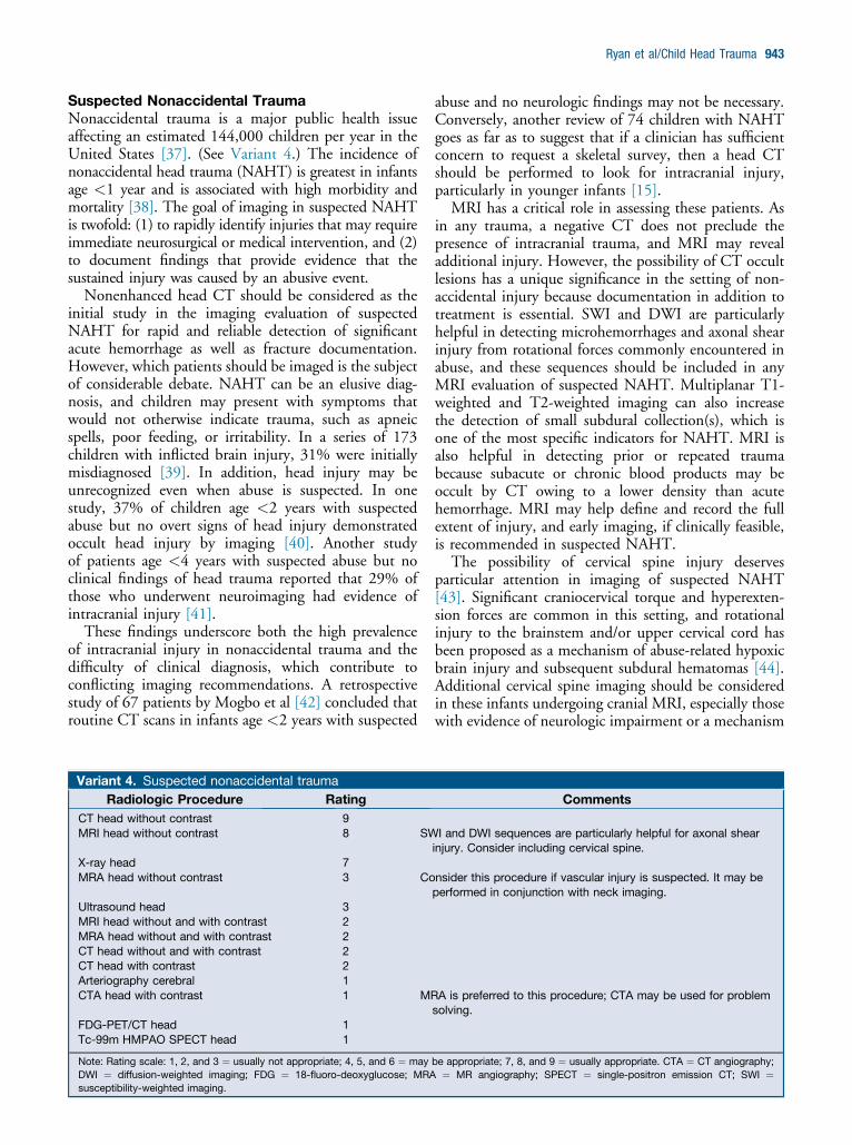
Variant 4. Suspected nonaccidental traumaRadiologic Procedure Rating
CT head without contrast 9MRI head without contrast 8 SW
X-ray head 7MRA head without contrast 3 Co
Ultrasound head 3MRI head without and with contrast 2MRA head without and with contrast 2CT head without and with contrast 2CT head with contrast 2Arteriography cerebral 1CTA head with contrast 1 MR
FDG-PET/CT head 1Tc-99m HMPAO SPECT head 1
Note: Rating scale: 1, 2, and 3 ¼ usually not appropriate; 4, 5, and 6 ¼ mayDWI ¼ diffusion-weighted imaging; FDG ¼ 18-fluoro-deoxyglucose; MRAsusceptibility-weighted imaging.
abuse and no neurologic findings may not be necessary.Conversely, another review of 74 children with NAHTgoes as far as to suggest that if a clinician has sufficientconcern to request a skeletal survey, then a head CTshould be performed to look for intracranial injury,particularly in younger infants [15].
MRI has a critical role in assessing these patients. Asin any trauma, a negative CT does not preclude thepresence of intracranial trauma, and MRI may revealadditional injury. However, the possibility of CT occultlesions has a unique significance in the setting of non-accidental injury because documentation in addition totreatment is essential. SWI and DWI are particularlyhelpful in detecting microhemorrhages and axonal shearinjury from rotational forces commonly encountered inabuse, and these sequences should be included in anyMRI evaluation of suspected NAHT. Multiplanar T1-weighted and T2-weighted imaging can also increasethe detection of small subdural collection(s), which isone of the most specific indicators for NAHT. MRI isalso helpful in detecting prior or repeated traumabecause subacute or chronic blood products may beoccult by CT owing to a lower density than acutehemorrhage. MRI may help define and record the fullextent of injury, and early imaging, if clinically feasible,is recommended in suspected NAHT.
The possibility of cervical spine injury deservesparticular attention in imaging of suspected NAHT[43]. Significant craniocervical torque and hyperexten-sion forces are common in this setting, and rotationalinjury to the brainstem and/or upper cervical cord hasbeen proposed as a mechanism of abuse-related hypoxicbrain injury and subsequent subdural hematomas [44].Additional cervical spine imaging should be consideredin these infants undergoing cranial MRI, especially thosewith evidence of neurologic impairment or a mechanism
Comments
I and DWI sequences are particularly helpful for axonal shearinjury. Consider including cervical spine.
nsider this procedure if vascular injury is suspected. It may beperformed in conjunction with neck imaging.
A is preferred to this procedure; CTA may be used for problemsolving.
be appropriate; 7, 8, and 9 ¼ usually appropriate. CTA ¼ CT angiography;¼ MR angiography; SPECT ¼ single-positron emission CT; SWI ¼

944 Journal of the American College of Radiology/Vol. 11 No. 10 October 2014
of injury that might result in spinal injury [45]. Soft-tissue injury is more common than fracture, and MRIwith fluid-sensitive fat-suppressed sequences can bestidentify paraspinous muscle edema, ligamentous injury,or spinal cord trauma and may provide evidence that asignificant force was sustained [46,47].Radiographs of the skull are known to have a low
predictive value in determining intracranial injury [24].However, in contrast to accidental head trauma, forwhich radiographs have largely been replaced by CT,skull radiographs are still often performed as part of theskeletal survey in evaluation of suspected nonaccidentaltrauma. It has been generally accepted that skull radio-graphs and head CT are complementary examinations,as fractures in the plane of the transaxial CT image maynot be apparent on the head CT examination. Charac-terization of fracture morphology is particularly impor-tant in this setting because some calvarial injuries, suchas depressed, wide, or growing skull fractures andinvolvement of more than one cranial bone, have asignificantly greater association with NAHT [48]. Arecent study by Prabhu et al [49] reports that three-dimensional reconstructions can be more accurate infracture detection and differentiation from accessorysutures. The sensitivity of CT for calvarial fractures andthe utility of supplemental plain films is likely highlyvariable depending on institutional technique and theability to generate multiplanar reformations.Head ultrasound can easily be performed at the
bedside in young infants and can differentiate convexitysubdural collections from subarachnoid collections [50].In addition, ultrasound is sensitive in detecting subcor-tical white-matter tears in the neonate [51]. Thesetypically occur in the frontal and anterior parietal para-sagittal regions and represent a shear-type injury that ishighly suggestive of NAHT [52]. However, ultrasound isinsensitive for detecting small acute subdural hematomas,particularly within the interhemispheric fissure andposterior fossa [53], as well as other intracranial injuries,and generally has a limited role in assessing NAHT.
Variant 5. Subacute head injury with cognitive and/or neuroRadiologic Procedure Rating
MRI head without contrast 8 MRI is preferreCT head without contrast 7MRA head without contrast 3 Consider this pMRI head without and with contrast 2 This procedureMRA head without and with contrast 2CT head without and with contrast 2CTA head with contrast 2Arteriography cerebral 2X-ray head 1CT head with contrast 1Ultrasound head 1FDG-PET/CT head 1Tc-99m HMPAO SPECT head 1
Note: Rating scale: 1, 2, and 3 ¼ usually not appropriate; 4, 5, and 6 ¼ mayFDG ¼ 18-fluoro-deoxyglucose; MRA ¼ MR angiography; SPECT ¼ single-p
Subacute Closed Head Injury with Cognitiveand/or Neurological DeficitIn addition to initial acute intracranial trauma, injurymay be caused by secondary processes such as herniationfrom worsening parenchymal edema, ischemia, hydro-cephalus, and progressive or delayed hemorrhage. (SeeVariant 5.) During the subacute phase, up to 30% ofcontusions may cause worsening mass effect with edemafrom toxic metabolites released into the surroundingtissue and cerebral autoregulation dysfunction [54].Delayed complications of fractures, such as cerebrospi-nal fluid leak, leptomeningeal cyst, or meningitis, mayalso occur.
In an effort to avoid unnecessary radiation exposureand health care costs, it is important to determine whichpatients would benefit from repeated imaging in thesubacute setting. Patients with a significant change inneurologic status should be reimaged. However, theabsence of neurologic changes does not preclude thepossibility of secondary or progressive injury. In a retro-spective study of 397 pediatric patients diagnosed at timeof discharge with intracranial hemorrhage, 2.5% pre-sented with delayed hemorrhage, with only 20% of thoseexhibiting mental status changes [55]. Hollingworth et al[56] suggested that those who benefit from repeated ex-amination include patients with positive findings on priorCT as well as those with moderate or severe head injuryregardless of prior imaging. Using this screening methodin a retrospective analysis of more than 250 children, theyidentified 89% of patients with worsening CT injuriesand 100% of patients that proceed to neurosurgicalintervention.
MRI is important in evaluating persistent, unex-plained, or new neurological deficits in the subacutesetting. The greater sensitivity of MRI for blood prod-ucts, including small brainstem and infratentorial hem-orrhages as well as subacute extraaxial hemorrhage,which becomes less dense on CT over time, and thesuperior detection of nonhemorrhagic contusions andischemia may be helpful in the absence of findings on
logic signsComments
d, but imaging should not be delayed if it is not readily available.
rocedure if vascular injury is suspected.is not indicated unless there is a concern for infection.
be appropriate; 7, 8, and 9 ¼ usually appropriate. CTA ¼ CT angiography;ositron emission CT.

Ryan et al/Child Head Trauma 945
prior CT. However, MRI requires the patient to bestable enough to tolerate a lengthier examination.Contrast-enhanced sequences generally are not indicatedunless there is a concern for infection such as frompenetrating injury or fractures involving the sinuses.
Vascular InjuriesTrauma to the intracranial vessels is infrequently re-ported and believed to be relatively uncommon inchildren. Dissection, pseudoaneurysm, and other arterialinjuries most often occur extracranially in the cervicalregion or at the skull base [57] and are typicallyconsidered with neck imaging protocols. Still, vascularinjuries have been described in pediatric trauma of anyseverity or mechanism as well as without identifiableantecedent injury. Evaluation is primarily guided byclinical suspicion, and vascular imaging should beconsidered in patients with evidence of arterial stroke byexamination or by imaging, as well as those with frac-tures extending through the skull base vascular channels[58]. However, data are conflicting regarding whetherpediatric traumatic stroke can be predicted using thesame risk factors or clinical signs as in adults, leading tothe concern that vascular injury may be under-recognized in children [59,60]. Most vascular literaturein the pediatric population is confined to small series,and uncertainty remains regarding the true incidenceand natural history of these injuries [61].Noninvasive imaging with CT angiography or MR
angiography is considered the first-line imaging forarterial injury. CT angiography provides high spatialresolution and rapid assessment but exposes the patientto ionizing radiation, particularly when performed inaddition to a necessary noncontrast head CT to assessfor acute intracranial injury. MR angiography can eval-uate the intracranial vasculature without radiation andcan be performed in conjunction with MRI for assess-ment of hemorrhage and ischemia; however, it is alengthier examination and may be difficult to performemergently. Subtle injuries may be occult on eitherCTA or MRA, and conventional angiography remainsthe definitive diagnostic test for vascular injury. How-ever, given that conventional angiography entails aninvasive procedure, radiation, and sedation, and haslimited availability, it is usually reserved for problemsolving in cases with uncertain diagnostic imaging andhigh clinical suspicion.
Advanced ImagingInterest has been increasing in the use of higher-orderimaging techniques, such as PET, single-photon emissionCT perfusion, functional MRI, diffusion tensor imaging,and proton MR spectroscopy, to assess the functional andmicrostructural consequences of head trauma [17].Several small studies suggest that blood flow and
perfusion abnormalities on Tc-99m single-photonemission CT, and decreased metabolic activity on 18-fluoro-deoxyglucoseePET brain imaging, can indicate
subtle traumatic changes in children and often correlatewith outcomes, although the prognostic value variesdepending on imaging timing [62-66]. Functional MRIhas shown alterations in language and sensorimotor or-ganization in children with posttraumatic injury, butthese studies are confined to small series, and preinjuryfunctional MRI data for comparison are lacking.Transcranial Doppler ultrasound has been used to assessintracranial blood flow in some adult trauma victims.However, the cerebral hemodynamics of young childrendiffer from those of adults, and TCD evaluation forpediatric head trauma is relatively untested at this time.The high radiation doses associated with CT perfusionare typically unwarranted in this population, and MRperfusion has yet to be studied in a substantial pediatriccohort.
Preliminary data suggest that diffusion tensor imagingmay also reveal injuries not detected by conventionalMR sequences, and some studies have shown a strongcorrelation between decreased fractional anisotropy,particularly in the corpus callosum, and posttraumaticoutcomes in children [62,64,67]. MR spectroscopymay also provide insight into TBI, with metabolite ab-normalities such as increased lactate and decreased N-acetylaspartate correlating with severity of injury andprognosis in small studies, particularly in neonates andinfants [63,68,69]. However, although advanced imag-ing offers future promise for more thorough assessmentand understanding of TBI, these modalities remaingenerally understudied in pediatric head trauma. Thesmall sample sizes, variability in timing, and differencesin technique in the literature are particularly problematicin pediatric assessment, owing to the continuouslychanging normative values of the developing brain. Fewvalidated data support the routine use of these tech-niques in clinical evaluation.
SUMMARY
� Noncontrast CT is the primary study for emergentevaluation of suspected intracranial injury in children,although the decision to image should take into accountthe increased risks of ionizing radiation in this population.� Imaging is typically not indicated in children withminor head trauma and no signs or symptoms.However, neurologic examination can be difficult inyounger children, and imaging in this setting may beappropriate if the clinical assessment is unclear orindeterminate.
� MRI is more sensitive than CT for small parenchymallesions, ischemia, and shear injury, and may beparticularly helpful in patients whose clinical status isdiscordant with negative CT findings, those withmoderate or severe injury, and in cases of suspectednonaccidental trauma.
� Advanced imaging techniques may reveal injury that isoccult by standard imaging, but they remain relatively

946 Journal of the American College of Radiology/Vol. 11 No. 10 October 2014
untested in the pediatric population with few data tosupport routine clinical use.
ACKNOWLEDGEMENTSThis article is a revised version of the ACR Appro-priateness Criteria Head Trauma—Child. Practitionersare encouraged to refer to the complete version at www.acr.org/ac.
REFERENCES1. Pinto PS, Poretti A, Meoded A, Tekes A, Huisman TA. The unique
features of traumatic brain injury in children. Review of the character-istics of the pediatric skull and brain, mechanisms of trauma, patternsof injury, complications and their imaging findings—part 1.J Neuroimaging 2012;22:e1-17.
2. Faul M, Xu L, Wald MM, Coronado VG. Traumatic brain injury in theUnited States: emergency department visits, hospitalizations and deaths2002e2006. Centers for Disease Control and Prevention, NationalCenter for Injury Prevention and Control. Available at: http://www.cdc.gov/traumaticbraininjury/tbi_ed.html. Accessed September 16, 2013.
3. Schnadower D, Vazquez H, Lee J, Dayan P, Roskind CG. Controversiesin the evaluation and management of minor blunt head trauma inchildren. Curr Opin Pediatr 2007;19:258-64.
4. Willis AP, Latif SA, Chandratre S, Stanhope B, Johnson K. Not a NICECT protocol for the acutely head injured child. Clin Radiol 2008;63:165-9.
5. Schutzman SA, Greenes DS. Pediatric minor head trauma. Ann EmergMed 2001;37:65-74.
6. Haydel MJ, Shembekar AD. Prediction of intracranial injury in childrenaged five years and older with loss of consciousness after minorhead injury due to nontrivial mechanisms. Ann Emerg Med 2003;42:507-14.
7. Kuppermann N, Holmes JF, Dayan PS, et al. Identification of childrenat very low risk of clinically-important brain injuries after head trauma: aprospective cohort study. Lancet 2009;374:1160-70.
8. Tavarez MM, Atabaki SM, Teach SJ. Acute evaluation of pediatric pa-tients with minor traumatic brain injury. Curr Opin Pediatr 2012;24:307-13.
9. Maguire JL, Boutis K, Uleryk EM, Laupacis A, Parkin PC. Should ahead-injured child receive a head CT scan? A systematic review of clinicalprediction rules. Pediatrics 2009;124:e145-54.
10. Halley MK, Silva PD, Foley J, Rodarte A. Loss of consciousness: when toperform CT? Pediatr Crit Care Med 2004;5:230-3.
11. Pearce MS, Salotti JA, Little MP, et al. Radiation exposure from CTscans in childhood and subsequent risk of leukaemia and brain tumours:a retrospective cohort study. Lancet 2012;380:499-505.
12. Brenner DJ, Hall EJ. CT—an increasing source of radiation exposure.New Engl J Med 2007;357:2277-84.
13. How to develop CT protocols for children. Available at: http://spr.affiniscape.com/associations/5364/files/Protocols.pdf. Accessed September16, 2013.
14. Image Gently�. Available at: http://www.imagegently.org/. AccessedSeptember 16, 2013.
15. Datta S, Stoodley N, Jayawant S, Renowden S, Kemp A. Neuro-radiological aspects of subdural haemorrhages. Arch Dis Child 2005;90:947-51.
16. Kemp AM, Rajaram S, Mann M, et al. What neuroimaging should beperformed in children in whom inflicted brain injury (iBI) is suspected?A systematic review. Clin Radiol 2009;64:473-83.
17. Hunter JV, Wilde EA, Tong KA, Holshouser BA. Emerging imagingtools for use with traumatic brain injury research. J Neurotrauma2012;29:654-71.
18. Skandsen T, Kvistad KA, Solheim O, Strand IH, Folvik M, Vik A.Prevalence and impact of diffuse axonal injury in patients with moderateand severe head injury: a cohort study of early MRI findings and 1-yearoutcome. J Neurosurg 2010;113:556-63.
19. Tong KA, Ashwal S, Holshouser BA, et al. Hemorrhagic shearinglesions in children and adolescents with posttraumatic diffuse axonalinjury: improved detection and initial results. Radiology 2003;227:332-9.
20. Holmes JF, Borgialli DA, Nadel FM, et al. Do children with blunt headtrauma and normal cranial CT scan results require hospitalization forneurologic observation? Ann Emerg Med 2011;58:315-22.
21. Reed MJ, Browning JG, Wilkinson AG, Beattie T. Can we abolish skullx rays for head injury? Arch Dis Child 2005;90:859-64.
22. Nakahara K, Shimizu S, Utsuki S, et al. Linear fractures occult on skullradiographs: a pitfall at radiological screening for mild head injury.J Trauma 2011;70:180-2.
23. Crowe L, Anderson V, Babl FE. Application of the CHALICE clinicalprediction rule for intracranial injury in children outside the UK: impacton head CT rate. Arch Dis Child 2010;95:1017-22.
24. Dunning J, Daly JP, Lomas JP, Lecky F, Batchelor J, Mackway-Jones K.Derivation of the children’s head injury algorithm for the prediction ofimportant clinical events decision rule for head injury in children. ArchDis Child 2006;91:885-91.
25. Oman JA, Cooper RJ, Holmes JF, et al. Performance of a decision rule topredict need for CT among children with blunt head trauma. Pediatrics2006;117:e238-46.
26. Schachar JL, Zampolin RL, Miller TS, Farinhas JM, Freeman K,Taragin BH. External validation of the New Orleans Criteria (NOC),the Canadian CT Head Rule (CCHR) and the National EmergencyX-Radiography Utilization Study II (NEXUS II) for CT scanning inpediatric patients with minor head injury in a non-trauma center. PediatrRadiol 2011;41:971-9.
27. Bainbridge J, Khirwadkar H, Hourihan MD. Vomiting—is this a goodindication for CT head scans in patients with minor head injury? Brit JRadiol 2012;85:183-6.
28. Nigrovic LE, Lee LK, Hoyle J, et al. Prevalence of clinically importanttraumatic brain injuries in children with minor blunt head trauma andisolated severe injury mechanisms. Arch Pediatr Adol Med 2012;166:356-61.
29. Pickering A, Harnan S, Fitzgerald P, Pandor A, Goodacre S. Clinicaldecision rules for children with minor head injury: a systematic review.Arch Dis Child 2011;96:414-21.
30. Mannix R, Meehan WP, Monuteaux MC, Bachur RG. Computedtomography for minor head injury: variation and trends in major USpediatric emergency departments. J Pediatr 2012;160:136-9 e1.
31. Gorelick MH, Atabaki SM, Hoyle J, et al. Interobserver agreement inassessment of clinical variables in children with blunt head trauma. AcadEmerg Med 2008;15:812-8.
32. Holmes JF, Palchak MJ, MacFarlane T, Kuppermann N. Performance ofthe pediatric Glasgow coma scale in children with blunt head trauma.Acad Emerg Med 2005;12:814-9.
33. Margulies SS, Thibault KL. Infant skull and suture properties: mea-surements and implications for mechanisms of pediatric brain injury.J Biomech Eng 2000;122:364-71.
34. Claret Teruel G, Palomeque Rico A, Cambra Lasaosa FJ, CatalaTemprano A, Noguera Julian A, Costa Clara JM. Severe head injuryamong children: CT evaluation as a prognostic factor. J Pediatr Surg2007;42:1903-6.
35. Sigmund GA, Tong KA, Nickerson JP, Wall CJ, Oyoyo U, Ashwal S.Multimodality comparison of neuroimaging in pediatric traumatic braininjury. Pediatr Neurol 2007;36:217-26.
36. Ball WS Jr. Nonaccidental craniocerebral trauma (child abuse): MRimaging. Radiology 1989;173:609-10.
37. Ashwal S, Wycliffe ND, Holshouser BA. Advanced neuroimaging inchildren with nonaccidental trauma. Dev Neurosci 2010;32:343-60.

Ryan et al/Child Head Trauma 947
38. Duhaime AC, Gennarelli TA, Thibault LE, Bruce DA, Margulies SS,Wiser R. The shaken baby syndrome. A clinical, pathological, andbiomechanical study. J Neurosurg 1987;66:409-15.
39. Jenny C, Hymel KP, Ritzen A, Reinert SE, Hay TC. Analysis of missedcases of abusive head trauma. JAMA 1999;281:621-6.
40. Rubin DM, Christian CW, Bilaniuk LT, Zazyczny KA, Durbin DR.Occult head injury in high-risk abused children. Pediatrics 2003;111:1382-6.
41. Laskey AL, Holsti M, Runyan DK, Socolar RR. Occult head trauma inyoung suspected victims of physical abuse. J Pediatr 2004;144:719-22.
42. Mogbo KI, Slovis TL, Canady AI, Allasio DJ, Arfken CL. Appropriateimaging in children with skull fractures and suspicion of abuse. Radi-ology 1998;208:521-4.
43. Brown RL, Brunn MA, Garcia VF. Cervical spine injuries in children: areview of 103 patients treated consecutively at a level 1 pediatric traumacenter. J Pediatr Surg 2001;36:1107-14.
44. Ghatan S, Ellenbogen RG. Pediatric spine and spinal cord injury afterinflicted trauma. Neurosurg Clinics N Am 2002;13:227-33.
45. Davis PC, Wippold FL II, Cornelius RS, et al. ACR AppropriatenessCriteria� head trauma. Available at: http://www.acr.org/w/media/ACR/Documents/AppCriteria/Diagnostic/HeadTrauma.pdf. Accessed July 29,2013.
46. Keiper MD, Zimmerman RA, Bilaniuk LT. MRI in the assessment ofthe supportive soft tissues of the cervical spine in acute trauma in chil-dren. Neuroradiology 1998;40:359-63.
47. Kadom N, Khademian Z, Vezina G, Shalaby-Rana E, Rice A, Hinds T.Usefulness of MRI detection of cervical spine and brain injuries in theevaluation of abusive head trauma. Pediatr Radiol 2014;44:839-48.
48. Hobbs CJ. Skull fracture and the diagnosis of abuse. Arch Dis Child1984;59:246-52.
49. Prabhu SP, Newton AW, Perez-Rossello JM, Kleinman PK. Three-dimensional skull models as a problem-solving tool in suspected childabuse. Pediatr Radiol 2013;43:575-81.
50. Chen CY, Chou TY, Zimmerman RA, Lee CC, Chen FH, Faro SH.Pericerebral fluid collection: differentiation of enlarged subarachnoidspaces from subdural collections with color Doppler ultrasound. Radi-ology 1996;201:389-92.
51. Amodio J, Spektor V, Pramanik B, Rivera R, Pinkney L, Fefferman N.Spontaneous development of bilateral subdural hematomas in an infantwith benign infantile hydrocephalus: color Doppler assessment of vesselstraversing extra-axial spaces. Pediatr Radiol 2005;35:1113-7.
52. Jaspan T, Narborough G, Punt JA, Lowe J. Cerebral contusional tears asa marker of child abuse—detection by cranial sonography. Pediatr Radiol1992;22:237-45.
53. Kemp AM, Jaspan T, Griffiths J, et al. Neuroimaging: What neurora-diological features distinguish abusive from non-abusive head trauma? Asystematic review. Arch Dis Child 2011;96:1103-12.
54. Pinto PS, Meoded A, Poretti A, Tekes A, Huisman TA. The uniquefeatures of traumatic brain injury in children. Review of the character-istics of the pediatric skull and brain, mechanisms of trauma, patternsof injury, complications, and their imaging findings—part 2.J Neuroimaging 2012;22:e18-41.
55. Hamilton M, Mrazik M, Johnson DW. Incidence of delayed intracranialhemorrhage in children after uncomplicated minor head injuries. Pedi-atrics 2010;126:e33-9.
56. Hollingworth W, Vavilala MS, Jarvik JG, et al. The use of repeated headCT in pediatric blunt head trauma: factors predicting new and worseningbrain injury. Pediatr Crit Care Med 2007;8:348-56; CEU quiz 57.
57. Stence NV, Fenton LZ, Goldenberg NA, Armstrong-Wells J,Bernard TJ. Craniocervical arterial dissection in children: diagnosis andtreatment. Curr Treat Options Neurol 2011;13:636-48.
58. Sepelyak K, Gailloud P, Jordan LC. Athletics, minor trauma, and pe-diatric arterial ischemic stroke. Eur J Pediatr 2010;169:557-62.
59. Jones TS, Burlew CC, Kornblith LZ, et al. Blunt cerebrovascular injuriesin the child. Am J Surg 2012;204:7-10.
60. Kopelman TR, Berardoni NE, O’Neill PJ, et al. Risk factors for bluntcerebrovascular injury in children: Do they mimic those seen in adults?J Trauma 2011;71:559-64; disc 64.
61. Mortazavi MM, Verma K, Tubbs RS, Harrigan M. Pediatric traumaticcarotid, vertebral and cerebral artery dissections: a review. Childs NervSyst 2011;27:2045-56.
62. Aoki Y, Inokuchi R, Gunshin M, Yahagi N, Suwa H. Diffusion tensorimaging studies of mild traumatic brain injury: a meta-analysis. J NeurolNeurosurg Psychiatr 2012;83:870-6.
63. Munson S, Schroth E, Ernst M. The role of functional neuroimaging inpediatric brain injury. Pediatrics 2006;117:1372-81.
64. Wilde EA, McCauley SR, Hunter JV, et al. Diffusion tensor imaging of acutemild traumatic brain injury in adolescents. Neurology 2008;70:948-55.
65. Worley G, Hoffman JM, Paine SS, et al. 18-Fluorodeoxyglucose PET inchildren and adolescents with traumatic brain injury. Dev Med ChildNeurol 1995;37:213-20.
66. Goshen E, Zwas ST, Shahar E, Tadmor R. The role of 99Tcm-HMPAObrain SPET in paediatric traumatic brain injury. Nucl Med Commun1996;17:418-22.
67. Ewing-Cobbs L, Prasad MR, Swank P, et al. Arrested development anddisrupted callosal microstructure following pediatric traumatic brain injury:relation to neurobehavioral outcomes. NeuroImage 2008;42:1305-15.
68. Ashwal S, Babikian T, Gardner-Nichols J, Freier MC, Tong KA,Holshouser BA. Susceptibility-weighted imaging and proton MR spec-troscopy in assessment of outcome after pediatric traumatic brain injury.Arch Phys Med Rehabil 2006;87:S50-8.
69. Walz NC, Cecil KM, Wade SL, Michaud LJ. Late proton MR spectros-copy following traumatic brain injury during early childhood: relationshipwith neurobehavioral outcomes. J Neurotrauma 2008;25:94-103.


