Embed Size (px)
Citation preview

Activation and repression of a sN-dependent promoternaturally lacking upstream activation sequencesmmi_6779 419..433
Odil Porrúa,1,2 Vicente García-González,1,2†
Eduardo Santero,1,2 Victoria Shingler3 andFernando Govantes1,2*1Centro Andaluz de Biología del Desarrollo, UniversidadPablo de Olavide/CSIC, Carretera de Utrera, Km. 1.41013 Sevilla, Spain.2Departamento de Biología Molecular e IngenieríaBioquímica, Universidad Pablo de Olavide, Carreterade Utrera, Km. 1. 41013 Sevilla, Spain.3Department of Molecular Biology, Umeå University,901 87 Umeå, Sweden.
Summary
The Pseudomonas sp. strain ADP protein AtzR is aLysR-type transcriptional regulator required for acti-vation of the atzDEF operon in response to nitrogenlimitation and cyanuric acid. Transcription of atzR isdirected by the sN-dependent promoter PatzR, acti-vated by NtrC and repressed by AtzR. Here we usein vivo and in vitro approaches to address the mecha-nisms of PatzR activation and repression. Activationby NtrC did not require any promoter sequencesother than the sN recognition motif both in vivo andin vitro, suggesting that NtrC activates PatzR in anupstream activation sequences-independent fashion.Regarding AtzR-dependent autorepression, our invitro transcription experiments show that the concen-tration of AtzR required for repression of the PatzRpromoter in vitro correlates with AtzR affinity for itsbinding site. In addition, AtzR prevents transcriptionfrom PatzR when added to a preformed E-sN–PatzRclosed complex, but isomerization to an opencomplex prevents repression. Gel mobility shift andDNase I footprint assays indicate that DNA-boundAtzR and E-sN are mutually exclusive. Taken together,these results strongly support the notion that AtzRrepresses transcription from PatzR by competingwith E-sN for their overlapping binding sites. There
are no previous reports of a similar mechanism forrepression of sN-dependent transcription.
Introduction
The alternative s factor sN, also known as s54, is ubiqui-tous among bacteria. RNA polymerase loaded with sN
(E-sN) displays features unlike any other form of theholoenzyme. Transcription by E-sN is initiated at promot-ers with conserved elements around positions -24 and-12 that differ substantially from the usual -35 and -10boxes in promoters recognized by members of the s70
family. E-sN readily binds DNA at this type of promoterregions to form a binary closed complex. However, thesN-bearing holoenzyme is unable to elicit DNA strandseparation at the promoter to form an open complex.Because of this defect, all sN-dependent promoters aresubjected to positive regulation by a member of a familyof bacterial transcriptional activators known as enhancer-binding proteins (EBPs) (Kustu et al., 1989; Morett andSegovia, 1993; Buck et al., 2000).
Enhancer-binding proteins are modular proteinsharbouring an N-terminal regulatory domain involvedin signal response, a highly conserved catalytic centraldomain with ATPase activity and a DNA-bindingC-terminal domain featuring a helix–turn–helix motif.EBPs bind to DNA over 80 bp upstream from the E-sN
recognition elements they control, at sites known asenhancer-like elements or upstream activation sequences(UAS) (Kustu et al., 1989). An EBP bound to its cognateUAS contacts promoter-bound E-sN via formation of aspontaneous or protein-induced DNA loop at the interven-ing sequences. Interaction of an EBP with E-sN resultsin isomerization to an open complex in a reaction thatrequires ATP hydrolysis by the conserved central domainof the EBP. The structure and function of EBPs and themolecular mechanisms behind sN-dependent promoteractivation have been thoroughly reviewed elsewhere(Morett and Segovia, 1993; Buck et al., 2000; Zhanget al., 2002; Studholme and Dixon, 2003; Schumacheret al., 2006; Wigneshweraraj et al., 2008). In addition topositive control, a handful of sN-dependent promotersare subjected to negative control by regulatory proteinsother than their cognate EBPs. Interestingly, in the fewcharacterized examples, repression appears to occur via
Accepted 18 June, 2009. *For correspondence. E-mail [email protected]; Tel. (+34) 954 977877; Fax (+34) 954 349376. †Presentaddress: Oryzon Genomics, Josep Samitier 1-5, 08028 Barcelona,Spain.
Molecular Microbiology (2009) 73(3), 419–433 � doi:10.1111/j.1365-2958.2009.06779.xFirst published online 7 July 2009
© 2009 The AuthorsJournal compilation © 2009 Blackwell Publishing Ltd

an anti-activation mechanism in which the repressorprevents productive interactions between the EBP andRNA polymerase (Feng et al., 1995; Martin-Verstraeteet al., 1995; Wang et al., 1998; Mao et al., 2007).
LysR-type transcriptional regulators (LTTRs) representthe most abundant family of transcriptional regulatory pro-teins in bacteria. LTTRs are typically encoded by genesadjacent and divergent from the promoters they activate.Most proteins in this family repress their own transcription.Activation and autorepression are exerted from a singleasymmetrical binding site located within the divergent pro-moter region. LTTR binding sites contain a palindromicrecognition element, harbouring the consensus T-N11-A,designated the repressor binding site or RBS. The RBS isnormally centred at position -65 relative to the activatedpromoter transcription start site, and often overlaps therepressed promoter. The RBS is considered the majorsequence determinant for LTTR binding, and is requiredfor both activation and repression. A second sequenceelement, the activator binding site or ABS, is locatedbetween the RBS and the activated promoter. The ABS isan accessory element for DNA binding, but it is strictlyrequired for activation. Many mechanistic details on tran-scriptional activation by several members of the LTTRfamily have been revealed over the last 15 years, anda general model for inducer-dependent activation, thesliding dimer model, has been proposed (reviewed bySchell, 1993; Tropel and van der Meer, 2004). In contrast,the phenomenon of autorepression has received littleattention, and it is generally assumed that LTTR bindingto the strong RBS element is sufficient for repression(Schell, 1993).
AtzR is an LTTR involved in activation of the cyanuricacid utilization operon atzDEF of Pseudomonas sp. ADPin response to cyanuric acid and nitrogen limitation. AtzRis encoded in a monocistronic operon divergently tran-scribed from atzDEF (Martinez et al., 2001). The PatzRpromoter is one of the rare examples of sN-dependentpromoters subjected to both positive and negative control.PatzR is activated by the nitrogen limitation-responsiveEBP NtrC, and repressed by AtzR irrespective of thepresence of cyanuric acid (García-González et al., 2005).The role of NtrC and the signalling cascade for nitrogenlimitation have been extensively characterized in entero-bacteria (Merrick and Edwards, 1995; Arcondeguy et al.,2001; Reitzer and Schneider, 2001; Reitzer, 2003). NtrCis an archetypical activator of sN-dependent promotersthat binds to UAS elements located far upstream fromthe E-sN recognition element. However, no sequenceresembling the proposed Pseudomonas putida NtrCbinding motif (Hervás et al., 2008) is present at the diver-gent atzR–atzDE promoter region, and we have shownthat deletion of all sequences upstream from the E-sN
recognition element has no effect on NtrC-dependent
activation of PatzR in vivo, suggesting that NtrC does notrequire any specific sequences upstream from the pro-moter to activate atzR transcription (Porrúa et al., 2007).We have also shown that AtzR binds a single site atthe divergent atzR–atzDEF promoter region. The AtzRbinding site contains a typical RBS, centred at position-65 relative to the atzDEF transcriptional start, whichoverlaps the sN-dependent PatzR promoter. We havecharacterized the function of the RBS as the major deter-minant for high-affinity DNA binding of AtzR that is essen-tial for both activation and autorepression, and our workhas revealed an important role for the ABS in negativeautoregulation (Porrúa et al., 2007).
The present work focuses on the mechanisms oftranscriptional regulation at the PatzR promoter. Here, weexplore further the requirement of cis-acting sequencesfor NtrC-dependent activation of PatzR and dissect theinterplay between E-sN, AtzR and their respective re-cognition sites at the atzR promoter region. Our resultsstrongly suggest that AtzR represses atzR transcription bycompeting with E-sN for DNA binding to effectively dimin-ish E-sN occupancy of the PatzR promoter. We proposethat this mechanism is an adaptation to the fact that theatzR promoter region lacks a UAS for NtrC, which makesNtrC-dependent activation inefficient and thus dependenton a high occupancy rate of the PatzR promoter by E-sN.
Results
Activation of the PatzR promoter does not requireupstream or downstream sequences
In contrast to most sN-promoters, the PatzR promoterregion lacks any sequences resembling the UAS con-sensus for its cognate EBP, NtrC. In addition, a PatzR–lacZ fusion lacking all DNA sequences upstream from the-24/-12 E-sN recognition element was still induced inresponse to nitrogen limitation in a similar fashion toa wild-type fusion (Porrúa et al., 2007), indicating thatNtrC binding to upstream sequences is not required forPatzR activation in vivo. Because the PatzR–lacZ fusionscontain 139 bp downstream from the atzR transcrip-tional start site, it is feasible that an enhancer-like elementmay be located in this region, as described for othersN-dependent promoters (Belitsky and Sonenshein, 1999;Jyot et al., 2002). To test this possibility, transcriptionalfusion plasmids pMPO232, containing the full-length atzRpromoter region (positions -250 to +139), and pMPO237,containing a minimal PatzR promoter that includes onlythe -24/-12 E-sN recognition boxes and the trans-criptional start site (positions -27 to +1), were constructed(Fig. 1). Both plasmids, along with the empty trans-criptional fusion vector (pMPO234), were transferred toP. putida KT2442 and its DntrC derivative MPO201 by
420 O. Porrúa et al. �
© 2009 The AuthorsJournal compilation © 2009 Blackwell Publishing Ltd, Molecular Microbiology, 73, 419–433

mating, and expression was monitored by means ofb-galactosidase assays (Table 1).
Expression from the wild-type atzR promoter regionin pMPO232 was low in cells grown under nitrogen suf-ficiency, and was significantly increased (14-fold) innitrogen-limited medium. This result is consistent with thepreviously observed induction of PatzR under nitrogenlimitation (García-González et al., 2005; Porrúa et al.,2007). Expression from the minimal PatzR promoter inpMPO237 was somewhat lower (twofold) in ammonium-containing medium. Nevertheless, an induction ratio com-parable to that in the full-length construct (10-fold) wasobserved with this construct under nitrogen limitation.Low, unregulated activity was obtained with the trans-
criptional fusion vector pMPO234 under both growthconditions. Expression from both PatzR–lacZ fusions wasdecreased similarly (seven- to eightfold) when assayedin the DntrC mutant MPO201 grown on serine, whilethe basal expression levels obtained in ammonium-containing medium were largely unaffected, indicatingthat NtrC is responsible for nitrogen limitation-responsiveactivation with both constructs. Analysis of certain E-sN-dependent promoters in plasmid constructs has beenshown to lead to enhanced UAS-independent activationthat may overrule the need for weak UAS elements(Drummond et al., 1983). To rule out the possibility thatthis phenomenon is an artefact due to the use of plasmid-borne fusions, two plasmids were constructed (pMPO166
Fig. 1. Fusion constructs and in vitro transcription templates used in this work.A. Sequence of the atzR–atzDEF promoter region with the end-points of the PatzR inserts in the different constructs indicated by bent arrows.Conserved positions of the PatzR promoter are underlined, boxes indicate the location of the RBS (shaded) and the proposed ABS (open)and an asterisk denotes the PatzR transcriptional start site.B. Schematic of the atzR–atzDEF promoter region, indicating the extension of the sequence included in the transcriptional fusion plasmids(solid bars) and in vitro transcription templates (shaded bars). Co-ordinates are relative to the transcriptional start of atzR. Solid arrowsindicate the location of the PatzR and PatzDEF promoters. Bent arrows denote the atzDEF and atzR transcriptional start points. Shadedand open boxes represent the RBS and the proposed ABS respectively.
Table 1. Expression of PatzR–lacZ transcriptional fusions.
Fusion plasmidPatzR promoterfragmenta
P. putida geneticbackground
Nitrogen source
Ammonium Serineb-Galactosidase activity
pMPO234 None Wild type 156 � 33 138 � 15pMPO232 -250 to +139 Wild type 447 � 92 6190 � 662
DntrC 444 � 45 880 � 134pMPO237 -27 to +1 Wild type 254 � 31 2590 � 316
DntrC 249 � 11 336 � 26
a. Co-ordinates are relative to the atzR transcriptional start site. Values are b-galactosidase activity (Miller units) of exponentially growing cultures,and represent the average and standard deviation of at least three independent measurements.
Complex regulation of a sN-dependent promoter 421
© 2009 The AuthorsJournal compilation © 2009 Blackwell Publishing Ltd, Molecular Microbiology, 73, 419–433

and pMPO167) that harbour the wild-type and minimalPatzR promoters fused to lacZ between the ends of theTn7 transposon. These plasmids, which are not replica-tive in P. putida, were used to insert the fusions at the Tn7insertion site in the chromosome of P. putida KT2442 andits DntrC mutant derivative MPO201. Expression fromboth fusions was low in nitrogen excess and was inducedunder nitrogen limitation (seven- and fivefold in the wild-type and minimal promoter respectively) in the wild-typestrain (Fig. S1). However, induction was not observedin the DntrC background, thus confirming the resultsobtained with plasmid-borne fusions. Taken together,these results strongly suggest that NtrC binding to a spe-cific recognition element either upstream or downstreamfrom the PatzR promoter is not required in vivo foractivation.
To test NtrC-dependent activation of PatzR further,in vitro transcription was performed using P. putida E-sN
reconstituted from purified core RNA polymerase and sN.A P. putida NtrC derivative bearing the D55E and S161Fsubstitutions was used as the activator. These mutationscorrespond to D54E and S160F in Escherichia coliNtrC, which render the protein constitutively active, thusbypassing the requirement for NtrB-dependent phospho-rylation (Klose et al., 1993). The constitutive phenotype ofNtrCD55E,S161F was verified in vivo by testing activation ofthe Klebsiella pneumoniae PnifLA promoter (A.B. Herváset al., submitted). Supercoiled plasmids containing PatzRpromoter fragments cloned in vector pTE103, whichefficiently terminates transcription at the strong T7 termi-nator, were used as templates.
Activation of PatzR by NtrCD55E,S161F was assessed inmultiround in vitro transcription assays using pMPO156(designated PatzR-wt), harbouring the complete PatzRpromoter region from -239 to +25 as a template (Fig. 1).Initially, the effect of a range of NtrCD55E,S161F concentra-tions on transcription from the PatzR-wt template wastested. Transcription was not detected in the absence ofNtrCD55E,S161F. However, increasing amounts of a specifictranscript were obtained as the NtrCD55E,S161F concentra-tion was increased from 1.25 to 5 mM (Fig. 2A). Theseresults indicate that unphosphorylated NtrCD55E,S161F canactivate transcription from the PatzR promoter in vitro,consistent with its constitutive phenotype. To test theeffect of upstream sequences on in vitro activation ofPatzR, transcription from the PatzR-wt template wascompared with that from plasmid pMPO162 (designatedPatzR-Dmin), which harbours the promoter fragmentbetween positions -29 and +25 (Fig. 1). For theseassays, an NtrCD55E,S161F concentration of 2.5 mM waschosen, as it was shown above that the response of thepromoter was not saturated at this concentration of theactivator.
Transcription from the PatzR-wt or PatzR-Dmin tem-plates was not detected in the absence of activator.Similar levels of specific transcripts of indistinguishablesizes were obtained with both constructs in reactions con-taining NtrCD55E,S161F (Fig. 2B), indicating that the PatzRpromoter can be similarly activated by NtrC regardless ofthe presence of upstream sequences. These results areconsistent with our in vivo observations and indicate thatsequences between -29 and +25 are sufficient for PatzR
Fig. 2. Analysis of PatzR activation in vitro.A. Multiround in vitro transcription from thewild-type (PatzR-wt) promoter region inthe presence of 0, 1.25, 2.5 and 5 mMNtrCD55E,S161F.B and C. Multiround in vitro transcription fromthe wild-type (PatzR-wt) and minimal(PatzR-Dmin) PatzR promoter regions in thepresence of 2.5 mM NtrCD55E,S161F(B) or 5 mMC-DmpR-His (C). Top: Images of the scannedgels. Bottom: The average of the valuesobtained with PatzR-wt in the presence ofNtrCD55E,S161F was set as 1. Transcript levelsare expressed relative to this value. Barsrepresent the average and standard deviationof four independent measurements.
422 O. Porrúa et al. �
© 2009 The AuthorsJournal compilation © 2009 Blackwell Publishing Ltd, Molecular Microbiology, 73, 419–433

activation in vitro. As an additional control, similar reac-tions were performed using histidine-tagged C-DmpR(C-DmpR-His) as the activator (Fig. 2C). C-DmpR-Hisis a truncated variant of the DmpR activator for thesN-Po promoter of the P. putida CF600 phenol utilizationpathway (Shingler, 2003). C-DmpR-His lacks its cognateN-terminal regulatory domain and C-terminal DNA-binding domain, and is consequently constitutively activebut can only activate transcription from solution. As shownabove with NtrCD55E,S161F, transcription was strictly depen-dent on the addition of C-DmpR-His, and similar trans-cription levels were obtained from the PatzR-wt or PatzR-Dmin templates, indicating that efficient activation ofPatzR may be achieved in the absence of DNA binding.Taken together, our in vivo and in vitro results are fullyconsistent with the notion that the atzR promoter regiondoes not contain any sequence determinants for specificNtrC binding. However, although activation by C-DmpR-His occurs necessarily from solution, it would be prema-ture to conclude that NtrC can also activate transcriptionwithout the aid of DNA binding (as opposed to activationfrom weak or non-specific sites). Thus, the similar activa-tion levels obtained with both proteins may be the resultof two different mechanisms, which may be explained onthe basis of differences in affinity for the closed complex,oligomerization rate, ATPase activity or other properties.
AtzR represses PatzR transcription in vitro
AtzR represses atzR transcription in vivo upon binding toa site that overlaps PatzR (Porrúa et al., 2007). In order totest AtzR autorepression in vitro, multiround transcriptionreactions were performed in the presence of histidine-tagged AtzR (AtzR-His6). In these assays, increasingconcentrations of AtzR-His6 were incubated with setconcentrations of E-sN, ATP and the PatzR-wt tem-plate plasmid pMPO156. NtrCD55E,S161F was subsequentlyadded to the reactions to allow open complex formationprior to the addition of NTPs (see Experimental proce-dures for details). A clear AtzR-His6 concentration-dependent decrease of PatzR activity was observed,indicating that AtzR efficiently represses PatzR transcrip-tion in vitro (Fig. 3A and C). The AtzR-His6 concentrationrequired for 50% repression was around 20 nM, which isin the range of the observed Kd of AtzR-His6 for its bindingsite at the atzR–atzDE promoter region (34 nM) (Porrúaet al., 2007).
Previous work has shown that deletion of sequencesupstream from -50, which eliminates most of the pro-posed ABS region, provokes a threefold increase in theapparent dissociation constant (Kd) of AtzR for the PatzRpromoter region. AtzR failed to repress a PatzR–lacZfusion bearing this deletion in vivo. However, this effectwas suppressed when the intracellular concentration of
AtzR was artificially increased, suggesting that the re-pression defect of the deleted promoter region is a con-sequence of decreased binding affinity for AtzR (Porrúaet al., 2007). To test this hypothesis directly, multiroundin vitro transcription was also performed on templateplasmid pMPO161 (designated PatzR-DABS), whichlacks all sequences upstream from -50 (see Fig. 1).AtzR-His6 concentration-dependent repression was alsoobserved with the PatzR-DABS template, but a three- tofourfold higher AtzR-His6 concentration than with thewild-type template was required to achieve 50% repres-sion levels (Fig. 3B and C). The extent of this increasein repressor concentration is similar to, and apparentlycompensates, the decrease in binding affinity of AtzR forthe mutant promoter (Porrúa et al., 2007). Taken together,our previous and current results strongly support thenotion that the ABS has a role in repression that correlateswith its contribution to the overall affinity of AtzR for theatzR–atzDE promoter region.
Fig. 3. Analysis of AtzR autorepression in vitro. Multiround in vitrotranscription from the wild-type promoter (PatzR-wt, A and C) andthe ABS-deleted variant (PatzR-DABS, B and C) in the presenceof 5 mM NtrC and 0–240 nM AtzR-His6.A and B. Images of the scanned gels.C. Average values of the transcript levels from at least threeindependent experiments plotted against the concentration of AtzR.The values obtained in the absence of AtzR were set as 100%.Transcript levels are expressed relative to this value. Error barsrepresent standard deviation.
Complex regulation of a sN-dependent promoter 423
© 2009 The AuthorsJournal compilation © 2009 Blackwell Publishing Ltd, Molecular Microbiology, 73, 419–433
Isomerization to the open complex prevents AtzRrepression
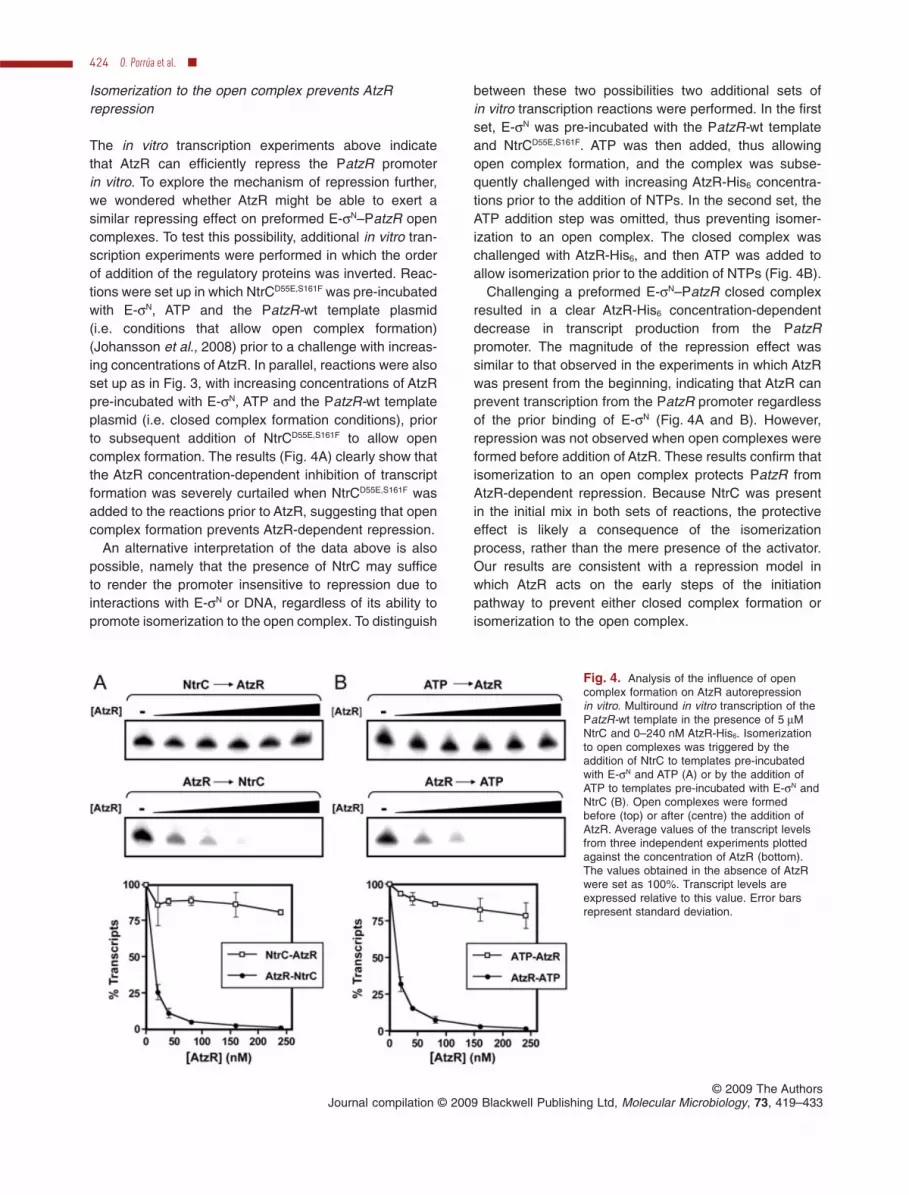
The in vitro transcription experiments above indicatethat AtzR can efficiently repress the PatzR promoterin vitro. To explore the mechanism of repression further,we wondered whether AtzR might be able to exert asimilar repressing effect on preformed E-sN–PatzR opencomplexes. To test this possibility, additional in vitro tran-scription experiments were performed in which the orderof addition of the regulatory proteins was inverted. Reac-tions were set up in which NtrCD55E,S161F was pre-incubatedwith E-sN, ATP and the PatzR-wt template plasmid(i.e. conditions that allow open complex formation)(Johansson et al., 2008) prior to a challenge with increas-ing concentrations of AtzR. In parallel, reactions were alsoset up as in Fig. 3, with increasing concentrations of AtzRpre-incubated with E-sN, ATP and the PatzR-wt templateplasmid (i.e. closed complex formation conditions), priorto subsequent addition of NtrCD55E,S161F to allow opencomplex formation. The results (Fig. 4A) clearly show thatthe AtzR concentration-dependent inhibition of transcriptformation was severely curtailed when NtrCD55E,S161F wasadded to the reactions prior to AtzR, suggesting that opencomplex formation prevents AtzR-dependent repression.
An alternative interpretation of the data above is alsopossible, namely that the presence of NtrC may sufficeto render the promoter insensitive to repression due tointeractions with E-sN or DNA, regardless of its ability topromote isomerization to the open complex. To distinguish
between these two possibilities two additional sets ofin vitro transcription reactions were performed. In the firstset, E-sN was pre-incubated with the PatzR-wt templateand NtrCD55E,S161F. ATP was then added, thus allowingopen complex formation, and the complex was subse-quently challenged with increasing AtzR-His6 concentra-tions prior to the addition of NTPs. In the second set, theATP addition step was omitted, thus preventing isomer-ization to an open complex. The closed complex waschallenged with AtzR-His6, and then ATP was added toallow isomerization prior to the addition of NTPs (Fig. 4B).
Challenging a preformed E-sN–PatzR closed complexresulted in a clear AtzR-His6 concentration-dependentdecrease in transcript production from the PatzRpromoter. The magnitude of the repression effect wassimilar to that observed in the experiments in which AtzRwas present from the beginning, indicating that AtzR canprevent transcription from the PatzR promoter regardlessof the prior binding of E-sN (Fig. 4A and B). However,repression was not observed when open complexes wereformed before addition of AtzR. These results confirm thatisomerization to an open complex protects PatzR fromAtzR-dependent repression. Because NtrC was presentin the initial mix in both sets of reactions, the protectiveeffect is likely a consequence of the isomerizationprocess, rather than the mere presence of the activator.Our results are consistent with a repression model inwhich AtzR acts on the early steps of the initiationpathway to prevent either closed complex formation orisomerization to the open complex.
Fig. 4. Analysis of the influence of opencomplex formation on AtzR autorepressionin vitro. Multiround in vitro transcription of thePatzR-wt template in the presence of 5 mMNtrC and 0–240 nM AtzR-His6. Isomerizationto open complexes was triggered by theaddition of NtrC to templates pre-incubatedwith E-sN and ATP (A) or by the addition ofATP to templates pre-incubated with E-sN andNtrC (B). Open complexes were formedbefore (top) or after (centre) the addition ofAtzR. Average values of the transcript levelsfrom three independent experiments plottedagainst the concentration of AtzR (bottom).The values obtained in the absence of AtzRwere set as 100%. Transcript levels areexpressed relative to this value. Error barsrepresent standard deviation.
424 O. Porrúa et al. �
© 2009 The AuthorsJournal compilation © 2009 Blackwell Publishing Ltd, Molecular Microbiology, 73, 419–433

AtzR competes with E-sN for overlapping binding sites
We have shown above that PatzR repression occursduring the initial steps of transcription initiation prior to theisomerization step. Overlapping of the AtzR binding siteand the -24 element of the promoter and the fact that themagnitude of repression correlates with binding affinity ofAtzR for its site strongly suggest that repression mayoccur by direct competition between AtzR and E-sN forDNA binding. To test this hypothesis further, gel mobilityshift analysis was performed with purified AtzR-His6 andP. putida E-sN using the wild-type PatzR promoter regionas a probe. To promote DNA binding by both proteins,buffer conditions were similar to those used for in vitrotranscription. The results are depicted in Fig. 5.
As previously shown (Porrúa et al., 2007), addition ofAtzR-His6 to the PatzR promoter region probe resultedin the appearance of a retarded band, indicative of AtzRbinding (Fig. 5A). In addition, a second, lower mobilityband was observed at higher AtzR-His6 concentrations.This is likely due to the change in binding buffer, as thisAtzR-His6 preparation yielded a single complex in ourroutinely used buffer (Porrúa et al., 2007). E-sN retardedthe PatzR promoter probe to produce a single very slowlymigrating band, consistent with the high molecular weightof the RNA polymerase holoenzyme. Yet, smearing ofthe radiolabel signal along the lane suggests that theE-sN–PatzR closed complex may not be very stable in ourelectrophoretic conditions (Fig. 5B). Remarkably, additionof AtzR-His6 to a preformed E-sN–PatzR closed complex
resulted in the gradual substitution of the E-sN-containingretarded band by the faster-migrating AtzR–PatzRcomplexes. A supershifted lower mobility band consis-tent with a ternary AtzR–E-sN–PatzR complex was notobserved, strongly suggesting that AtzR and E-sN bindingto the PatzR promoter region are mutually exclusive.
Since E-sN binding to the PatzR promoter regionappears to be unstable in the gel mobility shift assaysabove, we sought to obtain further support for this modelusing independent experimental evidence. To achieve thisgoal, DNase I footprinting was performed with AtzR-His6
and E-sN using the top strand-labelled PatzR promoterregion as a probe (Fig. 6). AtzR-His6 continuously pro-tected a 27 bp region encompassing the RBS andspanning positions -41 to -14 relative to the atzR tran-scriptional start site. Additional discontinuous protection
Fig. 5. Gel mobility shift assay of E-sN and AtzR at the atzRpromoter region. E-sN concentration, when present, was 150 nM(lanes 6–9) and AtzR-His6 concentrations were 0 (lanes 1, 5 and6), 40 (lanes 2 and 7), 80 (lanes 3 and 8) and 160 nM (lanes 4and 9). Closed arrowheads denote the AtzR–DNA complexes.An open arrowhead denotes the E-sN–DNA complex.
Fig. 6. DNase I footprint of E-sN and AtzR at the atzR promoterregion. E-sN concentration, when present, was 220 nM (lanes 2–5)and AtzR-His6 concentrations were 0 (lanes 1 and 2), 100 (lane 3),200 (lane 4) and 400 nM (lanes 5 and 6). The RBS and the ABSare represented by open boxes and a hatched box respectively.The -24 and -12 elements of PatzR are shown by closed boxes.Co-ordinates indicated are relative to the atzR transcriptional start.E-sN- and AtzR-protected regions are represented by a black and agrey bar respectively. Stars denote hypersensitive positions due toE-sN and asterisks denote hypersensitive positions due to AtzR.
Complex regulation of a sN-dependent promoter 425
© 2009 The AuthorsJournal compilation © 2009 Blackwell Publishing Ltd, Molecular Microbiology, 73, 419–433

was observed in the ABS region between positions -71and -42, and two sets of hypersensitive bands, indicativeof DNA bending, occurred around positions -54 and -44.Despite the fact that AtzR forms two complexes in gel shiftassays in these conditions (Fig. 5), the footprinting patternwas identical to that observed in the original binding buffer(Porrúa et al., 2007). On the other hand, E-sN protected acontinuous region spanning positions -40 to -2, withhypersensitive bands at positions -42, -41, -35 and -33.Addition of increasing amounts of AtzR-His6 to a bindingreaction containing PatzR-bound E-sN resulted in thegradual substitution of the E-sN polymerase footprint withthat of AtzR. This result strongly suggests that AtzR com-petes with E-sN for binding to the PatzR promoter region.Inversion of the order of addition of AtzR-His6 and E-sN
resulted in a mirror image of the previous result: the foot-printing pattern of AtzR was gradually substituted by thatof E-sN (data not shown), suggesting that both proteinsundergo fast exchange at the PatzR promoter to reachan equilibrium that is a function of their concentrationsin the binding reaction. Taken together, our results supporta model in which AtzR represses the PatzR promoterby diminishing promoter occupancy by E-sN via compe-tition for a binding site that overlaps the sN recognitionelement.
Discussion
The conserved mechanism of sN-dependent promoteractivation represents a well-established paradigm inbacterial gene expression (Kustu et al., 1989; Buck et al.,2000; Wigneshweraraj et al., 2008). While all sN-dependent promoters are subjected to positive regulation,a few of them are also targets for negative control thatinvariably operates by tampering with the activationmechanism (Rojo, 1999; 2001). In the present work, wepresent evidence that positive and negative regulation ofthe sN-dependent promoter PatzR occur by mechanismsthat represent a major departure from the establishedmodels, but have probably co-evolved to optimize regu-latory efficiency.
Our results show that P. putida NtrC does not requireany specific sequences other than the E-sN recognitionmotif to stimulate transcription from the atzR promoterin vivo, from a plasmid or inserted in the P. putida chro-mosome, or in vitro (Table 1, Fig. 2, Fig. S1). UAS ele-ments have been shown to contribute to activation via twodifferent mechanisms: tethering the EBP to increase itslocal concentration in the vicinity of the regulated pro-moter (Buck and Cannon, 1989; Wedel et al., 1990), andpromoting oligomerization that is required for ATPaseactivity (Wyman et al., 1997; Porter et al., 1993).However, it has long been known that DNA binding ofEBPs is not a strict requirement for transcriptional activa-
tion (Reitzer and Magasanik, 1986; Ninfa et al., 1987),Several EBP derivatives lacking their DNA binding deter-minants have been shown to activate transcription in vivoand in vitro, albeit higher protein concentrations in vitroand overproduction in vivo were usually required (Hualaand Ausubel, 1989; Huala et al., 1992; Berger et al., 1994;1995; Xu et al., 2004). A few examples of other naturallyoccurring sN-dependent promoters lacking a functionalUAS have been documented (Schmitz et al., 1988; Wuet al., 1999; Martínez et al., 2004; Burtnick et al., 2007).Whether these promoters are activated by their EBPsfrom solution or bound to low-affinity binding sites thatmay occur elsewhere in the DNA molecules used hasnot been resolved. However, the recent findings thatHelicobacter pylori FlgR (Brahmachary et al., 2004) andRhodobacter sphaeroides FleT (Poggio et al., 2005)activate sN-dependent transcription in vivo despite thefact that they lack the conserved C-terminal DNA-bindingdomain reinforce the notion that certain sN-dependentpromoters can be activated by EBPs in solution underphysiological conditions. A number of sequences encod-ing putative EBPs that lack a DNA-binding domain havebeen found in several bacterial genomes (Beck et al.,2007; Brahmachary et al., 2004) including that of P. putidaKT2440 (Cases et al., 2003) suggesting that this may notbe such an unusual phenomenon.
Upstream activation sequence-independent activationis fostered by high occupancy of the promoter by E-sN
(Buck and Cannon, 1989; Morett and Buck, 1989;Hoover et al., 1990). PatzR appears to be a strongsN-dependent promoter, as evidenced by its strong simi-larity to the consensus E-sN recognition motif (CGGCAC-N5-TTGCT versus TGGCAC-N5-TTGCA). High similarityto the consensus, with strict conservation of the centralGGCAC-N5-TTGA core of the sN-dependent promoterconsensus, has been shown at the UAS-independentRhizobium leguminosarum hupF P3 (Martínez et al.,2004), Klebsiella oxytoca nasR (Wu et al., 1999) andBorrelia burgdoferi rpoS (Burtnick et al., 2007) promot-ers. In addition, the fact that closed complex formation isreadily detected by DNase I footprinting assays also sup-ports the notion that PatzR is a strong E-sN binding site(Fig. 5). Similarity to the consensus and production of adetectable DNase I footprint have been shown to corre-late with high E-sN occupancy and increased UAS-independent activation (Morett and Buck, 1988; 1989;Hoover et al., 1990).
A recent paper (Bernardo et al., 2009) has shownthat P. putida E-sN has a higher affinity for sN-dependentpromoters than its E. coli counterpart, resulting in morerobust transcriptional activation in vivo and in vitro. Asdiscussed above, this trait is predicted to favour UAS-independent activation in P. putida. Two sets of observa-tions suggest that this may indeed be the case. First, we
426 O. Porrúa et al. �
© 2009 The AuthorsJournal compilation © 2009 Blackwell Publishing Ltd, Molecular Microbiology, 73, 419–433
have observed that the PatzR promoter fails to respond tonitrogen limitation in E. coli. However, it is activated nor-mally in a P. putida strain in which the E. coli ntrC codingsequence is precisely inserted in the chromosome in lieuof its P. putida counterpart, thus indicating that E. coli NtrCis not inherently defective for activation at this promoter(V. García-González and F. Govantes, unpubl. results).Second, deletion analyses performed in our laboratoryon several P. putida NtrC- and sN-dependent promotersshowed substantial levels of activation after removal ofthe cognate enhancer-like elements (A.B. Hervás, I.Canosa, A.I. Platero and F. Govantes, unpubl. results).Since a high concentration of the activator has also beenshown to contribute to UAS-independent activation (Ninfaet al., 1987; Popham et al., 1989; Schneider et al., 1991;Molina-Lopez and Santero, 1999; Brahmachary et al.,2004), the above results are also compatible with unusu-ally high NtrC levels in P. putida. However, our attempts toobtain a rigorous estimation of the intracellular NtrC con-centration in P. putida have been so far unsuccessful. Thecontribution of E-sN and NtrC to the apparently high rate ofUAS-independent activation in P. putida NtrC-dependentpromoters will be pursued further in the future.
The main drawback of UAS-independent activation isthe loss of promoter specificity (Beck et al., 2007). SinceNtrC supports UAS-independent activation of PatzR, itmay similarly activate other sN-dependent P. putida pro-moters in a UAS-independent fashion. However, as highoccupancy of the promoter by E-sN is required for efficientactivation from solution, a weaker promoter, in combina-tion with strong binding sites for a specific activator and/ordependence on IHF, could ensure the fidelity of activation(Buck and Cannon, 1989; Morett and Buck, 1989; Hooveret al., 1990). Conversely, another negative consequenceof the mechanism of PatzR activation could be the cross-activation by other EBPs. Our in vitro transcription ex-periments have demonstrated that the isolated centraldomain of DmpR can activate the PatzR promoter in vitro(Fig. 2). In addition, we have observed that PatzR canbe activated in vivo by NifA and XylR when these EBPsare expressed at high levels from plasmids in E. coli(V. García-González and F. Govantes, unpubl. results).Notwithstanding the artificial nature of the conditions inthese experiments, we consider it plausible that promis-cuous activation of PatzR in response to non-cognatephysiological cues may occur. In this regard, the factthat nitrogen limitation also modulates AtzR activity inactivation of the atzDEF promoter (García-Gonzálezet al., 2005) may be envisioned as a safety mechanism toprevent unwanted synthesis of the cyanuric acid degra-dative enzymes should sufficient AtzR be produced undernitrogen-sufficient conditions (also see below).
Like most members of the LysR family (Schell, 1993;Tropel and van der Meer, 2004), AtzR represses its
own synthesis (García-González et al., 2005). Our resultsshow that repression of the PatzR promoter takes placeprior to the formation of the open complex (Fig. 4) and thatAtzR and E-sN binding to the atzR promoter region aremutually exclusive (Fig. 5). Accordingly, we propose thatAtzR prevents open complex formation by competitionwith E-sN for binding to their overlapping recognitionelements. Although a mechanism of repression based oncompetition with RNA polymerase for DNA binding hasbeen largely suggested for other LTTRs (Ostrowski andKredich, 1991; Schell, 1993; Kovacikova and Skorupski,2001), this is the first time experimental evidence support-ing this hypothesis has been provided. In addition, ourprevious and present results indicate that integrity of theABS is critical for efficient autorepression. The defect inrepression provoked by a deletion of most of the ABS issuppressed by increasing AtzR concentration both in vivo(Porrúa et al., 2007) and in vitro (Fig. 3), indicating thatthe role of the ABS in repression is a consequence of itscontribution to the affinity of AtzR for its binding site. Theobserved correlation between the magnitude of repres-sion and the affinity of the regulator for its binding site isan expected outcome of the mechanism of autorepres-sion, given the fact that AtzR is present in the cell at a verylow concentration when produced from its own feedback-regulated promoter (Porrúa et al., 2007). A similar contri-bution of the ABS to the binding affinity has also beenobserved in the case of CatR binding site at catR–catBCpromoter region in P. putida (Parsek et al., 1992) and theIlvY binding site at ilvY–ilvC promoter region in E. coli(Wek and Hatfield, 1988). A role for the ABS in autore-pression has been demonstrated for E. coli OxyR andBacillus subtilis GltC (Toledano et al., 1994; Belitsky et al.,1995), albeit in both cases the ABS actually overlaps the-35 box of the repressed promoters, a situation that doesnot occur with PatzR.
The PatzR promoter is unusual in that it is asN-dependent promoter subjected to negative control.Only a few examples of negatively regulated sN-dependent promoters have been reported thus far and, inall cases, repression has been proposed to occur via ananti-activation mechanism (i.e. by interfering with the acti-vation process). At the dctA promoter, the cAMP receptorprotein (CRP) from E. coli appears to interact with E-sN
to lock the closed complex in a conformation that is lesssusceptible to activation by DctD (Wang et al., 1998). Analternative model in which DNA bending by the repressorhinders productive interactions between the UAS-boundEBP and the E-sN has been proposed for repressionof E. coli glnAp2 by CRP (Mao et al., 2007), down-regulation of the B. subtilis levanase operon by CcpA(Martin-Verstraete et al., 1995) and autorepression bythe Klebsiella aerogenes LTTR Nac (Feng et al., 1995).In contrast, AtzR prevents transcription from PatzR by
Complex regulation of a sN-dependent promoter 427
© 2009 The AuthorsJournal compilation © 2009 Blackwell Publishing Ltd, Molecular Microbiology, 73, 419–433

competing with E-sN for DNA binding. This is the first timethat a repressor affecting the capability of E-sN to bindits recognition sequences has been described for asN-dependent promoter. This unusual strategy may be aconsequence of the fact that NtrC does not bind specifi-cally to any sequences within the atzR promoter region. Inthis scenario, other possible mechanisms based on inter-ference with DNA binding of the activator or DNA loopingwould not be suitable. However, because high occupancyof the promoter by the RNA polymerase is critical foractivation from solution, competing with E-sN for DNAbinding appears to be a viable and appropriate mecha-nism to efficiently repress transcription.
Autorepression of LTTRs is proposed to be a mecha-nism to avert wasteful expenditure of energy by keepingsynthesis of the regulatory proteins at a low rate (Schell,1993). Indeed, we have previously shown that even wheninduced under nitrogen limitation conditions, AtzR is pro-duced in vivo in limiting amounts that are still sufficientfor physiological regulation of the cyanuric acid degrada-tive operon atzDEF (García-González et al., 2005; Porrúaet al., 2007). Similarly. the decrease in PatzR promoteroccupancy by E-sN caused by competition with AtzR mayalso minimize the amount of AtzR synthesized by cross-activation by other EBPs present in the cell. Thus, nitro-gen regulation of AtzR activity and autorepression may bemeans to compensate for the potential non-specific acti-vation of the cyanuric acid utilization pathway ensued bycross-activation of PatzR by other EBPs.
Experimental procedures
Bacterial strains and growth conditions
Bacterial strains used in this work and their relevantgenotypes are summarized in Table 2. Minimal mediumcontaining 25 mM sodium succinate as the sole carbonsource was used for in vivo gene expression analysis(Mandelbaum et al., 1993). Nitrogen sources were ammoniumchloride or L-serine (1 g l-1). Luria–Bertani (LB) mediumwas used as rich medium (Sambrook et al., 2000). Liquidcultures were grown in culture tubes or flasks with shak-ing (180–200 r.p.m.) at 30°C or 37°C (for P. putida orE. coli strains respectively). For solid media, Bacto-Agar(Difco, Detroit, Michigan) was added to a final concentra-tion of 18 g l-1. Antibiotics and other additions were used,when required, at the following concentrations: ampicillin(100 mg l-1), kanamycin (20 mg l-1), carbenicillin (500 mg l-1),rifampicin (10 mg l-1), chloramphenicol (15 mg l-1) and5-bromo-4-chloro-3-indoyl-b-D-galactopyranoside (X-gal)(25 mg l-1). All reagents were purchased from Sigma-Aldrich.
Plasmid and strain construction
Plasmids and oligonucleotides used in this work are sum-marized in Table 2. All DNA manipulations were performedaccording to standard procedures (Sambrook et al., 2000).
Plasmid DNA was transferred to E. coli and P. putida strainsby transformation (Inoue et al., 1990) or by triparental mating(Espinosa-Urgel et al., 2000). E. coli DH5a was used as ahost in all cloning procedures.
The broad-host-range lacZ transcriptional fusion vectorpMPO234 was constructed by cloning a BamHI–SacI frag-ment from pRS415 containing the lacZ transcriptional fusionjunction and the 5′ end of lacZ into BamHI- and SacI-digestedpMPO200. To obtain the atzR–lacZ transcriptional fusionplasmid pMPO232, pMPO104 was cleaved with EcoRI andBamHI and the resulting ~0.4 kb fragment containing thecomplete atzR–atzDE promoter region was then ligated intoEcoRI- and BamHI-treated pMPO234. The deleted derivativepMPO237 was constructed using the overlapping oligonucle-otides PatzR-EcoRI and PatzR-BglII. Primers were mixedand allowed to anneal by heating for 5 min at 85°C and thencooling down slowly to room temperature. The resultingduplex, which harbours overhanging ends compatible withEcoRI and BamHI, was cloned into EcoRI- and BamHI-digested pMPO234.
Plasmids pMPO166 and pMPO167 harbouring atzR–lacZfusions ready to integrate in the P. putida chromosome wereconstructed by excising an EcoRI–SalI fragment containingthe atzR–lacZ fusion from pMPO232 or pMPO237, respec-tively, and cloning into NotI-linearized pBK-miniTn7-WGm(Koch et al., 2001). Orientation of lacZ transcription towardsthe Tn7R end was tested by PCR. Integration of the fusionsin pMPO166 and pMPO167 at the Tn7 insertion site inP. putida KT2442 and MPO201 was performed by electropo-ration of each plasmid, along with the Tn7 helper plasmidpUX-BF13 in the desired strains, essentially as described(Koch et al., 2001). Correct integration was tested by PCRusing primers Tn7-GlmS and Tn7R109.
The in vitro transcription template plasmid pMPO156harbouring PatzR was constructed by PCR amplificationof the atzR–atzDE promoter region with primers atzR-EBamand atzR-BEco using pMPO104 as a template. The PCRproduct was cleaved with EcoRI and BamHI and ligated intopTE103 digested with the same enzymes. The deletedderivatives pMPO161 and pMPO162 were constructedsimilarly: PCR was performed using primers del-ABS andatzR-BEco, with plasmids pMPO121 or pMPO122, respec-tively, as templates. The EcoRI–BamHI fragments of thePCR products were then cloned into EcoRI- and BamHI-cleaved pTE103.
b-Galactosidase assays
Steady-state b-galactosidase assays were used to examinethe expression of atzR–lacZ fusions in P. putida KT2442 andMPO201. Pre-inocula of bacterial strains harbouring the rel-evant plasmids were grown to saturation in minimal mediumunder nitrogen-sufficient conditions (ammonium chloride,1 g l-1) and cells were then diluted in minimal medium con-taining the appropriate nitrogen sources (1 g l-1 ammoniumchloride for nitrogen excess, 1 g l-1 L-serine for nitrogenlimitation). Diluted cultures were shaken for 16–24 h to mid-exponential phase (OD600 = 0.25–0.5). Growth was thenstopped and b-galactosidase activity was determined fromSDS- and chloroform-permeabilized cells as previouslydescribed (Miller, 1992).
428 O. Porrúa et al. �
© 2009 The AuthorsJournal compilation © 2009 Blackwell Publishing Ltd, Molecular Microbiology, 73, 419–433

Protein purification
AtzR-His6 was purified from the overproducing strainNCM631 harbouring pMPO135 and pIZ227 by nickel affinitychromatography as previously described (Porrúa et al.,2007). NtrCD55E,S161F, purified by a salting-out procedure usingammonium sulphate as the precipitating agent, was a kindgift of A.B. Hervás (A.B. Hervás et al., submitted). NativeP. putida core RNA polymerase, sN, and C-DmpR-His weregifts from member of V. Shingler’s laboratory and were puri-fied by previously established protocols (Johansson et al.,2008). In brief, P. putida KT2440 core RNA polymerase
was purified from cellular extracts by sequential Polymin Pprecipitation, DNA agarose chromatography and Mono Qion exchange chromatography as described for E. coli coreRNA polymerase (Hager et al., 1990). Native P. putida sN wasoverproduced in an E. coli rpoN mutant strain from the cIts/lPL promoter of pVI903 and subsequently purified by theprocedure of Cannon et al. (1996). The C-DmpR-His fusionprotein, consisting of a methionine residue followed by resi-dues L219 to G496 of DmpR fused to the peptide SHHHHHH,was expressed from the PT7 promoter of pVI645 and purifiedby nickel affinity chromatography as previously described forother His-tagged DmpR derivatives (Wikstrom et al., 2001).
Table 2. Bacterial strains, plasmids and oligonucleotides used in this work.
Phenotype/genotype Reference/source
Bacterial strainsE. coli DH5a f80dlacZDM15 D(lacZYA-argF)U169 recA1 endA1 hsdR17 (rk
- mk+) supE44 thi-1
gyrA relA1Hanahan (1983)
E. coli NCM631 hsdS gal lDE3:lacI lacUV5:gen1(T7 RNA polymerase) Dlac linked to Tn10 Govantes et al. (1996)P. putida KT2442 mt-2 hsdR1 (r - m+) Rifr Franklin et al. (1981)P. putida MPO201 mt-2 hsdR1 (r - m+) Rifr DntrC::Tcr García-González et al. (2005)
PlasmidspBK-miniTn7-WGm Delivery plasmid for Tn7 transposase-dependent chromosomal insertion. Gmr Koch et al. (2001)pIZ227 pACYC184 containing lacI q and the T7 lysozyme gene. Cmr Govantes et al. (1996)pMPO104 atzR–lacZ translational fusion in pMPO200 carrying the sequence between positions
-250 and +139. AprGarcía-González et al. (2005)
pMPO121 atzR–lacZ translational fusion in pMPO200 carrying the sequence between positions-50 and +139. Apr
Porrúa et al. (2007)
pMPO122 atzR–lacZ translational fusion in pMPO200 carrying the sequence between positions-29 and +139. Apr
Porrúa et al. (2007)
pMPO135 pET23b plasmid derivative overexpressing AtzR-His6. Apr Porrúa et al. (2007)pMPO156 pTE103 harbouring atzR promoter sequences between positions -239 and +25. Apr This workpMPO161 pTE103 harbouring atzR promoter sequences between positions -29 and +25. Apr This workpMPO162 pTE103 harbouring atzR promoter sequences between positions -50 and +25. Apr This workpMPO166 atzR–lacZ transcriptional fusion in pBK-miniTn7-WGm carrying the sequence between
positions -250 and +139 between the Tn7 ends. Gmr AprThis work
pMPO167 atzR–lacZ transcriptional fusion in pBK-miniTn7-WGm carrying the sequence betweenpositions -27 and +1 between the Tn7 ends. Gmr Apr
This work
pMPO200 Broad-host-range lacZ translational fusion vector, based on pBBR1MCS-4. Apr García-González et al. (2005)pMPO202 atzD–lacZ translational fusion in pMPO200 bearing the sequence between positions
-475 and +152. AprGarcía-González et al. (2005)
pMPO232 atzR–lacZ transcriptional fusion in pMPO234 carrying the sequence between positions-250 and +139. Apr
This work
pMPO234 Broad-host-range lacZ transcriptional fusion vector, based on pMPO200. Apr This workpMPO237 atzR–lacZ transcriptional fusion in pMPO234 carrying the sequence between positions
-27 and +1. AprThis work
pRK2013 Helper plasmid in conjugation. Kmr Tra+ Figurski and Helinski (1979)pRS415 lacZ transcriptional fusion vector, based on pBR322. Apr Simons et al. (1987)pTE103 Vector for in vitro transcription assays. Apr Elliott and Geiduschek (1984)pUX-BF13 Helper plasmid providing the Tn7 transposase proteins for chromosomal insertion. Apr Bao et al. (1991)pVI903 cIts/lPL-rpoN expression plasmid for purification of P. putida KT2440 sN Johansson et al. (2008)pVI645 PT7-C-dmpR-His expression plasmid for purification of C-DmpR-His Johansson et al. (2008)
Oligonucleotides Sequence (5′ to 3′)atzR-BEco AGAGATGGATCCTATCGCTGACGatzR-EBam GATCCGAATTCCTGTGGCAAGGdel-ABS TATCAGGGTTATTGTCTCATFP-SalI TCGACCAAGGCGATTAAGTTGGGTAoligo-UP GTATTCCAGATCCTGGACGCPatzR-BglII GATCTCATGCGAGTCAAAGCAAGATCGGTGCCGPatzR-EcoRI ATTCGGCACCGATCTTGCTTTGACTCGCATGASeq-ClaI CGATGTAATGAAGAAAGCGTTn7-GlmS AATCTGGCCAAGTCGGTGACTn7R109 CAGCATAACTGGACTGATTTCAG
Co-ordinates indicated in plasmid descriptions are relative to the atzR transcriptional start. Oligonucleotide positions altered from the originalsequences are underlined.
Complex regulation of a sN-dependent promoter 429
© 2009 The AuthorsJournal compilation © 2009 Blackwell Publishing Ltd, Molecular Microbiology, 73, 419–433

In vitro transcription
Multiround in vitro transcription reactions were performed ina final volume of 20 ml containing 35 mM Tris-acetate, pH 7.9;70 mM potassium acetate; 20 mM ammonium acetate; 5 mMmagnesium acetate; 1 mM DTT; 10% glycerol; 250 mg l-1
BSA; 20 nM E-sN and 0.5 mg of supercoiled plasmid templatecontaining PatzR. Open complex formation was stimulatedby the addition of NtrCD55E,S161F or C-DmpR-His and 4 mM ATPand incubation for 10–20 min at 30°C. In repression experi-ments, AtzR (0–240 nM) was added either before or after theformation of open complex and subsequently incubated withthe reaction mixture for the same time as NtrCD55E,S161F. Afterincubation, a mixture of ATP, GTP, CTP (final concentration0.4 mM each), UTP (0.07 mM) and [a-32P]-UTP (0.33 mM,Amersham) was added to initiate multiround in vitrotranscription. Re-initiation was prevented after 5 min by theaddition of heparin (0.1 mg ml-1 final concentration), and5 min later the reactions were terminated by adding 5 ml ofstop/loading buffer (150 mM EDTA, 1.05 M NaCl, 14 M urea,3% glycerol, 0.075% xylene cyanol and 0.075% bromophenolblue). Samples were run on a 5% polyacrilamide-urea dena-turing gel in Tris-borate-EDTA buffer at room temperature.Transcripts were visualized with a Typhoon 9410 scannerand analysed using the ImageQuant software (Amersham).
Gel mobility shift assays
Probes for gel mobility shift assays were obtained by PCRamplification using pMPO202 as the template and primerspair oligo-UP and FP-SalI. The PCR products were subse-quently digested with ClaI and gel purified. DNA fragmentswere strand-specifically labelled by filling in 5′ overhangingends using the Klenow fragment in a reaction mixturecontaining [a-32P]-dCTP. Unincorporated nucleotides wereremoved using the MSB Spin PCRapace kit (Invitek).
Binding reactions were performed in binding buffer(35 mM Tris-acetate, pH 7.9; 70 mM potassium acetate;20 mM ammonium acetate; 2 mM magnesium acetate; 1 mMcalcium chloride; 1 mM DTT; 10% glycerol; 100 mg ml-1
salmon sperm DNA; 250 mg ml-1 BSA), containing 20 ng ofthe radiolabelled probe and 3 ml (150 nM final concentration)of E-sN, or an equivalent volume of dilution buffer (10 mMTris-HCl; 100 mM NaCl; 0.1 mM EDTA; 0.1 mM DTT; 50%glycerol), in a reaction volume of 18 ml. After 10 min incuba-tion at room temperature, 2 ml (0–160 nM final concentration)of AtzR was added and the mixture was incubated foran additional 10 min. Reactions were stopped with 4 ml ofloading buffer [0.125% w/v bromophenol blue, 0.125% w/vxylene cyanol, 10 mM Tris HCl (pH 8), 1 mM EDTA, 30%glycerol] and samples were separated on a 5% polyacryla-mide native gel in Tris-borate-EDTA buffer at 4°C. Gels weredried in a dryer DrygelSr SE1160 (Hoeffer) and analysed asdescribed above.
DNase I footprinting
Probes for DNase I footprinting were obtained as shownabove for gel mobility shift assays. Binding reactions wereperformed in binding buffer (35 mM Tris acetate, pH 7.9;
70 mM potassium acetate; 20 mM ammonium acetate; 2 mMmagnesium acetate; 1 mM calcium chloride; 1 mM DTT; 10%glycerol; 100 mg ml-1 salmon sperm DNA; 250 mg ml-1 BSA),containing 20 ng of the radiolabelled probe and 3 ml (220 nMfinal concentration) of E-sN in a reaction volume of 10 ml. After10 min incubation at room temperature, 0–400 nM AtzRwas added, the mixture was incubated for an additional10 min and partial digestion of the DNA was initiated by theaddition of 1 ml of an empirically determined dilution (typically10-2 to 10-3) of a DNase I stock solution (10 U ml-1, RocheDiagnostics). Incubation was continued for 30 additionalseconds and reactions were stopped by the addition of 5 mlof stop buffer (1.5 M sodium acetate, pH 5.2; 130 mM EDTA;1 mg ml-1 salmon sperm DNA; 2.4 mg ml-1 glycogen). DNAwas subsequently ethanol precipitated, re-suspended in 5 mlof loading buffer (0.125% bromophenol blue, 0.125% xylenecyanol, 20 mM EDTA, 95% formamide) and separated bygel electrophoresis on a 6% polyacrylamide-6 M urea dena-turing sequencing gel. A sequencing reaction was performedwith the Sequenase 2.0 kit (USB) using primer Seq-ClaI andwas run in parallel as a size marker. Gels were processedand analysed as described above for in vitro transcriptionassays.
Acknowledgements
We thank Ana B. Hervás (CABD, Universidad Pablo deOlavide), Linda U.M. Johansson, Lisandro M.D. Bernardoand Eleonore Skärfstad (Umeå University) for purifiedproteins; Sydney Kustu (University of California, Berkeley)for critical reading of the manuscript; Guadalupe Martínand Nuria Pérez for technical help; and all members ofthe Santero and Shingler laboratories for their insights andhelpful suggestions. This work was supported by GrantsBIO2004-01354 and BIO2007-63754 (Ministerio de Edu-cación y Ciencia, Spain), fellowships from the I3P (CSIC/Ministerio de Educación y Ciencia, Spain) and FPU(Ministerio de Educación y Cultura, Spain) programmes,awarded to O.P. and V.G.-G., respectively, and a short-termEMBO fellowship awarded to O.P. to visit the laboratoryof VS.
References
Arcondeguy, T., Jack, R., and Merrick, M. (2001) P(II) signaltransduction proteins, pivotal players in microbial nitrogencontrol. Microbiol Mol Biol Rev 65: 80–105.
Bao, Y., Lies, D.P., Fu, H., and Roberts, G.P. (1991) Animproved Tn7-based system for the single-copy insertionof cloned genes into the chromosomes of Gram-negativebacteria. Gene 109: 167–168.
Beck, L.L., Smith, T.G., and Hoover, T.R. (2007) Look,no hands! Unconventional transcriptional activators inbacteria. Trends Microbiol 15: 530–537.
Belitsky, B.R., Janssen, P.J., and Sonenshein, A.L. (1995)Sites required for GltC-dependent regulation of Bacillussubtilis glutamate synthase expression. J Bacteriol 177:5686–5695.
Belitsky, B.R., and Sonenshein, A.L. (1999) An enhancerelement located downstream of the major glutamate
430 O. Porrúa et al. �
© 2009 The AuthorsJournal compilation © 2009 Blackwell Publishing Ltd, Molecular Microbiology, 73, 419–433

dehydrogenase gene of Bacillus subtilis. Proc Natl AcadSci USA 96: 10290–10295.
Berger, D.K., Narberhaus, F., and Kustu, S. (1994) Theisolated catalytic domain of NIFA, a bacterial enhancer-binding protein, activates transcription in vitro: activation isinhibited by NIFL. Proc Natl Acad Sci USA 91: 103–107.
Berger, D.K., Narberhaus, F., Lee, H.S., and Kustu, S. (1995)In vitro studies of the domains of the nitrogen fixationregulatory protein NIFA. J Bacteriol 177: 191–199.
Bernardo, L.M., Johansson, L.U., Skärfstad, E., and Shingler,V. (2009) s54-promoter discrimination and regulation byppGpp and DksA. J Biol Chem 284: 828–838.
Brahmachary, P., Dashti, M.G., Olson, J.W., and Hoover,T.R. (2004) Helicobacter pylori FlgR is an enhancer-independent activator of s54-RNA polymerase holoenzyme.J Bacteriol 186: 4535–4542.
Buck, M., and Cannon, W. (1989) Mutations in the RNApolymerase recognition sequence of the Klebsiellapneumoniae nifH promoter permitting transcriptional acti-vation in the absence of NifA binding to upstream activatorsequences. Nucleic Acids Res 17: 2597–2612.
Buck, M., Gallegos, M.T., Studholme, D.J., Guo, Y., andGralla, J.D. (2000) The bacterial enhancer-dependentsigma(54) (sigma(N)) transcription factor. J Bacteriol 182:4129–4136.
Burtnick, M.N., Downey, J.S., Brett, P.J., Boylan, J.A., Frye,J.G., Hoover, T.R., and Gherardini, F.C. (2007) Insightsinto the complex regulation of rpoS in Borrelia burgdorferi.Mol Microbiol 65: 277–293.
Cannon, W., Missailidis, S., Austin, S., Moore, M., Drake, A.,and Buck, M. (1996) Purification and activities of theRhodobacter capsulatus RpoN (sigma N) protein. MolMicrobiol 21: 233–245.
Cases, I., Ussery, D.W., and de Lorenzo, V. (2003) The s54
regulon (sigmulon) of Pseudomonas putida. Environ Micro-biol 5: 1281–1293.
Drummond, M., Clements, J., Merrick, M., and Dixon, R.(1983) Positive control and autogenous regulation of thenifLA promoter in Klebsiella pneumoniae. Nature 301:302–307.
Elliott, T., and Geiduschek, E.P. (1984) Defining a bacte-riophage T4 late promoter: absence of a ‘-35’ region. Cell36: 211–219.
Espinosa-Urgel, M., Salido, A., and Ramos, J.L. (2000)Genetic analysis of functions involved in adhesion ofPseudomonas putida to seeds. J Bacteriol 182: 2363–2369.
Feng, J., Goss, T.J., Bender, R.A., and Ninfa, A.J. (1995)Repression of the Klebsiella aerogenes nac promoter.J Bacteriol 177: 5535–5538.
Figurski, D.H., and Helinski, D.R. (1979) Replication of anorigin-containing derivative of plasmid RK2 dependent ona plasmid function provided in trans. Proc Natl Acad SciUSA 76: 1648–1652.
Franklin, F.C., Bagdasarian, M., Bagdasarian, M.M., andTimmis, K.N. (1981) Molecular and functional analysis ofthe TOL plasmid pWWO from Pseudomonas putida andcloning of genes for the entire regulated aromatic ring metacleavage pathway. Proc Natl Acad Sci USA 78: 7458–7462.
García-González, V., Govantes, F., Porrúa, O., and Santero,E. (2005) Regulation of the Pseudomonas sp. strain ADP
cyanuric acid degradation operon. J Bacteriol 187: 155–167.
Govantes, F., Molina-López, J.A., and Santero, E. (1996)Mechanism of coordinated synthesis of the antagonisticregulatory proteins NifL and NifA of Klebsiella pneumoniae.J Bacteriol 178: 6817–6823.
Hager, D.A., Jin, D.J., and Burgess, R.R. (1990) Use of MonoQ high-resolution ion-exchange chromatography to obtainhighly pure and active Escherichia coli RNA polymerase.Biochemistry 29: 7890–7894.
Hanahan, D. (1983) Studies on transformation of Escherichiacoli with plasmids. J Mol Biol 166: 557–580.
Hervás, A.B., Canosa, I., and Santero, E. (2008) Transcrip-tome analysis of Pseudomonas putida in response to nitro-gen availability. J Bacteriol 190: 416–420.
Hoover, T.R., Santero, E., Porter, S., and Kustu, S. (1990)The integration host factor stimulates interaction of RNApolymerase with NIFA, the transcriptional activator fornitrogen fixation operons. Cell 63: 11–22.
Huala, E., and Ausubel, F.M. (1989) The central domain ofRhizobium meliloti NifA is sufficient to activate transcriptionfrom the R. meliloti nifH promoter. J Bacteriol 171: 3354–3365.
Huala, E., Stigter, J., and Ausubel, F.M. (1992) The centraldomain of Rhizobium leguminosarum DctD functionsindependently to activate transcription. J Bacteriol 174:1428–1431.
Inoue, H., Nojima, H., and Okayama, H. (1990) Highefficiency transformation of Escherichia coli with plasmids.Gene 96: 23–28.
Johansson, L.U., Solera, D., Bernardo, L.M., Moscoso, J.A.,and Shingler, V. (2008) Sigma(54)-RNA polymerasecontrols sigma(70)-dependent transcription from a non-overlapping divergent promoter. Mol Microbiol 70: 709–723.
Jyot, J., Dasgupta, N., and Ramphal, R. (2002) FleQ, themajor flagellar gene regulator in Pseudomonas aerugi-nosa, binds to enhancer sites located either upstream oratypically downstream of the RpoN binding site. J Bacteriol184: 5251–5260.
Klose, K.E., Weiss, D.S., and Kustu, S. (1993) Glutamateat the site of phosphorylation of nitrogen-regulatory pro-tein NTRC mimics aspartyl-phosphate and activates theprotein. J Mol Biol 232: 67–78.
Koch, B., Jensen, L.E., and Nybroe, O. (2001) Apanel ofTn7-based vectors for insertion of the gfp marker geneor for delivery of cloned DNA into Gram-negative bacteriaat a neutral chromosomal site. J Microbiol Methods 45:187–195.
Kovacikova, G., and Skorupski, K. (2001) Overlappingbinding sites for the virulence gene regulators AphA, AphBand cAMP-CRP at the Vibrio cholerae tcpPH promoter. MolMicrobiol 41: 393–407.
Kustu, S., Santero, E., Keener, J., Popham, D., and Weiss, D.(1989) Expression of sigma 54 (ntrA)-dependent genes isprobably united by a common mechanism. Microbiol Rev53: 367–376.
Mandelbaum, R.T., Wackett, L.P., and Allan, D.L. (1993)Mineralization of the s-triazine ring of atrazine by stablebacterial mixed cultures. Appl Environ Microbiol 59: 1695–1701.
Complex regulation of a sN-dependent promoter 431
© 2009 The AuthorsJournal compilation © 2009 Blackwell Publishing Ltd, Molecular Microbiology, 73, 419–433

Mao, X.J., Huo, Y.X., Buck, M., Kolb, A., and Wang, Y.P.(2007) Interplay between CRP-cAMP and PII-Ntr systemsforms novel regulatory network between carbon metabo-lism and nitrogen assimilation in Escherichia coli. NucleicAcids Res 35: 1432–1440.
Martinez, B., Tomkins, J., Wackett, L.P., Wing, R., and Sad-owsky, M.J. (2001) Complete nucleotide sequence andorganization of the atrazine catabolic plasmid pADP-1 fromPseudomonas sp. strain ADP. J Bacteriol 183: 5684–5697.
Martínez, M., Brito, B., Imperial, J., and Ruiz-Argüeso, T.(2004) Characterization of a new internal promoter (P3) forRhizobium leguminosarum hydrogenase accessory geneshupGHIJ. Microbiology 150: 665–675.
Martin-Verstraete, I., Stulke, J., Klier, A., and Rapoport, G.(1995) Two different mechanisms mediate cataboliterepression of the Bacillus subtilis levanase operon.J Bacteriol 177: 6919–6927.
Merrick, M.J., and Edwards, R.A. (1995) Nitrogen control inbacteria. Microbiol Rev 59: 604–622.
Miller, J.H. (1992) A Short Course in Bacterial Genetics: ALaboratory Manual. Cold Spring Harbor, NY: Cold SpringHarbor Laboratory Press.
Molina-Lopez, J.A., and Santero, E. (1999) An artificialenhancer with multiple response elements stimulatesprokaryotic transcriptional activation medicated by variousregulatory proteins. Mol Gen Genet 262: 291–301.
Morett, E., and Buck, M. (1988) NifA-dependent in vivo pro-tection demonstrates that the upstream activator sequenceof nif promoters is a protein binding site. Proc Natl Acad SciUSA 85: 9401–9405.
Morett, E., and Buck, M. (1989) In vivo studies on the inter-action of RNA polymerase-s54 with the Klebsiella pneumo-niae and Rhizobium meliloti nifH promoters. The role ofNifA in the formation of an open promoter complex. J MolBiol 210: 65–77.
Morett, E., and Segovia, L. (1993) The sigma 54 bacterialenhancer-binding protein family: mechanism of actionand phylogenetic relationship of their functional domains.J Bacteriol 175: 6067–6074.
Ninfa, A.J., Reitzer, L.J., and Magasanik, B. (1987) Initiationof transcription at the bacterial glnAp2 promoter by purifiedE. coli components is facilitated by enhancers. Cell 50:1039–1046.
Ostrowski, J., and Kredich, N.M. (1991) Negative autoregu-lation of cysB in Salmonella typhimurium: in vitro interac-tions of CysB protein with the cysB promoter. J Bacteriol173: 2212–2218.
Parsek, M.R., Shinabarger, D.L., Rothmel, R.K., and Chakra-barty, A.M. (1992) Roles of CatR and cis,cis-muconatein activation of the catBC operon, which is involved inbenzoate degradation in Pseudomonas putida. J Bacteriol174: 7798–7806.
Poggio, S., Osorio, A., Dreyfus, G., and Camarena, L. (2005)The flagellar hierarchy of Rhodobacter sphaeroides is con-trolled by the concerted action of two enhancer-bindingproteins. Mol Microbiol 58: 969–983.
Popham, D.L., Szeto, D., Keener, J., and Kustu, S. (1989)Function of a bacterial activator protein that binds totranscriptional enhancers. Science 243: 629–635.
Porrúa, O., García-Jaramillo, M., Santero, E., and Govantes,F. (2007) The LysR-type regulator AtzR binding site: DNA
sequences involved in activation, repression and cyanu-ric acid-dependent repositioning. Mol Microbiol 66: 410–427.
Porter, S.C., North, A.K., Wedel, A.B., and Kustu, S. (1993)Oligomerization of NTRC at the glnA enhancer is requiredfor transcriptional activation. Genes Dev 7: 2258–2273.
Reitzer, L. (2003) Nitrogen assimilation and global regulationin Escherichia coli. Annu Rev Microbiol 57: 155–176.
Reitzer, L.J., and Magasanik, B. (1986) Transcription of glnAin E. coli is stimulated by activator bound to sites far fromthe promoter. Cell 45: 785–792.
Reitzer, L., and Schneider, B.L. (2001) Metabolic context andpossible physiological themes of sigma(54)-dependentgenes in Escherichia coli. Microbiol Mol Biol Rev 65: 422–444.
Rojo, F. (1999) Repression of transcription initiation inbacteria. J Bacteriol 181: 2987–2991.
Rojo, F. (2001) Mechanisms of transcriptional repression.Curr Opin Microbiol 4: 145–151.
Sambrook, J., Russell, D.W., and Russell, D. (2000) Mole-cular Cloning, a Laboratory Manual. Cold Spring Harbor,NY: Cold Spring Harbor Laboratory Press.
Schell, M.A. (1993) Molecular biology of the LysR family oftranscriptional regulators. Annu Rev Microbiol 47: 597–626.
Schmitz, G., Nikaido, K., and Ames, G.F. (1988) Regulationof a transport operon promoter in Salmonella typhimurium:identification of sites essential for nitrogen regulation. MolGen Genet 215: 107–117.
Schneider, B.L., Shiau, S.P., and Reitzer, L.J. (1991) Role ofmultiple environmental stimuli in control of transcriptionfrom a nitrogen-regulated promoter in Escherichia coli withweak or no activator-binding sites. J Bacteriol 173: 6355–6363.
Schumacher, J., Joly, N., Rappas, M., Zhang, X., and Buck,M. (2006) Structures and organisation of AAA+ enhancerbinding proteins in transcriptional activation. J Struct Biol156: 190–199.
Shingler, V. (2003) Integrated regulation in response toaromatic compounds: from signal sensing to attractivebehaviour. Environ Microbiol 5: 1226–1241.
Simons, R.W., Houman, F., and Kleckner, N. (1987)Improved single and multicopy lac-based cloning vectorsfor protein and operon fusions. Gene 53: 85–96.
Studholme, D.J., and Dixon, R. (2003) Domain architecturesof s54-dependent transcriptional activators. J Bacteriol 185:1757–1767.
Toledano, M.B., Kullik, I., Trinh, F., Baird, P.T., Schneider,T.D., and Storz, G. (1994) Redox-dependent shift ofOxyR–DNA contacts along an extended DNA-binding site:a mechanism for differential promoter selection. Cell 78:897–909.
Tropel, D., and van der Meer, J.R. (2004) Bacterialtranscriptional regulators for degradation pathways of aro-matic compounds. Microbiol Mol Biol Rev 68: 474–500.
Wang, Y.P., Kolb, A., Buck, M., Wen, J., O’Gara, F., andBuc, H. (1998) CRP interacts with promoter-bound s54 RNApolymerase and blocks transcriptional activation of thedctA promoter. EMBO J 17: 786–796.
Wedel, A., Weiss, D.S., Popham, D., Droge, P., and Kustu, S.(1990) A bacterial enhancer functions to tether a trans-
432 O. Porrúa et al. �
© 2009 The AuthorsJournal compilation © 2009 Blackwell Publishing Ltd, Molecular Microbiology, 73, 419–433

criptional activator near a promoter. Science 248: 486–490.
Wek, R.C., and Hatfield, G.W. (1988) Transcriptional activa-tion at adjacent operators in the divergent-overlapping ilvYand ilvC promoters of Escherichia coli. J Mol Biol 203:643–663.
Wigneshweraraj, S., Bose, D., Burrows, P.C., Joly, N.,Schumacher, J., Rappas, M., et al. (2008) Modus operandiof the bacterial RNA polymerase containing the s54
promoter-specificity factor. Mol Microbiol 68: 538–546.Wikstrom, P., O’Neill, E., Ng, L.C., and Shingler, V. (2001) The
regulatory N-terminal region of the aromatic-responsivetranscriptional activator DmpR constrains nucleotide-triggered multimerisation. J Mol Biol 314: 971–984.
Wu, S.Q., Chai, W., Lin, J.T., and Stewart, V. (1999) Generalnitrogen regulation of nitrate assimilation regulatory genenasR expression in Klebsiella oxytoca M5al. J Bacteriol181: 7274–7284.
Wyman, C., Rombel, I., North, A.K., Bustamante, C., andKustu, S. (1997) Unusual oligomerization required for
activity of NtrC, a bacterial enhancer-binding protein.Science 275: 1614–1616.
Xu, H., Gu, B., Nixon, B.T., and Hoover, T.R. (2004)Purification and characterization of the AAA+ domainof Sinorhizobium meliloti DctD, a s54-dependent trans-criptional activator. J Bacteriol 186: 3499–3507.
Zhang, X., Chaney, M., Wigneshweraraj, S.R., Schumacher,J., Bordes, P., Cannon, W., and Buck, M. (2002) Mecha-nochemical ATPases and transcriptional activation. MolMicrobiol 45: 895–903.
Supporting information
Additional supporting information may be found in the onlineversion of this article.
Please note: Wiley-Blackwell are not responsible for thecontent or functionality of any supporting materials suppliedby the authors. Any queries (other than missing material)should be directed to the corresponding author for the article.
Complex regulation of a sN-dependent promoter 433
© 2009 The AuthorsJournal compilation © 2009 Blackwell Publishing Ltd, Molecular Microbiology, 73, 419–433


