Embed Size (px)
Citation preview
AHA Scientific Statement
Indications for Cardiac Catheterization and Intervention inPediatric Cardiac Disease
A Scientific Statement From the American Heart Association
Endorsed by the American Academy of Pediatrics and Society for Cardiovascular Angiographyand Intervention
Timothy F. Feltes, MD, FAHA, Chair; Emile Bacha, MD; Robert H. Beekman III, MD, FAHA;John P. Cheatham, MD; Jeffrey A. Feinstein, MD, MPH; Antoinette S. Gomes, MD, FAHA;
Ziyad M. Hijazi, MD, MPH, FAHA; Frank F. Ing, MD; Michael de Moor, MBBCh;W. Robert Morrow, MD; Charles E. Mullins, MD, FAHA; Kathryn A. Taubert, PhD, FAHA;Evan M. Zahn, MD; on behalf of the American Heart Association Congenital Cardiac Defects
Committee of the Council on Cardiovascular Disease in the Young, Council on Clinical Cardiology,and Council on Cardiovascular Radiology and Intervention
Table of Contents
Preamble . . . . . . . . . . . . . . . . . . . . . . . . . . . . . . . . . . . .26081. Preparation for Cardiac Catheterization. . . . . . . . . .26082. Indications for Diagnostic Catheterization. . . . . . . .26093. Opening of Atrial Communications. . . . . . . . . . . . .2612
3.1. Transseptal Techniques . . . . . . . . . . . . . . . . . .26123.2. Atrial Septostomy. . . . . . . . . . . . . . . . . . . . . . .2613
4. Transcatheter Device Closure of Septal Defects . . .26154.1. Secundum ASD . . . . . . . . . . . . . . . . . . . . . . . .26154.2. Ventricular Septal Defects . . . . . . . . . . . . . . . .26164.3. Fontan Fenestration and Baffle Leak Closure . . .2617
5. Transcatheter Balloon Dilation of Cardiac Valves . . .26175.1. Pulmonary Valvuloplasty . . . . . . . . . . . . . . . . .26175.2. Aortic Valvuloplasty . . . . . . . . . . . . . . . . . . . .26185.3. Mitral Valvuloplasty. . . . . . . . . . . . . . . . . . . . .26195.4. Tricuspid Valvuloplasty . . . . . . . . . . . . . . . . . .2621
6. Transcatheter Balloon Angioplasty and/or StentPlacement for Obstructive Lesions . . . . . . . . . . . . .2621
6.1. Native Coarctation and Recoarctation . . . . . . .26216.2. Pulmonary Artery Angioplasty and
Stent Placement . . . . . . . . . . . . . . . . . . . . . . . .26236.3. Systemic Venous Balloon Angioplasty
and Stenting . . . . . . . . . . . . . . . . . . . . . . . . . . .26256.4. Pulmonary Veins . . . . . . . . . . . . . . . . . . . . . . .26266.5. Patent Ductus Arteriosus Stenting . . . . . . . . . .26276.6. Conduit Intervention. . . . . . . . . . . . . . . . . . . . .2628
7. Transcatheter Vascular Occlusion . . . . . . . . . . . . . .26287.1. Patent Ductus Arteriosus . . . . . . . . . . . . . . . . .26287.2. Aortopulmonary Collateral Vessels . . . . . . . . .26307.3. Surgically Created Systemic-to–Pulmonary
Artery Shunts . . . . . . . . . . . . . . . . . . . . . . . . . .26317.4. Transcatheter Occlusion of Other
Vascular Abnormalities . . . . . . . . . . . . . . . . . .26327.5. Paravalvar Leaks . . . . . . . . . . . . . . . . . . . . . . .26337.6. Venovenous Channels . . . . . . . . . . . . . . . . . . .2633
8. Transcatheter Pulmonary Valve Replacement . . . . .26349. Hybrid Procedures. . . . . . . . . . . . . . . . . . . . . . . . . .2635
The American Heart Association makes every effort to avoid any actual or potential conflicts of interest that may arise as a result of an outsiderelationship or a personal, professional, or business interest of a member of the writing panel. Specifically, all members of the writing group are requiredto complete and submit a Disclosure Questionnaire showing all such relationships that might be perceived as real or potential conflicts of interest.
This statement was approved by the American Heart Association Science Advisory and Coordinating Committee on February 22, 2011. A copy of thestatement is available at http://my.americanheart.org/statements by selecting either the “By Topic” link or the “By Publication Date” link. To purchaseadditional reprints, call 843-216-2533 or E-mail [email protected]
The American Heart Association requests that this document be cited as follows: Feltes TF, Bacha E, Beekman RH 3rd, Cheatham JP, Feinstein JA,Gomes AS, Hijazi ZM, Ing FF, de Moor M, Morrow WR, Mullins CE, Taubert KA, Zahn EM; on behalf of the American Heart Association CongenitalCardiac Defects Committee of the Council on Cardiovascular Disease in the Young, Council on Clinical Cardiology, and Council on CardiovascularRadiology and Intervention. Indications for cardiac catheterization and intervention in pediatric cardiac disease: a scientific statement from the AmericanHeart Association. Circulation. 2011;123:2607–2652.
Expert peer review of AHA Scientific Statements is conducted at the AHA National Center. For more on AHA statements and guidelines development,visit http://my.americanheart.org/statements and select the “Policies and Development” link.
Permissions: Multiple copies, modification, alteration, enhancement, and/or distribution of this document are not permitted without the expresspermission of the American Heart Association. Instructions for obtaining permission are located at http://www.heart.org/HEARTORG/General/Copyright-Permission-Guidelines_UCM_300404_Article.jsp. A link to the “Permission Request Form” appears on the right side of the page.
(Circulation. 2011;123:2607-2652.)© 2011 American Heart Association, Inc.
Circulation is available at http://circ.ahajournals.org DOI: 10.1161/CIR.0b013e31821b1f10
2607
by guest on March 18, 2017
http://circ.ahajournals.org/D
ownloaded from
by guest on M
arch 18, 2017http://circ.ahajournals.org/
Dow
nloaded from
by guest on March 18, 2017
http://circ.ahajournals.org/D
ownloaded from
by guest on M
arch 18, 2017http://circ.ahajournals.org/
Dow
nloaded from
by guest on March 18, 2017
http://circ.ahajournals.org/D
ownloaded from
by guest on M
arch 18, 2017http://circ.ahajournals.org/
Dow
nloaded from
by guest on March 18, 2017
http://circ.ahajournals.org/D
ownloaded from
by guest on M
arch 18, 2017http://circ.ahajournals.org/
Dow
nloaded from
by guest on March 18, 2017
http://circ.ahajournals.org/D
ownloaded from
by guest on M
arch 18, 2017http://circ.ahajournals.org/
Dow
nloaded from
by guest on March 18, 2017
http://circ.ahajournals.org/D
ownloaded from
by guest on M
arch 18, 2017http://circ.ahajournals.org/
Dow
nloaded from
by guest on March 18, 2017
http://circ.ahajournals.org/D
ownloaded from
by guest on M
arch 18, 2017http://circ.ahajournals.org/
Dow
nloaded from
by guest on March 18, 2017
http://circ.ahajournals.org/D
ownloaded from
by guest on M
arch 18, 2017http://circ.ahajournals.org/
Dow
nloaded from
by guest on March 18, 2017
http://circ.ahajournals.org/D
ownloaded from
by guest on M
arch 18, 2017http://circ.ahajournals.org/
Dow
nloaded from
by guest on March 18, 2017
http://circ.ahajournals.org/D
ownloaded from
by guest on M
arch 18, 2017http://circ.ahajournals.org/
Dow
nloaded from
by guest on March 18, 2017
http://circ.ahajournals.org/D
ownloaded from
by guest on M
arch 18, 2017http://circ.ahajournals.org/
Dow
nloaded from
by guest on March 18, 2017
http://circ.ahajournals.org/D
ownloaded from
by guest on M
arch 18, 2017http://circ.ahajournals.org/
Dow
nloaded from
by guest on March 18, 2017
http://circ.ahajournals.org/D
ownloaded from
by guest on M
arch 18, 2017http://circ.ahajournals.org/
Dow
nloaded from
by guest on March 18, 2017
http://circ.ahajournals.org/D
ownloaded from
by guest on M
arch 18, 2017http://circ.ahajournals.org/
Dow
nloaded from
by guest on March 18, 2017
http://circ.ahajournals.org/D
ownloaded from
by guest on M
arch 18, 2017http://circ.ahajournals.org/
Dow
nloaded from
by guest on March 18, 2017
http://circ.ahajournals.org/D
ownloaded from
by guest on M
arch 18, 2017http://circ.ahajournals.org/
Dow
nloaded from
by guest on March 18, 2017
http://circ.ahajournals.org/D
ownloaded from
by guest on M
arch 18, 2017http://circ.ahajournals.org/
Dow
nloaded from
by guest on March 18, 2017
http://circ.ahajournals.org/D
ownloaded from
by guest on M
arch 18, 2017http://circ.ahajournals.org/
Dow
nloaded from
by guest on March 18, 2017
http://circ.ahajournals.org/D
ownloaded from
by guest on M
arch 18, 2017http://circ.ahajournals.org/
Dow
nloaded from
by guest on March 18, 2017
http://circ.ahajournals.org/D
ownloaded from
by guest on M
arch 18, 2017http://circ.ahajournals.org/
Dow
nloaded from
by guest on March 18, 2017
http://circ.ahajournals.org/D
ownloaded from
by guest on M
arch 18, 2017http://circ.ahajournals.org/
Dow
nloaded from
by guest on March 18, 2017
http://circ.ahajournals.org/D
ownloaded from
by guest on M
arch 18, 2017http://circ.ahajournals.org/
Dow
nloaded from
by guest on March 18, 2017
http://circ.ahajournals.org/D
ownloaded from
by guest on M
arch 18, 2017http://circ.ahajournals.org/
Dow
nloaded from
by guest on March 18, 2017
http://circ.ahajournals.org/D
ownloaded from
by guest on M
arch 18, 2017http://circ.ahajournals.org/
Dow
nloaded from
by guest on March 18, 2017
http://circ.ahajournals.org/D
ownloaded from
9.1. HLHS and Complex Single-Ventricle Physiology . .26359.2. Perventricular Device Closure of MVSDs . . . .26369.3. Stent Implantation . . . . . . . . . . . . . . . . . . . . . .2637
10. Unique Catheter/Interventional Care Considerations . .263810.1. Postoperative Patients, Including Those on
Extracorporeal Membrane Oxygenation . . . . .263810.2. Decompression of a Fontan Circuit . . . . . . . .263810.3. Endocarditis Prophylaxis Issues in Cardiac
Catheterization and Intervention . . . . . . . . . . .2639References . . . . . . . . . . . . . . . . . . . . . . . . . . . . . . . . . .2641
PreambleSince publication of the last American Heart Association(AHA) scientific statement on this topic in 1998,1 devicetechnology, advances in interventional techniques, and aninnovative spirit have opened the field of congenital hearttherapeutic catheterization. Unfortunately, studies testing thesafety and efficacy of catheterization and transcatheter ther-apy are rare in the field because of the difficulty in identifyinga control population, the relatively small number of pediatricpatients with congenital heart disease (CHD), and the broadspectrum of clinical expression. This has resulted in thealmost exclusive “off-label” use of transcatheter devices,initially developed for management of adult diseases, for thetreatment of CHD.
The objective of the present writing group, which includedrepresentatives of the AHA and endorsements from theSociety for Cardiovascular Angiography and Interventionsand the American Academy of Pediatrics, was not only toprovide the reader with an inventory of diagnostic cathe-terization and interventional treatment options but also tocritically review the literature and formulate relative rec-ommendations that are based on key opinion leader exper-tise and level of evidence. The writing group was chargedwith the task of performing an assessment of the evidenceand giving a classification of recommendations and a levelof evidence to each recommendation. The American Col-lege of Cardiology/AHA classification system was used, asfollows:
Classification of Recommendations
● Class I: Conditions for which there is evidence and/orgeneral agreement that a given procedure or treatment isbeneficial, useful, and effective.
● Class II: Conditions for which there is conflicting evidenceand/or a divergence of opinion about the usefulness/efficacy of a procedure or treatment.
— Class IIa: Weight of evidence/opinion is in favor ofusefulness/efficacy.
— Class IIb: Usefulness/efficacy is less well establishedby evidence/opinion.
● Class III: Conditions for which there is evidence and/orgeneral agreement that a procedure/treatment is not useful/effective and in some cases may be harmful.
Level of Evidence:
● Level of Evidence A: Data derived from multiple random-ized clinical trials or meta-analyses.
● Level of Evidence B: Data derived from a single random-ized trial or nonrandomized studies.
● Level of Evidence C: Only consensus opinion of experts,cases studies, or standard of care.
For the practice recommendations provided in this statement,the classification of recommendations and the level of evi-dence determinations were taken from data available fromclinical trials or registries about the usefulness/efficacy indifferent subpopulations. A recommendation with level ofevidence B or C does not imply that the recommendation isweak. Many important clinical questions addressed in theindications do not lend themselves to clinical trials. Althoughrandomized trials may not be available, there may be a veryclear clinical consensus that a particular test or therapy isuseful or effective. In 2003, the American College of Cardi-ology/AHA Task Force on Practice Guidelines developed alist of suggested phrases to use when writing recommenda-tions. All recommendations have been written in full sen-tences that express a complete thought, such that a recom-mendation, even if separated and presented apart from the restof the document (including headings above sets of recom-mendations), would still convey the full intent of the recom-mendation. It is hoped that this will increase readers’ com-prehension of the indications and allow queries at theindividual recommendation level.
In pediatrics, there are not likely to be any randomizedcontrolled trials for devices, which posed a challenge for thewriting group in compiling recommendations. Therefore,many of the indications are based on consensus opinion. Incases of strong consensus that an intervention be consideredas standard-of-care practice with scientific evidence, inter-ventions were designated as Class I indications. In thesecases, the wording often used was “is indicated.” The writinggroup acknowledges that these recommendations may notexclude surgical management of the cardiac condition. There-fore, we opted to use the term “is indicated” versus “shouldbe performed” in recognition of the fact that individualcenters must assess their own capabilities and limitations. Adevice use may have a Class I indication but not necessarilybe preferred at an individual center. Cardiac lesions, forinstance, do not always exist in isolation, and patients withsolitary or multiple lesions may be deemed better served bysurgery. Cardiologists and surgeons must work together toformulate an interventional approach to patients based onindividuality of the program. The writing group adhered toconflict of interest rules set by the AHA, and members wereobliged to reveal all potential conflicts of interest. The writinggroup was chaired by a member of the Congenital CardiacDefects Committee of the AHA who had no conflicts relatedto the topic of this scientific statement.
1. Preparation for Cardiac CatheterizationCardiac catheterization has long served as the “gold standard”for the anatomic and physiological assessment of patientswith CHD. Real-time fluoroscopy with contrast injectioncoupled with rapid digital angiography has provided thehigh-resolution images of the heart necessary for successfulsurgical management of these patients. The direct measure-
2608 Circulation June 7, 2011
by guest on March 18, 2017
http://circ.ahajournals.org/D
ownloaded from
ment of pressures within cardiac chambers and great vesselshelps to stratify patients according to risk, assists in evalua-tion of medical therapy, and helps to indicate a need forintervention.
Although it still serves a valuable role in this respect,cardiac catheterization has its limitations. Angiographic im-aging for the most part is limited to 2 simultaneous imagingplanes, and 3-dimensional reconstruction from angiographicdata is still in development. The invasive nature of cardiaccatheterization and common need for anesthesia forces theclinician to at least consider noninvasive alternatives for datacollection. Fortunately, advances in noninvasive imaginghave allowed cardiac catheterization to become increasingly acatheter-based therapeutic option rather than a diagnostictool. Two-dimensional echocardiography and 3-dimensionalimaging by echocardiography, magnetic resonance imaging(MRI), and computed tomography (CT) in many cases hasreplaced the need for cardiac catheterization.2–7 These samemodes of noninvasive imaging can improve the success ofcatheter-based therapies and help to minimize the risks ofinvasive procedures by helping to target the data collection ortherapy intended by catheterization. Thus, dialogue betweenthe interventionalist, surgeon, and managing cardiologist isparamount to the provision of quality care for the patient withCHD.
General Recommendations for CardiacCatheterization Preparation
Class I1. Complete cardiac echocardiographic imaging or alter-
native noninvasive imaging modes such as MRI or CTare indicated before invasive cardiac catheterization tofacilitate planning of data collection and performanceof an intervention (Level of Evidence: A).
2. Indications for Diagnostic CatheterizationCardiac catheterization and angiography have transformedthe care of children with CHD and have greatly increased thesafety and efficacy of surgery for CHD. As a result ofadvances in noninvasive imaging as outlined in section 1,“Preparation for Cardiac Catheterization,” diagnostic cathe-terization is no longer indicated in the routine preoperativeevaluation of most congenital defects, such as ventricularseptal defects (VSDs), atrial septal defects (ASDs), atrioven-tricular canal, tetralogy of Fallot, double-outlet right ventricle(RV), coarctation of the aorta, hypoplastic left heart syn-drome (HLHS), and other complex CHD. Details of anatomy,including situs, venous, and arterial connections; septal in-tegrity; severity of valvular stenosis or insufficiency; size ofpulmonary arteries; coronary artery origins; and aortic archanatomy are easily established with echocardiography to thedegree of certainty required for surgical intervention. Assess-ment of patient hemodynamics and, when necessary, assess-ment of anatomy by angiography should be accomplishedbefore interventional catheterization to confirm congenital oracquired heart disease in infants and children.
The availability of adequate noninvasive imaging is notuniform at all centers. At centers where MRI and CT imagingfor congenital diagnosis are not state-of-the-art and where
echocardiography does not provide sufficient detail, diagnos-tic catheterization is indicated. However, diagnostic catheter-ization should not be considered routine for diagnosis ofcongenital defects, and the performance of complete right-and left-sided heart studies may subject patients to unneces-sary risk and exposure to radiation. All diagnostic catheter-izations may lead to the necessity for an interventionalprocedure. As a consequence, the capability to proceed withthe interventional procedure should be a requirement of theindividual and institution that will perform the diagnosticportion of the catheterization. Individual centers must assesstheir abilities as they relate to this ability when determiningwhich catheterization cases they might perform.
It is not the purpose of the present statement to provide acomprehensive list of all indications for diagnostic catheter-ization; however, there are certain broad categories in whichthe need for diagnostic catheterization is well established incurrent practice. In most instances, these indications havebeen developed empirically, and there are no prospectivestudies, controlled or otherwise, that prove the superiority ofcatheterization over other diagnostic modalities. Therefore,most indications are supported by patient series, retrospectivecase reviews, or opinion of authorities. Diagnostic catheter-ization is or may be indicated in the circumstances outlined inthe following section.
Cardiac catheterization should be used in any circumstancein which the anatomy of a child with CHD is inadequatelydefined by noninvasive means. Although many congenitalcardiac lesions are diagnosed and referred directly to surgeryon the basis of noninvasive studies, there are some occasions,particularly in very complex lesions, on which more specificdetails about the anatomy or hemodynamics are necessary.The diagnostic cardiac catheterization remains the “finalauthority” for this definitive anatomic and hemodynamicinformation for many of the very complex lesions. Somecircumstances that suggest the need for diagnostic catheter-ization include the following:
1. High-flow or low-flow physiology associated withsemilunar valve stenosis can lead to inaccurate gradi-ents determined by noninvasive Doppler assessment.Combined aortic stenosis (AS) and insufficiency andpulmonary stenosis and insufficiency, especially withmultiple levels of obstruction of both the systemic andpulmonary circulation, can be confusing when exam-ined on the basis of echocardiographic studies alone. Adiagnostic catheterization is necessary to determine truepeak-to-peak gradient measurements, which still repre-sent the standard by which the need for both surgicaland catheter interventions is determined in some con-ditions. In many cases, noninvasive determination ofpressure gradients is sufficient to proceed with surgicalintervention; however, catheterization may be requiredto resolve conflicting noninvasive data.
2. Low-flow lesions for which calculation of pulmonaryarteriolar resistance can be misleading may be beststudied by combining angiographic assessment of pul-monary artery anatomy and transit time with pulmonaryartery pressures. Details of anatomy alone withoutcorresponding pressure data can be misleading. This isoften the case in the assessment of patients after
Feltes et al Cardiac Catheterization in Pediatrics 2609
by guest on March 18, 2017
http://circ.ahajournals.org/D
ownloaded from
cavopulmonary anastomosis and after Fontan comple-tion. Diagnostic catheterization is useful in these pa-tients in the evaluation for proceeding with completionof Fontan, revision of Fontan, or transplantation. In addi-tion, assessment of a fenestrated Fontan with trial occlu-sion is useful in patients with excessive cyanosis and inmaking the decision to proceed with closing the fenestra-tion. The ability to proceed with closure of fenestrations isrequired. Similar to the above category of patients, thepostoperative patient who is not following an anticipatedclinical course merits strong consideration for cardiaccatheterization (further discussed in section 10, “UniqueCatheter/Interventional Care Considerations”).
There are many potential problems that can occur aftersurgery for congenital defects for which diagnostic catheter-ization is indicated because of the inability of noninvasivetests to provide the diagnosis. As noted above, the diagnosticprocedure may become an interventional procedure, andcardiologists performing these studies should be prepared toproceed with intervention. Examples of some but certainlynot all of the more common indications in this setting areprovided below:
1. In situations in which excessive desaturation occursafter a systemic-to–pulmonary artery shunt has beencreated, catheterization is useful to exclude branchpulmonary artery stenosis and shunt stenosis or occlu-sion. Such a situation may arise if the patient is toounstable to tolerate an MRI or MRI/magnetic resonanceangiography (MRA), which can readily show pulmo-nary artery stenosis and shunt occlusion.
2. If excessive systemic desaturation is present after cavo-pulmonary anastomosis, catheterization is indicated toexclude venovenous, venoatrial, or pulmonary arterio-venous malformations. The ability to proceed withvenous collateral occlusion is a requirement in thissetting. State-of-the-art MRA and CT angiography mayhelp to detect and define the presence of these lesions.
3. If an excessive residual left-to-right shunt is suspectedafter closure of a VSD that included atrioventricularcanal repair, catheterization is indicated to calculateshunt size and severity.
4. If the presence of excessive aortopulmonary collateralflow is suspected, diagnostic catheterization should beperformed. The ability to proceed to occlusion of theoffending systemic-to-pulmonary collateral arteries isrequired.
5. When there is suspected RV outflow tract obstructionafter surgery for tetralogy of Fallot or double-outlet RVand MRI is either not available or the patient is unableto tolerate MRI, diagnostic catheterization is indicated.
6. If there is an unexplained need for extracorporealmembrane oxygenator support or failure to wean with-out obvious reasons, catheterization should be per-formed to identify problems and direct care.
In the past, the utility of diagnostic catheterization forproviding a definitive diagnosis in the setting of unexplainedheart failure with diminished systolic function was limited.Endomyocardial biopsy with routine light microscopic exam-ination is useful when positive findings of myocarditis arefound, but in most cases, light microscopic findings of
myocarditis or specific criteria by light and electron micros-copy that suggest a specific diagnosis of cardiomyopathy areabsent. The Dallas criteria for myocarditis are rarely met.8
MRI with assessment of myocardial edema and delayedenhancement is useful in this situation but does not provide adefinitive diagnosis.9 Although there may be situations inwhich the clinical condition of the patient precludes cathe-terization, catheterization with biopsy should be consideredwhenever possible, and if performed, appropriate samples ofmyocardium should be obtained for histological diagnosisand viral polymerase chain reaction.10 In addition, in someinstances, examination of biopsy material for abnormalities incontractile proteins and for other molecular abnormalitiesmay enable a definitive diagnosis of cardiomyopathy. Thelatter should occur in the setting of a comprehensive evalu-ation of potential heritable causes of cardiomyopathy.
Cardiac catheterization continues to play an important rolein the assessment of pulmonary hypertension and its respon-siveness to medical therapy. This may be the case in patientswith CHD in whom an accurate assessment of pulmonaryresistance is needed to make surgical and medical decisions.In addition, catheterization to assess resistance is essential inthe management of patients with pulmonary vascular diseaseto assess response to pharmacological agents. Laboratoriesthat perform catheterization to assess reactivity of the pulmo-nary vascular bed must have the capability to deliver inhalednitric oxide or other pulmonary vasodilators.11
A preoperative evaluation of pulmonary resistance is com-monly performed in patients undergoing a staged Fontanapproach to single-ventricle palliation. Some authorities donot recommend catheterization in this setting, but for most,catheterization to assess patients with single-ventricle physi-ology before stage 2 and 3 palliation remains commonpractice. However, the presence of elevated pulmonary arte-riolar resistance, unsuspected pulmonary venous obstruction,and elevation of ventricular filling pressures all impact theoutcome of second- and third-stage Fontan palliation.12 Cath-eterization in this setting is justified on this basis untilprospective studies demonstrate the safety of its omissionfrom preoperative assessment of these patients.
In specific cases, such as patients with pulmonary valveatresia, the pulmonary arterial supply of the lungs may becomplex. Catheterization in patients with pulmonary atresia–VSD or pulmonary atresia and complex ventricular anatomyis directed toward determining details of the aortopulmonarycollateral supply to the lungs and the size of the truepulmonary arteries. Accurate characterization of dual supplyby collaterals and true pulmonary arteries versus singlesupply by collaterals is important in planning the surgicalapproach to be used.13,14 The ability to perform selectivecatheterization of collaterals, balloon occlusion angiography,and pulmonary venous wedge angiography is essential. Non-invasive imaging such as MRI/MRA may be helpful indelineating these vessels.
Likewise, the coronary artery anatomy in patients withpulmonary valve atresia and an intact ventricular septum caninfluence the success of the intervention (catheter-based orsurgical) and patient survival. Although the presence ofRV-to–coronary artery fistulae in patients with pulmonary
2610 Circulation June 7, 2011
by guest on March 18, 2017
http://circ.ahajournals.org/D
ownloaded from
atresia and severe RV hypoplasia can be recognized withechocardiography, an RV-dependent coronary circulationcannot. The exclusion of RV dependence of the coronarycirculation is essential before procedures are performed todecompress the RV and establish RV-to–pulmonary arterycontinuity either through a transcatheter or surgical approach.Catheterization for this indication requires the ability toperform RV angiography in the hypoplastic RV, aorticangiography, and in some instances selective coronaryangiography.15,16
Diagnostic catheterization is indicated in the evaluation ofpatients for listing for cardiac transplantation in most in-stances, both in patients with congenital heart defects and inthose with cardiomyopathy. Catheterization is indicated forendomyocardial biopsy, determination of filling pressures,and assessment of pulmonary resistance.17 Many patients areclinically compromised, and catheterization may need to bedelayed or omitted. Catheterization is part of a multimodalityevaluation and should be used in combination with echocar-diography, MRI, and multislice CT. Likewise, situations mayexist in which prior catheterization data suffice for transplan-tation listing (eg, the postoperative patient being listed whohad a preoperative catheterization).
Graft vasculopathy in cardiac transplant patients is areflection of chronic rejection and occurs eventually in mostpatients. Graft vasculopathy is less common in children,although with time, most will develop some degree ofcoronary disease.18 Coronary angiography is performed rou-tinely in the assessment of heart transplant patients to monitorfor graft vasculopathy. It is well recognized that coronaryangiography is an insensitive method for the detection ofgraft vasculopathy; it is usually only positive in the setting ofmoderate to severe disease. Intravascular ultrasound, how-ever, is highly sensitive in detecting vasculopathy even in itsearliest stages.19 Although intravascular ultrasound is estab-lished as the definitive procedure in adults, its use in childrenhas been limited. Several reports have demonstrated thatintravascular ultrasound can be performed safely in children�6 years of age.20,21 The addition of intravascular ultrasoundto surveillance in pediatric heart transplantation programs hasbeen limited by inexperience and lack of training and, in part,because of the size of the catheters and sheaths that arerequired. Yet intravascular ultrasound should be used increas-ingly as part of the routine annual or semiannual evaluation ofpediatric heart transplant recipients, especially given theprogressive nature of coronary disease, the potential forsudden death or need for retransplantation, and increasingevidence that changing or augmenting immunotherapy mayslow the process of intimal proliferation in the early stages ofgraft vasculopathy.
Despite promising new diagnostic methods, such as sur-veillance that includes biomarkers (brain natriuretic peptide),catheterization with endomyocardial biopsy is the mainstayof rejection identification. Endomyocardial biopsy does notdiagnose rejection in all cases of unexplained deterioration ingraft function but remains the “gold standard.” Samplesobtained at catheterization should be studied for both cellularand antibody-mediated rejection. Criteria for the diagnosis ofboth are well developed at this time, including the Interna-
tional Society for Heart and Lung Transplantation revisedcriteria for cellular rejection and newly published criteria fordiagnosis of humoral rejection.22,23 Diagnosis of antibody-mediated rejection by biopsy requires both light microscopicfindings and fluorescent microscopy for recognition of depo-sition of complement fragments C4D and C3D.
Risks/ComplicationsCardiac catheterizations are not without risk to the patient.The following is a listing of the more common complications.The reader is referred to one of the cited references for moreinformation.24–26
● Exposure to ionized radiation (decreasing with newerequipment)
● Risk of general anesthesia (when used)● Hypothermia (especially in small infants)● Aggravation of hypoxia● Arrhythmias (temporary instability or even permanent, as
in heart block)● Vascular injury/perforations/tears● Cardiac perforation● Cardiac valve injury● Blood loss that requires transfusion● Allergic reactions to contrast, drugs, or anesthetics● Renal insufficiency caused by contrast material● Diffuse central nervous system injury● Stroke● Death
Recommendations for Diagnostic Catheterization
Class I1. It is recommended that hemodynamic and anatomic
data be obtained (via angiography when necessary)at the time of a planned interventional cardiaccatheterization (Level of Evidence: A).
2. It is recommended that cardiac catheterization beused to assess pulmonary resistance and reversibilityof pulmonary hypertension in patients with CHDor primary pulmonary hypertension when accu-rate assessment of pulmonary resistance is neededto make surgical and medical decisions (Level ofEvidence: B).
3. Cardiac catheterization is indicated in patients withcomplex pulmonary atresia for the detailed charac-terization of lung segmental pulmonary vascularsupply, especially when noninvasive imaging meth-ods incompletely define pulmonary artery anatomy(Level of Evidence: B).
4. Cardiac catheterization is indicated in determina-tion of coronary circulation in pulmonary atresiawith intact septum (Level of Evidence: B).
5. Cardiac catheterization is indicated in patients beingassessed for cardiac transplantation unless the pa-tient’s risk for catheterization outweighs the poten-tial benefit (Level of Evidence: C).
6. Cardiac catheterization is recommended for surveil-lance of graft vasculopathy after cardiac transplan-tation (Level of Evidence: B).
Feltes et al Cardiac Catheterization in Pediatrics 2611
by guest on March 18, 2017
http://circ.ahajournals.org/D
ownloaded from
Class IIa1. It is reasonable to perform a cardiac catheterization
to determine pulmonary pressure/resistance andtranspulmonary gradient in palliated single-ventricle patients before a staged Fontan procedure(Level of Evidence: B).
2. Cardiac catheterization is reasonable in any CHDpatient in whom complete diagnosis cannot be ob-tained by noninvasive testing or in whom such testingyields incomplete information (Level of Evidence: C).
3. Cardiac catheterization is reasonable for the assess-ment of cardiomyopathy or myocarditis (Level ofEvidence: B).
4. Cardiac catheterization is reasonable for the assess-ment of coronary circulation in some cases of Kawa-saki disease in which coronary involvement is sus-pected or requires further delineation or in theassessment of suspected congenital coronary arteryanomalies (Level of Evidence: B).
5. Cardiac catheterization is reasonable to perform forthe assessment of anatomy and hemodynamics inpostoperative cardiac patients when the early post-operative course is unexpectedly complicated andnoninvasive imaging techniques (eg, MRA, CT an-giography) fail to yield a clear explanation (Level ofEvidence: C).
3. Opening of Atrial Communications3.1. Transseptal TechniquesAn atrial transseptal approach is indicated whenever access tothe left side of the heart, and in particular the left atrium, isdesired and a preexisting communication between the rightand left heart is not present or, when present, cannot becrossed readily from the right side of the heart. An atrialtransseptal puncture/perforation from the right atrium into theleft atrium provides dependable, direct access to the leftatrium in the presence of a previously intact atrial septum.With the use of an atrial transseptal approach in the investi-gation of the entire left side of the heart, the potential forfurther compromise of an artery from a retrograde study isreduced or even eliminated, because most of the “left heart”information can be obtained by way of the transseptalprocedure while arterial pressure is monitored simultaneouslythrough a small-caliber, indwelling arterial line.27 In the eventa retrograde study is necessary for specific aortic root orarterial information, collection of data from the left side of theheart by the transseptal approach may allow the retrogradestudy to be performed with a smaller-diameter catheter andrequires a much shorter arterial cannulation time. For assess-ment of mitral valve disease, the transseptal approach may beused. Although the widespread use of pulmonary capillarywedge pressure with simultaneous left ventricular end-diastolicpressure monitoring usually proves sufficient for the assessmentof mitral valve disease, atrial pressures obtained by the trans-septal approach are generally of higher fidelity and thereforeprovide optimal waveforms. The final decision about this ap-proach rests with the experienced interventionalist.
After the performance of complex congenital heart surgicalrepairs, such as a Mustard or Senning venous switch proce-dure for transposition of the great arteries or the lateral tunnelFontan procedure for single ventricle, the most reasonable
access to critical locations in the pulmonary venous circula-tion of these patients is by a transseptal puncture through thebaffle.28 Biplane fluoroscopy is essential for the safe, depend-able performance of a transseptal atrial puncture/perforation,particularly in small patients. Biplane fluoroscopy allows aconstant 3-dimensional mental reconstruction of the intracar-diac structures that provides depth, as well as a simultaneousside-to-side relationship within the heart during the puncture/perforation. The use of single-plane fluoroscopy, even with arotating C-arm, is considered only under extenuating circum-stances, only for operators who have a thorough knowledgeof the atrial septum and its variations, and only for those whoare experienced and skilled in the transseptal technique. Avariety of adjunct procedures, including various types ofsimultaneous echocardiograms, special angled views in asingle plane, and the use of marker catheters in the aorta, havebeen proposed as substitutes for the use of biplane fluoros-copy, but none of these provide an equal substitute. Asingle-plane system should not even be considered in a verysmall patient or, conversely, in larger patients in the presenceof either a very large or a very small left atrium, a largedilated aortic root, no inferior vena cava access to the atrialseptum, or in the presence of any abnormal cardiac chamberor great vessel positional abnormalities, all of which add tothe hazards of a transseptal technique.29
The most common puncture/perforation of the atrial sep-tum is performed with the Brockenbrough transseptal nee-dle.30 These are used in conjunction with the Mullins longtransseptal sheath/dilator sets (Medtronic or Cook Medical).31
The tips of the dilators of the original system have a very tightfit and, in turn, a better, smoother taper over the fine distaltips of the Brockenbrough needles. The long, thin-walledsheath, in turn, fits very tightly over the dilator. The very tighttolerances of these junctions allow the sheath/dilator combi-nations to pass over the needle and through the septum withminimal force after needle puncture of the septum. Modernradiographic imaging allows precise, clear visualization ofthe needle and the sheath/dilator set and clearly shows therelationships of the sheath, dilator, and needle to each otherand to surrounding structures during the entire procedure. Thelong sheath of the available systems remains as access to theleft side of the heart for any type of catheters or devicesdesired, such as blade septostomy catheters, dilation balloons,or stents. The long sheath adds to the versatility and depend-ability of the transseptal procedure.31
Radiofrequency transseptal perforation is a technique thatwas developed relatively recently.32 Radiofrequency energyis used to perforate the atrial septum; this technique isparticularly useful in very small patients with small left atria(such as the newborn with a hypoplastic left heart) or whenthere is no direct femoral approach or ability for a needle toimpinge on or be pushed forcefully through the atrial septum.The use of radiofrequency energy for the perforation allowsthe perforating wire to be positioned against the septum byuse of a preformed guiding catheter, which can be curvedacutely (as much as 90°) to conform to a circuitous approachto the septum. This adjunct for perforation of the septum isespecially useful when the approach to the atrial transseptalprocedure is from the jugular vein and there is no enlargement
2612 Circulation June 7, 2011
by guest on March 18, 2017
http://circ.ahajournals.org/D
ownloaded from
of the left atrium or no bulge of the atrial septum into the rightatrium. A special radiofrequency generator is used for septalperforation in contrast to the generators used for electrophys-iological radiofrequency ablation. The energy necessary forperforation is a low-power (5 W), high-intensity (150 to 180V) electric current, which is administered for a very shorttime (0.4 second) through a very tiny-diameter (1.3F) elec-trode.32,33 This energy causes breakdown and perforation ofthe tissues that are immediately in front of and in contactwith the electrode. Essentially no “force” is required toaccomplish the perforation with the radiofrequency wire, butat the same time, some side-to-side control over the tip is lostcompared with a needle puncture. Once the radiofrequencywire has perforated through the atrial septum, a fine coaxialcatheter is advanced over the radiofrequency wire, whichenables the radiofrequency wire to be exchanged for stifferand more supportive wire. A fine-tipped dilator or a MullinsTransseptal Introducer Sheath (Medtronic, Inc) can then beintroduced into the left atrium over this wire. To overcomethe side-to-side control issues with the standard radiofre-quency wire perforation, several recent reports have appearedthat report the use of radiofrequency or electrosurgicalcautery energy in combination with a transseptal needle.34
These are still isolated reports, and the technique requiresfurther investigation before it can be considered as a recom-mended approach.
Risks/ComplicationsIt should be understood that transseptal puncture proceduresare not without risk. Cardiac perforation (especially if the leftatrium is of small volume), entry into the aorta, and airembolization associated with catheter and needle exchangesare all potential complications of this procedure.
Recommendations for Atrial Transseptal Punctureor Radiofrequency Perforation
Class I1. Transseptal puncture is indicated for any patient for
whom a transcatheter intervention is optimally per-formed from a left atrial approach (eg, pulmonaryvein dilation or stenting, closure of a paramitralvalve leak) and in whom no interatrial communica-tion exists (Level of Evidence: B).
2. Transseptal puncture is indicated for the hemody-namic assessment of patients with suspected leftventricular outflow tract obstruction when retro-grade crossing of the aortic valve is difficult orcannot be performed and no interatrial communica-tion exists (Level of Evidence: B).
3. Transseptal puncture is indicated for the hemody-namic assessment of patients with clinically signifi-cant mitral valve stenosis when simultaneous pulmo-nary artery wedge pressure and left ventricularend-diastolic pressure are considered inadequate(Level of Evidence: C).
4. Transseptal perforation is recommended when theatrial septum is intact and entry to the left atrium isnecessary for electrophysiological study or therapy(Level of Evidence: B).
Class IIa1. It is reasonable to consider transseptal puncture for
hemodynamic assessment of patients with hypertro-phic cardiomyopathy and in whom no interatrialcommunication exists (Level of Evidence: C).
2. It is reasonable to consider transseptal puncture foraortic valvuloplasty and in cases in which no inter-atrial communication exists (Level of Evidence: C).
3. It is reasonable to perform transseptal puncture inthe hemodynamic assessment of patients with orsuspected of having mitral stenosis, pulmonary veinstenosis, or pulmonary hypertension if no interatrialcommunication exists (Level of Evidence: C).
4. It is reasonable to perform a radiofrequency perfo-ration of the atrial septum when either the hemody-namics of the left atrium or access to the left side ofthe heart for diagnostic or therapeutic purposescannot otherwise be obtained by routine transseptalpuncture (Level of Evidence: B).
5. It is reasonable to perform a transseptal puncture toobtain a pulmonary venous saturation in the patientbeing considered for lung, heart, or heart-lungtransplantation (Level of Evidence: C).
Class IIb1. It may be reasonable to consider transseptal punc-
ture when performing balloon angioplasty/stent im-plantation for coarctation of the aorta if no inter-atrial communication exists (Level of Evidence: B).
3.2. Atrial SeptostomyThe survival of patients with certain forms of CHD (eg,tricuspid valve atresia, hypoplastic left heart) depends onunrestricted communication between the left and right atria.The presence of an interatrial shunt may be important toaugment cardiac output in obstructive lesions of the right sideof the heart (eg, tricuspid atresia, severe pulmonary valvestenosis or atresia); to enhance mixing in patients withtransposition of the great vessels; to off-load the right side ofthe heart in pulmonary vascular obstructive physiology; torelieve left atrial hypertension in left-sided obstructive lesions(eg, HLHS); and to decompress the right atrium in postoper-ative RV failure.
Few techniques have been developed over the years to createor enlarge an interatrial communication. These include balloonatrial septostomy, blade atrial septostomy, static balloon dilationof the septum, and radiofrequency perforation or transseptalpuncture of the septum that then leads to 1 of the aboveprocedures. These techniques provide a temporary solution; forlonger-lasting mixing/relief of obstruction, stent implantation inthe septum may provide a more durable solution.35
The creation of an atrial communication is typically per-formed in the cardiac catheterization laboratory by trainedinterventional pediatric cardiologists. The exception is rou-tine balloon atrial septostomy in newborns performed at thebedside with echocardiographic guidance. The creation of anatrial communication should include surgical and circulatorysupport backup. The surgical backup does not necessarilymean a standby operating room but rather the availability ofsurgeons and anesthesiologists who can manage neonates andinfants in case of complications from the procedure.
Feltes et al Cardiac Catheterization in Pediatrics 2613
by guest on March 18, 2017
http://circ.ahajournals.org/D
ownloaded from
Balloon atrial septostomy is perhaps the oldest interven-tional cardiac procedure, first performed by Rashkind andMiller in 1966 on a neonate with transposition of the greatvessels and severe desaturation.36 Access is obtained fromeither the umbilical vein or the femoral vein by use of anappropriate-sized sheath. If the baby is stable, routine hemo-dynamic assessment may be performed, followed by theseptostomy. Currently, there are 4 different catheters that canbe used for this purpose: Miller catheter (Edwards Life-sciences), Rashkind balloon catheter (USCI-CR Bard), Fog-arty (Paul) balloon catheter (Edwards Lifesciences), and theNuMED septostomy catheter (NuMED). The latter catheter isthe only one with an end hole that enables the operator toadvance it over a guidewire and to confirm position byinjecting contrast in the left atrium. Once the balloon ispositioned in the left atrium and the position is confirmed (byfluoroscopy and/or echocardiography), the balloon is inflatedwith the appropriate volume of saline/contrast mixture (80%/20%), and a forceful jerk/pull is applied to the catheter tobring the balloon to the junction of the right atrium andinferior vena cava. Care must be exercised as to howvigorously the balloon is pulled into the inferior vena cava.This process is repeated at least once until there is noresistance to passage of the full balloon across the defect. Agradient across the septum may be measured, and if stillsignificant, a balloon atrial septostomy may be repeated asabove. Alternatively, 2-dimensional echocardiography alongwith Doppler assessment of the residual gradient may be usedto determine the adequacy of the septostomy.
The indications for a blade atrial septostomy are the sameas for the balloon atrial septostomy except that blade septo-stomy is used when the patient’s atrial septum is either intactor thick and resistant to a pull-through type of proce-dure.29,37,38 This includes newborn patients in whom there isa significant gradient between the 2 atria and in whom theatrial septum appears thickened. These conditions can beanticipated in infants �4 to 6 weeks of age or any olderinfants/children who require the creation of an atrial commu-nication. When the atrial septum is totally intact, a long-sheath transseptal technique is indicated to introduce theblade catheter into the left atrium. In patients with theseindications, the blade atrial septostomy should be performedbefore any attempt at balloon septostomy is made. In thepresence of a thickened septum in all of the aforementionedcircumstances, the septum often can undergo a balloonseptostomy with only a ballooning (pull-through or dilation),but in that circumstance, the septum is stretched rather thantorn, and in turn, only a temporary defect is created. Once aninitial stretched opening is created in the septum with aballoon pull-through or dilation, a subsequent blade septos-tomy only elongates the defect to accommodate the width ofthe opened blade, so that the opened blade apparatus slidesthrough the stretched opening and does not incise the septaltissues at all. In addition, by first making 1 or more smallincisions in the septum with the blade, less wall tension on theballoon is needed to create a “tear,” and there is more controlover the opening created with a dilation balloon.
The small “incisions” in the septum created by the bladewithdrawal through a small, tight septal opening become the
starting point for more controlled linear tears in the specificdirections of the cuts as they are split or “torn” further withthe balloon septostomy/dilation. As a consequence, a con-trolled tear using less force is accomplished instead of thewhole circumference of the opening in the septum beingstretched until it forcefully “explodes” in any or multipledirections. A dilation septostomy after an initial blade septos-tomy is very effective when a very controlled diameter of theopening is desired. Controlled static balloon dilation of the atrialseptum or stenting of the atrial septum may be necessary inclinical situations in which the atrial septum is thickened andsubject to recoil restriction with conventional balloon septos-tomy or when blade septostomy is difficult.39–41
Atrial septostomy as a palliative measure for the treatmentof pulmonary hypertension has been advocated for patientswho are nonresponsive to medical therapy.42 Septostomy isregarded as a “last resort” procedure, akin to lung transplan-tation.43 Atrial septostomy is not without risk in these patientsbut may afford temporary improvement in functional class.
Risks/ComplicationsThe potential complications that can be encountered duringatrial septostomy include the following:
1. Balloon rupture and embolization of balloon fragments:Retrieval can be attempted, and if unsuccessful, surgicalretrieval may be necessary.44
2. Failure of balloon deflation: This is a rare complica-tion.45 One can attempt passage of a stylet wire in theballoon lumen to clear it from any material that may beobstructing the lumen.
3. Misjudgment of the position of the balloon: This can beavoided by use of echocardiographic or biplane fluoro-scopic guidance to confirm position. Contrast can beinjected into the left atrium if an end-hole catheter isused.
4. Cardiac perforation or damage that might include rup-ture of the atrial appendage, which may occur inpatients with juxtaposition of the atrial appendage: Thisis a serious complication that can be avoided by use ofbiplane fluoroscopy or echocardiography to confirmposition before the septostomy. Another example of thistype of complication would be mitral valve injury.
5. Vascular injury: This complication is rare, especiallywith use of the umbilical vein or small catheters.Nonetheless, vascular injuries have been reported withballoon atrial septostomy and include pulmonary veinavulsion and tear of the inferior vena cava.
6. Embolic complications such as stroke may be encountered.
Recommendations for Atrial Septostomy(Including Balloon Atrial Septostomy, Blade AtrialSeptostomy and Static Balloon Atrial SeptalDilation, and Atrial Septal Stenting)
Class I1. Atrial septostomy is indicated to enhance atrial
mixing (eg, transposition of the great vessels withrestrictive/intact atrial communication) or to decom-press the left atrium (Level of Evidence: B).
2. Atrial septostomy is indicated for the decompressionof left atrial hypertension, for example, in patientson extracorporeal membrane oxygenation support
2614 Circulation June 7, 2011
by guest on March 18, 2017
http://circ.ahajournals.org/D
ownloaded from
and with evidence of severe pulmonary edema or ifthere is poor cardiac return from the extracorporealmembrane oxygenation circuit and low venous sat-urations (Level of Evidence: C).
Class IIa1. Atrial septostomy is reasonable to perform in an
effort to decompress a hypertensive pulmonary ve-nous chamber in HLHS with an intact or restrictiveatrial communication (Level of Evidence: B).
2. It is reasonable to attempt transseptal puncture andstatic balloon dilation of interatrial synthetic or bio-prosthetic material (eg, Gore-Tex) if there is a need todecompress the left atrium (Level of Evidence: C).
Class IIb1. Atrial septostomy may be considered for decompres-
sion of a hypertensive pulmonary or systemic venouschamber (eg, tricuspid atresia with restrictive atrialcommunication; pulmonary atresia with intact ven-tricular septum and restrictive atrial communica-tion; total anomalous pulmonary venous connectionwith restrictive atrial communication) if neededbefore surgery (Level of Evidence: B).
2. In select patients with pulmonary arterial hyperten-sion who have been unresponsive to medical man-agement, atrial septostomy may be considered toallow preservation of cardiac output in severe pul-monary vascular disease at the expense of increasingsystemic desaturation (Level of Evidence: C).
4. Transcatheter Device Closure ofSeptal Defects
4.1. Secundum ASDASDs account for 7% of all congenital heart defects. Themost common ASD is a secundum defect versus defectslocated in the septum primum, sinus venosus defects, orunroofed coronary sinus. If left untreated, these defects mayresult in right-sided heart failure, arrhythmia, and pulmonaryhypertension. Although surgical closure of ASDs is safe,effective, and time-tested, it still requires open heart surgeryand hospitalization.46,47
Transcatheter closure of secundum ASD was first de-scribed in 1976 by Mills and King, who successfully treated5 patients with defects as large as 26 mm using a “doubleumbrella device.”48 A 27-year follow-up report showed 4 ofthe 5 patients to be alive and well with no residual shunt orlong-term sequelae.49 The large delivery system and some-what cumbersome nature of this device precluded its wide-spread application; however, the ensuing 30 years saw aplethora of innovative developments in devices designed totreat secundum ASD. All currently existing transcatheterASD closure devices are designed for use only for secundumASDs, and all other anatomic subtypes of ASD continue torequire surgical repair.
Several studies have shown outcomes from transcatheterdevice closure of secundum ASD to be comparable tosurgical outcome in carefully selected adult and pediatricpatients.46,47,50 Device closure of secundum ASD is associ-ated with low complication rates, short anesthetic times, andshort hospitalizations. When conditions are favorable, trans-
catheter secundum ASD closure has become the treatment ofchoice rather than surgery in many institutions.
Echocardiography, either intracardiac or transesophageal(TEE), plays a significant role in the guidance of theseprocedures and in the assessment of the final result. Researchefforts are ongoing to examine other imaging modalities, suchas MRI, as a means of guiding transcatheter ASD closure.
In the United States, currently only the AMPLATZERseptal occluder (AGA Medical) and HELEX septal occluder(WL Gore and Associates) are approved for secundum ASDclosure. Both devices were approved after prospective clini-cal trials comparing outcomes with traditional surgical clo-sure revealed favorable results.33,51 The AMPLATZER septaloccluder is composed of a nitinol wire mesh encasing Dacronfabric. The device is available in a wide range of sizes andfeatures a left and right atrial disk connected by a self-centering connecting waist. This device is suitable for allsubtypes of secundum ASD and has successfully closeddefects as large as 38 mm in diameter. Before release fromthe delivery cable, the device can be repositioned or removedeasily. The Gore HELEX occluder comprises a single strandof nitinol bonded to a piece of microporous expandedpolytetrafluoroethylene. When deployed, the device forms 2opposing disks that are locked into place across the atrialseptum. The HELEX occluder is suitable for closure of small-to moderate-sized defects (�18 mm in diameter) and is easilyrepositionable or removable even after release from thecatheter delivery system. Both devices can be implantedsuccessfully in children �2 years of age, although commonpractice suggests that a weight �15 kg may offer sometechnical advantages and simplify the procedure. With thesenewer devices, it is no longer necessary to have a septal rimpresent along the entire margin of the defect. Several reportsdescribe successful closure of secundum ASD with deficien-cies of the inferior, posterior, and superior rims.
Risks/ComplicationsThe following risks are associated with this technique:Device migration; device malposition; cardiac erosion/perfo-ration leading to tamponade and death; atrioventricular block;and the complications encountered from a cardiac catheter-ization, including air embolism, infection, and hematomas.The overall risk of complications with transcatheter deviceclosure of secundum ASDs with the AMPLATZER devicewas 7.2% (32 of 442 cases).44,52 The major complication ratewas 1.6% (7/442), including device embolism with surgicalremoval in 3 cases, cardiac arrhythmia requiring majortreatment in 2 patients, marker band (located at the tip of thedelivery sheath) embolism with surgical removal in 1 patient,and cerebral embolism with extremity numbness in 1 patient.There were no patient deaths. The minor complication ratewas 6.1% (27/442), including cardiac arrhythmia that re-quired minor treatment in 15 patients, thrombus formation in3, allergic reaction to drug in 2, device embolism withpercutaneous removal in 2, headaches with possible transientischemic attack in 2, marker band embolism in 2, and urinarytract disturbance in 1 patient. The HELEX device has notbeen associated with device erosion; however, there havebeen numerous cases of wire-frame fracture, with a quoted
Feltes et al Cardiac Catheterization in Pediatrics 2615
by guest on March 18, 2017
http://circ.ahajournals.org/D
ownloaded from
incidence between 5% and 7%. Although these fracturestypically cause no clinical sequelae, rare cases of damage tothe mitral valve have been reported.
Currently, there are no transcatheter devices designed forclosure of sinus venosus, primum, or coronary sinus ASDs.
Recommendations for Transcatheter DeviceClosure of Secundum ASD
Class I1. Transcatheter secundum ASD closure is indicated in
patients with hemodynamically significant ASD withsuitable anatomic features (Level of Evidence: B).*
Class IIa1. It is reasonable to perform transcatheter secundum
ASD closure in patients with transient right-to-leftshunting at the atrial level who have experiencedsequelae of paradoxical emboli such as stroke orrecurrent transient ischemic attack (Level ofEvidence: B).
2. It is reasonable to perform transcatheter secundumASD closure in patients with transient right-to-leftshunting at the atrial level who are symptomaticbecause of cyanosis and who do not require such acommunication to maintain adequate cardiac output(Level of Evidence: B).
Class IIb1. Transcatheter closure may be considered in patients
with a small secundum ASD who are believed to beat risk of thromboembolic events (eg, patients with atransvenous pacing system or chronically indwellingintravenous catheters, patients with hypercoagula-ble states) (Level of Evidence: C).
Class III1. Transcatheter secundum ASD closure is not indi-
cated in patients with a small secundum ASD of nohemodynamic significance and with no other riskfactors (Level of Evidence: B).
2. Transcatheter ASD closure should not be performedwith currently available devices in patients withASDs other than those of the secundum variety. Thiswould include defects of septum primum, sinusvenosus defects, and unroofed coronary sinus defects(Level of Evidence: C).
3. Transcatheter ASD closure is contraindicated in themanagement of patients with a secundum ASD andadvanced pulmonary vascular obstructive disease(Level of Evidence: C).
4.2. Ventricular Septal DefectsVSDs account for �20% of all forms of CHD.53 Theventricular septum can be divided into 4 regions: Membra-nous, inlet, trabecular, and outlet. VSDs can be single in anyof the mentioned regions or multiple (“Swiss cheese”) in themuscular part of the septum.
Perimembranous VSD is the most common type, account-ing for �80% of all VSDs, followed by the muscular type,which accounts for 5% to 20% of all defects. Supracristal(subarterial) VSD is a rare type, accounting for fewer than 5%of all defects, except in the Asian population, in whom theincidence is as high as 15% to 20%. Inlet VSDs account for�5% of all defects. Transthoracic echocardiography is the bestdiagnostic imaging tool with which to characterize the type, size,and number of defects and can estimate the pulmonary arterypressure. Surgical closure for patients with perimembranousVSD is safe and effective and is considered the therapeuticoption of choice in neonates, infants, and children �3 yearsof age. Currently, there are no approved devices with whichto close perimembranous VSD in the United States, but theutility of membranous VSD occluding devices is being testedinternationally.54 In the absence of device availability, thewriting committee has no recommendations on the use oftranscatheter device closure for perimembranous VSDs at thepresent time.
Patients with hemodynamically significant muscular VSDs(MVSDs) may be offered percutaneous or hybrid-approachdevice closure.55–58 Children �5 kg in weight and withfavorable anatomy are considered candidates for percutane-ous closure. For infants who weigh �5 kg or for patients withabnormal septal wall planes, percutaneous MVSD deviceclosure carries additional risk beyond the procedure- anddevice-related adverse events. Therefore, surgery or an alter-native method of device delivery (ie, hybrid perventricular) istypically considered. This topic is discussed further in section9, “Hybrid Procedures.”
We would like to emphasize that there are wide variationsin practice between centers in the approach to MVSDintervention. Many surgeons prefer to repair midmuscularVSDs in the operating room using traditional techniques.However, for apical MVSDs, some surgeons prefer a hybridapproach. Patients are selected for MVSD occlusion deviceon the basis of transthoracic echocardiography. Exclusioncriteria include weight �3.0 kg (unless the hybrid perven-tricular approach is used in this case); distance of �4 mmbetween the VSD and the aortic, pulmonic, mitral, or tricus-pid valves; pulmonary vascular resistance �7 indexed Woodunits; sepsis; and patients with conditions that would beexpected to be exacerbated by the use of aspirin, unless otherantiplatelet agents could be used for 6 months. MVSDs mostamenable to device closure are those located in the mid,apical, posterior, or anterior muscular septum.
Risks/ComplicationsRisks encountered during device closure of MVSD includedevice migration/embolization, tricuspid and mitral valveregurgitation, hemolysis, transient ischemic attack/stroke,ventricular tachycardia, and the rare chance of atrioventricu-lar heart block.
Recommendations for Device Closure of MVSDs
Class IIa1. It is reasonable for infants who weigh >5 kg,
children, and adolescents with hemodynamically sig-nificant (left ventricular or left atrial volume over-
*Hemodynamic significance is defined as evidence of right-sided heart volumeoverload, right-sided heart failure, and/or elevation of right-sided heart pressuressecondary to left-to-right shunting at the atrial level. Suitable anatomic defects are thosedefects in which there is enough septal rim surrounding the defect so as to ensure devicestability, with suitable patient size. Additionally, anatomically suitable defects allow forseating of a device without interference from any vital cardiac structures such as theatrioventricular valves or pulmonary veins.
2616 Circulation June 7, 2011
by guest on March 18, 2017
http://circ.ahajournals.org/D
ownloaded from
load or pulmonary-to-systemic blood flow ratio>2:1) MVSD to undergo percutaneous VSD deviceclosure (Level of Evidence: B).
Class IIb1. Neonates, infants who weigh <5 kg, and children
with hemodynamically significant (left ventricularor left atrial volume overload or pulmonary-to-systemic blood flow ratio >2:1) MVSD and associ-ated cardiac defects requiring cardiopulmonary by-pass may be considered for performance of hybridperventricular closure of the VSD off bypass, fol-lowed by surgical repair of the remaining defects ordevice placement during cardiopulmonary bypass(Level of Evidence: B).
Class III1. Neonates, infants, and children with hemodynam-
ically significant (left ventricular or left atrial vol-ume overload or pulmonary-to-systemic blood flowratio >2:1) inlet MVSDs with inadequate spacebetween the defect and the atrioventricular or semi-lunar valves should not undergo device closure(hybrid or percutaneous) (Level of Evidence: B).
2. Neonates, infants, and children with a small tomoderate-sized MVSD (without symptoms or evi-dence of pulmonary hypertension) in whom there isa reasonable expectation that the defect will becomesmaller over time should be followed up expectantlyand do not need closure of the VSD (Level ofEvidence: B).
4.3. Fontan Fenestration and Baffle Leak Closure
Fontan FenestrationLateral tunnel Fontan baffle fenestration and subsequentcatheter closure was first described in 1990 as a method ofincreasing operative survival and decreasing postoperativemorbidity in patients identified as being at high risk forconversion to this type of circulation.59 The concept behindthis strategy is that the creation of a right-to-left shunt or“pop-off” in patients with borderline Fontan physiology oranatomy (eg, diminished ventricular function, pulmonaryartery distortion, elevated pulmonary vascular resistance)allows cardiac output to be maintained, albeit at the cost ofsystemic oxygen saturation.60 The subject of creating apop-off for the Fontan circuit in the postoperative Fontanpatient is addressed in section 10.2, “Decompression of aFontan Circuit.”
Although debate continues about which patients mightbenefit from fenestration creation, possible benefits includeimproved operative survival for high-risk patients, decreasedchest tube drainage in the postoperative period, and improvedcardiac output and a lower incidence of arrhythmia in latefollow-up.61–64 The ultimate goal in patients with a singleventricle, however, remains the partitioning of the systemicand pulmonary circulations. Additionally, there is someconcern that patients left with patent fenestrations long-termmay be at some increased risk for a paradoxical thromboem-bolic phenomenon secondary to an obligatory atrium-levelshunt in the setting of cyanosis and a low-flow venoussituation. To this end, numerous techniques have been devel-oped to close these surgically created communications using
devices primarily created for other indications, includingASD occluders, patent foramen ovale occluders, VSD oc-cluders, embolization coils, vascular plugs, and coveredstents.65–71 Typically, after a complete right and left hemo-dynamic catheterization and angiography are performed, thefenestration is crossed from a transvenous approach andtemporarily occluded for a period of time (typically between10 and 20 minutes). If hemodynamics are deemed acceptablewith the fenestration temporarily occluded (improved sys-temic oxygen saturation associated with only a modest rise inFontan pressure or fall in cardiac output), a device is selectedto use for permanent fenestration closure. The type of devicechosen may be influenced by the type of Fontan operationperformed (intracardiac versus extracardiac), patient size,specifics of the anatomy, and operator preference. Reportedresults have been encouraging, with a high rate of proceduralsuccess and a low rate of complications. In follow-up,increased systemic oxygen levels and improved clinicalsymptomatology tend to be sustained; however, data aremixed on whether objective exercise testing improves afterfenestration closure.72–76
Recommendations for Transcatheter Closure ofFontan Fenestrations
Class IIa1. It is reasonable to consider transcatheter closure of a
chronic (outside of the immediate postoperative pe-riod) Fontan fenestration if the patient has favorablehemodynamics and tolerates test occlusion (Level ofEvidence: C).
Fontan Baffle LeaksFontan baffle leaks typically occur at the suture margins thatanchor an intracardiac baffle to the atrial wall and result in anobligatory right-to-left shunt, which in turn results in cyano-sis. Numerous reports have described successful cathetertherapy for this problem with use of a variety of occluders,coils, and covered stents77–82; the reader is referred to thesearticles for technical details.
Risks/ComplicationsBecause of the location of these baffle leaks, there is a higherlikelihood that there will be incomplete closure of the baffleleak, and there is a small potential risk for vascular emboli-zation or valvular compromise. Such risk needs to be assessedat the time of device placement.
Recommendation for Transcatheter Closure ofFontan Baffle Leaks
Class IIa1. It is reasonable to consider transcatheter closure of a
chronic (outside of the immediate postoperative pe-riod) Fontan baffle leak for the purpose of relievingcyanosis if the patient has favorable hemodynamicsand tolerates test occlusion (Level of Evidence: C).
5. Transcatheter Balloon Dilation ofCardiac Valves
5.1. Pulmonary ValvuloplastyFirst reported in 1982, balloon valvuloplasty remains thetreatment of choice for valvar pulmonary stenosis in patients
Feltes et al Cardiac Catheterization in Pediatrics 2617
by guest on March 18, 2017
http://circ.ahajournals.org/D
ownloaded from
of all ages.83 The subgroup of patients with thickened,dysplastic pulmonary valves, as commonly seen in Noonansyndrome, has shown a lower success rate with balloonvalvuloplasty, but given the low incidence of complications,the procedure is usually attempted.84 Indications for balloonpulmonary valvuloplasty are similar to surgical indications,namely, symptoms or a resting gradient �40 mm Hg with thepatient sedated in the catheterization laboratory. Many willchoose to wait for an echocardiographic peak instantaneousgradient of 40 mm Hg or higher in asymptomatic patients beforereferring them for valvuloplasty.85 Echocardiographic peak in-stantaneous gradients have reliably correlated well with cathe-terization peak-to peak gradients. Patients with mild pulmonicstenosis rarely progress or require intervention.
Since the initial report, multiple studies have proven theeffectiveness and safety of valvuloplasty.86–90 Restenosisafter balloon dilation is rare, with few children ever requiringrepeat dilation.30,91 Pulmonary regurgitation after dilation iscommon, occurring in 10% to 40% of patients.92 Previously,it was believed the insufficiency was not an issue, butincreasingly, children are being referred for pulmonary valveimplantation as the RV shows signs of dilation and dysfunc-tion, as seen with transannular patch repair of tetralogy ofFallot. Compared with surgical valvotomy, however, theincidence and severity of regurgitation were lower in thegroup treated with valvuloplasty.93 Some have now chosen touse balloons smaller than the previously recommended 120%to 140% of the annulus to try to reduce this incidence ofpulmonary insufficiency.
Balloon pulmonary valve dilation has been used success-fully in patients with tetralogy of Fallot and other forms ofcyanotic heart disease with clinically significant valvularpulmonic stenosis. These procedures may cause hypercya-notic episodes, but in some rare situations, they may be areasonable option for palliation.94,95 Balloon dilation is notuseful for treatment of infundibular pulmonary stenosis un-associated with pulmonary valve stenosis.
Taking the process one step further, multiple techniqueshave been used to perforate the valve in selected patients withpulmonary atresia. Using the stiff end of a guidewire,transseptal needles, lasers, and, most recently, radiofrequencyperforation catheters or wires, the atretic pulmonary valve canbe crossed.96–99 This procedure requires great caution, andsome institutions have incorporated a hybrid strategy to theprocedure.
Risks/ComplicationsComplications, specifically perforation of the RV outflowtract, were fairly common early in the pulmonary valvulo-plasty experience, but recent reports are encouraging, withhigher success rates and lower numbers of complications.Once a communication is made between the RV and thepulmonary arteries, a wire can be placed across the valve, andthe valve can be dilated in the usual fashion. It is not yet clearwhich patients with pulmonary atresia and an intact ventric-ular septum will potentially benefit from this procedure. Onegroup that clearly needs to be excluded are patients withRV-dependent coronary circulation, because they have shownhigher mortality rates after surgical decompression.100
Recommendations for Pulmonary Valvuloplasty
Class I1. Pulmonary valvuloplasty is indicated for a patient
with critical valvar pulmonary stenosis (defined aspulmonary stenosis present at birth with cyanosisand evidence of patent ductus arteriosus depen-dency), valvar pulmonic stenosis, and a peak-to-peakcatheter gradient or echocardiographic peak instan-taneous gradient of >40 mm Hg or clinically signif-icant pulmonary valvar obstruction in the presenceof RV dysfunction (Level of Evidence: A).
Class IIa1. It is reasonable to perform pulmonary valvuloplasty
on a patient with valvar pulmonic stenosis whomeets the above criteria in the setting of a dysplasticpulmonary valve (Level of Evidence: C).
2. It is reasonable to perform pulmonary valvuloplastyin newborns with pulmonary valve atresia and intactventricular septum who have favorable anatomythat includes the exclusion of RV-dependent coro-nary circulation (Level of Evidence: C).
Class IIb1. Pulmonary valvuloplasty may be considered as a
palliative procedure in a patient with complex cya-notic CHD, including some rare cases of tetralogy ofFallot (Level of Evidence: C).
Class III1. Pulmonary valvuloplasty should not be performed in
patients with pulmonary atresia and RV-dependentcoronary circulation (Level of Evidence: B).
5.2. Aortic ValvuloplastyFirst described in the early 1980s, balloon dilation hasreplaced open surgical valvotomy as the treatment of choicefor children with moderate to severe congenital valvar AS inthe majority of centers. A relatively large literature now existsdocumenting the safety and effectiveness of balloon dilationfor congenital AS.101–125 This literature consists primarily ofsingle-institution case series, with few multicenter reports andno randomized clinical trials comparing balloon dilation withalternative therapies. Nevertheless, the technique is widelyregarded as the therapy of first choice for children with valvarAS of sufficient severity to warrant intervention.
In infants, children, and adolescents with congenital aorticvalve stenosis, a technically adequate balloon dilation willtypically reduce the catheter peak-to-peak systolic valvegradient to 20 to 35 mm Hg. Severe aortic regurgitationoccurs uncommonly. The VACA (Valvuloplasty and Angio-plasty of Congenital Anomalies) Registry reported the resultsof aortic balloon dilation in 606 children who underwent theprocedure between 1984 and 1992 at 23 institutions.115,126 Forthe group as a whole, the procedure achieved a 60% reductionin the peak systolic aortic valve gradient. Identified riskfactors for a suboptimal outcome (defined as a residualsystolic gradient �60 mm Hg, major morbidity, or mortality)included patient age �3 months, a higher predilation valvegradient, a ratio of balloon-annulus diameters �0.9, thepresence of an unrepaired coarctation, and an earlier proce-dural date. In this data set, it appeared that the optimal ratio
2618 Circulation June 7, 2011
by guest on March 18, 2017
http://circ.ahajournals.org/D
ownloaded from
of balloon-annulus diameters was 0.9 to 1.0. Larger-diameterratios were associated with a significantly greater risk ofaortic regurgitation after the procedure.
The longer-term outcome after a successful balloon dila-tion of congenital aortic valve stenosis in childhood is good,but late restenosis and valve regurgitation eventually neces-sitate reintervention in the majority of children.113,117,122,123,125
For example, Pedra and colleagues122 reported the late out-comes after aortic valve dilation in 87 children �6 months ofage at the time of the procedure (median age 6.9 years) andwho were followed up for an average of 6.3 years. Thefreedom from reintervention was 86% after 1 year, 67% after5 years, and 46% at 12 years. For newborns who undergovalve dilation for critical AS, the reintervention rate is higherduring follow-up, as pointed out by McCrindle et al120 andMcElhinney et al124 in 2 reports that both described areintervention-free survival rate of 48% at 5 years in thispopulation. These studies underscore the palliative nature ofballoon dilation for congenital aortic valve stenosis.
Risks/ComplicationsComplications of the procedure include suboptimal relief ofvalve obstruction (more common with a balloon-annulusdiameter ratio �0.9, smaller annulus Z values, or valve leafletcalcification) and creation of significant aortic valve regurgi-tation (which occurs more often with a balloon-annulus ratio�1.0). Other possible adverse events include femoral vascu-lar injury, thromboembolic stroke, injury to the mitral valve,and in newborns, myocardial perforation. Mortality is uncom-mon in children and adolescents. Infants represent a higher-risk group, and individual risk-benefit analysis of surgeryversus catheterization must be weighed, especially in theinfant who is ductal dependent and has significant leftventricular dysfunction.120,124,125,127
A diagnostic cardiac catheterization typically precedesthe valve dilation procedure and is intended to document thevalve gradient, the degree of valve regurgitation, the aorticvalve annulus dimension, and left ventricular function. Pa-tients are heparinized unless a medical contraindication ex-ists. Aortic valve dilation can be performed with an antegradevenous approach or a retrograde approach from the femoral,umbilical, or carotid artery. In newborns with critical AS,care must be taken when crossing the valve in a retrogradedirection to avoid inadvertent valve leaflet perforation. If anantegrade technique is used, care must be taken to avoid wireinjury to the anterior mitral valve leaflet during ballooninflation. A balloon is chosen with an inflated diameter 80%to 100% of the aortic valve annulus diameter; the use of aballoon larger than the annulus diameter risks causing impor-tant valve regurgitation. A single balloon inflation willgenerally suffice if the balloon is well positioned across theaortic valve. In children with good ventricular function, rapidRV pacing can be helpful to stabilize the balloon position byminimizing balloon ejection from the ventricle during infla-tion. After dilation, repeat hemodynamics are measured, andan aortic root contrast injection is performed to document thedegree of aortic regurgitation that may have been induced.
Balloon dilation provides excellent palliation for mostchildren with congenital valvar AS. It cannot be considered
curative (as is the case with all current treatment options),because important valve regurgitation will occur in somepatients, and valve restenosis will eventually occur in themajority of patients late after the dilation procedure.
Our recommendations reflect the understanding that bal-loon dilation is a palliative intervention and are consistentwith the published natural history studies.127,128
Recommendations for Aortic Valvuloplasty
Class I1. Aortic valvuloplasty is indicated regardless of valve
gradient in the newborn with isolated critical valvarAS who is ductal dependent or in children withisolated valvar AS who have depressed left ventric-ular systolic function (Level of Evidence: B).
2. Aortic valvuloplasty is indicated in children withisolated valvar AS who have a resting peak systolicvalve gradient (by catheter) of >50 mm Hg† (Levelof Evidence: B).
3. Aortic valvuloplasty is indicated in children withisolated valvar AS who have a resting peak systolicvalve gradient (by catheter) of >40 mm Hg† if thereare symptoms of angina or syncope or ischemicST-T-wave changes on electrocardiography at restor with exercise (Level of Evidence: C).
Class IIb1. Aortic valvuloplasty may be considered in a child or
adolescent with a resting peak systolic valve gradient(by catheter) of >40 mm Hg† but without symptomsor ST–T-wave changes if the patient desires tobecome pregnant or to participate in strenuouscompetitive sports (Level of Evidence: C).
2. Aortic valvuloplasty may be considered for asymp-tomatic patients with a catheter-obtained peak sys-tolic gradient of <50 mm Hg when the patient isheavily sedated or anesthetized if a nonsedatedDoppler study finds the mean valve gradient to be>50 mm Hg (Level of Evidence: C).
Class III1. Aortic valvuloplasty is not indicated in children with
isolated valvar AS who have a resting peak systolicvalve gradient (by catheter) of <40 mm Hg† andwho have no symptoms or ST-T-wave changes onelectrocardiography (Level of Evidence: C).
2. Aortic valve balloon dilation is not indicated inchildren with isolated valvar AS who also have adegree of aortic regurgitation that warrants surgicalaortic valve replacement or repair (Level of Evi-dence: C).
5.3. Mitral ValvuloplastyMitral valve stenosis in children can be separated into 2 broadcategories: rheumatic and congenital. Although rheumaticmitral valve stenosis is commonly due to the development ofthickened mitral leaflets and fused commissures, congenitalmitral stenosis encompasses a broad spectrum of anatomicvariants, including the “typical” variant with thickened leaf-lets, shortened chordae, and decreased interchordal spaces;
†These refer to pressure gradients measured with a patient sedated during cardiaccatheterization. Catheter gradients obtained with a patient under general anesthesia arelikely to be somewhat lower.
Feltes et al Cardiac Catheterization in Pediatrics 2619
by guest on March 18, 2017
http://circ.ahajournals.org/D
ownloaded from
supramitral valve ring; double mitral orifices; parachutemitral valve; and the hypoplastic mitral valve associated withHLHS. Each variant may respond differently to balloondilation. Not uncommonly, congenital mitral stenosis pres-ents with a combination of these variants along with otherassociated cardiac defects. Balloon dilation to treat rheumaticmitral stenosis was first reported by Inoue in 1984 and is wellreported in the adult literature with excellent results.129–132
Balloon dilation can separate the thickened rheumatic valvewith fused commissures, leaving a relatively intact nonregur-gitant valve. The first transcatheter valvuloplasty for rheu-matic mitral valve stenosis in children was reported in1985,133 and others soon reported their experience.134–137 Thisprocedure was extended to patients with congenital mitralvalve stenosis.138–142 The double-transseptal technique wasdeveloped to minimize vascular trauma in infants and smallerchildren.143,144 The Inoue balloon (Toray International Amer-ica) was first developed for mitral valve dilations in adultsand later used in older patients with congenital mitral stenosisand double-orifice mitral stenosis.129,131,145 The latter rarevariant of mitral stenosis is often described as a fibrous bridgebetween the leaflets that can be successfully split or torn byballoon dilation.131
Although there have been reports of large series of balloondilations for rheumatic mitral stenosis, the majority of reportsfor congenital mitral valve stenosis are limited to small series.The largest report is from Children’s Hospital–Boston (Bos-ton, MA), comprising 64 patients.142 Because surgical repairof mitral valve stenosis in infants and young children has highmortality and morbidity, the less invasive transcatheter tech-nique remains a reasonable alternative, although the resultsremain variable, in part because of the varied morphology ofthe mitral valve stenosis.141,146 Balloon dilation procedurescan have good immediate and even midterm relief of gradi-ents, but progressive mitral regurgitation and restenosisremain problematic. Torn mitral leaflet or chordal attach-ments and papillary muscle rupture are the most commoncauses of regurgitation after dilation.147 In selected cases,transcatheter dilation may delay the need for surgical mitralvalve replacement. Successful transcatheter dilation to treatmitral restenosis after surgical repair of rheumatic mitralstenosis has also been reported.131 Success is based onaccurate preintervention diagnosis of the anatomic substrateof the mitral stenosis, and associated left-sided heart obstruc-tions are important factors to consider when deciding whethera patient should undergo balloon dilation or surgical inter-vention. In general, balloon dilation is more favorable inthose variants of mitral stenosis with commissural fusion andmore balanced chordal attachments, and worse outcome isseen with parachute mitral valves, supramitral rings, or smallmitral annulus; in younger patients; and in those who developsignificant mitral regurgitation.138,140
In the adult literature, the severity of mitral stenosis isbased on a constellation of data that include symptomatology(at rest and at exercise testing), mitral valve gradient, mitralvalve area, and pulmonary artery systolic pressure. Classifi-cations include mild (mitral valve area �1.5 cm2, mean valvegradient �5 mm Hg, pulmonary artery systolic pressure�30 mm Hg), moderate (mitral valve area 1 to 1.5 cm2, mean
valve gradient 5 to 10 mm Hg, pulmonary artery systolicpressure 30 to 50 mm Hg), and severe (mitral valve area �1cm2, valve gradient �10 mm Hg, pulmonary artery systolicpressure �50 mm Hg).127 In the “ACC/AHA 2006 Guide-lines for the Management of Patients With Valvular HeartDisease,”127 Class I indications for percutaneous balloonvalvuloplasty are for symptomatic adult patients with mod-erate to severe mitral stenosis and favorable valve morphol-ogy and asymptomatic patients with moderate to severemitral stenosis and pulmonary hypertension (�50 mm Hg atrest or �60 mm Hg with exercise). Class IIa indications arefor symptomatic adult patients with moderate to severe mitralstenosis who have a calcified valve and are high-risk candi-dates for surgery. Class IIb indications are for asymptomaticadult patients with moderate to severe mitral stenosis whohave new onset of atrial fibrillation in the absence of left atrialthrombi or moderate to severe mitral regurgitation; symptom-atic patients with mitral valve area �1.5 cm but withsignificant mitral valve stenosis based on pulmonary arterysystolic pressure �60 mm Hg, pulmonary artery wedgepressure of �25 mm Hg, or a mean mitral gradient of�15 mm Hg with exercise; or symptomatic patients withmoderate to severe mitral valve stenosis who have a calcifiedmitral valve, as an alternative to surgery.127 One largemulticenter adult series that involved 738 patients (most ofwhom had rheumatic mitral stenosis) indicated the predomi-nant symptom before the procedure was heart failure (94%),with 64% of patients in New York Heart Association class IIIor IV.135 The analysis was broken down into 2 groups basedon single- versus double-balloon technique. For the single-balloon group, echocardiographic data showed the mitralvalve gradient decreased from 9�4 to 6�3 mm Hg and themitral valve area increased from 1.1�0.3 to 1.6�0.4 cm2.Catheterization data showed the mitral valve gradient decreasedfrom 14�6 to 7�3 mm Hg. Mitral valve area increased from0.9�0.4 to 1.7�0.7 cm2, left atrial pressure decreased from26�7 to 19�6 mm Hg, and pulmonary artery pressure de-creased from 40�14 to 34�12 mm Hg. For the double-balloongroup, the mitral valve gradient decreased from 10�4 to5�3 mm Hg and mitral valves area increased from 1.1�0.3 to1.7�0.5 cm2. Catheterization data showed the mitral valvegradient decreased from 14�6 to 6�3 mm Hg. Mitral valve areaincreased from 1.0�0.3 to 2.0�0.8 cm2, left atrial pressuredecreased from 25�7 to 16�7 mm Hg, and pulmonary arterypressure decreased from 35�13 to 29�11 mm Hg.135
Unfortunately, there are few comparative data for infantsand children with rheumatic or congenital mitral valvestenosis in either the surgical or interventional literature.Although the hemodynamic classifications of severity ofmitral stenosis may be similar, symptoms in infants andyoung children may be limited to tachypnea, dyspnea, and/orfailure to thrive. One surgical series for congenital mitralvalve stenosis reported the average age at operation was5.1�3.2 years, with 54% in functional class III and 24% inclass IV. The average preoperative pulmonary arterial pres-sure was 76/37 mm Hg (mean 52 mm Hg), with a meancapillary wedge pressure of 21 mm Hg.148 Fawzy et al136
reported a series of 30 children with rheumatic mitral stenosiswho underwent balloon valvuloplasty. Although indications
2620 Circulation June 7, 2011
by guest on March 18, 2017
http://circ.ahajournals.org/D
ownloaded from
were not discussed, the cohort that was included had meanleft atrial pressure of 25�5 mm Hg, mean mitral gradient of16�4 mm Hg, and mitral area of 0.7 cm2 by catheterizationand 0.8 cm2 by echocardiography. Another small series of 8patients with congenital mitral stenosis who underwent bal-loon valvuloplasty had a mean mitral gradient of 18 mm Hgand left atrial pressure of 25 mm Hg.143 McElhinney et al142
reported the combined outcomes of transcatheter and surgicaltherapy of 108 infants and children with congenital mitralstenosis. The hemodynamic profile of the 64 patients whounderwent balloon dilation were as follows: Echocardiographypeak and mean gradients were 21.8�7.1 and 15.1�4.5 mm Hg,respectively; mean left atrial pressure was 24.0�5.8 mm Hg; thecalculated mitral valve area was 0.9�0.3 cm2; and meanpulmonary arterial pressures were in the mid-40s. Given the lackof data on indications, current publications would suggest thatthe hemodynamic profile of an infant or child with at leastmoderate mitral stenosis who met indications for a transcatheterintervention would include peak transmitral gradients of20 mm Hg or higher, mean transmitral gradient of 15 mm Hg orhigher, near-systemic pulmonary artery pressure, and calculatedmitral valve area of �1 cm/m2 with respiratory symptoms orfailure to thrive.
Risks/ComplicationsThe most important risk and complication for this procedureis iatrogenic mitral regurgitation. Although rheumatic mitralstenosis is often due to fusion of the commissures, which canbe safely widened with a balloon, the anatomic causes ofcongenital mitral stenosis are often more complicated, anddilation may result in torn chordae or even a flailed leafletwith significant mitral regurgitation. Other major complica-tions reported include cardiac arrest, atrial or ventricularperforation, transient rhythm abnormalities, stroke, and fem-oral artery or vein trauma. On the other hand, surgical repairfor congenital mitral stenosis also carries significant mortalityand morbidity, especially in infants and small children �5years of age. When mitral valve repair is not possible,replacement becomes the only option, which also carriessignificant risks in the young patient. The decision to proceedwith transcatheter balloon dilation must be weighed againstthe alternative risks and benefits of surgery.
Recommendations for Mitral Valvuloplasty
Class I1. Transcatheter balloon valvuloplasty is indicated for
symptomatic patients with moderate to severe iso-lated rheumatic mitral stenosis or asymptomaticpatients with moderate to severe rheumatic mitralvalve stenosis/restenosis associated with pulmonaryhypertension (Level of Evidence: B).
Class IIa1. Transcatheter balloon valvuloplasty is reasonable
for the treatment of symptomatic children >5 yearsof age with congenital mitral stenosis or restenosiswho have mitral valve morphology that is favorablefor balloon valvuloplasty (ie, thickened leaflets andfused commissure) (Level of Evidence: B).
Class IIb1. Transcatheter balloon valvuloplasty may rarely be
considered at centers of expertise for patients whoare <5 years of age with congenital mitral valvestenosis with moderate to severe residual stenosis orwith valve restenosis who have already undergonesurgical valvuloplasty at centers with expertise inmitral valve intervention in young children (Level ofEvidence: C).
2. Transcatheter balloon valvuloplasty at centers withsurgical and interventional expertise in mitral valveprocedures may be considered in the management ofinfants and young children <5 years of age withmoderate to severe congenital mitral valve stenosiswho would otherwise undergo mitral valve replace-ment or when mitral valve replacement is thought tobe problematic (Level of Evidence: C).
3. Transcatheter balloon valvuloplasty may be consid-ered as a palliative measure in the management ofpatients with moderate to severe congenital mitralvalve stenosis (ie, thickened leaflets, shortened chor-dae, and decreased interchordal spaces) (Level ofEvidence: B).
Class III1. Transcatheter balloon valvuloplasty is not indicated
for patients with congenital mitral valve stenosis dueto supramitral valve ring or associated with hyp-oplastic left ventricle (Level of Evidence: B).
5.4. Tricuspid ValvuloplastyGiven the rarity of tricuspid valve stenosis as an isolatedcongenital lesion and the lack of literature on tricuspid valveinterventions in the pediatric age group, no specific recom-mendations are offered by the authors.
6. Transcatheter Balloon Angioplasty and/orStent Placement for Obstructive Lesions
There is controversy and uncertainty with regard to transcath-eter treatment in coarctation of the aorta. The reader isreminded, as stated in the Preamble, that transcatheter treat-ment does not necessarily replace surgical management. Inseveral instances, both transcatheter and surgical interventionare viable treatment options.
6.1. Native Coarctation and RecoarctationCoarctation of the aorta is a common form of CHD, account-ing for �6% to 8% of all cardiac defects. The prevalence ofcoarctation is increased in certain disorders, such as Turnersyndrome. The most common associated cardiac anomaly isbicuspid aortic valve, which is present in �30% to 40% of allcases.149 The most important noncardiac associated anomalyis intracerebral aneurysm (berry aneurysm), present in �10%of all cases.150 The usual location of coarctation is juxtaduc-tal, just distal to the left subclavian artery; less often, thecoarctation is proximal to the origin of the left subclavianartery.151 Clinical manifestations of coarctation are age de-pendent. Neonates with coarctation of the aorta may presentwith signs and symptoms of low cardiac output and shockonce the ductus arteriosus closes. Older infants and childrenmay present with signs and symptoms of failure to thrive, and
Feltes et al Cardiac Catheterization in Pediatrics 2621
by guest on March 18, 2017
http://circ.ahajournals.org/D
ownloaded from
older children and adults may present with hypertension,headaches, and claudication. The diagnosis of coarctation issuggested by the presence of a blood pressure gradientbetween an arm and leg and the presence of weak femoralpulses. Transthoracic echocardiography confirms the diagno-sis. Other imaging modalities (CT/MRI angiograms, cardiaccatheterization) may not be obtained routinely unless thediagnosis is in doubt or additional information is needed toplan a surgical or catheter intervention; however, the infor-mation obtained by these studies will likely impact ourlong-term understanding of coarctation repair outcome. Non-invasive MRI/MRA and CT angiography provide a compre-hensive view of the thoracic aorta, including the arch,coarctation site, and associated collateral vessels. MRI/MRAis particularly valuable in the posttreatment follow-up ofpatients and permits monitoring of coarctation outcome.
Treatment of coarctation of the aorta has evolved over theyears. For native coarctation of the aorta, initially, surgicalrepair (extended resection with an end-to-end anastomosis)has been the primary treatment at most centers and remainsthe “gold standard” therapeutic option. However, in recentyears, balloon angioplasty with or without stent implantationhas emerged as an alternative, less invasive option.152–155 Formost patients with discrete recurrent/residual coarctation aftersurgical repair, balloon angioplasty has been shown to be thebest therapeutic option. Balloon angioplasty for native coarc-tation can also be performed safely and successfully beyondthe neonatal period. Young patients (�1 month but �6months of age) with discrete narrowing and no evidence ofarch hypoplasia may benefit from balloon angioplasty. Thiscriterion applies to relatively few patients in this age group,because arch hypoplasia commonly accompanies the coarc-tation of the aorta. However, the recurrence rate is higher foryounger patients (�6 months of age), and there is a small butimportant incidence of aneurysm formation after balloondilation of native coarctation at any age.156–159 Stent implan-tation for native coarctation or recoarctation of the aorta hasalso emerged as a beneficial therapeutic option for patientswho can receive a stent that can be expanded to an adult size(minimum of 2 cm in diameter). Many interventionalistsperform primary stent implantation in patients with suitableanatomy in place of balloon angioplasty because of theperceived superior beneficial short-term results of stent im-plantation; however, the downside to this procedure is theneed for future procedures to expand the stent as the patientgrows in size. Newer stent technology may mitigate thesestent size issues. In theory, stent implantation provides thepotential for long-term repair with less chance of coarctationrecurrence or aneurysm formation, but of course, the long-term benefit has yet to be proven.146–148 The reader is referredto excellent review articles for full technical details of balloonangioplasty and stent implantation.106,107
Risks/ComplicationsComplications encountered during or after balloon angio-plasty for both native and recurrent coarctation includefemoral artery injury, dissection, and aneurysm formation atthe site of angioplasty.160 The potential need for stent reex-pansion in growing children limits the use of stent implanta-
tion to patients in whom stents can be implanted that canreach an eventual adult size. Complications encounteredduring or after stent implantation are similar to those encoun-tered after balloon angioplasty, with the added potential riskof stent malposition. The incidence of aneurysm formationafter stent implantation is less than for balloon angioplastyalone.152,153 Although short- and medium-term data are avail-able in humans and animals, the long-term implications ofstent placement in the aorta are unknown.
Recommendations for Transcatheter BalloonAngioplasty of Coarctation/Recoarctation ofthe Aorta
Class I1. Balloon angioplasty of recoarctation is indicated when
associated with a transcatheter systolic coarctationgradient of >20 mm Hg and suitable anatomy, irre-spective of patient age (Level of Evidence: C).
2. Balloon angioplasty of recoarctation is indicated whenassociated with a transcatheter systolic coarctationgradient of <20 mm Hg and in the presence of signif-icant collateral vessels and suitable angiographic anat-omy, irrespective of patient age, as well as in patientswith univentricular heart or with significant ventricu-lar dysfunction (Level of Evidence: C).
Class IIa1. It is reasonable to consider balloon angioplasty of
native coarctation as a palliative measure to stabilizea patient irrespective of age when extenuating cir-cumstances are present such as severely depressedventricular function, severe mitral regurgitation,low cardiac output, or systemic disease affected bythe cardiac condition (Level of Evidence: C).
Class IIb1. Balloon angioplasty of native coarctation may be
reasonable in patients beyond 4 to 6 months of agewhen associated with a transcatheter systolic coarc-tation gradient >20 mm Hg and suitable anatomy(Level of Evidence: C).
2. Balloon angioplasty of native or recurrent coarcta-tion of the aorta might be considered in patients withcomplex coarctation anatomy or systemic conditionssuch as connective tissue disease or Turner syn-drome but should be scrutinized on a case-by-casebasis (Level of Evidence: C).
Recommendations for Stent Placement in NativeCoarctation and Recoarctation of the Aorta
Class I1. Stent placement is indicated in patients with recur-
rent coarctation who are of sufficient size for safestent placement, in whom the stent can be expandedto an adult size, and who have a transcathetersystolic coarctation gradient >20 mm Hg (Level ofEvidence: B).
Class IIa1. It is reasonable to consider placement of a stent that
can be expanded to an adult size for the initial
2622 Circulation June 7, 2011
by guest on March 18, 2017
http://circ.ahajournals.org/D
ownloaded from
treatment of native or recurrent coarctation of theaorta in patients with:• a transcatheter systolic coarctation gradient of
>20 mm Hg (Level of Evidence: B).• a transcatheter systolic coarctation gradient of
<20 mm Hg but with systemic hypertension asso-ciated with an anatomic narrowing that explainsthe hypertension (Level of Evidence: C).
• a long-segment coarctation with a transcathetersystolic coarctation gradient >20 mm Hg (Levelof Evidence: B).
2. Stent implantation for the treatment of coarctation(native or recurrent) is reasonable in patients inwhom balloon angioplasty has failed, as long as astent that can be expanded to an adult size can beimplanted (Level of Evidence: B).
Class IIb1. It may be reasonable to consider stent implantation
for the treatment of coarctation in infants andneonates when complex aortic arch obstructionexists despite surgical or catheter-mediated at-tempts to relieve this obstruction and when furthersurgery is regarded as high risk. Implantation of astent with less than adult-sized potential implies acommitment on the part of the surgical team toremove or enlarge this stent at a later date when thefinal diameter of this device is no longer adequate tomaintain unobstructed aortic flow (Level of Evi-dence: C).
2. It may be reasonable to consider placement of astent that can be expanded to an adult size for theinitial treatment of native or recurrent coarctationof the aorta in patients with:
• a transcoarctation gradient of <20 mm Hg but withan elevated left ventricular end-diastolic pressureand an anatomic narrowing (Level of Evidence: C).
• a transcoarctation gradient of <20 mm Hg butin whom significant aortic collaterals exist,which results in an underestimation of the coarctation(Level of Evidence: C).
6.2. Pulmonary Artery Angioplasty andStent Placement
Pulmonary Artery AngioplastyBalloon angioplasty alone is indicated for both severe mainpulmonary artery and severe branch pulmonary artery steno-sis, particularly in very small patients or in those withpulmonary arteries with very complicated anatomy, in whom,in either case, primary stent implantation is not a viableoption. Significant stenosis is obvious when there are mea-sureable gradients of �20 to 30 mm Hg across the stenosisarea, when there is elevation of the RV or proximal mainpulmonary artery pressure to greater than one half to twothirds of systemic pressure secondary to the more distalobstruction, or when there is relative flow discrepancybetween the 2 lungs of 35%/65% or worse. However, thesignificance of the stenosis is often more subtle and isdetermined by the subjective anatomic (angiographic) ap-pearance of narrowing or by the discrepant blood flow awayfrom specific areas rather than by a specific gradient across a
segment. In low-pulmonary-flow situations such as withGlenn shunts and Fontan circulations, gradients in the pul-monary bed are notoriously unreliable determinants of thedegree of stenosis. Likewise, underestimation of obstructionin the pulmonary circuit, even in the setting of normalpulmonary blood flow, can exist because of decompressionrunoff. In the case of isolated pulmonary arterial obstructionrunoff to the opposite lung or adjacent segments of the lung,very significant (�90%) unilateral or isolated segments ofbranch stenosis may result in minimal or even no pressuredrop across the obstruction.
Balloon angioplasty is applicable for both congenital andacquired pulmonary artery stenosis; however, balloon dilationalone has rarely been effective in the long term for suchlesions. Balloon angioplasty of these lesions frequently doesproduce up to 50% improvement in the diameter of theinvolved vessels and a 50% decrease in the gradient acrossthe lesion, as well as comparable increases in the blood flowin the involved vessel, but dilation alone seldom producescomplete or permanent resolution of the obstruction or normal-ization of flow to the area, and the improvement achieved isoften transient. There is little evidence that balloon dilation aloneproduces persistence of significant improvement over the longterm, with no published long-term data.161–164 More recentstudies on dilation of pulmonary artery stenosis have concen-trated on either balloon angioplasty augmented by cuttingballoons or primary stent implants, both of which are covered inthe specific indications for cutting balloons and for pulmonaryartery stent implants.165
Pulmonary artery stents are indicated in main or branchpulmonary artery stenosis that is not expected to have, or hasnot had, an adequate or persistent response to primarypulmonary artery balloon dilation.166,167 Intravascular stentsare effective for congenital stenosis, surgically created steno-sis of vessels, or stenosis due to compression from anadjacent structure. Single stents, multiple tandem stents, andbifurcating stents all are effective and can be used whenindicated by the underlying pulmonary artery anatomy.168
Risks/ComplicationsThe inherent risks associated with any complex cathetermanipulations are present in those required to position wiresand balloons for a balloon angioplasty. The risks for pulmo-nary angioplasty particularly include vessel perforation andarrhythmia. The major additional risks associated with angio-plasty of the main or branch pulmonary arteries are injuries tothe pulmonary vessels. To achieve a successful dilation, it isnecessary to significantly overdilate both the area of stenosisand the adjacent vessel. To achieve a more effective dilation,at least a tear in the intima of the wall of the vessel isconsidered necessary. Generally, the pulmonary arteries arevery compliant, which allows overdistension to even 2 to 3times their normal diameter. However, the stenotic areas arenot normal tissue and generally have lower compliance,which makes the vessels more susceptible to tears and canlead to vessel rupture, which can result in massive bleedingwith hemothorax and even death. Although not considered a“complication,” most pulmonary arteries and branches areonly dilated to �50% of the vessel’s nominal diameter, and
Feltes et al Cardiac Catheterization in Pediatrics 2623
by guest on March 18, 2017
http://circ.ahajournals.org/D
ownloaded from
many dilated pulmonary arteries and pulmonary arterybranches experience restenosis within a relatively short timeand often recontract to their original degree of stenosis.169
Pulmonary Artery Stent PlacementStent implantation for central and proximal pulmonary arter-ies in growing patients should only be accomplished withballoon-dilatable stents. The stents must have adequate wallstrength to support the vessel, have some flexibility, and belarger-diameter stents that are capable of eventual dilation tothe largest potential adult diameters of the pulmonary arteriesin that location of that particular patient.29,170 It is typically inthe best interest of patients to have stents with adult-sizepotential implanted into the central branch pulmonary arter-ies, and this should always be considered as the first and idealoption. In certain instances, such as small patient size, smallvessel size, or proximity in time to recent heart surgery,smaller stents without adult-size potential have been usedsuccessfully to treat branch pulmonary artery stenosis. Use ofthese smaller stents must be recognized as a palliativeprocedure, and a commitment must be made before implan-tation that surgical removal or enlargement of these stentswill be needed at a future date. This can often be accom-plished at the time of a planned future operation, such asRV–pulmonary artery conduit replacement or Fontan com-pletion. It is recommended that use of such smaller stents beconsidered only with consultation with a pediatric cardiacsurgeon.
Routine predilation of the stenosis in a pulmonary artery isnot recommended because of the risk of vascular rupture;however, there are 2 circumstances in which predilation of asignificant stenosis in a pulmonary artery is definitely rec-ommended before stent implantation. The first circumstanceis a stenosis of a pulmonary artery that could not be expandedto the desired diameter with the available balloons during aprevious catheterization. The second indication for predila-tion is when the diameter of the area of stenosis is so tight thatthe delivery sheath/dilator set cannot be advanced through thestenosis without predilation of the area. This degree ofstenosis would be �2 to 3 mm in diameter. There may beother circumstances in lesions �8 mm in size in which theoperator believes that dilation alone or cutting balloon dila-tion may relieve significant stenosis without a stent, and thedilation may be considered before a stent is implanted.
Risks/ComplicationsLarger and stiffer wires and delivery systems are required forimplantation of stents into the pulmonary arteries. Thisslightly increases the general risks of the wire/sheath manip-ulation; however, the risk of tearing a pulmonary arteryduring implantation of stents into pulmonary arteries is lessthan the risk of balloon dilation alone because overdilation ofthe individual vessels is not necessary.
When the balloon/stent is not sized properly or is notpositioned exactly in the stenotic lesion during implantation, thestent can be misplaced or migrate after implantation. This canresult in abnormal positioning in the vessel or in “jailing” ofbranch or side vessels. Abnormal positioning of the stent canalso lead to late abnormal intimal proliferation and restenosis of
the vessel. Gross undersizing or malpositioning of the stent canlead to retrograde embolization to the main pulmonary artery oreven the RV, which may require surgical removal.
Recommendations for Pulmonary Angioplasty
Class I1. Pulmonary angioplasty is indicated for the treat-
ment of significant peripheral branch pulmonaryartery stenosis (see text for definition of “significant”stenosis) or for pulmonary artery stenosis in verysmall patients in whom primary stent implantationis not an option (Level of Evidence: B).
Class IIa1. Pulmonary angioplasty is reasonable to consider for
treatment of significant distal arterial stenosis (asdefined in the introduction to “Pulmonary ArteryAngioplasty and Stent Placement”) or for stenosis inlarger, more proximal branch pulmonary arteriesthat do not appear to be amenable to primary stentimplantation (Level of Evidence: B).
Class IIb1. Pulmonary angioplasty may be considered for treat-
ment of significant main pulmonary artery stenosisthat results in an elevation of pressure to more thantwo thirds of systemic pressure in the proximalpulmonary artery segment or in the RV (in theabsence of pulmonary valve stenosis). This stenosis isusually a form of supravalvar pulmonic stenosis,which is not particularly responsive to balloon dila-tion alone (Level of Evidence: C).
Recommendations for Pulmonary ArteryStent Placement
Class I1. Primary intravascular stent implantation is indi-
cated for the treatment of significant proximal ordistal branch pulmonary artery stenosis when thevessel/patient is large enough to accommodate astent that is capable of being dilated to the adultdiameter of that vessel (Level of Evidence: B).
Class IIa1. It is reasonable to consider pulmonary artery stent
implantation in critically ill postoperative cardiacpatients when it has been determined that significantbranch pulmonary artery stenosis is resulting in adefinite hemodynamic compromise in a patient/ves-sel of any size, particularly if balloon dilation isunsuccessful (Level of Evidence: B).
2. Primary intravascular stent implantation is reason-able in the treatment of significant stenosis of themain pulmonary artery segment that results inelevation of the RV pressure, provided that the stentdefinitely will not compromise a functioning pulmo-nary valve and will not impinge on the pulmonaryartery bifurcation (Level of Evidence: B).
Class IIb1. It may be reasonable to implant small pulmonary
artery stents that lack the potential to achieve adultsize in small children as part of a cooperative
2624 Circulation June 7, 2011
by guest on March 18, 2017
http://circ.ahajournals.org/D
ownloaded from
surgical strategy to palliate severe branch pulmo-nary artery stenosis. These stents may need to beenlarged surgically or removed during a futureplanned operation (eg, conduit replacement, Fontancompletion) (Level of Evidence: C).
6.3. Systemic Venous Balloon Angioplastyand Stenting
Systemic VeinsSystemic venous stenosis occurs as a congenital defect,subsequent to surgery on or about the venous system, fromindwelling lines in the veins or from external compression.Because of the low-pressure venous circulation, “significant”gradients usually are not present, and even in the presence ofcomplete obstruction of a venous channel, only a 1- to2-mm Hg gradient may be present. The areas of significantobstruction are easily visible. There often will be distensionof the veins proximal to the obstruction, as well as multiplecollateral venous channels around the obstruction. The ve-nous obstruction may demonstrate congestion and swelling oftissues proximal to the area of significant obstruction.
Isolated balloon angioplasty is seldom indicated as thedefinitive treatment of systemic venous stenosis/occlusions,with most of these lesions currently being treated withintravascular stents. As is the case with balloon angioplasty ofpulmonary artery stenosis, some short-term improvementusually is achieved, with both an increase in the diameter ofthe involved vessel and an increase in flow through the area;however, significant long-term improvement of either diam-eter or flow seldom is maintained.171,172 This applies tocongenital venous stenosis and the much more commonstenosis of peripheral veins that develops in particular withthe presence of indwelling lines, as well as the stenosis ofcentral venous channels after surgical venous reconstructions(Mustard, Senning, and Fontan-type procedures). Similar toballoon angioplasty alone for pulmonary artery stenosis,isolated balloon angioplasty may be appropriate in very smallveins in small patients or in complex areas of stenosis such asthe more peripheral systemic veins, where the obstructedveins are in areas of the body that are subject to bending orflexing (eg, peripheral veins in the neck, axilla, or groin) and,as a consequence, not applicable for implantation of theavailable intravascular stents. Initial balloon angioplasty ofsystemic veins also is often necessary to allow access througha very tight stenosis in the veins to deliver the larger sheathsneeded for the implantation of intravascular stents into thearea. In very small (peripheral) vein occlusions, previouslydilated veins frequently reocclude totally, which makes ac-cess to the vessel even more difficult.
Risks/ComplicationsThe dilation of systemic veins is associated with very fewcomplications. Most stenotic veins have fairly straightfor-ward access and do not require the more dangerous complexcatheter manipulations to access. The risk of vascular perfo-ration is possible when it is necessary to access a totallyoccluded vein by purposeful perforation.
The normal and perhaps the stenotic systemic vein may bevery compliant, allowing dilation to 3 to 4 times its nominal
diameter without obvious damage to the vein; however,restenosis of dilated veins is common, probably even more sothan in the pulmonary arteries. The risk of venous rupturedoes exist, but in practice, this has been extremely rare.Thrombosis of dilated systemic veins is common if adequateflow is not established through the dilated vein.
Systemic Venous StentingIntravascular stents are indicated in the treatment of all typesof systemic vein stenosis and total occlusions, includinglesions in the iliofemoral vein, innominate veins, centralsubclavian vein, superior vena cava, and inferior vena cava,and in central venous baffle stenosis, such as that found inpostoperative Mustard/Senning and Fontan types of proce-dures. Because of the historical lack of persistent success ofballoon dilation alone and the frequent difficulty in accessingor traversing these systemic venous lesions, primary stentimplantation is indicated as the treatment at the time theselesions are encountered.
The majority of the favorable experience in the use ofstents in systemic vein stenosis/obstruction has come with theuse of balloon-expandable stents. As with all stents ingrowing patients, the stents implanted should be capable ofeventual dilation to the adult diameters of the vessels in-volved. The stents used currently in systemic veins should bepositioned to avoid areas of repeated flexion of the vessel toprevent bending or breaking of the stent. Persistent patency ofstents placed in systemic veins depends on the establishmentof good flow through the stented vessel at the end of theprocedure.170
There have been no controlled or randomized trials in theuse of stents in the systemic veins. Like stents in thepulmonary arteries, stents were adapted for use in the sys-temic veins after repeated failure of dilation alone to providesustained relief of systemic vein stenosis, and they haveshown favorable immediate and midterm results.173
Risks/ComplicationsAs with the dilation of systemic veins, access to stenoticsystemic veins for the placement of stents is usually moredirect, with less complex manipulations and less risk incurredduring the implantation. Because of the compliance of thesystemic veins, displacement of stents during implantation,including remote embolization of the stent, is more likelythan in other areas. There is a very low risk of venous rupturewith stent implantation. The embolized stent in a systemicvein has the likelihood of passing centrally into the rightatrium or RV, which makes surgical removal potentially moredifficult and dangerous. Systemic venous stents can collapseif exposed to any flexing or bending and then are very proneto reobstruction. If adequate flow is not established throughsystemic venous stents at the time of implantation, they willoften become thrombosed. The risk of venous stent thrombo-sis appears to be greater during the perioperative period.
Recommendations for Systemic VenousBalloon Angioplasty
Class IIa1. Venous balloon angioplasty is reasonable to consider
for the management of peripheral venous obstruction
Feltes et al Cardiac Catheterization in Pediatrics 2625
by guest on March 18, 2017
http://circ.ahajournals.org/D
ownloaded from
or “complex” venous stenosis in cases in which noviable alternative therapy exists (Level of Evidence: C).
Class IIb1. Venous balloon angioplasty may be reasonable in the
management of central venous obstruction, but itsusefulness as a definitive therapy has not beenestablished (Level of Evidence: C).
Recommendations for Systemic Venous Stenting
Class I1. Stenting is indicated for the relief of significant sys-
temic venous obstruction inferior to the clavicles andabove the inguinal ligaments (Level of Evidence: B).
6.4. Pulmonary VeinsPulmonary vein stenosis can present as an isolated congenitallesion or in association with other cardiac defects.174–181 Itcan also be acquired after corrective surgery for variousanomalous pulmonary venous connections174,182–189 or afterlung transplantation.190–193 Adult patients can acquire pulmo-nary vein stenosis after radiofrequency ablation for atrialfibrillation.194–209 More rare causes include external compres-sion from tumors, sarcoidosis, and mediastinitis.210,211 Thislesion is often progressive and fatal if it occurs bilater-ally.179,180,184,187,212 The use of stents for treatment of pulmo-nary vein stenosis was first reported in 1991.166 Since then,the literature on pulmonary vein stenting has been limited tosmall series and tends to be divided into 2 major categories:Adults who acquire pulmonary vein stenosis after radiofre-quency ablation therapy for atrial arrhythmias174,194–200,208
and children who either develop pulmonary vein stenosis aftersurgical repair of associated CHD174–176,178,184,187–189,213–218 or thosewith congenital pulmonary vein stenosis.174,181,215,218,219 Other treat-ment modalities include surgery,175,176,183–186,188,216 balloon angio-plasty,177,179 and use of cutting balloons,212,220,221 but to date, thereis not a single procedure that produces a high rate of patencyconsistently and does not require reintervention.
Comparison data of balloon angioplasty versus stent dila-tion suggest that stents achieve better results and have longerpatency rates.199,200,222 Furthermore, final stent diameter is afactor in delay of restenosis.174,196,219,223 This is particularlytrue when the stents are dilated to �8 to 10 mm in the caseof adults with acquired pulmonary vein stenosis after pulmo-nary vein ablation for atrial fibrillation. Prieto et al222 founda 42% success rate and 72% restenosis rate for angioplastycompared with a 95% success rate and 33% restenosis rate forstents. There was freedom from restenosis if the stent diam-eter was �10 mm. Similarly, Neumann et al199 found arestenosis rate as low as 23% when the stents were dilated to�10 mm. Kluge et al199,209 showed that 6 mm is the “criticaldiameter” for a low-flow state in the pulmonary veins inadults. Even so, other studies have shown a restenosis rate ofup to 60% whether angioplasty or stents were used.194,199,221
In the pediatric literature, the surgical data indicate the“sutureless marsupialization” technique to be most effective,but freedom from reoperation or death still hovers around50%.175,188,216 Restenosis rates after angioplasty and stentdata are similarly high and particularly worse for congenital
pulmonary vein stenosis.177,178,213,214 Because surgery afterstent implantation is very difficult, it is important to implanta stent that can be serially dilated because of restenosis or toaccommodate growth of the child to an adult. Often, reinter-ventions are performed as a bridge to lung transplantation asthe disease progresses into the intraparenchymal veins. It is inthe context of poor therapy options that our recommendationsare made.
CT imaging can overestimate the rate of complete occlu-sion. Pulmonary artery wedge angiograms are more sensitiveto diagnose patency, which permits at least an attempt tosalvage the pulmonary vein.200
The transcatheter technique is straightforward and welldescribed in the literature. A transseptal puncture is requiredif the atrial septum is intact.182 The course from the femoralveins to the pulmonary veins is not tortuous in general, andthe delivery systems are well tolerated by patients.
Risks/ComplicationsComplications are similar to stenting in other vascular struc-tures and include vessel dissection, stent malposition, andembolization. Because the procedure is performed in the leftatrium, an embolized stent can have devastating conse-quences. Alternatively, the stent implantation can be per-formed intraoperatively under direct vision.178,213,217 Theadvantages of this technique are the ability for precisepositioning of the stent within the left atrium, which permitsfuture easy access for reinterventions, and its suitability ininfants and small children. Although immediate results areexcellent, midterm and long-term results are less optimal,with restenosis being the major issue.
Recommendations for Pulmonary VenousAngioplasty and Stenting
Class I1. Pulmonary venous angioplasty and stenting are in-
dicated for the management of acquired significantpulmonary vein stenosis after radiofrequency abla-tion procedures (Level of Evidence: C).
2. Pulmonary venous angioplasty and stenting are in-dicated for the management of acquired pulmonaryvein stenosis after lung transplantation or for exter-nal compression due to tumors in older children andadolescents (Level of Evidence: B).
Class IIa1. Pulmonary venous angioplasty is reasonable to con-
sider in the management of pulmonary vein stenosisafter surgical repair for anomalous pulmonary veinconnections (Level of Evidence: C).
Class IIb1. Pulmonary venous angioplasty and stenting may be
considered in the management of isolated congenitalpulmonary vein stenosis (Level of Evidence: C).
2. Pulmonary venous angioplasty and stenting may beconsidered for the management of acquired pulmo-nary vein stenosis after lung transplantation or forexternal compression due to tumors in infants andyoung children (Level of Evidence: C).
2626 Circulation June 7, 2011
by guest on March 18, 2017
http://circ.ahajournals.org/D
ownloaded from
3. Pulmonary venous stenting may be considered forthe management of postoperative pulmonary veinstenosis after surgical repair for anomalous pulmo-nary vein connections (Level of Evidence: C).
Class III1. Pulmonary venous angioplasty and stenting should
not be considered in the management of pulmonaryvein stenosis associated with other CHD that re-quires surgical intervention (Level of Evidence: C).
6.5. Patent Ductus Arteriosus StentingStenting of the ductus arteriosus as a means to establish areliable source of pulmonary blood flow for palliation ofcyanotic heart disease is a relatively new transcatheter inter-vention. Stenting of the ductus arteriosus for the purpose ofestablishing systemic blood flow will be addressed later in thepresent statement. (The reader is referred to Section 9 forstenting of the patent ductus arteriosus [PDA]) to augmentsystemic blood flow.) Compared with surgical alternatives,ductal stenting is attractive because it can avoid complicationssuch as chylothorax, phrenic, or recurrent laryngeal nerve injuryor pulmonary artery distortion, which are well described afterplacement of a surgical aortopulmonary shunt. However, datadescribing the outcomes of ductal stenting in newborns withcyanotic CHD are currently available from relatively few single-institution case series. The advantages and disadvantages of thisprocedure, therefore, are not yet fully understood.
The initial results of ductal stenting were not favorablebecause of technical failures and early ductal restenosis.224,225
During the past decade, however, results have improved forductal stenting in select infants with cyanotic CHD.226–232
The improved outcomes described in more recent reports aredue in part to the availability of flexible, low-profile stentsdesigned for coronary artery use that can be delivered safelythrough a 4F to 5F sheath. Patient selection has also improvedwith the identification of those newborns who are more likelyto benefit from ductal stenting. Currently, the literaturesuggests that the balance of benefits to risks with ductalstenting is most favorable for newborns with a relativelystraight ductus (no more than 1 to 2 bends) who requirereliable palliation for no more than 3 to 6 months. Thesecriteria are typically met in infants with critical pulmonarystenosis or pulmonary atresia with intact ventricular septumthat may require an additional source of pulmonary bloodflow after a pulmonary valve dilation procedure. Newbornswith a more tortuous ductus (�2 turns) present a technicalchallenge to ductal stenting, and therefore, although success-ful stenting of such ducts has been reported, a higher rate ofprocedural failure or early restenosis may be expected.
Early restenosis is common in the stented ductus, and conse-quently, the palliation provided by this procedure is less reliableand often of shorter duration than can be expected from asurgical aortopulmonary shunt. Alwi and colleagues,227 forexample, reported the outcome of ductal stenting in 51 infantswith cyanotic CHD. Freedom from reintervention was 89% at 6months and decreased to 55% at 12 months. Restenosis mostcommonly is caused by in-stent tissue ingrowth but also occurspredictably at the aortic or pulmonary artery end of the ductus ifeven a 1- to 2-mm length is left unstented. In fact, most reports
have stressed the critical importance of stenting the entire lengthof the duct, from its pulmonary artery to aortic end, to avoidearly restenosis.
Ductal stenting can be performed from an antegrade or aretrograde approach. In newborns with critical pulmonarystenosis or pulmonary atresia after pulmonary valve dilation,who typically have a relatively short and straight ductus, thetransvenous antegrade approach is preferable; this approachcan enable an arterial catheter to be used for angiography toassist in stent positioning. A retrograde arterial approach isused in newborns without a patent RV outflow tract and hasbeen described from the femoral, umbilical, or axillary artery.It is important that the ductus be somewhat restrictive, toprovide a stable site for anchoring the stent. For this reason,prostaglandin E1 infusion may need to be stopped severalhours before the procedure in some patients.
Flexible premounted stents designed for coronary arteryuse are used for ductal stenting. These can be inserted through4F to 5F sheaths over a 0.014-inch wire. The necessary stentlength will vary depending on ductal anatomy, but to avoidearly restenosis, it is important that the stent cover the entireductus from pulmonary artery to aortic end. If a single stentdoes not provide sufficient length, then 2 overlapping stentscan be implanted. In full-term newborns, a stent diameter of3.5 to 4 mm generally provides adequate palliation ofpulmonary blood flow without leading to excessive pulmo-nary blood flow. In small newborns, a diameter of 3.0 mmmay be adequate. To help diminish in-stent restenosis, long-term antiplatelet therapy is generally recommended after theprocedure.
Ductal stenting may provide acceptable short-term pallia-tion in cyanotic newborns who have another source ofpulmonary blood flow, such as patients with critical pulmo-nary stenosis or pulmonary atresia with intact ventricularseptum after RV outflow tract dilation. Such patients oftenrequire only short-term additional pulmonary blood flowfrom the stented duct, and they also typically have a relativelystraight ductus, which is most amenable to stenting. Patientswith a more tortuous ductus (�2 turns) may also benefit fromductal stenting, but the literature suggests that stenting ofsuch ducts is technically challenging, and the proceduralrisk-benefit ratio is currently not as favorable. Becauserestenosis is relatively common, ductal stenting may not be asappropriate in patients who are absolutely dependent onductal patency for pulmonary blood flow and who thereforerequire a secure and reliable palliation. Certainly, if such anewborn is deemed to be at too high a risk for a surgicalshunt, then ductal stenting may be reasonable. However, verycareful follow-up is mandatory, because early and unpredict-able restenosis of the stented duct makes such palliation lessreliable and potentially hazardous in such a patient.
Risks/ComplicationsComplications of the procedure include those associated withcardiac catheterization in newborns, particularly femoralvessel injury or occlusion. In patients who are absolutely“ductal dependent,” the interventional team should be pre-pared for the possible need for extracorporeal membraneoxygenation or bypass should the ductus unexpectedly spasm
Feltes et al Cardiac Catheterization in Pediatrics 2627
by guest on March 18, 2017
http://circ.ahajournals.org/D
ownloaded from
or be injured during manipulation. The PDA stenting proce-dure itself can traumatize the ductus during wire/catheterpositioning and can lead to ductus perforation or occlusionbefore stenting. Stent malposition or embolization can occurand may require a difficult retrieval procedure. As notedabove, if any portion of the ductus remains uncovered by thestent, then early ductal stenosis is common and may lead toimportant arterial hypoxemia in a cyanotic infant. Shuntingthrough a stented PDA may be generous, which may lead tocomplications of excessive pulmonary blood flow. The stent-ing of the PDA does add to the surgical complexity at the timeof the second-stage palliation (eg, control of pulmonary bloodflow, need for pulmonary angioplasty). Close collaborationwith the surgical and interventional teams in adopting thisstrategy is necessary.
Recommendations for PDA Stenting for thePurpose of Augmenting Pulmonary Blood FlowThe recommendations that follow reflect the understandingthat despite avoiding surgical morbidities, palliation obtainedfrom ductal stenting is less reliable and of shorter durationthan that expected from a surgical aortopulmonary shunt. Thereader is referred to section 9 on PDA stenting to augmentsystemic blood flow.
Class IIa1. It is reasonable to stent an anatomically suitable
ductus arteriosus in an infant with cyanotic CHDwho has >1 source of pulmonary blood flow (eg,antegrade pulmonary blood flow or collateral bloodflow) but who requires additional pulmonary bloodflow from the stented ductus for a relatively shortperiod of time (3 to 6 months) (Level of Evidence: B).
Class IIb1. It might be reasonable to stent an anatomically
suitable PDA in an infant with cyanotic CHD whosesole source of pulmonary blood flow is the ductus(Level of Evidence: C).
Class III1. Ductal stenting should not be performed in an infant
with cyanotic CHD who has obvious proximal pul-monary artery stenosis in the vicinity of the ductalinsertion (Level of Evidence: C).
6.6. Conduit InterventionSurgically implanted conduits commonly are used to estab-lish RV-to–pulmonary artery continuity and for the extracar-diac completion of Fontan repairs. Obstruction of theseconduits occurs secondary to multiple factors. With a conduitimplanted in smaller patients, the conduit becomes obstructedwith growth of the patient. Additionally, kinking or externalcompression of the conduit, stenosis of the bioprostheticvalve within the conduit, and intimal proliferation and calci-fication within the conduit all can cause significant obstruc-tion. The definitive treatment for conduit obstruction issurgical replacement of the conduit.
However, significant obstructions can be palliated bydilation or stenting of the conduit, and this represents analternative treatment in patients with conduit obstruction
and RV pressures greater than two thirds of the systemicpressure. This palliation will create or aggravate pulmo-nary regurgitation, which must be considered. However,successful enlargement of the obstructive diameter oftenwill suffice for many months or even years until a moreinvasive and definitive surgical replacement must be performed.Balloon dilation alone may be successful short-term but seldomproduces sustained results, so that most interventional therapy ofconduit obstruction now includes the implantation of a stent tomaintain the dilated diameter. The reader is referred to section 7for a discussion of intravascular stent implantation in associationwith a transcatheter pulmonary valve implantation.
Risks/ComplicationsThe risks of balloon dilation/stent implantation in conduitsinclude not only the creation of pulmonary regurgitation butthe risk of a tear or even rupture of the conduit. To reduce thisrisk, the dilation/stent should not exceed the original diameterof the conduit. Coronary artery compression may occur as aresult of RV-to–pulmonary artery conduit stenting; it is bestavoided by careful assessment of coronary artery proximity tothe conduit and the use of coronary angiography duringconduit test dilation.
Recommendation for Balloon Dilation ofConduit Obstruction
Class IIb1. As a palliative measure, balloon dilation of signifi-
cant conduit obstruction may be indicated to relieveobstruction or to test the compliance of the obstruc-tion before stent implantation (Level of Evidence: C).
Recommendation for Intravascular StentImplantation for Conduit Obstruction
Class I1. Primary intravascular stent implantation is indi-
cated for the treatment of significant stenosis of anRV-to–main pulmonary artery/pulmonary arteryconduit when there is a predominant and significantstenosis of the conduit and it is believed that (a) thestent will significantly prolong the life of the conduitbefore further intervention becomes necessary, (b)the pulmonary regurgitation created will be toler-ated better than the stenosis, and (c) the stentdefinitely will not impinge on the pulmonary arterybifurcation or compromise the coronary circulationby compression (Level of Evidence: B).
7. Transcatheter Vascular Occlusion7.1. Patent Ductus ArteriosusOcclusion of the PDA was first described in 1971 with anIvalon plug.233 This was followed by Rashkind et al, whoused a hooked single-disk device in 1979 and a double-diskdevice in 1987.234,235 These systems used rather large deliverysystems.235 In 1992, Cambier et al236 reported a small seriesusing Gianturco coils to occlude small PDAs. The majoradvantage of this technique was the use of small (5F) deliverysystems. Since then, transcatheter occlusion of PDAs with avariety of devices and innovative techniques has been re-
2628 Circulation June 7, 2011
by guest on March 18, 2017
http://circ.ahajournals.org/D
ownloaded from
ported.237–295 Both safety and efficacy have been establishedin many series, and this treatment modality has become astandard of care at many centers except in very low birth-weight patients or those with unsuitable anatomy, such as thetype B (also known as the AP window type) PDA.296 In themid-1990s, the PDA Coil Registry, which represented 46institutions, reported a 95% success rate in a series of 535patients with a median minimal PDA diameter of 2.0 mm,with complete occlusion achieved in 75% within 24 hours.240
The European Pediatric Cardiology Registry reported a largeseries of 1291 attempted PDA coil occlusions in 1258patients with an immediate occlusion rate of 59%, whichincreased to 95% at 1-year follow-up.241 Complete closurehas approached 100% in late-term follow-up in later series.Subsequently, moderate-sized and large PDAs were closedsuccessfully with multiple coils or other devices by use of avariety of techniques.237,244,253,261,262,264,270,280,283,284,286,288–290,295
One study reported a series of 104 patients with moderateto large PDAs who underwent coil occlusion with a success rateof 100% and 98% complete occlusion at 2- to 16-monthfollow-up, respectively.237 With the recent US Food andDrug Administration approval of the AMPLATZER DuctOccluder in 2003, PDAs as large as 16 mm can be closedrather easily with no long-term residual shunting. Al-though original recommendations from the manufacturerfor ductal occlusion exclude patients who weigh �6 kg,successful use in infants as small as 2.5 kg has beenreported, although such patients are more challengingtechnically.
The most common devices used currently are various kindsof coils and the AMPLATZER ductal occluder device. Manyprograde and antegrade techniques have been developed todeliver coils to maximize occlusion and to minimize the riskof coil embolization.237,242,297–302 The AMPLATZER ductaloccluder device is generally implanted by the antegradeapproach. Standard delivery techniques are well described inthe literature.
Indications for PDA occlusion are elimination of pulmo-nary overcirculation and subsequent development of obstruc-tive pulmonary vascular disease, as well as prevention ofendocarditis/endarteritis. There is controversy related to oc-clusion of so-called silent ductus. Endocarditis in the silentductus has been found only in single-case reports.303 Ingeneral, there are few data on the benefits of occluding thesilent ductus because of its small size and presumably lack ofsignificant flow turbulence and endothelial damage.
In those patients with a large PDA and bidirectional flowdue to pulmonary vascular disease, occlusion may be bene-ficial only if the pulmonary lung bed shows some reactivity topulmonary vasodilator therapy.304,305 These patients shouldundergo hemodynamic assessment and pulmonary vasoreac-tivity testing before consideration for ductal occlusion. How-ever, data on this group of patients are scant, and long-termfollow-up data are unknown. Should pulmonary vasculardisease continue to progress, the ductus will no longer beavailable to prevent the RV pressures from becomingsuprasystemic.
Finally, in older patients who have developed Eisenmengersyndrome due to an unrestrictive ductus, occlusion of the
ductus is contraindicated. At the other end of the spectrum,small infants (�2.4 kg) would benefit from elimination oftheir ductus, but risks of the transcatheter approach renderthis option less desirable than surgical ligation and division.
Risks/ComplicationsThe PDA occlusion procedure is relatively straightforward.Rare complications have been reported, including inadvertentdevice embolization into the pulmonary and systemic circu-lation; device obstruction to aortic (creating an iatrogeniccoarctation) or pulmonary flow, especially in small infants;transient left ventricular systolic dysfunction; hemolysis; andrecanalization.237,241,252,278,279,285,289,306–312 Careful ductal andampulla measurements for device selection and postimplan-tation evaluation before device release are of the utmostimportance to minimize these risks. Residual shunting aftercoil occlusion may require additional coils. Although it iscommon to see initial residual shunting through an AM-PLATZER PDA occluder, a multicenter trial indicated 99.7%complete occlusion at 1-year follow-up.252 More challenginganatomy includes the type B PDA and the calcified ductus inthe elderly, whereas small infants and patients with pulmo-nary vascular disease pose another set of issues related toPDA occlusion.
Recommendations for TranscatheterPDA Occlusion
Class I1. Transcatheter PDA occlusion is indicated for the
treatment of a moderate-sized or large PDA withleft-to-right shunt that results in any of the follow-ing: Congestive heart failure, failure to thrive, pul-monary overcirculation (with or without pulmonaryhypertension), or an enlarged left atrium or leftventricle, provided the anatomy and patient size aresuitable (Level of Evidence: B).
Class IIa1. Transcatheter PDA occlusion is reasonable in the
presence of a small left-to-right shunt withnormal-sized heart chambers when the PDA isaudible by standard auscultation techniques(Level of Evidence: C).
Class IIb1. In rare instances, transcatheter PDA occlusion may
be considered in the presence of a bidirectional PDAshunt due to pulmonary hypertension and obstruc-tive pulmonary vascular disease but reversible topure left-to-right shunting with pulmonary vasodi-lator therapy (Level of Evidence: C).
2. Transcatheter PDA occlusion may be considered in aPDA associated with a small left-to-right shunt withnormal heart size and an inaudible murmur (Levelof Evidence: C).
Class III1. Transcatheter PDA occlusion should not be at-
tempted in a patient with a PDA with severe pulmo-nary hypertension associated with bidirectional orright-to-left shunting that is unresponsive to pulmo-nary vasodilator therapy (Level of Evidence: C).
Feltes et al Cardiac Catheterization in Pediatrics 2629
by guest on March 18, 2017
http://circ.ahajournals.org/D
ownloaded from
7.2. Aortopulmonary Collateral VesselsAortopulmonary collaterals can be found in association withvarious CHDs, from simple lesions such as the PDA to verycomplex malformed hearts, resulting in varying degrees ofleft-to-right shunting. There are 3 major categories of patientsin whom occlusion of these vessels may be considered whenfound.
Aortopulmonary collaterals are often found in single-ventricle patients who have undergone a Glenn shunt proce-dure or Fontan repair. The resultant left-to-right shunting maybe detrimental to the single-ventricle physiology, for exam-ple, by causing increased volume load and filling pressure tothe single ventricle, and it may act as competitive flow in thepulmonary circulation and increase pulmonary pressures inthe Glenn or Fontan circulation.313–326 In the early postoper-ative period, the increased left-to-right shunting can increaseventilator time and prolong the patient’s stay in the intensivecare unit. These collaterals may contribute to the develop-ment of pleural effusions or protein-losing enteropathy, butthe nature and extent of the contribution are not clear becausemany other hemodynamic, anatomic, and neurohumoral fac-tors are involved. There are many reports in the literature thatdocument the clinical improvement of ill single-ventriclepatients after occlusion of these vessels, but the benefits ofroutine occlusion of aortopulmonary collaterals during pre-Glenn or pre-Fontan cardiac catheterization in the otherwiseasymptomatic patient are not well defined.327–330 Certainbenefits of collateral occlusion for the surgical team includereducing the pulmonary venous blood return during cardio-pulmonary bypass, thus improving visibility and effectivetissue perfusion during cardiopulmonary bypass.
Patients with tetralogy of Fallot and pulmonary atresia withVSD commonly have associated aortopulmonary collater-als.315,316,319,331–334 Interestingly, in the presence of cyanosisand decreased pulmonary flow, the left-to-right shunt of theaortopulmonary collateral may be beneficial in augmentingpulmonary flow. This is especially true when aortopulmonarycollaterals are associated with hypoplastic native pulmonaryarteries. The anatomy of aortopulmonary collaterals in thisgroup of patients varies widely. They can be the major sourceof pulmonary flow and require surgical unifocalization pro-cedures to be incorporated into the pulmonary arterial circu-lation, or they may contribute to only a few segments of thepulmonary circulation. They may or may not have dualsupply with the native pulmonary arterial tree. The origins ofthe aortopulmonary collaterals can arise anywhere along theaorta or its major side branches, including the coronaryarteries. The decision to occlude these aortopulmonary col-laterals depend on several factors: Degree of left-to-rightshunting, degree of cyanosis and effective pulmonary flow,and degree of dual supply between the aortopulmonarycollateral and the native pulmonary artery to the segmentalbranches and pulmonary vascular bed. Collaboration andcareful planning with the surgeons are important, with theultimate goal of maximizing and evenly distributing truepulmonary flow and minimizing pulmonary pressure.
Aortopulmonary collaterals can be found in patients withother types of CHD that result in excessive left-to-rightshunting.316,331,335–338 Often, they are masked by another
predominant cardiac lesion and are not discovered until aftersurgical repair of the major lesion. The typical situation is apostoperative patient who remains in congestive heart failurewith pulmonary overcirculation of unclear origin. Echocardi-ography may or may not discover the presence of theaortopulmonary collateral. Further workup with cardiac cath-eterization is required when the aortopulmonary collateral isdiscovered and occluded. Further workup with MRI or CTmay be helpful with subsequent catheterization and occlusionof the aortopulmonary collateral vessels at the time of cardiaccatheterization.
Transcatheter occlusion of vascular structures has been reportedsince the 1970s with a variety of devices and materials, which fallinto 4 categories: Particulate agents, liquids, detachable balloons orplugs, and coils.315,316,319,321,332,333,339–356 At the present time,transcatheter occlusion of aortopulmonary collaterals is per-formed most commonly with coils and AMPLATZER vas-cular plug occluders. Techniques of occlusion are welldescribed in the literature. It is important to recognize thataortopulmonary collaterals may have multiple sources ofarterial supply, and occlusion devices should be delivered asselectively and as deeply into the lesion as possible to blockall potential arterial supply to the final pulmonary exit point.
Risks/ComplicationsThe risks of occluding aortopulmonary collaterals are relatedto inadvertent device embolization into an important systemicartery. Because many of these collaterals arise from the headvessels off the aortic arch, such as the carotid or subclavianartery, it is important to select a device that can be accom-modated by the collateral vessel such that it does notinadvertently fall out of the collateral during implantation.Rarely, extravasation of contrast material is reported withmanipulation of catheters into small collaterals.
Recommendations for AortopulmonaryCollateral Occlusion
Class I1. Transcatheter occlusion of the aortopulmonary col-
lateral vessels is indicated for treatment of aortopul-monary collateral vessels with documented largeleft-to-right shunting in biventricular or single-ventricle physiology that results in congestive heartfailure, pulmonary overcirculation, and respiratorycompromise, or development of pleural effusion orprotein-losing enteropathy (Level of Evidence: B).
Class IIb1. Transcatheter occlusion of aortopulmonary collat-
eral vessels may be considered in the presence ofmoderate-sized collaterals found in asymptomaticsingle-ventricle patients undergoing routine pre-Glenn or pre-Fontan cardiac catheterization (Levelof Evidence: B).
2. Transcatheter occlusion of aortopulmonary collat-eral vessels may be considered in association withsurgical consultation in patients with pulmonaryatresia and aortopulmonary collaterals that haveadequate dual supply from native pulmonary arteries(Level of Evidence: B).
2630 Circulation June 7, 2011
by guest on March 18, 2017
http://circ.ahajournals.org/D
ownloaded from
Class III1. Transcatheter occlusion is not recommended for the
presence of aortopulmonary collaterals of any size inbiventricle or single-ventricle patients who havesignificant cyanosis due to decreased pulmonaryflow (Level of Evidence: C).
2. Transcatheter occlusion is not recommended forpatients with pulmonary atresia with aortopulmo-nary collaterals that can be unifocalized into nativepulmonary arteries (Level of Evidence: C).
7.3. Surgically Created Systemic-to–PulmonaryArtery ShuntsThe use of surgical aortopulmonary shunts such as theBlalock-Taussig shunt (BTS) for palliation of cyanotic CHDhas waned in favor of more definitive surgical repair. Use ofso-called Potts and Waterston shunts was discontinued be-cause of pulmonary artery distortion and a high risk ofpulmonary hypertension but is still occasionally seen inadults with palliated CHD. As patients progress through moreadvanced surgical correction, alternate flow may becomeunnecessary, and with prolonged patency, shunts may com-promise more anatomically and physiologically (surgically)corrected blood flow. Further surgical intervention to ligate orocclude these shunts is associated with risk of morbidities,which include injury to the phrenic nerve, recurrent laryngealnerve, and thoracic duct. Surgical occlusion of the shuntrequires an incision, usually requires a longer hospital stay,and may be associated with morbidity because of prolongeddissection and a long operation. With the development ofmethods and available devices, several literature studies andreports have shown that percutaneous transcatheter interven-tions provide an effective, reproducible, and safe route ofshunt closure.357,358 The use of the percutaneous route isadvantageous overall for shunt closure when it is the sole stepremaining for elimination of extraneous sources of pulmo-nary blood flow, which may put the patient at long-term riskof pulmonary hypertension, ventricular volume overload, andinfective endocarditis.
The literature consists of small series of patients or casereports. The largest series is from Sivakumar et al,358 of 22patients who had occlusion of BTS before surgical correctionof tetralogy of Fallot. Earlier, Moore et al357 had reportedpercutaneous closure of BTS in 18 patients from 1993 to1998 using both coils and vascular occlusion devices; of the19 closures, all were complete and without complications,embolism, or the need for further corrective surgical inter-ventions. These studies support the overall feasibility andhigh rate of success associated with the percutaneous routefor transcatheter closure of surgically created shunts.
Shunt occlusion is performed during cardiac catheteriza-tion either under general anesthesia or with sedation. Antibi-otics are given, patients are fully heparinized, and continuousheparinized fluid infusion is flushed through large sheaths toprevent thrombus formation. Larger sheaths may kink be-cause of acute angles created by sharp angles at the takeoffand insertion of surgically created shunts.170
Various devices have been used for shunt occlusions. Mostcommonly, coils are chosen for BTS occlusion170,357–362
because of the smaller catheter and sheath requirements for
deployment from the arterial end of the shunt. Coils may beeither the standard Gianturco coil (0.038-inch or the sturdier0.052-inch), the sturdier 0.052-inch pushable coils (withcontrolled-release mechanisms that use a bioptome), theMReye Flipper or Jackson coil (Cook Medical), or the“DuctOcclud” coil (PFM Medical, Cologne, Germany).Controlled-release coils are preferable when there is nostenosis within the shunt to ensure safe deployment beforerelease. In older patients or if venous access to the shuntcan be achieved, a larger sheath can be used to deliver anAMPLATZER vascular plug or AMPLATZER duct oc-cluder.353,357,358,363–365 Other devices that have been used forBTS occlusion in the past include the Gianturco-Grifkavascular occlusion device (Cook),366 the Rashkind double-umbrella occluding device,367 and detachable balloons.368
The risk of embolization of a device placed in a BTS(especially where there is no stenosis in the shunt) can beminimized either by stent placement in the pulmonary arteryat the shunt anastomosis site170,357 or by temporary occlusionof the distal end of the shunt with a balloon catheter ortemporary balloon occlusion of the pulmonary artery acrossthe area of the shunt.170,358–360 Techniques of BTS occlusionare sometimes easier when an exchange length wire intro-duced into the shunt from the arterial end is snared andexteriorized out of the femoral vein to form an arteriovenousloop.170,359
The Potts shunt is probably best closed with a coveredaortic endoluminal stent graft.369 Use of an AMPLATZERMVSD occluder (AGA) has been reported,370 as well as otherdevices.170
For direct ascending aorta–to–pulmonary artery connec-tions (Waterston anastomosis), double-umbrella devices(Rashkind PDA, Lock Clamshell, and CardioSEAL ASDdevices [NMT Medical]) and AMPLATZER ASD deviceshave all been used.170,371 A through-and-through arterio-venous exchange length wire helps prevent kinking of thelong delivery sheath in the unfavorable angles on thisshunt.170
Risks/ComplicationsThe high rate of flow through shunts from the arterial system(eg, BTS) or directly from the aorta to the pulmonary system(Potts and Waterston shunts) increases the risk of pulmonaryembolism.372 However, technical modifications to controleither flow into the shunt or distal flow exiting the shunt, bycatheter, balloon, or stent, are effective in minimizing suchcomplications.357–360
Nineteen shunt occlusions reported in 2000 by Moore etal357 from 2 institutions resulted in no procedural complica-tions, no embolization of any device or coil, and no protru-sion into pulmonary or systemic arteries. In a more recentseries,358 transcatheter closure with various coils (bioptomereleased) or detachable devices was successful in 21 of 22patients, with 1 coil embolization and 1 procedural failure dueto acute angle takeoff of the shunt. In this latter series,reported procedural time, fluoroscopic time, and radiationdose were reasonable (respective means: 42 minutes, 8.5minutes, and 14.2 Gy�cm2).
Feltes et al Cardiac Catheterization in Pediatrics 2631
by guest on March 18, 2017
http://circ.ahajournals.org/D
ownloaded from
Heparinization is used at 100 U/kg to prevent thrombus inthe sheaths and femoral artery occlusion. However, it hasbeen shown to have no measurable effect on occlusion ratewhen coils are used.373
An important consideration in BTS transcatheter deviceocclusion is the long-term consideration of leaving behind afixed length of Gore-Tex tube, which has the potential tocause “tenting” upward of the pulmonary artery in a growingchild. Long-term follow-up of this potential complication istherefore required. Tenting of the pulmonary artery was notreported in the 2 larger series of transcatheter occlusion ofBTS or from institutions that surgically clip or ligate BTSrather than dividing the shunt.357,358
Recommendations for Transcatheter DeviceOcclusion of Surgically Created Shunts
Class I1. Transcatheter occlusion of BTS is indicated for pa-
tients with pulmonary atresia with intact ventricularseptum or critical pulmonary stenosis who have previ-ously undergone a decompressive surgical or interven-tional procedure (eg, pulmonary valvuloplasty) butdemonstrate evidence of no longer being dependent onthe systemic-to-pulmonary shunt for maintenance ofadequate oxygenation (Level of Evidence: C).
2. Transcatheter coil or device occlusion of BTS (orPotts or Waterston shunts) is indicated after surgicalcorrection of a congenital heart defect in which aprevious palliative surgical shunt remains patentwith a residual significant left-to-right shunt (Levelof Evidence: C).
Class IIa1. Transcatheter coil or device occlusion of a palliative
BTS (or Potts or Waterston shunt) is reasonable justbefore corrective surgery if the surgeon predicts diffi-cult or increased risk with surgical closure of the shuntand the patient does not become significantly desatu-rated during test occlusion (Level of Evidence: C).
Class III1. Transcatheter coil or device occlusion of a BTS (or
Potts or Waterston shunt) is not recommendedbefore the cardiac defect has been corrected if thepatient develops unsatisfactory hypoxemia with bal-loon occlusion of the shunt (Level of Evidence: C).
7.4. Transcatheter Occlusion of OtherVascular Abnormalities
Coronary FistulaCoronary fistulae may arise from the right or left coronaryartery and drain most commonly into the right atrium,ventricle, or pulmonary artery. With the advent of Dopplerechocardiography, these previously rare connections are be-ing detected with increasing frequency. Most patients areasymptomatic, and spontaneous regression has been re-ported.374 In the adult population, the frequency of symptomsincreases. Thrombosis within the fistula is rare but may causeacute myocardial infarction, paroxysmal atrial fibrillation,and ventricular arrhythmias. Embolization can be performedwith coils or other occluding devices.375–377 Complications
may include incomplete occlusion with residual shunting,myocardial ischemia if a more distal coronary artery isinadvertently occluded, and distal embolization of a coil tothe right side of the heart or the pulmonary artery, whichrequires retrieval. Late recanalization or endarteritis has notbeen reported after coil embolization of coronary arteryfistulae.
Pulmonary Arteriovenous MalformationsPulmonary arteriovenous malformations (PAVMs) are abnor-mal direct connections between the pulmonary arteries andveins (bypassing the capillaries); although uncommon, theymay be life-threatening. PAVMs may be congenital andisolated or associated with liver disease, palliated CHD (inwhich no hepatic flow travels through the lung), cancer, ortrauma. There is a strong association between PAVMs andOsler-Weber-Rendu syndrome (hereditary hemorrhagic tel-angiectasia); 50% to 80% of PAVMs are associated with thissyndrome, and 5% to 15% of patients with hereditary hem-orrhagic telangiectasia have PAVMs. Manifestations ofPAVMs include cyanosis (most commonly), paradoxicalembolization, brain abscess, and rupture. Lesions are gener-ally classified on the basis of size (small versus large) andwhether an aneurysm or anomalous drainage exists.378 Trans-catheter occlusion is recommended for all symptomatic pa-tients and for asymptomatic patients with discrete lesionswith feeding arteries �3 mm in diameter to prevent neuro-logical complications and progressive cyanosis.379 It shouldbe understood that generalized diffuse microscopic PAVMsare not typically amenable to transcatheter occlusion. Trans-catheter techniques have proven to be safe and effective, withexcellent long-term results.380 The development of new vas-cular occluding devices has expanded the number of optionsfor closure and has allowed for successful closure of evenlarge PAVMs.381–384
Risks/ComplicationsComplications, although infrequent, may include incompleteocclusion, thrombus formation in the venous channels orvenous “collecting system,” and malposition/embolization ofthe closure devices. Care must be taken to avoid the occlusionof nearby nonfistulous branches.
Recommendations for Transcatheter Occlusion ofCoronary and Pulmonary Arteriovenous Fistulas
Class I1. Transcatheter occlusion is indicated for closure of
discrete PAVMs in patients who have evidence ofsignificant cyanosis or for patients at risk for or whohave a documented history of a systemic embolicevent (Level of Evidence: B).
2. Transcatheter occlusion is indicated for patientswith symptomatic coronary artery fistulae (Level ofEvidence: B).
Class IIa1. Transcatheter occlusion is reasonable for the man-
agement of patients with moderate or large coronaryartery fistulae without clinical symptoms (Level ofEvidence: C).
2632 Circulation June 7, 2011
by guest on March 18, 2017
http://circ.ahajournals.org/D
ownloaded from
Class III1. Transcatheter device occlusion is not indicated for
patients with clinically insignificant coronary arterio-venous fistulae (eg, normal-sized cardiac chambers)(Level of Evidence: C).
7.5. Paravalvar LeaksOcclusion of paravalvar leak (PVL) from a surgically placedartificial mitral or aortic valve is indicated for a significantleak that causes heart failure (New York Heart Associationclass III or greater) or hemolysis that requires recurrent bloodtransfusions.385 Surgical reoperation for PVL carries a risk ofmorbidity and mortality.386 In patients deemed unfit forsurgery because of comorbidity, transcatheter occlusion isconsidered feasible. The more recent development of percu-taneous transcatheter techniques potentially mitigates theneed for further surgical intervention, presenting a valid, iftechnically challenging, and nonsurgical solution for closureof these leaks.386,387
The devices that are currently available for percutaneoustranscatheter occlusion of PVLs are not specifically tailoredto this purpose. In addition, the procedure is time-consuming,and a second procedure may be necessary. Overall, there isreasonable technical and clinical success.388,389
Various devices such as Gianturco coils, the AMPLATZERductal occluder device, atrial or muscular septal occlusiondevices, and the Gianturco-Grifka vascular occlusion de-vice have been used to occlude PVLs.389–396 The AMPLATZERvascular plug is generally not suitable because it lacksretention disks.385 For periaortic lesions, the retrograde fem-oral approach is used with coronary diagnostic catheters tocross the leak with echocardiographic (TEE or transthoracicechocardiography) guidance. Perimitral PVL occlusion ismore technically challenging. General anesthesia and TEEguidance are usually required. The transseptal puncture ispositioned carefully to allow the best approach to the PVL.The PVL is crossed either from the left atrium or from the leftventricle by a retrograde approach. Once the PVL is crossed,an exteriorized arteriovenous rail is often created by snaringthe end of the wire and pulling it through either to the femoralvein (snared in the left atrium) or to the femoral artery (snaredin the aorta), depending on whether the approach has beenretrograde or prograde. Over this long wire, a sizing balloonand then a large sheath can be positioned across the PVL toallow device deployment.
OutcomeBoth surgery and transcatheter PVL occlusion have limita-tions in this unstable and ill group of patients.386–389 In thelargest series most recently published,389 27 patients under-went attempted percutaneous closure of mitral PVL with anAMPLATZER ductal occluder device. The device was im-planted successfully in 63%, and the degree of regurgitationwas reduced in 50%. There was significant comorbidity(�60%) in this group of patients. Another smaller series oftranscatheter PVL occlusion has shown better proceduralsuccess, with mild or no residual regurgitation in 17 of 21attempts.385 Five of 10 patients reported in 2006 had sus-tained symptomatic improvement after 1 year, but 4 of the 10required a second procedure.388
Risks/ComplicationsThere is currently no device specifically developed forclosure of PVL through a transcatheter approach. With thehigh regurgitant flow and varying diameter of the PVL, thereis a risk of device dislodgement.397,398 Large defects areassociated with greater risk of embolization, especially afterattempted closure with coil devices. Impingement of thedevice on prosthetic valvular leaflets or residual leak mayremain after closure. The procedures tend to be difficult, withprolonged radiation and procedural times. These risks may bemitigated by the thorough use of advanced imaging tech-niques to assess the exact conformation of the leak and toconfirm proper and thorough placement of the device ordevices within the defect.389 Adequate closure is also depen-dent on choice of an appropriate device. Residual shunt is notuncommon and sometimes necessitates a second procedure.Mortality is usually related to the comorbidities of the patient.
Recommendation for Transcatheter DeviceOcclusion of PVLs
Class I1. Transcatheter device occlusion of PVL is indicated
for patients with PVL-associated hemolysis, recur-rent blood transfusions, or hemodynamically signif-icant heart failure and is deemed at high risk forsurgical intervention after consultation with surgicalcolleagues (Level of Evidence: C).
Class III1. Transcatheter device occlusion of PVLs is not rec-
ommended for a small (hemodynamically insignifi-cant) PVL or when hemolysis is mild or nonexistent(Level of Evidence: C).
2. Transcatheter device occlusion of a PVL is contra-indicated when it is determined that there is inade-quate space in which to seat the device withoutimpairing valvar function (Level of Evidence: C).
7.6. Venovenous ChannelsVenovenous collaterals are found most often in children witha univentricular heart after a bidirectional Glenn or modifiedFontan procedure. The resulting increased systemic venouspressure after these palliations allows for flow through thepulmonary vasculature. The development of collaterals fromthe “high-pressure” systemic venous system to the low-pressure venous system serves as a right-to-left shunt andmay lead to (significant) cyanosis. The cause is unclear butmay represent either de novo angiogenesis or reopening ofpreviously existing channels. Venovenous collaterals occur inapproximately one third of patients after Glenn or Fontanprocedures, and approximately one third of those requireintervention. The only predictor of development of collateralsis a higher transpulmonary gradient or higher systemicvenous pressure.399–404
Collateral channels to the pulmonary venous atrium (eg,directly to the heart, pulmonary veins, or coronary sinus)should be embolized at the time of diagnosis. Collateralsfound to drain from the upper-body systemic venous return tobelow the diaphragm should be embolized preoperatively(pre-Glenn). There is no need to embolize collateral channels
Feltes et al Cardiac Catheterization in Pediatrics 2633
by guest on March 18, 2017
http://circ.ahajournals.org/D
ownloaded from
that drain below the diaphragm in an asymptomatic or mildlycyanotic pre-Fontan patient if a Fontan completion isplanned. Once the Fontan procedure is completed, venouscollateral channels that drain below the heart are of nophysiological significance.
Risks/ComplicationsAs with other embolization procedures, the most commoncomplications are incomplete occlusion and device migra-tion/embolization with the need for retrieval. Late recanali-zation may occur if coils are placed too proximally in thevessel to allow other collaterals to reenter the occluded vesseldistal to the occlusion point.
Recommendations for Transcatheter DeviceOcclusion of Venovenous Anomalous Channels
Class I1. Transcatheter embolization of a venovenous collat-
eral vessel that drains to the pulmonary venousatrium either directly or via the pulmonary veins orcoronary sinus is indicated if the collateral vesselsare clinically relevant (eg, significant cyanosis, riskfor or documentation of a systemic embolic event)(Level of Evidence: C).
2. Transcatheter embolization is indicated for the man-agement of venovenous collaterals that have devel-oped after the modified Glenn procedure and thatdrain below the diaphragm, leading to clinicallyimportant cyanosis, unless Fontan completion isimminent (Level of Evidence: C).
Class IIa1. It is reasonable to consider transcatheter emboliza-
tion of venovenous collateral vessels not associatedwith significant cyanosis when such vessels are foundat the time of pre-Fontan catheterization (Level ofEvidence: C).
Class IIb1. Venovenous collaterals that drain below the dia-
phragm in a pre-Glenn patient may be embolized(Level of Evidence: B).
Class III1. Venovenous collaterals that drain below the dia-
phragm in a patient scheduled to undergo Fontancompletion need not be embolized (Level ofEvidence: C).
8. Transcatheter PulmonaryValve Replacement
Pulmonary regurgitation may result in progressive RV dila-tion and failure, arrhythmias, and death. Resurrection of thepulmonic valve at an appropriate age may restore RVfunction and improve symptoms. Surgical valve implantationrequires cardiopulmonary bypass, which may aggravate analready failing RV.405,406 Currently, surgeons use one ofseveral types of valved conduits to replace the pulmonicvalve: Homografts from cadavers, valved conduits, and theContegra bovine jugular vein graft or a bioprosthetic valveimplanted directly in the RV outflow tract. The primary modeof failure in all 3 is stenosis; however, regurgitation can
occur, and often, the reason for failure is a combination ofstenosis and regurgitation. Tweddell and colleagues407 re-ported on factors that affect homograft longevity and discov-ered that by approximately 4 to 5 years after homograftreplacement, �25% of patients would require reinterventionon these conduits.
Bonhoeffer et al408 were the first to report on a novelpercutaneous pulmonary valve. Since the first report,hundreds of patients have received this valve with goodresults.409 The Bonhoeffer valve (Melody transcatheterpulmonary valve) is made of a bovine jugular vein sewninside a Cheatham-Platinum stent (NuMED Inc). Thevalve is hand-crimped on a BIB (balloon-in-balloon)catheter and is delivered while protected by a retractablecover that requires a 22F delivery system. Garay andcolleagues410 reported on the use of another percutaneousvalve, the Edwards-Cribier transcatheter heart valve (Ed-wards Lifesciences), in a patient with a failed homograft.This valve is made of 3 bovine pericardial leaflets sewninside a stainless steel stent. The valve is available in 2sizes, 23 or 26 mm in diameter, and requires a 22F to 24Fdelivery sheath. The valve is crimped by use of a crimper.The delivery sheath is 35 cm long; therefore, it is deliveredunprotected by the sheath. The valve and delivery systemare being modified to allow deployment in a smallerdelivery system (18F); furthermore, a 20- and 29-mm-diameter valve will be added to the trial in the future.
The Melody transcatheter pulmonary valve (Medtronic)received US Food and Drug Administration approval forhumanitarian device exemption use in January 2010.411 TheEdwards SAPIEN transcatheter heart valve is still in clinicaltrials in the United States, and patients are eligible to receivethese valves if they meet certain inclusion and exclusioncriteria. The current transcatheter valve systems are designedto treat conduit and bioprosthetic valve failure only at thistime. They are not intended to be used to treat patients whohave undergone RV outflow tract reconstruction by a transan-nular patch technique. There were some members of thewriting committee who believed that recent publications andexperience with the transcatheter pulmonary valve warranteda Class I recommendation for its use.412–419 The committeeultimately elected to assign this a Class IIa recommendationgiven the relatively recent release for its use in the UnitedStates and the still limited number of centers that provide theprocedure.
Risks/ComplicationsThere are potential risks associated with transfemoral accessand potential risks in those patients who require generalanesthesia. Risks uniquely associated with the use of thepulmonic valve include the following:
1. Device instability and/or dislodgement: This may leadto percutaneous device retrieval and redeployment ormay necessitate urgent surgery to retrieve the device.Therefore, such procedures should be performed incenters where surgical backup is available.
2. Coronary compression due to stent placement: Coro-nary artery compression can be avoided by careful
2634 Circulation June 7, 2011
by guest on March 18, 2017
http://circ.ahajournals.org/D
ownloaded from
CT/MRA before the procedure to assess the distancebetween the conduit (and site of the stent valve implan-tation) and the coronary artery. In addition, at the timeof the procedure, one should perform balloon inflationin the RV outflow tract during aortic root angiographyor selective coronary angiography.
3. Pulmonary artery obstruction: Right or left pulmonaryartery obstruction can be avoided by careful preproce-dural CT/MRI assessment and meticulous stent andvalve placement with fluoroscopy and angiography atthe time of implantation. The appropriate-length stentshould be positioned away from the origin of eitherbranch pulmonary artery.
4. Homograft rupture: Factors related to rupture includedegree of deterioration of the conduit (eg, severe calci-fication) and aggressive oversizing of balloon dilation.The importance of appropriate sizing for multiple mo-dalities (CT/MRI, echocardiography, balloon-sizing an-giography) cannot be overemphasized. Furthermore, theavailability of covered stents for bailout in such circum-stances is of crucial importance.
5. Stent fracture: This complication can lead to an increasein RV outflow tract gradient and RV pressure.409,420 Theincidence of this complication was as high as 21% in 1series that used the Melody valve.409 In that series, repeatinterventions after pulmonic valve implantation were per-formed predominantly because of stent fractures. Nord-meyer and colleagues420 have devised a risk-stratification,systemic classification and anticipatory management strat-egy to effectively manage stent fracture after percutaneouspulmonary valve implantation.
6. Local vascular complications due to the large sizerequired for valve delivery: Currently, some centersadvocate venous cut-down and repair of the vein tobetter manage the vessel after intervention. Other cen-ters use femoral vessel closure devices to achievehemostasis after valve delivery. Several brands areavailable.
Recommendations for Percutaneous PulmonaryValve Replacement
Class IIa1. It is reasonable to consider percutaneous pulmonary
valve replacement in a patient with an RV-to–pulmonary artery conduit with associated moderateto severe pulmonary regurgitation or stenosis pro-vided the patient meets inclusion/exclusion criteriafor the available valve (Level of Evidence: B).
9. Hybrid Procedures9.1. HLHS and ComplexSingle-Ventricle PhysiologyThe “hybrid” approach to CHD combines traditional surgicaland interventional catheterization procedures into 1 strategy.Successful execution of a hybrid procedure necessitates aclose and dynamic working relationship between the inter-ventionalist and surgeon that begins before and continuesthroughout and after the hybrid procedures. Perhaps in nocardiac lesion is the hybrid approach more embraced than inHLHS.
HLHS is a fatal disease, with �90% of patients dyingwithin the first month of life without some sort of
intervention.421,422 The objective of stage I palliation forHLHS or any single-ventricle physiology is to provide anunobstructed systemic outflow tract, unrestrictive inter-atrial communication, a controlled source of pulmonaryblood flow, and a reliable source of coronary blood flow.The Norwood procedure described in 1981423 and the Sanomodification of the Norwood procedure described in2001424 both meet the above objectives. Although individ-ual centers worldwide have had success with 1 or more ofthe surgical strategies, the learning curve has been steep,and in recent years, survival through all 3 stages ofpalliation has appeared to plateau.425,426
In 1992, Gibbs and colleagues224 proposed palliating anewborn with HLHS by percutaneous PDA stent implanta-tion and bilateral pulmonary artery branch banding duringoff-pump surgery. The next year, Ruiz et al427 used the samepalliation as a bridge to cardiac transplantation in infants withHLHS. However, similar to the Norwood operation, theinitial results of off-pump transcatheter surgical palliationwere poor. A more contemporary attempt at a similar ap-proach (selective pulmonary artery bandings without cardio-pulmonary bypass followed by percutaneous PDA stentimplantation and balloon dilation of the atrial septum) wasmade by Akintuerk et al in 2002.428 This group in Giessen,Germany, has found an overall actuarial survival rate aftertranscatheter surgery of 83%, with a 21% combined mortalityrate for patients with a stage I, interstage, and stage IIrepair.428–431
Numerous reports have been published by Galantowiczand Cheatham related to the evolution of the hybrid approachto HLHS.432–437 Their hybrid approach consists of an initialplacement of surgical pulmonary artery bands through a smallmedian sternotomy off cardiopulmonary bypass, with PDAstent delivery through a sheath placed directly through apurse-string suture in the main pulmonary artery above thepulmonary valve. A balloon atrial septostomy is performedlater as a percutaneous therapy before discharge of thepatient, which allows the left atrium to enlarge and a largerballoon to be used to give long-lasting results. These authorsadvocate that this hybrid technique minimizes comorbiditiesof HLHS stage I palliation such as small patient size, severityof ventricular dysfunction or tricuspid regurgitation, size ofthe ascending aorta, and multisystem organ failure. For thisreason, many centers regard the hybrid stage I as a “rescue”procedure for high-risk HLHS and single-ventricle patients oras a bridge to heart transplantation in infants withHLHS.438,439 Only retrograde aortic arch obstruction with thePDA fully open is considered a contraindication to the hybridstage I palliation because of potential acute obstruction tocoronary flow, with the cells of the stent covering the tinyretrograde orifice. To avoid hemodynamic issues related toretrograde obstruction, one center has advocated the use ofthe “reverse” BTS at the time of hybrid palliation to protectcoronary blood flow, but this is not applied universally inhybrid palliation.440,441
A focus for patients, parents, and healthcare professionalsalike is the long-term neurodevelopmental outcome andquality of life for HLHS patients. Multiple studies have raisedconcern about abnormal IQ and neurodevelopmental testing
Feltes et al Cardiac Catheterization in Pediatrics 2635
by guest on March 18, 2017
http://circ.ahajournals.org/D
ownloaded from
after the Norwood procedure.224,427,428,442–445 The impact thatthe hybrid approach will have on this issue remains unknown.
Risks/ComplicationsThe immediate risks of the hybrid palliation include stentembolization, development of retrograde coarctation, anddevelopment of proximal stenosis of uncovered ductus. Pul-monary artery banding may result in pulmonary arterialdistortion and need for angioplasty at the time of the secondpalliation. Balloon atrial septostomy in these patients mayresult in inadequate decompression or need for repeat balloonatrial septostomy. As noted above, reinterventions are com-mon among hybrid-palliated HLHS patients. Heralded by adeterioration in RV pressure, increasing tricuspid regurgita-tion, or both, in-stent stenosis of the PDA stent or develop-ment of a retrograde coarctation may be observed. Likewise,close observation for restenosis of the atrial septum isnecessary.
Recommendations for a Hybrid Approach toHLHS and Complex Single Ventricle
Class IIa1. It is reasonable to perform hybrid stage I palliation,
consisting of right and left pulmonary artery band-ing, PDA stent implantation, and creation of anunrestrictive atrial communication, by combiningtranscatheter and surgical techniques without car-diopulmonary bypass as an alternative to conven-tional surgery in neonates with HLHS or complexsingle ventricle, in high-risk surgical candidates, andas a bridge to heart transplantation (Level of Evi-dence: B).
Class IIb1. Hybrid stage I palliation may not be indicated in a
patient who has significant retrograde aortic archobstruction at the time of initial diagnosis that mightbe further compromised by placement of the PDAstent. This decision should be a collaborative deci-sion between the interventional cardiologist and acongenital heart surgeon (Level of Evidence: C).
9.2. Perventricular Device Closure of MVSDsThe surgical and percutaneous device closure of some formsof MVSDs remains a challenging problem. In the US feasi-bility trial evaluating the AMPLATZER MVSD occluder forpercutaneous closure, a weight of �5 kg was significantlycorrelated with procedure- or device-related complications(53.8% compared with 38.1% in infants �5 kg).55 Thereasons for this increase in complication rate are attributed tothe necessary arterial venous loop required for percutaneousdelivery, the relatively stiff exchange guidewire needed, thestiff delivery cable, the less than ideal angle of devicedelivery to MVSD anatomy, and the necessity for deliveryacross the tricuspid valve, which causes tricuspid regurgita-tion. All of these obstacles are magnified in the small infantand result in hemodynamic instability during device delivery.
In 1999, Amin et al446 described an alternative method ofdevice closure of MVSD and perimembranous VSD using aleft thoracotomy incision and performing “perventricular”closure without the use of cardiopulmonary bypass in Yu-
catan pigs, which raised the potential for a hybrid approach toMVSD. Previously, there were reports of intraoperativedevice closure of MVSD while patients were on cardiopul-monary bypass, but a perventricular approach through amedian sternotomy could potentially avoid cardiopulmonarybypass, and such a hybrid approach could be used in babiesand small infants.447 In 2003, Bacha et al448 described thetechnique and results of perventricular MVSD closure in 6consecutive infants without cardiopulmonary bypass. Either asmall median sternotomy or a subxiphoid minimally invasiveincision was performed. Using TEE, the free wall of the RVwas punctured with a needle through a purse-string sutureafter a suitable location was established by the surgeon bypushing on the RV free wall with his or her finger under TEEguidance; this allowed a perpendicular approach to theMVSD and avoided the papillary muscles. Under continuousTEE guidance, a soft angled-tip guidewire was introducedthrough the needle and across the MVSD into the leftventricle and then directed into either the aorta or left atrium.An appropriately sized short sheath and dilator were thenintroduced over the guidewire through the small perventric-ular purse-string suture and placed in the left ventricle. AnAMPLATZER MVSD occluder 1 to 2 mm larger than theTEE-measured diameter of the MVSD was chosen andattached to the delivery cable, then transferred from theloading sheath into the short delivery sheath. Using TEE, theleft ventricular disk was deployed, avoiding the mitral valvechordal attachments, and pulled against the left ventricularside of the septum, which allowed the middle waist to expandand fill the defect, with eventual deployment of the RV disk.Once the device was believed to be secure, the releasemechanism was engaged and the device released. After that,the sheath was withdrawn from the perventricular entry site.It is possible that multiple MVSDs may be closed by use ofthis technique. With this technique, there is no hemodynamiccompromise because the atrioventricular and semilunarvalves are not crossed; there is no tension on the deliverycable because the device is delivered perpendicular to thedefect; and there is virtually no blood loss. On completion ofthe procedure, the purse-string suture is tightened to close theentry site in the RV free wall, and the small mediansternotomy or subxiphoid incision is closed. There were nocomplications in the initial 6 patients, with no significantshunting across the MVSD on discharge transthoracic echo-cardiography. In fact, 4 of the 6 patients still requiredcardiopulmonary bypass for additional complex CHD repair,but this hybrid strategy shortened their time on cardiopulmo-nary bypass and resulted in excellent clinical outcomes incomplicated small infants.
By 2005, a multicenter experience with perventriculardevice closure of MVSDs was reported in 12 infants, withcomplete closure in 10 of the 12 and small shunts in 2, whichdemonstrated that this technology could be easily transferredto other hybrid teams.449 In 2007, a report was published thatdescribed AMPLATZER MVSD occluder closure of MVSDsin infants �1 year of age.56
That same year, successful perventricular closure ofMVSDs using the CardioSEAL occluder was reported ininfants who weighed �8 kg, with 80% complete closure.450
2636 Circulation June 7, 2011
by guest on March 18, 2017
http://circ.ahajournals.org/D
ownloaded from
All of these reports were from centers within the UnitedStates, but by 2008, perventricular device closure of MVSDwas being performed worldwide with excellent results and ahigh percentage of complete closure or trivial residualflow.451,452 Challenges remain for teams that perform perven-tricular MVSD closure. Defects either in the posterior,anterior, or apical septum can be difficult to access from thefree wall of the RV and may require an even more in-depthhybrid approach.453 With a combined percutaneous approachto cross these defects from the left ventricle and place theguidewire into the pulmonary artery or RA, a perventricularsheath can be placed through the RV to facilitate snarecapture of the guidewire and exteriorization through the RVsheath. The AMPLATZER MVSD occluder can then bedelivered by a perventricular approach to maximize theoptimal delivery angle for successful implantations.
MVSDs that would be suitable for hybrid closure includethose with a hemodynamically significant shunt; a lowlikelihood of spontaneous closure; location of the defect in ananatomic area accessible by this method that would allowdevice closure without impingement on other intracardiacstructures, such as the atrioventricular valves; and of a sizeand location that would indicate that the device will remain ina stable position (ie, with acceptable rims surrounding thedefect).
Risks/ComplicationsThe perventricular delivery of an MVSD device carries therisk of device embolization and inability to adequately anchorthe device (which could lead to an aborted attempt, residualinterventricular shunt, or atrioventricular valve compromise),similar to that seen in transvascular device placement. Al-though these intraprocedural complications are minimized byuse of a hybrid perventricular approach that avoids hemody-namic compromise, arrhythmias, device malposition, andblood transfusions, unusual late complications have beenreported.454 Nearly 10 months after successful perventricularclosure, a left ventricular pseudoaneurysm was identifiedduring cardiac catheterization to close additional defects. Thepseudoaneurysm was believed to be secondary to a stiffguidewire, sheath, or dilator injury of the left ventricular freewall at the time of implantation. The pseudoaneurysm wastreated successfully by transcatheter embolization, and thiscomplication could be avoided with careful placement of theguidewire, sheath, dilator, and device under expert TEEguidance.
Recommendations for Hybrid PerventricularDevice Closure of MVSDs
Class IIa1. It is reasonable to perform hybrid perventricular
device closure of MVSDs in small infants, therebyavoiding the cardiopulmonary bypass required insurgical closure and minimizing the adverse eventsassociated with percutaneous device delivery (Levelof Evidence: B).
Class IIb1. Hybrid perventricular device closure of an MVSD in
patients may be considered if there are concomitant
surgical procedures that have additional risks or ifthe defect location might be better approached bythis technique, as determined by a joint decisionbetween the interventional cardiologist and cardiacsurgeon (Level of Evidence: C).
9.3. Stent ImplantationEndovascular stents have become a valuable tool in thetreatment of a variety of vascular stenoses in patients withCHD.166,168,455–457 Stent placement has been used to success-fully treat vascular narrowing in the systemic and pulmonaryarterial and venous circulations, as well as a variety ofprosthetic tubes such as systemic-pulmonary shunts andRV-to–pulmonary artery conduits.458–460 Although the effec-tiveness of stent therapy is well accepted, the technical demandsof implantation have at times limited the applicability of thesedevices, particularly in small, critically ill children. Althoughimprovements in delivery technique and balloon, stent, andsheath technology have made percutaneous stent implantation amore accomplishable procedure,461–465 there are numerous clin-ical scenarios in which intraoperative or hybrid stent implanta-tion may be preferable.
Intraoperative stent implantation into the pulmonary arter-ies was first reported in 1992.466 The technique of implanta-tion has evolved and varies between centers but typicallyinvolves either placement under direct vision, videoscopicguidance with cardiopulmonary bypass, or angiographic im-age guidance performed in a beating heart without by-pass.213,217,467–472 When placed under direct vision or with theaid of videoscopic guidance, balloon-expandable stents of apredetermined size are delivered to the target area without theuse of an introducer sheath and expanded to a predetermineddiameter. The stent dimensions and implantation balloon sizeare determined by preprocedural imaging (angiography, MRI,or CT scan). Hybrid stent implantation with surgical sheathplacement (eg, delivery sheath surgically placed into the RVoutflow tract to deliver adult-sized pulmonary artery stents ina small infant) uses serial angiography through the side armof the sheath to determine stent and implantation balloon size.This technique allows for postimplantation imaging and theperformance of preintervention and postintervention hemo-dynamic measurements. Although the greatest experiencewith hybrid stent implantation has been with branch pulmo-nary artery stenosis, more recently, this technique has beenused to treat recurrent aortic arch obstruction, systemic–pul-monary artery shunt obstruction, and central venous stenosesand for the maintenance of ductal patency. The majoradvantage of these techniques is freedom from sheath con-straints, which allows stents with adult-sized growth potentialto be placed in young infants and children. Additionalbenefits of hybrid stent placement include a simplifiedcatheter course, which may result in improved speed and easeof implantation, decreased exposure to ionizing radiation, im-proved hemodynamic stability during implantation, and theability to combine 2 procedures (surgery and interventionalcatheterization) under 1 anesthetic, thereby reducing cumulativemorbidity. Because of these benefits, intraoperative imaging canbe helpful on completion of the surgical procedure.
Feltes et al Cardiac Catheterization in Pediatrics 2637
by guest on March 18, 2017
http://circ.ahajournals.org/D
ownloaded from
Risks/ComplicationsThe risks of hybrid placement of endovascular stents are thesame as those outlined for individual use of transcatheterstent placement in the text.
Recommendations for Hybrid Stent Implantation
Class IIa1. It is reasonable to perform hybrid stent implantation
when a patient is undergoing an open cardiac sur-gical procedure and a stenotic lesion has been iden-tified, either preoperatively or intraoperatively, thatcould be treated concomitantly with stent implanta-tion (Level of Evidence: C).
10. Unique Catheter/InterventionalCare Considerations
10.1. Postoperative Patients, Including Those onExtracorporeal Membrane OxygenationPatients who experience difficulty recovering after congenitalheart surgery have a high incidence of residual structurallesions that may be amenable to further therapy (eithersurgical, catheter-directed, or hybrid).473–475 This is particu-larly true for patients who cannot be separated from cardio-pulmonary bypass or who require mechanical cardiopulmo-nary support in the early postoperative period. Notsurprisingly, these patients have a hospital mortality ratesignificantly higher than that for similar patients with anuncomplicated postoperative course.476–482 Reported survivalto discharge in patients who require mechanical cardiopul-monary support after congenital heart surgery ranges between33% and 55%. Noninvasive diagnostic imaging modalities(eg, transthoracic echocardiography, TEE, CT scan, andMRI) may be useful in determining the presence or absenceof hemodynamically significant residual anatomic lesions,but these techniques may be limited in this setting. Diagnosticcardiac catheterization can be performed safely in the earlypostoperative period and often results in the discovery ofpreviously undiagnosed important anatomic lesions that re-quire further surgical or catheter intervention.483–485
Although interventional catheterization has become amainstay in the treatment of many late postoperative lesions,such as branch pulmonary artery stenosis, residual shunts,and recurrent coarctation of the aorta, these interventionshave historically been avoided in the early postoperativeperiod. Reasons for this include concerns about difficultiestransporting these critically ill patients, concerns about wors-ening clinical status as a result of the procedure, hemody-namic instability during the catheterization procedure, andfear of disruption of recently placed suture lines. It iscommonly believed that 6 weeks must pass after cardiacsurgery to allow for adequate formation of scar tissue aroundnew anastomotic sites before a catheter intervention such asangioplasty or stent implantation can be performed safely.Recent data, however, suggest that angioplasty, stent implan-tation, and device occlusion of intracardiac and vascularshunts can all be performed safely, with acceptable results, in theearly postoperative period (�6 weeks after surgery).159,474,486
Furthermore, performance of successful catheter intervention in
the early postoperative period may improve survival to dischargefor patients who require mechanical cardiopulmonary sup-port.459 Prerequisites for the safe performance of these proce-dures include (1) the involvement of a multidisciplinary teamthat includes a dedicated interventional cardiologist, congenitalheart surgeon, cardiac intensivist, cardiac anesthesiologist, and,when indicated, perfusion team, and (2) a catheterization labo-ratory with substantial experience in the treatment of critically illpatients and complex CHD.
Recommendations for Catheterization andTranscatheter Interventions onPostoperative Patients
Class I1. Cardiac catheterization with the potential for inter-
vention is indicated early in the postoperative periodin critically ill patients when there is a high index ofsuspicion for the presence of a residual structurallesion(s) but noninvasive diagnostics fail to identify acause for hemodynamic and/or clinical compromise(Level of Evidence: B).
2. Cardiac catheterization with potential for interven-tion is indicated early in the postoperative period inany patient who requires mechanical cardiopulmo-nary support without a clear cause (Level ofEvidence: B).
Class IIa1. It is reasonable to perform cardiac catheterization
with potential for catheter intervention early in thepostoperative period in patients who are not pursu-ing a typical postoperative course (eg, prolongedmechanical ventilation, systemic oxygen desatura-tion) and in whom noninvasive diagnostic testingdoes not determine a reason for this course (Level ofEvidence: C).
10.2. Decompression of a Fontan CircuitAlthough sparsely reported in the literature, atrial septos-tomy has also been used in patients with Fontan circuits,both soon after surgery to augment cardiac output andchronically for treatment of such complications as a failingFontan physiology or protein-losing enteropathy.487– 492
Preoperative catheterization of Fontan candidates usuallyidentifies those patients liable to encounter early postop-erative issues due to elevated pulmonary resistance. Thesepatients often undergo a fenestrated Fontan. However,pulmonary resistance may increase in patients postopera-tively for a number of unforeseen reasons that mightjeopardize the Fontan physiology. Likewise, a fenestrationmay be inadequate in size or may even close soon aftersurgery in patients at risk. The creation or enlargement ofa fenestration in the Fontan circuit (or in the case of anextracardiac Fontan, stenting of the fenestration) can leadto improved Fontan hemodynamics by relieving the sys-temic venous hypertension and providing added preload tothe single ventricle. The concept works much the sameway for patients with chronically failing Fontan physiol-ogy. In both scenarios, however, the benefits may becountered by the negative effects of increased hypoxemiaor increased load on a failing ventricle. The increasing use
2638 Circulation June 7, 2011
by guest on March 18, 2017
http://circ.ahajournals.org/D
ownloaded from
of the extracardiac conduit Fontan limits this as an option.Notably, radiofrequency perforation is not applicable forperforation through artificial materials used in many intra-cardiac baffles.
Recommendations for Decompression of aFontan Circuit
Class IIa1. As a means to augment cardiac output, atrial septo-
stomy is indicated for the immediate postoperativepatient with a nonfenestrated Fontan procedure (orfenestrated with a small or occluded fenestration)with a low cardiac output state secondary to elevatedpulmonary vascular resistance that is unresponsiveor minimally responsive to therapy (Level ofEvidence: C).
2. It is reasonable to consider atrial septostomy in aneffort to augment cardiac output in patients with achronically failing Fontan physiology (Level of Evi-dence: C).
10.3. Endocarditis Prophylaxis Issues in CardiacCatheterization and InterventionMost children with uncorrected or incompletely correctedCHD are at increased risk for developing infective endocar-ditis. This increased risk goes away after successful repair ofthe cardiac defect, although in other children, the risk islifelong. In the past, it was recommended that all individualswith an increased risk for infective endocarditis receiveantimicrobial prophylaxis before certain dental or gastroin-testinal/genitourinary surgeries or procedures. In 2007, theAHA made major changes to the recommendations forendocarditis prophylaxis.493 They concluded that only anextremely small number of cases of infective endocarditismight be prevented by antibiotic prophylaxis even if suchprophylactic therapy were 100% effective. The recommenda-tion for routine prophylaxis was deemed reasonable only forpatients with underlying cardiac conditions associated withthe highest risk of adverse outcome from infective endocar-ditis. These cardiac conditions associated with the highestrisk of adverse outcome from infective endocarditis and forwhich prophylaxis before certain dental procedures includethe following:
● Prosthetic cardiac valve or prosthetic material used forcardiac valve repair
● Previous infective endocarditis● CHD, including the following:
— Unrepaired cyanotic CHD, including palliative shuntsand conduits
— Completely repaired congenital heart defect with pros-thetic material or device, whether placed by surgery orby catheter intervention, during the first 6 months afterthe procedure (endothelialization of prosthetic materialoccurs within 6 months after the procedure)
— Repaired CHD with residual defects at the site oradjacent to the site of a prosthetic patch or prostheticdevice (which inhibit endothelialization)
● Cardiac transplantation recipients who develop cardiacvalvulopathy. For children with these underlying cardiacconditions, prophylaxis is reasonable for all dentalprocedures that involve manipulation of gingival tissueor the periapical region of teeth or perforation of the oralmucosa. Specific antibiotic regimens are listed in theTable. Antimicrobial prophylaxis solely to prevent en-docarditis is no longer recommended for any patientswho undergo a gastrointestinal/genitourinary tractprocedure.
● Infective endocarditis as a complication of cardiaccatheterization is exceedingly rare, and routine antibioticprophylaxis is not recommended.493,494 Bacteremia israrely observed during diagnostic cardiac catheteriza-tion, and infective endocarditis as a complication afterpediatric cardiac catheterization is rare.494,495 Trauma tothe endothelium of a valve or to the endocardium duringcatheterization can induce deposition of platelets andfibrin, which leads to a nonbacterial thrombotic endo-cardial lesion that makes the site vulnerable to infection.In addition, a congenitally abnormal structure within theheart or great vessels provides a site that could becomeinfected. Without associated bacteremia, however, en-docarditis will not occur. This points to the need forstrict attention to sterile technique at the wound entrysite.
● With regard to placement of intracardiac and intravascularprosthetic devices such as stents, coils, and occludingdevices, it remains the practice of most interventionalcardiologists to provide antibiotic coverage (typically acephalosporin) during and after the case.
Table. Regimens for Infective Endocarditis Prophylaxis Beforea Dental Procedure
Situation Agent Pediatric Dose*
Oral Amoxicillin 50 mg/kg
Unable to take oralmedication
AmpicillinOR
Cefazolin or ceftriaxone
50 mg/kg IM or IV
50 mg/kg IM or IV
Allergic to penicillinsor ampicillin—oral
Cephalexin†‡OR
ClindamycinOR
Azithromycin or clarithromycin
50 mg/kg
20 mg/kg
15 mg/kg
Allergic to penicillinsor ampicillin andunable to take oralmedication
Cefazolin or ceftriaxone‡OR
Clindamycin
50 mg/kg IM or IV
20 mg/kg IM or IV
IM indicates intramuscular; IV, intravenous.*Medication given as single dose 30 to 60 minutes before the procedure.
Total pediatric dose should not exceed total adult dose (see reference 493 foradult doses).
†Or other first- or second-generation oral cephalosporin in equivalentpediatric dosage.
‡Cephalosporins should not be used in an individual with a history ofanaphylaxis, angioedema, or urticaria with penicillins or ampicillin.
Adapted from Wilson et al.493 Copyright © 2007, American Heart Associa-tion, Inc.
Feltes et al Cardiac Catheterization in Pediatrics 2639
by guest on March 18, 2017
http://circ.ahajournals.org/D
ownloaded from
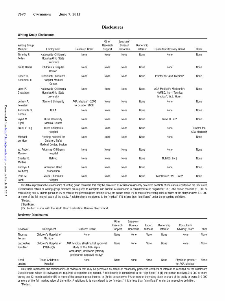
Disclosures
Writing Group Disclosures
Writing GroupMember Employment Research Grant
OtherResearchSupport
Speakers’Bureau/
HonorariaOwnership
Interest Consultant/Advisory Board Other
Timothy F.Feltes
Nationwide Children’sHospital/Ohio State
University
None None None None None None
Emile Bacha Children’s HospitalBoston
None None None None None None
Robert H.Beekman III
Cincinnati Children’sHospital Medical
Center
None None None None Proctor for AGA Medical* None
John P.Cheatham
Nationwide Children’sHospital/Ohio State
University
None None None None AGA Medical*; Medtronic*;NuMED, Inc†; ToshibaMedical*; W.L. Gore†
None
Jeffrey A.Feinstein
Stanford University AGA Medical* (2006to October 2008)
None None None None None
Antoinette S.Gomes
UCLA None None None None None None
Ziyad M.Hijazi
Rush UniversityMedical Center
None None None None NuMED, Inc* None
Frank F. Ing Texas Children’sHospital
None None None None None Proctor forAGA Medical†
Michaelde Moor
Floating Hospital forChildren, Tufts
Medical Center, Boston
None None None None None None
W. RobertMorrow
Arkansas Children’sHospital
None None None None None None
Charles E.Mullins
Retired None None None None NuMED, Inc† None
Kathryn A.Taubert‡
American HeartAssociation
None None None None None None
Evan M.Zahn
Miami Children’sHospital
None None None None Medtronic*; W.L. Gore* None
This table represents the relationships of writing group members that may be perceived as actual or reasonably perceived conflicts of interest as reported on the DisclosureQuestionnaire, which all writing group members are required to complete and submit. A relationship is considered to be “significant” if (1) the person receives $10 000 ormore during any 12-month period or 5% or more of the person’s gross income; or (2) the person owns 5% or more of the voting stock or share of the entity or owns $10 000or more of the fair market value of the entity. A relationship is considered to be “modest” if it is less than “significant” under the preceding definition.
*Modest.†Significant.‡Dr. Taubert is now with the World Heart Federation, Geneva, Switzerland.
Reviewer Disclosures
Reviewer Employment Research Grant
OtherResearchSupport
Speakers’Bureau/
HonorariaExpert
WitnessOwnership
InterestConsultant/
Advisory Board Other
ThomasForbes
Children’s Hospital ofMichigan
None None None None None None None
JacquelineKreutzer
Children’s Hospital ofPittsburgh
AGA Medical (Postmarket approvalstudy of the AGA septal
occluder)*; Medtronic (Melodypostmarket approval study)*
None None None None None None
HenriJustino
Texas Children’sHospital
None None None None None Physician proctorfor AGA Medical*
None
This table represents the relationships of reviewers that may be perceived as actual or reasonably perceived conflicts of interest as reported on the DisclosureQuestionnaire, which all reviewers are required to complete and submit. A relationship is considered to be “significant” if (1) the person receives $10 000 or moreduring any 12-month period or 5% or more of the person’s gross income; or (2) the person owns 5% or more of the voting stock or share of the entity or owns $10 000or more of the fair market value of the entity. A relationship is considered to be “modest” if it is less than “significant” under the preceding definition.
*Modest.
2640 Circulation June 7, 2011
by guest on March 18, 2017
http://circ.ahajournals.org/D
ownloaded from
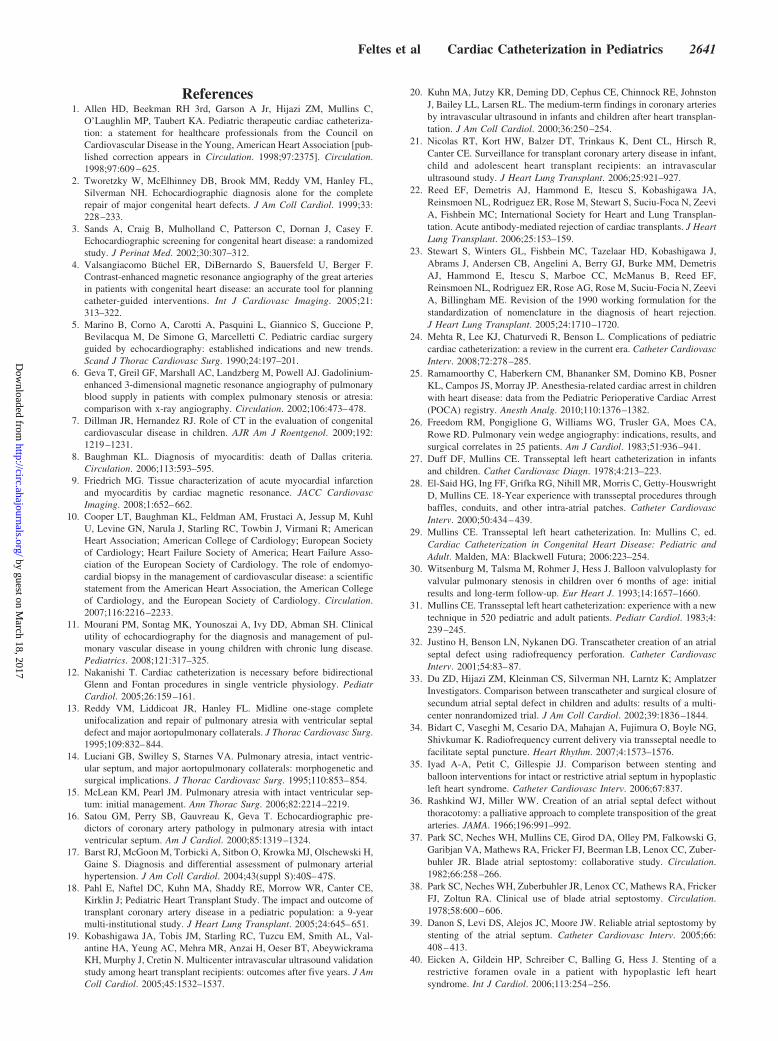
References1. Allen HD, Beekman RH 3rd, Garson A Jr, Hijazi ZM, Mullins C,
O’Laughlin MP, Taubert KA. Pediatric therapeutic cardiac catheteriza-tion: a statement for healthcare professionals from the Council onCardiovascular Disease in the Young, American Heart Association [pub-lished correction appears in Circulation. 1998;97:2375]. Circulation.1998;97:609–625.
2. Tworetzky W, McElhinney DB, Brook MM, Reddy VM, Hanley FL,Silverman NH. Echocardiographic diagnosis alone for the completerepair of major congenital heart defects. J Am Coll Cardiol. 1999;33:228–233.
3. Sands A, Craig B, Mulholland C, Patterson C, Dornan J, Casey F.Echocardiographic screening for congenital heart disease: a randomizedstudy. J Perinat Med. 2002;30:307–312.
4. Valsangiacomo Buchel ER, DiBernardo S, Bauersfeld U, Berger F.Contrast-enhanced magnetic resonance angiography of the great arteriesin patients with congenital heart disease: an accurate tool for planningcatheter-guided interventions. Int J Cardiovasc Imaging. 2005;21:313–322.
5. Marino B, Corno A, Carotti A, Pasquini L, Giannico S, Guccione P,Bevilacqua M, De Simone G, Marcelletti C. Pediatric cardiac surgeryguided by echocardiography: established indications and new trends.Scand J Thorac Cardiovasc Surg. 1990;24:197–201.
6. Geva T, Greil GF, Marshall AC, Landzberg M, Powell AJ. Gadolinium-enhanced 3-dimensional magnetic resonance angiography of pulmonaryblood supply in patients with complex pulmonary stenosis or atresia:comparison with x-ray angiography. Circulation. 2002;106:473–478.
7. Dillman JR, Hernandez RJ. Role of CT in the evaluation of congenitalcardiovascular disease in children. AJR Am J Roentgenol. 2009;192:1219–1231.
8. Baughman KL. Diagnosis of myocarditis: death of Dallas criteria.Circulation. 2006;113:593–595.
9. Friedrich MG. Tissue characterization of acute myocardial infarctionand myocarditis by cardiac magnetic resonance. JACC CardiovascImaging. 2008;1:652–662.
10. Cooper LT, Baughman KL, Feldman AM, Frustaci A, Jessup M, KuhlU, Levine GN, Narula J, Starling RC, Towbin J, Virmani R; AmericanHeart Association; American College of Cardiology; European Societyof Cardiology; Heart Failure Society of America; Heart Failure Asso-ciation of the European Society of Cardiology. The role of endomyo-cardial biopsy in the management of cardiovascular disease: a scientificstatement from the American Heart Association, the American Collegeof Cardiology, and the European Society of Cardiology. Circulation.2007;116:2216–2233.
11. Mourani PM, Sontag MK, Younoszai A, Ivy DD, Abman SH. Clinicalutility of echocardiography for the diagnosis and management of pul-monary vascular disease in young children with chronic lung disease.Pediatrics. 2008;121:317–325.
12. Nakanishi T. Cardiac catheterization is necessary before bidirectionalGlenn and Fontan procedures in single ventricle physiology. PediatrCardiol. 2005;26:159–161.
13. Reddy VM, Liddicoat JR, Hanley FL. Midline one-stage completeunifocalization and repair of pulmonary atresia with ventricular septaldefect and major aortopulmonary collaterals. J Thorac Cardiovasc Surg.1995;109:832–844.
14. Luciani GB, Swilley S, Starnes VA. Pulmonary atresia, intact ventric-ular septum, and major aortopulmonary collaterals: morphogenetic andsurgical implications. J Thorac Cardiovasc Surg. 1995;110:853–854.
15. McLean KM, Pearl JM. Pulmonary atresia with intact ventricular sep-tum: initial management. Ann Thorac Surg. 2006;82:2214–2219.
16. Satou GM, Perry SB, Gauvreau K, Geva T. Echocardiographic pre-dictors of coronary artery pathology in pulmonary atresia with intactventricular septum. Am J Cardiol. 2000;85:1319–1324.
17. Barst RJ, McGoon M, Torbicki A, Sitbon O, Krowka MJ, Olschewski H,Gaine S. Diagnosis and differential assessment of pulmonary arterialhypertension. J Am Coll Cardiol. 2004;43(suppl S):40S–47S.
18. Pahl E, Naftel DC, Kuhn MA, Shaddy RE, Morrow WR, Canter CE,Kirklin J; Pediatric Heart Transplant Study. The impact and outcome oftransplant coronary artery disease in a pediatric population: a 9-yearmulti-institutional study. J Heart Lung Transplant. 2005;24:645–651.
19. Kobashigawa JA, Tobis JM, Starling RC, Tuzcu EM, Smith AL, Val-antine HA, Yeung AC, Mehra MR, Anzai H, Oeser BT, AbeywickramaKH, Murphy J, Cretin N. Multicenter intravascular ultrasound validationstudy among heart transplant recipients: outcomes after five years. J AmColl Cardiol. 2005;45:1532–1537.
20. Kuhn MA, Jutzy KR, Deming DD, Cephus CE, Chinnock RE, JohnstonJ, Bailey LL, Larsen RL. The medium-term findings in coronary arteriesby intravascular ultrasound in infants and children after heart transplan-tation. J Am Coll Cardiol. 2000;36:250–254.
21. Nicolas RT, Kort HW, Balzer DT, Trinkaus K, Dent CL, Hirsch R,Canter CE. Surveillance for transplant coronary artery disease in infant,child and adolescent heart transplant recipients: an intravascularultrasound study. J Heart Lung Transplant. 2006;25:921–927.
22. Reed EF, Demetris AJ, Hammond E, Itescu S, Kobashigawa JA,Reinsmoen NL, Rodriguez ER, Rose M, Stewart S, Suciu-Foca N, ZeeviA, Fishbein MC; International Society for Heart and Lung Transplan-tation. Acute antibody-mediated rejection of cardiac transplants. J HeartLung Transplant. 2006;25:153–159.
23. Stewart S, Winters GL, Fishbein MC, Tazelaar HD, Kobashigawa J,Abrams J, Andersen CB, Angelini A, Berry GJ, Burke MM, DemetrisAJ, Hammond E, Itescu S, Marboe CC, McManus B, Reed EF,Reinsmoen NL, Rodriguez ER, Rose AG, Rose M, Suciu-Focia N, ZeeviA, Billingham ME. Revision of the 1990 working formulation for thestandardization of nomenclature in the diagnosis of heart rejection.J Heart Lung Transplant. 2005;24:1710–1720.
24. Mehta R, Lee KJ, Chaturvedi R, Benson L. Complications of pediatriccardiac catheterization: a review in the current era. Catheter CardiovascInterv. 2008;72:278–285.
25. Ramamoorthy C, Haberkern CM, Bhananker SM, Domino KB, PosnerKL, Campos JS, Morray JP. Anesthesia-related cardiac arrest in childrenwith heart disease: data from the Pediatric Perioperative Cardiac Arrest(POCA) registry. Anesth Analg. 2010;110:1376–1382.
26. Freedom RM, Pongiglione G, Williams WG, Trusler GA, Moes CA,Rowe RD. Pulmonary vein wedge angiography: indications, results, andsurgical correlates in 25 patients. Am J Cardiol. 1983;51:936–941.
27. Duff DF, Mullins CE. Transseptal left heart catheterization in infantsand children. Cathet Cardiovasc Diagn. 1978;4:213–223.
28. El-Said HG, Ing FF, Grifka RG, Nihill MR, Morris C, Getty-HouswrightD, Mullins CE. 18-Year experience with transseptal procedures throughbaffles, conduits, and other intra-atrial patches. Catheter CardiovascInterv. 2000;50:434–439.
29. Mullins CE. Transseptal left heart catheterization. In: Mullins C, ed.Cardiac Catheterization in Congenital Heart Disease: Pediatric andAdult. Malden, MA: Blackwell Futura; 2006:223–254.
30. Witsenburg M, Talsma M, Rohmer J, Hess J. Balloon valvuloplasty forvalvular pulmonary stenosis in children over 6 months of age: initialresults and long-term follow-up. Eur Heart J. 1993;14:1657–1660.
31. Mullins CE. Transseptal left heart catheterization: experience with a newtechnique in 520 pediatric and adult patients. Pediatr Cardiol. 1983;4:239–245.
32. Justino H, Benson LN, Nykanen DG. Transcatheter creation of an atrialseptal defect using radiofrequency perforation. Catheter CardiovascInterv. 2001;54:83–87.
33. Du ZD, Hijazi ZM, Kleinman CS, Silverman NH, Larntz K; AmplatzerInvestigators. Comparison between transcatheter and surgical closure ofsecundum atrial septal defect in children and adults: results of a multi-center nonrandomized trial. J Am Coll Cardiol. 2002;39:1836–1844.
34. Bidart C, Vaseghi M, Cesario DA, Mahajan A, Fujimura O, Boyle NG,Shivkumar K. Radiofrequency current delivery via transseptal needle tofacilitate septal puncture. Heart Rhythm. 2007;4:1573–1576.
35. Iyad A-A, Petit C, Gillespie JJ. Comparison between stenting andballoon interventions for intact or restrictive atrial septum in hypoplasticleft heart syndrome. Catheter Cardiovasc Interv. 2006;67:837.
36. Rashkind WJ, Miller WW. Creation of an atrial septal defect withoutthoracotomy: a palliative approach to complete transposition of the greatarteries. JAMA. 1966;196:991–992.
37. Park SC, Neches WH, Mullins CE, Girod DA, Olley PM, Falkowski G,Garibjan VA, Mathews RA, Fricker FJ, Beerman LB, Lenox CC, Zuber-buhler JR. Blade atrial septostomy: collaborative study. Circulation.1982;66:258–266.
38. Park SC, Neches WH, Zuberbuhler JR, Lenox CC, Mathews RA, FrickerFJ, Zoltun RA. Clinical use of blade atrial septostomy. Circulation.1978;58:600–606.
39. Danon S, Levi DS, Alejos JC, Moore JW. Reliable atrial septostomy bystenting of the atrial septum. Catheter Cardiovasc Interv. 2005;66:408–413.
40. Eicken A, Gildein HP, Schreiber C, Balling G, Hess J. Stenting of arestrictive foramen ovale in a patient with hypoplastic left heartsyndrome. Int J Cardiol. 2006;113:254–256.
Feltes et al Cardiac Catheterization in Pediatrics 2641
by guest on March 18, 2017
http://circ.ahajournals.org/D
ownloaded from
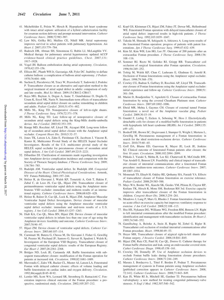
41. Michelfelder E, Polzin W, Hirsch R. Hypoplastic left heart syndromewith intact atrial septum: utilization of a hybrid catheterization facilityfor cesarean section delivery and prompt neonatal intervention. CatheterCardiovasc Interv. 2008;72:983–987.
42. Law MA, Grifka RG, Mullins CE, Nihill MR. Atrial septostomyimproves survival in select patients with pulmonary hypertension. AmHeart J. 2007;153:779–784.
43. Badesch DB, Abman SH, Simonneau G, Rubin LJ, McLaughlin VV.Medical therapy for pulmonary arterial hypertension: updated ACCPevidence-based clinical practice guidelines. Chest. 2007;131:1917–1928.
44. Vogel JH. Balloon embolization during atrial septostomy. Circulation.1970;42:155–156.
45. Ellison RC, Plauth WH Jr, Gazzaniga AB, Fyler DC. Inability to deflatecatheter balloon: a complication of balloon atrial septostomy. J Pediatr.1970;76:604–606.
46. Suchon E, Pieculewicz M, Tracz W, Przewlocki T, Sadowski J, PodolecP. Transcatheter closure as an alternative and equivalent method to thesurgical treatment of atrial septal defect in adults: comparison of earlyand late results. Med Sci Monit. 2009;15:CR612–CR617.
47. Kaya MG, Baykan A, Dogan A, Inanc T, Gunebakmaz O, Dogdu O,Uzum K, Eryol NK, Narin N. Intermediate-term effects of transcathetersecundum atrial septal defect closure on cardiac remodeling in childrenand adults. Pediatr Cardiol. 2010;31:474–482.
48. Mills NL, King TD. Nonoperative closure of left-to-right shunts.J Thorac Cardiovasc Surg. 1976;72:371–378.
49. Mills NL, King TD. Late follow-up of nonoperative closure ofsecundum atrial septal defects using the King-Mills double-umbrelladevice. Am J Cardiol. 2003;92:353–355.
50. Knepp MD, Rocchini AP, Lloyd TR, Aiyagari RM. Long-term followup of secundum atrial septal defect closure with the Amplatzer septaloccluder. Congenit Heart Dis. 2010;5:32–37.
51. Jones TK, Latson LA, Zahn E, Fleishman CE, Jacobson J, Vincent R,Kanter K; Multicenter Pivotal Study of the HELEX Septal OccluderInvestigators. Results of the U.S. multicenter pivotal study of theHELEX septal occluder for percutaneous closure of secundum atrialseptal defects. J Am Coll Cardiol. 2007;49:2215–2221.
52. Dibardino DJ. Clarification of statements made regarding investigationinto Amplatzer device complication incidence and comparison with theSociety of Thoracic Surgery database. J Thorac Cardiovasc Surg. 2009;138:784–785.
53. Rudolph AM. Ventricular septal defect. In: Rudolph AM. CongenitalDiseases of the Heart: Clinical-Physiological Considerations. Armonk,NY: Futura Publishing; 2001:197–244.
54. Holzer R, de Giovanni J, Walsh KP, Tometzki A, Goh T, Hakim F,Zabal C, de Lezo JS, Cao QL, Hijazi ZM. Transcatheter closure ofperimembranous ventricular septal defects using the Amplatzer mem-branous VSD occluder: immediate and midterm results of an interna-tional registry. Catheter Cardiovasc Interv. 2006;68:620–628.
55. Holzer R, Balzer D, Cao QL, Lock K, Hijazi ZM; Amplatzer MuscularVentricular Septal Defect Investigators. Device closure of muscularventricular septal defects using the Amplatzer muscular ventricularseptal defect occluder: immediate and mid-term results of a U.S.registry. J Am Coll Cardiol. 2004;43:1257–1263.
56. Diab KA, Cao QL, Mora BN, Hijazi ZM. Device closure of muscularventricular septal defects in infants less than one year of age using theAmplatzer devices: feasibility and outcome. Catheter Cardiovasc Interv.2007;70:90–97.
57. Hijazi ZM. Device closure of ventricular septal defects. Catheter Car-diovasc Interv. 2003;60:107–114.
58. Carminati M, Butera G, Chessa M, De Giovanni J, Fisher G, GewilligM, Peuster M, Piechaud JF, Santoro G, Sievert H, Spadoni I, Walsh K;Investigators of the European VSD Registry. Transcatheter closure ofcongenital ventricular septal defects: results of the European Registry.Eur Heart J. 2007;28:2361–2368.
59. Bridges ND, Lock JE, Castaneda AR. Baffle fenestration with sub-sequent transcatheter closure: modification of the Fontan operation forpatients at increased risk. Circulation. 1990;82:1681–1689.
60. Mavroudis C, Zales VR, Backer CL, Muster AJ, Latson LA. FenestratedFontan with delayed catheter closure: effects of volume loading andbaffle fenestration on cardiac index and oxygen delivery. Circulation.1992;86(suppl):II-85–II-92.
61. Lemler MS, Scott WA, Leonard SR, Stromberg D, Ramaciotti C. Fen-estration improves clinical outcome of the Fontan procedure: a pro-spective, randomized study. Circulation. 2002;105:207–212.
62. Kopf GS, Kleinman CS, Hijazi ZM, Fahey JT, Dewar ML, HellenbrandWE. Fenestrated Fontan operation with delayed transcatheter closure ofatrial septal defect: improved results in high-risk patients. J ThoracCardiovasc Surg. 1992;103:1039–1047.
63. Takeda M, Shimada M, Sekiguchi A, Ishizawa A. Long-term results ofthe fenestrated Fontan operation: progress of patients with patent fen-estrations. Jpn J Thorac Cardiovasc Surg. 1999;47:432–439.
64. Kim SJ, Kim WH, Lim HG, Lee JY. Outcome of 200 patients after anextracardiac Fontan procedure. J Thorac Cardiovasc Surg. 2008;136:108–116.
65. Sommer RJ, Recto M, Golinko RJ, Griepp RB. Transcatheter coilocclusion of surgical fenestration after Fontan operation. Circulation.1996;94:249–252.
66. Tofeig M, Walsh KP, Chan C, Ladusans E, Gladman G, Arnold R.Occlusion of Fontan fenestrations using the Amplatzer septal occluder.Heart. 1998;79:368–370.
67. Cowley CG, Badran S, Gaffney D, Rocchini AP, Lloyd TR. Transcath-eter closure of Fontan fenestrations using the Amplatzer septal occluder:initial experience and follow-up. Catheter Cardiovasc Interv. 2000;51:301–304.
68. Marini D, Boudjemline Y, Agnoletti G. Closure of extracardiac Fontanfenestration by using the covered Cheatham Platinum stent. CatheterCardiovasc Interv. 2007;69:1002–1006.
69. Ebeid MR, Mehta I, Gaymes CH. Closure of external tunnel Fontanfenestration: a novel use of the Amplatzer vascular plug. PediatrCardiol. 2009;30:15–19.
70. Gundel F, Liebig T, Eicken A, Sebening W, Hess J. Electrolyticallydetachable coils for closure of a modified baffle fenestration in patientswith hypoplastic left heart syndrome (HLHS). Int J Cardiol. 2009;135:e49–e51.
71. Boshoff DE, Brown SC, Degiovanni J, Stumper O, Wright J, Mertens L,Gewillig M. Percutaneous management of a Fontan fenestration: insearch for the ideal restriction-occlusion device. Catheter CardiovascInterv. 2010;75:60–65.
72. Goff DA, Blume ED, Gauvreau K, Mayer JE, Lock JE, JenkinsKJ. Clinical outcome of fenestrated Fontan patients after closure: thefirst 10 years. Circulation. 2000;102:2094–2099.
73. Pihkala J, Yazaki S, Mehta R, Lee KJ, Chaturvedi R, McCrindle BW,Van Arsdell G, Benson LN. Feasibility and clinical impact of transcath-eter closure of interatrial communications after a fenestrated Fontanprocedure: medium-term outcomes. Catheter Cardiovasc Interv. 2007;69:1007–1014.
74. Momenah TS, Eltayb H, Oakley RE, Qethamy HA, Faraidi YA. Effectsof transcatheter closure of Fontan fenestration on exercise tolerance.Pediatr Cardiol. 2008;29:585–588.
75. Mays WA, Border WL, Knecht SK, Gerdes YM, Pfriem H, Claytor RP,Knilans TK, Hirsch R, Mone SM, Beekman RH 3rd. Exercise capacityimproves after transcatheter closure of the Fontan fenestration inchildren. Congenit Heart Dis. 2008;3:254–261.
76. Meadows J, Lang P, Marx G, Rhodes J. Fontan fenestration closure hasno acute effect on exercise capacity but improves ventilatory response toexercise. J Am Coll Cardiol. 2008;52:108–113.
77. Hsu HS, Nykanen DG, Williams WG, Freedom RM, Benson LN. Rightto left interatrial communications after the modified Fontan procedure:identification and management with transcatheter occlusion. Br Heart J.1995;74:548–552.
78. Gamillscheg A, Beitzke A, Stein JI, Rupitz M, Zobel G, Rigler B.Transcatheter coil occlusion of residual interatrial communications afterFontan procedure. Heart. 1998;80:49–53.
79. Hoyer MH, Transcatheter closure of atypical right-to-left shunts afterFontan surgery. J Invasive Cardiol. 2006;18:E61–E65.
80. Hijazi ZM, Ruiz CE, Patel H, Cao QL, Dorros G. Catheter therapy forFontan baffle obstruction and leak, using an endovascular covered stent.Cathet Cardiovasc Diagn. 1998;45:158–161.
81. Crowley DI, Donnelly JP. Use of Amplatzer occlusion devices toocclude Fontan baffle leaks during fenestration closure procedures.Catheter Cardiovasc Interv. 2008;71:244–249.
82. Masura J, Bordacova L, Tittel P, Berden P, Podnar T. Percutaneousmanagement of cyanosis in Fontan patients using Amplatzer occluders[published correction appears in Catheter Cardiovasc Interv. 2008;72:141]. Catheter Cardiovasc Interv. 2008;71:843–849.
83. Kan JS, White RI Jr, Mitchell SE, Gardner TJ. Percutaneous balloonvalvuloplasty: a new method for treating congenital pulmonary-valvestenosis. N Engl J Med. 1982;307:540–542.
2642 Circulation June 7, 2011
by guest on March 18, 2017
http://circ.ahajournals.org/D
ownloaded from
84. Ballerini L, Mullins CE, Cifarelli A, Pasquini L, De Simone G, GiannicoS, Guccione P, Di Donato R, Di Carlo D. Percutaneous balloon valvu-loplasty of pulmonary valve stenosis, dysplasia, and residual stenosisafter surgical valvotomy for pulmonary atresia with intact ventricularseptum: long-term results. Cathet Cardiovasc Diagn. 1990;19:165–169.
85. Silvilairat S, Cabalka AK, Cetta F, Hagler DJ, O’Leary PW. Echocar-diographic assessment of isolated pulmonary valve stenosis: which out-patient Doppler gradient has the most clinical validity? J Am SocEchocardiogr. 2005;18:1137–1142.
86. Stanger P, Cassidy SC, Girod DA, Kan JS, Lababidi Z, Shapiro SR.Balloon pulmonary valvuloplasty: results of the Valvuloplasty andAngioplasty of Congenital Anomalies Registry. Am J Cardiol. 1990;65:775–783.
87. Lababidi Z, Wu JR. Percutaneous balloon pulmonary valvuloplasty.Am J Cardiol. 1983;52:560–562.
88. Kveselis DA, Rocchini AP, Snider AR, Rosenthal A, Crowley DC, DickM 2nd. Results of balloon valvuloplasty in the treatment of congenitalvalvar pulmonary stenosis in children. Am J Cardiol. 1985;56:527–532.
89. Rey C, Marache P, Francart C, Dupuis C. Percutaneous transluminalballoon valvuloplasty of congenital pulmonary valve stenosis, with aspecial report on infants and neonates. J Am Coll Cardiol. 1988;11:815–820.
90. Rao PS, Fawzy ME, Solymar L, Mardini MK.. Long-term results ofballoon pulmonary valvuloplasty of valvar pulmonic stenosis. AmHeart J. 1988;115:1291–1296.
91. McCrindle BW. Independent predictors of long-term results afterballoon pulmonary valvuloplasty. Valvuloplasty and Angioplasty ofCongenital Anomalies (VACA) Registry Investigators. Circulation.1994;89:1751–1759.
92. Masura J, Burch M, Deanfield JE, Sullivan ID. Five-year follow-up afterballoon pulmonary valvuloplasty. J Am Coll Cardiol. 1993;21:132–136.
93. O’Connor BK, Beekman RH, Lindauer A, Rocchini A. Intermediate-termoutcome after pulmonary balloon valvuloplasty: comparison with amatched surgical control group. J Am Coll Cardiol. 1992;20:169–173.
94. Rao PS, Wilson AD, Thapar MK, Brais M. Balloon pulmonary valvu-loplasty in the management of cyanotic congenital heart defects. CathetCardiovasc Diagn. 1992;25:16–24.
95. Kreutzer J, Perry SB, Jonas RA, Mayer JE, Castaneda AR, Lock JE.Tetralogy of Fallot with diminutive pulmonary arteries: preoperativepulmonary valve dilation and transcatheter rehabilitation of pulmonaryarteries. J Am Coll Cardiol. 1996;27:1741–1747.
96. Justo RN, Nykanen DG, Williams WG, Freedom RM, Benson LN.Transcatheter perforation of the right ventricular outflow tract as initialtherapy for pulmonary valve atresia and intact ventricular septum in thenewborn. Cathet Cardiovasc Diagn. 1997;40:408–413.
97. Gibbs JL, Blackburn ME, Uzun O, Dickinson DF, Parsons JM, ChatrathRR. Laser valvotomy with balloon valvoplasty for pulmonary atresiawith intact ventricular septum: five years’ experience. Heart. 1997;77:225–228.
98. Walsh MA, Lee KJ, Chaturvedi R, Van Arsdell GS, Benson LN. Radio-frequency perforation of the right ventricular outflow tract as a palliativestrategy for pulmonary atresia with ventricular septal defect. CatheterCardiovasc Interv. 2007;69:1015–1020.
99. Humpl T, Soderberg B, McCrindle BW, Nykanen DG, Freedom RM,Williams WG, Benson LN. Percutaneous balloon valvotomy in pulmo-nary atresia with intact ventricular septum: impact on patient care.Circulation. 2003;108:826–832.
100. Giglia TM, Mandell VS, Connor AR, Mayer JE Jr, Lock JE. Diagnosisand management of right ventricle-dependent coronary circulation inpulmonary atresia with intact ventricular septum. Circulation. 1992;86:1516–1528.
101. Beekman RH, Rocchini AP, Andes A. Balloon valvuloplasty for criticalaortic stenosis in the newborn: influence of new catheter technology.J Am Coll Cardiol. 1991;17:1172–1176.
102. Fischer DR, Ettedgui JA, Park SC, Siewers RD, del Nido PJ. Carotidartery approach for balloon dilation of aortic valve stenosis in theneonate: a preliminary report. J Am Coll Cardiol. 1990;15:1633–1636.
103. Lababidi Z, Wu JR, Walls JT. Percutaneous balloon aortic valvulo-plasty: results in 23 patients. Am J Cardiol. 1984;53:194–197.
104. Walls JT, Lababidi Z, Curtis JJ, Silver D. Assessment of percutaneousballoon pulmonary and aortic valvuloplasty. J Thorac Cardiovasc Surg.1984;88:352–356.
105. Choy M, Beekman RH, Rocchini AP, Crowley DC, Snider AR, Dick M2nd, Rosenthal A. Percutaneous balloon valvuloplasty for valvar aorticstenosis in infants and children. Am J Cardiol. 1987;59:1010–1013.
106. Helgason H, Keane JF, Fellows KE, Kulik TJ, Lock JE. Balloon dilationof the aortic valve: studies in normal lambs and in children with aorticstenosis. J Am Coll Cardiol. 1987;9:816–822.
107. Sholler GF, Keane JF, Perry SB, Sanders SP, Lock JE. Balloon dilationof congenital aortic valve stenosis: results and influence of technical andmorphological features on outcome. Circulation. 1988;78:351–360.
108. Zeevi B, Keane JF, Castaneda AR, Perry SB, Lock JE. Neonatal criticalvalvar aortic stenosis: a comparison of surgical and balloon dilationtherapy. Circulation. 1989;80:831–839.
109. Meliones JN, Beekman RH, Rocchini AP, Lacina SJ. Balloon valvulo-plasty for recurrent aortic stenosis after surgical valvotomy in childhood:immediate and follow-up studies. J Am Coll Cardiol. 1989;13:1106–1110.
110. Vogel M, Benson LN, Burrows P, Smallhorn JF, Freedom RM. Balloondilatation of congenital aortic valve stenosis in infants and children:short term and intermediate results. Br Heart J. 1989;62:148–153.
111. Keane JF, Perry SB, Lock Je. Balloon dilation of congenital valvularaortic stenosis. J Am Coll Cardiol. 1990;16:457–458.
112. Shaddy RE, Boucek MM, Sturtevant JE, Ruttenberg HD, Orsmond GS.Gradient reduction, aortic valve regurgitation and prolapse after balloonaortic valvuloplasty in 32 consecutive patients with congenital aorticstenosis. J Am Coll Cardiol. 1990;16:451–456.
113. O’Connor BK, Beekman RH, Rocchini AP, Rosenthal A. Intermediate-termeffectiveness of balloon valvuloplasty for congenital aortic stenosis: a pro-spective follow-up study. Circulation. 1991;84:732–738.
114. Mosca RS, Iannettoni MD, Schwartz SM, Ludomirsky A, Beekman RH3rd, Lloyd T, Bove EL. Critical aortic stenosis in the neonate: a com-parison of balloon valvuloplasty and transventricular dilation. J ThoracCardiovasc Surg. 1995;109:147–154.
115. McCrindle BW; Valvuloplasty and Angioplasty of CongenitalAnomalies (VACA) Registry Investigators. Independent predictors ofimmediate results of percutaneous balloon aortic valvotomy in children.Am J Cardiol. 1996;77:286–293.
116. Magee AG, Nykanen D, McCrindle BW, Wax D, Freedom RM, BensonLN. Balloon dilation of severe aortic stenosis in the neonate: comparisonof anterograde and retrograde catheter approaches. J Am Coll Cardiol.1997;30:1061–1066.
117. Galal O, Rao PS, Al-Fadley F, Wilson AD. Follow-up results of balloonaortic valvuloplasty in children with special reference to causes of lateaortic insufficiency. Am Heart J. 1997;133:418–427.
118. Egito ES, Moore P, O’Sullivan J, Colan S, Perry SB, Lock JE, Keane JF.Transvascular balloon dilation for neonatal critical aortic stenosis: earlyand midterm results. J Am Coll Cardiol. 1997;29:442–447.
119. Cowley CG, Dietrich M, Mosca RS, Bove EL, Rocchini AP, Lloyd TR.Balloon valvuloplasty versus transventricular dilation for neonatalcritical aortic stenosis. Am J Cardiol. 2001;87:1125–1127, A10.
120. McCrindle BW, Blackstone EH, Williams WG, Sittiwangkul R, SprayTL, Azakie A, Jonas RA. Are outcomes of surgical versus transcatheterballoon valvotomy equivalent in neonatal critical aortic stenosis?Circulation. 2001;104(suppl 1):I-152–I-158.
121. Latiff HA, Sholler GF, Cooper S. Balloon dilatation of aortic stenosis ininfants younger than 6 months of age: intermediate outcome. PediatrCardiol. 2003;24:17–26.
122. Pedra CA, Sidhu R, McCrindle BW, Nykanen DG, Justo RN, FreedomRM, Benson LN. Outcomes after balloon dilation of congenital aorticstenosis in children and adolescents. Cardiol Young. 2004;14:315–321.
123. Reich O, Tax P, Marek J, Razek V, Gilík J, Tomek V, Chaloupecky V,Bartakova H, Skovranek J. Long term results of percutaneous balloonvalvoplasty of congenital aortic stenosis: independent predictors ofoutcome. Heart. 2004;90:70–76.
124. McElhinney DB, Lock JE, Keane JF, Moran AM, Colan SD. Left heartgrowth, function, and reintervention after balloon aortic valvuloplastyfor neonatal aortic stenosis. Circulation. 2005;111:451–458.
125. Fratz S, Gildein HP, Balling G, Sebening W, Genz T, Eicken A, Hess J.Aortic valvuloplasty in pediatric patients substantially postpones theneed for aortic valve surgery: a single-center experience of 188 patientsafter up to 17.5 years of follow-up. Circulation. 2008;117:1201–1206.
126. Rocchini AP, Beekman RH, Ben Shachar G, Benson L, Schwartz D,Kan JS. Balloon aortic valvuloplasty: results of the Valvuloplasty andAngioplasty of Congenital Anomalies Registry. Am J Cardiol. 1990;65:784–789.
127. Bonow RO, Carabello BA, Kanu C, de Leon AC Jr, Faxon DP, FreedMD, Gaasch WH, Lytle BW, Nishimura RA, O’Gara PT, O’Rourke RA,Otto CM, Shah PM, Shanewise JS, Smith SC Jr, Jacobs AK, Adams CD,Anderson JL, Antman EM, Faxon DP, Fuster V, Halperin JL, Hiratzka
Feltes et al Cardiac Catheterization in Pediatrics 2643
by guest on March 18, 2017
http://circ.ahajournals.org/D
ownloaded from
LF, Hunt SA, Lytle BW, Nishimura R, Page RL, Riegel B. ACC/AHA2006 guidelines for the management of patients with valvular heartdisease: a report of the American College of Cardiology/American HeartAssociation Task Force on Practice Guidelines (Writing Committee toRevise the 1998 Guidelines for the Management of Patients WithValvular Heart Disease) [published corrections appear in Circulation.2010;121:e443 and Circulation. 2007;115:e409]. Circulation. 2006;114:e84–e231.
128. Keane JF, Driscoll DJ, Gersony WM, Hayes CJ, Kidd L, O’Fallon WM,Pieroni DR, Wolfe RR, Weidman WH. Second natural history study ofcongenital heart defects: results of treatment of patients with aorticvalvar stenosis. Circulation. 1993;87(suppl):I-16–I-27.
129. Inoue K, Owaki T, Nakamura T, Kitamura F, Miyamoto N. Clinicalapplication of transvenous mitral commissurotomy by a new ballooncatheter. J Thorac Cardiovasc Surg. 1984;87:394–402.
130. Babic UU, Pejcic P, Djurisic Z, Vucinic M, Grujicic SN. Percutaneoustransarterial balloon mitral valvuloplasty: 30 months experience. Herz.1988;13:91–99.
131. Lee CY, Lau KW, Ding ZP, Tan A, Chan C, Koh TH, Quek S, Chee TS,Ng A, Johan A. Percutaneous balloon valvuloplasty in mitral restenosisafter previous surgical commissurotomy. Singapore Med J. 1995;36:474–478.
132. Baz JA, Pinar E, Albarran A, Mauri J; Spanish Society of CardiologyWorking Group on Cardiac Catheterization and Interventional Car-diology. Spanish Cardiac Catheterization and Coronary InterventionRegistry: 17th official report of the Spanish Society of CardiologyWorking Group on Cardiac Catheterization and Interventional Car-diology (1990–2007). Rev Esp Cardiol. 2008;61:1298–1314.
133. Lock JE, Khalilullah M, Shrivastava S, Bahl V, Keane JF. Percutaneouscatheter commissurotomy in rheumatic mitral stenosis. N Engl J Med.1985;313:1515–1518.
134. Levine MJ, et al. Long-term follow-up in 105 patients undergoingpercutaneous balloon valvuloplasty. J Am Coll Cardiol. 1989;13:18A.Abstract.
135. The National Heart, Lung, and Blood Institute Balloon ValvuloplastyRegistry Participants. Multicenter experience with balloon mitral com-missurotomy. NHLBI Balloon Valvuloplasty Registry Report onimmediate and 30-day follow-up results. Circulation. 1992;85:448–461.
136. Fawzy ME, Mimish L, Awad M, Galal O, el-Deeb F, Khan B. Mitralballoon valvotomy in children with Inoue balloon technique: immediateand intermediate-term result. Am Heart J. 1994;127:1559–1562.
137. Unal N, Mese T, Hudaoglu S, Celikkol B, Yunus S, Saylam GS, AkcoralA. Percutaneous transvenous balloon mitral valvuloplasty: mid-termresults in adolescents. Turk J Pediatr. 1999;41:341–348.
138. Kveselis DA, Rocchini AP, Beekman R, Snider AR, Crowley D, DickM, Rosenthal A. Balloon angioplasty for congenital and rheumaticmitral stenosis. Am J Cardiol. 1986;57:348–350.
139. Alday LE, Juaneda E. Percutaneous balloon dilatation in congenitalmitral stenosis. Br Heart J. 1987;57:479–482.
140. Spevak PJ, Bass JL, Ben-Shachar G, Hesslein P, Keane JF, Perry S,Pyles L, Lock JE. Balloon angioplasty for congenital mitral stenosis.Am J Cardiol. 1990;66:472–476.
141. Moore P, Adatia I, Spevak PJ, Keane JF, Perry SB, Castaneda AR, LockJE. Severe congenital mitral stenosis in infants. Circulation. 1994;89:2099–2106.
142. McElhinney DB, Sherwood MC, Keane JF, del Nido PJ, Almond CS,Lock JE. Current management of severe congenital mitral stenosis:outcomes of transcatheter and surgical therapy in 108 infants andchildren. Circulation. 2005;112:707–714.
143. Grifka RG, O’Laughlin MP, Nihill MR, Mullins CE. Double-transseptal,double-balloon valvuloplasty for congenital mitral stenosis. Circulation.1992;85:123–129.
144. Jarrar M, Betbout F, Gamra H, Maatouk F, Ayari M, Farhat MB.Successful percutaneous double balloon valvuloplasty for congenitalmitral stenosis. Int J Cardiol. 1996;56:193–196.
145. Lo PH, Hung JS, Lau KW, Kim MH, Ku PM, Krayyem M. Inoue-balloon mitral valvuloplasty in double-orifice mitral stenosis. J InvasiveCardiol. 2003;15:301–303.
146. Collins-Nakai RL, Rosenthal A, Castaneda AR, Bernhard WF, NadasAS. Congenital mitral stenosis: a review of 20 years’ experience.Circulation. 1977;56:1039–1047.
147. Acar C, Jebara VA, Grare P, Chachques JC, Dervanian P, Vahanian A,Carpentier A. Traumatic mitral insufficiency following percutaneous
mitral dilation: anatomic lesions and surgical implications. Eur J Car-diothorac Surg. 1992;6:660–663.
148. Carpentier A. Congenital malformations of the mitral valve. In: Stark J,deLeval M, eds. Surgery for Congenital Heart Defects. Philadelphia,PA: Saunders; 1994:599–614.
149. Gotzsche CO, Krag-Olsen B, Nielsen J, Sørensen KE, Kristensen BO.Prevalence of cardiovascular malformations and association withkaryotypes in Turner’s syndrome. Arch Dis Child. 1994;71:433–436.
150. Connolly HM, Huston J 3rd, Brown RD Jr, Warnes CA, Ammash NM,Tajik AJ. Intracranial aneurysms in patients with coarctation of theaorta: a prospective magnetic resonance angiographic study of 100patients. Mayo Clin Proc. 2003;78:1491–1499.
151. Brickner ME, Hillis LD, Lange RA. Congenital heart disease in adults:first of two parts. N Engl J Med. 2000;342:256–263.
152. Forbes TJ, Garekar S, Amin Z, Zahn EM, Nykanen D, Moore P, QureshiSA, Cheatham JP, Ebeid MR, Hijazi ZM, Sandhu S, Hagler DJ, SievertH, Fagan TE, Ringewald J, Du W, Tang L, Wax DF, Rhodes J, JohnstonTA, Jones TK, Turner DR, Pedra CA, Hellenbrand WE; CongenitalCardiovascular Interventional Study Consortium (CCISC). Proceduralresults and acute complications in stenting native and recurrent coarc-tation of the aorta in patients over 4 years of age: a multi-institutionalstudy. Catheter Cardiovasc Interv. 2007;70:276–285.
153. Forbes TJ, Moore P, Pedra CA, Zahn EM, Nykanen D, Amin Z, GarekarS, Teitel D, Qureshi SA, Cheatham JP, Ebeid MR, Hijazi ZM, SandhuS, Hagler DJ, Sievert H, Fagan TE, Ringwald J, Du W, Tang L, WaxDF, Rhodes J, Johnston TA, Jones TK, Turner DR, Pass R, Torres A,Hellenbrand WE. Intermediate follow-up following intravascularstenting for treatment of coarctation of the aorta. Catheter CardiovascInterv. 2007;70:569–577.
154. Rothman A, Galindo A, Evans WN, Collazos JC, Restrepo H. Effec-tiveness and safety of balloon dilation of native aortic coarctation inpremature neonates weighing �or � 2,500 grams. Am J Cardiol. 2010;105:1176–1180.
155. Marshall AC, Perry SB, Keane JF, Lock JE. Early results andmedium-term follow-up of stent implantation for mild residual orrecurrent aortic coarctation. Am Heart J. 2000;139:1054–1060.
156. Morrow WR, Vick GW 3rd, Nihill MR, Rokey R, Johnston DL, HedrickTD, Mullins CE. Balloon dilation of unoperated coarctation of the aorta:short- and intermediate-term results. J Am Coll Cardiol. 1988;11:133–138.
157. Fletcher SE, Nihill MR, Grifka RG, O’Laughlin MP, Mullins CE.Balloon angioplasty of native coarctation of the aorta: midtermfollow-up and prognostic factors. J Am Coll Cardiol. 1995;25:730–734.
158. Patel HT, Madani A, Paris YM, Warner KG, Hijazi ZM. Balloonangioplasty of native coarctation of the aorta in infants and neonates: isit worth the hassle? Pediatr Cardiol. 2001;22:53–57.
159. Fawzy ME, Fathala A, Osman A, Badr A, Mostafa MA, Mohamed G,Dunn B. Twenty-two years of follow-up results of balloon angioplastyfor discreet native coarctation of the aorta in adolescents and adults. AmHeart J. 2008;156:910–917.
160. Tynan M, Finley JP, Fontes V, Hess J, Kan J. Balloon angioplasty forthe treatment of native coarctation: results of Valvuloplasty and Angio-plasty of Congenital Anomalies Registry. Am J Cardiol. 1990;65:790–792.
161. Bush DM, Hoffman TM, Del Rosario J, Eiriksson H, Rome JJ. Fre-quency of restenosis after balloon pulmonary arterioplasty and itscauses. Am J Cardiol. 2000;86:1205–1209.
162. Rothman A, Perry SB, Keane JF, Lock JE. Early results and follow-upof balloon angioplasty for branch pulmonary artery stenoses. J Am CollCardiol. 1990;15:1109–1117.
163. Hosking MC, Thomaidis C, Hamilton R, Burrows PE, Freedom RM,Benson LN. Clinical impact of balloon angioplasty for branch pulmo-nary arterial stenosis. Am J Cardiol. 1992;69:1467–1470.
164. Mullins CE. Intravascular stent implant-pulmonary branch stenosis. In:Mullins C, ed. Cardiac Catheterization in Congenital Heart Disease:Pediatric and Adult. Malden, MA: Blackwell Futura; 2006:597–622.
165. Butera G, Antonio LT, Massimo C, Mario C. Expanding indications forthe treatment of pulmonary artery stenosis in children by using cuttingballoon angioplasty. Catheter Cardiovasc Interv. 2006;67:460–465.
166. O’Laughlin MP, Perry SB, Lock JE, Mullins CE. Use of endovascularstents in congenital heart disease. Circulation. 1991;83:1923–1939.
167. Trant CA Jr, O’Laughlin MP, Ungerleider RM, Garson A Jr. Cost-effectiveness analysis of stents, balloon angioplasty, and surgery for thetreatment of branch pulmonary artery stenosis. Pediatr Cardiol. 1997;18:339–344.
2644 Circulation June 7, 2011
by guest on March 18, 2017
http://circ.ahajournals.org/D
ownloaded from
168. O’Laughlin MP, Slack MC, Grifka RG, Perry SB, Lock JE, Mullins CE.Implantation and intermediate-term follow-up of stents in congenitalheart disease. Circulation. 1993;88:605–614.
169. Bartolomaeus G, Radtke WA. Patterns of late diameter change afterballoon angioplasty of branch pulmonary artery stenosis: evidence forvascular remodeling. Catheter Cardiovasc Interv. 2002;56:533–540.
170. Mullins CE. Cardiac Catheterization in Congenital Heart Disease:Pediatric and Adult. Malden, MA: Blackwell Futura; 2006:924.
171. Dev V, Kaul U, Jain P, Reddy S, Sharma S, Pandey G, Rajani M.Percutaneous transluminal balloon angioplasty for obstruction of thesuprahepatic inferior vena cava and cavo-atrial graft stenosis. Am JCardiol. 1989;64:397–399.
172. Tzifa A, Marshall AC, McElhinney DB, Lock JE, Geggel RL. Endo-vascular treatment for superior vena cava occlusion or obstruction in apediatric and young adult population: a 22-year experience. J Am CollCardiol. 2007;49:1003–1009.
173. Frazer JR, Ing FF. Stenting of stenotic or occluded iliofemoral veins,superior and inferior vena cavae in children with congenital heart dis-ease: acute results and intermediate follow up. Catheter CardiovascInterv. 2009;73:181–188.
174. Latson LA, Prieto LR. Congenital and acquired pulmonary vein stenosis.Circulation. 2007;115:103–108.
175. Devaney EJ, Chang AC, Ohve RG, Bove EL. Management of congenitaland acquired pulmonary vein stenosis. Ann Thorac Surg. 2006;81:992–995.
176. van Son JA, Danielson GK, Puga FJ, Edwards WD, Driscoll DJ. Repairof congenital and acquired pulmonary vein stenosis. Ann Thorac Surg.1995;60:144–150.
177. Lock JE, Bass JL, Castaneda-Zuniga W, Fuhrman BP, Rashkind WJ,Lucas RV Jr. Dilation angioplasty of congenital or operative narrowingsof venous channels. Circulation. 1984;70:457–464.
178. Cullen S, ho SY, Shore D, Lincoln C, Redington A. Congenital stenosisof pulmonary veins: failure to modify natural history of intraoperativeplacement of stents. Cardiol Young. 1994;4:395–398.
179. Driscoll DJ, Hesslein PS, Mullins CE. Congenital stenosis of individualpulmonary veins: clinical spectrum and unsuccessful treatment by trans-venous balloon dilation. Am J Cardiol. 1982;49:1767–1772.
180. Breinholt JP, Hawkins JA, Minich LA, Tani LY, Orsmond GS, Ritter S,Shaddy RE. Pulmonary vein stenosis with normal connection: associatedcardiac abnormalities and variable outcome. Ann Thorac Surg. 1999;68:164–168.
181. Sadr IM, Tan PE, Kieran MW, Jenkins KJ. Mechanism of pulmonaryvein stenosis in infants with normally connected veins. Am J Cardiol.2000;86:577–579, A10.
182. Dieter RS, Nelson B, Wolff MR, Thornton F, Grist TM, Cohen DMTransseptal stent treatment of anastomotic stricture after repair of partialanomalous pulmonary venous return. J Endovasc Ther. 2003;10:838–842.
183. Hyde JA, Stumper O, Barth MJ, Wright JG, Silove ED, de Giovanni JV,Brawn WJ, Sethia B. Total anomalous pulmonary venous connection:outcome of surgical correction and management of recurrent venousobstruction. Eur J Cardiothorac Surg. 1999;15:735–740.
184. Caldarone CA, Najm HK, Kadletz M, Smallhorn JF, Freedom RM,Williams WG, Coles JG. Relentless pulmonary vein stenosis after repairof total anomalous pulmonary venous drainage. Ann Thorac Surg. 1998;66:1514–1520.
185. Spray TL, Bridges ND. Surgical management of congenital and acquiredpulmonary vein stenosis. Semin Thorac Cardiovasc Surg Pediatr CardSurg Annu. 1999;2:177–188.
186. Mukadam ME, Khandeparkar JM, Agarwal NB, Kumar LJ, TendolkarAG, Magotra RA. Surgical considerations in management of left hemi-anomalous pulmonary venous connections. Thorac Cardiovasc Surg.1995;43:48–51.
187. Lamb RK, Qureshi SA, Wilkinson JL, Arnold R, West CR, HamiltonDI. Total anomalous pulmonary venous drainage: seventeen-yearsurgical experience. J Thorac Cardiovasc Surg. 1988;96:368–375.
188. Yun TJ, Coles JG, Konstantinov IE, Al-Radi OO, Wald RM, Guerra V,de Oliveira NC, Van Arsdell GS, Williams WG, Smallhorn J, CaldaroneCA. Conventional and sutureless techniques for management of thepulmonary veins: evolution of indications from postrepair pulmonaryvein stenosis to primary pulmonary vein anomalies. J Thorac Car-diovasc Surg. 2005;129:167–174.
189. Hancock Friesen CL, Zurakowski D, Thiagarajan RR, Forbess JM, delNido PJ, Mayer JE, Jonas RA. Total anomalous pulmonary venous
connection: an analysis of current management strategies in a singleinstitution. Ann Thorac Surg. 2005;79:596–606.
190. Zimmermann GS, Reithmann C, Strauss T, Hatz R, Brenner P, UberfuhrA, Frey L, Nikolaou K, Behr J. Successful angioplasty and stenttreatment of pulmonary vein stenosis after single-lung transplantation.J Heart Lung Transplant. 2009;28:194–198.
191. Clark SC, Levine AJ, Hasan A, Hilton CJ, Forty J, Dark JH. Vascularcomplications of lung transplantation. Ann Thorac Surg. 1996;61:1079–1082.
192. Malden ES, Kaiser LR, Gutierrez FR. Pulmonary vein obstruction fol-lowing single lung transplantation. Chest. 1992;102:645–647.
193. Michel-Cherqui M, Brusset A, Liu N, Raffin L, Schlumberger S,Ceddaha A, Fischler M. Intraoperative transesophageal echocardio-graphic assessment of vascular anastomoses in lung transplantation: areport on 18 cases. Chest. 1997;111:1229–1235.
194. Packer DL, Keelan P, Munger TM, Breen JF, Asirvatham S, PetersonLA, Monahan KH, Hauser MF, Chandrasekaran K, Sinak LJ, HolmesDR Jr. Clinical presentation, investigation, and management of pulmonaryvein stenosis complicating ablation for atrial fibrillation. Circulation. 2005;111:546–554.
195. Qureshi AM, Prieto LR, Latson LA, Lane GK, Mesia CI, Radvansky P,White RD, Marrouche NF, Saad EB, Bash DL, Natale A, Rhodes JF.Transcatheter angioplasty for acquired pulmonary vein stenosis afterradiofrequency ablation. Circulation. 2003;108:1336–1342.
196. Holmes DR Jr, Monahan KH, Packer D. Pulmonary vein stenosis com-plicating ablation for atrial fibrillation: clinical spectrum and interven-tional considerations. JACC Cardiovasc Interv. 2009;2:267–276.
197. Purerfellner H, Aichinger J, Martinek M, Nesser HJ, Cihal R, Gsch-wendtner M, Dierneder J. Incidence, management, and outcome insignificant pulmonary vein stenosis complicating ablation for atrialfibrillation. Am J Cardiol. 2004;93:1428–1431, A10.
198. Tsao HM, Chen SA. Evaluation of pulmonary vein stenosis aftercatheter ablation of atrial fibrillation. Card Electrophysiol Rev. 2002;6:397–400.
199. Neumann T, Sperzel J, Dill T, Kluge A, Erdogan A, Greis H, Hansel J,Berkowitsch A, Kurzidim K, Kuniss M, Hamm CW, Pitschner HF.Percutaneous pulmonary vein stenting for the treatment of severe ste-nosis after pulmonary vein isolation. J Cardiovasc Electrophysiol. 2005;16:1180–1188.
200. Skanes AC, Gula LJ, Yee R, Krahn AD, Klein GJ. Pulmonary veinstenosis: intervene early and carry a big stent. J Cardiovasc Electro-physiol. 2008;19:679–680.
201. Schneider C, Ernst S, Malisius R, Bahlmann E, Lampe F, Broemel T,Krause K, Boczor S, Antz M, Kuck KH. Transesophageal echocardiog-raphy: a follow-up tool after catheter ablation of atrial fibrillation andinterventional therapy of pulmonary vein stenosis and occlusion. J IntervCard Electrophysiol. 2007;18:195–205.
202. Dong J, Vasamreddy CR, Jayam V, Dalal D, Dickfeld T, Eldadah Z,Meininger G, Halperin HR, Berger R, Bluemke DA, Calkins H.Incidence and predictors of pulmonary vein stenosis following catheterablation of atrial fibrillation using the anatomic pulmonary vein ablationapproach: results from paired magnetic resonance imaging. J Car-diovasc Electrophysiol. 2005;16:845–852.
203. Saad EB, Rossillo A, Saad CP, Martin DO, Bhargava M, Erciyes D,Bash D, Williams-Andrews M, Beheiry S, Marrouche NF, Adams J,Pisano E, Fanelli R, Potenza D, Raviele A, Bonso A, Themistoclakis S,Brachmann J, Saliba WI, Schweikert RA, Natale A. Pulmonary veinstenosis after radiofrequency ablation of atrial fibrillation: functionalcharacterization, evolution, and influence of the ablation strategy.Circulation. 2003;108:3102–3107.
204. Cappato R, Calkins H, Chen SA, Davies W, Iesaka Y, Kalman J, KimYH, Klein G, Packer D, Skanes A. Worldwide survey on the methods,efficacy, and safety of catheter ablation for human atrial fibrillation.Circulation. 2005;111:1100–1105.
205. Robbins IM, Colvin EV, Doyle TP, Kemp WE, Loyd JE, McMahon WS,Kay GN. Pulmonary vein stenosis after catheter ablation of atrial fibril-lation. Circulation. 1998;98:1769–1775.
206. Yu WC, Hsu TL, Tai CT, Tsai CF, Hsieh MH, Lin WS, Lin YK, TsaoHM, Ding YA, Chang MS, Chen SA. Acquired pulmonary vein stenosisafter radiofrequency catheter ablation of paroxysmal atrial fibrillation.J Cardiovasc Electrophysiol. 2001;12:887–892.
207. Yang HM, Lai CK, Patel J, Moore J, Chen PS, Shivkumar K, FishbeinMC. Irreversible intrapulmonary vascular changes after pulmonary veinstenosis complicating catheter ablation for atrial fibrillation. CardiovascPathol. 2007;16:51–55.
Feltes et al Cardiac Catheterization in Pediatrics 2645
by guest on March 18, 2017
http://circ.ahajournals.org/D
ownloaded from
208. Chen SA, Hsieh MH, Tai CT, Tsai CF, Prakash VS, Yu WC, Hsu TL,Ding YA, Chang MS. Initiation of atrial fibrillation by ectopic beatsoriginating from the pulmonary veins: electrophysiological character-istics, pharmacological responses, and effects of radiofrequencyablation. Circulation. 1999;100:1879–1886.
209. Kluge A, Dill T, Ekinci O, Hansel J, Hamm C, Pitschner HF, BachmannG. Decreased pulmonary perfusion in pulmonary vein stenosis afterradiofrequency ablation: assessment with dynamic magnetic resonanceperfusion imaging. Chest. 2004;126:428–437.
210. den Bakker MA, Thomeer M, Maat AP, Groeninx van Zoelen CE.Life-threatening hemoptysis caused by chronic idiopathic pulmonaryhilar fibrosis with unilateral pulmonary vein occlusion. Ann DiagnPathol. 2005;9:319–322.
211. Nunes H, Humbert M, Capron F, Brauner M, Sitbon O, Battesti JP,Simonneau G, Valeyre D. Pulmonary hypertension associated with sar-coidosis: mechanisms, haemodynamics and prognosis. Thorax. 2006;61:68–74.
212. McMahon CJ, McDermott M, Walsh KP. Failure of cutting balloonangioplasty to prevent restenosis in childhood pulmonary venous steno-sis. Catheter Cardiovasc Interv. 2006;68:763–766.
213. Mendelsohn AM, Bove EL, Lupinetti FM, Crowley DC, Lloyd TR,Fedderly RT, Beekman RH 3rd. Intraoperative and percutaneousstenting of congenital pulmonary artery and vein stenosis. Circulation.1993;88(part 2):II-210–II-217.
214. Coles JG, Yemets I, Najm HK, Lukanich JM, Perron J, Wilson GJ,Rabinovitch M, Nykanen DG, Benson LN, Rebeyka IM, Trusler GA,Freedom RM, Williams WG. Experience with repair of congenital heartdefects using adjunctive endovascular devices. J Thorac CardiovascSurg. 1995;110:1513–1519.
215. Bini RM, Cleveland DC, Ceballos R, Bargeron LM Jr, Pacifico AD,Kirklin JW. Congenital pulmonary vein stenosis. Am J Cardiol. 1984;54:369–375.
216. Najm HK, Caldarone CA, Smallhorn J, Coles JG. A sutureless techniquefor the relief of pulmonary vein stenosis with the use of in situ peri-cardium. J Thorac Cardiovasc Surg. 1998;115:468–470.
217. Ungerleider RM, Johnston TA, O’Laughlin MP, Jaggers JJ, Gaskin PR.Intraoperative stents to rehabilitate severely stenotic pulmonary vessels.Ann Thorac Surg. 2001;71:476–481.
218. Michel-Behnke I, Luedemann M, Hagel KJ, Schranz D. Serial stentimplantation to relieve in-stent stenosis in obstructed total anomalouspulmonary venous return. Pediatr Cardiol. 2002;23:221–223.
219. Tomita H, Watanabe K, Yazaki S, Kimura K, Ono Y, Yagihara T,Echigo S. Stent implantation and subsequent dilatation for pulmonaryvein stenosis in pediatric patients: maximizing effectiveness. Circ J.2003;67:187–190.
220. Seale AN, Daubeney PE, Magee AG, Rigby ML. Pulmonary veinstenosis: initial experience with cutting balloon angioplasty. Heart.2006;92:815–820.
221. Cook AL, Prieto LR, Delaney JW, Rhodes JF. Usefulness of cuttingballoon angioplasty for pulmonary vein in-stent stenosis. Am J Cardiol.2006;98:407–410.
222. Prieto LR, Schoenhagen P, Arruda MJ, Natale A, Worley SE. Com-parison of stent versus balloon angioplasty for pulmonary vein stenosiscomplicating pulmonary vein isolation. J Cardiovasc Electrophysiol.2008;19:673–678.
223. Hosking M, Redmond M, Allen L, Broecker L, Keaney M, Lebeau J,Walley V. Responses of systemic and pulmonary veins to the presenceof an intravascular stent in a swine model. Cathet Cardiovasc Diagn.1995;36:90–96.
224. Gibbs JL, Rothman MT, Rees MR, Parsons JM, Blackburn ME, RuizCE. Stenting of the arterial duct: a new approach to palliation forpulmonary atresia. Br Heart J. 1992;67:240–245.
225. Gibbs JL, Uzun O, Blackburn ME, Wren C, Hamilton JR, WattersonKG. Fate of the stented arterial duct. Circulation. 1999;99:2621–2625.
226. Gewillig M, Boshoff DE, Dens J, Mertens L, Benson LN. Stenting theneonatal arterial duct in duct-dependent pulmonary circulation: newtechniques, better results. J Am Coll Cardiol. 2004;43:107–112.
227. Alwi M, Choo KK, Latiff HA, Kandavello G, Samion H, Mulyadi MD.Initial results and medium-term follow-up of stent implantation of patentductus arteriosus in duct-dependent pulmonary circulation. J Am CollCardiol. 2004;44:438–445.
228. Michel-Behnke I, Akintuerk H, Thul J, Bauer J, Hagel KJ, Schranz D.Stent implantation in the ductus arteriosus for pulmonary blood supplyin congenital heart disease. Catheter Cardiovasc Interv. 2004;61:242–252.
229. Boshoff DE, Michel-Behnke I, Schranz D, Gewillig M. Stenting theneonatal arterial duct. Expert Rev Cardiovasc Ther. 2007;5:893–901.
230. Celebi A, Yalcin Y, Erdem A, Zeybek C, Akdeniz C, Polat TB. Stentimplantation into the patent ductus arteriosus in cyanotic congenitalheart disease with duct-dependent or diminished pulmonary circulation.Turk J Pediatr. 2007;49:413–417.
231. Hussain A, Al-Zharani S, Muhammed AA, Al-Ata J, Galal OM.Midterm outcome of stent dilatation of patent ductus arteriosus inductal-dependent pulmonary circulation. Congenit Heart Dis. 2008;3:241–249.
232. Santoro G, Gaio G, Palladino MT, Iacono C, Carrozza M, Esposito R,Russo MG, Caianiello G, Calabro R. Stenting of the arterial duct innewborns with duct-dependent pulmonary circulation. Heart. 2008;94:925–929.
233. Porstmann W, Wierny L, Warnke H, Gerstberger G, Romaniuk PA.Catheter closure of patent ductus arteriosus: 62 cases treated withoutthoracotomy. Radiol Clin North Am. 1971;9:203–218.
234. Rashkind WJ. Therapeutic interventional procedures in congenital heartdisease. Radiol Diagn (Berl). 1987;28:449–460.
235. Rashkind WJ, Cuaso CC. Transcatheter closure of a patent ductusarteriosus: successful use in a 3.5 kg infant. Pediatr Cardiol. 1979;1:3–7.
236. Cambier PA, Kirby WC, Wortham DC, Moore JW. Percutaneousclosure of the small (less than 2.5 mm) patent ductus arteriosus usingcoil embolization. Am J Cardiol. 1992;69:815–816.
237. Ing FF, Sommer RJ. The snare-assisted technique for transcatheter coilocclusion of moderate to large patent ductus arteriosus: immediate andintermediate results. J Am Coll Cardiol. 1999;33:1710–1718.
238. Ing FF, Bierman FZ. Percutaneous transcatheter coil occlusion of thepatent ductus arteriosus aided by the nitinol snare: further observations.Cardiovasc Intervent Radiol. 1995;18:222–226.
239. Ing FF, Mullins CE, Rose M, Shapir Y, Bierman FZ. Transcatheterclosure of the patent ductus arteriosus in adults using the Gianturco coil.Clin Cardiol. 1996;19:875–879.
240. Lloyd TR, Beekman RH, Moore JW, Hijazi ZM, Hellenbrand WE,Sommer RJ, Wiggins JW, Zamora R, Vincent RN; for the PDA CoilRegistry Investigators. The PDA Coil Registry: report of the first 535procedures. Circulation. 1995;92(suppl I):I-380. Abstract.
241. Magee AG, Huggon IC, Seed PT, Qureshi SA, Tynan M; on behalf ofthe Association for European Paediatric Cardiology. Transcatheter coilocclusion of the arterial duct: results of the European Registry. EurHeart J. 2001;22:1817–1821.
242. Ing FF, Recto MR, Saidi A, Denfield S, Mullins CE. A method pro-viding bidirectional control of coil delivery in occlusions of patentductus arteriosus with shallow ampulla and Pott’s shunts. Am J Cardiol.1997;79:1561–1563.
243. Wang JK, Liau CS, Huang JJ, Hsu KL, Lo PH, Hung JS, Wu MH, LeeYT. Transcatheter closure of patent ductus arteriosus using Gianturcocoils in adolescents and adults. Catheter Cardiovasc Interv. 2002;55:513–518.
244. Sivakumar K, Francis E, Krishnan P. Safety and feasibility of transcath-eter closure of large patent ductus arteriosus measuring �or�4 mm inpatients weighing �or�6 kg. J Interv Cardiol. 2008;21:196–203.
245. Tomita H, Uemura S, Haneda N, Soga T, Matsuoka T, Nishioka T,Yazaki S, Hatakeyama K, Takamuro M, Horita N. Coil occlusion ofPDA in patients younger than 1 year: Risk factors for adverse events.J Cardiol. 2009;53:208–213.
246. Grifka RG, Vincent JA, Nihill MR, Ing FF, Mullins CE. Transcatheterpatent ductus arteriosus closure in an infant using the Gianturco-GrifkaVascular Occlusion Device. Am J Cardiol. 1996;78:721–723.
247. Celiker A, Aypar E, Karagoz T, Dilber E, Ceviz N. Transcatheterclosure of patent ductus arteriosus with Nit-Occlud coils. CatheterCardiovasc Interv. 2005;65:569–576.
248. Thanopoulos BV, Tzannos KA, Eleptherakis N, Stefanadis C. Com-parison and results of transcatheter closure of patent ductus arteriosususing the swivel-disk device versus plug occluder in children. Am JCardiol. 2008;102:486–490.
249. Thanopoulos BD, Tsaousis GS, Djukic M, Al Hakim F, EleftherakisNG, Simeunovic SD. Transcatheter closure of high pulmonary arterypressure persistent ductus arteriosus with the Amplatzer muscular ven-tricular septal defect occluder. Heart. 2002;87:260–263.
250. Grifka RG, Fenrich AL, Tapio JB. Transcatheter closure of patent ductusarteriosus and aorto-pulmonary vessels using non-ferromagnetic InconelMReye embolization coils. Catheter Cardiovasc Interv. 2008;72:691–695.
2646 Circulation June 7, 2011
by guest on March 18, 2017
http://circ.ahajournals.org/D
ownloaded from
251. Glatz AC, Petit CJ, Gillespie MJ. Novel use of a modified AmplatzerVascular Plug to occlude a patent ductus arteriosus in two patients.Catheter Cardiovasc Interv. 2008;72:82–86.
252. Pass RH, Hijazi Z, Hsu DT, Lewis V, Hellenbrand WE. MulticenterUSA Amplatzer patent ductus arteriosus occlusion device trial: initialand one-year results. J Am Coll Cardiol. 2004;44:513–519.
253. Shabbir M, Akhtar K, Maadullah, Ahmed W. Transcatheter closure oflarge sized patent ductus arteriosus using the Amplatzer duct occluderdevice. J Coll Physicians Surg Pak. 2009;19:275–278.
254. Chen Z, Chen L, Wu L. Transcatheter Amplatzer occlusion and surgicalclosure of patent ductus arteriosus: comparison of effectiveness andcosts in a low-income country. Pediatr Cardiol. 2009;30:781–785.
255. Forsey J, Kenny D, Morgan G, Hayes A, Turner M, Tometzki A, MartinR. Early clinical experience with the new Amplatzer Ductal Occluder IIfor closure of the persistent arterial duct. Catheter Cardiovasc Interv.2009;74:615–623.
256. Morgan G, Tometzki AJ, Martin RP. Transcatheter closure of longtubular patent arterial ducts: the Amplatzer Duct Occluder II: a new andvaluable tool. Catheter Cardiovasc Interv. 2009;73:576–580.
257. Thanopoulos B, Eleftherakis N, Tzannos K, Stefanadis C. Transcatheterclosure of the patent ductus arteriosus using the new Amplatzer ductoccluder: initial clinical applications in children. Am Heart J. 2008;156:917.e1–917.e6.
258. Zhang JF, Huang D, Yang YN, Gao XM, Ma YT. Percutaneous trans-catheter closure of patent ductus arteriosus with an Amplatzer ductoccluder using retrograde guidewire-established femoral arteriovenousloop. Clin Exp Pharmacol Physiol. 2008;35:606–610.
259. Wang JK, Wu MH, Hwang JJ, Chiang FT, Lin MT, Lue HC. Trans-catheter closure of moderate to large patent ductus arteriosus with theAmplatzer duct occluder. Catheter Cardiovasc Interv. 2007;69:572–578.
260. Al-Hamash S. Transcatheter closure of patent ductus arteriosus andinterruption of inferior vena cava with azygous continuation using anAmplatzer duct occluder. Pediatr Cardiol. 2006;27:618–620.
261. Vijayalakshmi IB, Chitra N, Rajasri R, Vasudevan K. Initial clinicalexperience in transcatheter closure of large patent arterial ducts ininfants using the modified and angled Amplatzer duct occluder. CardiolYoung. 2006;16:378–384.
262. Pass RH. Amplatzer Duct Occluder device: a new technology for theclosure of the moderate-to-large-sized patent ductus arteriosus. ExpertRev Med Devices. 2006;3:291–296.
263. Masura J, Tittel P, Gavora P, Podnar T. Long-term outcome of trans-catheter patent ductus arteriosus closure using Amplatzer duct occluders.Am Heart J. 2006;151:755.e7–755.e10.
264. Jan SL, Hwang B, Fu YC, Chi CS. Transcatheter closure of a largepatent ductus arteriosus in a young child using the Amplatzer ductoccluder. Pediatr Cardiol. 2005;26:703–706.
265. Vijayalakshmi IB, Chitra N, Rajasri R, Prabhudeva AN. Amplatzerangled duct occluder for closure of patent ductus arteriosus larger thanthe aorta in an infant. Pediatr Cardiol. 2005;26:480–483.
266. Al-Ata J, Arfi Am, Hussain A, Kouatli AA, Jalal MO. The efficacy andsafety of the Amplatzer ductal occluder in young children and infants.Cardiol Young. 2005;15:279–285.
267. Hsin HT, Lin LC, Hwang JJ, Ho SG, Tseng CD, Chiang FT. Retrogradewire-assisted percutaneous transcatheter closure of persistent ductusarteriosus with Amplatzer duct occluder in the elderly: a new appli-cation. Catheter Cardiovasc Interv. 2004;61:264–267.
268. Masura J, Gavora P, Podnar T. Transcatheter occlusion of patent ductusarteriosus using a new angled Amplatzer duct occluder: initial clinicalexperience. Catheter Cardiovasc Interv. 2003;58:261–267.
269. Hong TE, Hellenbrand WE, Hijazi ZM; Amplatzer Investigators. Trans-catheter closure of patent ductus arteriosus in adults using the Amplatzerduct occluder: initial results and follow-up. Indian Heart J. 2002;54:384–389.
270. Hoyer MH. Closure of a very large patent ductus arteriosus using theAmplatzer duct occluder. J Invasive Cardiol. 2002;14:531–534.
271. Ebeid MR, Masura J, and Hijazi ZM. Early experience with theAmplatzer ductal occluder for closure of the persistently patent ductusarteriosus. J Interv Cardiol. 2001;14:33–36.
272. Thanopoulos BD, Hakim FA, Hiari A, Tsaousis GS, Paphitis C, HijaziZM. Patent ductus arteriosus equipment and technique: Amplatzer ductoccluder: intermediate-term follow-up and technical considerations.J Interv Cardiol. 2001;14:247–254.
273. Lee CH, Leung YL, Chow WH. Transcatheter closure of the patentductus arteriosus using an Amplatzer duct occluder in adults. JpnHeart J. 2001;42:533–537.
274. Fischer G, Stieh J, Uebing A, Grabitz R, Kramer HH. Transcatheterclosure of persistent ductus arteriosus in infants using the Amplatzerduct occluder. Heart. 2001;86:444–447.
275. Kong H, Gu X, Bass JL, Titus J, Urness M, Kim TH, Hunter DW.Experimental evaluation of a modified Amplatzer duct occluder.Catheter Cardiovasc Interv. 2001;53:571–576.
276. Waight DJ, Cao QL, Hijazi ZM. Transcatheter closure of patent ductusarteriosus using the Amplatzer duct occluder. Curr Interv Cardiol Rep.2001;3:263–267.
277. Sandhu SK, King TD, Troutman WB, Hixon RL 3rd, Kiel EA,Bourgeois KV. Transcatheter closure of patent ductus arteriosus with theAmplatzer duct occluder: short-term follow-up. J Invasive Cardiol.2001;13:298–302.
278. Bilkis AA, Alwi M, Hasri S, Haifa AL, Geetha K, Rehman MA,Hasanah I. The Amplatzer duct occluder: experience in 209 patients.J Am Coll Cardiol. 2001;37:258–261.
279. Faella HJ, Hijazi ZM. Closure of the patent ductus arteriosus with theAmplatzer PDA device: immediate results of the international clinicaltrial. Catheter Cardiovasc Interv. 2000;51:50–54.
280. Marwah A, Radhakrishnan S, Shrivastava S. Immediate and early resultsof closure of moderate to large patent arterial ducts using the newAmplatzer device. Cardiol Young. 2000;10:208–211.
281. Thanopoulos BD, Hakim FA, Hiari A, Goussous Y, Basta E, ZarayelyanAA, Tsaousis GS. Further experience with transcatheter closure of thepatent ductus arteriosus using the Amplatzer duct occluder. J Am CollCardiol. 2000;35:1016–1021.
282. Hakim F, Hawelleh AA, Goussous Y, Hijazi ZM. Simultaneous stentimplantation for coarctation of the aorta and closure of patent ductusarteriosus using the Amplatzer duct occluder. Catheter CardiovascInterv. 1999;47:36–38.
283. Masura J, Walsh KP, Thanopoulous B, Chan C, Bass J, Goussous Y,Gavora P, Hijazi ZM. Catheter closure of moderate- to large-sized patentductus arteriosus using the new Amplatzer duct occluder: immediate andshort-term results. J Am Coll Cardiol. 1998;31:878–882.
284. Huang TC, Chien KJ, Hsieh KS, Lin CC, Lee CL. Comparison of0.052-inch coils vs Amplatzer duct occluder for transcatheter closure ofmoderate to large patent ductus arteriosus. Circ J. 2009;73:356–360.
285. Gudausky TM, Hirsch R, Khoury PR, Beekman RH 3rd. Comparison oftwo transcatheter device strategies for occlusion of the patent ductusarteriosus. Catheter Cardiovasc Interv. 2008;72:675–680.
286. Wang JK, Hwang JJ, Chiang FT, Wu MH, Lin MT, Lee WL, Lue HC.A strategic approach to transcatheter closure of patent ductus: Gianturcocoils for small-to-moderate ductus and Amplatzer duct occluder forlarge ductus. Int J Cardiol. 2006;106:10–15.
287. Szkutnik M, Menacho-Delgadillo R, Palmero-Zilveti E, Bialkowski J.Transcatheter closure of patent ductus arteriosus among native high-altitude habitants. Pediatr Cardiol. 2008;29:624–627.
288. Santoro G, Bigazzi MC, Carrozza M, Palladino MT, Sarubbi B, ScarpatiC, Dalto M, Russo MG, Calabro R. Percutaneous treatment of moderate-to-large patent ductus arteriosus with different devices: early andmid-term results. Ital Heart J. 2005;6:396–400.
289. Santoro G, Bigazzi MC, Palladino MT, Russo MG, Carrozza M, CalabroR. Comparison of percutaneous closure of large patent ductus arteriosusby multiple coils versus the Amplatzer duct occluder device. Am JCardiol. 2004;94:252–255.
290. Godart F, Rey C, Devos P, Breviere GM, Francart C. Transcatheterocclusion of moderate to large patent arterial ducts, having a diameterabove 2.5 mm, with the Amplatzer Duct Occluder: comparisons with theRashkind, buttoned devices, and coils in 116 consecutive patients.Cardiol Young. 2003;13:413–419.
291. Saliba Z, El-rassi I, Helou D, Abou-Jaoudeh P, Chehab G, Daou L,Khater D, Gerbaka B, Jebara V. Development of catheter-basedtreatment of patent ductus arteriosus: a medium-sized centre experience.Arch Cardiovasc Dis. 2009;102:111–118.
292. Giroud JM, Jacobs JP, Evolution of strategies for management of thepatent arterial duct. Cardiol Young. 2007;17(suppl 2):68–74.
293. Shen XQ, Zhou SH, Fang ZF, Hu XQ, Qi SS, Chen GR, Wang C.Transcatheter closure for patent ductus arteriosus in children [inChinese]. Zhong Nan Da Xue Xue Bao Yi Xue Ban. 2006;31:782–785.
294. Brockmeier K, Hallscheidt P, Haller C. Closure of a patent arterial ductin a septuagenarian with atrial fibrillation. Cardiol Young. 2004;14:328–329.
Feltes et al Cardiac Catheterization in Pediatrics 2647
by guest on March 18, 2017
http://circ.ahajournals.org/D
ownloaded from
295. Lin CC, Hsieh KS, Huang TC, Weng KP. Closure of large patent ductusarteriosus in infants. Am J Cardiol. 2009;103:857–861.
296. Krichenko A, Benson LN, Burrows P, Moes CA, McLaughlin P,Freedom RM. Angiographic classification of the isolated, persistentlypatent ductus arteriosus and implications for percutaneous catheterocclusion. Am J Cardiol. 1989;63:877–880.
297. Hays MD, Hoyer MH, Glasow PF. New forceps delivery technique forcoil occlusion of patent ductus arteriosus. Am J Cardiol. 1996;77:209–211.
298. Grifka RG, Jones TK. Transcatheter closure of large PDA using 0.052�Gianturco coils: controlled delivery using a bioptome catheter through a4 French sheath. Catheter Cardiovasc Interv. 2000;49:301–306.
299. Owada CY, Moore P. A new controlled release system for standard0.052 and 0.038 Gianturco coils. J Am Coll Cardiol. 1998;31(suppl A):153A. Abstract.
300. Berdjis F, Moore JW. Balloon occlusion delivery technique for closureof patent ductus arteriosus. Am Heart J. 1997;133:601–604.
301. Kuhn MA, Latson LA. Transcatheter embolization coil closure of patentductus arteriosus–modified delivery for enhanced control during coilpositioning. Cathet Cardiovasc Diagn. 1995;36:288–290.
302. Prieto LR, Latson LA, Dalvi B, Arbetman MM, Ebeid MR, LamorgeseTT. Transcatheter coil embolization of abnormal vascular connectionsusing a new type of delivery catheter for enhanced control. Am JCardiol. 1999;83:981–983, A10.
303. Balzer DT, Spray TL, McMullin D, Cottingham W, Canter CE. Endar-teritis associated with a clinically silent patent ductus arteriosus. AmHeart J. 1993;125:1192–1193.
304. Ussia GP, Mule M, Caruso E, Aiello R, Tamburino C. Combinedendothelin receptor antagonist and transcatheter interventional therapyof patent ductus arteriosus with severe pulmonary artery hypertension.Int J Cardiol. 2007;116:427–429.
305. Gorenflo M, Ulmer HE, Brockmeier K. Successful closure of the arterialduct in the setting of rubella syndrome. Cardiol Young. 2002;12:200–202.
306. Kramoh EK, Miro J, Bigras JL, Turpin S, Lambert R, Lapierre C, Jin W,Dahdah N. Differential pulmonary perfusion scan after percutaneousocclusion of the patent ductus arteriosus: one-decade consecutive lon-gitudinal study from a single institution. Pediatr Cardiol. 2008;29:918–922.
307. Vavuranakis M, Tzannos KA, Thanopoulos BD, Vlasis K, Stefanadis C.Severe hemolysis complicating transcatheter occlusion of a patentductus arteriosus: the importance of elimination of residual flow.Hellenic J Cardiol. 2007;48:373–376.
308. Galal MO, Arfi MA, Nicole S, Payot M, Hussain A, Qureshi S. Leftventricular systolic dysfunction after transcatheter closure of a largepatent ductus arteriosus. J Coll Physicians Surg Pak. 2005;15:723–725.
309. Pfammatter JP, Meier B. Successful repositioning of an Amplatzer ductoccluder immediately after inadvertent embolization in the descendingaorta. Catheter Cardiovasc Interv. 2003;59:83–85.
310. Joseph G, Mandalay A, Zacharias TU, George B. Severe intravascularhemolysis after transcatheter closure of a large patent ductus arteriosususing the Amplatzer duct occluder: successful resolution by intradevicecoil deployment. Catheter Cardiovasc Interv. 2002;55:245–249.
311. Godart F, Rodes J, Rey C. Severe haemolysis after transcatheter closureof a patent arterial duct with the new Amplatzer duct occluder. CardiolYoung. 2000;10:265–267.
312. Duke C, Chan KC. Aortic obstruction caused by device occlusion ofpatent arterial duct. Heart. 1999;82:109–111.
313. Kaulitz R, Ziemer G, Paul T, Peuster M, Bertram H, Hausdorf G.Fontan-type procedures: residual lesions and late interventions. AnnThorac Surg. 2002;74:778–785.
314. Triedman JK, Bridges ND, Mayer JE Jr, Lock JE. Prevalence and riskfactors for aortopulmonary collateral vessels after Fontan and bidirec-tional Glenn procedures. J Am Coll Cardiol. 1993;22:207–215.
315. Rothman A, Tong AD. Percutaneous coil embolization of superfluousvascular connections in patients with congenital heart disease. AmHeart J. 1993;126:206–213.
316. Beekman RH 3rd, Shim D, Lloyd TR. Embolization therapy in pediatriccardiology. J Interv Cardiol. 1995;8:543–556.
317. Ichikawa H, Yagihara T, Kishimoto H, Isobe F, Yamamoto F, NishigakiK, Matsuki O, Fujita T. Extent of aortopulmonary collateral blood flowas a risk factor for Fontan operations. Ann Thorac Surg. 1995;59:433–437.
318. Spicer RL, Uzark KC, Moore JW, Mainwaring RD, Lamberti JJ. Aor-topulmonary collateral vessels and prolonged pleural effusions aftermodified Fontan procedures. Am Heart J. 1996;131:1164–1168.
319. Moore JW, Berdjis F. Coil occlusion of congenital vascular malfor-mation and surgical shunts. Prog Pediatr Cardiol. 1996;6:149–159.
320. Rychik J. Management of protein-losing enteropathy after the Fontanprocedure. Semin Thorac Cardiovasc Surg Pediatr Card Surg Annu.1998;1:15–22.
321. Rothman A. Pediatric cardiovascular embolization therapy. PediatrCardiol. 1998;19:74–84.
322. Kanter KR, Vincent RN, Raviele AA. Importance of acquired systemic-to-pulmonary collaterals in the Fontan operation. Ann Thorac Surg.1999;68:969–974.
323. Ascuitto RJ, Ross-Ascuitto NT. Systematic-to-pulmonary collaterals: asource of flow energy loss in Fontan physiology. Pediatr Cardiol.2004;25:472–481.
324. Lim DS, Graziano JN, Rocchini AP, Lloyd TR. Transcatheter occlusionof aortopulmonary shunts during single-ventricle surgical palliation.Catheter Cardiovasc Interv. 2005;65:427–433.
325. Eicken A, Genz T, Wild F, Balling G, Schreiber C, Hess J. Resolutionof persistent late postoperative chylothorax after coil occlusion of aor-topulmonary collaterals. Int J Cardiol. 2007;115:e80–e82.
326. Stern HJ. The argument for aggressive coiling of aortopulmonary col-laterals in single ventricle patients. Catheter Cardiovasc Interv. 2009;74:897–900.
327. Bradley SM, McCall MM, Sistino JJ, Radtke WA. Aortopulmonarycollateral flow in the Fontan patient: does it matter? Ann Thorac Surg.2001;72:408–415.
328. Bradley SM. Management of aortopulmonary collateral arteries inFontan patients: routine occlusion is not warranted. Semin Thorac Car-diovasc Surg Pediatr Card Surg Annu. 2002;5:55–67.
329. Berdat PA, Belli E, Lacour-Gayet F, Planche C, Serraf A. Additionalpulmonary blood flow has no adverse effect on outcome after bidirec-tional cavopulmonary anastomosis. Ann Thorac Surg. 2005;79:29–36.
330. McElhinney DB, Reddy VM, Tworetzky W, Petrossian E, Hanley FL,Moore P. Incidence and implications of systemic to pulmonary col-laterals after bidirectional cavopulmonary anastomosis. Ann ThoracSurg. 2000;69:1222–1228.
331. Perry SB, Radtke W, Fellows KE, Keane JF, Lock JE. Coil embolizationto occlude aortopulmonary collateral vessels and shunts in patients withcongenital heart disease. J Am Coll Cardiol. 1989;13:100–108.
332. Reidy JF, Jones OD, Tynan MJ, Baker EJ, Joseph MC. Embolisationprocedures in congenital heart disease. Br Heart J. 1985;54:184–192.
333. Hijazi ZM. New device for percutaneous closure of aortopulmonarycollaterals. Catheter Cardiovasc Interv. 2004;63:482–485.
334. Brawn WJ, Jones T, Davies B, Barron D. How we manage patients withmajor aorta pulmonary collaterals. Semin Thorac Cardiovasc SurgPediatr Card Surg Annu. 2009;12:152–157.
335. Ing FF, Laskari C, Bierman FZ. Additional aortopulmonary collateralsin patients referred for coil occlusion of a patent ductus arteriosus.Cathet Cardiovasc Diagn. 1996;37:5–8.
336. Krishnan US, Lamour JM, Hsu DT, Kichuk MR, Donnelly CM,Addonizio LJ. Management of aortopulmonary collaterals in childrenfollowing cardiac transplantation for complex congenital heart disease.J Heart Lung Transplant. 2004;23:564–569.
337. Santoro G, Carrozza M, Russo MG, Calabro R. Symptomatic aorto-pul-monary collaterals early after arterial switch operation. Pediatr Cardiol.2008;29:838–841.
338. Veshti A, Vida VL, Padalino MA, Stellin G. The role of aortopulmonarycollaterals after an arterial switch operation: a word of caution. PediatrCardiol. 2009;30:347–348.
339. Lin SR, LaDow CS Jr, Tatoian JA, Go EB. Angiographic demonstrationand silicone pellet embolization of facial hemangiomas of bone.Neuroradiology. 1974;7:201–204.
340. Chuang VP, Reuter SR, Schmidt RW. Control of experimental traumaticrenal hemorrhage by embolization with autogenous blood clot.Radiology. 1975;117:55–58.
341. Gianturco C, Anderson JH, Wallace S. Mechanical devices for arterialocclusion. Am J Roentgenol Radium Ther Nucl Med. 1975;124:428–435.
342. Anderson JH, Wallace S, Gianturco C. Transcatheter intravascular coilocclusion of experimental arteriovenous fistulas. AJR Am J Roentgenol.1977;129:795–798.
2648 Circulation June 7, 2011
by guest on March 18, 2017
http://circ.ahajournals.org/D
ownloaded from
343. Wallace S, Schwarten DE, Smith DC, Gerson LP, Davis LJ. Intrarenalarteriovenous fistulas: transcatheter steel coil occlusion. J Urol. 1978;120:282–286.
344. Castaneda-Zuniga WR, Sanchez R, Amplatz K. Experimental obser-vations on short and long-term effects of arterial occlusion with Ivalon.Radiology. 1978;126:783–785.
345. White RI Jr, Kaufman SL, Barth KH, DeCaprio V, Strandbert JD.Embolotherapy with detachable silicone balloons: technique and clinicalresults. Radiology. 1979;131:619–627.
346. White RI Jr, Ursic TA, Kaufman SL, Barth KH, Kim W, Gross GS.Therapeutic embolization with detachable balloons: physical factorsinfluencing permanent occlusion. Radiology. 1978;126:521–523.
347. Anderson JH, Wallace S, Gianturco C, Gerson LP. ”Mini“ Gianturcostainless steel coils for transcatheter vascular occlusion. Radiology.1979;132:301–303.
348. Bank WO, Kerber CW. Gelfoam embolization: a simplified technique.AJR Am J Roentgenol. 1979;132:299–301.
349. Freeny PC, Mennemeyer R, Kidd CR, Bush WH. Long-term radiograph-ic-pathologic follow-up of patients treated with visceral transcatheterocclusion using isobutyl 2-cyanoacrylate (Bucrylate). Radiology. 1979;132:51–60.
350. Cromwell LD, Kerber CW. Modification of cyanoacrylate for thera-peutic embolization: preliminary experience. AJR Am J Roentgenol.1979;132:799–801.
351. Terry PB, Barth KH, Kaufman SL, White RI Jr. Balloon embolizationfor treatment of pulmonary arteriovenous fistulas. N Engl J Med. 1980;302:1189–1190.
352. Sieverding L, Breuer J. Interventional occlusion of congenital vascularmalformations with the detachable Cook coil system. J Interv Cardiol.2001;14:313–318.
353. Hill SL, Hijazi ZM, Hellenbrand WE, Cheatham JP. Evaluation of theAMPLATZER vascular plug for embolization of peripheral vascularmalformations associated with congenital heart disease. Catheter Car-diovasc Interv. 2006;67:113–119.
354. Tissot C, da Cruz E, Beghetti M, Aggoun Y. Successful use of a newAmplatzer Vascular plug for percutaneous closure of a large aortopul-monary collateral artery in a pulmonary atresia with ventricular septaldefect prior to complete repair. Int J Cardiol. 2007;116:e39–e41.
355. Petko C, Gray RG, Cowley CG. Amplatzer occlusion of accessoryventriculopulmonary connections. Catheter Cardiovasc Interv. 2009;73:105–108.
356. Mullins CE. Vascular occlusion. In: Cardiac Catheterization in Con-genital Heart Disease: Pediatric and Adult. Malden, MA: BlackwellFutura; 2006:661–692.
357. Moore JW, Ing FF, Drummond D, Berdjis F, Clapp SK, Grifka RG,Nihill MR, Mullins CE. Transcatheter closure of surgical shunts inpatients with congenital heart disease. Am J Cardiol. 2000;85:636–640.
358. Sivakumar K, Krishnan P, Pieris R, Francis E. Hybrid approach tosurgical correction of tetralogy of Fallot in all patients with functioningBlalock Taussig shunts. Catheter Cardiovasc Interv. 2007;70:256–264.
359. Limsuwan A, Sklansky MS, Kashani IA, Shaughnessy RD, Lucas VW,Rothman A. Wire-snare technique with distal flow control for coilocclusion of a modified Blalock-Taussig shunt. Catheter CardiovascInterv. 2000;49:51–54.
360. Ebeid MR, Joransen JA, Gaymes CH. Transhepatic closure of atrialseptal defect and assisted closure of modified Blalock/Taussig shunt.Catheter Cardiovasc Interv. 2006;67:674–678.
361. Lane GK, Lucas VW, Sklansky MS, Kashani IA, Rothman A. Percuta-neous coil occlusion of ascending aorta to pulmonary artery shunts. Am JCardiol. 1998;81:1389–1391.
362. Tometzki AJ, Houston AB, Redington AN, Rigby ML, Redel DA,Wilson N. Closure of Blalock-Taussig shunts using a new detachablecoil device. Br Heart J. 1995;73:383–384.
363. Ramakrishnan S, Kothari SS. Amplatzer vascular plug closure of aBlalock-Taussig shunt through a Glenn shunt. Catheter CardiovascInterv. 2008;72:413–415.
364. Kenny D, Walsh KP. Transcatheter occlusion of a classical BT shuntwith the Amplatzer Duct Occluder II. Catheter Cardiovasc Interv. 2008;72:841–843.
365. Jang GY, Son CS, Lee JW. Transcatheter occlusion of a modifiedBlalock-Taussig shunt using the Amplatzer vascular plug with thecatheter-snare technique. Pediatr Cardiol. 2008;29:670–672.
366. Hoyer MH, Leon RA, Fricker FJ. Transcatheter closure of modifiedBlalock-Taussig shunt with Gianturco-Grifka Vascular OcclusionDevice. Catheter Cardiovasc Interv. 1999;48:365–367.
367. Houde C, Zahn EM, Benson LN. Transcatheter closure of Blalock-Taussig shunts with a modified Rashkind umbrella delivery system. BrHeart J. 1993;69:56–58.
368. Reidy JF, Baker E, Tynan M. Transcatheter occlusion of a Blalock-Taussig shunt with a detachable balloon in a child. Br Heart J. 1983;50:101–103.
369. Ahmar W, Aggarwal A, Skillington P, Atkinson N. Closure of patentPotts shunt with aortic endoluminal stent graft. Cardiovasc Revasc Med.2006;7:192–194.
370. Boshoff D, Budts W, Daenen W, Gewillig M. Transcatheter closure ofa Potts’ shunt with subsequent surgical repair of tetralogy of Fallot.Catheter Cardiovasc Interv. 2005;64:121–123.
371. Jux C, Schneider M, Paul T. Retrograde use of the Cardioseal/Starflexoccluder device. Catheter Cardiovasc Interv. 2005;65:448–454.
372. Burrows PE, Edwards TC, Benson LN. Transcatheter occlusion ofBlalock-Taussig shunts: technical options. J Vasc Interv Radiol. 1993;4:673–680.
373. Johnson WH Jr, Peterson RK, Howland DF, Lock JE. Systemic hepa-rinization does not prevent clot formation in coil embolization. CathetCardiovasc Diagn. 1990;20:267–270.
374. Sherwood MC, Rockenmacher S, Colan SD, Geva T. Prognostic signif-icance of clinically silent coronary artery fistulas. Am J Cardiol. 1999;83:407–411.
375. Armsby LR, Keane JF, Sherwood MC, Forbess JM, Perry SB, Lock JE.Management of coronary artery fistulae: patient selection and results oftranscatheter closure. J Am Coll Cardiol. 2002;39:1026–1032.
376. Behera SK, Danon S, Levi DS, Moore JW. Transcatheter closure ofcoronary artery fistulae using the Amplatzer Duct Occluder. CatheterCardiovasc Interv. 2006;68:242–248.
377. Brown MA, Balzer D, Lasala J. Multiple coronary artery fistulae treatedwith a single Amplatzer vascular plug: check the back door when thefront is locked. Catheter Cardiovasc Interv. 2009;73:390–394.
378. Anabtawi IN, Ellison RG, Ellison LT. Pulmonary arteriovenous aneu-rysms and fistulas: anatomical variations, embryology, and classifi-cation. Ann Thorac Surg. 1965;122:277–285.
379. Gupta S, Faughnan ME, Bayoumi AM. Embolization for pulmonaryarteriovenous malformation in hereditary hemorrhagic telangiectasia: adecision analysis. Chest. 2009;136:849–858.
380. Faughnan ME, Thabet A, Mei-Zahav M, Colombo M, Maclusky I,Hyland RH, Pugash RA, Chait P, Henderson KJ, White RI Jr. Pulmo-nary arteriovenous malformations in children: outcomes of transcatheterembolotherapy. J Pediatr. 2004;145:826–831.
381. Ferro C, Rossi UG, Bovio G, Seitun S, Rossi GA. Percutaneous trans-catheter embolization of a large pulmonary arteriovenous fistula with anAmplatzer vascular plug. Cardiovasc Intervent Radiol. 2007;30:328–331.
382. Beck A, Dagan T, Matitiau A, Bruckheimer E. Transcatheter closure ofpulmonary arteriovenous malformations with Amplatzer devices.Catheter Cardiovasc Interv. 2006;67:932–937.
383. Apostolopoulou SC, Kelekis NL, Papagiannis J, Hausdorf G, RammosS. Transcatheter occlusion of a large pulmonary arteriovenous malfor-mation with use of a Cardioseal device. J Vasc Interv Radiol. 2001;12:767–769.
384. Gamillscheg A, Schuchlenz H, Stein JI, Beitzke A. Interventionalocclusion of a large pulmonary arteriovenous malformation with anAmplatzer septal occluder. J Interv Cardiol. 2003;16:335–339.
385. Sorajja P, Cabalka AK, Hagler DJ, Reeder GS, Chandrasekaran K, CettaF, Rihal CS. Successful percutaneous repair of perivalvular prostheticregurgitation. Catheter Cardiovasc Interv. 2007;70:815–823.
386. Akins CW, Bitondo JM, Hilgenberg AD, Vlahakes GJ, Madsen JC,MacGillivray TE. Early and late results of the surgical correction ofcardiac prosthetic paravalvular leaks. J Heart Valve Dis. 2005;14:792–799.
387. Hijazi ZM. Transcatheter management of paravalvular mitral leaks: farfrom ideal. Catheter Cardiovasc Interv. 2004;61:552–553.
388. Pate GE, Al Zubaidi A, Chandavimol M, Thompson CR, Munt BI,Webb JG. Percutaneous closure of prosthetic paravalvular leaks: caseseries and review. Catheter Cardiovasc Interv. 2006;68:528–533.
389. Cortes M, Garcıa E, Garcıa-Fernandez MA, Gomez JJ, Perez-David E,Fernandez-Aviles F. Usefulness of transesophageal echocardiography inpercutaneous transcatheter repairs of paravalvular mitral regurgitation.Am J Cardiol. 2008;101:382–386.
390. Hein R, Wunderlich N, Robertson G, Wilson N, Sievert H. Catheterclosure of paravalvular leak. EuroIntervention. 2006;2:318–325.
Feltes et al Cardiac Catheterization in Pediatrics 2649
by guest on March 18, 2017
http://circ.ahajournals.org/D
ownloaded from
391. Webb JG, Pate GE, Munt BI. Percutaneous closure of an aortic pros-thetic paravalvular leak with an Amplatzer duct occluder. CatheterCardiovasc Interv. 2005;65:69–72.
392. Moore JD, Lashus AG, Prieto LR, Drummond-Webb J, Latson LA.Transcatheter coil occlusion of perivalvular mitral leaks associated withsevere hemolysis. Catheter Cardiovasc Interv. 2000;49:64–67.
393. Eisenhauer AC, Piemonte TC, Watson PS. Closure of prosthetic paraval-vular mitral regurgitation with the Gianturco-Grifka vascular occlusiondevice. Catheter Cardiovasc Interv. 2001;54:234–238.
394. Shapira Y, Hirsch R, Kornowski R, Hasdai D, Assali A, Vaturi M,Sievert H, Hein R, Battler A, Sagie A. Percutaneous closure of perival-vular leaks with Amplatzer occluders: feasibility, safety, and shorttermresults. J Heart Valve Dis. 2007;16:305–313.
395. Kuehl M, Schreieck J, Burgstahler C. Percutaneous closure of aperiprosthetic leakage after mitral valve reoperation due to recurrentendocarditis. Catheter Cardiovasc Interv. 2009;73:838–841.
396. Kort HW, Sharkey AM, Balzer DT. Novel use of the Amplatzer ductoccluder to close perivalvar leak involving a prosthetic mitral valve.Catheter Cardiovasc Interv. 2004;61:548–551.
397. Alfirevic A, Koch CG. Failed closure of paravalvular leak with anAmplatzer occluder device after mitral valve replacement. Anesth Analg.2009;108:439–440.
398. Ussia GP, Scandura S, Calafiore AM, Mangiafico S, Meduri R, GalassiAR, Tamburino C. Images in cardiovascular medicine. Late devicedislodgement after percutaneous closure of mitral prosthesis paravalvu-lar leak with Amplatzer muscular ventricular septal defect occluder.Circulation. 2007;115:e208–e210.
399. Gross GJ, Jonas RA, Castaneda AR, Hanley FL, Mayer JE Jr, BridgesND. Maturational and hemodynamic factors predictive of increasedcyanosis after bidirectional cavopulmonary anastomosis. Am J Cardiol.1994;74:705–709.
400. McElhinney DB, Reddy VM, Hanley FL, Moore P. Systemic venouscollateral channels causing desaturation after bidirectional cavopulmo-nary anastomosis: evaluation and management. J Am Coll Cardiol.1997;30:817–824.
401. Magee AG, McCrindle BW, Mawson J, Benson LN, Williams WG,Freedom RM. Systemic venous collateral development after the bidi-rectional cavopulmonary anastomosis: prevalence and predictors. J AmColl Cardiol. 1998;32:502–508.
402. Heinemann M, Breuer J, Steger V, Steil E, Sieverding L, Ziemer G.Incidence and impact of systemic venous collateral development afterGlenn and Fontan procedures. Thorac Cardiovasc Surg. 2001;49:172–178.
403. Weber HS. Incidence and predictors for the development of significantsupradiaphragmatic decompressing venous collateral channels followingcreation of Fontan physiology. Cardiol Young. 2001;11:289–294.
404. Sugiyama H, Yoo SJ, Williams W, Benson LN. Characterization andtreatment of systemic venous to pulmonary venous collaterals seen afterthe Fontan operation. Cardiol Young. 2003;13:424–430.
405. Therrien J, Siu SC, McLaughlin PR, Liu PP, Williams WG, Webb GD.Pulmonary valve replacement in adults late after repair of tetralogy ofFallot: are we operating too late? J Am Coll Cardiol. 2000;36:1670–1675.
406. d’Udekem Y, Rubay J, Ovaert C. Failure of right ventricular recovery ofFallot patients after pulmonary valve replacement: delay of reoperationor surgical technique? J Am Coll Cardiol. 2001;37:2008–2009.
407. Tweddell JS, Pelech AN, Frommelt PC, Mussatto KA, Wyman JD,Fedderly RT, Berger S, Frommelt MA, Lewis DA, Friedberg DZ,Thomas JP Jr, Sachdeva R, Litwin SB. Factors affecting longevity ofhomograft valves used in right ventricular outflow tract reconstructionfor congenital heart disease. Circulation. 2000;102(suppl 3):III-130–III-135.
408. Bonhoeffer P, Boudjemline Y, Saliba Z, Hausse AO, Aggoun Y, BonnetD, Sidi D, Kachaner J. Transcatheter implantation of a bovine valve inpulmonary position: a lamb study. Circulation. 2000;102:813–816.
409. Lurz P, Coats L, Khambadkone S, Nordmeyer J, Boudjemline Y,Schievano S, Muthurangu V, Lee TY, Parenzan G, Derrick G, Cullen S,Walker F, Tsang V, Deanfield J, Taylor AM, Bonhoeffer P. Percutane-ous pulmonary valve implantation: impact of evolving technology andlearning curve on clinical outcome. Circulation. 2008;117:1964–1972.
410. Garay F, Webb J, Hijazi ZM. Percutaneous replacement of pulmonaryvalve using the Edwards-Cribier percutaneous heart valve: first report ina human patient. Catheter Cardiovasc Interv. 2006;67:659–662.
411. McElhinney DB, Hellenbrand WE, Zahn EM, Jones TK, Cheatham JP,Lock JE, Vincent JA. Short- and medium-term outcomes after trans-
catheter pulmonary valve placement in the expanded multicenter USMelody valve trial. Circulation. 2010;122:507–516.
412. Zahn EM, Hellenbrand WE, Lock JE, McElhinney DB. Implantation ofthe Melody transcatheter pulmonary valve in patients with a dysfunc-tional right ventricular outflow tract conduit early results from the U.S.clinical trial. J Am Coll Cardiol. 2009;54:1722–1729.
413. Khambadkone S, Coats L, Taylor A, Boudjemline Y, Derrick G, TsangV, Cooper J, Muthurangu V, Hegde SR, Razavi RS, Pellerin D,Deanfield J, Bonhoeffer P. Percutaneous pulmonary valve implantationin humans: results in 59 consecutive patients. Circulation. 2005;112:1189–1197.
414. Coats L, Khambadkone S, Derrick G, Hughes M, Jones R, Mist B,Pellerin D, Marek J, Deanfield JE, Bonhoeffer P, Taylor AM. Physio-logical consequences of percutaneous pulmonary valve implantation: thedifferent behaviour of volume- and pressure-overloaded ventricles. EurHeart J. 2007;28:1886–1893.
415. Lurz P, Nordmeyer J, Coats L, Taylor AM, Bonhoeffer P,Schulze-Neick I. Immediate clinical and haemodynamic benefits ofrestoration of pulmonary valvar competence in patients with pulmonaryhypertension. Heart. 2009;95:646–650.
416. Romeih S, Kroft LJ, Bokenkamp R, Schalij MJ, Grotenhuis H,Hazekamp MG, Groenink M, de Roos A, Blom NA. Delayedimprovement of right ventricular diastolic function and regression ofright ventricular mass after percutaneous pulmonary valve implantationin patients with congenital heart disease. Am Heart J. 2009;158:40–46.
417. Momenah TS, El Oakley R, Al Najashi K, Khoshhal S, Al Qethamy H,Bonhoeffer P. Extended application of percutaneous pulmonary valveimplantation. J Am Coll Cardiol. 2009;53:1859–1863.
418. Vezmar M, Chaturvedi R, Lee KJ, Almeida C, Manlhiot C, McCrindleBW, Horlick EM, Benson LN. Percutaneous pulmonary valve implan-tation in the young 2-year follow-up. JACC Cardiovasc Interv. 2010;3:439–448.
419. Asoh K, Walsh M, Hickey E, Nagiub M, Chaturvedi R, Lee KJ, BensonLN. Percutaneous pulmonary valve implantation within bioprostheticvalves. Eur Heart J. 2010;31:1404–1409.
420. Nordmeyer J, Khambadkone S, Coats L, Schievano S, Lurz P, ParenzanG, Taylor AM, Lock JE, Bonhoeffer P. Risk stratification, systematicclassification, and anticipatory management strategies for stent fractureafter percutaneous pulmonary valve implantation. Circulation. 2007;115:1392–1397.
421. Noonan JA, Nadas AS. The hypoplastic left heart syndrome: an analysisof 101 cases. Pediatr Clin North Am. 1958;5:1029–1056.
422. Morris CD, Outcalt J, Menashe VD. Hypoplastic left heart syndrome:natural history in a geographically defined population. Pediatrics. 1990;85:977–983.
423. Norwood WI, Lang P, Casteneda AR, Campbell DN. Experience withoperations for hypoplastic left heart syndrome. J Thorac CardiovascSurg. 1981;82:511–519.
424. Sano S, Ishino K, Kawada M, Arai S, Kasahara S, Asai T, Masuda Z,Takeuchi M, Ohtsuki S. Right ventricle-pulmonary artery shunt infirst-stage palliation of hypoplastic left heart syndrome. J Thorac Car-diovasc Surg. 2003;126:504–509.
425. Gutgesell HP, Massaro TA. Management of hypoplastic left heartsyndrome in a consortium of university hospitals. Am J Cardiol. 1995;76:809–811.
426. Ashburn DA, McCrindle BW, Tchervenkov CI, Jacobs ML, LoflandGK, Bove EL, Spray TL, Williams WG, Blackstone EH. Outcomes afterthe Norwood operation in neonates with critical aortic stenosis or aorticvalve atresia. J Thorac Cardiovasc Surg. 2003;125:1070–1082.
427. Ruiz CE, Gamra H, Zhang HP, García EJ, Boucek MM. Brief report:stenting of the ductus arteriosus as a bridge to cardiac transplantation ininfants with the hypoplastic left-heart syndrome. N Engl J Med. 1993;328:1605–1608.
428. Akintuerk H, Michel-Behnke I, Valeske K, Mueller M, Thul J, Bauer J,Hagel KJ, Kreuder J, Vogt P, Schranz D. Stenting of the arterial duct andbanding of the pulmonary arteries: basis for combined Norwood stage Iand II repair in hypoplastic left heart. Circulation. 2002;105:1099–1103.
429. Akinturk H, Michel-Behnke I, Valeske K, Mueller M, Thul J, Bauer J,Hagel KJ, Schranz D. Hybrid transcatheter-surgical palliation: basis foruniventricular or biventricular repair: the Giessen experience. PediatrCardiol. 2007;28:79–87.
430. Michel-Behnke I, Akintuerk H, Marquardt I, Mueller M, Thul J, BauerJ, Hagel KJ, Kreuder J, Vogt P, Schranz D. Stenting of the ductusarteriosus and banding of the pulmonary arteries: basis for various
2650 Circulation June 7, 2011
by guest on March 18, 2017
http://circ.ahajournals.org/D
ownloaded from
surgical strategies in newborns with multiple left heart obstructivelesions. Heart. 2003;89:645–650.
431. Muller M, Akinturk H, Schindler E, Brau M, Scholz S, Valeske K,Michel-Behnke I, Thul J, Schranz D, Hempelmann G. A combined stage1 and 2 repair for hypoplastic left heart syndrome: anaesthetic consid-erations. Paediatr Anaesth. 2003;13:360–365.
432. Hill SL, Galantowicz M, Cheatham JP. Hybrid stage I palliation. PediatrCardiol Today. 2003;1:1–4.
433. Galantowicz M, Cheatham JP. Lessons learned from the development ofa new hybrid strategy for the management of hypoplastic left heartsyndrome. Pediatr Cardiol. 2005;26:190–199.
434. Galantowicz M, Cheatham JP, Phillips A, Cua CL, Hoffman TM, HillSL, Rodeman R. Hybrid approach for hypoplastic left heart syndrome:intermediate results after the learning curve. Ann Thorac Surg. 2008;85:2063–2070.
435. Holzer RJ, Wood A, Chisolm JL, Hill SL, Phillips A, Galantowicz M,Cheatham JP. Atrial septal interventions in patients with hypoplastic leftheart syndrome. Catheter Cardiovasc Interv. 2008;72:696–704.
436. Fenstermaker B, Berger GE, Rowland DG, Hayes J, Hill SL, CheathamJP, Galantowicz M, Cua CL. Interstage echocardiographic changes inpatients undergoing hybrid stage I palliation for hypoplastic left heartsyndrome. J Am Soc Echocardiogr. 2008;21:1222–1228.
437. Holzer RJ, Green J, Bergdall V, Chisolm JL, Hill SL, Galantowicz M,Cheatham JP, Phillips A. An animal model for hybrid stage I palliationof hypoplastic left heart syndrome. Pediatr Cardiol. 2009;30:922–927.
438. Artrip JH, Campbell DN, Ivy DD, Almodovar MC, Chan KC, MitchellMB, Clarke DR, Lacour-Gayet F. Birth weight and complexity aresignificant factors for the management of hypoplastic left heartsyndrome. Ann Thorac Surg. 2006;82:1252–1257.
439. Bacha EA, Daves S, Hardin J, Abdulla RI, Anderson J, Kahana M,Koenig P, Mora BN, Gulecyuz M, Starr JP, Alboliras E, Sandhu S,Hijazi ZM. Single-ventricle palliation for high-risk neonates: theemergence of an alternative hybrid stage I strategy. J Thorac CardiovascSurg. 2006;131:163–171.e2.
440. Caldarone CA, Benson LN, Holtby H, Van Arsdell GS. Main pulmonaryartery to innominate artery shunt during hybrid palliation of hypoplasticleft heart syndrome. J Thorac Cardiovasc Surg. 2005;130:e1–e2.
441. Caldarone CA, Benson L, Holtby H, Li J, Redington AN, Van ArsdellGS. Initial experience with hybrid palliation for neonates with single-ventricle physiology. Ann Thorac Surg. 2007;84:1294–1300.
442. Glauser TA, Rorke LB, Weinberg PM, Clancy RR. Congenital brainanomalies associated with the hypoplastic left heart syndrome.Pediatrics. 1990;85:984–990.
443. Mahle WT, Tavani F, Zimmerman RA, Nicolson SC, Galli KK, GaynorJW, Clancy RR, Montenegro LM, Spray TL, Chiavacci RM, WernovskyG, Kurth CD. An MRI study of neurological injury before and aftercongenital heart surgery. Circulation. 2002;106(suppl 1):I-109–I-114.
444. Rychik J. Hypoplastic left heart syndrome: from in-utero diagnosis toschool age. Semin Fetal Neonatal Med. 2005;10:553–566.
445. Ohye RG, Gaynor JW, Ghanayem NS, Goldberg CS, Laussen PC,Frommelt PC, Newburger JW, Pearson GD, Tabbutt S, Wernovsky G,Wruck LM, Atz AM, Colan SD, Jaggers J, McCrindle BW, Prakash A,Puchalski MD, Sleeper LA, Stylianou MP, Mahony L; Pediatric HeartNetwork Investigators. Design and rationale of a randomized trial com-paring the Blalock-Taussig and right ventricle-pulmonary artery shuntsin the Norwood procedure. J Thorac Cardiovasc Surg. 2008;136:968–975.
446. Amin Z, Gu X, Berry JM, Titus JL, Gidding SS, Rocchini AP. Perven-tricular [correction of Periventricular] closure of ventricular septaldefects without cardiopulmonary bypass [published correction appearsin Ann Thorac Surg. 2000;69:71]. Ann Thorac Surg. 1999;68:149–153.
447. Chaturvedi RR, Shore DF, Yacoub M, Redington AN. Intraoperativeapical ventricular septal defect closure using a modified Rashkinddouble umbrella. Heart. 1996;76:367–369.
448. Bacha EA, Cao QL, Starr JP, Waight D, Ebeid MR, Hijazi ZM. Perven-tricular device closure of muscular ventricular septal defects on thebeating heart: technique and results. J Thorac Cardiovasc Surg. 2003;126:1718–1723.
449. Bacha EA, Cao QL, Galantowicz ME, Cheatham JP, Fleishman CE,Weinstein SW, Becker PA, Hill SL, Koenig P, Alboliras E, Abdulla R,Starr JP, Hijazi ZM. Multicenter experience with perventricular deviceclosure of muscular ventricular septal defects. Pediatr Cardiol. 2005;26:169–175.
450. Lim DS, Forbes TJ, Rothman A, Lock JE, Landzberg MJ. Transcatheterclosure of high-risk muscular ventricular septal defects with the Car-
dioSEAL occluder: initial report from the CardioSEAL VSD registry.Catheter Cardiovasc Interv. 2007;70:740–744.
451. Crossland DS, Wilkinson JL, Cochrane AD, d’Udekem Y, Brizard CP,Lane GK. Initial results of primary device closure of large muscularventricular septal defects in early infancy using perventricular access.Catheter Cardiovasc Interv. 2008;72:386–391.
452. Gan C, Lin K, An Q, Tang H, Song H, Lui RC, Tao K, Zhuang Z, ShiY. Perventricular device closure of muscular ventricular septal defectson beating hearts: initial experience in eight children. J Thorac Car-diovasc Surg. 2009;137:929–933.
453. Diab KA, Hijazi ZM, Cao QL, Bacha EA. A truly hybrid approach toperventricular closure of multiple muscular ventricular septal defects.J Thorac Cardiovasc Surg. 2005;130:892–893.
454. Breinholt JP, Rodefeld MD, Hoyer MH. Successful embolization of aleft ventricular pseudoaneurysm after perventricular ventricular septaldefect device closure. Catheter Cardiovasc Interv. 2009;74:624–626.
455. Coe JY, Olley PM. A novel method to maintain ductus arteriosuspatency. J Am Coll Cardiol. 1991;18:837–841.
456. Rosenthal E, Qureshi SA. Stent implantation in congenital heart disease.Br Heart J. 1992;67:211–212.
457. Bulbul ZR, Bruckheimer E, Love JC, Fahey JT, Hellenbrand WE.Implantation of balloon-expandable stents for coarctation of the aorta:implantation data and short-term results. Cathet Cardiovasc Diagn.1996;39:36–42.
458. Hosking MC, Benson LN, Nakanishi T, Burrows PE, Williams WG,Freedom RM. Intravascular stent prosthesis for right ventricular outflowobstruction. J Am Coll Cardiol. 1992;20:373–380.
459. Zahn EM, Chang AC, Aldousany A, Burke RP. Emergent stentplacement for acute Blalock-Taussig shunt obstruction after stage 1Norwood surgery. Cathet Cardiovasc Diagn. 1997;42:191–194.
460. Brown SC, Boshoff DE, Heying R, Gorenflo M, Rega F, Eyskens B,Meyns B, Gewillig M. Stent expansion of stretch Gore-Tex grafts inchildren with congenital heart lesions. Catheter Cardiovasc Interv.2010;75:843–848.
461. McMahon CJ, El Said HG, Vincent JA, Grifka RG, Nihill MR, Ing FF,Fraley JK, Mullins CE. Refinements in the implantation of pulmonaryarterial stents: impact on morbidity and mortality of the procedure overthe last two decades. Cardiol Young. 2002;12:445–452.
462. Pass RH, Hsu DT, Garabedian CP, Schiller MS, Jayakumar KA, Hel-lenbrand WE. Endovascular stent implantation in the pulmonary arteriesof infants and children without the use of a long vascular sheath.Catheter Cardiovasc Interv. 2002;55:505–509.
463. Forbes TJ, Rodriguez-Cruz E, Amin Z, Benson LN, Fagan TE, Hel-lenbrand WE, Latson LA, Moore P, Mullins CE, Vincent JA. TheGenesis stent: a new low-profile stent for use in infants, children, andadults with congenital heart disease. Catheter Cardiovasc Interv. 2003;59:406–414.
464. Ashwath R, Gruenstein D, Siwik E. Percutaneous stent placement inchildren weighing less than 10 kilograms. Pediatr Cardiol. 2008;29:562–567.
465. Stanfill R, Nykanen DG, Osorio S, Whalen R, Burke RP, Zahn EM.Stent implantation is effective treatment of vascular stenosis in younginfants with congenital heart disease: acute implantation and long-termfollow-up results. Catheter Cardiovasc Interv. 2008;71:831–841.
466. Houde C, Zahn EM, Benson LN, Coles J, Williams WG, Trusler GA.Intraoperative placement of endovascular stents. J Thorac CardiovascSurg. 1992;104:530–532.
467. Bokenkamp R, Blom NA, De Wolf D, Francois K, Ottenkamp J,Hazekamp MG. Intraoperative stenting of pulmonary arteries. EurJ Cardiothorac Surg. 2005;27:544–547.
468. Mitropoulos FA, Laks H, Kapadia N, Gurvitz M, Levi D, Williams R,Plunkett M. Intraoperative pulmonary artery stenting: an alternativetechnique for the management of pulmonary artery stenosis. Ann ThoracSurg. 2007;84:1338–1341.
469. Holzer RJ, Chisolm JL, Hill SL, Olshove V, Phillips A, Cheatham JP,Galantowicz M. ”Hybrid“ stent delivery in the pulmonary circulation.J Invasive Cardiol. 2008;20:592–598.
470. Menon SC, Cetta F, Dearani JA, Burkhart HA, Cabalka AK, Hagler DJ.Hybrid intraoperative pulmonary artery stent placement for congenitalheart disease. Am J Cardiol. 2008;102:1737–1741.
471. Schmitz C, Esmailzadeh B, Herberg U, Lang N, Sodian R, Kozlik-Feldmann R, Welz A, Breuer J. Hybrid procedures can reduce the riskof congenital cardiovascular surgery. Eur J Cardiothorac Surg. 2008;34:718–725.
Feltes et al Cardiac Catheterization in Pediatrics 2651
by guest on March 18, 2017
http://circ.ahajournals.org/D
ownloaded from
472. Davenport JJ, Lam L, Whalen-Glass R, Nykanen DG, Burke RP,Hannan R, Zahn EM. The successful use of alternative routes of vascularaccess for performing pediatric interventional cardiac catheterization.Catheter Cardiovasc Interv. 2008;72:392–398.
473. Chaturvedi RR, Macrae D, Brown KL, Schindler M, Smith EC, DavisKB, Cohen G, Tsang V, Elliott M, de Leval M, Gallivan S, GoldmanAP. Cardiac ECMO for biventricular hearts after paediatric open heartsurgery. Heart. 2004;90:545–551.
474. Hwang B, Lee PC, Fu YC, Jan SL, Kao CC, Wang PY, Lien CH, WengZC, Meng CC. Transcatheter implantation of intravascular stents forpostoperative residual stenosis of peripheral pulmonary artery stenosis.Angiology. 2004;55:493–498.
475. Asoh K, Hickey E, Dorostkar PC, Chaturvedi R, van Arsdell G, HumplT, Benson LN. Outcomes of emergent cardiac catheterization followingpediatric cardiac surgery. Catheter Cardiovasc Interv. 2009;73:933–940.
476. Ziomek S, Harrell JE Jr, Fasules JW, Faulkner SC, Chipman CW, MossM, Frazier E, Van Devanter SH. Extracorporeal membrane oxygenationfor cardiac failure after congenital heart operation. Ann Thorac Surg.1992;54:861–867.
477. Walters HL 3rd, Hakimi M, Rice MD, Lyons JM, Whittlesey GC, KleinMD. Pediatric cardiac surgical ECMO: multivariate analysis of riskfactors for hospital death. Ann Thorac Surg. 1995;60:329–336.
478. Aharon AS, Drinkwater DC Jr, Churchwell KB, Quisling SV, ReddyVS, Taylor M, Hix S, Christian KG, Pietsch JB, Deshpande JK,Kambam J, Graham TP, Chang PA. Extracorporeal membrane oxygen-ation in children after repair of congenital cardiac lesions. Ann ThoracSurg. 2001;72:2095–2101.
479. Hamrick SE, Gremmels DB, Keet CA, Leonard CH, Connell JK,Hawgood S, Piecuch RE. Neurodevelopmental outcome of infants sup-ported with extracorporeal membrane oxygenation after cardiac surgery.Pediatrics. 2003;111(part 1):e671–e675.
480. Balasubramanian SK, Tiruvoipati R, Amin M, Aabideen KK, Peek GJ,Sosnowski AW, Firmin RK. Factors influencing the outcome of pae-diatric cardiac surgical patients during extracorporeal circulatorysupport. J Cardiothorac Surg. 2007;2:4.
481. Baslaim G, Bashore J, Al-Malki F, Jamjoom A. Can the outcome ofpediatric extracorporeal membrane oxygenation after cardiac surgery bepredicted? Ann Thorac Cardiovasc Surg. 2006;12:21–27.
482. Suzuki Y, Yamauchi S, Daitoku K, Fukui K, Fukuda I. Extracorporealmembrane oxygenation circulatory support after congenital cardiacsurgery. ASAIO J. 2009;55:53–57.
483. desJardins SE, Crowley DC, Beekman RH, Lloyd TR. Utility of cardiaccatheterization in pediatric cardiac patients on ECMO. Catheter Car-diovasc Interv. 1999;46:62–67.
484. Booth KL, Roth SJ, Perry SB, del Nido PJ, Wessel DL, Laussen PC.Cardiac catheterization of patients supported by extracorporealmembrane oxygenation. J Am Coll Cardiol. 2002;40:1681–1686.
485. Ettedgui JA, Fricker FJ, Park SC, Fischer DR, Siewers RD, Nido PJ.Cardiac catheterization in children on extracorporeal membrane oxygen-ation. Cardiol Young. 1996;6:59–61.
486. Rosales AM, Lock JE, Perry SB, Geggel RL. Interventional catheter-ization management of perioperative peripheral pulmonary stenosis:balloon angioplasty or endovascular stenting. Catheter CardiovascInterv. 2002;56:272–277.
487. Bhole V, Wright JG, De Giovanni JV, Dhillon R, Miller PA, Desai T,Chikermane A, Jones T, Barron DJ, Brawn WJ, Stumper O. Transcath-eter interventions in the early postoperative period after the Fontanprocedure. Catheter Cardiovasc Interv. 2011;77:92–98.
488. Michel-Behnke I, Luedemann M, Bauer J, Hagel KJ, Akintuerk H,Schranz D. Fenestration in extracardiac conduits in children aftermodified Fontan operation by implantation of stent grafts. PediatrCardiol. 2005;26:93–96.
489. Stumper O, Gewillig M, Vettukattil J, Budts W, Chessa M, ChaudhariM, Wright JG. Modified technique of stent fenestration of the atrialseptum. Heart. 2003;89:1227–1230.
490. Veldtman GR, Norgard G, Wåhlander H, Garty Y, Thabit O, McCrindleBW, Lee KJ, Benson LN. Creation and enlargement of atrial defects incongenital heart disease. Pediatr Cardiol. 2005;26:162–168.
491. Rychik J, Rome JJ, Jacobs ML. Late surgical fenestration for compli-cations after the Fontan operation. Circulation. 1997;96:33–36.
492. Vyas H, Driscoll DJ, Cabalka AK, Cetta F, Hagler DJ. Results oftranscatheter Fontan fenestration to treat protein losing enteropathy.Catheter Cardiovasc Interv. 2007;69:584–589.
493. Wilson W, Taubert KA, Gewitz M, Lockhart PB, Baddour LM, LevisonM, Bolger A, Cabell CH, Takahashi M, Baltimore RS, Newburger JW,Strom BL, Tani LY, Gerber M, Bonow RO, Pallasch T, Shulman ST,Rowley AH, Burns JC, Ferrieri P, Gardner T, Goff D, Durack DT.Prevention of infective endocarditis: guidelines from the AmericanHeart Association: a guideline from the American Heart AssociationRheumatic Fever, Endocarditis, and Kawasaki Disease Committee,Council on Cardiovascular Disease in the Young, and the Council onClinical Cardiology, Council on Cardiovascular Surgery and Anesthesia,and the Quality of Care and Outcomes Research InterdisciplinaryWorking Group [published correction appears in Circulation. 2007;116:e376–e377]. Circulation. 2007;116:1736–1754.
494. Baddour LM, Bettmann MA, Bolger AF, Epstein AE, Ferrieri P, GerberMA, Gewitz MH, Jacobs AK, Levison ME, Newburger JW, Pallasch TJ,Wilson WR, Baltimore RS, Falace DA, Shulman ST, Tani LY, TaubertKA. Nonvalvular cardiovascular device-related infections. Circulation.2003;108:2015–2031.
495. Cassidy SC, Schmidt KG, Van Hare GF, Stanger P, Teitel DF. Com-plications of pediatric cardiac catheterization: a 3-year study. J Am CollCardiol. 1992;19:1285–1293.
KEY WORDS: AHA Scientific Statements � catheterization � pediatrics �heart defects, congenital � valvuloplasty, balloon � cardiovascular diseases
2652 Circulation June 7, 2011
by guest on March 18, 2017
http://circ.ahajournals.org/D
ownloaded from
Morrow, Charles E. Mullins, Kathryn A. Taubert and Evan M. ZahnFeinstein, Antoinette S. Gomes, Ziyad M. Hijazi, Frank F. Ing, Michael de Moor, W. Robert
Timothy F. Feltes, Emile Bacha, Robert H. Beekman III, John P. Cheatham, Jeffrey A.Scientific Statement From the American Heart Association
Indications for Cardiac Catheterization and Intervention in Pediatric Cardiac Disease: A
Print ISSN: 0009-7322. Online ISSN: 1524-4539 Copyright © 2011 American Heart Association, Inc. All rights reserved.
is published by the American Heart Association, 7272 Greenville Avenue, Dallas, TX 75231Circulation doi: 10.1161/CIR.0b013e31821b1f10
2011;123:2607-2652; originally published online May 2, 2011;Circulation.
http://circ.ahajournals.org/content/123/22/2607World Wide Web at:
The online version of this article, along with updated information and services, is located on the
http://circ.ahajournals.org/content/suppl/2016/04/13/CIR.0b013e31821b1f10.DC1Data Supplement (unedited) at:
http://circ.ahajournals.org//subscriptions/
is online at: Circulation Information about subscribing to Subscriptions:
http://www.lww.com/reprints Information about reprints can be found online at: Reprints:
document. Permissions and Rights Question and Answer this process is available in the
click Request Permissions in the middle column of the Web page under Services. Further information aboutOffice. Once the online version of the published article for which permission is being requested is located,
can be obtained via RightsLink, a service of the Copyright Clearance Center, not the EditorialCirculationin Requests for permissions to reproduce figures, tables, or portions of articles originally publishedPermissions:
by guest on March 18, 2017
http://circ.ahajournals.org/D
ownloaded from
Rovatcím
241
TartalomPreambulum . . . . . . . . . . . . . . . . . . . . . . . . . . . . . . . . . . . 2421. Előkészítés a szívkatéteres beavatkozásra . . . . . . . . . . 2422. A diagnosztikus katéterezés indikációi . . . . . . . . . . . . 2433. A pitvari összeköttetések megnyitása . . . . . . . . . . . . . 246
3.1. Transseptalis technikák . . . . . . . . . . . . . . . . . . . . . 2463.2. Pitvari septostomia . . . . . . . . . . . . . . . . . . . . . . . . 248
4. Septumdefektusok transzkatéteres eszközös zárása . . . 2494.1. Szekundum ASD . . . . . . . . . . . . . . . . . . . . . . . . . . 2494.2. Kamrai septumdefektusok . . . . . . . . . . . . . . . . . . 2504.3. A Fontan-fenesztráció
és a terelőfolt-szivárgás zárása . . . . . . . . . . . . . . . 2515. A szívbillentyűk transzkatéteres ballonos tágítása . . . . 252
5.1. Pulmonalis valvuloplastica . . . . . . . . . . . . . . . . . . 2525.2. Aortavalvuloplastica . . . . . . . . . . . . . . . . . . . . . . . 2535.3. Mitralis valvuloplastica . . . . . . . . . . . . . . . . . . . . 2545.4. Tricuspidalis valvuloplastica . . . . . . . . . . . . . . . . 256
6. Transzkatéteres ballonos angioplastica és/vagystentbeültetés az obstruktív elváltozások kezelésére . . 2576.1. Natív coarctatio és recoarctatio . . . . . . . . . . . . . . 2576.2. Arteria pulmonalis angioplastica
és stentbeültetés . . . . . . . . . . . . . . . . . . . . . . . . . . 2586.3. Szisztémás vénás ballonos angioplastica
és stentbeültetés . . . . . . . . . . . . . . . . . . . . . . . . . . 2606.4. Pulmonalis vénák . . . . . . . . . . . . . . . . . . . . . . . . . 261
6.5. Stent beültetése a nyitott ductus arteriosusba . . . 2626.6. A conduiten végzett beavatkozások . . . . . . . . . . 263
7. Érösszeköttetések katéteres elzárása . . . . . . . . . . . . . 2647.1. Nyitott ductus arteriosus . . . . . . . . . . . . . . . . . . . 2647.2. Aortopulmonalis kollaterálisok . . . . . . . . . . . . . 2657.3. Sebészi úton kialakított
szisztemopulmonalis söntök . . . . . . . . . . . . . . . . 2667.4. Egyéb vascularis eltérések katéteres zárása . . . . 2687.5. Paravalvularis hasadék . . . . . . . . . . . . . . . . . . . . 2687.6. Venovenosus csatornák . . . . . . . . . . . . . . . . . . . . 259
8. Katéteres pulmonalisbillentyű-csere . . . . . . . . . . . . . 2709. Hibrid beavatkozások . . . . . . . . . . . . . . . . . . . . . . . . 271
9.1. Hypoplasiás bal szívfél szindróma és komplexegykamrás keringés . . . . . . . . . . . . . . . . . . . . . . 271
9.2. A muscularis kamrai septumdefektusokperiventricularis eszközös zárása . . . . . . . . . . . . 272
9.3. Stentbeültetés . . . . . . . . . . . . . . . . . . . . . . . . . . . 27310. Különleges megfontolások a katéteres/intervenciós
kezelésekkel kapcsolatban . . . . . . . . . . . . . . . . . . . . . 27410.1. Műtét után álló, köztük extracorporealis
membránoxigenizációs kezelésbenrészesülő betegek . . . . . . . . . . . . . . . . . . . . . . . 274
10.2. A Fontan-keringés dekompressziója . . . . . . . . . 27510.3. Endocarditisprofilaxis a szívkatéteres
vizsgálatok és beavatkozások során . . . . . . . . . 275Irodalom . . . . . . . . . . . . . . . . . . . . . . . . . . . . . . . . . . . . . . 277
Az AHA tudományos állásfoglalása
Az American Heart Association minden lehetséges lépést megtesz azért, hogy elkerülje a már fennálló vagy lehetséges érdekütközéseket, amelyek azirányelv megírását végző testület tagjainak külső kapcsolatai, illetve személyi, szakmai és üzleti érdekei miatt jelentkezhetnének. Pontosabban, azirányelv megírását végző testület minden tagját megkértük arra, hogy töltse ki és adja be az érdekeltségekre vonatkozó kérdőívet, amely bemutatja azösszes olyan kapcsolatot, ami valódi vagy lehetséges érdekütközésnek tekinthető.Ezt a nyilatkozatot az American Heart Association Tudományos Tanácsadó és Koordinálóbizottsága (Science Advisory and Coordinating Committee)
2011. február 22-én hagyta jóvá. A nyilatkozat másolata a http://my.americanheart.org/statements oldalon keresztül a „by topic” (témánként) vagy „by pub-lication date” (a közlés dáuma alapján) címkére kattintva érhető el.
Az American Heart Association kérésére a dokumentumot a következőképpen kell idézni: Feltes TF, Bacha E, Beekman RH 3rd, Cheatham JP, Feins-tein JA, Gomes AS, Hijazi ZM, Ing FF, de Moor M, Morrow WR, Mullins CE, Taubert KA, Zahn EM; on behalf of the American Heart Association Con-genital Cardiac Defects Committee of the Council on Cardiovascular Disease in the Young, Council on Clinical Cardiology, and Council on Cardiovascu-lar Radiology and Intervention. Indications for cardiac catheterization and intervention in pediatric cardiac disease: a scientific statement from the Ameri-can Heart Association. Circulation. 2011;123:2607-2652.
Az AHA tudományos nyilatkozatainak szakértői átnézését az AHA Országos Központjában végeztük. Az AHA nyilatkozatainak és irányelveinek meg-írásáról további részletek a http://my.americanheart.org/statements internetes oldalon, a „Policies and Development” (Irányelvek és fejlesztés) hivatkozásalatt olvashatók.
Engedélyek: A dokumentum sokszorosítása, módosítása, megváltoztatása, bővítése és/vagy terjesztése az American Heart Association kifejezett engedélyenélkül tilos. Az engedély igénylésére vonatkozó információk a http://www.heart.org/HEARTORG/General/Copyright-Permission-Guidelines_UCM_300404_Article.jsp weboldalon találhatók. A „Permission Request Form” („Engedélyigénylő nyomtatvány”) hivatkozás a weboldal jobb oldalán jelenik meg.
(Eredeti megjelenés: Circulation. 2011;123:2607–2652.)© 2011 American Heart Association, Inc.1. évfolyam, 4. szám, 2011. december Circulation – Magyar Kiadás 241–287
A szívkatéterezés és a katéteres beavatkozások indikációigyermekkardiológiai betegségekben
Az American Heart Association tudományos állásfoglalásaTámogatja az American Academy of Pediatrics
és a Society for Cardiovascular Angiography and Intervention
Timothy F. Feltes, MD, FAHA, Chair; Emile Bacha, MD; Robert H. Beekman III, MD, FAHA;John P. Cheatham, MD; Jeffrey A. Feinstein, MD, MPH; Antoinette S. Gomes, MD, FAHA;
Ziyad M. Hijazi, MD, MPH, FAHA; Frank F. Ing, MD; Michael de Moor, MBBCh;W. Robert Morrow, MD; Charles E. Mullins, MD, FAHA; Kathryn A. Taubert, PhD, FAHA;
Evan M. Zahn, MD; az American Heart Association Congenital Cardiac Defects Committee részérőla Council on Cardiovascular Disease in the Young, a Council on Clinical Cardiology
és a Council on Cardiovascular Radiology and Intervention
242 Circulation – Magyar Kiadás 2011. december
PreambulumMióta az American Heart Association (AHA) 1998-ban köz-zétette a legutóbbi tudományos állásfoglalását ebben a téma-körben,1 a műszertechnika fejlődése, az intervenciós mód-szerekben bekövetkezett előrelépések és az innovatív szellemúj területeket nyitott meg a veleszületett szívbetegségekbenvégzett terápiás katéteres beavatkozások számára. A katéte-rezés, valamint a katéteres kezelések biztonságosságával éshatékonyságával foglalkozó vizsgálatok száma sajnos ala-csony ezen a területen a kontrollpopuláció kijelölésének ne-hézségei, a veleszületett szívbetegségekben szenvedő gyer-mek betegek viszonylag alacsony száma és a klinikai megje-lenés változatossága miatt. Ennek köszönhető az, hogy azeredetileg felnőttkori betegségek kezelésére kifejlesztett ka-téteres eszközöket szinte minden esetben a hivatalos indiká-ciós területen kívül használják a veleszületett szívbetegsé-gek kezelésére.
Az AHA képviselőiből létrehozott, illetve a Society forCardiovascular Angiography and Intervention és az AmericanAcademy of Pediatrics által támogatott írócsoport célja diag-nosztikus katéterezések és intervenciós kezelési lehetőségekbemutatásán túl ajánlások létrehozása volt az irodalom kriti-kai áttekintése és a legjelentősebb véleményformálók szakér-telme, illetve az evidenciák szintje alapján. Az írócsoportmegbízást kapott arra, hogy végezze el az evidenciák vizsgá-latát, valamint osztályozza az ajánlásokat és állapítsa meg azevidencia szintjét az egyes ajánlásokra vonatkozóan. Enneksorán az American College of Cardiology/AHA osztályozásirendszerét használták az alábbiak szerint:
Az ajánlások osztályozása• I. osztály: Olyan betegségek, amelyek esetében bizonyíté-
kok és/vagy általános egyetértés támasztja alá azt, hogy azadott beavatkozás, illetve kezelés előnyös, hasznos és ha-tékony.
• II. osztály: Olyan betegségek, amelyek esetében a bizonyí-tékok ellentmondásosak és/vagy a vélemények eltérnek azadott beavatkozás vagy kezelés hasznosságára/hatékony-ságára vonatkozóan.– IIa osztály: A bizonyítékok/vélemények többsége a hasz-
nosság/hatékonyság mellett szól.– IIb osztály: A bizonyítékok/vélemények kevésbé tá-
masztják alá a beavatkozás vagy kezelés hasznossá-gát/hatékonyságát.
• III. osztály: Olyan betegségek, amelyek esetében bizonyí-tékok és/vagy általános egyetértés támasztják alá azt, hogyaz adott beavatkozás/kezelés nem hatékony/hasznos, és bi-zonyos esetekben akár káros is lehet.
Az evidenciák szintje:• A szintű evidencia: Több randomizált klinikai vizsgálatból
vagy metaanalízisből származó adat.• B szintű evidencia: Egy randomizált vizsgálatból vagy
nem randomizált vizsgálatokból származó adat.• C szintű evidencia: Csak a szakértők véleménye, esetta-
nulmányok vagy a szokásos kezelés.
A jelen állásfoglalásban bemutatott gyakorlati ajánlások lét-rehozásakor az ajánlások osztályozását és az evidenciákszintjének meghatározását, továbbá a klinikai vizsgálatokbólvagy a különböző alpopulációkban észlelt hasznosságra/hatékonyságra vonatkozó regiszterekből származó elérhető
adatokat használtuk. Ha egy ajánlás B vagy C szintű, ez nemjelenti azt, hogy az adott ajánlás gyenge. Az indikációknáltárgyalt számos, fontos klinikai kérdés nem vizsgálható kli-nikai vizsgálatok keretében. Annak ellenére, hogy az adottvizsgálattal vagy kezelési móddal kapcsolatban esetleg nemállnak rendelkezésre randomizált vizsgálatok eredményei,egyértelmű klinikai konszenzus jöhet létre annak hasznossá-gára vagy hatékonyságára vonatkozóan. Az American Col-lege of Cardiology/AHA által a gyakorlati irányelvek kiala-kítása céljából létrehozott munkacsoport 2003-ban kidolgoz-ta az ajánlások megírásakor használandó kifejezések listáját.Minden ajánlást teljes mondatban fogalmaztunk meg, amely-ben egy teljes gondolatot fejtettünk ki, így az adott ajánlásakkor is közvetíti annak teljes eredeti szándékát, ha a doku-mentum egészéből kiragadva, attól függetlenül idézik (ide-értve az ajánláscsoportok fölött olvasható fejszövegeket is).Reményeink szerint az olvasók így jobban megérthetik az in-dikációkat, és lehetségessé válik az egyedi ajánlási szintekmegvitatása.
A gyermekgyógyászatban az eszközökkel kapcsolatban ál-talában nem történtek randomizált kontrollált vizsgálatok,ami nehézséget okozott az írócsoport számára az ajánlásokkialakításakor. Így számos javaslatot közös megegyezés alap-ján állítottak fel. Azokban az esetekben, amikor széles körűegyetértés volt arra vonatkozóan, hogy egy beavatkozást aszokásos klinikai gyakorlat részének kell tekinteni, és ezt tu-dományos bizonyítékok is alátámasztották, az adott beavat-kozásokat az I. osztályú ajánlások közé soroltuk be. Ezekbenaz esetekben gyakran a „javasolt” megfogalmazást használ-tuk. Az írócsoport tudomásul veszi, hogy ezek az ajánlásoknem zárják ki az adott szívbetegség sebészi kezelését. Ezértáltalában a „javasolt” kifejezést használtuk a „kell végezni”kifejezés helyett, mivel felismertük azt a tényt, hogy az egyesintézményeknek fel kell mérniük a saját lehetőségeiket éskorlátaikat. Az adott központnak akkor sem kell feltétlenül azeszköz használatát választani, ha az I. osztályú besorolást ka-pott. A szívbetegségek például nem mindig izolált formábanfordulnak elő, és az izolált vagy összetett rendellenességekkelélő betegek esetében esetleg hasznosabb lehet a sebészi be-avatkozás elvégzése. A program egyénre szabott megközelí-tésének megfelelően a kardiológusoknak és a sebészeknekegyütt kell működniük a betegek intervenciós ellátásával kap-csolatos megközelítés kidolgozásában. Az írócsoport betar-totta az AHA által kialakított érdekütközési szabályokat, ésminden tagja köteles volt közölni a nála fennálló összes le-hetséges érdekütközést. Az írócsoport elnöke az AHA Vele-született Szívbetegségek Bizottságának egyik tagja volt, aki-nél semmilyen érdekütközés sem állt fent a jelen tudományosállásfoglalás témájával kapcsolatban.
1. Előkészítés a szívkatéteres beavatkozásraA szívkatéterezés sokáig a veleszületett szívbetegségben szen-vedők anatómiai és élettani eltéréseinek tisztázására használtlegalapvetőbb módszer volt. A képerősítő mellett, kontraszt-anyaggal végzett valós idejű átvilágítás gyors, digitális angiog-ráfiával kiegészítve nagy felbontású képeket hoz létre a szív-ről, ami szükséges a betegek sikeres sebészi ellátásához. A szívüregeiben és a nagyerekben fennálló nyomás közvetlen méréseelősegíti a betegek csoportosítását a kockázat alapján, a gyógy-szeres kezelés hatékonyságának felmérését, valamint a beavat-kozás szükségességének megítélését.
Bár a szívkatéterezés ebből a szempontból még mindigfontos szerepet tölt be, ennek a módszernek is megvannak a
maga korlátai. Ezek közül a legfontosabb, hogy az angiográ-fiás képalkotás során egyidejűleg csak két síkban lehet képe-ket létrehozni, és az angiográfiás adatok háromdimenziós re-konstrukciója jelenleg is fejlesztés alatt áll. A szívkatéterezésinvazív jellege és az altatás gyakori szükségessége arra kész-teti a gyakorló orvosokat, hogy legalábbis fontolják meg azadatgyűjtés egyéb, nem invazív módjainak alkalmazását. Sze-rencsére a nem invazív képalkotás fejlődése lehetővé tette azt,hogy a szívkatéterezés diagnosztikai eszköz helyett egyre in-kább katéterezésen alapuló terápiás lehetőséggé váljon. A két-dimenziós echokardiográfia, illetve a háromdimenziós képal-kotású echokardiográfia, mágneses rezonanciás képalkotás(MRI) és komputertomográfia (CT) alapján végzett három-dimenziós képalkotás számos esetben kiváltotta a szívkatéte-rezést.2–7 A képalkotás itt felsorolt módszerei javíthatják a ka-téteres beavatkozások sikerességét, valamint segíthetik a katé-terezéssel végzett adatgyűjtés vagy kezelés megtervezését.Így az intervenciós kardiológus, a sebész és a kezelőorvos kö-zötti párbeszéd alapvető fontosságú a veleszületett szívbeteg-ségben szenvedő betegek megfelelő ellátásának biztosításaszempontjából.
Általános ajánlások a szívkatéterezéselőkészítésére vonatkozóanI. osztály
1. Az invazív szívkatéterezés előtt teljes echokardiográ-fiás vizsgálat vagy egyéb nem invazív képalkotó vizsgá-lat, például CT- vagy MR-vizsgálat, elvégzése javasoltaz adatgyűjtés megtervezésének és a beavatkozás el-végzésének elősegítése céljából. (Az evidencia szintje: A)
2. A diagnosztikus katéterezés indikációiA szívkatéterezés és az angiográfia átformálta a veleszületettszívbetegségben szenvedő gyermekek kezelését, és jelentősmértékben javította a veleszületett szívbetegségek miattvégzett műtétek biztonságosságát és hatékonyságát. A neminvazív képalkotásnak az 1. szakaszban („Előkészítés a szív-katéteres beavatkozásra”) leírt fejlődésével a legtöbb vele-született szívfejlődési rendellenesség, például kamrai sep-tumdefektusok (VSD), pitvari septumdefektusok (ASD), azatriventricularis septumdefektus, Fallot-tetralógia, kettős ki-áramlású jobb kamra (DORV), coarctatio aortae, hypoplasiásbalszívfél-szindróma (HLHS) és más összetett szívfejlődésirendellenességek esetén már nem szükséges rutinszerűendiagnosztikus katéterezést végezni a műtét előtti kivizsgálásrészeként. Az anatómia részletei, például a situs, a vénás és azartériás összeköttetések, a septum épsége, a valvularis steno-sis vagy insufficientia foka, a tüdőartériák mérete, a koszorú-erek eredése és az aortaív anatómiája a sebészi beavatkozáselvégzéséhez szükséges bizonyossággal könnyen megítél-hető echokardiográfiás vizsgálattal is. Az intervenciós katé-terezés előtt el kell végezni a hemodinamikai jellemzők ésszükség esetén az anatómia angiográfiás kivizsgálását a cse-csemők és gyermekek veleszületett vagy szerzett szívbeteg-ségének igazolása céljából.
A megfelelő nem invazív képalkotó módszerek nem min-den központban érhetőek el egyenlő mértékben. Azokban aközpontokban, ahol az MR- és CT-vizsgálatok a veleszületettszívbetegségek diagnosztikájában nem megfelelő színvona-lúak, és az echokardiográfiás vizsgálattal nem lehet elég rész-letet látni, diagnosztikus katéterezés elvégzése javasolt. A diag-nosztikus katéterezés azonban nem tekintendő rutinbeavatko-
zásnak a veleszületett szívfejlődési rendellenességek diagnosz-tikája során, és a teljes jobb- és balszívfél-vizsgálatok a bete-get felesleges kockázatnak és sugárzásnak tehetik ki. Mindendiagnosztikus katéterezés során felmerülhet az intervenciószükségessége. Emiatt a katéterezés diagnosztikus szakaszátvégző személynek és intézménynek képesnek kell lennie azintervenciós beavatkozás elvégzésére is. Az egyes közpon-toknak fel kell mérniük a lehetőségeiket, mivel ezekre a lehe-tőségekre támaszkodnak, amikor eldöntik, hogy mely esetek-ben végezhetnek katéteres beavatkozást.
A jelen állásfoglalásnak nem célja a diagnosztikus katéte-rezés összes indikációjának felsorolása, azonban létezik né-hány tágabb kategória, amelyekben a diagnosztikus katétere-zés szükségessége megalapozott a jelenlegi klinikai gyakor-latban. A legtöbb esetben ezek az indikációk tapasztalati útonjöttek létre, és nem történtek kontrollált vagy egyéb típusúprospektív vizsgálatok annak igazolására, hogy a szívkatéte-rezés ezekben az esetekben előnyösebb, mint a többi diag-nosztikus módszer. Így az indikációk többségét esettanul-mány-sorozatok, retrospektív esettanulmányok és szaktekin-télyek véleménye támasztja alá. A diagnosztikus katéterezésjavasolt vagy javasolt lehet a következő szakaszban leírt kö-rülmények között.
Szívkatéterezést kell végezni minden olyan esetben, ami-kor a veleszületett szívfejlődési rendellenességben szenvedőgyermek anatómiája nem megfelelően tisztázható a nem in-vazív módszerek alkalmazásával. Bár számos veleszületettszívfejlődési rendellenesség diagnosztizálható és közvetlenültovábbirányítható a sebészhez a nem invazív vizsgálatokalapján, előfordulhatnak olyan esetek, különösen nagyon ösz-szetett elváltozások esetén, amikor szükséges további, speci-fikus részletek megismerése az anatómiával és a hemodina-mikai jellemzőkkel kapcsolatban. Az így szerzett definitívanatómiai és hemodinamikai adatok alapján a diagnosztikusszívkatéterezés képezi a „végső döntés” alapját az összetettelváltozások jelentős részében. A diagnosztikus katéterezésszükségességét felvető körülmények a következők lehetnek:
1. A semilunaris billentyűk szűkületével járó alacsonyvagy magas áramlás esetén a nem invazív Doppler-vizs-gálatokkal meghatározott gradiensek pontatlanok lehet-nek. Az aortastenosis és insufficientia, valamint a pul-monalis stenosis és insufficientia kombinációja esetén,különösen akkor, ha mind a szisztémás, mind a pulmo-nalis keringésben több szinten van jelen szűkület, a ka-pott eredmények ellentmondóak lehetnek, amennyibena vizsgálat csak az echokardiográfiás vizsgálatok alap-ján történik. A valódi csúcsgradiens (peak-to-peak) meg-határozásához diagnosztikus katéterezés szükséges, ésbizonyos betegségekben jelenleg is ez alapján döntik ela sebészi vagy katéteres beavatkozások szükségességét.A nyomásgradiensek nem invazív meghatározása szá-mos esetben elegendő a sebészi beavatkozások elvégzé-séhez, azonban a nem invazív módszerekkel mért ada-tok ellentmondásossága esetén katéterezés is szükséges-sé válhat.
2. Az olyan alacsony áramlással járó elváltozások, ame-lyeknél a pulmonalis artériák rezisztenciájának kiszámí-tásakor kapott eredmények félrevezetők lehetnek, a leg-jobban a tüdőartériák anatómiájának angiográfiás vizsgá-lata és a tranzitidő, illetve a pulmonalis artériás nyomásokmeghatározásának kombinációjával tanulmányozhatók.Félrevezető lehet, ha csak az anatómia részleteit ismer-
Feltes és mtsai Szívkatéterezés gyermekkardiológiai betegségekben 243
jük a megfelelő nyomásadatok nélkül. Ez gyakran igaz acavopulmonalis anastomosis vagy teljes Fontan-műtét-sorozat után álló betegekre. A diagnosztikus katéterezésgyakran hasznos ezekben a betegekben a teljes Fontan-műtét, a Fontan-revízió vagy a transzplantáció szüksé-gességének elbírálásához. Emellett a súlyos cyanosis-ban szenvedő, fenesztrált Fontan-műtéten átesett bete-gekben is hasznos a fenesztráció átmeneti elzárásánakhatására kialakuló hemodinamikai hatások értékelésé-hez és a fenesztráció definitív zárásának elbírálásához.A vizsgálatot végzőnek képesnek kell lennie a definitívzárás elvégzésére is. A fenti betegcsoporthoz hasonlóanazokban a betegekben is alaposan mérlegelni kell a szív-katéterezés szükségességét, akikben a műtétet követőennem a várható klinikai lefolyás észlelhető (további rész-letek a 10. szakaszban olvashatók „Különleges megfon-tolások a katéteres/intervenciós kezelésekkel kapcsolat-ban” címmel).
A veleszületett szívbetegségek miatt végzett műtétek utánszámos olyan probléma jelentkezhet, ami diagnosztikus katé-terezést tesz szükségessé, mivel nem invazív vizsgálatokkalnem lehet diagnózishoz jutni. Mint korábban már megjegyez-tük, a diagnosztikus beavatkozás menet közben intervenciósbeavatkozássá válhat, így a vizsgálatot végző kardiológusnakfelkészültnek kell lennie arra, hogy szükség esetén továbblép-jen az intervenció irányába. Az ilyen körülmények között fel-merülő leggyakoribb indikációk többek között, de nem kizá-rólag, a következők lehetnek:
1. Azokban a helyzetekben, amikor kifejezett deszaturációalakul ki az artériás szisztemopulmonalis sönt kialakítá-sa után, a katéteres vizsgálat elvégzése hasznos lehet apulmonaliság-stenosis, illetve a söntstenosis vagy -elzá-ródás kizárásához. Ilyen helyzetek akkor alakulnak ki,ha a beteg állapota túl instabil az MRI vagy MRI/mág-neses rezonanciás angiográfia (MRA) elvégzéséhez,ami egyébként gyorsan kimutatná az arteria pulmonalisszűkületét vagy a sönt elzáródását.
2. Azokban a helyzetekben, amikor kifejezett szisztémásdeszaturáció áll fenn a cavopulmonalis anastomosis ki-alakítása után, katéterezés elvégzése javasolt a venove-nosus, venoatrialis vagy pulmonalis arteriovenosus mal-formációk kizárására. Ebben a helyzetben a vizsgálatotvégző személynek képesnek kell lennie a vénás kollate-rálisok elzárására is. A modern MRA és CT-angiográfiasegítséget nyújthat az ilyen elváltozások kimutatásábanés helyzetének pontos meghatározásában.
3. Ha kifejezett residualis bal-jobb sönt fennállása valószí-nűsíthető az atrioventricularis septumdefektus-műtét ré-szeként elvégzett kamrai septumdefektus zárását köve-tően, katéteres vizsgálat elvégzése javasolt a sönt mére-tének és súlyosságának kiszámításához.
4. Ha kifejezett aortopulmonalis kollaterális áramlás fenn-állása valószínűsíthető, diagnosztikus katéterezést kellvégezni. A vizsgálatot végző személynek szükség eseténképesnek kell lennie az áramlást mutató szisztemopul-monalis kollaterális artériák elzárásának elvégzésére is.
5. Ha Fallot-tetralógia vagy kettős kiáramlású jobb kamramiatt végzett műtét után jobb kamrai kiáramlási szűkü-let fennállása valószínűsíthető, és az MR-vizsgálat nemelérhető vagy a beteg nem tolerálná az MR-vizsgálat el-végzését, diagnosztikus katéterezés elvégzése javasolt.
6. Ha a betegben nem magyarázható az extracorporalismembránoxigenátor-kezelés szükségessége vagy a le-szoktatás sikertelen, és ennek nincs nyilvánvaló oka, ka-téterezést kell végezni a probléma azonosítása és a köz-vetlen ellátás elvégzése céljából.
A múltban a diagnosztikus katéterezés csak korlátozottmértékben volt használható a definitív diagnózis felállításáracsökkent szisztolés funkcióval járó, megmagyarázhatatlanszívelégtelenség esetén. A rutin fénymikroszkópos vizsgálat-tal kísért endomyocardialis biopszia hasznos azokban az ese-tekben, amikor myocarditisre utaló pozitív eredmények iga-zolódnak, de a legtöbb esetben hiányoznak a myocarditisreutaló fénymikroszkópos eltérések, illetve a cardiomyopathiaegyértelmű diagnózisának felállításához szükséges fény- éselektronmikroszkópos elváltozások. A myocarditis Dallas-kritériumai csak ritkán teljesülnek.8 Az MR-vizsgálattal ész-lelt myocardialis oedema és a késői halmozás ezekben az ese-tekben hasznosnak bizonyulhat, azonban ez sem biztosítegyértelmű diagnózist.9 Bár előfordulhatnak olyan helyzetek,amikor a beteg klinikai állapota nem teszi lehetővé a katétere-zés elvégzését, a katéterezés és a biopszia elvégzését lehető-leg mindig fontolóra kell venni, és amennyiben sor kerül avizsgálatra, megfelelő myocardiummintát kell venni a szövet-tani diagnózis felállításához és a vírus kimutatására végzettpolimeráz láncreakció elvégzéséhez.10 Bizonyos esetekbenemellett a biopszia során nyert mintából a kontraktilis fehér-jék eltérései és egyéb molekuláris eltérések kimutatása lehe-tővé teheti a cardiomyopathia definitív diagnózisának felállí-tását. Ez utóbbit a feltételezhetően öröklött cardiomyopathiásesetek részletes kivizsgálása során kell elvégezni.
A szívkatéterezés változatlanul fontos szerepet játszik apulmonalis hypertonia és az annak gyógyszeres kezeléséreadott terápiás válasz kivizsgálásában. Idetartozhatnak azok aveleszületett szívbetegségben szenvedő betegek, akikben apulmonalis rezisztencia pontos megítélése szükséges a sebé-szi beavatkozásra és gyógyszeres kezelésre vonatkozó dönté-sek meghozatalához. Emellett a rezisztencia mérésére végzettkatéterezés alapvető fontosságú a pulmonalis érbetegségbenszenvedő betegek kezelésében is, a gyógyszeres kezelésreadott terápiás válasz megítéléséhez. Azokban a laboratóriu-mokban, ahol katéterezést végeznek a pulmonalis érrendszerreaktivitásának vizsgálata céljából, lehetőséget kell biztosíta-ni inhalációs nitrogén-monoxid vagy egyéb pulmonalis vaso-dilatatorok adására.11
A pulmonalis rezisztencia preoperatív vizsgálatát gyakranvégzik az egykamrás keringés többlépcsős Fontan-műtéttelvégzett palliatív ellátása előtt álló betegekben. Egyes szakte-kintélyek nem javasolják a katéterezés elvégzését ebben a kli-nikai helyzetben, de továbbra is gyakran alkalmazott módszermaradt az egykamrás keringéssel élő betegek katéteres vizs-gálata a beavatkozás második vagy harmadik lépcsőfokaelőtt. A pulmonalis arteriolák emelkedett rezisztenciája, a felnem ismert pulmonalis vénás elzáródás és a kamrai töltőnyo-mások emelkedése mind hatással lehet a palliatív Fontan-mű-tét második és harmadik lépcsőfokának kimenetelére.12 Eb-ben a helyzetben emiatt indokolt a katéterezés elvégzése,amíg prospektív vizsgálatok nem igazolják, hogy a katéteresvizsgálat biztonsággal elhagyható az ilyen betegek preopera-tív kivizsgálásából.
Bizonyos körülmények között, például pulmonalis atresiaesetén a tüdők artériás vérellátása igen összetett lehet. A pul-monalis atresiában és kamrai septumdefektusban vagy pul-
244 Circulation – Magyar Kiadás 2011. december
Feltes és mtsai Szívkatéterezés gyermekkardiológiai betegségekben 245
monalis atresiában és komplex kamrai eltérésekben szenvedőbetegekben a katéterezés célja a tüdők felé irányuló aortopul-monalis kollaterális vérellátás részleteinek tisztázása és a va-lódi pulmonalis artériák méretének meghatározása. A kollate-rálisokon és valódi pulmonalis artériákon keresztül történőkettős vérellátás és a kizárólagosan a kollaterálisokon keresz-tül történő egyszeres vérellátás pontos kimutatásával a katéte-rezés fontos szerepet játszik az alkalmazandó sebészi megkö-zelítés kiválasztásában.13,14 A vizsgálat során a vizsgálónakmindenképpen képesnek kell lennie a kollaterálisok szelektívkatéterezésére, valamint a ballonos okklúziós angiográfia és apulmonalis vénás ék (wedge) angiográfia elvégzésére. A neminvazív képalkotó eljárások, például MRI/MRA, hasznosaklehetnek az ilyen erek megjelenítésében.
Hasonlóképpen, az ép kamrai septummal járó pulmonalisatresiában szenvedő betegekben a koszorúerek anatómiájabefolyásolhatja a (katéteres vagy sebészi) beavatkozás sikerétés a beteg túlélését. Bár a jobb kamrai koszorúér-fisztulákechokardiográfiás vizsgálattal is felismerhetők a pulmonalisatresiában vagy súlyos jobb kamrai hypoplasiában szenvedőbetegekben, a jobbkamra-dependens koszorúér-keringés nemismerhető fel. A koszorúér-keringés jobbkamra-dependenciá-jának kizárása alapvető fontosságú a jobb kamra terhelésénekcsökkentésére a jobb kamra és a pulmonalis artéria közöttifolytonosság megteremtésére végzett katéteres vagy sebészibeavatkozás előtt. Az ilyen indikációval katéterezést végzőszemélynek képesnek kell lennie a hypoplasiás jobb kamrá-ban jobbkamra-angiográfiát, aorta-angiográfiát és bizonyosesetekben szelektív coronaria-angiográfiát végezni.15,16
A legtöbb esetben diagnosztikus katéterezés elvégzése ja-vasolt a szívtranszplantációs listára kerülő betegek kivizsgálá-sa során, mind a veleszületett szívfejlődési rendellenességben,mind a cardiomyopathiában szenvedők esetében. Katéterezéselvégzése javasolt az endomyocardialis biopszia vételéhez,valamint a töltőnyomás és a pulmonalis rezisztencia meghatá-rozásához.17 Számos beteg klinikailag rossz állapotban van,ezért a katéterezést esetleg el kell halasztani vagy teljesen elkell hagyni. A katéterezés egy több módszerrel végzett kivizs-gálás része, amelyet az echokardiográfia, az MRI és a többsze-letes CT-vizsgálat eredményével együtt kell értékelni. Hason-lóképpen, olyan helyzetek is fennállhatnak, amikor a korábbikatéterezés eredménye elegendő a transzplantációs listára ke-rüléshez (például műtét után transzplantációs listára kerülőbeteg, akinél a műtét előtt megtörtént a katéteres vizsgálat).
A szívtranszplantáción átesett betegekben a graftvasculo-pathia a krónikus rejekció megjelenési formája, és előbb-utóbb a legtöbb betegben megjelenik. A graftvasculopathiagyermekekben kevésbé gyakori, de idővel általában náluk ismegjelenik valamilyen súlyosságú koszorúér-betegség.18 A co-ronaria-angiográfiát rutinszerűen elvégzik a szívtranszplan-táción átesett betegek kivizsgálása során a graftvasculopathiakialakulásának ellenőrzése céljából. Jól ismert tény, hogy a co-ronaria-angiográfia nem érzékeny módszer a graftvasculopa-thia kimutatására, és általában csak közepesen súlyos vagy sú-lyos betegség esetén bizonyul pozitívnak. Ezzel ellentétben,az intravascularis ultrahang igen érzékeny módszer a vasculo-pathia kimutatására már a legkorábbi stádiumban is.19 Bár fel-nőttekben az intravascularis ultrahang használata jól megala-pozott és széles körben elterjedt, gyermekekben ez a módszercsak korlátozottan alkalmazható. Számos jelentés bizonyítottamár, hogy az intravascularis ultrahang hat év feletti gyerme-kekben is biztonsággal elvégezhető.20,21 A tapasztalat és a kép-zés hiánya, valamint részben a szükséges katéterek és hüve-
lyek mérete korlátozza az intravascularis ultrahangvizsgálatbeillesztését a gyermekkori szívtranszplantációs program so-rán alkalmazott utánkövetésbe. Mindemellett az intravascu-laris ultrahangot egyre gyakrabban használják a szívtransz-plantáción átesett gyermek betegek évente vagy féléventerutinszerűen végzett kivizsgálása során, különösen a koszo-rúér-betegségek progresszív jellege, valamint a hirtelen ha-lál esélye vagy a retranszplantáció esetleges szükségességemiatt, valamint figyelembe véve az egyre gyarapodó bizo-nyítékokat arra vonatkozóan, hogy a graftvasculopathia ko-rai stádiumaiban az immunkezelés megváltoztatása vagyemelése lelassíthatja az intimaproliferáció folyamatát.
Az ígéretes új diagnosztikus módszerek, például a biomar-kereket (agyi natriureticus peptid) is felhasználó betegköve-tés ellenére az endomyocardialis biopsziával kombinált katé-terezés marad a rejekció felismerésének alappillére. Az endo-myocardialis biopszia segítségével a kilökődés nem mindenindokolatlan graftműködés-romlás esetén diagnosztizálható,de továbbra is ezt tekintjük a kivizsgálás „arany standardjá-nak”. A katéterezés során nyert mintákon mind a celluláris,mint a humorális immunrendszer által kiváltott rejekciót vizs-gálni kell. Mára mindkét típusú rejekció diagnosztikus krité-riumait jól kidolgozták, ilyenek például az International Soci-ety for Heart and Lung Transplantation celluláris rejekcióravonatkozó átdolgozott kritériumai és a humorális rejekcióravonatkozó, közelmúltban publikált kritériumok.22,23 Az ellen-anyagok által kiváltott rejekció diagnózisának felállításához afénymikroszkópos lelet és a C4D és C3D komplement frag-mentumok lerakódásának kimutatására végzett fluoreszcensmikroszkópos vizsgálat eredménye egyaránt szükséges.
Kockázatok/szövődményekA szívkatéterezés kockázatokkal járhat a betegre nézve. Azalábbiakban felsoroljuk a leggyakoribb szövődményeket.Részletes információt az idézett hivatkozásokban találhatnakaz olvasók.24–26
• Ionizáló sugárzás (a modern készülékek alkalmazásávalegyre kisebb mértékben)
• Az altatás (ha szükséges) kockázatai• Hypothermia (különösen kis csecsemőkben)• A hipoxia súlyosbodása• Aritmiák (átmeneti instabilitás vagy akár tartós aritmia,
például ingerületvezetési blokk)• Érsérülés/-perforáció/-szakadás• Szívperforáció• Szívbillentyű-sérülés• Transzfúziót igénylő vérvesztés• Allergiás reakció a kontrasztanyagra, a gyógyszerekre
vagy az altatószerekre• Veseelégtelenség a kontrasztanyag hatására• Diffúz központi idegrendszeri sérülés• Stroke• Halál
Ajánlások a diagnosztikus katéterezésrevonatkozóanI. osztály
1. A tervezett intervenciós szívkatéterezés időpontjá-ban a hemodinamikai és anatómiai adatok megszer-zése ajánlott (szükség esetén angiográfia segítségé-vel). (Az evidencia szintje: A)
2. A veleszületett szívbetegségekben vagy primer pul-monalis hypertoniában szenvedő betegekben szívka-téterezés elvégzése javasolt a pulmonalis rezisztenciaés a pulmonalis hypertonia reverzibilitásának meg-határozására, ha a sebészi és belgyógyászati dönté-sek meghozatalához szükséges a pulmonalis rezisz-tencia pontos mérése. (Az evidencia szintje: B)
3. Szívkatéterezés elvégzése javasolt a komplex pulmo-nalis atresiában szenvedő betegekben a tüdő szeg-mentális érellátásának részletes feltérképezése céljá-ból, különösen akkor, ha a nem invazív módszereksegítségével nem teljesen látható a pulmonalis arté-riák anatómiája. (Az evidencia szintje: B)
4. Szívkatéterezés elvégzése javasolt az ép septum mel-lett fennálló pulmonalis atresiában szenvedő bete-gekben a koszorúér-keringés vizsgálatára. (Az evi-dencia szintje: B)
5. Szívkatéterezés elvégzése javasolt a szívtranszplantá-ció előtt álló betegek kivizsgálása keretében, kivéveabban az esetben, ha a katéterezés kockázatai megha-ladják a lehetséges előnyöket. (Az evidencia szintje: C)
6. Szívkatéterezés elvégzése ajánlott az esetleges graft-vasculopathia kimutatására a szívtranszplantációnátesett betegekben. (Az evidencia szintje: B)
IIa osztály1. Indokolt a szívkatéterezés elvégzése a pulmonalis nyo-
más/rezisztencia és a transzpulmonalis gradiens meg-határozása céljából a palliációs beavatkozáson átesett,egykamrás keringéssel élő betegekben a többlépcsősFontan-beavatkozás előtt. (Az evidencia szintje: B)
2. Indokolt a szívkatéterezés elvégzése minden veleszü-letett szívfejlődési rendellenességben szenvedő be-tegben, ha a nem invazív vizsgálatok során nem lehetfelállítani a teljes diagnózist, vagy a nem invazívvizsgálatokkal nyert eredmények egyértelműen hiá-nyosak. (Az evidencia szintje: C)
3. Indokolt a szívkatéterezés elvégzése a cardiomyopa-thia vagy myocarditis kivizsgálása során. (Az eviden-cia szintje: B)
4. Indokolt a szívkatéterezés elvégzése a koszorúér-ke-ringés kivizsgálása céljából a Kawasaki-betegség bi-zonyos eseteiben, amikor a koszorúerek érintettségevalószínűsíthető vagy további kivizsgálást igényel, il-letve a feltételezett veleszületett koszorúér-elváltozá-sok kivizsgálása során. (Az evidencia szintje: B)
5. Indokolt a szívkatéterezés elvégzése a műtét után ál-ló szívbetegek anatómiai és hemodinamikai kivizsgá-lása céljából, ha a posztoperatív időszakban váratlankomplikáció jelentkezik, és a nem invazív képalkotómódszerek (például MRA, CT-angiográfia) nem ad-nak erre egyértelmű magyarázatot. (Az evidenciaszintje: C)
3. A pitvari összeköttetések megnyitása3.1. Transseptalis technikákPitvari transseptalis megközelítés javasolt minden olyan eset-ben, amikor hozzáférés szükséges a bal szívfélhez, különösen abal pitvarhoz, és a jobb, illetve bal szívfél között magzati kor-ban fennálló összeköttetés már nincs jelen, vagy ha jelen van,de azon keresztül nem lehet átjutni a jobb szívfélből a bal szív-félbe. A jobb pitvarból a bal pitvar irányába végzett pitvari
transseptalis punkció/perforáció megbízható, közvetlen hozzá-férést biztosít a bal pitvarhoz a korábban ép pitvari septummellett. A teljes bal szívfél kivizsgálása során a pitvari trans-septalis megközelítés használatával csökkenthető vagy telje-sen megszüntethető az artériák esetleges további károsodásá-nak a retrográd vizsgálatból származó kockázata, mivel a balszívfélre vonatkozó információ nagy része a transseptalis bea-vatkozás útján is megszerezhető, miközben az artériás nyomásegyidejűleg egy kis kaliberű beültetett artériás hozzáférés útjánmonitorizálható.27 Ha kifejezetten az aortagyökre avagy az ar-tériákra vonatkozó információ gyűjtése céljából retrográd vizs-gálat elvégzése szükséges, a bal szívfélre vonatkozó adatoktransseptalis megközelítéssel végzett gyűjtésével a retrográdvizsgálatot kisebb átmérőjű katéterrel lehet elvégezni, illetveaz artériás kanülálási idő jelentősen lerövidül.Amitralis billen-tyű betegségeinek vizsgálatához transseptalis megközelítés ishasználható. Bár a pulmonalis kapilláris éknyomás és a balkamrai végdiasztolés nyomás egyidejű monitorizálása mintszéles körben alkalmazott módszer, általában elegendő a mitra-lis billentyű betegségeinek kivizsgálására, a transseptalis meg-közelítéssel nyert pitvari nyomásadatok általában megbízha-tóbbak és így optimális hullámformákat hoznak létre. A trans-septalis megközelítés alkalmazásáról szóló végső döntés atapasztalt intervenciós kardiológus kezében van.
A komplex szívbetegségek sebészi megoldását, például anagyér-transzpozíció miatt végzett Mustard vagy Senning vénásswitch műtétet vagy az univentricularis szív miatt végzett latera-lis csatornaképzéssel járó Fontan-beavatkozást követően a pul-monalis vénás keringés legkritikusabb pontjai legésszerűbben aterelőfolton keresztül végzett transseptalis punkcióval közelít-hetők meg.28 A transseptalis pitvari punkció/perforáció bizton-ságos, megbízható elvégzéséhez kétirányú röntgenátvilágítószükséges, különösen a kicsi betegek esetén. A kétirányú rönt-genátvilágítás segítségével a beavatkozást végző folyamatosanmaga elé képzelheti az intracardialis képletek háromdimenziósrekonstrukcióját, ezzel a képletek mélységükben is „életre kel-nek”, valamint a punkció/perforáció ideje alatt egyidejűleg lát-hatóvá válik a szíven belüli képletek térbeli viszonya.Az egyirá-nyú röntgenátvilágítás használata még C-ívvel is csak különle-ges körülmények között jöhet szóba, és a beavatkozást ilyenesetben csak olyan intervenciós szakember végezheti, aki rész-letesen ismeri a pitvari septumot és annak variációit, valaminttapasztalt és képzett a transseptalis technika alkalmazásában.A kétirányú képerősítő helyett számos kiegészítő eljárást java-soltak, például különböző típusú egyidejű elektrokardiográfiásvizsgálatok, speciális síkok ábrázolása az egyirányú átvilágításesetén és jelölőkatéterek felvezetése az aortába, de ezek egyikesem bizonyult egyenrangú megoldásnak. A nagyon nagy vagynagyon kicsi bal kamrával élő nagyon kicsi vagy éppen ellenke-zőleg, nagyon nagy betegekben, nagy, tág aortagyök esetében,valamint, ha a vena cava inferior irányából nem lehet hozzáférnia kamraseptumhoz, illetve a szívüregeknél vagy a nagyerek el-helyezkedésében bármilyen kóros eltérés áll fenn, az egyirányúképerősítő mellett végzett beavatkozás szóba sem jöhet, mivelezek mind fokozzák a transseptalis módszer kockázatait.29
A pitvari septum punkciója/perforációja leggyakrabbanBrockenbrough-féle transseptalis tűvel történik.30 Ezt Mul-lins-féle hosszú transseptalis hüvely/dilatátor-készletekkel(Medtronic vagy Cook Medical) szokták együtt használni.31
Az eredeti készletekben található dilatátorok hegyei nagyonszorosan illeszkednek és jobban, simábban vékonyodnak el,mint a Brockenbrough-tűk finom distalis hegyei. A hosszú,vékony falú hüvelyek pedig szorosan ráfekszenek a dilatátor-
246 Circulation – Magyar Kiadás 2011. december
Feltes és mtsai Szívkatéterezés gyermekkardiológiai betegségekben 247
ra. Az összekapcsolódások nagyon szoros illeszkedése lehe-tővé teszi, hogy a hüvely/dilatátor kombinációk minimáliserő alkalmazásával is átjussanak a tűn és keresztülhatoljanaka septumon, miután a tűvel átszúrtuk a septumot. A modernradiológiai képalkotás segítségével pontosan láttatható a tű ésa hüvely/dilatátor-készlet, és egyértelműen elkülöníthető ahüvely, a dilatátor és a tű, valamint a környező képletek a tel-jes beavatkozás ideje alatt. Az elérhető rendszerek hosszú hü-velye segítségével hozzáférhetővé válik a bal szívfél mindenkatéter vagy eszköz számára, például „pengés” (blade) sep-tostomiás katéterek, dilatációs ballonok vagy stentek. A hosz-szú hüvely emellett megteremti a transseptalis beavatkozásrugalmasságát és megbízhatóságát is.31
A rádiófrekvenciás transseptalis perforáció technikáját a kö-zelmúltban dolgozták ki.32 Ennek során rádiófrekvenciás ener-giát használnak a perforáció létrehozására a pitvari septumon;ez a módszer különösen hasznos nagyon kis bal pitvarral élő, ki-csi betegekben (például hypoplasiás bal szívfél szindrómábanszenvedő újszülöttekben), illetve abban az esetben, ha nem lehetközvetlen femoralis megközelítést alkalmazni, illetve a tűt ráhe-lyezni a pitvari septumra vagy erővel átszúrni azon. Ha a perfo-rációt rádiófrekvenciás energia használatával végezzük, lehet-ségessé válik, hogy a perforálódrótot egy előre kialakított veze-tőkatéter segítségével irányítsuk rá a pitvari septumra, amelynekpontosan beállíthatjuk az ívét (akár 90° is lehet), így alkalmaz-kodhatunk a pitvari septum nehézkes megközelítéséhez. A sep-tum perforációjához alkalmazott ilyen segédeszköz különösenhasznos abban az esetben, ha a pitvari transseptalis beavatkozásta vena jugularison keresztül végezzük, és nincs balpitvar-meg-nagyobbodás, vagy nem öblösödik ki a pitvari septum a jobb pit-varba. A septumperforáció elvégzéséhez egy speciális rádió-frekvencia-generátort használunk, nem az elektrofiziológiaiablatióhoz alkalmazott rádiófrekvenciás generátorokat.Aperfo-ráció elvégzéséhez kis teljesítményű (5 W), nagyfeszültségű(150–180 V) elektromos áram szükséges, amelyet igen rövididő (0,4 másodperc) alatt kell leadni egy igen kis átmérőjű(1,3 F) elektródán keresztül.32,33 Ez az energia a közvetlenül azelektróda előtt található és az azzal érintkező szövetek pusztulá-sát és perforációját okozza. A rádiófrekvenciás dróttal végzettperforáció elvégzéséhez „erő” igazából nem szükséges, viszonta tűvel végzett punkcióhoz képest csökken a hegy oldalirányúirányíthatósága. Miután a rádiófrekvenciás drót átjutott a pitvariseptumon, egy finom koaxiális katétert vezetünk keresztül a rá-diófrekvenciás dróton, ezzel a rádiófrekvenciás drót egy mere-vebb és nagyobb kitámasztást biztosító drótra cserélhető. Ez-után egy finom hegyű dilatátort vagy egy Mullins-féle transsep-talis bevezetőhüvelyt (Medtronic, Inc) lehet bevezetni a balpitvarba ezen a dróton át.Arádiófrekvenciás dróttal végzett per-foráció során az oldalirányú irányíthatósággal kapcsolatban fel-merülő problémák leküzdéséről a közelmúltban számos beszá-moló látott napvilágot, amelyek a rádiófrekvencia vagy a sebé-szeti elektromos kauter energiája és a transseptalis tű kombinálthasználatáról számolnak be.34 Ezek a beszámolók jelenleg mégelszigeteltek, és a technika további vizsgálatokat igényel, mi-előtt ajánlott módszernek tekinthetnénk.
Kockázatok/szövődményekFontos megértenünk, hogy a transseptalis beavatkozásnakmegvannak a maga szövődményei. A szívperforáció (különö-sen abban az esetben, ha a bal pitvar kis térfogatú), az aortábatörténő behatolás, a katéter és tűcserékkel kapcsolatos légem-bóliák kialakulása a beavatkozás lehetséges szövődményei.
Ajánlások a pitvari transseptalis punkcióravagy a rádiófrekvenciás perforációra vonatkozóanI. osztály
1. Transseptalis punkció elvégzése javasolt mindenolyan betegben, akiben a katéteres beavatkozás leg-jobban bal pitvari megközelítésből végezhető el (pél-dául tüdővéna-tágítás vagy stent, paramitralis val-vularis leak), és akiben nem áll fenn összeköttetés apitvarok között. (Az evidencia szintje: B)
2. Transseptalis punkció elvégzése javasolt a feltéte-lezett bal kamrai kiáramlási pálya szűkületébenszenvedő betegek hemodinamikai kivizsgálása so-rán, akikben az aortabillentyűn retrográd úton ne-hezen vagy egyáltalán nem lehet átjutni, és nem állfenn összeköttetés a pitvarok között. (Az evidenciaszintje: B)
3. Transseptalis punkció elvégzése javasolt a klinikai-lag jelentős mitralis billentyűstenosisban szenvedőbetegek hemodinamikai kivizsgálása során, ha azegyidejűleg mért pulmonalis artériás éknyomás ésbal kamrai végdiasztolés nyomás nincs összhangban.(Az evidencia szintje: C)
4. Transseptalis perforáció elvégzése ajánlott, ha a pit-vari septum ép, és bal pitvari behatolás szükséges azelektrofiziológiai vizsgálathoz vagy kezeléshez. (Azevidencia szintje: B)
IIa osztály1. Indokolt megfontolni a transseptalis punkció elvég-
zését a hypertrophiás cardiomyopathiában szenvedőolyan betegek hemodinamikai kivizsgálása során,akikben nem áll fenn összeköttetés a pitvarok között.(Az evidencia szintje: C)
2. Indokolt megfontolni a transseptalis punkció elvég-zését az aorta-valvuloplastica elvégzéséhez olyan be-tegekben, akikben nem áll fenn összeköttetés a pitva-rok között. (Az evidencia szintje: C)
3. Indokolt transseptalis punkciót végezni a feltétele-zett mitralis stenosisban, vena pulmonalis stenosis-ban vagy pulmonalis stenosisban szenvedő betegekhemodinamikai kivizsgálása során, amennyibennem áll fenn náluk összeköttetés a pitvarok között.(Az evidencia szintje: C)
4. Indokolt rádiófrekvenciás pitvari septumperforációtvégezni, amennyiben a bal pitvar hemodinamikaijellemzői vagy a bal szívfél megközelítése diagnoszti-kus vagy terápiás célból egyéb módon nem oldhatómeg rutin transseptalis punkcióból. (Az evidenciaszintje: B)
5. Indokolt transseptalis punkciót végezni a vena pul-monalis oxigénszaturációjának mérésére azokbana betegekben, akikben felmerül a tüdő-, szív- vagyszív-tüdő -transzplantáció lehetősége. (Az evidenciaszintje: C)
IIb osztály1. Indokolt lehet a transseptalis punkció lehetőségét
megfontolni, amikor ballonos angioplastica/stentbe-ültetés történik coarctatio aortae miatt, amennyibennem áll fenn összeköttetés a pitvarok között. (Az evi-dencia szintje: B)
248 Circulation – Magyar Kiadás 2011. december
3.2. Pitvari septostomiaBizonyos típusú veleszületett szívbetegségekben (például tri-cuspidalis billentyűatresia, hypoplasiás bal szívfél szindró-ma) szenvedő betegek túlélése attól függ, hogy a bal és a jobbpitvar közötti összeköttetés kellő nagyságú-e. A pitvarok kö-zötti sönt fennállása fontos lehet a perctérfogat növeléséhezjobbszívfél-obstrukcióval járó vitiumok (például tricuspidalisatresia, súlyos pulmonalis billentyű stenosis vagy atresia)esetében, a vérkeveredés elősegítésére nagyér-transzpozíció-ban szenvedő betegekben, a jobb szívfél terhelésének csök-kentésére a tüdőerek szűkületével járó betegségekben, a balpitvari nyomás csökkentésére a bal szívfelet érintő obstruktívlaesiókban (például hypoplasiás bal szívfél szindróma), vala-mint a jobb pitvar dekompressziója céljából, posztoperatívjobbkamra-elégtelenség fennállásakor.
A pitvarok közötti összeköttetés kialakítására vagy növelé-sére az évek során csak néhány technikát dolgoztak ki. Idetar-tozik a ballonos pitvari septostomia, blade pitvari septosto-mia, a septum statikus ballonos tágítása, valamint a septumrádiófrekvenciás perforációja vagy transseptalis punkciója,amelyet az előbb felsorolt beavatkozások valamelyike követ.Ezek a módszerek csak átmeneti megoldást biztosítanak; avérkeveredés tartós biztosítására/az obstrukció következmé-nyeinek csökkentésére a septumba helyezett stent beültetésebiztosít tartósabb megoldást.35
A pitvari összeköttetés kialakítását általában egy képzettintervenciós gyermekkardiológus végzi szívkatéteres labora-tóriumban. Ez alól kivételt jelent a rutin ballonos pitvari sep-tostomia újszülöttekben, amelyet echokardiográfiás irányításmellett a betegágy mellett is el lehet végezni. A pitvari össze-köttetések kialakításához sebészi és keringéstámogatási hát-tér szükséges. A sebészi háttér nem feltétlenül jelenti azt,hogy egy műtőnek készenlétben kell állnia, csak a sebészek-nek és az aneszteziológusnak kell elérhetőnek lenniük, akikképesek a beavatkozás következtében kialakuló szövődmé-nyek kezelésére újszülöttek és csecsemők esetében is.
A ballonos pitvari septostomia talán a legrégibb interven-ciós kardiológiai beavatkozás, amelyet 1966-ban Rashkind ésMiller végzett először egy nagyér-transzpozícióban szenvedősúlyosan hipoxiás újszülöttön.36 A behatolás vagy a vena um-bilicalison vagy a vena femoralison keresztül történik egymegfelelő méretű hüvely használatával. Ha az újszülött stabilállapotú, rutin hemodinamikai kivizsgálást lehet végezni, eztköveti a septostomia. Erre a célra jelenleg négy különbözőkatétert lehet használni: Miller-katétert (Edwards Lifescien-ces), Rashkind-ballonkatétert (USCI-CR Bard), Fogarty-(Paul-) ballonkatétert (Edwards Lifesciences) vagy NuMEDseptostomiás katétert (NuMED). Az utóbbi az egyetlen vég-lyukas katéter, amelyet ezáltal a beavatkozást végző orvos avezetődróton végigvezethet, majd a bal pitvarba adott kont-rasztanyag segítségével ellenőrizheti a helyzetét. Miután aballont bejuttattuk a bal pitvarba és ellenőriztük a helyzetét(átvilágítással vagy echokardiográfiával), a ballont sóoldat éskontrasztanyag megfelelő arányú (80%/20%) keverékévelfeltöltjük, majd erősen megrántjuk/meghúzzuk a katétert, ésígy eljuttatjuk a ballont a jobb pitvar és a vena cava inferiorhatárára. Figyelni kell, hogy milyen erővel húzzuk meg a bal-lont, amikor áthúzzuk a vena cava inferiorba. Ezt az eljárástlegalább egyszer meg kell ismételni, amíg a felfújt ballonminden ellenállás nélkül át nem jut a defektuson. A defektuskét oldala közötti gradiens megmérhető, és ha továbbra is je-lentős, a ballonos pitvari septostomiát a fentiek szerint meg
lehet ismételni. Alternatív megoldásként a Doppler-vizsgálat-tal kombinált kétdimenziós echokardiográfia során is meg-mérhetjük a residualis gradienst a septostomia megfelelősé-gének meghatározására.
A blade pitvari septostomia indikációi megegyeznek a bal-lonos pitvari septostomiáéval, kivéve, hogy blade pitvari sep-tostomiát végzünk akkor, ha a beteg pitvari septuma ép vagyvastag, és ellenáll az eszköz áthúzásával végzett beavatkozás-nak.29,37,38 Idetartoznak azok az újszülöttek, akikben a két pit-var között jelentős gradiens mérhető és a pitvari septum vas-tagnak tűnik. Ezek a jellemzők a négy-hat hétnél idősebb, apitvari összeköttetés létrehozását igénylő csecsemőknél, illet-ve gyermekeknél várhatóan fennállnak. Amennyiben a pitva-ri septum teljesen ép, hosszú hüvellyel végzett transseptalistechnika alkalmazása javasolt ahhoz, hogy bejuttassuk a pen-gét a bal pitvarba. Ilyen indikációk esetén blade septostomiátkell végezni, mielőtt bármilyen kísérletet tennénk a ballonosseptostomiára. Ha a korábban már leírt körülmények bárme-lyike mellett megvastagodott septum észlelhető, a septumongyakran ballonos septostomiát lehet végezni csak a ballonhasználatával (áthúzás vagy tágítás), de ebben az esetben aseptumot inkább szétfeszítjük és nem átszakítjuk, és így csakátmeneti defektust hozunk létre. Miután a ballon áthúzásávallétrehoztuk az első, szétfeszített nyílást, az ezt követő bladeseptostomiával meghosszabbítjuk a defektust, hogy széltébenbefogadja a nyitott pengét, így a nyitott pengét is magába fog-laló eszköz átcsúszik a szétfeszített nyíláson, és egyáltalánnem vág bele a septum szöveteibe. Emellett azáltal, hogy apengével először egy vagy több kisebb bemetszést ejtünk aseptumon, a „szakadás” létrehozásához a fal kisebb nyomástfog kifejteni a ballonra, így jobban lehet befolyásolni a tágí-tóballonnal létrehozott nyílás méretét.
A kis „bemetszések” – amelyek a septumon keletkeztek,miközben a pengét visszahúztuk a septum kicsi, szoros nyílá-sán – lesznek az irányítottabb módon, meghatározott iránybanvégzett vágásokkal létrehozott lineáris szakadások kiinduló-pontjai, amelyeket a ballonos septostomia/tágítás során besza-kítunk vagy „továbbhasítunk”. Ennek következményeként ki-sebb erővel létrehozott irányított szakadást kapunk ahelyett,hogy a septumban található nyílás teljes kerületét addig feszí-tenénk, amíg erővel „fel nem robban” bármelyik vagy többirányban. A blade septostomiát követően végzett tágításos sep-tostomia nagyon hatékony abban az esetben, ha szeretnénkpontosan meghatározni a létrehozandó nyílás átmérőjét. A pit-vari septumon végzett kontrollált statikus ballonos tágításvagy a pitvari septum stentelése is szükséges lehet azokban aklinikai helyzetekben, amikor a pitvari septum megvastago-dott, és a hagyományos ballonos septostomia esetén fennállannak az esélye, hogy a nyílás a beavatkozás után visszaszű-kül, és a blade septostomia elvégzése bonyolulttá válik.39–41
Többen is érveltek a pulmonalis hypertonia kezeléséreszolgáló palliatív pitvari septostomia elvégzése mellett azok-ban a betegekben, akik nem reagáltak a gyógyszeres kezelés-re.42 A septostomiát „utolsó mentsvárként” elvégzett beavat-kozásnak tekintjük, a tüdőtranszplantációhoz hasonlóan.43
A pitvari septostomiának is megvannak a kockázatai ezekbena betegekben, de átmeneti javulást eredményezhet a betegekfunkcionális besorolásában.
Kockázatok/szövődményekA pitvari septostomiás beavatkozás során az alábbi lehetségesszövődmények jelentkezhetnek:
1. A ballon szakadása és embólia kialakulása a ballon da-rabjai miatt. Meg lehet kísérelni a ballon visszahúzását,ha ez sikertelen, sebészi eltávolításra lehet szükség.44
2. A ballon leeresztésének sikertelensége. Ez egy ritka szö-vődmény.45 Meg lehet próbálni, hogy egy vékony hegy-gyel ellátott drótot juttassunk be a ballon lumenébe, hogykitisztítsuk belőle a lument esetleg elzáró anyagokat.
3. A ballon helyzetének téves megítélése. Ez a ballonhely-zet igazolására végzett echokardiográfia vagy kétirányúátvilágítás segítségével elkerülhető. Véglyukas katéterhasználata esetén kontrasztanyagot is fecskendezhetünka bal kamrába.
4. Szívperforáció vagy sérülés, például a bal fülcse sérü-lése, ami azokban a betegekben alakulhat ki, akikben abal fülcse juxtapozíciója áll fenn. Ez egy súlyos szö-vődmény, ami elkerülhető abban az esetben, ha a sep-tostomia elvégzése előtt kétirányú átvilágítással vagyechokardiográfiával igazoljuk a ballon helyzetét. A sé-rüléses szövődmények egy másik példája a mitralis bil-lentyű sérülése.
5. Érsérülés: Ez a szövődmény ritka, különösen abban azesetben, ha a behatolás a vena umbilicalison keresztültörténik vagy kisméretű katétereket használunk. Mind-emellett, a ballonos pitvari septostomia kapcsán jelen-tett érsérülések közé tartozik a vena pulmonalis vagy avena cava inferior szakadása.
6. Előfordulhatnak embóliás szövődmények, például stroke is.
Ajánlások a pitvari septostomiára vonatkozóan(közte ballonos pitvari septostomia, blade pitvariseptostomia, illetve statikus ballonos pitvariseptumtágítás és pitvari septumstentelés)I. osztály
1. Pitvari septostomia elvégzése javasolt a pitvari keve-redés fokozására (például nagyér-transzpozíció rest-riktív pitvari összeköttetéssel/ép kamrai septummal)vagy a bal pitvar terhelésének csökkentésére. (Az evi-dencia szintje: B)
2. Pitvari septostomia elvégzése javasolt a bal pitvarinyomás csökkentésére például extracorporealismembránoxigenizációs kezelés alatt álló betegekben,ha súlyos tüdőödémára utaló jelek állnak fenn, vagyaz extracorporealis membránoxigenátor-körbőlelégtelen a visszafolyás, és alacsony vénás oxigénsza-turáció észlelhető. (Az evidencia szintje: C)
IIa osztály1. Pitvari septostomia elvégzése indokolt a magas nyo-
mású vénás üreg nyomásának csökkentése érdekébenaz ép septummal vagy restriktív pitvari összekötte-téssel társuló hypoplasiás bal szívfél szindrómábanszenvedő betegekben. (Az evidencia szintje: B)
2. Indokolt megkísérelni a transseptalis punkció és sta-tikus ballonos tágítás elvégzését a szintetikus vagybiológiai anyagból készült prosztetikus anyag (pél-dául Gore-Tex) esetén, ha szükség van a bal pitvarinyomás csökkentésére. (Az evidencia szintje: C)
IIb osztály1. Szükség esetén megfontolható a pitvari septostomia
elvégzése a magas nyomású pulmonalis vagy sziszté-más vénás üreg nyomásának csökkentésére (példáultricuspidalis atresia restriktív pitvari kommuniká-
cióval; pulmonalis atresia ép kamrai septummal ésrestriktív pitvari összeköttetéssel; teljes tüdővéna-transzpozíció restriktív pitvari kommunikációval) aműtétet megelőzően. (Az evidencia szintje: B)
2. A pulmonalis artériás hypertoniában szenvedő bete-gek egy részében, akik nem reagáltak a gyógyszereskezelésre, megfontolható a pitvari septostomia elvég-zése a perctérfogat megőrzésére a súlyos tüdőérbeteg-ségben, a szisztémás oxigenizáció csökkenése árán.(Az evidencia szintje: C)
4. Septumdefektusok transzkatétereseszközös zárása
4.1. Szekundum ASDA pitvari septumdefektusok (ASD – atrial septum defect) azösszes veleszületett szívfejlődési rendellenesség 7%-át teszikki. A pitvari septumdefektusok leggyakoribb típusa a szekun-dum típusú defektus, szemben a ritkább, a primer septumon el-helyezkedő, a sinus venosus típusú és a sinus coronarius defek-tusokkal szemben.Akezelés elmaradása esetén ezek a defektu-sok jobbszívfél-elégtelenséget, szívritmuszavart és pulmonalishypertoniát okozhatnak. Bár a pitvari septumdefektusok sebé-szi zárása biztonságos, hatékony és kipróbált módszer, nyitottszívműtétet és hosszú kórházi kezelést tesz szükségessé.46,47
Aszekundum típusú pitvari septumdefektusok transzkatéte-res zárásáról először Mills és King számoltak be 1976-ban,akik öt, legfeljebb 26 mm-es defektusban szenvedő beteget ke-zeltek sikeresen egy „dupla esernyő alakú eszközzel”.48 A 27évvel később végzett utánkövetés során az öt betegből négymég mindig élt és nem állt fenn náluk residualis sönt vagyhosszú távú szövődmény.49 A segédrendszer mérete és az esz-köz ormótlan jellege megakadályozta a széles körű alkalmazá-sát, azonban a rákövetkező 30 évben elárasztották a piacot aszekundum típusú pitvari septumdefektus kezelésére tervezetteszközök és a társuló innovatív fejlesztések. A pitvari septum-defektusok zárására jelenleg létező összes transzkatéteres esz-közt kizárólag a szekundum típusú pitvari septumdefektusokzárására tervezték, és a pitvari septumdefektusok összes többianatómiai altípusa továbbra is sebészi beavatkozást igényel.
Számos tanulmány igazolta, hogy a szekundum típusú pit-vari septumdefektusok transzkatéteres eszközzel végzett zá-rása során elért eredmények a gondosan kiválasztott felnőtt ésgyermek betegekben megegyeznek a sebészi beavatkozásokeredményeivel.46,47,50 A szekundum típusú pitvari septumde-fektusok eszközös zárása esetén ritkán alakulnak ki szövőd-mények, illetve rövid az altatási idő és a kórházi kezelés idő-tartama. Kedvező körülmények esetén a pitvari septumdefek-tusok transzkatéteres zárása a választandó kezelési mód alegtöbb intézményben.
A beavatkozások irányításában és a végső eredmény érté-kelésében az intracardialis vagy transoesophagealis (TEE)echokardiográfia fontos szerepet játszik. Jelenleg is folynakkutatások az egyéb képalkotó módszerek segítségével, pél-dául MR-irányítással végzett transzkatéteres pitvari septum-defektus-zárással kapcsolatban.
Az Amerikai Egyesült Államokban jelenleg csak az AMP-LATZER septalis occluder (AGA Medical) és a HELEX septa-lis occluder (WL Gore and Associates) használata engedélye-zett a szekundum típusú pitvari septumdefektusok zárása során.Mindkét eszköz engedélyezése prospektív klinikai vizsgálatokután történt, amelyek során az eredményeknek a hagyományossebészi beavatkozásokkal történt összehasonlítása kedvezőeredményeket igazolt.33,51 Az AMPLATZER septalis occluder
Feltes és mtsai Szívkatéterezés gyermekkardiológiai betegségekben 249
egy dacron anyagot körülvevő nitinol dróthálóból áll. Az esz-köz igen változatos méretskálában elérhető, és egy bal, illetvejobb pitvari korongból és a kettőt összekötő szárból áll, amimagától elhelyezkedik a defektus közepén. Ez az eszköz aszekundum típusú pitvari septumdefektusok összes altípusá-nak kezelésére alkalmas, és segítségével akár 38 mm átmérőjűdefektusok is sikeresen zárhatóak. Az eszköz könnyen újrapo-zicionálható vagy eltávolítható, amíg nem választottuk le a be-vezetésére használt drótról. A Gore HELEX occluder egyetlennitinolhoz kötött mikroporózus kiterjesztett politetrafluoroeti-lén szálból készült. A kinyitás után az eszköz két szemben állókorongból áll, amelyet a pitvari septum két oldalán rögzítünk.AHELEX occluder kicsi vagy közepes méretű (18 mm alatti át-mérőjű) defektusok zárására alkalmas, és könnyen újrapozício-nálható vagy eltávolítható akár azt követően is, hogy leválasz-tottuk a katéteres bejuttató rendszerről. Mindkét eszközt sikere-sen beültették két év alatti gyermekekbe is, bár a leggyakrabbanalkalmazott gyakorlat szerint a 15 kg feletti testsúly bizonyostechnikai előnyökkel jár, és egyszerűbbé teszi a beavatkozást.Az újabb eszközök használatával már nem szükséges, hogy adefektus körül körben septalis perem legyen jelen. Számos je-lentés beszámol a szekundum típusú pitvari septumdefektusoksikeres zárásáról az alsó, hátsó és felső perem hiányában is.
Kockázatok/szövődményekA beavatkozás a következő kockázatokkal jár: az eszköz el-mozdulása, az eszköz rossz helyzete; szíverózió/-perforáció,ami tamponád kialakulásához és halálhoz vezethet; atrioventri-cularis blokk; valamint a szívkatéterezésből származó kompli-kációk, például embólia, fertőzések és haematomák. A szekun-dum típusú pitvari septumdefektusok transzkatéteres eszközöszárása során jelentkező szövődmények teljes kockázata azAMPLATZER eszköz használata esetén 7,2% volt (442 eset-ből 32).44,52 A jelentős komplikációk gyakorisága 1,6%-nak bi-zonyult (442 esetből hét), ideértve a három betegben az eszközdarabjai miatt kialakult, sebészi eltávolítást igénylő embóliajelentkezését, a két betegben fellépő, jelentős kezelést igénylőszívritmuszavart és a jelölőcsík (ami a bejuttató hüvely csúcsá-nál található) sebészi ellátást igénylő embóliáját egy betegben,valamint a végtagok zsibbadásával járó agyi embóliát egy be-tegben. Egyetlen beteg sem halt meg. A kisebb szövődményekgyakorisága 6,1% volt (442 esetből 27), ideértve kisebb keze-lést igénylő szívritmuszavarokat 15 betegben, thrombus kiala-kulását három betegben, a gyógyszerrel szembeni allergiás re-akciót két betegben, perkután úton eltávolítható, az eszköz mi-att kialakult embóliát két betegben, fejfájással társuló esetlegestranziens ischaemiás rohamot két betegben, a jelölőcsík embó-liáját két betegben és húgyúti panaszokat egy betegben. A HE-LEX eszköz használata esetén nem tapasztaltak az eszköz mi-att kialakuló eróziót, azonban számos esetben eltört a fémke-ret, ennek leírt gyakorisága 5-7% között mozgott. Bár ezek atörések jellemzően nem járnak klinikai következményekkel,ritka esetekben beszámoltak a mitralis billentyű sérüléséről.
Jelenleg nem áll rendelkezésre transzkatéteres eszköz a si-nus venosus, primum és sinus coronarius típusú pitvari sep-tumdefektusok zárására
Ajánlások a szekundum típusúpitvari septumdefektusok transzkatétereseszközös zárására vonatkozóanI. osztály
1. Aszekundum típusú pitvari septumdefektusok transz-katéteres zárása javasolt azokban a hemodinamikai-
lag jelentős pitvari septumdefektusban szenvedő bete-gekben, akik anatómiailag alkalmasak a beavatkozás-ra. (Az evidencia szintje: B)*
IIa osztály1. Indokolt elvégezni a szekundum típusú pitvari sep-
tumdefektus transzkatéteres zárását azokban a bete-gekben, akikben pitvari szinten átmeneti jobb-balsönt áll fenn, és korábban már jelentkeztek a para-dox embolisatio következményei, például stroke vagytranziens ischaemiás roham. (Az evidencia szintje: B)
2. Indokolt elvégezni a szekundum típusú pitvari sep-tumdefektus transzkatéteres zárását azokban a bete-gekben, akikben pitvari szinten átmeneti jobb-balsönt áll fenn, cianotikusak, és nincs szükségük a pit-vari összeköttetésre a perctérfogat fenntartásához.(Az evidencia szintje: B)
IIb osztály1. A transzkatéteres zárás elvégzése megfontolható a
kisméretű szekundum típusú pitvari septumdefektus-ban szenvedő betegekben, akikben feltételezhetőenfennáll a thromboemboliás szövődmények kockázata(például transvenosusan behelyezett pacemakerrelvagy beültetett vénás katéterrel élő betegek vagy hi-perkoagulációs állapotok). (Az evidencia szintje: C)
III. osztály1. Nem indokolt elvégezni a szekundum típusú pitvari
septumdefektus transzkatéteres zárását azokban abetegekben, akik kicsi, hemodinamikailag nem szig-nifikáns szekundum típusú pitvari septumdefektus-ban szenvednek, és nem áll fenn náluk más kockázatitényező. (Az evidencia szintje: B)
2. A jelenleg elérhető eszközökkel a szekundum típusúpitvari septumdefektus és annak altípusain kívülmás típusú pitvari septumdefektusok zárása nem vé-gezhető el. Idetartoznak a septum primum, sinus ve-nosus és nem fedett sinus coronarius defektusok. (Azevidencia szintje: C)
3. Apitvari septumdefektus transzkatéteres zárása kont-raindikált a szekundum típusú pitvari septumdefek-tusban és előrehaladott pulmonalis érbetegségbenszenvedő betegek kezelésében. (Az evidencia szintje: C)
4.2. Kamrai septumdefektusokA kamrai septumdefektusok (VSD – ventricular septal defect)az összes veleszületett szívbetegség körülbelül 20%-át teszikki.53 A kamrai septum négy részre osztható: membranosus,beáramlási, trabecularis és kiáramlási. A kamrai septumde-fektusok lehetnek az itt felsorolt régiók bármelyikében elhe-lyezkedő egyszeres vagy septum muscularis részén elhelyez-kedő többszörös laesiók („svájcisajt-defektus”).
A leggyakoribb típus a perimembranosus kamrai septum-defektus, ezek az összes kamrai septumdefektus körülbelül
250 Circulation – Magyar Kiadás 2011. december
* Hemodinamikailag jelentősnek tekintjük a jobb szívfél volumen túlter-helését, a jobb szívfél elégtelenséget és/vagy pitvari bal-jobb sönt következté-ben kialakult jobb szívfélbeli nyomásemelkedést. Adekvát anatómiai defek-tusnak nevezzük azt a jelenséget, ahol a beteg méreteinek megfelelő és stabileszköz beültetéséhez elegendő perem szegélyezi a defektust. Továbbá, ezek azanatómiai defektusok a létfontosságú képletek (atrioventrikuláris billentyűk,pulmonalis vénák) akadályozása nélkül teszik lehetővé az eszköz beültetését.
80%-át teszik ki. Ezt gyakoriságban a muscularis típus követi,amely az összes defektusok 5–20%-ért felelős. A szuprakrisztá-lis (subarterialis) VSD ritka típus, az összes defektus kevesebbmint 5%-át adja, az ázsiai populáció kivételével, ahol az inci-denciája akár 15-20% is lehet. A beáramlási kamrai septumde-fektusok az összes defektus körülbelül 5%-át képezik. A trans-thoracicus echokardiográfia a legjobb diagnosztikus eszköz,amelynek segítségével azonosítani lehet a defektus típusát, mé-retét, a defektusok számát, és meg lehet becsülni a pulmonalisartériákban fennálló nyomást. A perimembranosus kamrai sep-tumdefektusban szenvedő betegekben a sebészi zárás biztonsá-gos, hatékony, és újszülöttekben, csecsemőkben és három évalatti gyermekekben ezt tekintjük a választandó kezelési mód-nak. Jelenleg az Amerikai Egyesült Államokban egyetlen esz-köz sincs törzskönyvezve a perimembranosus kamrai septum-defektusok transzkatéteres zárására, a membranosus kamraiseptumdefektusok zárására szolgáló eszközöket azonban többországban vizsgálják.54 Elérhető eszközök hiányában az ajánlástíró bizottság jelenleg nem tesz javaslatot a perimembranosuskamrai septumdefektusok transzkatéteres eszközös zárására.
A hemodinamikailag szignifikáns muscularis kamrai sep-tumdefektusban (MVSD – muscular ventricular septum de-fect) szenvedő betegeknek fel lehet ajánlani a perkután vagyhidrid módszerrel végzett eszközös zárást.55–58 A több mint5 kg testsúlyú és kedvező anatómiai helyzettel rendelkező be-tegeket megfelelő jelölteknek tekintjük a perkután zárásra. Az5 kg alatti testsúlyú csecsemőkben és azokban a betegekben,akikben a septalis falak nem a szokásos síkokban helyezked-nek el, a muscularis kamrai septumdefektus perkután eszközöszárása a beavatkozással és az eszközzel kapcsolatos szövőd-mények mellett más kockázatokat is hordoz. Ezért ilyenkor ál-talában sebészi beavatkozást vagy az eszköz bejuttatásánakmás módját (például hibrid perventricularis) módját választ-ják. Ezt a témát a 9. szakaszban, amelynek címe „Hibrid bea-vatkozások”, részletesebben bemutatjuk.
Hangsúlyozni kívánjuk, hogy a különböző központokbannagymértékben különbözik a muscularis kamrai septumde-fektusok miatt végzett beavatkozások megközelítése. Számossebész a műtőben, hagyományos technikával szereti helyreál-lítani a midmuscularis kamrai septumdefektusokat. A csúcsimuscularis kamrai septumdefektusok esetében azonban soksebész a hibrid megközelítést alkalmazza. A muscularis kam-rai septumdefektus eszközös zárására alkalmas betegekettransthoracicus echokardiográfia alapján választják ki. Kizárókritériumok a 3,0 kg alatti testsúly (kivéve, ha ebben az eset-ben hibrid perventricularis megközelítést alkalmaznak), a ki-sebb, mint 4 mm-es távolság a kamrai septumdefektus és azaorta, pulmonalis, mitralis vagy tricuspidalis billentyű között,a hét indexált Wood-egység feletti pulmonalis vascularis re-zisztencia; valamint olyan betegségben szenvedő gyermekek,akiknek betegsége várhatóan fellángol az aszpirinkezelés ha-tására, kivéve, ha hat hónapon át egyéb thrombocytaaggregá-ció-gátló szereket lehet alkalmazni. Az eszközös zárásra leg-inkább alkalmas muscularis kamrai septumdefektusok általá-ban a muscularis septum középső részén, a csúcsban, a hátsórészben vagy az elülső részben helyezkednek el.
Kockázatok/szövődményekA muscularis kamrai septumdefektusok eszközös zárásánakkockázatai közé tartozik az eszköz elmozdulása/az eszköz miattkialakuló embolisatio, tricuspidalis vagy mitralis regurgitatio,hemolízis, tranziens ischaemiás roham/stroke, kamrai tachycar-dia és nagyon ritkán atrioventricularis blokk kialakulása.
Ajánlások a muscularis septumdefektusok eszközöszárására vonatkozóanIIa osztály
1. A hemodinamikailag szignifikáns (bal kamrai vagybal pitvari volumentúlterhelés, vagy a pulmonalis ésszisztémás véráramlás aránya >2:1) musculariskamrai septumdefektusban szenvedő 5 kg felettitestsúlyú csecsemők, gyermekek és fiatalok esetébenindokolt elvégezni a kamrai septumdefektus perk-után eszközös zárását. (Az evidencia szintje: B)
IIb osztály1. Az 5 kg alatti testsúlyú újszülöttek és csecsemők, va-
lamint a hemodinamikailag szignifikáns (bal kamraivagy bal pitvari volumentúlterhelés, vagy a pulmo-nalis és szisztémás véráramlás aránya >2:1), muscu-laris kamrai septumdefektusban és cardiopulmona-lis bypasst szükségessé tévő egyéb szívfejlődési rend-ellenességben szenvedő gyermekekben meg lehetfontolni a kamrai septumdefektus hibrid perventri-cularis megközelítéssel végzett zárását bypass nél-kül, amelyet a fennmaradó defektusok sebészi kor-rekciója vagy eszközök beültetése követ cardiopul-monalis bypass mellett. (Az evidencia szintje: B)
III. osztály1. A hemodinamikailag szignifikáns (bal kamrai vagy
bal pitvari volumentúlterhelés vagy a pulmonalis ésszisztémás véráramlás aránya >2:1), beáramlási kam-rai septumdefektusban szenvedő újszülöttekben, cse-csemőkben és gyermekekben, ha a defektus és azatrioventricularis vagy semilunaris billentyűk közöttnincs elég távolság, eszközös zárás (hibrid vagy per-kután) nem végezhető. (Az evidencia szintje: B)
2. A kis vagy közepes méretű muscularis kamrai sep-tumdefektusban szenvedő újszülöttek, csecsemők ésgyermekek esetében (ha nem állnak fenn pulmonalishypertoniára utaló tünetek vagy vizsgálati eredmé-nyek), akikben a defektus mérete az idő múlásávalvárhatóan csökkenni fog, indokolt a rendszeres kont-rollok melletti várakozás, és nincs szükség a kamraiseptumdefektus zárására. (Az evidencia szintje: B)
4.3. A Fontan-fenesztrációés a terelőfolt-szivárgás zárásaFontan-fenesztrációA lateralis csatorna képzésével végzett Fontan-műtét terelő-foltjának fenesztrációját és annak későbbi katéteres zárását1990-ben írták le az ilyen típusú keringés létrehozása utánmagas kockázatúnak tekintett betegek műtéti túlélésének ja-vítására és posztoperatív szövődményeinek csökkentésére.59
A módszer bevezetése mögött az az elképzelés áll, hogy jobb-bal sönt vagy „elvezetés” létrehozásával a Fontan-szerű ke-ringéssel vagy anatómiával (például csökkent kamrafunkció,arteria pulmonalis disztorzió, emelkedett pulmonalis vascula-ris rezisztencia) élő betegekben lehetővé válik a perctérfogatfenntartása, jóllehet, a szisztémás oxigénszaturáció csökkené-se árán.60 A Fontan-műtét után álló betegek Fontan-keringésé-ből történő elvezetés létrehozásának célja a 10.2 szakaszban,„A Fontan-keringés dekompressziója” cím alatt olvasható.
Bár jelenleg is folynak viták arról, hogy mely betegek szá-mára lehet előnyös a fenesztráció létrehozása, a lehetséges elő-nyök közé tartozik a magas kockázatú betegek műtéti túlélésé-
Feltes és mtsai Szívkatéterezés gyermekkardiológiai betegségekben 251
252 Circulation – Magyar Kiadás 2011. december
nek javulása, a mellkasi csöveken elvezetett váladék mennyi-ségének csökkenése a posztoperatív időszakban, a perctérfogatjavulása, valamint az aritmiák gyakoriságának csökkenése azutánkövetés késői időszakában.61–64 A végső cél az egykamráskeringéssel élő betegekben azonban továbbra is a szisztémás éspulmonalis keringés elválasztása marad. Emellett felmerül an-nak a lehetősége is, hogy azokban a betegekben, akikben tartó-san fenntartjuk a fenesztráció átjárhatóságát, fokozódhat a cya-nosis és az alacsony vénás visszaáramlás mellett kötelezőenfennálló pitvari szintű sönt miatt másodlagosan kialakuló para-dox thromboemboliás szövődmények kockázata. Ennek meg-előzése érdekében számos technikát dolgoztak ki a sebésziúton létrehozott összeköttetések zárására. Ehhez olyan eszkö-zöket használtak, amelyeket eredetileg egyéb célokra fejlesz-tettek ki, mint például pitvari septumdefektus-occluderek, fo-ramenovale-occluderek, kamrai septumdefektus-occluderek,embolisatiós coilok, vascularis elzáróeszközök és fedett sten-tek.65–71 Általában a teljes jobb és bal oldali hemodinamikai ka-téterezés és angiográfia elvégzése után a fenesztráción transve-nosus megközelítéssel jutunk át, és azt egy időre (általában 10-20 percre) átmenetileg elzárjuk. Ha a beteg hemodinamikaiértékeit a fenesztráció átmeneti elzárása után is elfogadhatónaktartjuk (javuló szisztémás oxigénszaturáció, amit csak a Fon-tan-nyomás kismértékű emelkedése és a perctérfogat kismérté-kű csökkenése kísér), kiválasztjuk a megfelelő eszközt a fe-nesztráció tartós zárására. A választott eszköz típusa az elvég-zett Fontan-műtét típusától (intrakardiális vagy extrakardiális),a beteg méretétől, az anatómiai jellemzőktől és a beavatkozástvégző sebész szokásaitól függ. A beszámolókban leírt eredmé-nyek biztatóak, a beavatkozás nagy arányban sikeres volt, ésritkán fordultak elő szövődmények. Az utánkövetés során azemelkedett szisztémás oxigénszintek és a javuló klinikai tüne-tek tartósan fennmaradnak, azonban az adatok vegyesek azzalkapcsolatban, hogy az objektív terheléses vizsgálatok eredmé-nye javul-e a fenesztráció zárása után.72–76
Ajánlások a Fontan-fenesztrációtranszkatéteres zárására vonatkozóanIIa osztály
1. Indokolt megfontolni a krónikus (a közvetlen poszt-operatív időszak után is fenntartott) Fontan-feneszt-ráció transzkatéteres zárását, ha a beteg hemodina-mikai értékei kedvezőek, és az okklúziós tesztet jóltolerálja. (Az evidencia szintje: C)
Fontan-terelőfoltok szivárgásaFontan-terelőfoltok szivárgása általában az intrakardiális te-relőfoltot a pitvarfalhoz rögzítő varratok szélén jelentkezik,és obligát jobb-bal söntöt hoz létre, ami cyanosist okoz. Szá-mos beszámolóban leírták a probléma sikeres katéteres keze-lését, amelynek során számos, különböző occludert, coilt ésfedett stentet használtak;77–82 a technikai részletekkel kapcso-latban utalunk ezekre a cikkekre.
Kockázatok/szövődményekA terelőfolt szivárgásának helyzete miatt magas a terelőfolt-szivárgás inkomplett zárásának valószínűsége, illetve ki-sebb eséllyel vascularis embolisatio és billentyűsérülés isfelléphet. Ezeket a kockázatokat az eszköz elhelyezésekorkell értékelni.
Ajánlások Fontan-terelőfolt-szivárgástranszkatéteres zárására vonatkozóanIIa osztály
1. Indokolt megfontolni a krónikus (a közvetlen poszt-operatív időszakon túl is fennálló) Fontan-terelőfolt-szivárgások transzkatéteres zárását a cyanosis csök-kentése érdekében, ha a beteg hemodinamikai érté-kei kedvezőek, és az okklúziós tesztet jól tolerálja.(Az evidencia szintje: C)
5. A szívbillentyűk transzkatéteresballonos tágítása
5.1. Pulmonalis valvuloplasticaAz először 1982-ben leírt ballonos valvuloplastica továbbra isaz elsőként választandó kezelési mód a valvularis pulmonalisstenosis kezelésére a betegek életkorától függetlenül.83 A bete-gek egy alcsoportjában, akik megvastagodott, dysplasiás pul-monalis billentyűkkel rendelkeznek, mint például a Noonan-szindrómában szenvedő betegekben, gyakran látható, hogy aballonos valvuloplastica sikerének aránya alacsonyabb, demivel szövődmények igen ritkán jelentkeznek, a beavatkozástáltalában esetükben is megkísérlik.84 A ballonos pulmonalisvalvuloplastica indikációi hasonlóak a sebészi beavatkozásindikációihoz, vagyis akkor indokolt, ha a beteg tüneteket mu-tat vagy a nyugalmi gradiens >40 Hgmm a katéteres laborató-riumban fekvő szedált betegben mérve. Sok szakember a tü-netmentes betegeit csak akkor irányítja valvuloplasticára, ha apillanatnyi csúcsgradiens 40 Hgmm vagy magasabb.85 Aecho-kardiográfiával mért pillanatnyi nyomások megbízhatóanegyeznek a katéterezés során mért csúcsgradiensekkel. Azenyhe pulmonalis stenosisban szenvedő betegek állapota rit-kán progrediál vagy igényel beavatkozást.
Az első beszámoló megszületése óta számos vizsgálat iga-zolta a valvuloplastica hatékonyságát és biztonságosságát.86–90
Restenosis ritkán alakul ki a ballonos tágítást követően, éscsak néhány gyermekben szükséges a tágítás megismétlé-se.30,91 Gyakran alakul ki pulmonalis regurgitatio a tágításután, ez a betegek 10–40%-ában jelentkezik.92 Korábban úgygondoltuk, hogy a billentyű elégtelensége nem jelent problé-mát, azonban egyre nagyobb számban utalják a gyermekeketpulmonalisbillentyű-beültetésre a jobb kamra tágulata és mű-ködészavara miatt, hasonlóképpen, mint az a Fallot-tetralógiamiatt transannularis folttal végzett korrekciós műtéten átesettgyermekeknél tapasztalható. A valvuloplasticával kezelt bete-gekben mindemellett a sebészi valvulotomiához képest ala-csonyabb a regurgitatio incidenciája és súlyossága.93 Néhányszakember az elmúlt időszakban úgy döntött, hogy a koráb-ban javasolt, az anulus méretének 120–140%-ával megegye-ző méretű ballonok helyett kisebbeket használ, ezzel próbáljameg csökkenteni a pulmonalis billentyű elégtelenségénekgyakoriságát.
A ballonos pulmonalis billentyűtágítást sikeresen alkal-mazták a Fallot-tetralógiában és egyéb, klinikailag szignifi-káns pulmonalis stenosissal járó cianotikus szívbetegségek-ben szenvedő betegek kezelésére. Ezek a beavatkozások cia-notikus rohamokat válthatnak ki, azonban ritka esetben apalliatív műtét ésszerű alternatíváját képezhetik.94,95 A ballo-nos tágítás nem alkalmas a valvularis pulmonalis stenosissaltársuló infundibularis pulmonalis stenosis kezelésére.
A beavatkozás továbbfejlesztéseként számos technikát al-kalmaztak már a pulmonalis atresiában szenvedő betegekben
a billentyű átszakítására. A vezetődrót merev végét, transsep-talis tűket, lézert vagy – a közelmúltban született beszámolókszerint – rádiófrekvenciás perforációs katétereket vagy dróto-kat használva át lehet jutni az atreticus pulmonalis billen-tyűn.96–99 A beavatkozás nagy odafigyelést igényel, és bizo-nyos intézményekben a hibrid megközelítést alkalmazó be-avatkozás részeként végzik el.
Kockázatok/szövődményekA szövődmények, különösen a jobb kamrai kiáramlási pályaperforációja viszonylag gyakori volt a pulmonalis valvulo-plastica alkalmazásának elején, azonban a közelmúltbanmegjelent beszámolók eredményei biztatóak, a sikerességaránya magasabb és a szövődmények száma alacsonyabb lett.Miután kialakítottuk az összeköttetést a jobb kamra és az ar-teria pulmonalis között, a drótot átvezethetjük a billentyűn, ésa billentyű tágítását a szokásos módon elvégezhetjük. Mégnem tisztázott, hogy az ép kamrai septummal társuló pulmo-nalis atresiában szenvedő betegek számára előnyös lehet-e eza beavatkozás. A jobbkamra-dependens koszorúér-keringés-sel élő betegek az egyik olyan betegcsoport, akikben ez a be-avatkozás nem végezhető el, mivel körükben a sebészi de-kompressziót követően magasabb a mortalitás.100
Ajánlások a pulmonalis valvuloplasticáravonatkozóanI. osztály
1. Pulmonalis valvuloplastica elvégzése javasolt kriti-kus valvularis pulmonalis stenosisban (a cyanosissalés ductusdependens keringés bizonyítékaival társulópulmonalis stenosis a megszületéstől kezdve jelenvan), 40 Hgmm feletti katéteres csúcsgradiensselvagy echokardiográfiával mért pillanatnyi csúcsgra-dienssel járó valvularis pulmonalis stenosisban vagyjobb kamrai működészavarral társuló klinikailag je-lentős valvularis pulmonalis obstrukcióban szenvedőbetegekben. (Az evidencia szintje: A)
IIa osztály1. Indokolt pulmonalis valvuloplasticát végezni azokon
a valvularis pulmonalis stenosisban szenvedő betege-ken, akik teljesítik a fenti kritériumokat, amennyi-ben pulmonalis billentyűjük diszplasztikus. (Az evi-dencia szintje: C)
2. Indokolt pulmonalis valvuloplasticát végezni azokonaz ép kamrai septummal társuló pulmonalis billen-tyűatresiában szenvedő újszülötteken, akikben ked-vező anatómiai helyzet észlelhető, többek között ki-zárható a jobbkamra-dependens koszorúér-keringésfennállása. (Az evidencia szintje: C)
IIb osztály1. Megfontolható a pulmonalis valvuloplastica elvégzé-
se palliatív beavatkozásként a komplex cyanoticusveleszületett szívfejlődési rendellenességekben szen-vedő betegekben, ideértve a Fallot-tetralógia bizo-nyos ritka eseteit is. (Az evidencia szintje: C)
III. osztály1. Nem végzendő pulmonalis valvuloplastica a jobb-
kamra-dependens koszorúér-keringéssel társulópulmonalis atresiában szenvedő betegekben. (Az evi-dencia szintje: B)
5.2. AortavalvuloplasticaAz először az 1980-as évek elején leírt ballonos tágítás át-vette a nyitott sebészi valvulotomia helyét a közepesen sú-lyos vagy súlyos veleszületett valvularis aortastenosisbanszenvedő gyermekek kezelésében, és így az elsőként válasz-tandó kezeléssé vált a központok többségében. Mára vi-szonylag nagy mennyiségű irodalmi adattal dokumentálták aveleszületett aortastenosis kezelésére használt ballonos tágí-tás biztonságosságát és hatékonyságát.101–125 Ezek az irodal-mi hivatkozások elsősorban egy intézményből származóesetsorozatok bemutatását jelentik, csak néhány, több köz-pont tapasztalatai alapján készített beszámolót olvashatunk,és nem található közöttük egyetlen randomizált klinikai vizs-gálat sem, ami összehasonlítaná a ballonos tágítást az egyéblehetséges kezelési módokkal. Mindezek mellett széles kör-ben ezt a módszert tekintik elsőként választandó kezelésimódnak a beavatkozást igénylő súlyosságú valvularis aorta-stenosisban szenvedő gyermekek esetében.
A veleszületett aortabillentyű-stenosisban szenvedő cse-csemők, gyermekek és fiatalok esetében a technikailag meg-felelő ballonos tágítás általában 20–35 Hgmm-re csökkenti akatéteres szisztolés csúcsgradienst a billentyű felett. Súlyosaortaregurgitatio csak ritkán alakul ki. A VACA-regiszterben(Valvuloplastica és angiográfia a veleszületett rendellenessé-gek kezelésére – Valvuloplasty and Angioplasty of Congeni-tal Anomalies) 606 ballonos aortatágításon átesett gyermekadatait mutatták be, akiken 1984 és 1992 között, 23 intéz-ményben végezték el ezt a beavatkozást.115,126 A teljes csopor-tot figyelembe véve a beavatkozás segítségével 60%-os csök-kenést értek el az aortabillentyűn mért szisztolés csúcsgra-diensben. Az optimálisnál rosszabb kimenetel (60 Hgmm-tmeghaladó residualis szisztolés gradiens, jelentős társuló be-tegség vagy halálozás) azonosított kockázati tényezői a há-rom hónap alatti életkor, a tágítás előtt mért magasabb gradiens,a ballon és az anulus átmérőjének 0,9 alatti aránya, nem kor-rigált coarctatio fennállása és régen elvégzett beavatkozás.Ebben az adathalmazban a ballon és az anulus átmérőjénekaránya optimálisan 0,9–1,0 volt. Az átmérők arányának emel-kedésével szignifikánsan emelkedett az aortaregurgitatio koc-kázata a beavatkozást követően.
A veleszületett aortabillentyű-stenosis miatt végzett ballo-nos tágítás utáni hosszú távú kimenetel gyermekkorban jó, dea késői restenosis és billentyűregurgitatio a gyermekek több-ségében előbb-utóbb szükségessé teszi a beavatkozás megis-métlését.113,117,122,123,125 Pedra és munkatársai122 például beszá-moltak 87, a beavatkozás időpontjában hat hónapnál idősebbés átlagosan 6,3 éven át követett gyermek (medián életkor 6,9év) késői kimeneteléről aortatágítást követően. Az ismételt be-avatkozást nem igénylő betegek aránya egy év után 86%, öt évután 67%, 12 év után pedig 46% volt. Azokban az újszülöttek-ben, akikben kritikus aortastenosis miatt szükséges billentyű-tágítást végezni, az utánkövetés során a beavatkozás megis-métlésének gyakorisága magasabb volt, mint azt McCrindle ésmunkatársai120 és McElhinney és munkatársai124 is leszögeztékbeszámolóikban, amelyekben a beavatkozásmentes túléléstebben a populációban egyöntetűen 48%-nak írták le. Ezek avizsgálatok hangsúlyozzák a veleszületett aortabillentyű-ste-nosisban végzett ballonos tágítás palliatív jellegét.
Kockázatok/szövődményekA beavatkozás szövődményei közé tartozik a billentyű obst-rukciójának elégtelen oldása (gyakoribb abban az esetben, haa ballon és az anulus átmérőjének aránya kisebb, mint 0,9 mm,
Feltes és mtsai Szívkatéterezés gyermekkardiológiai betegségekben 253
alacsonyabb Z-értékek mérhetők, vagy a billentyű vitorlájameszes) és jelentős aortabillentyű-regurgitatio kialakulása (ezgyakrabban észlelhető abban az esetben, ha a ballon és azanulus átmérőjének aránya magasabb, mint 1,0). Az egyéb le-hetséges szövődmények közé tartozik a femoralis ér sérülése,thromboembolia következtében kialakuló stroke, a mitralisbillentyű sérülése és újszülöttekben a myocardium perforáci-ója. Gyermekekben és fiatalokban a halálozás ritka. A cse-csemők egy magasabb kockázatú csoportot képeznek, ígyminden betegben egyedileg kell értékelni a sebészi beavatko-zás és a katéterezés kockázatainak és előnyeinek arányát, kü-lönösen azokban a gyermekekben, akik ductusdependens ke-ringéssel élnek, és jelentős a bal kamrai működészavar koc-kázata.120,124,125,127
A diagnosztikus szívkatéterezés általában megelőzi a bil-lentyűtágításos beavatkozást, és célja a billentyű gradiensé-nek, az aortaregurgitatio fokának, az aortabillentyű-anulusméreteinek rögzítése és a bal kamra funkciójának mérése.A betegek heparinkezelést kapnak, kivéve, ha ennek valami-lyen orvosi ellenjavallata van. Az aortabillentyű tágítását an-terográd vénás úton vagy az arteria femoralis, umbilicalisvagy carotison keresztül retrográd megközelítéssel is el lehetvégezni. A kritikus aortastenosisban szenvedő újszülöttekbena billentyűn történő áthatoláskor a retrográd megközelítés al-kalmazása esetén különösen oda kell figyelni, hogy elkerül-hessük a billentyű vitorlájának véletlen átszakítását. Az ante-rográd technika használatakor gondosan kell eljárni annak ér-dekében, hogy a ballon felfújása során a drót ne sértse meg amitralis billentyű elülső vitorláját. A ballont úgy választjuk ki,hogy felfújt állapotban átmérője az aortabillentyű-anulus át-mérőjének 80–100%-a legyen; az anulus átmérőjénél na-gyobb ballon használata jelentős billentyűregurgitatiót okoz.Amennyiben a ballont sikerült megfelelően pozícionálni azaortabillentyűben, a ballon egyszeri felfújása általában ele-gendőnek bizonyul. A jó kamrafunkcióval rendelkező gyer-mek gyors jobb kamrai ingerlése a ballon kimozdulásának mi-nimalizálásával segíthet stabilizálni a ballon pozícióját a fel-fújás alatt. A tágítást követően megmérjük a hemodinamikaiértékeket, és az aortagyökbe adott kontrasztanyag segítségé-vel dokumentáljuk az esetleg kialakult aortaregurgitatio fokát.
A ballonos tágítás kiváló palliatív beavatkozás a veleszüle-tett valvularis aortastenosisban szenvedő gyermekek többsé-gének kezelésére. Nem tekinthető kuratív módszernek (mint ajelenleg alkalmazott terápiás módszerek egyike sem), mivelbizonyos betegekben jelentős billentyűregurgitatio jelentke-zik, és jóval a tágítás után a billentyű restenosisa is előbb-utóbb kialakul a betegek többségében.
Ajánlásaink azt a felismerést tükrözik, hogy a ballonostágítás palliatív beavatkozás, amely összhangban áll a ter-mészetes kórlefolyást bemutató, korábban már közölt vizs-gálatokkal.127,128
Ajánlások az aortavalvuloplasticára vonatkozóanI. osztály
1. Aortavalvuloplastica elvégzése javasolt a billentyűnmért gradienstől függetlenül az izolált, klinikailagjelentős, ductusdependens keringéssel járó valvula-ris aortastenosisban szenvedő újszülöttekben, illetvea csökkent szisztolés balkamra-funkcióval társulóizolált valvularis aortastenosisban szenvedő gyer-mekekben. (Az evidencia szintje: B)
2. Aortavalvuloplastica elvégzése javasolt azokban azizolált valvularis aortastenosisban szenvedő gyer-
mekekben, akiken 50 Hgmm feletti nyugalmi sziszto-lés csúcsgradiens mérhető (katéteres vizsgálattal).*(Az evidencia szintje: B)
3. Aortavalvuloplastica elvégzése javasolt azokban azizolált valvularis aortastenosisban szenvedő gyer-mekekben, akikben 40 Hgmm feletti nyugalmi szisz-tolés csúcsgradiens mérhető (katéteres vizsgálattal),amennyiben anginás vagy syncopés tüneteik vannak,illetve a nyugalmi vagy terheléses EKG-vizsgálat so-rán ischaemiás ST-T hullám elváltozások láthatók.(Az evidencia szintje: C)
IIb osztály1. Az aortavalvuloplastica elvégzése megfontolható
azokban a gyermekekben vagy fiatalokban, akikben40 Hgmm feletti nyugalmi szisztolés csúcsgradiens1
mérhető a billentyűn (katéteres vizsgálattal), de nin-csenek tüneteik vagy ischaemiás ST-T elváltozásaik,amennyiben a beteg teherbe szeretne esni vagy nagyerőkifejtést igénylő versenysportot szeretne űzni. (Azevidencia szintje: C)
2. Az aortavalvuloplastica elvégzése megfontolhatóazokban a tünetmentes betegekben, akikben az erősszedálás vagy altatás mellett katéterrel mért sziszto-lés csúcsgradiens kisebb, mint 50 Hgmm, amennyi-ben a szedálás nélkül végzett Doppler-vizsgálattal abillentyű átlagos gradiense 50 Hgmm felett van. (Azevidencia szintje: C)
III. osztály1. Aortavalvuloplastica elvégzése nem javasolt az izo-
lált valvularis aortastenosisban szenvedő gyermek-ekben, amennyiben a nyugalomban mért szisztoléscsúcsgradiens 40 Hgmm alatt van, és nincsenek tü-neteik vagy ST-T hullám elváltozások az elektrokar-diográfiás vizsgálat során. (Az evidencia szintje: C)
2. Nem javasolt az aortabillentyű ballonos tágításaazokban az izolált valvularis aortastenosisban szen-vedő gyermekekben, akikben az aortaregurgitatiosúlyossága sebészi aortabillentyű-cserét vagy -kor-rekciót tesz szükségessé. (Az evidencia szintje: C)
5.3. Mitralis valvuloplasticaA gyermekkori mitralis stenosis két tágabb csoportba sorol-ható: reumás eredetű és veleszületett. Míg a reumás mitralisbillentyű stenosisa általában a vitorlák megvastagodása és acomissurák fúziója miatt jön létre, a veleszületett mitralis ste-nosis az anatómiai variánsok széles spektrumát öleli fel, köz-tük a „típusos” variánst, amelyre megvastagodott vitorlák, rö-vid ínhúrok és az ínhúrok közötti tér csökkenése jellemző;supramitralis valvularis gyűrű, kettős szájadékú mitralis gyű-rű, parachute mitralis billentyű és a mitralis billentyű hypo-plasiás bal szívfél szindrómával társuló hypoplasiája. Mindenvariáns másképp reagál a ballonos tágításra. A veleszületettmitralis stenosis nemritkán a fenti variánsok kombinációja-ként jelentkezik és más szívfejlődési rendellenességekkel istársul. A reumás láz következtében kialakult mitralis stenosis
254 Circulation – Magyar Kiadás 2011. december
* Ezek a számok a szedálás mellett végzett szívkatéteres vizsgálat so-rán mért nyomásgradiensekre vonatkoznak. Az altatásban mért nyomás-gradiensek várhatóan valamivel alacsonyabbak lennének.
Feltes és mtsai Szívkatéterezés gyermekkardiológiai betegségekben 255
ballonos tágításáról először Inoue számolt be 1984-ben, ésa felnőttekkel kapcsolatos szakirodalomban igen sok beszá-molót olvashatunk, amelyek kiváló eredményeket mutatnakbe.129–132 A ballonos tágítás segítségével szétválasztjuk a meg-vastagodott reumás billentyűket és az összetapadt comissurá-kat, ennek eredményeként egy viszonylag ép, regurgitatiótnem mutató billentyű marad vissza. A gyermekkori reumáseredetű mitralisbillentyű-stenosis transzkatéteres valvulop-lasticájáról először 1985-ben számoltak be,133 majd nem sok-kal később mások is leírták tapasztalataikat.134–137 Ezt az eljá-rást veleszületett mitralisbillentyű-stenosisban szenvedő be-tegekre is kiterjesztették.138–142 A kettős transseptalis technikakidolgozásának a célja az volt, hogy csecsemőkben és kis-gyermekekben csökkenthessék a vascularis traumát.143,144 AzInoue-ballont (Toray International America) kezdetben a fel-nőttek mitralis billentyűjének ballonos tágításához fejlesztet-ték ki, majd később a viszonylag idősebb betegek veleszüle-tett mitralis stenosisának és a kettős szájadékú stenoticus mit-ralis billentyűnek a kezelésére is használták.129,131,145 Utóbbi amitralis stenosis egy ritka variánsa, amelyet gyakran úgy ír-nak le, mintha egy kötőszövetes híd lenne a vitorlák között,amit a ballonos tágítás során sikeresen szét lehet hasítani vagyel lehet szakítani.131
Bár elérhető néhány beszámoló a reumás láz következtébenkialakuló mitralis stenosis ballonos tágításáról nagy esetszámúmintán, a veleszületett mitralis stenosisra vonatkozó beszámo-lók többsége korlátozott számú vagy kevés beteget ölel csakfel. A legnagyobb beszámoló a bostoni Children’s Hospitalból(Boston, MA) származik, és 64 beteg adatait dolgozza fel.142
Mivel a mitralisbillentyű-stenosis sebészi kezelése csecse-mőkben és kisgyermekekben magas halálozással és sok szö-vődménnyel jár, a kevésbé invazív transzkatéteres technika to-vábbra is ésszerű alternatívát jelent, annak ellenére, hogy azeredmények változóak, ami részben a mitralisbillentyű-steno-sis változó morfológiájának tudható be.141,146 A ballonos tágítássegítségével a gradiens azonnal és középtávon megfelelőencsökkenthető, de a progresszív mitralis regurgitatio és a reste-nosis továbbra is problémát jelent. A tágítást követően a mitra-lis vitorla és az ínhúrok, valamint a papillaris izmok tapadásá-nak szakadása a regurgitatio leggyakoribb oka.147 Válogatottesetekben a transzkatéteres tágítás segítségével elodázható amitralis műbillentyű beültetése. Beszámoltak már a reumás lázmiatt kialakult mitralis stenosis sebészi megoldása után kiala-kult mitralis restenosis sikeres transzkatéteres tágításáról is.131
A siker azon múlik, hogy sikerül-e a beavatkozás előtt megfe-lelően feltérképezni a mitralis stenosis anatómiai jellemzőit, ésa társuló balszívfél-obstrukciók szintén fontos tényezők, ame-lyeket figyelembe kell venni, amikor eldöntjük, hogy az adottbetegen ballonos tágítást vagy sebészi beavatkozást végzünk-e.Általánosságban, a ballonos tágítás kedvezőbb a mitralis steno-sis olyan variánsaiban, ahol a comissurák fúziója észlelhető ésaz ínhúrok tapadása jobban megtartott, és a kimenetel rosszabba parachute mitralis billentyűk, supramitralis gyűrű és kis mit-ralis anulus esetén, fiatalabb betegekben, valamint azokban,akikben jelentős mitralis regurgitatio alakul ki.138,140
A felnőttekre vonatkozó szakirodalomban a mitralis steno-sis súlyosságát több adat, köztük a tünetek (nyugalomban ésterhelésre), a mitralis billentyűgradiens, a mitralis billentyű-szájadék nagysága és a pulmonalis artériában mérhető szisz-tolés nyomás együttes értelmezése alapján állapítják meg. Azosztályozásban megkülönböztetünk enyhe (a mitralis billen-tyűszájadék mérete >1,5 cm2, a billentyűn mérhető átlagos
gradiens <5 Hgmm, a pulmonalis artériában mérhető sziszto-lés nyomás <30 Hgmm), közepes súlyosságú (a mitralis bil-lentyűszájadék mérete 1–1,5 cm2, a billentyűn mérhető átla-gos gradiens 5–10 Hgmm, a pulmonalis artériában mérhetőszisztolés nyomás 30–50 Hgmm), és súlyos (a mitralis billen-tyűszájadék mérete <1 cm2, a billentyűn mérhető gradiens>10 Hgmm, a pulmonalis artériában mérhető szisztolés nyo-más >50 Hgmm) mitralis stenosist.127 Az ACC/AHA 2006-os,a valvularis szívbetegségben szenvedők kezelésére vonatko-zó irányelve127 szerint a perkután ballonos valvuloplastica I.osztályú indikációit a tüneteket mutató közepesen súlyosvagy súlyos mitralis stenosisban és pulmonalis hypertoniában(nyugalomban >50 Hgmm, terhelésre >60 Hgmm) szenvedőfelnőtt betegek képezik. A IIa osztályú indikációt a tüneteketmutató, közepesen súlyos vagy súlyos mitralis stenosisbanszenvedő felnőtt betegek képezik, akikben a billentyű mesze-sedése áll fenn, és magas a szívműtét kockázata. A IIb osztá-lyú indikációk a tünetmentes, közepesen súlyos vagy súlyosmitralis stenosisban szenvedő betegekre vonatkoznak, akik-ben a közelmúltban pitvarfibrilláció alakult ki, nem mutatha-tó ki pitvari thrombus vagy közepesen súlyos vagy súlyosmitralis regurgitatio; valamint idetartoznak a tüneteket muta-tó, >1,5 cm-es mitralis billentyűszájadékkal rendelkező, de apulmonalis artériákban mérhető >60 Hgmm-es szisztolésnyomás, a pulmonalis artériákban mérhető 25 Hgmm-es ék-nyomás és a terhelés mellett >15 Hgmm-es átlagos mitralisgradiens alapján jelentős mitralisbillentyű-stenosisban szen-vedő betegek; valamint azok a tüneteket mutató, közepesensúlyos vagy súlyos mitralisbillentyű-stenosisban szenvedőbetegek, akikben meszes mitralis billentyű mutatható ki, a se-bészi beavatkozás alternatívájaként.127 Egy felnőtteken (akiktöbbsége reumás láz miatt kialakult mitralis stenosisban szen-vedett) végzett nagy esetszámú, 738 beteget felölelő esetta-nulmány-sorozat azt mutatta, hogy a vezető tünet a beavatko-zást megelőzően a szívelégtelenség volt (94%), és a betegek64%-a tartozott a New York Heart Association besorolásaszerinti III. vagy IV. osztályba.135 Az alkalmazott technika(szimpla vagy dupla ballon) alapján az elemzéshez két cso-portba sorolták a betegeket. A szimpla ballonnal kezelt cso-portban az echokardiográfiás adatok szerint a mitralis billen-tyűn mérhető gradiens 9±4 Hgmm-ről 6±3 Hgmm-re csök-kent, és a mitralis billentyűszájadék mérete 1,1±0,3 cm2-ről1,6±0,4 cm2-re emelkedett. A katéterezés során nyert adatokazt mutatták, hogy a mitralis billentyűn mért gradiens 14±6Hgmm-ről 7±3 Hgmm-re csökkent. A mitralis billentyűszája-dék mérete 0,9±0,4 cm2-ről 1,7±0,7 cm2-re emelkedett, a balpitvari nyomás 26±7 Hgmm-ről 19±6 Hgmm-re csökkent, apulmonalis artériában mért nyomás pedig 40±14 Hgmm-ről34±12 Hgmm-re csökkent. A dupla ballonnal kezelt csoport-ban a mitralis billentyűn mérhető gradiens 10±4 Hgmm-ről5±3 Hgmm-re csökkent, és a mitralis billentyűszájadék mére-te 1,1±0,3 cm2-ről 1,7±0,5 cm2-re emelkedett. A katéterezéssorán nyert adatok azt mutatták, hogy a mitralis billentyűnmért gradiens 14±6 Hgmm-ről 6±3 Hgmm-re emelkedett. Amitralis billentyűszájadék mérete 1,0±0,3 cm2-ről 2,0±0,8cm2-re emelkedett, a bal pitvari nyomás 25±7 Hgmm-ről16±7 Hgmm-re csökkent, a pulmonalis artériában mért nyo-más pedig 35±13 Hgmm-ről 29±11 Hgmm-re csökkent.135
Sajnos csak kevés összehasonlítható adat érhető el a reu-más eredetű vagy veleszületett mitralisbillentyű-stenosisbanszenvedő csecsemőkről és gyermekekről a sebészi vagy az in-tervenciós irodalomban. Bár a mitralis stenosis súlyossági be-
sorolásához használt hemodinamikai kritériumok hasonlóaklehetnek, a csecsemők és kisgyermekek esetében a tünetek atachypnoéra, dyspnoéra és gyarapodási elégtelenségre korlá-tozódhatnak. A veleszületett mitralisbillentyű-stenosissal fog-lalkozó egyik sebészi esetriport-sorozat arról számolt be,hogy a betegek átlagéletkora a műtét időpontjában 5,1±3,2 évvolt, 54%-uk tartozott a III. és 24%-uk tartozott a IV. funkcio-nális osztályba. Az átlagos preoperatív arteria pulmonalisnyomás 76/37 Hgmm (átlagos nyomás 52 Hgmm), az átlagoskapilláris éknyomás pedig 21 Hgmm volt.148 Fawzy és munka-társai136 egy 30, reumás eredetű mitralis stenosisban szenvedőgyermekből álló esetsorozatról számoltak be, akiken ballonosvalvuloplasticát végeztek. Bár az indikációkat nem részle-tezték, a bevont kohorsz átlagos bal pitvari nyomása 25±5Hgmm, átlagos mitralis gradiense 16±4 Hgmm volt, és mitra-lis billentyűszájadékuk mérete a katéterezés során végzett mé-rés során 0,7 cm2, az echokardiográfiás vizsgálat során pedig0,8 cm2 volt. Egy másik, kis esetszámot, pontosan nyolc, vele-született mitralis stenosisban szenvedő és ballonos valvulo-plasticával kezelt beteg adatait feldolgozó esettanulmány-soro-zatban 18 Hgmm-es átlagos mitralis gradiens és 25 Hgmm-esbal kamrai nyomást mértek.143 McElhinney és munkatársai142
108, veleszületett mitralis stenosisban szenvedő csecsemő ésgyermek adatainak értékelésével számoltak be a transzkatéte-res és sebészi kombinált kezelés kimeneteléről. A ballonos tá-gításon átesett 64 beteg hemodinamikai jellemzői a követke-zők voltak: az echokardiográfiával mért csúcs- és átlagos gra-diens 21,8±7,1, illetve 15,1±4,5 Hgmm; az átlagos bal kamrainyomás 24,0±5,8 Hgmm; a mitralis billentyűszájadék számí-tott mérete 0,9±0,3 cm2; az átlagos artériás pulmonalis nyo-más pedig 45 körül volt. Tekintve, hogy hiányoztak az indiká-ciókra vonatkozó adatok, a friss közlemények felvethetikmajd, hogy a közepes súlyosságú vagy súlyosabb mitralis ste-nosisban szenvedő olyan csecsemők és gyermekek hemodina-mikai profiljában, akikben fennáll a transzkatéteres beavatko-zás indikációja, 20 Hgmm-es vagy azt meghaladó transmitra-lis csúcsgradiensnek, 15 Hgmm-es vagy azt meghaladóátlagos transmitralis gradiensnek, közel szisztémás arteriapulmonalis nyomásnak és 1 cm/m2 alatt számított mitralisszá-jadék-területnek is szerepelnie kell, amelyek légzési tünetek-kel vagy gyarapodási elégtelenséggel társulnak.
Kockázatok/szövődményekA beavatkozás legfontosabb kockázata és szövődménye a iat-rogén mitralis regurgitatio. Míg a reumás eredetű mitralis ste-nosis gyakran a commissurák fúziója miatt alakul ki, ami aballon segítségével biztonsággal kitágítható, a veleszületettmitralis stenosis anatómiai okai viszont sok esetben ennél ösz-szetettebbek, így a tágítás az ínhúrok szakadásához és akár avitorlák visszacsapódásához, és ezáltal jelentős mitralis regur-gitatióhoz vezethet. A leírt egyéb jelentős szövődmények közétartozik a szívmegállás, a pitvar vagy kamra perforációja, át-meneti ritmuszavarok, stroke, illetve az arteria vagy vena fe-moralis sérülése. Másrészről, a veleszületett mitralis stenosissebészi korrekciója is jelentős mortalitással és morbiditássaljár, különösen csecsemőkben és öt év alatti kisgyermekekben.Ha a mitralis billentyű korrekciós műtétje nem végezhető el, aműbillentyű-beültetés marad az egyetlen lehetőség, ami szin-tén jelentős kockázattal jár a fiatal betegre nézve. A transzka-téteres ballonos tágítás elvégzéséről szóló döntés meghozat-alakor össze kell vetni a katéteres és a sebészi beavatkozáskockázatait és előnyeit.
Ajánlások a mitralis valvuloplasticára vonatkozóanI. osztály
1. Transzkatéteres ballonos valvuloplastica elvégzésejavasolt azokban a tüneteket mutató betegekben,akik közepes súlyosságú vagy súlyos izolált reumáseredetű mitralis stenosisban szenvednek, és azokbana tünetmentes betegekben, akik pulmonalis hyperto-niával társuló közepesen súlyos vagy súlyos reumáseredetű mitralis billentyű stenosisban/restenosisbanszenvednek. (Az evidencia szintje: B)
IIa osztály1. Transzkatéteres ballonos valvuloplastica elvégzése
indokolt azon tüneteket mutató, veleszületett mitra-lis stenosisban vagy restenosisban szenvedő öt év fe-letti életkorú gyermekek kezelése során, akikben amitralis billentyű anatómiai viszonyai kedvezőek aballonos valvuloplastica szempontjából (vagyis meg-vastagodott vitorlák és fuzionált commissurák ész-lelhetők). (Az evidencia szintje: B)
IIb osztály1. A transzkatéteres ballonos valvuloplastica elvégzése
ritka esetekben megfontolható azokban a közpon-tokban, ahol tapasztalattal rendelkeznek a veleszüle-tett mitralisbillentyű-stenosisban szenvedő öt évalatti gyermekek kezelésében, akikben közepes fokúvagy súlyos residualis stenosis vagy billentyűresteno-sis áll fenn, és már átestek sebészi valvuloplasticán akisgyermekek mitralisbillentyű-stenosisának kezelé-sében tapasztalattal rendelkező központban. (Az evi-dencia szintje: C)
2. A mitralis billentyűn végzett sebészi és intervenciósbeavatkozásokban tapasztalt központokban megfon-tolható a transzkatéteres ballonos valvuloplastica el-végzése a közepesen súlyos vagy súlyos veleszületettmitralisbillentyű-stenosisban szenvedő csecsemőkönés öt év alatti kisgyermekeken, akikbe egyébkéntmitralis műbillentyűt kellene beültetni, és ez a felté-telezések szerint problémákat jelentene. (Az eviden-cia szintje: C)
3. A transzkatéteres ballonos valvuloplastica elvégzésepalliatív megoldásként megfontolható a közepesensúlyos vagy súlyos mitralisbillentyű-stenosisban (pél-dául megvastagodott vitorlák, megrövidült ínhúrokés az ínhúrok közötti tér csökkenése) szenvedő bete-gek kezelése során. (Az evidencia szintje: B)
III. osztály1. Transzkatéteres ballonos valvuloplastica elvégzése
nem javasolt a supramitralis gyűrű miatt kialakultveleszületett mitralis stenosisban vagy hypoplasiásbal kamrával társuló mitralis stenosisban szenvedőbetegekben. (Az evidencia szintje: B)
5.4. Tricuspidalis valvuloplasticaMivel a tricuspidalis billentyű stenosisa izolált elváltozáskéntigen ritkán fordul elő, és nem lelhetők fel irodalmi adatok agyermekgyógyászati betegekben a tricuspidalis billentyűnvégzett beavatkozással kapcsolatban, a szerzők nem fogal-maznak meg célzott ajánlásokat.
256 Circulation – Magyar Kiadás 2011. december
6. Transzkatéteres ballonos angioplasticaés/vagy stentbeültetés az obstruktív elváltozások
kezeléséreA coarctatio aortae transzkatéteres kezelése viták és bizonyta-lanságok forrása. Emlékeztetjük olvasóinkat, hogy a Pream-bulumban megfogalmazottaknak megfelelően a transzkatéte-res kezelés nem feltétlenül helyettesíti a sebészi beavatkozást.Számos esetben mind a transzkatéteres, mind a sebészi keze-lés megfelelő megoldást jelent.
6.1. Natív coarctatio és recoarctatioA coarctatio aortae a veleszületett szívfejlődési rendellenessé-gek gyakori formája, ami az összes szívfejlődési rendellenességkörülbelül 6-8%-áért tehető felelőssé. Bizonyos betegségek-ben, például Turner-szindrómában, emelkedett a coarctatioprevalenciája.Aleggyakoribb társuló szívfejlődési rendellenes-ség a bicuspidalis aortabillentyű, ami az összes eset több mint30-40%-ában megfigyelhető.149 A legfontosabb, nem a szívetérintő társuló rendellenesség az intracerebrális aneurizma (bo-gyószerű aneurizma), ami az esetek körülbelül 10%-ában vanjelen.150 A coarctatio szokásos megjelenési helye a juxtaductalispozícióban van, közvetlenül a bal arteria subclavia után, ritkáb-ban a bal arteria subclaviától proximalisan helyezkedik el.151
A coarctatio tünetei a beteg életkorától függenek. A coarctatioaortaéban szenvedő újszülöttekben alacsony perctérfogat éssokk tünetei jelentkeznek a ductus arteriosus záródása után.Idősebb csecsemőkben és kisgyermekekben a súlygyarapodásielégtelenség a vezető tünet, míg az idősebb gyermekekbenhypertonia, fejfájás és sántítás hívhatja fel a figyelmet a beteg-ségre. A coarctatio diagnózisát a karokon és lábakon mért vér-nyomásértékek közötti különbség és a gyenge femoralis pulzu-sok alapján vethetjük fel. A transthoracicus echokardiográfiamegerősíti a diagnózist. Az egyéb képalkotó vizsgálatokat (CT/MRI angiogram, szívkatéterezés) nem feltétlenül kell rutinsze-rűen elvégezni, kivéve, ha kétséges a diagnózis vagy további in-formációk szükségesek a sebészi vagy katéteres beavatkozástervének kidolgozásához; az ilyen vizsgálatok során szerzett in-formációk mindemellett valószínűleg hosszú távon segítenekmajd megismerni a coarctatio miatt végzett korrekciós beavat-kozások kimenetelét. A nem invazív MRI/MRA és CT-angiog-ráfiás vizsgálatok segítségével teljes egészében áttekinthető amellkasi aorta, benne az aortaív, a coarctatio helye és a társulókollaterális erek.Az MRI/MRAkülönösen értékes a beteg keze-lés utáni utánkövetésében, és lehetővé teszi a coarctatio kime-netelének ellenőrzését.
A coarctatio aortae kezelése az elmúlt években sokat fejlő-dött. Natív coarctatio aortae fennállásakor kezdetben a sebészikorrekció (kiterjesztett reszekció és vég a véghez anasztomózis)volt az elsőként választott kezelési módszer a legtöbb központ-ban, és továbbra is ez jelenti a kezelés „arany standardját”.Akö-zelmúltban azonban a stentimplantációval vagy anélkül végzettballonos angioplastica egyre inkább előtérbe került mint alterna-tív, kevésbé invazív kezelési lehetőség.152–155Asebészi beavatko-zás után újra kialakuló vagy residualis coarctatióban szenvedőlegtöbb beteg esetében igazoltan a ballonos angioplastica a leg-megfelelőbb módszer. Az újszülöttkor után a natív coarctatioballonos angioplasticája szintén biztonsággal és sikeresen elvé-gezhető.Afiatal (egy és hat hónap közötti életkorú) betegek szá-mára, akiknél enyhe szűkület áll fenn és nincs ívhypoplasiárautaló jel, előnyös lehet a ballonos angioplastica. Ennek a krité-riumnak ebben a korcsoportban viszonylag kevés beteg felelmeg, mivel az aorta coarctatiójához gyakran társul ívhypoplasia.Mindemellett a kiújulás is gyakoribb a fiatal (hat hónap alatti)
betegekben, valamint minden életkorban ritka, de fontos szö-vődmény az aneurizma kialakulása a natív coarctatio ballonostágítását követően.156–159 Emellett a natív coarctatio vagy re-coarctatio esetén a stentimplantáció lehetősége is felmerült mintelőnyös terápiás lehetőség azokban a betegekben, akikbe be le-het ültetni egy olyan stentet, ami kitágítható a felnőttméretig (ezlegalább 2 cm átmérőt jelent). Számos intervenciós szakemberballonos angioplastica helyett primer stentbeültetést végez amegfelelő anatómiai helyzettel rendelkező betegeken, mivel astentbeültetés rövid távú eredményeit előnyösebbnek tekintik,azonban ennek a módszernek az a hátránya, hogy később, a be-teg növekedésével további beavatkozások válnak szükségessé astent tágítása céljából.Az újabb stenttechnológiák csökkenthetika stentekkel kapcsolatos problémákat. A stentbeültetéssel elmé-letileg hosszú távú korrekció érhető el, és kisebb a coarctatio ki-újulásának vagy aneurizma kialakulásának veszélye, de a hosszútávú előnyöket természetesen még nem sikerült igazolni.146–148
Felhívjuk az olvasók figyelmét a ballonos angioplasticáról ésstentbeültetésről szóló kiváló összefoglaló közleményekre.106,107
Kockázatok/szövődményekA natív vagy kiújult coarctatio ballonos angioplasticája alatt ésazt követően észlelt szövődmények közé tartozik az arteria fe-moralis sérülése, dissectio és aneurizmakialakulás az angiop-lastica helyén.160 Anövekedésben lévő gyermekekben a stent is-mételt tágításának esetleges szükségessége miatt a stentimplan-táció csak azokon a betegeken végezhető el, akiknél a beültetettstent később fokozatosan kitágítható a felnőttméretre. A stent-implantáció során és azt követően észlelt szövődmények meg-egyeznek a ballonos angioplastica során tapasztaltakkal, ezek-hez társul a stent elmozdulásának kockázata is.Astentbeültetéstkövetően az aneurizma kialakulásának kockázata alacsonyabb,mint a kizárólagos angioplastica esetén.152,153 Bár rövid és köze-pes távú utánkövetésre vonatkozó adatok emberekben és állat-kísérletekből is elérhetők, jelenleg még nem ismertek az aortábahelyezett stent beültetésének hosszú távú következményei.
Ajánlások a coarctatio/recoarctatioaortae transzkatéters ballonos angioplasticájáravonatkozóanI. osztály
1. A recoarctatio ballonos angioplasticájának elvégzésejavasolt, ha a 20 Hgmm-t meghaladó katéteres útonmért szisztolés coarctatio gradienssel társul, és azanatómiai helyzet megfelelő, a beteg életkorától füg-getlenül. (Az evidencia szintje: C)
2. A recoarctatio ballonos angioplasticájának elvégzésejavasolt, ha a katéteres úton mért szisztolés coarcta-tiogradiens 20 Hgmm alatt van, jelentős kollaterálisérhálózat mutatható ki, és az anatómiai helyzet meg-felelő, a beteg életkorától függetlenül, akkor is, ha abeteg egykamrás keringéssel él, vagy jelentős kamraiműködészavar mutatható ki. (Az evidencia szintje: C)
IIa osztály1. Natív coarctatio esetén palliatív beavatkozásként, a
beteg stabilizálása céljából indokolt megfontolni aballonos angioplastica elvégzését a beteg életkorátólfüggetlenül, ha a körülmények, például súlyosan ká-rosodott kamrafunkció, súlyos mitralis regurgitatio,alacsony perctérfogat vagy a szívbetegség következ-tében kialakult szisztémás betegség sürgetővé teszika beavatkozás elvégzését. (Az evidencia szintje: C)
Feltes és mtsai Szívkatéterezés gyermekkardiológiai betegségekben 257
IIb osztály1. Ésszerű lehet a ballonos angioplastica elvégzése a
négy–hat hónap közötti életkorú, natív coarctatió-ban szenvedő betegeken, ha katéteres úton 20Hgmm-t meghaladó szisztolés coarctatiogradiensmérhető, és az anatómiai helyzet megfelelő. (Az evi-dencia szintje: C)
2. A natív vagy kiújuló coarctatio aortae ballonos angi-oplastica elvégzése megfontolható azokon a betege-ken, akikben a coarctatio összetett szívfejlődésirendellenességgel vagy szisztémás elváltozással, pél-dául kötőszöveti betegséggel vagy Turner-szindró-mával társult, azonban ezeket az eseteket mindigegyedileg kell értékelni. (Az evidencia szintje: C)
Ajánlások a natív coarctatio aortae vagy recoarctatioaortae miatt végzett stentbeültetésre vonatkozóanI. osztály
1. Stentbeültetés elvégzése javasolt azokban a kiújulócoarctatióban szenvedő betegekben, akiknek testmé-retei lehetővé teszik a stent biztonságos beültetését,vagy a stent felnőttméretre tágítható, és a katéteresúton mért szisztolés coarctatiogradiens meghaladjaa 20 Hgmm-t. (Az evidencia szintje: B)
IIa osztály1. Ésszerű megfontolni a felnőttméretre tágítható stent
beültetését azokon a natív vagy kiújuló coarctatioaortaéban szenvedő betegeken az első kezelés során,akikben:• A katéteres úton mért szisztolés coarctatiogradiens
>20 Hgmm. (Az evidencia szintje: B)• A katéteres úton mért szisztolés coarctatiogradiens
<20 Hgmm, de a szisztémás hypertonia olyan ana-tómiai szűkülettel társul, ami magyarázza a hyper-tonia kialakulását. (Az evidencia szintje: C)
• Hosszú szakaszt érintő coarctatio, amelyben a katé-teres úton mért szisztolés coarctatiogradiens >20Hgmm. (Az evidencia szintje: B)
2. A coarctatio (natív vagy kiújuló) kezelése céljából al-kalmazott stentbeültetés elvégzése ésszerű azokon abetegeken, akikben a ballonos angioplastica sikerte-lennek bizonyult, amennyiben beültethető olyanstent, amely kitágítható felnőttméretre. (Az evidenciaszintje: B)
IIb osztály1. Indokolt lehet a stentbeültetés megfontolása a coarc-
tatio kezelésére azokon a csecsemőkön és újszülötte-ken, akikben összetett aortaív-szűkület áll fenn aszűkület megoldására végzett sebészi vagy katéteresbeavatkozások ellenére, amennyiben a további sebé-szi beavatkozások magas kockázatúnak minősülnek.A felnőtt méretűnél kisebb stent beültetése esetlegazt jelentheti, hogy a sebészcsoportnak később, ha azeszköz végső átmérője már nem alkalmas az aortaszűkületmentes véráramlásának biztosítására, el kelltávolítania vagy meg kell nagyobbítania ezt a stentet.(Az evidencia szintje: C)
2. Ésszerű lehet a felnőttméretűre tágítható stent beül-tetésének megfontolása a natív vagy kiújuló coarcta-tio aortaéban szenvedő betegekben a következő kö-rülmények fennállása esetén:
• A coarctatión mért gradiens <20 Hgmm, de emelke-dett bal kamrai végdiasztolés nyomás és anatómiaiszűkület mutatható ki. (Az evidencia szintje: C)
• A coarctatión mért gradiens <20 Hgmm, de jelentősaortakollaterálisok állnak fenn, amelynek követ-keztében a coarctatio súlyosságát alulbecsülhetjük.(Az evidencia szintje: C)
6.2. Arteria pulmonalis angioplasticaés stentbeültetésArteria pulmonalis angioplasticaA kizárólagos ballonangioplastica elvégzése javasolt mind azarteria pulmonalis főtörzs, mind az arteria pulmonalis ágak sú-lyos szűkülete esetén, különösen a nagyon kicsi betegeken, va-lamint azokon, akikben a pulmonalis artériák anatómiai elvál-tozásai igen összetettek, és a primer stentbeültetés nem megva-lósítható. A jelentős szűkület fennállása egyértelmű, ha amérhető gradiensek meghaladják a 20-30 Hgmm-es értéket astenoticus terület két oldalán, a distalis szűkület miatt a jobbkamrában vagy a pulmonalis artéria proximalis szakaszán mértartériás vérnyomás meghaladja a szisztémás vérnyomás felét-kétharmadát, vagy a két tüdőfélben mért áramlás aránya 35%/65% vagy rosszabb. A szűkület jelentősége azonban gyakrannehezebben megítélhető, és meghatározása a szűkület szubjek-tív anatómiai (angiográfiás) képe vagy bizonyos területek vér-ellátása közötti diszkrepancia alapján történik és nem az adottszakasz két oldala között mért gradiens segítségével. Ismerttény, hogy az alacsony pulmonalis áramlással járó anatómia,például Glenn-söntök vagy Fontan-keringés esetén a pulmona-lis ágban mért gradiensek alapján megbízhatatlanul lehet meg-határozni a szűkület súlyosságát. Hasonlóképpen, a tüdőkerin-gésben fennálló szűkületet normális pulmonalis áramlás mel-lett is alulbecsülhetjük a magas nyomás miatt kialakuló szökésjelensége miatt. Amennyiben izolált arteria pulmonalis szűkü-let esetén a vér az ellenoldali tüdő vagy a tüdő szomszédosszegmentumai felé vezetődik el, nagyon jelentős (>90%) egy-oldali vagy izolált ágstenosisok esetén is előfordulhat, hogyminimális nyomásesés mérhető a szűkület két oldala között,vagy egyáltalán nem alakul ki nyomásesés.
A ballonos angioplastica veleszületett és szerzett arteriapulmonalis stenosis esetén is elvégezhető, a ballonos tágításazonban csak ritkán jelent hosszú távú megoldást ezeknek azelváltozásoknak a kezelésére. Az elváltozások kezelésérevégzett ballonos angioplastica eredményeként az érintett erekátmérője akár 50%-kal nőhet, és a szűkület két oldalán mértnyomásgradiens pedig 50%-kal csökkenhet, valamint az érin-tett ér áramlása jelentősen emelkedhet, a tágítás azonban ön-magában csak ritkán jelenti a szűkület teljes vagy véglegesmegoldását és az adott ér által ellátott terület vérellátásánaknormalizálódását; az elért javulás pedig gyakran csak átmene-ti. Kevés bizonyíték utal csak arra, hogy a ballonos tágítás ön-magában hosszú távon tartósan jelentős javulást hozna létre,és hosszú távú utánkövetésre vonatkozó adatok nem találha-tóak az irodalomban.161–164 Az arteria pulmonalis stenosis tá-gításával kapcsolatban a közelmúltban elvégzett vizsgálatokvagy a vágóballonokkal kiegészített ballonos angioplasticáravagy a primer stentbeültetésekre vonatkoztak, ezekkel a vá-góballonokkal és az arteria pulmonalis stentbeültetés indiká-cióival foglalkozó részekben külön foglalkozunk.165
Az arteria pulmonalis stentek beültetése akkor javasolt azarteria pulmonalis főtörzsébe vagy az arteria pulmonalis ágai-ba, ha a szűkület várhatóan nem fog megfelelően és tartósan ja-vulni, vagy korábban nem javult megfelelően és tartósan az ar-
258 Circulation – Magyar Kiadás 2011. december
Feltes és mtsai Szívkatéterezés gyermekkardiológiai betegségekben 259
teria pulmonalison végzett ballonos tágítás hatására.166,167 Azintravascularis stentek alkalmasak a veleszületett szűkületek, asebészileg létrehozott szűkületek és a szomszédos képleteknyomása által létrehozott szűkületek kezelésére is. Az egysze-res stentek, többszörös stentek és az elágazó stentek egyaránthatékonyak, és mindegyik alkalmazható, ha ezt a pulmonalisartériák anatómiai viszonyai szükségessé teszik.168
Kockázatok/szövődményekAz összes összetett katéteres beavatkozással természetszerűentársuló kockázatok a ballonos angioplasticához szükséges dró-tok és ballonok behelyezése során is jelentkeznek. A pulmona-lis angioplastica kockázatai közül kiemelendő az erek perforá-ciója és a szívritmuszavar. Az arteria pulmonalis főtörzsönvagy ágakon végzett angioplasticával kapcsolatos legfonto-sabb további kockázat a pulmonalis erek sérülése. A sikeres tá-gításhoz jelentősen túl kell tágítani mind a szűkület által érin-tett területet, mind a szomszédos érszakaszokat. A még haté-konyabb tágításhoz szükségesnek tartjuk, hogy legalább egyszakadás keletkezzen az érfal intimájában. A pulmonalis arté-riák általában nagyon jól tágíthatók, így a túltágítás mértéke anormális átmérő akár két-háromszorosát is elérheti. A stenoti-cus területek azonban nem normális szövetből épülnek fel, ésáltalában kevésbé tágíthatók, emiatt nagyobb a szakadások ve-szélye, ami érrupturát és ennek következtében súlyos vérzést,haemothoraxot és akár halált okozhat. Bár nem tekintjük „szö-vődménynek”, a legtöbb pulmonalis artériát és pulmonaliságat csak a normális átmérőjének körülbelül 50%-áig tágítjuk,és a tágított pulmonalis artériák és arteria pulmonalis ágaknagy részén restenosis alakul ki viszonylag rövid idő alatt, éssok esetben az eredeti mértékre szűkülnek vissza.169
Arteria pulmonalis stent beültetéseA növekedésben lévő betegekben a centrális vagy proximalispulmonalis artériákba csak ballonnal tágítható stentekkel vé-gezhető stentbeültetés. A stentek falának elég erősnek kelllennie ahhoz, hogy ki tudja támasztani az eret, bizonyos fokighajlékonynak kell lennie és végső átmérőjüknek nagyobbnakkell lennie, mint a pulmonalis artériákban az adott helyzetbenfelnőttekben lehetséges legnagyobb átmérő az adott betegesetében.29,170 A betegek számára az a legelőnyösebb, ha acentrális pulmonalis artériákba beültetett stentek felnőttmé-retre tágíthatók, és mindig ezt kell tekinteni az első és ideálismegoldásnak. Bizonyos esetekben a beteg kicsi mérete, az érkis mérete vagy a közelmúltban elvégzett szívműtét közelsé-ge miatt kisebb, felnőttméretre nem tágítható stenteket is si-keresen használtak az arteria pulmonalis ágak stenosisánakkezelésére. A kisebb méretű stentek használatát palliatív bea-vatkozásnak kell tekinteni, és már a stent beültetése előtt fi-gyelembe kell venni, hogy a jövőben az ilyen stenteket sebé-szi úton el kell majd távolítani vagy meg kell nagyobbítani.Ezt gyakran egy tervezett későbbi műtét, például a jobb kam-ra-arteria pulmonalis conduit cseréjekor vagy a Fontan-mű-tét-sorozat utolsó lépésének elvégzésekor is meg lehet tenni.Azt javasoljuk, hogy a kisebb stentek használatára csak gyer-mekszívsebésszel végzett megbeszélés után kerüljön sor.A pulmonalis artéria rutinszerű előzetes tágítása nem javasoltaz érruptura veszélye miatt; van azonban két olyan helyzet,amikor az arteria pulmonalis jelentős szűkületének előzetestágítása egyértelműen javasolt a stent beültetése előtt. Az elsőilyen helyzet az, amikor a korábbi katéterezés során az elér-
hető ballonokkal nem sikerült a pulmonalis artéria szűkületéta kívánt átmérőjűre tágítani. Az előzetes tágítás második indi-kációját az jelenti, ha a szűkület olyan fokú, hogy a vezető hü-vely/dilatátor-készlet nem vezethető át a szűkületen a területelőzetes tágítása nélkül. Ehhez a szűkület átmérőjének 2-3mm alatt kell lennie. Előfordulhatnak egyéb helyzetek is a 8mm alatti átmérőjű szűkületek fennállásakor, amikor a bea-vatkozást végző véleménye szerint az önmagában vagy vágó-ballonnal végzett tágítás sentbeültetése nélkül is megoldhatjaa jelentős stenosist, ezekben az esetekben a tágítás elvégzéseszóba jön a stent beültetését megelőzően.
Kockázatok/szövődményekA pulmonalis artériákban végzett stentbeültetésekhez na-gyobb és merevebb drótok és hüvelyek szükségesek. Ez kismértékben fokozza a drótokkal/hüvelyekkel végzett manipu-lációk kockázatait; mindemellett stentbeültetések során ki-sebb a kockázata a pulmonalis artériák szakadásának, mint akizárólagos ballonos tágításkor, mivel az adott ér túltágításaebben az esetben nem szükséges.Ha a ballon/stent mérete nem megfelelő, vagy azt a beültetéssorán nem sikerült pontosan a stenoticus szakaszba helyezni,a stent rossz helyzetbe kerülhet, vagy a beültetést követőenelmozdulhat. Ez az ér kóros helyzetéhez vagy az oldalágak le-zárásához vezethet. A stent rossz helyzete miatt kóros intima-proliferáció indulhat el, és emiatt az ér visszaszűkülhet.A stent méretének jelentős alulbecslése vagy súlyosan kórospozíciója retrográd embolisatiót okozhat az artéria pulmona-lis főtörzsében vagy akár a jobb kamrában, amit esetleg sebé-szileg kell eltávolítani.
Ajánlások a pulmonalis angioplasticára vonatkozóanI. osztály
1. Pulmonalis angioplastica elvégzése javasolt az arte-ria pulmonalis perifériás ágainak jelentős szűkülete(a „jelentős” szűkület definíciója a szövegben olvas-ható) vagy az igen kicsi betegek arteria pulmonalisstenosisa esetén, akiken a primer stentbeültetés nemjöhet szóba. (Az evidencia szintje: B)
IIa osztály1. Ésszerű megfontolni a pulmonalis angioplastica el-
végzését a distalis artériák jelentős szűkületének (az„Arteria pulmonalis angioplastica és stentbeültetés”című szakasz bevezetésében leírt definíció szerint)vagy a nagyobb, proximalisabb arteria pulmonaliságak szűkületének esetében, ha az elváltozás primerstentbeültetéssel nem tűnik kezelhetőnek. (Az eviden-cia szintje: B)
IIb osztály1. Megfontolható pulmonalis angioplastica elvégzése az
arteria pulmonalis főtörzs jelentős szűkületének ke-zelésére, ha annak következtében az arteria pulmo-nalis proximalis szakaszában vagy a jobb kamrában(pulmonalis billentyűstenosis hiányában) a sziszté-más vérnyomás több mint kétharmadára emelkedika nyomás. Az ilyen szűkület általában supravalvula-ris pulmonalis stenosis következtében alakul ki, amikülönösen rosszul reagál a kizárólagos ballonos tágí-tásra. (Az evidencia szintje: C)
Ajánlások az arteria pulmonalisstentbeültetésére vonatkozóanI. osztály
1. Primer intravascularis stentbeültetés javasolt a pro-ximalis vagy distalis arteria pulmonalis ágak jelentősszűkületének kezelésére, ha az ér, illetve a beteg mé-rete elég nagy ahhoz, hogy befogadjon egy olyanstentet, amely később az adott ér felnőttkori méreté-ig kitágítható. (Az evidencia szintje: B)
IIa osztály1. Ésszerű megfontolni az arteria pulmonalis stent be-
ültetését az olyan kritikus állapotú vagy műtét utánálló szívbetegekbe, akikben az arteria pulmonalis ágjelentős szűkülete bizonyítottan felelős a hemodina-mikai eltérések kialakulásáért, a beteg, illetve az érméretétől függetlenül, különösen abban az esetben,ha a ballonos tágítás sikertelennek bizonyult. (Az evi-dencia szintje: B)
2. Ésszerű primer intravascularis stentimplantációt vé-gezni az arteria pulmonalis főtörzs egy szakaszán ész-lelhető jelentős szűkület kezeléseként, ha ez a jobbkamrai nyomás emelkedését okozza, amennyiben astent egyértelműen nem fogja akadályozni a pulmo-nalis billentyű működését vagy belógni az arteria pul-monalis bifurcatiójába. (Az evidencia szintje: B)
IIb osztály1. Ésszerű lehet kicsi, a felnőttméretre nem kitágítható
stenteket beültetni a pulmonalis artériába kicsi gyer-mekekben a kooperatív sebészi stratégia részeként asúlyos arteria pulmonalis ág stenosis palliatív megol-dásaként. Ezeket a stenteket a későbbi tervezett mű-tétek (conduitcsere, a Fontan-műtét utolsó lépése)során esetleg sebészileg meg kell nagyobbítani vagyel kell távolítani. (Az evidencia szintje: C)
6.3. Szisztémás vénás ballonos angioplasticaés stentbeültetésSzisztémás vénákA szisztémás vénák szűkülete veleszületett rendellenesség-ként, a vénás rendszeren vagy a körül végzett műtétek ered-ményeként, beültetett vénás katéterek miatt és külső komp-resszió hatására alakulhat ki. A vénás keringés alacsony nyo-mása miatt „jelentős” gradiensek általában nem mérhetők,még a vénás elvezetés teljes elzáródásakor is előfordulhat,hogy csak 1-2 Hgmm-es gradiens van jelen. A jelentős szűkü-let miatt érintett területek általában jól láthatók. A szűkülettőlproximalisan elhelyezkedő vénák gyakran tágak, valamint aszűkület körül számos vénás kollaterális figyelhető meg.A vénás szűkület gyakran a jelentős szűkület helyétől proxi-malisan elhelyezkedő területeken látható pangás és ödémaképében jelentkezik.
Kizárólagos ballonos angioplastica csak ritkán indikált aszisztémás vénás szűkület/elzáródás definitív ellátása során,mivel ezeket az elváltozásokat jelenleg leginkább intravascula-ris stentekkel kezelik. Mint a pulmonalis artériák szűkületénekmegoldására végzett ballonos angioplastica esetén, némi rövidtávú javulás ebben az esetben is elérhető az érintett ér átmérőjé-ben és az érintett terület véráramlásában, azonban mind az át-mérő, mind a véráramlás tekintetében csak ritkán marad fennjelentős hosszú távú javulás.171,172 Ez vonatkozik a vénák vele-született szűkületére, valamint azokra a sokkal gyakoribb ese-
tekre egyaránt, amikor a perifériás vénák szűkülete tartós vénáskatéterek vagy a centrális vénák sebészi rekonstrukciója (Mus-tard-, Senning-, Fontan-típusú műtétek) miatt alakul ki. A pul-monalis artéria szűkületének kezelésére végzett kizárólagosballonos angioplasticához hasonlóan a kizárólagos ballonos an-gioplastica itt is megfelelő módszer lehet nagyon kicsi betegeknagyon kicsi vénáiban, valamint abban az esetben, ha a szűkülethelye teszi komplexebbé az ellátást, például a perifériásabbanhelyezkedő vénák stenosisáról van szó, amikor az érintett vénákhajlításnak vagy feszítésnek kitett testtájakon futnak (például anyakon, hónaljban vagy a lágyék táján található perifériás vé-nák), és ennek következtében a jelenleg elérhető intravascularisstentek beültetése nem megoldható. A szisztémás vénák előze-tes ballonos angioplasticája szintén gyakran szükségessé válik,hogy ezáltal átjuthassunk a súlyos szűkületen és bejuttathassukaz érintett területre az intravascularis stentek beültetéséhezszükséges nagyobb hüvelyeket. A nagyon kicsi (perifériás) vé-nák szűkületekor a korábban kitágított vénák sokszor teljesenelzáródnak, ami még bonyolultabbá teszi a hozzáférést.
Kockázatok/szövődményekA szisztémás vénák tágítása igen kevés szövődménnyel jár.A legtöbb szűk vénához könnyen hozzá lehet férni, és nincsszükség ehhez veszélyesebb összetett katéteres beavatkozá-sokra. Az érperforáció veszélye áll fenn, ha egy teljesen elzártvénába kell behatolni célzott megnyitás céljából.A normális és feltételezhetően a stenoticus vénák is igen jólalkalmazkodnak a tágításhoz, ami lehetővé teszi, hogy nor-mális átmérőjük három-négyszeresére tágítsuk őket a vénaegyértelmű károsítása nélkül, a tágított vénák visszaszűkülé-se azonban gyakori, talán még gyakoribb, mint a pulmonalisartériák esetében. A vénaruptura veszélye létezik, de a gya-korlatban csak igen ritka esetekben fordul elő. A tágítottszisztémás vénákban gyakran alakul ki trombózis, amennyi-ben a rajtuk áthaladó véráram nagysága nem megfelelő.
Szisztémás vénás stentek beültetéseIntravascularis stentek beültetése javasolt minden szisztémásvénás szűkület vagy teljes elzáródás kezelésére, mint példáula vena iliofemoralis, vena anonyma, vena subclavia, vena ca-va superior és vena cava inferior elváltozásai, valamint acentrális vénás terelőfolt szűkülete esetén, ami például a Mus-tard/Senning vagy Fontan-típusú beavatkozásokat követőenfordul elő. Mivel a korábbi próbálkozások során következete-sen sikertelennek bizonyult a kizárólagos ballonos tágítás, ésgyakran bonyolult hozzáférni ezekhez a szisztémás vénás el-változásokhoz és átjutni a szűkületen, primer stentbeültetés-sel végzett kezelés javasolt már akkor, amikor ezeket az el-változásokat először feltárjuk.
A szisztémás vénák szűkületének/elzáródásának kezelésé-ben használt stentekkel kapcsolatos kedvező tapasztalatoktöbbségéről ballonnal tágítható stentek alkalmazásakor szá-moltak be. Mint a növekedésben lévő gyermekeknek beülte-tett stenteknél mindig, ebben az esetben is olyan stentet kellalkalmazni, ami később az érintett ér felnőttkori átmérőjéigkitágítható. A jelenleg a szisztémás vénákban használhatóstenteket olyan helyre kell beültetni, ahol nincs kitéve ismé-telt feszülésnek, hogy ezzel megakadályozzuk a stent elhaj-lását vagy törését. A szisztémás vénákba beültetett stentek tar-tós átjárhatósága attól függ, hogy sikerül-e megfelelő vér-áramlást létrehozni a stentelt érben a beavatkozás végére.170
Nem történtek kontrollált vagy randomizált vizsgálatok astentek használatáról a szisztémás vénákban. Mint a pulmo-
260 Circulation – Magyar Kiadás 2011. december
Feltes és mtsai Szívkatéterezés gyermekkardiológiai betegségekben 261
nalis artériákba beültetett stenteket, ezeket a stenteket is az-után kezdték el alkalmazni a szisztémás vénákban, miután akizárólagos dilatáció ismételten sikertelennek bizonyult,hogy ezáltal tartósan biztosítani lehessen a szisztémás vénákszűkületének oldását; ezzel a módszerrel kedvező azonnali ésközéptávú eredményeket láttunk.173
Kockázatok/szövődményekMint a szisztémás vénák tágítása esetén, a stenoticus sziszté-más vénákban történő stentbeültetéshez szükséges vénás hoz-záférés is általában közvetlenebb, kevésbé komplex beavatko-zásokat tesz szükségessé, és maga a stentbeültetés is kevesebbkockázattal jár. A szisztémás vénák jó alkalmazkodóképessé-ge miatt az egyéb területeken észlelteknél magasabb a stent ki-mozdulásának és a stent miatti távoli embóliák kialakulásánaka veszélye a beültetés közben. Bár nagyon alacsony, de fennálla kockázata a véna rupturájának a stentbeültetés alatt. A szisz-témás vénába helyezett stent embóliaként a jobb pitvarba vagyjobb kamrába juthat, ami a sebészi eltávolítását bonyolultabbáés veszélyesebbé teheti. A szisztémás vénás stentek feszítésvagy hajlítás hatására összerogyhatnak és gyakran újra elzá-ródnak. Ha a beültetés időpontjában nem sikerül rajtuk ke-resztül megfelelő véráramlást létrehozni, gyakran trombózisalakul ki bennük. A vénás stentekben kialakuló trombóziskockázata a perioperatív időszakban magasabbnak tűnik.
Ajánlások a szisztémás vénás ballonosangioplasticára vonatkozóanIIa osztály
1. Ésszerű megfontolni a vénás ballonos angioplasticaelvégzését a perifériás vénás elzáródás vagy „össze-tett” vénás szűkület esetén, amennyiben nem létezikmás alkalmazható terápiás módszer. (Az evidenciaszintje: C)
IIb osztály1. Ésszerű lehet vénás ballonos angioplasticát végezni a
centrális vénás elzáródások esetén, de ennek hasz-nosságát még nem bizonyították a vénás szűkületekés elzáródások definitív kezelésében. (Az evidenciaszintje: C)
Ajánlások a szisztémás vénásstentbeültetésre vonatkozóanI. osztály
1. Stentbeültetés javasolt a jelentős szisztémás vénás elzá-ródás kezelésére a kulcscsontok szintje alatt és az ingu-inális szalagok szintje felett. (Az evidencia szintje: B)
6.4. Pulmonalis vénákA pulmonalis vénák szűkülete izolált veleszületett rendellenes-ségként vagy egyéb szívfejlődési rendellenességekkel társulvais jelentkezhet.174–181 Emellett létrejöhet tüdővéna-transzpozí-ció sebészi korrekciójának következtében174,182–189 vagy tüdő-transzplantáció után is.190–193 Felnőtt betegekben a pulmonalisvénák szűkülete kialakulhat a pitvarfibrilláció miatt végzett rá-diófrekvenciás ablatiót követően is.194–209 Ritkább okai közé tar-tozik a tumorok, sarcoidosis vagy mediastinitis miatti külsőkompresszió.210,211 Az elváltozás gyakran progresszív, és a mind-két oldalt érintő elváltozások halálos kimenetelűek.179,180,184,187,212
Először 1991-ben írták le stentek alkalmazását a pulmonalis vé-nák szűkületének kezelésében.166 Azóta a pulmonalis vénákstentkezelésével kapcsolatos szakirodalom csak kis esetszámúesetsorozat-vizsgálatokra korlátozódik és leginkább két fő kate-góriát különít el: felnőttek, akikben a pulmonalis véna stenosisaa pitvari ritmuszavarok rádiófrekvenciás ablatióját követően ala-kult ki,174,194–200,208 és gyermekek, akikben a pulmonalis véna ste-nosisa a társuló veleszületett szívfejlődési rendellenesség sebé-szi korrekcióját követően jelentkezik174–176,178,184,187,189,213–218 vagya pulmonalis vénák veleszületett szűkületében szenved-nek.174,181,215,218,219 Az egyéb kezelési lehetőségek közé tartozik aműtét,175,176,183–186,188,216 ballonos angioplastica,177,179 és a vágóbal-lonok használata,212,220,221 de mostanáig nem sikerült azonosítaniolyan beavatkozást, amelyek kellően nagy arányban biztosíta-nák a pulmonalis vénák tartós átjárhatóságát, és elkerülhetővétennék a további beavatkozásokat.
A ballonos angioplasticára és a stentes tágításra vonatkozóadatok összehasonlítása alapján a stentekkel jobb eredmé-nyek és tartósabb átjárhatóság érhető el.199,200,222 Emellett astent mérete az egyik tényező, ami késleltetheti a szűkület is-mételt jelentkezését.174,196,219,223 Ez különösen igaz abban azesetben, ha a stentet 8-10 mm-nél tágabbra nyitjuk a pitvar-fibrilláció miatti pulmonalis véna ablatiója következtében ki-alakult vena pulmonalis szűkületében szenvedő felnőtt bete-gekben. Prieto és munkatársai222 eredményei szerint az angio-plasticát követően a beavatkozás sikerének aránya 42%, arestenosis aránya pedig 72% volt, szemben a stent alkalmazá-sa során tapasztalt 95%-os sikerességi és 33%-os restenosis-aránnyal. Ha a stent átmérője meghaladta a 10 mm-t, nem ala-kult ki restenosis. Hasonlóképpen, Neumann és munkatár-sai199 eredményei szerint a restenosis gyakorisága csak 23%volt, ha a stentet 10 mm-nél nagyobbra tágították. Kluge ésmunkatársai199,209 azt találták, hogy felnőttekben 6 mm a „kri-tikus átmérő” alacsony pulmonalis vénás áramlás esetén.Mindezek mellett más vizsgálatok akár 60%-os restenosis-arányt mutattak ki, attól függően, hogy angioplastica vagystentbeültetés történt.194,199,221 A gyermekgyógyászati iroda-lomban a sebészeti adatok szerint a „varratmentes marsupia-lisatio” módszere a leghatékonyabb, de a reoperációt nemigénylő túlélők aránya így is 50% körül marad.175,188,216 A res-tenosis aránya az angioplastica és a stentbeültetés után hason-lóan magas, és különösen rossz a veleszületett vena pulmona-lis szűkületben szenvedő betegekben.177,178,213,214 Mivel a sebé-szeti beavatkozás a stentbeültetés után nagyon bonyolult,fontos, hogy olyan stentet ültessünk be, amely a restenosisokesetén vagy a gyermek növekedéséhez alkalmazkodva ismé-telten tágítható. A beavatkozásokat a tüdőtranszplantáció el-odázása érdekében gyakran többször is meg kell ismételni,ahogy a betegség az intraparenchymalis vénák irányába pro-grediál. Ajánlásainkat a terápiás lehetőségek elégtelenségétfigyelembe véve tettük meg.
A CT-vizsgálat gyakran túlbecsüli a teljes elzáródás gya-koriságát. A pulmonalis artéria ék angiogram sokkal érzéke-nyebb módszer az átjárhatóság igazolására, ami lehetőségetad ara, hogy legalább megkíséreljük megmenteni a pulmona-lis vénát.200
A transzkatéteres technika magától értetődő és leírásamegtalálható a szakirodalomban. A pitvari septum épségeesetén transseptalis punkció szükséges.182 A femoralis vénákés a pulmonalis vénák közötti út általában nem tartalmazmegtöretéseket, és a stent bejuttatására szolgáló rendszert abetegek általában jól tolerálják.
262 Circulation – Magyar Kiadás 2011. december
Kockázatok/szövődményekA beavatkozás szövődményei hasonlóak az egyéb vascularisképleteken végzett stentbeültetés során észleltekhez, és ide-tartozik az ér dissectiója, a stent elmozdulása vagy az emboli-satio. Mivel a beavatkozást a bal pitvarban végzik, a stent mi-att kialakuló embolisatiónak igen súlyos következményei le-hetnek. A stent beültetését alternatív módon közvetlenszemellenőrzés mellett intraoperatív úton is elvégezhet-jük.178,213,217 Ez utóbbi technikai előnye az, hogy a stentet pon-tosan lehet pozícionálni a bal pitvarban, miáltal a későbbi is-mételt intervenciók során hozzá lehet férni a stenthez, ésmegfelelőbb a csecsemők és a kisgyermekek kezelésére is.Bár az azonnali eredmények kiválóak, a középtávú és hosszútávú eredmények kevésbé optimálisak, és a restenosis jelen-tős problémát jelent.
Ajánlások a pulmonalis vénás angioplasticáraés stentbeültetésre vonatkozóanI. osztály
1. Pulmonalis vénás angioplastica és stentbeültetés ja-vasolt a rádiófrekvenciás ablatiós beavatkozásokután kialakuló jelentős szerzett pulmonalis vénásstenosis kezelésére. (Az evidencia szintje: C)
2. Pulmonalis vénás angioplastica és stentbeültetés ja-vasolt a tüdőtranszplantáció után vagy daganatokmiatti külső kompresszió következtében kialakultszerzett pulmonalis vénás stenosis kezelésére idősebbgyermekben és fiatalokban. (Az evidencia szintje: B).
IIa osztály1. Ésszerű megfontolni a pulmonalis vénás angioplasti-
ca elvégzését a tüdővéna-transzpozíció sebészi kor-rekciója után kialakuló pulmonalis vénás stenosiskezelése során. (Az evidencia szintje: C)
IIb osztály1. Megfontolható pulmonalis vénás angioplastica és
stentbeültetés elvégzése az izolált veleszületett pul-monalisvéna-szűkület kezelése során. (Az evidenciaszintje: C)
2. Megfontolható pulmonalis vénás angioplastica ésstentbeültetés elvégzése a tüdőtranszplantáció vagydaganatok miatti külső kompresszió következtébenkialakuló szerzett pulmonalis véna szűkületének ke-zelésére. (Az evidencia szintje: C)
3. Megfontolható pulmonalis vénás stentbeültetés el-végzése a tüdővéna-transzpozíció miatti korrekciósműtét után kialakuló posztoperatív pulmonalis vé-nás stenosis kezelése során. (Az evidencia szintje: C)
III. osztály1. Nem jön szóba a pulmonalis vénás angioplastica és
stentbeültetés elvégzése a sebészi beavatkozástigénylő egyéb veleszületett szívfejlődési rendellenes-ségekkel társuló pulmonalis véna szűkület kezelésesorán. (Az evidencia szintje: C)
6.5. Stent beültetése a nyitott ductus arteriosusbaA ductus arteriosusba ültetett stent viszonylag új katéteres be-avatkozás a cyanoticus szívbetegségek palliatív kezelésében, amegbízható pulmonalis vérellátás biztosítása céljából. Állás-foglalásunk későbbi részében foglalkozunk a ductus arteriosus
stent kezelésével a szisztémás keringés fenntartása céljából.(A nyitott ductus arteriosusnak a szisztémás keringés javításacéljából végzett kezelésével kapcsolatos részletek a 9. sza-kaszban olvashatók.) A sebészi megoldásokhoz képest a duc-tus stentelése vonzó megoldást jelent, mivel ezáltal elkerülhe-tők a szövődmények, például chylothorax, nervus phrenicus ésnervus laryngealis recurrens sérülés vagy az arteria pulmona-lis megtöretése, amelyek az aortopulmonalis söntök kialakítá-sa céljából végzett műtétek jól ismert szövődményei. A vele-született cyanoticus szívfejlődési rendellenességekben szen-vedő újszülötteken végzett ductusstentelés kimenetelét leíróadatokat azonban jelenleg csak viszonylag kevés, egy-egy in-tézményben végzett esetsorozat-vizsgálat adatait bemutató ta-nulmányban olvashatjuk. Ennek következtében a beavatkozáselőnyei és hátrányai még nem teljesen tisztázottak.
A ductusstentelés eredményei kezdetben a technikai prob-lémák és a restenosis korai jelentkezése miatt nem voltak ked-vezőek.224,225 Az elmúlt évtizedben javultak a veleszületettcyanoticus szívbetegségben szenvedő csecsemőkön váloga-tott esetekben végzett ductusstentelés eredményei.226–232 Azújabb beszámolókban leírt javuló kimenetel részben a koszo-rúerek stentelésére tervezett flexibilis, alacsony profilú, 4–5 Fméretű hüvelyeken keresztül biztonságosan bejuttatható sten-tek megjelenésének tudható be. A betegek kiválasztása is ja-vult, miután sikerült azonosítani azokat az újszülötteket, akikszámára nagyobb valószínűséggel lesz előnyös a ductus sten-telése. Az irodalmi adatok alapján jelenleg úgy gondoljuk,hogy azokban az újszülöttekben a legkedvezőbb a ductus-stentelés kockázatainak és előnyeinek aránya, akiknek duc-tusa viszonylag egyenes (legfeljebb egy-két kanyarulat vanrajta), és legfeljebb csak három-hat hónapra igényelnekmegbízható palliatív beavatkozást. Ezek a feltételek jellem-zően a kritikus pulmonalis stenosisban vagy ép kamrai sep-tummal társuló pulmonalis atresiában szenvedő csecsemőkesetében teljesülnek, akiknél a pulmonalis billentyű tágításacéljából elvégzett beavatkozást követően egy további meg-bízható forrásra is szükség van a megfelelő pulmonalis áram-lás biztosítása céljából. A kanyargósabb (több mint két ka-nyarulat) ductusszal rendelkező újszülötteken a ductussten-telés technikai szempontból kihívást jelent, és ezért – bárilyen ductusok esetén is beszámoltak már sikeres stentbeül-tetésről – a sikertelenség és a restenosis aránya ebben azesetben várhatóan magasabb lesz.
A korai restenosis gyakori a ductus stentelése esetén, en-nek következtében a beavatkozással biztosított palliáció ke-vésbé megbízható és gyakran rövidebb ideig marad fenn,mint a sebészi aortopulmonalis söntök esetében várható len-ne. Alwi és munkatársai227 például 51, veleszületett cyanoti-cus szívbetegségben szenvedő csecsemőn végzett ductussten-telés eredményeiről számoltak be. Hat hónappal a beavatko-zást követően a betegek 66%-án nem kellett megismételni abeavatkozást, ez az érték a tizenkettedik hónapra 55%-racsökkent. A restenosis leggyakrabban a stentbe betüremkedőszövetnövekedések miatt alakul ki, de létrejöhet a ductus aor-ticus vagy pulmonalis végén is, amennyiben akár 1-2 mm-esszakasz stentelés nélkül marad. A legtöbb beszámoló hangsú-lyozza, hogy a korai restenosis elkerülése érdekében alapve-tően fontos a ductust teljes hosszában, a pulmonalis artériátólaz aorticus végéig stentelni.
A ductusstentelés anterográd és retrográd megközelítésselis elvégezhető. Azokban az újszülöttekben, akiken pulmona-lisbillentyű-tágítás történt pulmonalis stenosis vagy pulmona-lis atresia miatt, és általában viszonylag rövid és egyenes duc-
tusszal rendelkeznek, a transvenosus anterográd megközelí-tés választandó; ebből a megközelítésből artériás katétert le-het használni az angiográfia során a stent pozicionálásánakelősegítésére. Retrográd megközelítést kell használni azok-ban az újszülöttekben, akikben nincs megtartott jobb kamraikiáramlási pálya; ilyen beavatkozást a beszámolók alapjánvégeztek már az arteria femoralison, az arteria umbilicalisonvagy az arteria axillarison végrehajtott behatolásból is. Fon-tos, hogy a ductus valamilyen fokban restriktív legyen, mertígy lehet megfelelő pozíciót találni a stent rögzítésére. Emiattbizonyos betegekben a prosztaglandin E1-infúziót a beavat-kozás előtt néhány órával le kell állítani.
A ductus stentelésére a koszorúerek stentelésére tervezettflexibilis, előre felhelyezett stenteket használunk. Ezeket4–5 F méretű hüvelyeken át, 0,014” dróton be lehet juttatni.A szükséges stent hossza a ductus anatómiájától függően vál-tozik, de a korai restenosis elkerülése érdekében fontos, hogya stent a pulmonalis artériától az aorticus végéig a teljes duc-tust átérje. Ha egy stent nem elegendő a ductus teljes hosszá-nak áthidalására, két, egymást átfedő stentet is be lehet he-lyezni. Érett újszülöttekben a 3,5–4,5 mm átmérőjű stentekáltalában elegendő pulmonalis véráramlást biztosítanak anél-kül, hogy túlzott pulmonalis áramlást hoznának létre. Kicsiújszülöttekben a 3,0 mm átmérőjű stentek is elegendőek.A stentrestenosis kockázatának csökkentésére általában hosz-szú távú thrombocytaaggregáció-gátló kezelés javasolt a be-avatkozás után.
A ductus stentelése megfelelő rövid távú palliatív kezeléstjelenthet azokban a cyanoticus újszülöttekben, akikben a pul-monalis áramlás más forrásból is táplálkozik, mint például akritikus pulmonalis stenosisban vagy ép kamrai septummaltársuló pulmonalis atresia miatt végzett jobb kamrai kiáram-lási pálya tágítását követően. Ezekben a betegekben gyakrancsak rövid távon van szükség a pulmonalis véráramlás emelé-sére a stentelt ductuson keresztül, és az ilyen betegek ductusaáltalában viszonylag egyenes, ami alkalmassá teszi őket astentbeültetésre. A kanyargósabb ductusú (több mint két ka-nyarulat) betegek számára is előnyös lehet a ductus stentelé-se, de az irodalmi adatok alapján az ilyen ductusok esetében astent behelyezése technikailag nagyobb kihívást jelent, és abeavatkozás kockázat-előny aránya jelenleg nem annyirakedvező. Mivel a restenosis viszonylag gyakori, a ductusstentelése nem feltétlenül megfelelő módszer azokban a bete-gekben, akiknek a tüdőkeringése teljesen a ductus átjárható-ságától függ, és ezért biztonságos és megbízható palliatív be-avatkozást igényelnek. Amennyiben egy ilyen újszülöttbentúl magasnak tartjuk a sebészi sönt kialakításának kockázatát,a ductus stentelése indokolt lehet. Mindemellett, ilyenkor kö-telező a betegek igen gondos utánkövetése, mivel a stenteltductus korai és előre nem látható restenosisa miatt az ilyenbetegekben a palliáció kevésbé megbízható és esetleg veszé-lyes is lehet.
Kockázatok/szövődményekA beavatkozás szövődményei közé tartoznak az újszülöttekszívkatéterezésével társuló általános szövődmények, amelyekközül a legjelentősebb a femoralis ér sérülése vagy elzáródása.A teljesen „ductusdependens” betegek esetében az intervenci-ós munkacsoportnak fel kell készülnie az extracorporealismembrán oxigenizációs vagy bypasskezelésre, ami abban azesetben válhat szükségessé, ha a ductus váratlanul bezáródikvagy megsérül a beavatkozás ideje alatt. Maga a stentelés iskárosíthatja a ductus arteriosust a drót/katéter felvezetése so-
rán, vagy a ductus perforációjához vagy elzáródásához vezet-het a stentelést megelőzően. A stent kerülhet rossz helyzetbe,illetve embolisatiót okozhat, ennek eltávolítása bonyolult bea-vatkozást tehet szükségessé. Mint korábban már leírtuk, gyak-ran alakul ki a ductus korai restenosisa abban az esetben, ha aductus bármilyen rövid szakasza stentelés nélkül marad, amijelentős artériás hypoxiát okozhat a cyanoticus csecsemőben.A stentelt ductuson keresztül nagy mennyiségű vér áramolhatát, aminek következtében kialakulhatnak a fokozott pulmona-lis áramlás szövődményei. A nyitott ductus arteriosus stentelé-se bonyolultabbá teszi a sebészi beavatkozást a palliáció má-sodik lépésekor (például a pulmonalis áramlás mennyiségénekszabályozása, pulmonalis angioplastica szükségessége). A se-bészi és intervenciós munkacsoport szoros együttműködéseszükséges a beavatkozás alkalmazása esetén.
Ajánlások a nyitott ductus arteriosusnaka pulmonalis véráramlás fokozása céljából végzettstentelésére vonatkozóanAz alábbi ajánlások azt a felismerést tükrözik, hogy a sebé-szeti szövődmények elkerülése ellenére a ductus stentelésévelelért palliáció kevésbé megbízható és rövidebb ideig tart,mint a sebészi aortopulmonalis söntök esetén várható lenne.A nyitott ductus arteriosusnak a szisztémás keringés javításacéljából végzett kezelésével kapcsolatos részletek a 9. sza-kaszban olvashatók.
IIa osztály1. Ésszerű stentbeültetést végezni az anatómiailag al-
kalmas ductus arteriosusba azokon a veleszületettcyanoticus szívfejlődési rendellenességben szenvedőbetegeken, ahol a pulmonalis áramlás több mint egyforrásból táplálkozik (például anterográd pulmona-lis áramlás vagy kollaterális pulmonalis áramlás), deviszonylag rövid időre (három-hat hónap) továbbipulmonalis áramlás kialakítását igénylik a stenteltductuson keresztül. (Az evidencia szintje: B)
IIb osztály1. Ésszerű lehet stentbeültetést végezni az anatómiailag
alkalmas nyitott ductus arteriosusba azokon a vele-született cyanoticus szívfejlődési rendellenességbenszenvedő csecsemőkön, akikben a pulmonalis áram-lás kizárólagos forrása a ductus. (Az evidencia szint-je: C)
III osztály1. A ductus stentelését nem szabad elvégezni azokon a
veleszületett cyanoticus szívfejlődési rendellenesség-ben szenvedő betegeken, akikben nyilvánvaló proxi-malis pulmonalis artéria stenosis áll fenn a ductusbeszájadzása közelében. (Az evidencia szintje: C)
6.6. A conduiten végzett beavatkozásokA sebészi úton beültetett conduiteket széles körben alkalmaz-zák a jobb kamra és a pulmonalis artéria közötti összeköttetéskialakítására vagy a Fontan-típusú korrekciós műtétek extra-kardiális kiegészítésére. A conduitek számos okból elzáród-hatnak. A kisebb betegekbe beültetett conduitek a beteg növe-kedésével beszűkülhetnek. Emellett a conduit megtöretésevagy külső összenyomatása, a conduitbe beültetett biológiaiműbillentyű szűkülete és a conduiten belül kialakuló intima-proliferáció vagy -meszesedés mind jelentős szűkületet hoz-
Feltes és mtsai Szívkatéterezés gyermekkardiológiai betegségekben 263
264 Circulation – Magyar Kiadás 2011. december
hat létre. A conduit szűkületének definitív kezelését a conduitsebészi úton végzett cseréje jelenti.
A conduit tágítása vagy a conduitbe ültetett stent segítsé-gével azonban elvégezhető a jelentős szűkületek palliatív ke-zelése, és ez a módszer alternatív kezelési módot jelent azok-ban a betegekben, akikben a conduit szűkülete áll fenn, és ajobb kamrai nyomások meghaladják a szisztémás nyomáskétharmadát. A palliatív kezelés pulmonalis regurgitatiót hoz-hat létre vagy súlyosbíthatja a már fennálló pulmonalis regur-gitatiót; a döntés meghozatalakor ezt is figyelembe kell ven-ni. Mindemellett az elzáródás átmérőjének sikeres emelésehónapokkal vagy akár évekkel elodázhatja az invazívabb ésdefinitív megoldást jelentő sebészi csere szükségességénekidőpontját. Rövid távon a kizárólagos ballonos tágítás is sike-res lehet, ez azonban ritkán bizonyul tartósan eredményesnek,így a conduitszűkület esetében végzett intervenciós kezeléssorán napjainkban stentbeültetés is történik a tágításkor elértátmérő fenntartására. A transzkatéteres pulmonalisbillentyű-beültetéssel társuló intravascularis stentbeültetésről a 7. sza-kaszban olvashatnak részleteket.
Kockázatok/szövődményekA conduiteken végzett ballonos tágítás/stentbeültetés szövőd-ményeként a pulmonalis regurgitatio kialakulása mellettfennáll a conduit sérülésének vagy akár rupturájának kocká-zata is. A kockázat csökkentése céljából a tágítás/stent méretenem haladhatja meg a conduit eredeti átmérőjét. A jobb kam-ra-arteria pulmonalis conduit stentelése során kompresszióalá kerülhetnek a koszorúerek, ez leginkább a koszorúerek ésa conduit közelségének pontos vizsgálatával és a conduit pró-batágításával egy időben végzett coronaria-angioplastica al-kalmazásával kerülhető el.
Ajánlások a conduitszűkület ballonostágítására vonatkozóanIIb osztály
1. A jelentős conduitszűkület ballonos dilatációjánakelvégzése palliatív beavatkozásként indokolt lehet aszűkület csökkentésére vagy a szűkület tágulékony-ságának vizsgálatára a stentbeültetést megelőzően.(Az evidencia szintje: C)
Ajánlások a conduitszűkület miatt végzettstentbeültetésre vonatkozóanI. osztály
1. Primer intravascularis stentbeültetés javasolt a jobbkamra-arteria pulmonalis főág/arteria pulmonalisconduit jelentős szűkületének kezelésére, amennyi-ben a conduit domináló és jelentős szűkülete áll fenn,és a feltételezések szerint (a) a stent jelentősen javít-ja a conduit élettartamát, mielőtt további beavatko-zás válna szükségessé; (b) a létrejövő pulmonalis re-gurgitatio jobban tolerálható, mint maga a szűkület;(c) a stent biztosan nem fog ráfeküdni az arteria pul-monalis bifurcatiójára vagy az általa kifejtett külsőkompresszió miatt nem fogja rontani a koszorúér-keringést. (Az evidencia szintje: B)
7. Érösszeköttetések katéteres elzárása7.1. Nyitott ductus arteriosusA nyitott ductus arteriosus Ivalon-dugó használatával végzettzárását először 1971-ben közölték.233 Ezt Rashkind és munka-
társai követték, akik 1979-ben kampóval ellátott, egy darab ko-rongból álló eszközt, 1987-ben pedig két korongból álló esz-közt használtak.234,235 Ezeknek az eszközöknek a bejuttatásáhozviszonylag nagy bejuttatórendszerekre volt szükség.235 1992-ben Cambier és munkatársai236 egy kis létszámú, Gianturco-mikrospirálok használatával végzett esetsorozat-vizsgálatotközöltek a kisméretű nyitott ductus arteriosusok zárásáról.A technika legjelentősebb előnye a kisméretű (5 F) bejuttató-eszközök használata volt. Azóta a nyitott ductus arteriosusokkatéteres zárása kapcsán számos eszköz és innovatív technikaalkalmazásáról született beszámoló.237–295 A beavatkozás biz-tonságosságát és hatékonyságát számos esetsorozat-vizsgálat-ban igazolták, és ez a terápiás módszer elsőként választandókezelési móddá vált a legtöbb központban, az igen kis születé-si súlyú vagy nem megfelelő anatómiával rendelkező, példáulB típusú nyitott ductus arteriosusban (más néven aortopulmo-nalis ablak) szenvedő betegek kivételével.296 Az 1990-es évekközepén a 46 intézmény adatait bemutató PDA Coil Regiszter95%-os sikerességi arányról számolt be az 535, átlagosan leg-alább 2,0 mm-es átmérőjű nyitott ductus arteriosusban szenve-dő betegen végzett esetsorozat-vizsgálat során, amelyben 24órán belül 75%-os teljes elzáródási arányt írtak le.240 Az Euró-pai Gyermekkardiológiai Regiszter (European Pediatric Car-diology Registry) egy nagy esetszámú esetsorozat-vizsgálateredményeit mutatta be, amelyben 1258 betegen 1291 esetbenkísérelték meg nyitott ductus arteriosus zárását mikrospirálokhasználatával, és az azonnali elzárás aránya 59% volt, ami azegyéves utánkövetés során 95%-ra emelkedett.241 Egy nagyobbbetegszámú esetsorozatban a teljes zárás aránya megközelítet-te a 100%-ot. Ezt követően a közepes méretű vagy nagyméretűnyitott ductus arteriosusokat is sikeresen zártak többfajta mik-rospirál vagy egyéb eszköz és különböző technikák alkalmazá-sával.237,244,253,261,262,264,270,280,283,284,286,288–290,295 Egy tanulmány104 közepes vagy nagyméretű nyitott ductus arteriosusbanszenvedő, mikrospirállal végzett záráson átesett beteg adataitmutatta be, a sikeresség aránya két hónapos utánkövetés után100%, 16 hónappal később pedig 98% volt.237 Az AmerikaiEgyesült Államok Élelmiszer- és Gyógyszerügyi hatósága aközelmúltban, 2003-ban engedélyezte az AMPLATZER ducta-lis occluderek használatát, ezzel az akár 16 mm-es nyitott duc-tus arteriosusok is könnyedén és hosszú távú residualis söntfennmaradása nélkül zárhatóak. Bár a gyártók eredeti ajánlásaiszerint a ductus zárását 6 kg alatti testsúlyú betegeken nem le-het katéteres úton elvégezni, beszámoltak már beavatkozások-ról igen kicsi, akár 2,5 kg testsúlyú gyermekeken is, ugyanak-kor náluk ez a beavatkozás technikailag sokkal bonyolultabb.
A jelenleg leggyakrabban alkalmazott eszközök a külön-böző mikrospirálok és az AMPLATZER ductalis occluder.Sok prográd és anterográd technikát fejlesztettek ki a mikros-pirálok bejuttatására az elzáródás esélyének maximalizálásaés a mikrospirál miatt kialakuló embolisatio kockázatánakminimalizálása céljából.237,242,297–302 Az AMPLATZER ducta-lis occluder eszközt általában anterográd megközelítésből ül-tetjük be. A szakirodalomban részletesen elolvashatók a szo-kásos eszközbeviteli módszerek.
A nyitott ductus arteriosus elzárásának indikációját a pul-monalis keringés elárasztottsága és a következményes pul-monalis érbetegség kialakulása, valamint az endocarditis/en-darteritis megelőzése jelenti. Az úgynevezett tünetmentes(silent) ductus elzárásával kapcsolatban ellentmondóak a vé-lemények. Tünetmentes nyitott ductus arteriosus esetén kiala-kuló endocarditisről csak különálló eseteket bemutató beszá-molókban olvashatunk.303 Általánosságban elmondható, hogy

Feltes és mtsai Szívkatéterezés gyermekkardiológiai betegségekben 265
csak kevés olyan adat áll rendelkezésünkre a tünetmentes duc-tus bezárásának előnyeiről, mivel ennek a mérete kicsi és fel-tételezhetően nem okoz jelentős turbulenciát vagy endothel-károsodást.
A nagyméretű nyitott ductus arteriosus és pulmonalis vas-cularis betegség következtében kialakult bidirekcionálisáramlás esetén a ductus zárása csak abban az esetben előnyös,ha a pulmonalis érágy valamilyen szinten reagál a pulmonalisvasodilatator kezelésre.304,305 Ezeken a betegeken hemodina-mikai kivizsgálást és a pulmonalis vazoreaktivitás vizsgálatátkell elvégezni, mielőtt felmerülhetne a ductus zárásának lehe-tősége. Mindemellett az ilyen betegekről csak kevés adat állrendelkezésünkre, és a hosszú távú utánkövetésre vonatkozóadatok még nem ismertek. Amennyiben a pulmonalis érbe-tegség progrediál, a ductus már nem lesz jelen és nem tudjamegakadályozni, hogy a jobb kamrai nyomások a szisztémásnyomás fölé emelkedjenek.
Végül, az idősebb, a nem restriktív ductus következtébenkialakult Eisenmenger-szindrómában szenvedő betegekben aductus zárása ellenjavallt. A nagyon kicsi (2,4 kg alatti testsú-lyú) csecsemőkön pedig, akik számára előnyös lenne a ductusmegszüntetése, a kockázatok miatt ez a megközelítés kevésbékedvező, mint a sebészi lekötés és átvágás.
Kockázatok/szövődményekA nyitott ductus arteriosus zárása viszonylag egyszerű bea-vatkozás. Beszámoltak ritka szövődményekről, mint példáulaz eszköz miatt kialakuló véletlen embolisatio a pulmonalisés a szisztémás keringésben, az eszköz miatt kialakuló szűkü-let miatt az aorta (ami iatrogén coarctatiót hoz létre) vagy pul-monalis áramlás akadályozása, különösen fiatal csecsemők-ben, átmeneti bal kamrai szisztolés működészavar, hemolízisés a ductus rekanalizációja.237,241,252,278,279,285,289,306–312 A kocká-zatok minimalizálása érdekében kiemelkedően fontos a duc-tus és az ampulla méreteinek pontos mérése az eszköz kivá-lasztása során és a beültetést követően, de még az eszköz el-engedése előtt. A mikrospirállal végzett elzárást követőenfennmaradó sönt további mikrospirálok behelyezését tehetiszükségessé. Bár az AMPLATZER PDA occluderek haszná-lata esetén kezdetben gyakran megfigyelhető residualis sönt,egy multicentrikus vizsgálat 99,7%-os teljes elzáródási aránytigazolt az egyéves utánkövetés végén.252 A B típusú nyitottductus arteriosus és az idősek meszes ductusa bonyolíthatja abeavatkozás elvégzését, míg a kis csecsemők és a pulmonalisérbetegségben szenvedők nyitott ductus arteriosusának zárásamás típusú problémákat vethet fel.
Ajánlások a nyitott ductus arteriosuskatéteres zárására vonatkozóanI. osztály
1. A nyitott ductus arteriosus katéteres zárása javasolta bal-jobb sönttel járó, közepes vagy nagyméretűnyitott ductus arteriosus kezelésére, amennyiben an-nak következtében az alábbiak valamelyike jelentke-zik: pangásos szívelégtelenség, gyarapodási elégte-lenség, fokozott pulmonalis keringés (pulmonalishypertoniával vagy a nélkül), vagy balpitvar- vagybalkamra-megnagyobbodás, feltéve, hogy a ductusanatómiája és a beteg mérete lehetővé teszi a beavat-kozás elvégzését. (Az evidencia szintje: B)
IIa osztály1. A nyitott ductus arteriosus katéteres zárása indokolt
kis bal-jobb sönt és normális méretű szívüregek ese-tén, ha a nyitott ductus arteriosus a szokásos hallga-tózási technika mellett hallható zörejt okoz. (Az evi-dencia szintje: C)
IIb osztály1. Ritka esetekben megfontolható lehet a nyitott ductus
arteriosus katéteres zárása pulmonalis hypertoniakövetkeztében kialakult bidirekcionális sönt és obst-ruktív pulmonalis érbetegség esetén, ha pulmonalisvasodilatator kezelés hatására a sönt bal-jobb irá-nyúvá válik. (Az evidencia szintje: C)
2. A nyitott ductus arteriosus katéteres zárása megfon-tolható a kis bal-jobb sönttel, normális szívmérettelés nem hallható zörejjel járó nyitott ductus arterio-sus esetén. (Az evidencia szintje: C)
III. osztály1. A nyitott ductus arteriosus katéteres zárása nem kí-
sérelhető meg azokon a nyitott ductus arteriosusbanszenvedő betegeken, akikben bidirekcionális vagyjobb-bal sönttel járó súlyos pulmonalis hypertoniaáll fenn, ami nem reagál a pulmonalis vasodilatatorkezelésre. (Az evidencia szintje: C)
7.2. Aortopulmonalis kollaterálisokAortopulmonalis kollaterálisok számos veleszületett szívfej-lődési rendellenességgel társulhatnak, az egyszerű rendelle-nességektől, mint a nyitott ductus arteriosus, a nagyon össze-tett veleszületett szívfejlődési rendellenességekig, ami külön-böző fokú bal-jobb söntöt hoz létre. Három jelentősebbkategóriába sorolhatók azok a betegek, akikben a kimutatottkollaterálisok zárása szóba jöhet.
Aortopulmonalis kollaterálisok gyakran mutathatók kiegykamrás keringéssel élő, Glenn-műtéten vagy Fontan-mű-téten átesett betegekben. A következményes bal-jobb sönt azegyetlen kamrát terhelő emelkedett volumenterhelés és töltő-nyomások miatt káros lehet az egykamrás keringéssel élőkhemodinamikájára, és kompetitív áramlást hozhat létre a pul-monalis keringésben, illetve emelheti a nyomást a Glenn-vagy Fontan-keringésben.313–326 A korai posztoperatív idő-szakban az emelkedett bal-jobb sönt következtében a betegekhosszabban igényelnek gépi lélegeztetést és intenzív osztá-lyos kezelést. A kollaterálisok hozzájárulhatnak a pleuralisfolyadékgyülemek és a proteinvesztő enteropathia kialakulá-sához, de ennek oka és mértéke nem tisztázott, mivel ezek ki-alakulásában egyéb hemodinamikai, anatómiai és neurohu-morális tényezők is szerepet játszanak. A szakirodalombanszámos beszámoló olvasható az egykamrás keringéssel élőbetegek klinikai javulásáról a kollaterális elzárását követően,azonban nem határozták meg kellően, hogy milyen előnyök-kel jár az aortopulmonalis kollaterálisok rutinszerű zárása aGlenn- vagy Fontan-műtét előtt végzett szívkatéterezés soránaz egyébként tünetmentes betegekben.327–330 A kollaterálisokzárása a sebészeti munkacsoport számára jelent bizonyos elő-nyöket, mint például a cardiopulmonalis bypass során a pul-monalis vénás visszaáramlás csökkenése, ezzel a látási viszo-nyok javulása a műtéti területen és hatásos szöveti perfúzió acardiopulmonalis bypass során.
266 Circulation – Magyar Kiadás 2011. december
AFallot-tetralógiábanvagykamrai septumdefektussal társulópulmonalis atresiában szenvedő betegekben gyakran mutatha-tók ki társuló aortopulmonalis kollaterálisok.315,316,319,331–334 Ér-dekes módon cyanosis és csökkent pulmonalis áramlás eseténaz aortopulmonalis kollaterálisokon kialakuló bal-jobb sönt ja-víthatja a pulmonalis áramlást. Ez különösen igaz abban azesetben, ha az aortopulmonalis kollaterálisok hypoplasiás natívpulmonalis artériákkal társulnak.Az aortopulmonalis kollaterá-lisok anatómiája ebben a betegcsoportban igen változatos. Ezeklehetnek a pulmonalis vérellátás legfontosabb forrásai, és sebé-szi unifokalizációs beavatkozásokat tehetnek szükségessé an-nak érdekében, hogy bevonhassuk őket a pulmonalis artériáskeringésbe, de előfordulhat az is, hogy csak néhány szegmen-tum keringéséhez járulnak hozzá. Ezeknek a szegmentumok-nak lehet kettős vérellátása az aortopulmonalis kollaterálisok-ból és a natív pulmonalis artériás hálózatból. Az aortopulmona-lis kollaterálisok az aorta és jelentősebb mellékágai, köztük akoszorúerek bármelyik szakaszából eredhetnek. Az aortopul-monalis kollaterálisok elzárására vonatkozó döntés számos té-nyezőtől függ: a bal-jobb sönt mértéke, a cyanosis foka és a ha-tékony pulmonalis áramlás nagysága, az aortopulmonalis kolla-terálisokból és a natív pulmonalis artériákból származó kettősvérellátás aránya a szegmentális ágakban és a pulmonalis vas-cularis ágakban. A sebészekkel történő együttműködés és agondos tervezés alapvető fontosságú, és végső célja a valódipulmonalis áramlás maximalizálása és egyenletes elosztása, va-lamint a pulmonalis nyomás minimalizálása.
Aortopulmonalis kollaterálisok számos egyéb típusú vele-született szívfejlődési rendellenességgel társulva kimutatha-tók és kifejezett bal-jobb söntöt hoznak létre.316,331,335–338 Eztaz egyéb, domináló szívhibák gyakran elfedhetik, és a kolla-terálisok fennállása csak a fő elváltozás sebészi korrekciója-kor derül ki. Jellemző helyzet, hogy a betegben a műtét utánváltozatlanul fennmarad a pangásos szívelégtelenség és az is-meretlen eredetű fokozott tüdőkeringés. Az echokardiográfi-ás vizsgálat nem feltétlenül mutatja ki az aortopulmonaliskollaterálisok jelenlétét. További kivizsgálás és ennek kereté-ben szívkatéterezés szükséges az aortopulmonalis kollateráli-sok felfedezésekor és zárásakor. Hasznos lehet MRI vagy CT-vizsgálat elvégzése, majd ezt követően szívkatéterezés és az-zal egy időben az aortopulmonalis kollaterálisok zárása.
Az érképletek katéteres zárásáról 1970 óta születnek be-számolók, a beavatkozások során különböző eszközöket ésanyagokat használtak, amelyek négy különböző kategóriábasorolhatók: makrorészecskékből álló anyagok, folyadékok,leválasztható ballonok és dugók, valamint mikrospirá-lok.315,316,319,321,332,333,339–356 Jelenleg az aortopulmonalis kolla-terálisok katéteres zárását leggyakrabban mikrospirálokkalés AMPLATZER vascularis dugós occluderekkel végzik. Azelzárás technikája a szakirodalomban megtalálható. Fontosfelismerni, hogy az aortopulmonalis kollaterálisok többhelyről is kaphatnak artériás vérellátást, és az elzárásukrahasznált eszközöket a lehető legszelektívebben és lehetőlegmélyebben kell bejuttatni a kollaterálisba, hogy az artéri-ás vérellátás összes lehetséges forrását lezárjuk a pulmona-lis oldali végződésükig.
Kockázatok/szövődményekAz aortopulmonalis kollaterálisok elzárásának kockázatai azeszköz miatt véletlenül kialakuló szisztémás artériás emboli-satióval hozhatók kapcsolatba. Mivel a kollaterálisok nagy ré-sze az aortaív főbb ágaiból, például az arteria carotisból és az
arteria subclaviából erednek, fontos, hogy olyan eszközt vá-lasszunk, amely beültethető a kollaterális erekbe anélkül, hogya beültetés során véletlenül kimozdulna onnan. A kis kollate-rálisokban végzett katéteres beavatkozások során ritkán be-számoltak a kontrasztanyag extravasatiójáról.
Ajánlások az aortopulmonalis kollaterálisokzárására vonatkozóanI. osztály
1. Az aortopulmonalis kollaterálisok katéteres zárásajavasolt az aortopulmonalis kollaterálisok ellátásá-ra, amennyiben kétkamrás vagy egykamrás keringésmellett igazoltan jelentős bal-jobb sönt áll fent, amipangásos szívelégtelenséget, fokozott pulmonalis ke-ringést, légzészavart, illetve pleuralis folyadékgyü-lem vagy fehérjevesztő enteropathia kialakulásátokozza. (Az evidencia szintje: B)
IIb osztály1. Az aortopulmonalis kollaterálisok katéteres zárása
megfontolható abban az esetben, ha közepes méretűkollaterálisok mutathatók ki az egykamrás keringés-sel élő tünetmentes betegben, akinél a Glenn- vagyFontan-műtét előtti rutin szívkatéteres vizsgálatokrakerül sor. (Az evidencia szintje: B)
2. Az aortopulmonalis kollaterálisok katéteres zárása asebészi konzíliummal egyetértésben megfontolhatóazokon a pulmonalis atresiában és aortopulmonaliskollaterálisokban szenvedő betegeken, akikben apulmonalis keringés a natív pulmonalis artériákonkeresztül elegendő kettős vérellátást kap. (Az eviden-cia szintje: B)
III. osztály1. A katéteres zárás nem javasolt, amennyiben bármi-
lyen méretű aortopulmonalis kollaterálisok állnakfenn a kétkamrás vagy egykamrás keringéssel élő be-tegben, akiben a csökkent pulmonalis vérellátás miattjelentős cyanosis észlelhető. (Az evidencia szintje: C)
2. A katéteres zárás nem javasolt a pulmonalis atresiá-ban és aortopulmonalis atresiában szenvedő bete-gekben, amennyiben a kollaterálisok unifokalizálha-tók és bevezethetők a natív pulmonalis artériákba.(Az evidencia szintje: C)
7.3. Sebészi úton kialakított szisztemopulmonalis söntökA definitív sebészi korrekció térhódításával a sebészi úton ki-alakított aortopulmonalis söntöket, például a Blalock–Taus-sig-söntöt (BTS) egyre ritkábban alkalmazzuk a veleszületettcyanoticus szívbetegségek palliatív kezelésére. Az úgyneve-zett Potts- és Waterston-söntöket már egyáltalán nem alkal-mazzuk a pulmonalis artéria megcsavarodása és a pulmonalishypertonia magas kockázata miatt, de palliatív műtéten át-esett, veleszületett szívfejlődési rendellenességben szenvedőfelnőtt betegeknél időnként még látható. Ahogy a betegek egy-re fejlettebb sebészi korrekción esnek át, az alternatív áramlásfenntartása szükségtelenné válhat, és tartósan nyitott söntökkárosítják az anatómiai és élettani szempontból (sebészileg)korrigált véráramlást. A söntök zárására végzett további sebé-szeti beavatkozások szövődmények, például nervus phrenicusvagy nervus laryngealis recurrens, illetve ductus thoracicus sé-rülés kockázatával jár: a söntök sebészi zárása műtéti metszést
Feltes és mtsai Szívkatéterezés gyermekkardiológiai betegségekben 267
tesz szükségessé, általában hosszabb kórházi tartózkodássaljár és szövődményekkel társulhat az elhúzódó mellkasmegnyi-tás és a hosszú műtét miatt. A módszerek és elérhető eszközökfejlődésével a szakirodalomban számos tanulmány és beszá-moló igazolta, hogy a perkután katéteres beavatkozás haté-kony, reprodukálható és biztonságos módja a sönt zárásá-nak.357,358 A perkután behatolás különösen előnyös akkor, ha ezaz egyetlen lépés hiányzik csak ahhoz, hogy megszüntessük apulmonalis áramlás egyéb forrásait, amelyek egyébként a be-teget hosszú távon a pulmonalis hypertonia, kamrai volumen-terhelés és infektív endocarditis kockázatának tennék ki.
A szakirodalomban kis esetszámú esetsorozatokat és eset-bemutatásokat olvashatunk. Sivakumar és munkatársai358 kö-zölték a legnagyobb esetszámú esetsorozatot, ebben 22 bete-gen végezték el a Blalock–Taussig-sönt zárását a Fallot-tetra-lógia sebészi korrekcióját megelőzően. Korábban Moore ésmunkatársai357 írták le a Blalock–Taussig-sönt perkután zárá-sát 18 betegen, 1993 és 1998 között, mikrospirálok és vascu-laris okklúziós eszközök alkalmazásával. A 19 eszközös zárásmindegyike sikeres és szövődménymentes volt, nem alakultki embólia, és nem volt szükség további korrekciós sebészetibeavatkozásra. Ezek a vizsgálatok is alátámasztják, hogy asebészi úton létrehozott söntök katéteres zárása általánosság-ban jól megvalósítható, és magas sikerességi aránnyal jár.
A sönt zárása szívkatéterezés során altatásban vagy nyug-tató adása mellett történik. Antibiotikumokat adunk, a bete-gek teljes heparinizálásban részesülnek, majd a thrombuskép-ződés megelőzése érdekében a nagyméretű hüvelyeket folya-matos heparinos infúzióval mossuk át. A nagyobb hüvelyekmegtörhetnek a sebészileg létrehozott söntök beszájadzásánáltalálható éles szögek miatt.170
A söntök elzárására számos különböző eszközt használnak.A Blalock–Taussig-sönt zárására leggyakrabban mikrospirálo-kat választanak,170,357–362 mivel kisebb katéterek és hüvelyekszükségesek a sönt artériás végének rögzítéséhez. Az alkalma-zott mikrospirálok a szokásos Gianturco-mikrospirálok (0,97mm vagy vastagabb, 1,32 mm), vastagabb, 1,32 mm-es tolhatómikrospirálok (bioptomot használó szabályos elengedést biz-tosító mechanizmusok), MReye Flipper- vagy Jackson-mik-rospirálok (Cook Medical), vagy a „DuctOcclud-mikrospirál”(PFM Medical, Köln, Németország). A szabályozott elengedé-sű mikrospirálok választandóak abban az esetben, ha a söntönbelül nem áll fenn szűkület, hogy ezzel biztosíthassuk a bizton-ságos rögzítést az elengedést megelőzően. Idősebb betegekbenvagy ha a sönt a vénás oldalról érhető el, nagyobb hüvelyt kellhasználni az AMPLATZER vascularis dugó vagy az AMPLA-TER ductalis occluder bejuttatásához.353,357,358,363–365 A múltbanegyéb eszközöket is használtak a Blalock–Taussig-sönt elzárá-sára, mint például a Gianturco–Grifka vascularis okklúzióseszköz (Cook)366 vagy a Rashkind-féle kettős ernyőből állóokklúziós eszköz367 és a leválasztható ballonok.368
ABlalock–Taussig-söntbe helyezett eszköz miatt kialakulóembolisatio kockázata (különösen, ha a söntben nincsen szű-kület) minimalizálható azzal, ha stentet helyezünk pulmonalisartériába a söntanasztomózis helyén,170,357 egy ballonkatéterrelátmenetileg lezárjuk a sönt distalis végét vagy a pulmonalisartéria teljes keresztmetszetét a sönt magasságában.170,358–360
A Blalock–Taussig-sönt elzárásának technikája néha egysze-rűbb, ha az artériás vég irányából a söntbe vezetett hosszú cse-rélődrótot ráhurkoljuk és a femoralis vénán keresztül kihúz-zuk, egy arteriovenosus hurkot hozva létre.170,359
APott-söntöt valószínűleg fedett aortás endoluminalis stent-tel lehet legjobban zárni.369 BeszámoltakAMPLATZER MVSD
(multiplex kamrai septumdefektus) occluder (AGA)370 és máseszközök használatáról is.170
A közvetlen felszálló aorta-arteria pulmonalis összekötte-tések (Waterston-anasztomózis) esetén dupla ernyőt tartalma-zó eszközt (Rashkind PDA, Lock Clamshell és CardioSEALASD eszközök [NMT Medical]) és AMPLATZER ASD esz-közöket egyaránt alkalmaztak.170,371 Amennyiben a sönt szögekedvezőtlen, a hosszú bevezetőhüvely megtöretése arteriove-nosus cserélődrót segítségével megelőzhető.170
Kockázatok/szövődményekAz artériás rendszerből ((például Blalock–Taussig-sönt) vagyközvetlenül az aortából (Potts- és Waterston-söntök) a pulmo-nalis rendszerbe irányuló áramlás nagysága fokozza a tüdő-embólia kockázatát.372 A söntbe irányuló vagy a söntöt elha-gyó distalis áramlás szabályozása céljából kidolgozott techni-kai módosítások, például a katéteres, ballonos vagy stentesmódszerek segítségével az ilyen szövődmények kockázataminimalizálható.357–360
Moore és munkatársai357 2000-ben tizenkilenc söntzáráselvégzéséről számoltak be, amelyekre két intézményben ke-rült sor. A beavatkozás egyetlen esetben sem járt szövődmé-nyekkel, nem alakult ki embolisatio az eszköz vagy a mikro-spirál miatt, illetve nem alakult ki betüremkedés a pulmonalisvagy szisztémás artériákba. Egy frissebb esetsorozat-vizsgá-lat358 eredményei alapján a különböző (bioptommal elenge-dett) mikrospirálokkal vagy leválasztható eszközökkel vég-zett katéterzárás 22 betegből 21-ben sikeresnek bizonyult,egy esetben alakult ki embolisatio a mikrospirál miatt, és egybetegben a beavatkozás sikertelen volt a sönt szögének akutcsökkenése következtében. Az utóbbi esetsorozat-vizsgálat-ban a beavatkozás ideje, az átvilágítás ideje és az alkalmazottsugárzás dózisa elfogadható volt (értékük áltagosan az előbbisorrendben 42 perc, 8,5 perc és 14,2 Gycm2).
A hüvelyekben kialakuló thrombusok és az arteria femo-ralis elzáródás megelőzésére 100 E/kg dózisban heparinke-zelést alkalmazunk. Mindazonáltal, ennek nincs mérhetőhatása az elzáródások kialakulására a mikrospirálok haszná-lata esetén.373
A Blalock–Taussig-sönt katéteres eszközös zárásakor hosz-szú távon fontos figyelembe venni, hogy a beavatkozást köve-tően egy változatlan hosszúságú Gore-Tex cső marad a korábbisönt helyén, ami a gyermek növekedésével a pulmonalis artéri-át felfelé boltosíthatja. Emiatt hosszú távú követés szükséges,hogy kimutathassuk az utóbbi szövődmény esetleges kialaku-lását. A pulmonalis artériák felfelé boltosulásáról nem számol-tak be a Blalock–Taussig-sönt katéteres zárásáról készült kétnagyobb betegszámmal végzett esetsorozat-vizsgálatban ésazokban az intézményekben, ahol a Blalock–Taussig-söntötnem vágják át, csak klippekkel lezárják vagy lekötik.357,358
Ajánlások a sebészileg létrehozott söntök katétereseszközös zárására vonatkozóanI. osztály
1. A Blalock–Taussig-sönt katéteres zárása javasoltazokon a betegeken, akik ép kamrai septummal tár-suló pulmonalis atresiában vagy kritikus pulmona-lis stenosisban szenvednek, és korábban dekomp-ressziós sebészi vagy intervenciós beavatkozáson(például pulmonalis valvulopastica) estek át, demegfelelő oxigenizációjuk bizonyítottan fenntartha-tó a szisztemopulmonalis söntök nélkül is. (Az evi-dencia szintje: C)
2. A Blalock–Taussig-sönt (illetve Potts- vagy Waters-ton-sönt) katéteres mikrospirálos vagy eszközös zá-rása javasolt a veleszületett szívbetegség sebészi kor-rekcióját követően, amennyiben a korábbi sebésziúton létrehozott palliatív sönt továbbra is átjárhatómarad, és rajta jelentős residualis bal-jobb sönt mu-tatható ki. (Az evidencia szintje: C)
IIa osztály1. A palliatív célból létrehozott Blalock–Taussig-sönt
(illetve Potts- vagy Waterston-sönt) katéteres mik-rospirálos vagy eszközös zárása ésszerű lehet közvet-lenül a korrekciós műtétet megelőzően, ha a sebészúgy látja, hogy a sönt sebészi zárása bonyolult vagymagas kockázatú lehet, és a betegnél nem alakul kijelentős szaturációesés a sönt átmeneti zárása során.(Az evidencia szintje: C)
III. osztály1. A Blalock–Taussig-sönt (illetve Potts- vagy Waters-
ton-sönt) katéteres mikrospirálos vagy eszközös zá-rása nem javasolt a szívhiba korrekcióját megelőző-en, ha a betegben elfogadhatatlan hypoxia alakul kia sönt ballonos zárásakor. (Az evidencia szintje: C)
7.4. Egyéb vascularis eltérések katéteres zárásaKoszorúér-fisztulaA koszorúér-fisztulák a jobb vagy a bal oldali koszorúerekbőlis eredhetnek, és általában a jobb pitvar, a jobb kamra vagy apulmonalis artéria irányába vezetik a vért. A Doppler-echokar-diográfia bevezetésével egyre gyakrabban mutatjuk ki ezt akorábban ritkának tartott kóros összeköttetést. A legtöbb betegtünetmentes, és beszámoltak a fisztula spontán záródásáról.374
A felnőttpopulációban emelkedik a tünetek gyakorisága. A fisz-tulán belül kialakuló trombózis ritka, de akut myocardialisinfarctust, paroxysmalis pitvarfibrillációt és kamrai ritmusza-varokat okozhat. Az embolisatio mikrospirálokkal vagy másokklúziós eszközökkel is elvégezhető.375–377 A beavatkozás szö-vődményei közé tartozik az elégtelen zárás és a residualis söntfennmaradása, myocardialis infarctus, ha a distalisabb koszorú-eret véletlenül elzárjuk, valamint a mikrospirál miatt kialakulódistalis embolisatio a jobb szívfélben vagy a pulmonalis artéri-ában, ami sebészi eltávolítást tesz szükségessé. A koszorúér-fisztulák mikrospirállal végzett embolisatiója kapcsán nem szá-moltak be késői rekanalizációról vagy endarteritisről.
Pulmonalis arteriovenosus malformációkA pulmonalis arteriovenosus malformációk (PAVM) a pulmo-nalis artériák és vénák között fennálló kóros összeköttetések(amelyek miatt a vér elkerüli a kapillárisokat). Ezek az elvál-tozások ritkák, de akár életveszélyesek is lehetnek. A pulmo-nalis arteriovenosus malformációk lehetnek veleszületettekés izoláltak, vagy társulhatnak májbetegségekkel, veleszüle-tett szívbetegségek palliatív ellátásával (ebben az esetben amájból származó áramlás nem jut el a tüdőbe), rákkal vagysérülésekkel. Szoros kapcsolat áll fenn a pulmonalis arterio-venosus malformációk és az Osler–Weber–Rendu-szindróma(örökletes vérzéses hajszálértágulat) között a pulmonalis arte-riovenosus malformációk 50-80%-a ezzel a tünetegyüttesseltársul, míg az Osler–Weber–Rendu-szindrómában szenvedők5–15%-ában jelen vannak pulmonalis arteriovenosus malfor-mációk. A pulmonalis arteriovenosus malformációk tüneteiközé tartozik a cyanosis (ez a leggyakoribb), a paradox em-
bolisatio, agyi tályog és a ruptura. Az elváltozásokat általábana méretük (kicsi vagy nagy) vagy az alapján osztályozzuk,hogy fennáll-e aneurizma vagy kóros vérelvezetés.378 Katéte-res zárás javasolt minden tüneteket mutató betegen és azokona tünetmentes betegeken, akikben jól elkülöníthető elváltozá-sok mutathatók ki, amelyeket több mint 3 mm átmérőjű arté-riák táplálnak, a neurológiai szövődmények és a progresszívcyanosis megelőzésére.379 Fontos megérteni, hogy a generali-zált diffúz mikroszkópos pulmonalis arteriovenosus malfor-mációk jellemzően nem zárhatók katéteres úton. A katéteresmódszerek biztonságosnak és hatékonynak bizonyultak, éshosszú távú eredményeik kiválóak voltak.380 Az új vascularisokklúziós eszközök kifejlesztésével megnőtt a zárás lehetősé-geinek száma, és még a nagyméretű pulmonalis arterioveno-sus malformációk esetén is sikeresen elvégezhetővé vált a ka-téteres zárás.381–384
Kockázatok/szövődményekSzövődmények ugyan ritkán fordulnak elő, de idetartozik anem teljes zárás, thrombus kialakulása a vénás csatornábanvagy a vénás „gyűjtőrendszerben”, valamint a zárásra hasz-nált eszközök helytelen helyzete, illetve az eszközök miatt ki-alakuló embolisatio. Különös figyelmet kell fordítani arra,hogy ne zárjuk el a közeli, nem fisztulás érágakat.
Ajánlások a koszorúér és pulmonalis arteriovenosusfisztulák zárására vonatkozóanI. osztály
1. Katéteres zárás javasolt a jól elkülöníthető pulmona-lis arteriovenosus malformációk zárása céljából azo-kon a betegeken, akikben jelentős cyanosisra utalójelek mutathatók ki, vagy fokozott a szisztémás em-bóliás szövődmények kockázata, illetve kórtörténe-tében szisztémás embóliás szövődmények szerepel-nek. (Az evidencia szintje: B)
2. Katéteres zárás javasolt a tüneteket okozó koszorú-ér-fisztulában szenvedő betegeken. (Az evidenciaszintje: B)
IIa osztály1. A katéteres zárás elvégzése ésszerű azoknak a bete-
geknek a kezelése során, akikben közepes vagy nagy-méretű koszorúér-fisztulák mutathatók ki klinikaitünetek jelentkezése nélkül. (Az evidencia szintje: C)
III. osztály1. Katéteres eszközös zárás nem javasolt azokon a be-
tegeken, akikben klinikailag nem jelentős koszorúérarteriovenosus fisztulák állnak fenn (például normá-lis méretű szívüregek mutathatók ki). (Az evidenciaszintje: C)
7.5. Paravalvularis hasadékA sebészileg beültetett mitralis és aorta-műbillentyűk mellettkialakuló paravalvularis hasadék zárása javasolt, ha a hasa-dék következtében szívelégtelenség (New York Heart Asso-ciation III. osztály vagy e felett) vagy ismételt vörösvértest-transzfúziót igénylő hemolízis alakul ki.385 A paravalvularishasadékok sebészi ellátása kapcsán fennáll a szövődményekés a halálozás kockázata.386 Azokban a betegekben, akiket atársuló betegségeik miatt nem tartunk alkalmasnak a sebésze-ti beavatkozásra, a katéteres zárást megfelelő megoldásnaktekintjük. A közelmúltban a perkután katéteres technikák fej-
268 Circulation – Magyar Kiadás 2011. december
lődésével csökkent a további sebészeti beavatkozások irántiigény, hiszen a katéteres zárás megfelelő, ámde technikailagkihívást jelentő nem sebészi megoldást biztosít az ilyen hasa-dékok ellátására.386,387
A paravalvularis hasadékok perkután katéteres zárására je-lenleg elérhető eszközöket nem kifejezetten erre a célra fej-lesztették ki. A beavatkozás emellett időigényes, és másodikbeavatkozás elvégzése is szükségessé válhat. Összességébenezzel a módszerrel megfelelő technikai és klinikai sikerek ér-hetők el.388,389
Különböző eszközöket, például Gianturco-mikrospirálo-kat, AMPLATZER ductalis occluder eszközt, pitvari vagymuscularis septum occluder eszközöket és Gianturco–Grifkavascularis occluder eszközt használtak a paravalvularis hasa-dékok ellátására.389–396 Az AMPLATZER vascularis dugók ál-talában nem alkalmazhatók, mivel nincsenek rajtuk korongokaz eszköz helyben tartására.385 A periaorticus elváltozásokesetében retrográd femoralis megközelítést alkalmazunk ko-szorúér-diagnosztikus katéterekkel, hogy (transoesophagealisvagy transthoracicus) echokardiográfiás irányítás mellett át-jussunk a hasadékon. A perimitralis paravalvularis hasadékoktechnikai szempontból nagyobb kihívást jelentenek. A be-avatkozáshoz általában altatás és transoesophagealis echokar-diográfiás irányítás szükséges. A transseptalis punkció pontoshelyzetének biztosítása fontos a paravalvularis hasadék opti-mális megközelítéséhez. A paravalvularis hasadékon a balpitvarból vagy a bal kamrából jutunk át retrográd megközelí-tés segítségével. Miután átjutottunk a paravalvularis hasadé-kon, gyakran egy exteriorizált arteriovenosus korlátot hozunklétre a drót végének ráhurkolásával, és keresztülhúzzuk a fe-moralis vénán (ha a hurkolás a bal pitvarban történt) vagy afemoralis artérián (ha a hurkolás az aortában történt), attólfüggően, hogy retrográd vagy prográd megközelítést alkal-maztunk. A hosszú dróton keresztül átvezethető a mérőballonés a nagy hüvely a paravalvularis hasadékon, és lehetővé vá-lik az eszköz elengedése.
KimenetelA paravalvularis hasadékok sebészi és katéteres zárása egya-ránt csak korlátozottan végezhető el instabil és súlyos állapo-tú betegeken.386–389 A közelmúltban közölt legnagyobb esetso-rozat-tanulmányban389 27 betegen kísérelték meg a mitralisparavalvularis hasadék perkután zárását AMPLATZER duc-talis occluder eszközzel. Az eszközt a betegek 63%-ában ül-tették be sikeresen, a regurgitatio mértéke pedig 50%-ukbancsökkent. A társuló betegségek aránya jelentős (>60%) voltebben a betegcsoportban. A paravalvularis hasadékok katéte-res zárásával foglalkozó másik, kisebb esetszámú esetsoro-zat-vizsgálat során a beavatkozás nagyobb arányban bizo-nyult sikeresnek, 21 kísérletből 17 betegben enyhe residualisregurgitatio volt csak észlelhető, vagy egyáltalán nem volt ki-mutatható regurgitatio.385 A 2006-ban közölt 10 beteg közülötben tartós tüneti javulásról számoltak be, viszont négy be-tegben második próbálkozás is szükségessé vált.388
Kockázatok/szövődményekJelenleg nem áll rendelkezésünkre kifejezetten a paravalvula-ris hasadékok katéteres zárására kifejlesztett eszköz. A jelen-tős regurgitatiós áramlás és a paravalvularis hasadékok válto-zó átmérője miatt fennáll az eszköz elmozdulásának kockáza-ta.397,398 A nagy defektusok esetében magasabb az embolisatiokockázata, különösen abban az esetben, ha a zárást mikrospi-rálokkal próbáljuk elvégezni. A zárás után az eszköz ráfeküd-het a műbillentyű vitorláira, vagy residualis hasadék marad-
hat fenn. A beavatkozások általában bonyolultak, hosszú ideigszükséges a sugárzás használata, valamint maga a beavatko-zás is hosszú időt vesz igénybe. Ezek a kockázatok csökkent-hetők, ha a hasadék pontos igazolására és az eszköz vagy esz-közök megfelelő és gondos beültetésének ellenőrzésére mo-dern képalkotó technikákat használunk.389 A megfelelő zárása megfelelő eszköz kiválasztásától is függ. A residualis söntnem ritka, és bizonyos esetekben második beavatkozást teszszükségessé. A halálozás általában a beteg társuló betegsé-geivel hozható kapcsolatba.
Ajánlások a paravalvularis hasadékokkatéteres eszközös zárására vonatkozóanI. osztály
1. A paravalvularis hasadékok katéteres eszközös zárá-sa javasolt azokban a betegekben, akikben a para-valvularis hasadék hemolízissel, ismételt vörösvér-sejt-transzfúziókkal vagy hemodinamikailag jelen-tős szívelégtelenséggel társul, és a betegben asebészkollégákkal végzett megbeszélést követőenmagasnak tartják a sebészi beavatkozás kockázatát.(Az evidencia szintje: C)
III. osztály1. A paravalvularis hasadékok katéteres eszközös zárá-
sa nem javasolt a kicsi (hemodinamikailag nem je-lentős) paravalvularis hasadékok esetén, vagy hanem áll fenn hemolízis. (Az evidencia szintje: C)
2. A paravalvularis hasadékok katéteres eszközös zárá-sa ellenjavallt abban az esetben, ha a megítélés sze-rint nincs elég hely az eszköz beültetésére a billentyűműködésének akadályozása nélkül. (Az evidenciaszintje: C)
7.6. Venovenosus csatornákVenovenosus csatornák leggyakrabban a bidirekcionálisGlenn-műtét vagy módosított Fontan-beavatkozás után, egy-kamrás keringéssel élő gyermekekben mutathatók ki. Azilyen palliatív beavatkozásokat követően kialakuló következ-ményes emelkedett szisztémás vénás nyomás lehetővé tesziaz áramlást a pulmonalis érhálózaton keresztül. A „magasnyomású” szisztémás vénás rendszer és az „alacsony nyomá-sú” vénás rendszer jobb-bal söntöt hoz létre, ami (jelentős)cyanosishoz vezethet. Ennek oka nem tisztázott, de hátteré-ben de novo angiogenezis vagy a korábban is fennálló csator-nák ismételt megnyílása is állhat. Venovenosus kollaterálisoka Glenn- vagy Fontan-műtét után álló betegek körülbelül har-madában jelentkeznek, és további egyharmadukban beavat-kozást igényelnek. A kollaterális kialakulását csak a maga-sabb transpulmonalis gradiens és a magasabb szisztémás vé-nás nyomás jelzi.399–404
A pulmonalis vénás pitvarba (például közvetlenül a szívbe,a pulmonalis vénákba vagy a sinus coronariusba) vezető kol-lateráliscsatornákat a diagnózis felállításának időpontjábanembolizálni kell. Azokat a kollaterálisokat, amelyek a felsőtestfél szisztémás vénás visszaáramlása felől a rekesz alatti te-rületekre vezetnek, a műtét előtt (a Glenn-beavatkozást meg-előzően) kell embolizálni. Nem szükséges embolizálni azokata kollateráliscsatornákat, amelyek a rekesz alá vezetnek a tü-netmentes vagy enyhén cyanoticus, Fontan-műtét előtt állóbetegekben, ha tervezik a Fontan-műtét teljessé tételét. A Fon-tan-műtét-sorozat befejezését követően a szív alá vezető vé-nás kollateráliscsatornáknak nincs élettani jelentősége.
Feltes és mtsai Szívkatéterezés gyermekkardiológiai betegségekben 269
270 Circulation – Magyar Kiadás 2011. december
Kockázatok/szövődményekMint más embolisatiós beavatkozások esetén, a leggyakoribbszövődményeket ebben az esetben is a nem teljes zárás és azeszköz elmozdulása/eszköz miatt kialakuló embolisatio jelen-tik, amelyek eltávolítást tesznek szükségessé. Késői rekanali-záció jelentkezhet, ha a mikrospirált az érben túl proximalisanhelyezik el, így más kollaterálisok újra beszájadzhatnak az el-zárt érbe az elzárástól distalisan.
Ajánlások a kóros venovenosus csatornákkatéteres eszközös zárására vonatkozóanI. osztály
1. Javasolt a közvetlenül vagy a pulmonalis vénákonvagy sinus coronariuson keresztül a pulmonalis vé-nás pitvarba vezető venovenosus kollaterális erekkatéteres embolisatiója, amennyiben ezek a kollate-rális erek klinikailag jelentősek (például jelentőscyanosis áll fenn, fennáll a szisztémás embóliás szö-vődmények kockázata, vagy korábban már doku-mentáltan jelentkezett szisztémás embóliás szövőd-mény). (Az evidencia szintje: C)
2. Katéteres embolisatio javasolt a módosított Glenn-beavatkozás után jelentkező, a rekesz alá vezető ésklinikailag jelentős cyanosist okozó venovenosus kol-laterálisok kezeléseként, amennyiben a közeljövőbennem tervezik a Fontan-beavatkozás teljessé tételét.(Az evidencia szintje: C)
IIa osztály1. Ésszerű megfontolni a jelentős cyanosissal nem táru-
ló venovenosus kollaterálisok katéteres embolisatió-ját, ha ezek az erek a Fontan-műtét előtti katéterezéssorán igazolódnak. (Az evidencia szintje: C)
IIb osztály1. Elvégezhető a Glenn-műtét előtt álló betegeken a re-
kesz alá vezető venovenosus kollaterálisok embolisa-tiója. (Az evidencia szintje: B)
III. osztály1. A Fontan-műtét tervezett utolsó lépcsőfoka előtt álló
betegeken nem szükséges elvégezni a rekesz alá veze-tő venovenosus kollaterálisok embolisatióját. (Az evi-dencia szintje: C)
8. Katéteres pulmonalisbillentyű-csereA pulmonalis regurgitatio progresszív jobbkamra-tágulatot és-elégtelenséget, szívritmuszavart és halált okozhat. A pulmo-nalis billentyű megfelelő életkorban végzett kezelése rendez-heti a jobb kamra működését és javíthatja a tüneteket. A sebé-szi billentyűbeültetés cardiopulmonalis bypasst igényel, amitovább ronthatja a már egyébként is károsodott jobbkamra-funkciót.405,406 A sebészek jelenleg a különböző billentyűvelellátott conduitek valamelyikét használják a pulmonalis bil-lentyű cseréje során: a kadaverekből származó homografto-kat, a billentyűvel ellátott conduiteket és a Contegra szarvas-marhából származó jugularisvéna-graftot vagy a biológiaiműbillentyűket közvetlenül a jobb kamrai kiáramlási pályájá-ba ültetik be. A sebészi sikertelenség leggyakoribb oka mind-három esetben a szűkület, azonban regurgitatio is kialakulhat,és a sikertelenség oka gyakran a szűkület és a regurgitatiokombinációja. Tweddell és munkatársai407 beszámoltak azok-
ról a tényezőkről, amelyek befolyásolhatják a homograft élet-tartamát, és azt találták, hogy körülbelül négy-öt évvel a ho-mograft beültetése után a betegek körülbelül 25%-a igényelismételt beavatkozást ezeken a conduiteken.
Bonhoeffer és munkatársai408 számoltak be először a pul-monalis billentyűn végzett perkután beavatkozásról. Az elsőbeszámoló óta több száz beteg kapott ilyen billentyűt, és a be-avatkozás eredményei jók voltak.409 A Bonhoeffer-billentyű(Melody-katéteres pulmonalis billentyű) szarvasmarhák ju-gularis vénájából készül, amelyet egy Cheatham–Platinum-stent (NuMED Inc) belsejébe varrnak be. A billentyűt kézzelkell rögzíteni egy BIB (ballon a ballonban – balloon-in-balloon) katéterbe, és bevitel közben egy visszahúzható borí-tással védjük, ami egy 22 F-es beviteli rendszert tesz szüksé-gessé. Garay és munkatársai410 egy másik perkután billentyű,az Edwards–Cribier-katéteres szívbillentyű (Edwards Lifes-ciences) használatáról számoltak be egy betegben, akiben ahomograft működése nem volt megfelelő. Ez a billentyű szar-vasmarha-eredetű perikardiális vitorlákból készül, amelyetegy rozsdamentes acél stentbe varrnak be. A billentyű két mé-retben, 23 és 26 mm-es átmérővel kapható, és 22–24 F-es be-viteli hüvely használatát teszi szükségessé. A billentyűt egyspeciális eszköz segítségével rögzítjük. A beviteli hüvely 35cm hosszú, így az eszközt a hüvely védelme nélkül vezetjükbe. A billentyű és a beviteli rendszer jelenleg módosítás alattáll, hogy kisebb beviteli rendszer (18 F) is alkalmazható le-gyen, valamint a jövőben 20 mm-es és 29 mm-es átmérőjűbillentyűket is bevonnak majd a vizsgálatba.
A Melody katéteres pulmonalis billentyű (Medtronic) hasz-nálatát az Amerikai Egyesült Államok Élelmiszer- és Gyógy-szerügyi Hatósága 2010 januárjában engedélyezte, az érintettbetegek alacsony száma miatti mentességgel a klinikai vizsgá-latok alól.411 Az Edwards SAPIEN katéteres szívbillentyű azAmerikai Egyesült Államokban jelenleg is klinikai kipróbálásalatt áll, a betegek akkor kaphatnak ilyen billentyűt, ha megfe-lelnek bizonyos beválasztási és kizárási kritériumoknak. A je-lenleg elérhető katéteresbillentyű-rendszereket pillanatnyilagcsak a conduitek és a biológiai műbillentyűk elégtelen műkö-dése esetén lehet használni. Nem alkalmazhatóak olyan bete-gekben, akikben jobb kamrai kiáramlásipálya-rekonstrukciótörtént transanularis folttechnikával. Az ajánlásokat író bizott-ság néhány tagja úgy gondolta, hogy a katéteres pulmonalisbillentyűvel kapcsolatban a közelmúltban megjelent közlemé-nyek és tapasztalatok I. osztályú ajánlások kiadását teszikszükségessé annak használatával kapcsolatban.412–419 A bizott-ság végül úgy döntött, hogy IIa osztályú ajánlásokat adnak ki,mivel az eszköz az Amerikai Egyesült Államokban viszonylagnem olyan régen került használatba, és jelenleg is kevés köz-pontban végzik ezt a beavatkozást.
Kockázatok/szövődményekMind a femoralis behatolás, mind az altatás szövődmények-kel járhat. A kifejezetten a pulmonalis műbillentyű használa-tával kapcsolatos kockázatok a következők:1. Az eszköz instabilitása és/vagy elmozdulása. Emiatt szük-
ségessé válhat az eszköz perkután úton végzett eltávolításaés visszaültetése, vagy esetleg sürgős sebészi beavatkozástkell végezni az eszköz eltávolítása céljából. Emiatt azilyen beavatkozásokat olyan központokban kell végezni,ahol sebészi háttér is elérhető.
2. Koszorúér-kompresszió a stent behelyezése miatt. A ko-szorúerek kompressziója elkerülhető azzal, ha a beavatko-zást megelőzően CT-/MRA-vizsgálattal gondosan meg-
mérjük a conduit (a stentbillentyű beültetésének helye) ésa koszorúér közötti távolságot. Emellett a jobb kamrai ki-áramlási pálya tágítását az aortagyök angiográfiája vagyszelektív coronaria-angiográfia mellett kell végezni.
3. A pulmonalisartéria-elzáródás: a jobb vagy a bal pulmonalisartériák elzáródása a beavatkozást megelőzően végzett ala-pos CT/MRI kivizsgálás, valamint a stent és a billentyű gon-dos, képerősítő és angiográfia mellett végzett behelyezésesegítségével kerülhető el. A megfelelő hosszúságú stentet apulmonalis artéria ágainak eredésétől távol kell elhelyezni.
4. A homograft rupturája: a rupturához vezető tényezők aconduit károsodásának foka (például súlyos meszesedés)vagy a ballonos tágítás agresszív túlméretezése. Nem leheteléggé hangsúlyozni a több módszerrel (CT/MRI, echo-kardiográfia, mérőballonos angiográfia) végzett mérésekfontosságát. Továbbá a fedett stentek elérhetősége is alap-vetően fontos az ilyen helyzetekben.
5. A stent törése: ez a szövődmény a jobb kamrai kiáramlási-pálya-gradiens és a jobb kamrai nyomás emelkedéséhezvezet.409,420 A Melody-billentyű használata során az ilyenszövődmények gyakorisága akár a 21%-ot is elérheti.409
Abban az esetsorozatban a pulmonalisbillentyű-beültetésutáni ismételt beavatkozások oka döntően a stent törésevolt. Nordmeyer és munkatársai420 kidolgoztak egy kocká-zatbesorolási, szisztémás osztályozási és várakozó kezelé-si stratégiát a perkután pulmonalisbillentyű-beültetésekután kialakuló stenttörések kezelésére.
6. Az eszközök bejuttatásához szükséges nagyméretű bejut-tatórendszerek miatt helyi vascularis szövődmények ala-kulhatnak ki. Néhány központ jelenleg a vénák átvágása éskésőbbi korrekciója mellett érvel, így a beavatkozást köve-tően az ér jobban ellátható. Más központok femoralis érzá-ró eszközöket használnak a billentyű bevitele utáni vérzés-csillapításra.
Ajánlások a perkután pulmonalis billentyűcseréjére vonatkozóanIIa osztály
1. Indokolt megfontolni a perkután pulmonalis billen-tyű cseréjének elvégzését azokon a jobb kamra-arte-ria pulmonalis conduittel élő betegeken, akikben kö-zepesen súlyos vagy súlyos pulmonalis regurgitatiovagy stenosis áll fenn, feltéve, hogy a beteg teljesíti azelérhető billentyűkre vonatkozó beválasztási/kizárá-si kritériumokat. (Az evidencia szintje: B)
9. Hibrid beavatkozások9.1. Hypoplasiás bal szívfél szindróma és komplexegykamrás keringésA veleszületett szívfejlődési rendellenességek kezelésének„hibrid” megközelítése során a hagyományos sebészi és az in-tervenciós katéteres beavatkozásokat egy közös stratégiábanegyesítjük. A hibrid beavatkozások sikeres végrehajtása szük-ségessé teszi az intervenciós szakemberek és a sebészek kö-zötti szoros és dinamikus együttműködést, ami már a hibridbeavatkozások előtt megkezdődik, majd a hibrid beavatkozá-sok ideje alatt és azokat követően is folytatódik. Talán nincsmég egy olyan szívfejlődési rendellenesség, amelynek kezelé-sében nagyobb lelkesedéssel kezdték volna alkalmazni a hib-rid megközelítést, mint a hypoplasiás bal szívfél szindróma.
A hypoplasiás bal szívfél szindróma halálos betegség, azérintett betegek körülbelül 90%-a az első hónapban meghal,
ha nem történik valamilyen beavatkozás.421,422 A hypoplasiásbal szívfél szindróma vagy bármilyen más típusú egykamráskeringés palliatív ellátásának első lépcsőjében célunk a szű-kületmentes kiáramlási pálya, nem restriktív interatrialis ösz-szeköttetés, szabályozott pulmonalis áramlás és megbízhatókoszorúér-áramlás biztosítása. Az 1981-ben leírt Norwood-műtét és annak 2001-ben közölt Sano-féle módosítása424
egyaránt megfelel ezeknek a céloknak. Bár az egyes közpon-tok világszerte sikereket értek el egy vagy akár több sebészistratégia alkalmazásával, és a tanulási időszakban az eredmé-nyek meredeken javultak, az elmúlt néhány évben a túlélés apalliáció mindhárom lépcsőjénél stabilizálódni látszik.425,426
1992-ben Gibbs és munkatársai224 azt javasolták, hogyegy hypoplasiás bal szívfél szindrómában szenvedő újszü-löttbe perkután PDA-stentet ültessenek be, és mindkét pul-monalis artériaágyon bandműtétet végezzenek szívpumpaalkalmazása nélkül. A rákövetkező évben Ruiz és munkatár-sai427 ugyanezt a palliatív módszert használták egy hypopla-siás bal szívfél szindrómában szenvedő újszülött állapotánakstabilizálására a szívtranszplantációt megelőzően. A szív-pumpa nélkül végzett katéteres sebészi palliáció kezdetieredményei azonban a Norwood-műtéthez hasonlóan igenrosszak voltak. A közelmúltban, 2002-ben Akintuerk és mun-katársai egy újabb kísérletet tettek egy hasonló megközelítés(szelektív pulmonalis artéria stent cardiopulmonalis bypassnélkül, majd perkután PDA-stent-beültetés és a pitvari sep-tum ballonos tágítása) alkalmazására. Ennek a németországiGießenben működő munkacsoportnak az eredményei szerinta katéteres műtét utáni teljes túlélési arány 83% volt a kor-rekció első stádiumaként, a stádiumok között és a másodikstádiumként elvégzett korrekció összesített halálozása pedig21%.428–431
Galantowicz és Cheatham számos beszámolót közölt ahypoplasiás bal szívfél szindróma kezelésében alkalmazotthibrid megközelítés fejlődéséről.432–437 Az ő hibrid megközelí-tésük során először kis medián sternotomián keresztül, car-diopulmonalis bypass nélkül, sebészi úton végzett bandműtéttörténik a pulmonalis artérián, majd egy PDA-stentet juttat-nak be a közvetlenül a pulmonalis főtörzsbe helyezett do-hányzacskóöltéseken keresztül a pulmonalis billentyű fölé.A ballonos pitvari septostomiát később, a beteg hazaadásaelőtt perkután beavatkozásként végzik, ami lehetővé teszi abal pitvar méretének növekedését és egy nagyobb ballonhasználatát, ez tartósabb eredményeket biztosít. Ezek a szer-zők úgy érvelnek, hogy ez a hibrid technika minimalizálja ahypoplasiás bal szívfél szindróma palliatív ellátásának elsőlépcsőfokának elvégzését nehezítő társuló problémákat, minta beteg kis mérete, a kamrai működészavar vagy a tricuspida-lis regurgitatio súlyossága, a felszálló aorta nagysága és atöbbszervi elégtelenség. Emiatt a legtöbb központban az elsőlépcsőfok elvégzésére alkalmazott hibrid módszert alternatívkényszermegoldásnak tekintik a bal szívfél szindrómábanszenvedő vagy egykamrás keringéssel élő magas kockázatúbetegekben és a hypoplasiás bal szívfél szindrómában szen-vedő csecsemők állapotának stabilizálására a szívtranszplan-tációig.438,439 A palliáció hibrid módszerrel végzett első lépé-sének ellenjavallatát a teljesen nyitott ductus arteriosusszaltársuló retrogárd aortaív-szűkület jelenti, ugyanis ebben azesetben akutan csökkenhet a koszorúér-áramlás, mivel a stentsejtecskéi elzárhatják az apró retrográd szájadékot. A retrográdszűkülettel kapcsolatos hemodinamikai zavarok elkerüléséreegy központ azt javasolta, hogy a hibrid palliáció időpontjában„fordított” Blalock–Taussig-söntöt hozzanak létre a koszorú-
Feltes és mtsai Szívkatéterezés gyermekkardiológiai betegségekben 271
erek vérellátásának védelme érdekében, de ezt a megoldástnem alkalmazzák általánosan a hibrid palliáció során.440,441
A betegek, szülők és egészségügyi szakemberek figyelmé-nek központjában egyaránt a hypoplasiás bal szívfél szindró-mában szenvedők hosszú távú fejlődésneurológiai kimenete-le és életminősége áll. Számos tanulmány szerzői kifejeztékaggodalmukat a Norwood-beavatkozás utáni intellektuális ésfejlődésneurológiai zavarokkal kapcsolatban.224,427,428,442–445
Jelenleg még nem ismert a hibrid megközelítés hatása ezekrea problémákra.
Kockázatok/szövődményekA hibrid palliáció azonnali szövődményei közé tartozik astent miatt kialakuló embolisatio, a retrogárd coarctatio kiala-kulása és a nem fedett ductus proximalis szűkületének jelent-kezése. A pulmonalis artériákon végzett bandműtét a pulmo-nalis artéria torzulását okozhatja, ami a második palliatív mű-tét során angioplasticát tehet szükségessé. A ballonos pitvariseptostomia ezekben a betegekben elégtelen dekompressziótokozhat, emiatt esetleg ismételt ballonos pitvari septostomiátkell végezni. Mint korábban már jeleztük, az ismételt beavat-kozások gyakoriak a hibrid módszerrel végzett palliációs be-avatkozáson átesett hypoplasiás bal szívfél szindrómás bete-gekben. Megfigyelhető a jobb kamrai nyomások romlása ésfokozódó tricuspidalis regurgitatio vagy mindkettő által jel-zett, a nyitott ductus arteriosus stenten belül kialakuló szűkü-let vagy a retrográd coarctatio kialakulása. Hasonlóképpenszoros betegkövetés szükséges a pitvari septum esetleges re-stenosisának kimutatásához is.
Ajánlások a hypoplasiás bal szívfél szindrómaés az összetett egykamrás keringés hibridmegközelítésére vonatkozóanIIa osztály
1. Indokolt hibrid módszerrel végezni a palliáció elsőlépcsőfokát, vagyis jobb és bal oldali arteria pulmo-nalis bandműtétet és nyitott ductus arteriosus stent-beültetést végezni és nem restriktív pitvari kommu-nikációt kialakítani a katéteres és sebészi módszerekkombinálásával, cardiopulmonalis bypass alkalma-zása nélkül, a hagyományos sebészi beavatkozásokalternatívájaként azokon a hypoplasiás bal szívfélszindrómában szenvedő vagy komplex egykamráskeringéssel élő újszülötteken, akikben a sebészeti be-avatkozás magas kockázattal jár, valamint a betegállapotának stabilizálására a szívtranszplantációig.(Az evidencia szintje: B)
IIb osztály1. Nem feltétlenül indokolt hibrid módszerrel végezni a
palliáció első lépcsőfokát azokon a betegeken, akik-ben a diagnózis felállításakor jelentős retrográd aor-taív-szűkület áll fenn, amit a nyitott ductus arterio-susba helyezett stent tovább ronthat. Ezt a döntést azintervenciós kardiológusok és a veleszületett szívbe-tegségekkel foglalkozó gyermekszívsebészek megbe-szélése alapján kell meghozni. (Az evidencia szintje: C)
9.2. A muscularis kamrai septumdefektusokperventricularis eszközös zárásaA muscularis kamrai septumdefektus (MVSD – muscularventricular septal defect) bizonyos típusainak sebészi és per-
kután eszközös zárása továbbra is jelentős kihívást jelent. AzAmerikai Egyesült Államokban a perkután zárásra AMPLAT-ZER MVSD occluder eszközt alkalmazó megvalósíthatóságivizsgálat során azt találták, hogy az 5 kg alatti testsúly szigni-fikáns mértékben társult a beavatkozással és az eszközzel tár-suló szövődmények kockázatának emelkedésével (az 5 kgalatti testsúlyú gyermekeknél 53,8%-ban, míg az 5 kg felettitestsúlyú gyermekeknél 38,1%-ban alakult ki valamilyen szö-vődmény).55 Az ilyen szövődmények gyakoriságának emel-kedése a perkután bevitelhez szükséges arteriovenosus hurokhasználatának, a viszonylag merev vezetődrót szükségessé-gének, a merev beviteli kábelnek és a muscularis septumde-fektus anatómiai helyére bejutatott eszköz kevésbé ideálisszögének, valamint annak tudható be, hogy az eszközt a tri-cuspidalis billentyűn keresztül kell bejuttatni, ami regurgitati-ót okoz. Ezek a nehézségek kis csecsemőkben fokozott mér-tékben jelentkeznek, és az eszköz bevitele során hemodina-mikai instabilitást okoznak.
Amin és munkatársai446 1999-ben leírták a muscularis ésperimembranosus kamrai septumdefektusok eszközös zárásá-nak alternatív módszerét. A törpemalacokon végzett beavat-kozás során bal oldali thoracotomiás metszést alkalmaztak, és„perventricularis” zárást végeztek cardiopulmonalis bypassnélkül, ezáltal felmerült a muscularis kamrai septumdefektu-sok hibrid megközelítéssel végzett zárásának lehetősége. Ko-rábban is születtek már beszámolók a muscularis kamrai sep-tumdefektusok intraoperatív eszközös zárásáról cardiopulmo-nalis bypass mellett, de a medián sternotomián keresztülvégzett perventricularis megközelítés segítségével esetleg el-kerülhetővé válhat a cardiopulmonalis bypass használata, ésilyen hibrid megközelítés alkalmazható lehet újszülöttekbenés kis csecsemőkben is.447 2003-ban Bacha és munkatársai448
hat egymást követő, cardiopulmonalis bypass alkalmazásanélkül elvégzett perventricularis muscularis kamrai septum-defektus zárása kapcsán leírták a módszer technikáját és ered-ményeit csecsemőkben. Kis medián sternotomiát vagy akardnyúlvány alatt ejtett minimálisan invazív metszést alkal-maztak. Transoesophagealis echokardiográfia irányítása mel-lett a bal kamrán punkciót végeztek a dohányzacskóvarratonkeresztül, miután a sebész a jobb kamra szabad falát az ujjá-val a transoesophagealis echokardiográfia alá tolva megke-reste a punkció megfelelő helyét; ezzel lehetővé vált a mus-cularis kamrai septumdefektus felkeresése merőleges irány-ból és a papillaris izmok megkímélése. A lágy, hegyes végűvezetődrótot folyamatos transoesophagealis echokardiográfiairányítása mellett átvezették egy tűn és a muscularis kamraiseptumdefektuson egészen a bal kamráig, majd az aorta vagya bal pitvar felé irányították. Ezt követően a kis pervenrikulá-ris dohányzacskóvarraton keresztül egy megfelelő méretű rö-vid hüvelyt és dilatátort vezettek be a vezetődróton, és he-lyeztek be a bal kamrába. A muscularis kamrai septumdefek-tus transoesophagealis echokardiográfia során átmérőjénél1-2 mm-rel nagyobb AMPLATZER MVSD occludert válasz-tottak és rögzítettek a beviteli kábelen, amelyet a töltőhüvely-ről a rövid beviteli hüvelybe tettek át. A bal kamrai korongota transoesophagealis echokardiográfia irányítása mellett amitralis billentyű ínhúrjainak kapcsolódási pontjait kímélve ahelyére tették és ráfeszítették a septum bal kamrai oldalára,ennek hatására a középső, keskenyebb összekötő szakasz ki-tágulhatott és kitölthette a defektust, végül beillesztették ajobb kamrai korongot is. Miután az eszköz rögzítését stabil-nak találták, használatba vették a felengedő mechanizmust, éselengedték az eszközt. Ezt követően kihúzták a hüvelyt a per-
272 Circulation – Magyar Kiadás 2011. december

ventricularis behatolás helyéről. Lehetséges, hogy ezzel amódszerrel a többszörös muscularis kamrai septumdefektuso-kat is zárni lehet. A módszer alkalmazása során nem jelentke-zik hemodinamikai zavar, mivel az atrioventricularis és sze-milunáris billentyűkön nem kell áthatolni; nincs feszülés abeviteli kábelben, mivel az eszközt a defektusra merőlegesenjuttatjuk be, és gyakorlatilag vérvesztéssel sem kell számolni.A beavatkozás befejezésekor megfeszítik a dohányzacskóvar-ratot, és ezzel zárják a behatolás helyét a jobb kamra szabadfalán, majd zárják a kis medián sternotomiát vagy a kardnyúl-vány alatti metszést. A beavatkozáson átesett első hat beteg-ben nem észleltek semmilyen szövődményt, és a hazaadáskorvégzett transthoracicus echokardiográfia során nem mutattakki jelentős söntöt. Mindezek mellett a hat betegből négyencardiopulmonalis bypasst igényeltek a társuló összetett szív-fejlődési rendellenességük miatt végzett korrekciós műtét so-rán, de a hibrid stratégia alkalmazásával csökkenthető volt acardiopulmonalis bypasson töltött idő, és a végső klinikai ki-menetel kiváló volt ezekben az összetett szívfejlődési rendel-lenességben szenvedő kis csecsemőkben.
2005-ig több központ tapasztalatairól számoltak be a mus-cularis kamrai septumdefektusok perventricularis eszközöszárására vonatkozóan. Összesen 12 csecsemő esetét mutattákbe, közülük 10 betegben a defektus teljes zárása megtörtént,két esetben pedig csak kis söntök maradtak fenn, ami igazol-ta azt, hogy a módszer a hibrid beavatkozásokkal foglalkozótöbbi munkacsoportnak is könnyen átadható.449 2007-ben be-számolót közöltek, amelyben az egy év alatti csecsemők mus-cularis kamrai septumdefektusa miatt AMPLATZER MVSDoccluder eszközzel végzett zárással kapcsolatos tapasztalato-kat írtak le.56
Ugyanebben az évben beszámoló született a musculariskamrai septumdefektus sikeres perventricularis zárásárólCardioSEAL occluder használatával 8 kg-nál alacsonyabbtestsúlyú csecsemőkben, a teljes zárás aránya 80% volt.450 Azösszes itt szereplő beszámoló az Amerikai Egyesült Álla-mokban található központokból származik, és 2008-ra a vi-lág számos országában kiváló eredményekkel végezték amuscularis kamrai septumdefektusok perventricularis eszkö-zös zárását; a beavatkozásokat követően a betegek többsé-gében a defektus teljes zárását érték el vagy csak minimálisresidualis áramlás maradt vissza.451,452 A muscularis kamraiseptumdefektusok perventricularis zárását végző munkacso-portoknak további kihívásokkal kell szembenézniük. A pos-terior, anterior vagy apicalis septumon elhelyezkedő defektu-sokat a jobb kamra szabad falának irányából nehezen lehetmegközelíteni, és még összetettebb hibrid megközelítés al-kalmazását tehetik szükségessé.453 A kombinált perkutánmegközelítés alkalmazásával ezeken a defektusokon a balkamra irányából lehet átjutni, és a vezetődrótot be lehet ve-zetni a pulmonalis artériába vagy a jobb pitvarba, a pervent-ricularis hüvelyt pedig be lehet vezetni a jobb kamrán ke-resztül, rá lehet hurkolni a vezetődrótra, amit a jobb kamraihüvelyen keresztül ezután el lehet távolítani. Az AMPLAT-ZER MVSD occludert perventricularis megközelítésből jut-tathatjuk be, ezzel a bevitel szöge a lehető legoptimálisabbátehető a sikeres beültetés érdekében.
A hibrid módszerrel végzett zárásra azok a musculariskamrai septumdefektusok alkalmasak, amelyek hemodinami-kailag jelentős sönttel járnak, spontán várhatóan nem záród-nak, anatómiailag olyan helyzetben vannak, ami ezzel a mód-szerrel megközelíthető és lehetővé teszi a defektus eszközöszárását anélkül, hogy zavarná a szív más képleteinek, például
az atrioventricularis billentyűnek a működését, és a defektusmérete és helyzete arra utal, hogy az eszköz a beavatkozástkövetően stabil helyzetben marad (például a defektust elfo-gadható perem veszi körül).
Kockázatok/szövődményekA muscularis kamrai septumdefektusok kezelésére alkalma-zott eszközök perventricularis bejuttatása az eszköz miatt ki-alakuló embolisatio és az eszköz nem megfelelő rögzítésének(ami sikertelen zárási kísérletet, residualis interventricularissöntöt és atrioventricularis billentyűsérülést okozhat) kocká-zatával járhat, hasonlóképpen minden más transvasculariseszközbeültetéshez. Bár a beavatkozás során jelentkező szö-vődmények gyakorisága a hemodinamikai zavarok kialakulá-sát megelőző hibrid perventricularis megközelítés segítségé-vel minimálisra csökkenthető, beszámoltak ritmuszavarokról,az eszköz helytelen helyzetéről, vörösvérsejt-transzfúziókról,valamint szokatlan késői szövődményekről is.454 A sikeresperventricularis zárás után közel 10 hónappal egy esetben balkamrai pszeudoaneurizmát mutattak ki a további defektusokzárására végzett szívkatéterezés során. A pszeudoaneurizmakialakulását a merev vezetődrót, hüvely vagy dilatátor miatt abal kamra szabad falán a beültetés során kialakult sérülés kö-vetkezményének tartották. A pszeudoaneurizmát katéteresembolisatióval sikeresen kezelték, és ez a szövődmény a ve-zetődrót, a hüvely, a dilatátor és az eszköz óvatos mozgatásá-val és gyakorlott szakember által végzett transoesophagealisechokardiográfiás irányítással elkerülhető.
Ajánlások a muscularis kamrai septumdefektusokhibrid perventricularis eszközös zárására vonatkozóanIIa osztály
1. Ésszerű a muscularis kamrai septumdefektusok hib-rid perventricularis eszközös zárását elvégezni kiscsecsemőkön, ezzel elkerülhetővé téve a sebészi zá-ráshoz szükséges cardiopulmonalis bypass használa-tát és minimalizálva a perkután eszközbevitellel kap-csolatos nem kívánt szövődmények kockázatát. (Azevidencia szintje: B)
IIb osztály1. Megfontolható lehet a muscularis kamrai septumde-
fektusok hibrid perventricularis eszközös zárásánakelvégzése azokon a muscularis kamrai septumdefek-tusban szereplő betegeken, akiken egyidejűleg to-vábbi kockázatokat hordozó sebészi beavatkozáso-kat végeznek, vagy a defektus a helyzete miatt ezzel atechnikával jobban megközelíthető az intervencióskardiológus és a szívsebész közös véleménye alapján.(Az evidencia szintje: C)
9.3. StentbeültetésAz endovascularis stentek értékes eszközökké váltak a külön-böző érképletek szűkületének kezelésére a veleszületett szív-fejlődési rendellenességben szenvedő betegekben.166,168,455–457
A stentbeültetést sikeresen alkalmazták már a szisztémás és pul-monalis artériás és vénás keringésben, valamint a különbözőprosztetikus csövekben, például a sistemopulmonalis söntökbenés jobb kamra-arteria pulmonalis conduitekben kialakuló szű-kületek kezelésére.458–460 Bár a stentkezelés hatékonysága széleskörben elfogadott, a beültetés technikai követelményei időnkéntkorlátozzák az ilyen eszközök alkalmazhatóságát, különösen ki-
Feltes és mtsai Szívkatéterezés gyermekkardiológiai betegségekben 273
csi, kritikus állapotú gyermekekben. Bár a beviteli rendszerekés a ballon-, stent- és hüvelytechnológia fejlődésével a perkutánstentbeültetés könnyebben végrehajthatóvá vált,461–465 számosolyan klinikai helyzet létezik, amikor az intraoperatív vagy hib-rid stentbeültetés jelenti a kedvezőbb megoldást.
A pulmonalis artériákon végzett intraoperatív stentbeülte-tést először 1992-ben írták le.466 A beültetés technikája azótafejlődött és központonként változik, de a beavatkozás soránáltalában a közvetlen szemellenőrzés, videoszkópos irányításés cardiopulmonalis bypass vagy angiográfiás képalkotássalvégzett irányítás mellett működő szíven cardiopulmonalisbypass nélkül végzett beültetés történik.213,217,467–472 Ha köz-vetlen szemellenőrzés vagy videoszkópos irányítás alatt vé-gezzük a beültetést, előre meghatározott méretű, ballonnal tá-gítható stenteket juttatunk el a célterületre bevezető hüvelyekhasználata nélkül és kitágítjuk az előre meghatározott átmé-rőre. A stent méreteit és a beültetésre használt ballon méretéta beavatkozás előtt végzett képalkotó vizsgálatok során hatá-rozzuk meg (angiográfia, MR- vagy CT-vizsgálat). A sebészihüvelybehelyezéssel (például a bevezetőhüvelyt sebészileghelyezik be a jobb kamrai kiáramlási pályába, hogy ezáltalegy kis csecsemőbe felnőttméretű stentet lehessen beültetni)végzett hibrid stentbeültetés során sorozatos angiográfiás fel-vételeket készítünk a hüvely oldalcsatornáján keresztül astent és a beültetéshez használt ballon méretének meghatáro-zásához. Ezzel a módszerrel lehetségessé válik a beültetéstkövető képalkotó vizsgálatok és a beavatkozást megelőző éskövető hemodinamikai mérések elvégzése. Bár a hibrid stent-beültetésről a pulmonalis artériaágak szűkületének kezelésekapcsán szereztük a legtöbb tapasztalatot, ezt a módszert al-kalmazták már kiújuló aortaív-szűkület, sistemopulmonalissönt szűkülete és centrális vénás szűkületek kezelésére, vala-mint a ductus átjárhatóságának fenntartására. Az ilyen mód-szerek legfontosabb előnye, hogy a hüvelyek miatt fellépő ne-hézségekkel nem kell számolni, ezzel lehetségessé válik a fel-nőttméretű, növekedésre képes stentek beültetése fiatalcsecsemőkbe és gyermekekbe is. A hibrid módszerrel végzettstentbeültetés további előnyét jelenti a katéter útjának egy-szerűsödése, ennek következtében gyorsabbá és egyszerűbbéválik a beültetés, csökken a használt ionizáló sugárzás meny-nyisége, javulnak a hemodinamikai paraméterek a beültetésalatt, és két beavatkozás (sebészi és katéteres intervenció) iselvégezhetővé válik egy altatás alatt, ezzel csökkenthető a ku-mulatív morbiditás. A fenti előnyök alapján az intraoperatívképalkotás hasznos lehet a sebészi beavatkozás elvégzésekor.
Kockázatok/szövődményekAz endovascularis stentek hibrid módszerrel végzett behelyezé-sének kockázatai megegyeznek azokkal, amelyeket az egyes ka-téteres stentbehelyezések kapcsán leírtunk ebben a szövegben.
Ajánlások a hibrid módszerrelvégzett stentbeültetésre vonatkozóanIIa osztály
1. Ésszerű hibrid módszerrel stentbeültetést végezniazokon a betegeken, akik nyitott szívműtéten esnekát, és a műtétet megelőzően vagy a műtét közben va-lamilyen olyan szűkület fennállása igazolódik, ami aműtéttel egy időben stentbeültetéssel kezelhető. (Azevidencia szintje: C)
10. Különleges megfontolásoka katéteres/intervenciós kezelésekkel
kapcsolatban10.1. Műtét után álló, köztük extracorporealismembránoxigenizációs kezelésben részesülő betegekAzokban a betegekben, akiknek állapota nehezen javul a ve-leszületett szívfejlődési rendellenesség miatt végzett műté-tet követően, gyakran mutathatók ki residualis szerkezeti el-térések, amelyek további kezelést (sebészeti beavatkozást,katéteres intervenciót vagy hibrid módszert) tehetnek szük-ségessé.473–475 Ez különösen igaz azokban a betegekben,akik nem vehetők le a cardiopulmonalis bypassról vagy me-chanikus keringés- és légzéstámogatást igényelnek a koraiposztoperatív időszakban. Nem meglepő módon ezeknek abetegeknek szignifikánsan magasabb a kórházi mortalitása,mint azoknak, akikben szövődménymentes volt a posztope-ratív időszak.476–482 A veleszületett szívfejlődési rendellenes-ség miatt végzett szívműtétet követően mechanikus kerin-gés- és légzéstámogatást igénylő betegek kórházi túlélésea beszámolók szerint 33% és 55% között van. A nem invazívdiagnosztikus képalkotó módszerek (például transthora-cicus echokardiográfia, transoesophagealis echokardiográ-fia, CT-vizsgálat vagy MRI) hasznosak lehetnek annakeldöntésére, hogy fennállnak-e hemodinamikailag jelentősresidualis anatómiai eltérések, de ezek a módszerek ilyenkörülmények között csak korlátozottan alkalmazhatók.Diagnosztikus szívkatéterezés a korai posztoperatív idő-szakban is biztonsággal végezhető, és segítségével a koráb-ban nem diagnosztizált olyan anatómiai eltérések is igazol-hatók, amelyek további sebészeti vagy katéteres beavatko-zást igényelnek.483–485
Bár az intervenciós katéterezés számos posztoperatív elté-résben, például pulmonalisartériaág-szűkületben, residualissöntökben és kiújuló coarctatio aortaéban a legfontosabb ke-zelési móddá vált, ezeket a beavatkozásokat a korai poszt-operatív időszakban korábban kerülték. Ennek okai közé tar-tozik a kritikus állapotú betegek szállításának nehézsége, aklinikai állapot esetleges rosszabbodása a beavatkozás hatá-sára, hemodinamikai instabilitás a katéteres beavatkozás ide-je alatt és félelem a friss varratok kiszakadásától. Általánosvélekedés szerint hat hétnek kell eltelni szívműtét után ah-hoz, hogy kellő erősségű hegszövet alakuljon ki az új anasz-tomózis helyén, mielőtt valamilyen katéteres beavatkozástvagy stentbeültetést biztonsággal elvégezhetnénk. Frissebbadatok mindemellett azt mutatják, hogy az angioplastica,sentbeültetés, intrakardiális és vascularis söntök eszközöszárása biztonságosan és elfogadható eredményekkel elvé-gezhető a korai posztoperatív időszakban (a műtét utáni hathéten belül) is.159,474,486 A korai posztoperatív időszakban vég-zett sikeres katéteres beavatkozások továbbá javíthatják amechanikus keringés- és légzéstámogatást igénylő betegekkórházi túlélését is.459 Az ilyen beavatkozások biztonságoselvégzésének feltételei a következők: 1. egy kijelölt inter-venciós kardiológusból, veleszületett szívfejlődési rendelle-nességekkel foglalkozó szívsebészből, kardiológiai intenzívterapeutából és szükség esetén perfúziós munkacsoportbólálló multidiszciplináris munkacsoport bevonása a kezelésbe,és 2. olyan katéteres laboratórium, amely jelentős tapaszta-lattal rendelkezik a kritikus állapotú betegek és összetettszívfejlődési rendellenességek ellátásában.
274 Circulation – Magyar Kiadás 2011. december
Feltes és mtsai Szívkatéterezés gyermekkardiológiai betegségekben 275
Ajánlások műtét után álló betegek szívkatéterezéséreés katéteres beavatkozásaira vonatkozóanI. osztály
1. A szívkatéterezés és esetleges beavatkozás elvégzésejavasolt a korai posztoperatív időszakban azokon akritikus állapotú betegeken, akikben nagy valószínű-séggel residualis strukturális eltérések állnak fenn,de a nem invazív diagnosztikai módszerekkel nem si-került azonosítani a hemodinamikai zavar vagy akritikus állapot okát. (Az evidencia szintje: B) A szív-katéterezés és esetleges beavatkozás elvégzése java-solt a korai posztoperatív időszakban azokon a bete-geken, akik nyilvánvaló ok nélkül mechanikus kerin-gés- és légzéstámogatást igényelnek. (Az evidenciaszintje: B)
IIa osztály1. Ésszerű szívkatéterezést és esetleges beavatkozást
végezni a korai posztoperatív időszakban azokon abetegeken, akikben a betegség lefolyása a posztope-ratív időszakban nem a megszokott mintát követi(például hosszan tartó mechanikus lélegeztetés,szisztémás deszaturáció), és nem invazív diagnoszti-kus vizsgálatokkal nem sikerül azonosítani ennekokát. (Az evidencia szintje: C)
10.2. A Fontan-keringés dekompressziójaBár a szakirodalomban csak kevés esetben számoltak be róla,Fontan-keringéssel élő betegekben pitvari septostomiát is vé-geztek mind a műtétet követően a perctérfogat javítására,mind a különböző szövődmények, például a romló Fontan-hemodinamika és fehérjevesztő enteropathia kezelésére.487–492
A Fontan-műtét előtt álló betegek műtét előtti katéteres vizs-gálata segítségével általában azonosíthatóak azok a betegek,akikben az emelkedett pulmonalis rezisztencia miatt nagyobbvalószínűséggel alakulnak ki problémák a korai posztoperatívidőszakban. Ezekben a betegekben gyakran fenesztrált Fon-tan-beavatkozást végeznek. A pulmonalis rezisztencia azon-ban számos előre nem látható okból is emelkedhet, ami ve-szélyezteti a Fontan-hemodinamikát. Hasonlóképpen, a ma-gas kockázatú betegekben a fenesztráció mérete elégtelenlehet, vagy az nem sokkal a műtét után bezáródhat. A feneszt-rációk megnagyobbítása vagy létrehozása Fontan-keringés-ben (vagy extrakardiális Fontan-beavatkozás esetén a fe-nesztráció stentelése) a Fontan-hemodinamika javulásáhozvezethet a szisztémás vénás hypertonia csökkentése és azegyetlen kamra előterhelésének növelése révén. A módszeralapja ugyanaz, mint a krónikusan elégtelen Fontan-keringésesetén. A beavatkozás előnyeit azonban mindkét esetben el-lensúlyozhatják a fokozódó hypoxia és az elégtelen kamrátérő elégtelen terhelés miatti negatív hatások. Az extrakardiá-lis Fontan-conduit használata csökkenti a módszer alkalmaz-hatóságát. Meg kell még jegyeznünk azt is, hogy a rádiófrek-venciás perforáció nem alkalmas az intrakardiális terelőfoltoklétrehozására használt mesterséges anyagok jelentős részénekperforálására.
Ajánlások a Fontan-keringésdekompressziójára vonatkozóanIIa osztály
1. A perctérfogat növelése céljából pitvari septostomiaelvégzése javasolt a nem fenesztrált Fontan-műtéten
átesett (illetve kicsi vagy elzáródott fenesztrációvalrendelkező) betegeken, akikben az alacsony perctér-fogat a kezelésre nem vagy csak alig reagáló emelke-dett pulmonalis vascularis rezisztencia következmé-nye. (Az evidencia szintje: C)
2. Ésszerű megfontolni pitvari septostomia elvégzését aperctérfogat emelése céljából a krónikusan elégtelenFontan-keringéssel élő betegekben. (Az evidenciaszintje: C)
10.3. Endocarditisprofilaxisa szívkatéteres vizsgálatok és beavatkozások soránA legtöbb nem vagy nem teljesen korrigált veleszületett szív-fejlődési rendellenességben szenvedő gyermekben fokozottaz infektív endocarditis kialakulásának kockázata. Ez a koc-kázat a rendellenesség sikeres korrekciója után megszűnik,azonban más gyermekekben egy életen át fennmarad. Koráb-ban azt javasoltuk, hogy az infektív endocarditis fokozottkockázatával élő betegek bizonyos fogászati beavatkozások,valamint a gyomor- és bélrendszeri, húgyúti és genitális mű-tétek előtt részesüljenek profilaktikus antibiotikus kezelés-ben. 2007-ben az AHA jelentősen megváltoztatta az endocar-ditisprofilaxisra vonatkozó ajánlásokat.493 Arra a következte-tésre jutottak, hogy csak igen kevés infektív endocarditiselőzhető meg még abban az esetben is, ha a profilaktikus ke-zelés 100%-ban hatékony. A profilaxis rutinszerű alkalmazá-sát csak azokban a betegekben tartották indokoltnak, akikbena kardiológiai betegség az infektív endocarditis szövődmé-nyeinek fokozott kockázatával társul.
Táblázat. Az infektív endocarditis kezelésére vonatkozó javaslatoka fogászati beavatkozások elôtt
Rövidítések: im.: intramuscularis, iv.: intravénás* A gyógyszereket a beavatkozás elôtt 30—60 perccel egyszeri dózisban kell
beadni. A teljes gyermekgyógyászati dózis nem haladhatja meg a felnôttekbenalkalmazott dózist (a felnôtteknél alkalmazott dózisok a 493. hivatkozásbanolvashatók).
†Vagy más elsô vagy második generációs orális cefalosporin ennek megfe-lelô gyermekgyógyászati dózisban.
‡ A cefalosporinok nem alkalmazhatók azokban a betegekben, akikben ko-rábban anafilaxia, angioödéma vagy csalánkiütés jelentkezett a penicillinké-szítmények vagy ampicillin használata kapcsán.Átvéve Wilson et al.-tól.493 Copyright © 2007, American Heart Associa-
tion, Inc.
Helyzet Hatóanyag Gyermekgyógyászati dózis*
Orális Amoxicillin 50 mg/kg
A beteg szájon át nem tudjabevenni a gyógyszereket
AmpicillinVAGY
cefazolin vagy ceftriaxon
50 mg/kg im. vagy iv.
50 mg/kg im. vagy iv.
A beteg allergiása penicillinkészítményekrevagy az ampicillinre —orális gyógyszerelés
Cephalexin†‡
VAGYclindamycin
VAGYazithromycin
vagy clarithromycin
50 mg/kg
20 mg/kg
15 mg/kg
A beteg allergiás a penicillin-készítményekre vagyaz ampicillinre, és szájon átnem tudja bevennia gyógyszereket
Cefazolin vagy ceftriaxon‡
VAGYclindamycin
50 mg/kg im. vagy iv.
20 mg/kg im. vagy iv.
Az infektív endocarditis szövődményeinek fokozott koc-kázatával társuló és emiatt bizonyos fogászati beavatkozásokelőtt profilaxis adását szükségessé tévő szívbetegségek a kö-vetkezők:• Műbillentyűk vagy prosztetikus anyagok használata a
szívbillentyűn végzett korrekciós műtétek során.• Korábbi infektív endocarditis.• Veleszületett szívfejlődési rendellenesség, többek között a
következők:– Nem korrigált veleszületett cyanoticus szívfejlődési
rendellenesség, ideértve a palliatív söntöket és conduite-ket is.
– Prosztetikus anyagokkal vagy eszközökkel, sebészi vagyintervenciós úton teljes mértékben korrigált veleszületettszívfejlődési rendellenesség, a beavatkozást követő elsőhat hónapban (a prosztetikus anyagok endothelisatiója abeavatkozást követően hat hónappal fejeződik be).
– Korrigált veleszületett szívfejlődési rendellenességek resi-dualis defektusokkal a prosztetikus folttal vagy proszteti-kus eszközzel (amely megakadályozza az endothelisatiót).
• Szívtranszplantáción átesett betegek, akikben valvulopat-hia alakult ki. Az ilyen kardiológiai alapbetegségekbenszenvedő gyermekek számára profilaxis adása indokoltminden, az ínyeket vagy a fogak periapicalis területeit érin-tő, illetve a szájnyálkahártya sérülésével járó fogászati bea-
vatkozás előtt. Az antibiotikumok adásának pontos rendje atáblázatban látható. A kizárólag az endocarditis megelőzé-sére adott antibiotikus profilaxis a továbbiakban nem java-solt az összes betegben, akin gyomor- és bélrendszeri, hú-gyúti vagy genitális műtétet végeznek.
• Infektív endocarditis a szívkatéterezés következtében igenritkán alakul ki, így rutinszerű antibiotikus profilaxis nemajánlott.493,494 A diagnosztikus szívkatéterezés során ritkánmutatható ki bacteriaemia, és infektív endocarditis ritkánalakul ki gyermekkorban szívkatéterezés szövődménye-ként.494,495 A billentyűket fedő endothel, illetve az endocar-dium katéterezés közben kialakuló sérülése vérlemezkékés fibrin lerakódásához vezet, ami nem bakteriális trombo-tikus endocardialis károsodást okoz, ez pedig az adott te-rületet hajlamosítja az endocarditis kialakulására. Emelletta szívben és nagyerekben veleszületetten jelen lévő elvál-tozások szintén predilekciós helyei lehetnek a fertőzés ki-alakulásának. Társuló bacteriaemia nélkül endocarditisazonban nem alakul ki. Ennek alapján nem lehet eléggéhangsúlyozni a steril behatolás fontosságát.
• Az intracardialis és intravascularis prosztetikus eszközök,például stentek, mikrospirálok és elzáróeszközök beülteté-sekor a legtöbb intervenciós kardiológus továbbra is anti-biotikus védelmet biztosít (általában cefalosporinokkal) abeavatkozás közben és után.
276 Circulation – Magyar Kiadás 2011. december
A cikk szerzôinek érdekeltsége
Ebben a táblázatban bemutatjuk az irányelv megírását végzô munkacsoport tagjainak fennálló vagy megalapozottan feltételezhetô érdekütközéseit az érdeküt-közéseket felmérô kérdôívben leírtak alapján, amelyet az irányelv megírásában részt vett összes személynek ki kellett töltenie és be kellett adnia. A kapcsolatot ak-kor tekintjük „jelentôsnek”, ha 1. az adott személy legalább 10 000 dollárt kap vagy bruttó jövedelmének legalább 5%-át ebbôl a tevékenységbôl keresi meg bár-mely 12 hónapos idôszakban; vagy 2. az adott személy birtokában van az adott vállalkozás szavazásra jogosító részvényeinek legalább 5%-a vagy az adott vállal-kozás piaci értékébôl legalább 10 000 dollár. A kapcsolatot akkor tekintjük „közepes jelentôségûnek”, ha az elôzô definíció alapján nem éri el a „jelentôs” szintet.* Közepes jelentôségû.† Jelentôs.‡ Dr.Taubert jelenleg a genfi székhelyûWorld Heart Federationnél dolgozik.
A cikk szerzôi Munkahely Kutatói ösztöndíjEgyéb kutatásitámogatás
Elôadó/honorárium
Tulajdonjog/érdekeltség Tanácsadó testület Egyéb
Timothy F. Feltes Nationwide Children´s Hospital/Ohio StateUniversity
Nincs Nincs Nincs Nincs Nincs Nincs
Emile Bacha Children´s Hospital Boston Nincs Nincs Nincs Nincs Nincs Nincs
Robert H. Beekman III Cincinnati Children´sHospital Medical Center
Nincs Nincs Nincs Nincs Az AGA Medicalorvosi felügyelôje*
Nincs
John P. Cheatham Nationwide Children´s Hospital/Ohio StateUniversity
Nincs Nincs Nincs Nincs AGA Medical*;Medtronic*; NuMED,Inc†;Toshiba Medical*;
W.L. Gore†
Nincs
Jeffrey A. Feinstein Stanford University AGA Medical*(2006 és 2008.októbere között)
Nincs Nincs Nincs Nincs Nincs
Antoinette S. Gomes UCLA Nincs Nincs Nincs Nincs Nincs Nincs
Ziyad M. Hijazi Rush University Medical Center Nincs Nincs Nincs Nincs NuMED, Inc* Nincs
Frank F. Ing Texas Children´s Hospital Nincs Nincs Nincs Nincs Nincs Az AGA Medicalfelügyelôje†
Michael de Moor Floating Hospital for Children,Tufts Medical Center, Boston
Nincs Nincs Nincs Nincs Nincs Nincs
W. Robert Morrow Arkansas Children´s Hospital Nincs Nincs Nincs Nincs Nincs Nincs
Charles E. Mullins Nyugdíjas Nincs Nincs Nincs Nincs NuMED, Inc† Nincs
Kathryn A.Taubert‡ American Heart Association Nincs Nincs Nincs Nincs Nincs Nincs
Evan M. Zahn Miami Children's Hospital Nincs Nincs Nincs Nincs Medtronic*;W.L. Gore*
Nincs
Irodalom1. Allen HD, Beekman RH 3rd, Garson A Jr, Hijazi ZM, Mullins C, O’Laugh
lin MP, Taubert KA. Pediatric therapeutic cardiac catheterization:a statement for healthcare professionals from the Council on Cardiovas-cular Disease in the Young, American Heart Association [published cor-rection appears in Circulation. 1998;97:2375]. Circulation. 1998;97:609–625.
2. Tworetzky W, McElhinney DB, Brook MM, Reddy VM, Hanley FL,Silverman NH. Echocardiographic diagnosis alone for the complete re-pair of major congenital heart defects. J Am Coll Cardiol. 1999;33:228–233.
3. Sands A, Craig B, Mulholland C, Patterson C, Dornan J, Casey F.Echocardiographic screening for congenital heart disease: a randomizedstudy. J Perinat Med. 2002;30:307–312.
4. Valsangiacomo Büchel ER, DiBernardo S, Bauersfeld U, Berger F.Contrast-enhanced magnetic resonance angiography of the great arter-ies in patients with congenital heart disease: an accurate tool for plan-ning catheter-guided interventions. Int J Cardiovasc Imaging. 2005;21:313–322.
5. Marino B, Corno A, Carotti A, Pasquini L, Giannico S, Guccione P,Bevilacqua M, De Simone G, Marcelletti C. Pediatric cardiac surgeryguided by echocardiography: established indications and new trends.Scand J Thorac Cardiovasc Surg. 1990;24:197–201.
6. Geva T, Greil GF, Marshall AC, Landzberg M, Powell AJ. Gadolinium-enhanced 3-dimensional magnetic resonance angiography of pul-monary blood supply in patients with complex pulmonary stenosis oratresia: comparison with x-ray angiography. Circulation. 2002;106:473–478.
7. Dillman JR, Hernandez RJ. Role of CT in the evaluation of congenitalcardiovascular disease in children. AJR Am J Roentgenol. 2009;192:1219–1231.
8. Baughman KL. Diagnosis of myocarditis: death of Dallas criteria. Cir-culation. 2006;113:593–595.
9. Friedrich MG. Tissue characterization of acute myocardial infarctionand myocarditis by cardiac magnetic resonance. JACC Cardiovasc Im-aging. 2008;1:652–662.
10. Cooper LT, Baughman KL, Feldman AM, Frustaci A, Jessup M, Kuhl U,Levine GN, Narula J, Starling RC, Towbin J, Virmani R; AmericanHeart Association; American College of Cardiology; European Societyof Cardiology; Heart Failure Society of America; Heart Failure Asso-ciation of the European Society of Cardiology. The role of endomyo-cardial biopsy in the management of cardiovascular disease: a scientificstatement from the American Heart Association, the American Collegeof Cardiology, and the European Society of Cardiology. Circulation.2007;116:2216–2233.
11. Mourani PM, Sontag MK, Younoszai A, Ivy DD, Abman SH. Clinicalutility of echocardiography for the diagnosis and management of pul-monary vascular disease in young children with chronic lung disease.Pediatrics. 2008;121:317–325.
12. Nakanishi T. Cardiac catheterization is necessary before bidirectionalGlenn and Fontan procedures in single ventricle physiology. Pediatr-Cardiol. 2005;26:159–161.
13. Reddy VM, Liddicoat JR, Hanley FL. Midline one-stage complete uni-focalization and repair of pulmonary atresia with ventricular septal de-fect and major aortopulmonary collaterals. J Thorac Cardiovasc Surg.1995;109:832–844.
14. Luciani GB, Swilley S, Starnes VA. Pulmonary atresia, intact ventric-ular septum, and major aortopulmonary collaterals: morphogenetic andsurgical implications. J Thorac Cardiovasc Surg. 1995;110:853–854.
15. McLean KM, Pearl JM. Pulmonary atresia with intact ventricular sep-tum: initial management. Ann Thorac Surg. 2006;82:2214–2219.
16. Satou GM, Perry SB, Gauvreau K, Geva T. Echocardiographic predic-tors of coronary artery pathology in pulmonary atresia with intact ven-tricular septum. Am J Cardiol. 2000;85:1319–1324.
17. Barst RJ, McGoon M, TorbickiA, Sitbon O, Krowka MJ, Olschewski H,Gaine S. Diagnosis and differential assessment of pulmonary arterialhypertension. J Am Coll Cardiol. 2004;43(suppl S):40S-47S.
18. Pahl E, Naftel DC, Kuhn MA, Shaddy RE, Morrow WR, Canter CE,Kirklin J; Pediatric Heart Transplant Study. The impact and outcomeof transplant coronary artery disease in a pediatric population: a9-year multi-institutional study. J Heart Lung Transplant. 2005;24:645–651.
19. Kobashigawa JA, Tobis JM, Starling RC, Tuzcu EM, Smith AL, Valan-tine HA, Yeung AC, Mehra MR, Anzai H, Oeser BT, AbeywickramaKH, Murphy J, Cretin N. Multicenter intravascular ultrasound valida-tion study among heart transplant recipients: outcomes after five years.JAm Coll Cardiol. 2005;45:1532–1537.
20. Kuhn MA, Jutzy KR, Deming DD, Cephus CE, Chinnock RE, JohnstonJ, Bailey LL, Larsen RL. The medium-term findings in coronary arteriesby intravascular ultrasound in infants and children after heart transplan-tation. J Am Coll Cardiol. 2000;36:250–254.
21. Nicolas RT, Kort HW, Balzer DT, Trinkaus K, Dent CL, Hirsch R, Can-ter CE. Surveillance for transplant coronary artery disease in infant,child and adolescent heart transplant recipients: an intravascular ultra-sound study. J Heart Lung Transplant. 2006;25:921–927.
22. Reed EF, Demetris AJ, Hammond E, Itescu S, Kobashigawa JA, Reins-moen NL, Rodriguez ER, Rose M, Stewart S, Suciu-Foca N, Zeevi A,Fishbein MC; International Society for Heart and Lung Transplantation.Acute antibody-mediated rejection of cardiac transplants. J Heart LungTransplant. 2006;25:153–159.
23. Stewart S, Winters GL, Fishbein MC, Tazelaar HD, Kobashigawa J,Abrams J, Andersen CB, Angelini A, Berry GJ, Burke MM, DemetrisAJ, Hammond E, Itescu S, Marboe CC, McManus B, Reed EF, Reins-moen NL, Rodriguez ER, Rose AG, Rose M, Suciu-Focia N, Zeevi A,Billingham ME. Revision of the 1990 working formulation for the stan-dardization of nomenclature in the diagnosis of heart rejection. J HeartLung Transplant. 2005;24:1710–1720.
24. Mehta R, Lee KJ, Chaturvedi R, Benson L. Complications of pediatriccardiac catheterization: a review in the current era. Catheter CardiovascInterv. 2008;72:278–285.
25. Ramamoorthy C, Haberkern CM, Bhananker SM, Domino KB, PosnerKL, Campos JS, Morray JP. Anesthesia-related cardiac arrest in childrenwith heart disease: data from the Pediatric Perioperative Cardiac Arrest(POCA) registry. Anesth Analg. 2010;110:1376–1382.
26. Freedom RM, Pongiglione G, Williams WG, Trusler GA, Moes CA,Rowe RD. Pulmonary vein wedge angiography: indications, results, andsurgical correlates in 25 patients. Am J Cardiol. 1983;51:936–941.
27. Duff DF, Mullins CE. Transseptal left heart catheterization in infantsand children. Cathet Cardiovasc Diagn. 1978;4:213–223.
28. El-SaidHG, IngFF, GrifkaRG, NihillMR, MorrisC, Getty-HouswrightD, Mullins CE. 18-Year experience with transseptal procedures throughbaffles, conduits, and other intra-atrial patches. Catheter Cardiovasc In-terv. 2000;50:434–439.
29. Mullins CE. Transseptal left heart catheterization. In: Mullins C, ed.Cardiac Catheterization in Congenital Heart Disease: Pediatric andAdult. Malden, MA: Blackwell Futura; 2006:223–254.
30. Witsenburg M, Talsma M, Rohmer J, Hess J. Balloon valvuloplasty forvalvular pulmonary stenosis in children over 6 months of age: initial re-sults and long-term follow-up. Eur Heart J. 1993;14:1657–1660.
31. Mullins CE. Transseptal left heart catheterization: experience with anew technique in 520 pediatric and adult patients. Pediatr Cardiol.1983;4: 239–245.
32. Justino H, Benson LN, Nykanen DG. Transcatheter creation of an atrialseptal defect using radiofrequency perforation. Catheter Cardiovasc In-terv. 2001;54:83–87.
33. Du ZD, Hijazi ZM, Kleinman CS, Silverman NH, Larntz K; AmplatzerInvestigators. Comparison between transcatheter and surgical closure ofsecundum atrial septal defect in children and adults: results of a multi-center nonrandomized trial. J Am Coll Cardiol. 2002;39:1836–1844.
34. Bidart C, Vaseghi M, Cesario DA, Mahajan A, Fujimura O, Boyle NG,Shivkumar K. Radiofrequency current delivery via transseptal needle tofacilitate septal puncture. Heart Rhythm. 2007;4:1573–1576.
35. Iyad A-A, Petit C, Gillespie JJ. Comparison between stenting and bal-loon interventions for intact or restrictive atrial septum in hypoplasticleft heart syndrome. Catheter Cardiovasc Interv. 2006;67:837.
36. Rashkind WJ, Miller WW. Creation of an atrial septal defect withoutthoracotomy: a palliative approach to complete transposition of thegreat arteries. JAMA. 1966;196:991–992.
37. Park SC, Neches WH, Mullins CE, Girod DA, Olley PM, Falkowski G,Garibjan VA, Mathews RA, Fricker FJ, Beerman LB, Lenox CC, Zuber-buhler JR. Blade atrial septostomy: collaborative study. Circulation.1982;66:258–266.
38. ParkSC, NechesWH, ZuberbuhlerJR, LenoxCC, MathewsRA, FrickerFJ, Zoltun RA. Clinical use of blade atrial septostomy. Circulation.1978;58:600–606.
39. Danon S, Levi DS, Alejos JC, Moore JW. Reliable atrial septostomyby stenting of the atrial septum. Catheter Cardiovasc Interv. 2005;66:408–413.
40. Eicken A, Gildein HP, Schreiber C, Balling G, Hess J. Stenting of a re-strictive foramen ovale in a patient with hypoplastic left heart syndrome.Int J Cardiol. 2006;113:254–256.
41. Michelfelder E, Polzin W, Hirsch R. Hypoplastic left heart syndromewith intact atrial septum: utilization of a hybrid catheterization facilityfor cesarean section delivery and prompt neonatal intervention.CatheterCardiovasc Interv. 2008;72:983–987.
42. Law MA, Grifka RG, Mullins CE, Nihill MR. Atrial septostomy im-proves survival in select patients with pulmonary hypertension. AmHeart J. 2007;153:779–784.
43. Badesch DB, Abman SH, Simonneau G, Rubin LJ, McLaughlin VV.Medical therapy for pulmonary arterial hypertension: updated ACCP ev-idence-based clinical practice guidelines. Chest. 2007;131:1917–1928.
44. Vogel JH. Balloon embolization during atrial septostomy. Circulation.1970;42:155–156.
Feltes és mtsai Szívkatéterezés gyermekkardiológiai betegségekben 277
45. Ellison RC, Plauth WH Jr, Gazzaniga AB, Fyler DC. Inability to deflatecatheter balloon: a complication of balloon atrial septostomy. J Pediatr.1970;76:604–606.
46. Suchon E, Pieculewicz M, Tracz W, Przewlocki T, Sadowski J, PodolecP. Transcatheter closure as an alternative and equivalent method to thesurgical treatment of atrial septal defect in adults: comparison of earlyand late results. Med Sci Monit. 2009;15:CR612–CR617.
47. Kaya MG, Baykan A, Dogan A, Inanc T, Gunebakmaz O, Dogdu O,Uzum K, Eryol NK, Narin N. Intermediate-term effects of transcathetersecundum atrial septal defect closure on cardiac remodeling in childrenand adults. Pediatr Cardiol. 2010;31:474–482.
48. Mills NL, King TD. Nonoperative closure of left-to-right shunts. J Tho-rac Cardiovasc Surg. 1976;72:371–378.
49. Mills NL, King TD. Late follow-up of nonoperative closure of secun-dum atrial septal defects using the King-Mills double-umbrella device.Am J Cardiol. 2003;92:353–355.
50. Knepp MD, RocchiniAP, Lloyd TR,Aiyagari RM. Long-term follow upof secundum atrial septal defect closure with the Amplatzer septal oc-cluder. Congenit Heart Dis. 2010;5:32–37.
51. Jones TK, Latson LA, Zahn E, Fleishman CE, Jacobson J, Vincent R,Kanter K; Multicenter Pivotal Study of the HELEX Septal Occluder In-vestigators. Results of the U.S. multicenter pivotal study of the HELEXseptal occluder for percutaneous closure of secundum atrial septal de-fects. J Am Coll Cardiol. 2007;49:2215–2221.
52. Dibardino DJ. Clarification of statements made regarding investigationinto Amplatzer device complication incidence and comparison with theSociety of Thoracic Surgery database. J Thorac Cardiovasc Surg.2009;138:784–785.
53. Rudolph AM. Ventricular septal defect. In: Rudolph AM. CongenitalDiseases of the Heart: Clinical-Physiological Considerations. Armonk,NY: Futura Publishing; 2001:197–244.
54. Holzer R, de Giovanni J, Walsh KP, Tometzki A, Goh T, Hakim F, ZabalC, de Lezo JS, Cao QL, Hijazi ZM. Transcatheter closure of perimem-branous ventricular septal defects using the Amplatzer membranousVSD occluder: immediate and midterm results of an international reg-istry. Catheter Cardiovasc Interv. 2006;68:620–628.
55. Holzer R, Balzer D, Cao QL, Lock K, Hijazi ZM; Amplatzer MuscularVentricular Septal Defect Investigators. Device closure of muscularventricular septal defects using the Amplatzer muscular ventricular sep-tal defect occluder: immediate and mid-term results of a U.S. registry.J Am Coll Cardiol. 2004;43:1257–1263.
56. Diab KA, Cao QL, Mora BN, Hijazi ZM. Device closure of muscularventricular septal defects in infants less than one year of age using theAmplatzer devices: feasibility and outcome. Catheter Cardiovasc In-terv. 2007;70:90–97.
57. Hijazi ZM. Device closure of ventricular septal defects. Catheter Car-diovasc Interv. 2003;60:107–114.
58. Carminati M, Butera G, Chessa M, De Giovanni J, Fisher G, GewilligM, Peuster M, Piechaud JF, Santoro G, Sievert H, Spadoni I, Walsh K;Investigators of the European VSD Registry. Transcatheter closure ofcongenital ventricular septal defects: results of the European Registry.Eur Heart J. 2007;28:2361–2368.
59. Bridges ND, Lock JE, Castaneda AR. Baffle fenestration with subse-quent transcatheter closure: modification of the Fontan operation for pa-tients at increased risk. Circulation. 1990;82:1681–1689.
60. Mavroudis C, Zales VR, Backer CL, Muster AJ, Latson LA. FenestratedFontan with delayed catheter closure: effects of volume loading and baf-fle fenestration on cardiac index and oxygen delivery. Circulation.1992;86(suppl):II-85–II-92.
61. Lemler MS, Scott WA, Leonard SR, Stromberg D, Ramaciotti C. Fen-estration improves clinical outcome of the Fontan procedure: a prospec-tive, randomized study. Circulation. 2002;105:207–212.
62. Kopf GS, Kleinman CS, Hijazi ZM, Fahey JT, Dewar ML, HellenbrandWE. Fenestrated Fontan operation with delayed transcatheter closure ofatrial septal defect: improved results in high-risk patients. J Thorac Car-diovasc Surg. 1992;103:1039–1047.
63. Takeda M, Shimada M, Sekiguchi A, Ishizawa A. Long-term results ofthe fenestrated Fontan operation: progress of patients with patent fen-estrations. Jpn J Thorac Cardiovasc Surg. 1999;47:432–439.
64. Kim SJ, Kim WH, Lim HG, Lee JY. Outcome of 200 patients after anextracardiac Fontan procedure. J Thorac Cardiovasc Surg. 2008;136:108–116.
65. Sommer RJ, Recto M, Golinko RJ, Griepp RB. Transcatheter coil occlu-sion of surgical fenestration after Fontan operation. Circulation. 1996;94:249–252.
66. Tofeig M, Walsh KP, Chan C, Ladusans E, Gladman G, Arnold R. Oc-clusion of Fontan fenestrations using the Amplatzer septal occluder.Heart. 1998;79:368–370.
67. Cowley CG, Badran S, Gaffney D, Rocchini AP, Lloyd TR. Transcath-eter closure of Fontan fenestrations using theAmplatzer septal occluder:initial experience and follow-up. Catheter Cardiovasc Interv. 2000;51:301–304.
68. Marini D, Boudjemline Y, Agnoletti G. Closure of extracardiac Fontanfenestration by using the covered Cheatham Platinum stent. CatheterCardiovasc Interv. 2007;69:1002–1006.
69. Ebeid MR, Mehta I, Gaymes CH. Closure of external tunnel Fontan fen-estration: a novel use of the Amplatzer vascular plug. Pediatr Cardiol.2009;30:15–19.
70. Gundel F, Liebig T, Eicken A, Sebening W, Hess J. Electrolytically de-tachable coils for closure of a modified baffle fenestration in patientswith hypoplastic left heart syndrome (HLHS). Int J Cardiol. 2009;135:e49–e51.
71. Boshoff DE, Brown SC, Degiovanni J, Stumper O, Wright J, Mertens L,Gewillig M. Percutaneous management of a Fontan fenestration: insearch for the ideal restriction-occlusion device. Catheter CardiovascInterv. 2010;75:60–65.
72. Goff DA, Blume ED, Gauvreau K, Mayer JE, Lock JE, Jenkins KJ.Clinical outcome of fenestrated Fontan patients after closure: the first 10years. Circulation. 2000;102:2094–2099.
73. Pihkala J, Yazaki S, Mehta R, Lee KJ, Chaturvedi R, McCrindle BW,Van Arsdell G, Benson LN. Feasibility and clinical impact of transcath-eter closure of interatrial communications after a fenestrated Fontanprocedure: medium-term outcomes. Catheter Cardiovasc Interv. 2007;69:1007–1014.
74. Momenah TS, Eltayb H, Oakley RE, Qethamy HA, Faraidi YA. Effectsof transcatheter closure of Fontan fenestration on exercise tolerance. Pe-diatr Cardiol. 2008;29:585–588.
75. Mays WA, Border WL, Knecht SK, Gerdes YM, Pfriem H, Claytor RP,Knilans TK, Hirsch R, Mone SM, Beekman RH 3rd. Exercise capacityimproves after transcatheter closure of the Fontan fenestration in chil-dren. Congenit Heart Dis. 2008;3:254–261.
76. Meadows J, Lang P, Marx G, Rhodes J. Fontan fenestration closure hasno acute effect on exercise capacity but improves ventilatory responseto exercise. J Am Coll Cardiol. 2008;52:108–113.
77. Hsu HS, Nykanen DG, Williams WG, Freedom RM, Benson LN. Rightto left interatrial communications after the modified Fontan procedure:identification and management with transcatheter occlusion. Br Heart J.1995;74:548–552.
78. Gamillscheg A, Beitzke A, Stein JI, Rupitz M, Zobel G, Rigler B. Trans-catheter coil occlusion of residual interatrial communications afterFontan procedure. Heart. 1998;80:49–53.
79. Hoyer MH, Transcatheter closure of atypical right-to-left shunts afterFontan surgery. J Invasive Cardiol. 2006;18:E61–E65.
80. Hijazi ZM, Ruiz CE, Patel H, Cao QL, Dorros G. Catheter therapy forFontan baffle obstruction and leak, using an endovascular covered stent.Cathet Cardiovasc Diagn. 1998;45:158–161.
81. Crowley DI, Donnelly JP. Use of Amplatzer occlusion devices to oc-clude Fontan baffle leaks during fenestration closure procedures.Catheter Cardiovasc Interv. 2008;71:244–249.
82. Masura J, Bordacova L, Tittel P, Berden P, Podnar T. Percutaneous man-agement of cyanosis in Fontan patients using Amplatzer occluders [pub-lished correction appears in Catheter Cardiovasc Interv. 2008; 72:141].Catheter Cardiovasc Interv. 2008;71:843–849.
83. Kan JS, White RI Jr, Mitchell SE, Gardner TJ. Percutaneous balloonvalvuloplasty: a new method for treating congenital pulmonary-valvestenosis. N Engl J Med. 1982;307:540–542.
84. Ballerini L, Mullins CE, Cifarelli A, Pasquini L, De Simone G, Gianni-co S, Guccione P, Di Donato R, Di Carlo D. Percutaneous balloon valvu-loplasty of pulmonary valve stenosis, dysplasia, and residual stenosisafter surgical valvotomy for pulmonary atresia with intact ventricularseptum: long-term results. Cathet Cardiovasc Diagn. 1990;19:165–169.
85. Silvilairat S, Cabalka AK, Cetta F, Hagler DJ, O’Leary PW. Echocar-diographic assessment of isolated pulmonary valve stenosis: which out-patient Doppler gradient has the most clinical validity? J Am SocEchocardiogr. 2005;18:1137–1142.
86. Stanger P, Cassidy SC, Girod DA, Kan JS, Lababidi Z, Shapiro SR.Balloon pulmonary valvuloplasty: results of the Valvuloplasty and An-gioplasty of Congenital Anomalies Registry. Am J Cardiol. 1990;65:775–783.
87. Lababidi Z, Wu JR. Percutaneous balloon pulmonary valvuloplasty. AmJ Cardiol. 1983;52:560–562.
88. Kveselis DA, Rocchini AP, Snider AR, Rosenthal A, Crowley DC, DickM 2nd. Results of balloon valvuloplasty in the treatment of congenitalvalvar pulmonary stenosis in children. Am J Cardiol. 1985;56:527–532.
89. Rey C, Marache P, Francart C, Dupuis C. Percutaneous transluminal bal-loon valvuloplasty of congenital pulmonary valve stenosis, with a specialreport on infants and neonates. J Am Coll Cardiol. 1988;11:815–820.
90. Rao PS, Fawzy ME, Solymar L, Mardini MK. Long-term results of bal-loon pulmonary valvuloplasty of valvar pulmonic stenosis. Am Heart J.1988;115:1291–1296.
91. McCrindle BW. Independent predictors of long-term results after bal-loon pulmonary valvuloplasty. Valvuloplasty and Angioplasty of Con-genital Anomalies (VACA) Registry Investigators. Circulation. 1994;89:1751–1759.
278 Circulation – Magyar Kiadás 2011. december
92. Masura J, Burch M, Deanfield JE, Sullivan ID. Five-year follow-upafter balloon pulmonary valvuloplasty. J Am Coll Cardiol. 1993;21:132–136.
93. O’Connor BK, Beekman RH, Lindauer A, Rocchini A. Intermediate-term outcome after pulmonary balloon valvuloplasty: comparison witha matched surgical control group. J Am Coll Cardiol. 1992;20:169–173.
94. Rao PS, Wilson AD, Thapar MK, Brais M. Balloon pulmonary valvulo-plasty in the management of cyanotic congenital heart defects. CathetCardiovasc Diagn. 1992;25:16–24.
95. Kreutzer J, Perry SB, Jonas RA, Mayer JE, Castańeda AR, Lock JE.Tetralogy of Fallot with diminutive pulmonary arteries: preoperativepulmonary valve dilation and transcatheter rehabilitation of pulmonaryarteries. J Am Coll Cardiol. 1996;27:1741–1747.
96. Justo RN, Nykanen DG, Williams WG, Freedom RM, Benson LN.Transcatheter perforation of the right ventricular outflow tract as initialtherapy for pulmonary valve atresia and intact ventricular septum in thenewborn. Cathet Cardiovasc Diagn. 1997;40:408–413.
97. Gibbs JL, Blackburn ME, Uzun O, Dickinson DF, Parsons JM, ChatrathRR. Laser valvotomy with balloon valvoplasty for pulmonary atresiawith intact ventricular septum: five years’ experience. Heart. 1997;77:225–228.
98. Walsh MA, Lee KJ, Chaturvedi R, Van Arsdell GS, Benson LN. Ra-diofrequency perforation of the right ventricular outflow tract as a pal-liative strategy for pulmonary atresia with ventricular septal defect.Catheter Cardiovasc Interv. 2007;69:1015–1020.
99. Humpl T, Soderberg B, McCrindle BW, Nykanen DG, Freedom RM,Williams WG, Benson LN. Percutaneous balloon valvotomy in pulmo-nary atresia with intact ventricular septum: impact on patient care. Cir-culation. 2003;108:826–832.
100. Giglia TM, Mandell VS, Connor AR, Mayer JE Jr, Lock JE. Diagnosisand management of right ventricle-dependent coronary circulation inpulmonary atresia with intact ventricular septum. Circulation. 1992;86:1516–1528.
101. Beekman RH, Rocchini AP, Andes A. Balloon valvuloplasty for criticalaortic stenosis in the newborn: influence of new catheter technology. JAm Coll Cardiol. 1991;17:1172–1176.
102. Fischer DR, Ettedgui JA, Park SC, Siewers RD, del Nido PJ. Carotid ar-tery approach for balloon dilation of aortic valve stenosis in the neonate:a preliminary report. J Am Coll Cardiol. 1990;15:1633–1636.
103. Lababidi Z, Wu JR, Walls JT. Percutaneous balloon aortic valvuloplas-ty: results in 23 patients. Am J Cardiol. 1984;53:194–197.
104. Walls JT, Lababidi Z, Curtis JJ, Silver D. Assessment of percutaneousballoon pulmonary and aortic valvuloplasty. J Thorac Cardiovasc Surg.1984;88:352–356.
105. Choy M, Beekman RH, Rocchini AP, Crowley DC, Snider AR, Dick M2nd, Rosenthal A. Percutaneous balloon valvuloplasty for valvar aorticstenosis in infants and children. Am J Cardiol. 1987;59:1010–1013.
106. Helgason H, Keane JF, Fellows KE, Kulik TJ, Lock JE. Balloon dilationof the aortic valve: studies in normal lambs and in children with aorticstenosis. J Am Coll Cardiol. 1987;9:816–822.
107. Sholler GF, Keane JF, Perry SB, Sanders SP, Lock JE. Balloon dilationof congenital aortic valve stenosis: results and influence of technical andmorphological features on outcome. Circulation. 1988;78:351–360.
108. Zeevi B, Keane JF, Castaneda AR, Perry SB, Lock JE. Neonatal criticalvalvar aortic stenosis: a comparison of surgical and balloon dilationtherapy. Circulation. 1989;80:831–839.
109. Meliones JN, Beekman RH, Rocchini AP, Lacina SJ. Balloon valvulo-plasty for recurrent aortic stenosis after surgical valvotomy in child-hood: immediate and follow-up studies. J Am Coll Cardiol. 1989;13:1106–1110.
110. Vogel M, Benson LN, Burrows P, Smallhorn JF, Freedom RM. Balloondilatation of congenital aortic valve stenosis in infants and children:short term and intermediate results. Br Heart J. 1989;62:148–153.
111. Keane JF, Perry SB, Lock Je. Balloon dilation of congenital valvularaortic stenosis. J Am Coll Cardiol. 1990;16:457–458.
112. Shaddy RE, Boucek MM, Sturtevant JE, Ruttenberg HD, Orsmond GS.Gradient reduction, aortic valve regurgitation and prolapse after balloonaortic valvuloplasty in 32 consecutive patients with congenital aorticstenosis. J Am Coll Cardiol. 1990;16:451–456.
113. O’ConnorBK, Beekman RH, Rocchini AP, Rosenthal A. Intermediate-term effectiveness of balloon valvuloplasty for congenital aortic steno-sis: a prospective follow-up study. Circulation. 1991;84:732–738.
114. MoscaRS, Iannettoni MD, SchwartzSM, LudomirskyA, BeekmanRH3rd, Lloyd T, Bove EL. Critical aortic stenosis in the neonate: a compar-ison of balloon valvuloplasty and transventricular dilation. J ThoracCardiovasc Surg. 1995;109:147–154.
115. McCrindle BW; Valvuloplasty and Angioplasty of Congenital Anom-alies (VACA) Registry Investigators. Independent predictors of imme-diate results of percutaneous balloon aortic valvotomy in children. Am JCardiol. 1996;77:286–293.
116. MageeAG, NykanenD, McCrindleBW, WaxD, FreedomRM, BensonLN. Balloon dilation of severe aortic stenosis in the neonate: compari-
son of anterograde and retrograde catheter approaches. J Am Coll Cardi-ol. 1997;30:1061–1066.
117. Galal O, Rao PS, Al-Fadley F, Wilson AD. Follow-upresultsofballoonaortic valvuloplasty in children with special reference to causes of lateaortic insufficiency. Am Heart J. 1997;133:418–427.
118. Egito ES, Moore P, O’Sullivan J, Colan S, Perry SB, Lock JE, Keane JF.Transvascular balloon dilation for neonatal critical aortic stenosis: earlyand midterm results. J Am Coll Cardiol. 1997;29:442–447.
119. Cowley CG, Dietrich M, Mosca RS, Bove EL, Rocchini AP, Lloyd TR.Balloon valvuloplasty versus transventricular dilation for neonatal criti-cal aortic stenosis. Am J Cardiol. 2001;87:1125–1127, A10.
120. McCrindle BW, Blackstone EH, Williams WG, Sittiwangkul R, SprayTL, Azakie A, Jonas RA. Are outcomes of surgical versus transcatheterballoon valvotomy equivalent in neonatal critical aortic stenosis? Circu-lation. 2001;104(suppl 1):I-152–I-158.
121. LatiffHA, ShollerGF, CooperS. Balloondilatationofaorticstenosisin in-fants younger than 6 months of age: intermediate outcome. Pediatr Car-diol. 2003;24:17–26.
122. Pedra CA, Sidhu R, McCrindle BW, Nykanen DG, Justo RN, FreedomRM, Benson LN. Outcomes after balloon dilation of congenital aorticstenosis in children and adolescents. Cardiol Young. 2004;14:315–321.
123. Reich O, Tax P, Marek J, Razek V, Gilik J, Tomek V, ChaloupeckyV,Bartakova H, Skovranek J. Long term results of percutaneous balloonvalvoplasty of congenital aortic stenosis: independent predictors of out-come. Heart. 2004;90:70–76.
124. McElhinney DB, Lock JE, Keane JF, Moran AM, Colan SD. Leftheartgrowth, function, and reintervention after balloon aortic valvuloplastyfor neonatal aortic stenosis. Circulation. 2005;111:451–458.
125. Fratz S, Gildein HP, Balling G, Sebening W, Genz T, Eicken A, HessJ. Aortic valvuloplasty in pediatric patients substantially postponesthe need for aortic valve surgery: a single-center experience of 188patients after up to 17.5 years of follow-up. Circulation. 2008;117:1201–1206.
126. Rocchini AP, Beekman RH, Ben Shachar G, Benson L, Schwartz D,Kan JS. Balloon aortic valvuloplasty: results of the Valvuloplasty andAngioplasty of Congenital Anomalies Registry. Am J Cardiol. 1990;65:784–789.
127. Bonow RO, Carabello BA, Kanu C, de Leon AC Jr, Faxon DP, FreedMD, Gaasch WH, Lytle BW, Nishimura RA, O’Gara PT, O’Rourke RA,OttoCM, ShahPM, ShanewiseJS, SmithSCJr, JacobsAK, AdamsCD,Anderson JL, Antman EM, Faxon DP, Fuster V, Halperin JL, HiratzkaLF, Hunt SA, Lytle BW, Nishimura R, Page RL, Riegel B. ACC/AHA2006 guidelines for the management of patients with valvular heart dis-ease: a report of the American College of Cardiology/American HeartAssociation Task Force on Practice Guidelines (Writing Committee toRevise the 1998 Guidelines for the Management of Patients With Valvu-lar Heart Disease) [published corrections appear in Circulation. 2010;121:e443 and Circulation. 2007;115:e409]. Circulation. 2006;114:e84–e231.
128. Keane JF, Driscoll DJ, Gersony WM, Hayes CJ, Kidd L, O’Fallon WM,Pieroni DR, Wolfe RR, Weidman WH. Second natural history study ofcongenital heart defects: results of treatment of patients with aortic val-var stenosis. Circulation. 1993;87(suppl):I-16–I-27.
129. Inoue K, Owaki T, Nakamura T, Kitamura F, Miyamoto N. Clinical ap-plication of transvenous mitral commissurotomy by a new ballooncatheter. J Thorac Cardiovasc Surg. 1984;87:394–402.
130. Babic UU, Pejcic P, Djurisic Z, Vucinic M, Grujicic SN. Percutaneoustransarterial balloon mitral valvuloplasty: 30 months experience. Herz.1988;13:91–99.
131. Lee CY, Lau KW, Ding ZP, Tan A, Chan C, Koh TH, Quek S, Chee TS,Ng A, Johan A. Percutaneous balloon valvuloplasty in mitral restenosisafter previous surgical commissurotomy. Singapore Med J. 1995;36:474–478.
132. Baz JA, Pinar E, Albarran A, Mauri J; Spanish Society of CardiologyWorking Group on Cardiac Catheterization and Interventional Cardiol-ogy. Spanish Cardiac Catheterization and Coronary Intervention Reg-istry: 17th official report of the Spanish Society of Cardiology WorkingGroup on Cardiac Catheterization and Interventional Cardiology(1990–2007). Rev Esp Cardiol. 2008;61:1298–1314.
133. Lock JE, Khalilullah M, Shrivastava S, Bahl V, Keane JF. Percutaneouscatheter commissurotomy in rheumatic mitral stenosis. N Engl JMed.1985;313:1515–1518.
134. Levine MJ, et al. Long-term follow-up in 105 patients undergoing per-cutaneous balloon valvuloplasty. J Am Coll Cardiol. 1989;13:18A. Ab-stract.
135. The National Heart, Lung, and Blood Institute Balloon ValvuloplastyRegistry Participants. Multicenter experience with balloon mitral com-missurotomy. NHLBI Balloon Valvuloplasty Registry Report on imme-diate and 30-day follow-up results. Circulation. 1992;85:448–461.
136. Fawzy ME, Mimish L, Awad M, Galal O, el-Deeb F, Khan B. Mitral bal-loon valvotomy in children with Inoue balloon technique: immediateand intermediate-term result. Am Heart J. 1994;127:1559–1562.
Feltes és mtsai Szívkatéterezés gyermekkardiológiai betegségekben 279
137. Unal N, Mese T, Hudaoglu S, Celikkol B, Yunus S, Saylam GS, AkcoralA. Percutaneous transvenous balloon mitral valvuloplasty: mid-term re-sults in adolescents. Turk J Pediatr. 1999;41:341–348.
138. Kveselis DA, Rocchini AP, Beekman R, Snider AR, Crowley D, DickM, Rosenthal A. Balloon angioplasty for congenital and rheumatic mi-tral stenosis. Am J Cardiol. 1986;57:348–350.
139. Alday LE, Juaneda E. Percutaneous balloon dilatation in congenital mi-tral stenosis. Br Heart J. 1987;57:479–482.
140. Spevak PJ, Bass JL, Ben-Shachar G, Hesslein P, Keane JF, Perry S,Pyles L, Lock JE. Balloon angioplasty for congenital mitral stenosis.Am J Cardiol. 1990;66:472–476.
141. MooreP, AdatiaI, SpevakPJ, KeaneJF, PerrySB, CastanedaAR, LockJE. Severe congenital mitral stenosis in infants. Circulation. 1994;89:2099–2106.
142. McElhinney DB, Sherwood MC, Keane JF, del Nido PJ, Almond CS,Lock JE. Current management of severe congenital mitral stenosis: out-comes of transcatheter and surgical therapy in 108 infants and children.Circulation. 2005;112:707–714.
143. Grifka RG, O’Laughlin MP, Nihill MR, Mullins CE. Double-transsep-tal, double-balloon valvuloplasty for congenital mitral stenosis. Circu-lation. 1992;85:123–129.
144. Jarrar M, Betbout F, Gamra H, Maatouk F, Ayari M, Farhat MB. Suc-cessful percutaneous double balloon valvuloplasty for congenital mitralstenosis. Int J Cardiol. 1996;56:193–196.
145. Lo PH, Hung JS, Lau KW, Kim MH, Ku PM, Krayyem M. Inoue-bal-loon mitral valvuloplasty in double-orifice mitral stenosis. J InvasiveCardiol. 2003;15:301–303.
146. Collins-Nakai RL, Rosenthal A, Castaneda AR, Bernhard WF, NadasAS. Congenital mitral stenosis: a review of 20 years’ experience. Circu-lation. 1977;56:1039–1047.
147. Acar C, Jebara VA, Grare P, Chachques JC, Dervanian P, Vahanian A,Carpentier A. Traumatic mitral insufficiency following percutaneousmitral dilation: anatomic lesions and surgical implications. Eur J Car-diothorac Surg. 1992;6:660–663.
148. Carpentier A. Congenital malformations of the mitral valve. In: Stark J,deLeval M, eds. Surgery for Congenital Heart Defects. Philadelphia,PA: Saunders; 1994:599–614.
149. Gotzsche CO, Krag-Olsen B, Nielsen J, Sørensen KE, Kristensen BO.Prevalence of cardiovascular malformations and association with kary-otypes in Turner’s syndrome. Arch Dis Child. 1994;71:433–436.
150. Connolly HM, Huston J 3rd, Brown RD Jr, Warnes CA, Ammash NM,Tajik AJ. Intracranial aneurysms in patients with coarctation of the aor-ta: a prospective magnetic resonance angiographic study of 100 pa-tients. Mayo Clin Proc. 2003;78:1491–1499.
151. Brickner ME, Hillis LD, Lange RA. Congenital heartdisease inadults:first of two parts. N Engl J Med. 2000;342:256–263.
152. Forbes TJ, Garekar S, Amin Z, Zahn EM, Nykanen D, Moore P, QureshiSA, Cheatham JP, Ebeid MR, Hijazi ZM, Sandhu S, Hagler DJ, SievertH, Fagan TE, Ringewald J, Du W, Tang L, Wax DF, Rhodes J, JohnstonTA, Jones TK, Turner DR, Pedra CA, Hellenbrand WE; Congenital Car-diovascular Interventional Study Consortium (CCISC). Procedural re-sults and acute complications in stenting native and recurrent coarc-tation of the aorta in patients over 4 years of age: a multi-institutionalstudy. Catheter Cardiovasc Interv. 2007;70:276–285.
153. Forbes TJ, Moore P, Pedra CA, Zahn EM, Nykanen D, Amin Z, GarekarS, Teitel D, Qureshi SA, Cheatham JP, Ebeid MR, Hijazi ZM, Sandhu S,Hagler DJ, Sievert H, Fagan TE, Ringwald J, Du W, Tang L, Wax DF,Rhodes J, Johnston TA, Jones TK, Turner DR, Pass R, Torres A, Hellen-brand WE. Intermediate follow-up following intravascular stenting fortreatment of coarctation of the aorta. Catheter Cardiovasc Interv.2007;70:569–577.
154. Rothman A, Galindo A, Evans WN, Collazos JC, Restrepo H. Effec-tiveness and safety of balloon dilation of native aortic coarctation in pre-mature neonates weighing <or = 2,500 grams. Am J Cardiol. 2010; 105:1176–1180.
155. Marshall AC, Perry SB, Keane JF, Lock JE. Early results and medium-term follow-up of stent implantation for mild residual or recurrent aorticcoarctation. Am Heart J. 2000;139:1054–1060.
156. Morrow WR, Vick GW 3rd, Nihill MR, Rokey R, Johnston DL,Hedrick TD, Mullins CE. Balloon dilation of unoperated coarctation ofthe aorta: short- and intermediate-term results. J Am Coll Cardiol.1988;11:133–138.
157. Fletcher SE, Nihill MR, Grifka RG, O’Laughlin MP, Mullins CE. Bal-loon angioplasty of native coarctation of the aorta: midterm follow-upand prognostic factors. J Am Coll Cardiol. 1995;25:730–734.
158. Patel HT, Madani A, Paris YM, Warner KG, Hijazi ZM. Balloon angio-plasty of native coarctation of the aorta in infants and neonates: is itworth the hassle? Pediatr Cardiol. 2001;22:53–57.
159. Fawzy ME, Fathala A, Osman A, Badr A, Mostafa MA, Mohamed G,Dunn B. Twenty-two years of follow-up results of balloon angioplastyfor discreet native coarctation of the aorta in adolescents and adults. AmHeart J. 2008;156:910–917.
160. Tynan M, Finley JP, Fontes V, Hess J, Kan J. Balloon angioplasty for thetreatment of native coarctation: results of Valvuloplasty and Angio-plas-ty of Congenital Anomalies Registry. Am J Cardiol. 1990;65:790–792.
161. Bush DM, Hoffman TM, Del Rosario J, Eiriksson H, Rome JJ. Frequen-cy of restenosis after balloon pulmonary arterioplasty and its causes. AmJ Cardiol. 2000;86:1205–1209.
162. RothmanA, Perry SB, Keane JF, Lock JE. Early results and follow-up ofballoon angioplasty for branch pulmonary artery stenoses. J Am CollCardiol. 1990;15:1109–1117.
163. Hosking MC, Thomaidis C, Hamilton R, Burrows PE, Freedom RM,Benson LN. Clinical impact of balloon angioplasty for branch pulmo-nary arterial stenosis. Am J Cardiol. 1992;69:1467–1470.
164. Mullins CE. Intravascular stent implant-pulmonary branch stenosis. In:Mullins C, ed. Cardiac Catheterization in Congenital Heart Disease:Pediatric and Adult. Malden, MA: Blackwell Futura; 2006:597–622.
165. Butera G, Antonio LT, Massimo C, Mario C. Expanding indications forthe treatment of pulmonary artery stenosis in children by using cuttingballoon angioplasty. Catheter Cardiovasc Interv. 2006;67:460–465.
166. O’Laughlin MP, Perry SB, Lock JE, Mullins CE. Use of endovascularstents in congenital heart disease. Circulation. 1991;83:1923–1939.
167. Trant CA Jr., O’Laughlin MP, Ungerleider RM, Garson A Jr. Cost-effec-tiveness analysis of stents, balloon angioplasty, and surgery for the treat-ment of branch pulmonary artery stenosis. Pediatr Cardiol. 1997;18:339–344.
168. O’Laughlin MP, Slack MC, Grifka RG, Perry SB, Lock JE, Mullins CE.Implantation and intermediate-term follow-up of stents in congenitalheart disease. Circulation. 1993;88:605–614.
169. Bartolomaeus G, Radtke WA. Patterns of late diameter change after bal-loon angioplasty of branch pulmonary artery stenosis: evidence for vas-cular remodeling. Catheter Cardiovasc Interv. 2002;56:533–540.
170. Mullins CE. Cardiac Catheterization in Congenital Heart Disease: Pe-diatric and Adult. Malden, MA: Blackwell Futura; 2006:924.
171. Dev V, Kaul U, Jain P, Reddy S, Sharma S, Pandey G, Rajani M. Percu-taneous transluminal balloon angioplasty for obstruction of the supra-hepatic inferior vena cava and cavo-atrial graft stenosis. Am J Cardiol.1989;64:397–399.
172. Tzifa A, Marshall AC, McElhinney DB, Lock JE, Geggel RL. Endo-vascular treatment for superior vena cava occlusion or obstruction in apediatric and young adult population: a 22-year experience. J Am CollCardiol. 2007;49:1003–1009.
173. Frazer JR, Ing FF. Stenting of stenotic or occluded iliofemoral veins, su-perior and inferior vena cavae in children with congenital heart disease:acute results and intermediate follow up. Catheter Cardiovasc Interv.2009;73:181–188.
174. Latson LA, Prieto LR. Congenital and acquired pulmonary vein steno-sis. Circulation. 2007;115:103–108.
175. Devaney EJ, Chang AC, Ohve RG, Bove EL. Management of congeni-tal and acquired pulmonary vein stenosis. Ann Thorac Surg. 2006;81:992–995.
176. van Son JA, Danielson GK, Puga FJ, Edwards WD, Driscoll DJ. Repairof congenital and acquired pulmonary vein stenosis. Ann Thorac Surg.1995;60:144–150.
177. Lock JE, Bass JL, Castaneda-Zuniga W, Fuhrman BP, Rashkind WJ, Lu-cas RV Jr. Dilation angioplasty of congenital or operative narrowings ofvenous channels. Circulation. 1984;70:457–464.
178. Cullen S, ho SY, Shore D, Lincoln C, Redington A. Congenital stenosisof pulmonary veins: failure to modify natural history of intraoperativeplacement of stents. Cardiol Young. 1994;4:395–398.
179. Driscoll DJ, Hesslein PS, Mullins CE. Congenital stenosis of individualpulmonary veins: clinical spectrum and unsuccessful treatment bytrans-venous balloon dilation. Am J Cardiol. 1982;49:1767–1772.
180. Breinholt JP, Hawkins JA, Minich LA, Tani LY, Orsmond GS, Ritter S,Shaddy RE. Pulmonary vein stenosis with normal connection: associat-ed cardiac abnormalities and variable outcome. Ann Thorac Surg. 1999;68:164–168.
181. Sadr IM, Tan PE, Kieran MW, Jenkins KJ. Mechanism of pulmonaryvein stenosis in infants with normally connected veins. Am J Cardiol.2000;86:577–579, A10.
182. Dieter RS, Nelson B, Wolff MR, Thornton F, Grist TM, Cohen DMTransseptal stent treatment of anastomotic stricture after repair of partialanomalous pulmonary venous return. J Endovasc Ther. 2003;10:838–842.
183. Hyde JA, Stumper O, Barth MJ, Wright JG, Silove ED, de Giovanni JV,Brawn WJ, Sethia B. Total anomalous pulmonary venous connection:outcome of surgical correction and management of recurrent venous ob-struction. Eur J Cardiothorac Surg. 1999;15:735–740.
184. Caldarone CA, Najm HK, Kadletz M, Smallhorn JF, Freedom RM,Williams WG, Coles JG. Relentless pulmonary vein stenosis after repairof total anomalous pulmonary venous drainage. Ann Thorac Surg.1998;66:1514–1520.
185. Spray TL, Bridges ND. Surgical management of congenital and ac-quired pulmonary vein stenosis. Semin Thorac Cardiovasc Surg PediatrCard Surg Annu. 1999;2:177–188.
280 Circulation – Magyar Kiadás 2011. december
186. Mukadam ME, Khandeparkar JM, Agarwal NB, Kumar LJ, TendolkarAG, Magotra RA. Surgical considerations in management of left hemi-anomalous pulmonary venous connections. Thorac Cardiovasc Surg.1995;43:48–51.
187. Lamb RK, Qureshi SA, Wilkinson JL,Arnold R, West CR, Hamilton DI.Total anomalous pulmonary venous drainage: seventeen-year surgicalexperience. J Thorac Cardiovasc Surg. 1988;96:368–375.
188. Yun TJ, Coles JG, Konstantinov IE, Al-Radi OO, Wald RM, Guerra V,de Oliveira NC, Van Arsdell GS, Williams WG, Smallhorn J, CaldaroneCA. Conventional and sutureless techniques for management of the pul-monary veins: evolution of indications from postrepair pulmonary veinstenosis to primary pulmonary vein anomalies. J Thorac CardiovascSurg. 2005;129:167–174.
189. Hancock Friesen CL, Zurakowski D, Thiagarajan RR, Forbess JM, delNido PJ, Mayer JE, Jonas RA. Total anomalous pulmonary venous con-nection: an analysis of current management strategies in a single institu-tion. Ann Thorac Surg. 2005;79:596–606.
190. Zimmermann GS, Reithmann C, Strauss T, Hatz R, Brenner P, UberfuhrA, Frey L, Nikolaou K, Behr J. Successful angioplasty and stent treat-ment of pulmonary vein stenosis after single-lung transplantation. JHeart Lung Transplant. 2009;28:194–198.
191. Clark SC, Levine AJ, Hasan A, Hilton CJ, Forty J, Dark JH. Vascularcomplications of lung transplantation. Ann Thorac Surg. 1996;61:1079–1082.
192. Malden ES, Kaiser LR, Gutierrez FR. Pulmonary vein obstruction fol-lowing single lung transplantation. Chest. 1992;102:645–647.
193. Michel-Cherqui M, Brusset A, Liu N, Raffin L, Schlumberger S, Cedda-haA, Fischler M. Intraoperative transesophageal echocardio-graphic as-sessment of vascular anastomoses in lung transplantation: a report on 18cases. Chest. 1997;111:1229–1235.
194. Packer DL, Keelan P, Munger TM, Breen JF, Asirvatham S, PetersonLA, Monahan KH, Hauser MF, Chandrasekaran K, Sinak LJ, HolmesDR Jr. Clinical presentation, investigation, and management of pul-monary vein stenosis complicating ablation for atrial fibrillation. Circu-lation. 2005;111:546–554.
195. Qureshi AM, Prieto LR, Latson LA, Lane GK, Mesia CI, Radvansky P,White RD, Marrouche NF, Saad EB, Bash DL, Natale A, Rhodes JF.Transcatheter angioplasty for acquired pulmonary vein stenosis after ra-diofrequency ablation. Circulation. 2003;108:1336–1342.
196. Holmes DR Jr, Monahan KH, Packer D. Pulmonary vein stenosis com-plicating ablation for atrial fibrillation: clinical spectrum and interven-tional considerations. JACC Cardiovasc Interv. 2009;2:267–276.
197. Pu’rerfellner H, Aichinger J, Martinek M, Nesser HJ, Cihal R, Gsch-wendtner M, Dierneder J. Incidence, management, and outcome in sig-nificant pulmonary vein stenosis complicating ablation for atrial fibril-lation. Am J Cardiol. 2004;93:1428–1431, A10.
198. Tsao HM, Chen SA. Evaluation of pulmonary vein stenosis aftercatheter ablation of atrial fibrillation. Card Electrophysiol Rev. 2002;6:397–400.
199. Neumann T, Sperzel J, Dill T, Kluge A, Erdogan A, Greis H, Hansel J,Berkowitsch A, Kurzidim K, Kuniss M, Hamm CW, Pitschner HF. Per-cutaneous pulmonary vein stenting for the treatment of severe stenosisafter pulmonary vein isolation. J Cardiovasc Electrophysiol. 2005;16:1180–1188.
200. Skanes AC, Gula LJ, Yee R, Krahn AD, Klein GJ. Pulmonary veinstenosis: intervene early and carry a big stent. J Cardiovasc Electro-physiol. 2008;19:679–680.
201. Schneider C, Ernst S, Malisius R, Bahlmann E, Lampe F, Broemel T,Krause K, Boczor S, Antz M, Kuck KH. Transesophageal echocardiog-raphy: a follow-up tool after catheter ablation of atrial fibrillation andinterventional therapy of pulmonary vein stenosis and occlusion. J In-terv Card Electrophysiol. 2007;18:195–205.
202. Dong J, Vasamreddy CR, Jayam V, Dalal D, Dickfeld T, Eldadah Z,Meininger G, Halperin HR, Berger R, Bluemke DA, Calkins H. Inci-dence and predictors of pulmonary vein stenosis following catheter ab-lation of atrial fibrillation using the anatomic pulmonary vein ablationapproach: results from paired magnetic resonance imaging. J Cardio-vasc Electrophysiol. 2005;16:845–852.
203. Saad EB, Rossillo A, Saad CP, Martin DO, Bhargava M, Erciyes D,Bash D, Williams-Andrews M, Beheiry S, Marrouche NF, Adams J,Pisano E, Fanelli R, Potenza D, Raviele A, Bonso A, Themistoclakis S,Brachmann J, Saliba WI, Schweikert RA, Natale A. Pulmonary veinstenosis after radiofrequency ablation of atrial fibrillation: functionalcharacterization, evolution, and influence of the ablation strategy. Cir-culation. 2003;108:3102–3107.
204. Cappato R, Calkins H, Chen SA, Davies W, Iesaka Y, Kalman J, KimYH, Klein G, Packer D, Skanes A. Worldwide survey on the methods,efficacy, and safety of catheter ablation for human atrial fibrillation. Cir-culation. 2005;111:1100–1105.
205. Robbins IM, Colvin EV, Doyle TP, Kemp WE, Loyd JE, McMahon WS,Kay GN. Pulmonary vein stenosis after catheter ablation of atrial fibril-lation. Circulation. 1998;98:1769–1775.
206. Yu WC, Hsu TL, Tai CT, Tsai CF, Hsieh MH, Lin WS, Lin YK, TsaoHM, Ding YA, Chang MS, Chen SA. Acquired pulmonary vein stenosisafter radiofrequency catheter ablation of paroxysmal atrial fibrillation. JCardiovasc Electrophysiol. 2001;12:887–892.
207. Yang HM, Lai CK, Patel J, Moore J, Chen PS, Shivkumar K, FishbeinMC. Irreversible intrapulmonary vascular changes after pulmonary veinstenosis complicating catheter ablation for atrial fibrillation. CardiovascPathol. 2007;16:51–55.
208. Chen SA, Hsieh MH, Tai CT, Tsai CF, Prakash VS, Yu WC, Hsu TL,Ding YA, Chang MS. Initiation of atrial fibrillation by ectopic beatsoriginating from the pulmonary veins: electrophysiological character-istics, pharmacological responses, and effects of radiofrequency abla-tion. Circulation. 1999;100:1879–1886.
209. Kluge A, Dill T, Ekinci O, Hansel J, Hamm C, Pitschner HF, BachmannG. Decreased pulmonary perfusion in pulmonary vein stenosis after ra-diofrequency ablation: assessment with dynamic magnetic resonanceperfusion imaging. Chest. 2004;126:428–437.
210. Den Bakker MA, Thomeer M, Maat AP, Groeninx van Zoelen CE. Life-threatening hemoptysis caused by chronic idiopathic pulmonary hilar fi-brosis with unilateral pulmonary vein occlusion. Ann Diagn Pathol.2005;9:319–322.
211. Nunes H, Humbert M, Capron F, Brauner M, Sitbon O, Battesti JP, Si-monneau G, Valeyre D. Pulmonary hypertension associated with sar-coidosis: mechanisms, haemodynamics and prognosis. Thorax.2006;61:68–74.
212. McMahon CJ, McDermott M, Walsh KP. Failure of cutting balloon an-gioplasty to prevent restenosis in childhood pulmonary venous stenosis.Catheter Cardiovasc Interv. 2006;68:763–766.
213. Mendelsohn AM, Bove EL, Lupinetti FM, Crowley DC, Lloyd TR, Fed-derly RT, Beekman RH 3rd. Intraoperative and percutaneous stenting ofcongenital pulmonary artery and vein stenosis. Circulation. 1993;88(part 2):II-210–II-217.
214. Coles JG, Yemets I, Najm HK, Lukanich JM, Perron J, Wilson GJ, Rabi-novitch M, Nykanen DG, Benson LN, Rebeyka IM, Trusler GA, Free-dom RM, Williams WG. Experience with repair of congenital heart de-fects using adjunctive endovascular devices. J Thorac Cardiovasc Surg.1995;110:1513–1519.
215. Bini RM, Cleveland DC, Ceballos R, Bargeron LM Jr, Pacifico AD,Kirklin JW. Congenital pulmonary vein stenosis. Am J Cardiol. 1984;54:369–375.
216. Najm HK, Caldarone CA, Smallhorn J, Coles JG.Asutureless techniquefor the relief of pulmonary vein stenosis with the use of in situ pericardi-um. J Thorac Cardiovasc Surg. 1998;115:468–470.
217. Ungerleider RM, Johnston TA, O’Laughlin MP, Jaggers JJ, Gaskin PR.Intraoperative stents to rehabilitate severely stenotic pulmonary vessels.Ann Thorac Surg. 2001;71:476–481.
218. Michel-Behnke I, Luedemann M, Hagel KJ, Schranz D. Serial stent im-plantation to relieve in-stent stenosis in obstructed total anomalous pul-monary venous return. Pediatr Cardiol. 2002;23:221–223.
219. Tomita H, Watanabe K, Yazaki S, Kimura K, Ono Y, Yagihara T, EchigoS. Stent implantation and subsequent dilatation for pulmonary veinstenosis in pediatric patients: maximizing effectiveness. Circ J. 2003;67:187–190.
220. Seale AN, Daubeney PE, Magee AG, Rigby ML. Pulmonary vein steno-sis: initial experience with cutting balloon angioplasty. Heart. 2006;92:815–820.
221. Cook AL, Prieto LR, Delaney JW, Rhodes JF. Usefulness of cutting bal-loon angioplasty for pulmonary vein in-stent stenosis. Am J Cardiol.2006;98:407–410.
222. Prieto LR, Schoenhagen P, Arruda MJ, Natale A, Worley SE. Compari-son of stent versus balloon angioplasty for pulmonary vein stenosiscomplicating pulmonary vein isolation. J Cardiovasc Electrophysiol.2008;19:673–678.
223. Hosking M, Redmond M, Allen L, Broecker L, Keaney M, Lebeau J,Walley V. Responses of systemic and pulmonary veins to the presenceof an intravascular stent in a swine model. Cathet Cardiovasc Diagn.1995;36:90–96.
224. Gibbs JL, Rothman MT, Rees MR, Parsons JM, Blackburn ME, RuizCE. Stenting of the arterial duct: a new approach to palliation for pul-monary atresia. Br Heart J. 1992;67:240–245.
225. Gibbs JL, Uzun O, Blackburn ME, Wren C, Hamilton JR, WattersonKG. Fate of the stented arterial duct. Circulation. 1999;99:2621–2625.
226. Gewillig M, Boshoff DE, Dens J, Mertens L, Benson LN. Stenting theneonatal arterial duct in duct-dependent pulmonary circulation: newtechniques, better results. J Am Coll Cardiol. 2004;43:107–112.
227. Alwi M, Choo KK, Latiff HA, Kandavello G, Samion H, Mulyadi MD.Initial results and medium-term follow-up of stent implantation ofpatent ductus arteriosus in duct-dependent pulmonary circulation. J AmColl Cardiol. 2004;44:438–445.
228. Michel-Behnke I, Akintuerk H, Thul J, Bauer J, Hagel KJ, Schranz D.Stent implantation in the ductus arteriosus for pulmonary blood supply incongenital heart disease. Catheter Cardiovasc Interv. 2004;61:242–252.
Feltes és mtsai Szívkatéterezés gyermekkardiológiai betegségekben 281
229. Boshoff DE, Michel-Behnke I, Schranz D, Gewillig M. Stenting theneonatal arterial duct. Expert Rev Cardiovasc Ther. 2007;5:893–901.
230. Celebi A, Yalcin Y, Erdem A, Zeybek C, Akdeniz C, Polat TB. Stent im-plantation into the patent ductus arteriosus in cyanotic congenital heartdisease with duct-dependent or diminished pulmonary circulation. TurkJ Pediatr. 2007;49:413–417.
231. Hussain A, Al-Zharani S, Muhammed AA, Al-Ata J, Galal OM. Mid-term outcome of stent dilatation of patent ductus arteriosus in ductal-de-pendent pulmonary circulation. Congenit Heart Dis. 2008;3:241–249.
232. Santoro G, Gaio G, Palladino MT, Iacono C, Carrozza M, Esposito R,Russo MG, Caianiello G, Calabro R. Stenting of the arterial duct innewborns with duct-dependent pulmonary circulation. Heart. 2008;94:925–929.
233. Porstmann W, Wierny L, Warnke H, Gerstberger G, Romaniuk PA.Catheter closure of patent ductus arteriosus: 62 cases treated withoutthoracotomy. Radiol Clin North Am. 1971;9:203–218.
234. Rashkind WJ. Therapeutic interventional procedures in congenital heartdisease. Radiol Diagn (Berl). 1987;28:449–460.
235. Rashkind WJ, Cuaso CC. Transcatheter closure of a patent ductus arte-riosus: successful use in a 3.5 kg infant. Pediatr Cardiol. 1979;1:3–7.
236. Cambier PA, Kirby WC, Wortham DC, Moore JW. Percutaneous closureof the small (less than 2.5 mm) patent ductus arteriosus using coil em-bolization. Am J Cardiol. 1992;69:815–816.
237. Ing FF, Sommer RJ. The snare-assisted technique for transcatheter coilocclusion of moderate to large patent ductus arteriosus: immediate andintermediate results. J Am Coll Cardiol. 1999;33:1710–1718.
238. Ing FF, Bierman FZ. Percutaneous transcatheter coil occlusion of thepatent ductus arteriosus aided by the nitinol snare: further observations.Cardiovasc Intervent Radiol. 1995;18:222–226.
239. Ing FF, Mullins CE, Rose M, Shapir Y, Bierman FZ. Transcatheter clo-sure of the patent ductus arteriosus in adults using the Gianturco coil.Clin Cardiol. 1996;19:875–879.
240. Lloyd TR, Beekman RH, Moore JW, Hijazi ZM, Hellenbrand WE, Som-mer RJ, Wiggins JW, Zamora R, Vincent RN; for the PDA Coil RegistryInvestigators. The PDA Coil Registry: report of the first 535 procedures.Circulation. 1995;92(suppl I):I-380. Abstract.
241. Magee AG, Huggon IC, Seed PT, Qureshi SA, Tynan M; on behalf of theAssociation for European Paediatric Cardiology. Transcatheter coil oc-clusion of the arterial duct: results of the European Registry. Eur Heart J.2001;22:1817–1821.
242. Ing FF, Recto MR, SaidiA, Denfield S, Mullins CE.A method providingbidirectional control of coil delivery in occlusions of patent ductus arte-riosus with shallow ampulla and Pott’s shunts. Am J Cardiol. 1997;79:1561–1563.
243. Wang JK, Liau CS, Huang JJ, Hsu KL, Lo PH, Hung JS, Wu MH, LeeYT. Transcatheter closure of patent ductus arteriosus using Gianturcocoils in adolescents and adults. Catheter Cardiovasc Interv. 2002;55:513–518.
244. Sivakumar K, Francis E, Krishnan P. Safety and feasibility of transcath-eter closure of large patent ductus arteriosus measuring >or = 4mmin pa-tients weighing <or= 6 kg. J Interv Cardiol. 2008;21:196–203.
245. Tomita H, Uemura S, Haneda N, Soga T, Matsuoka T, Nishioka T, Yaza-ki S, Hatakeyama K, Takamuro M, Horita N. Coil occlusion of PDA inpatients younger than 1 year: Risk factors for adverse events. J Cardiol.2009;53:208–213.
246. Grifka RG, Vincent JA, Nihill MR, Ing FF, Mullins CE. Transcatheterpatent ductus arteriosus closure in an infant using the Gianturco-GrifkaVascular Occlusion Device. Am J Cardiol. 1996;78:721–723.
247. Celiker A, Aypar E, Karagoz T, Dilber E, Ceviz N. Transcatheter closureof patent ductus arteriosus with Nit-Occlud coils. Catheter CardiovascInterv. 2005;65:569–576.
248. Thanopoulos BV, Tzannos KA, Eleptherakis N, Stefanadis C. Compari-son and results of transcatheter closure of patent ductus arteriosus usingthe swivel-disk device versus plug occluder in children. Am J Cardiol.2008;102:486–490.
249. Thanopoulos BD, Tsaousis GS, Djukic M, Al Hakim F, EleftherakisNG, Simeunovic SD. Transcatheter closure of high pulmonary arterypressure persistent ductus arteriosus with the Amplatzer muscular ven-tricular septal defect occluder. Heart. 2002;87:260–263.
250. Grifka RG, FenrichAL, Tapio JB. Transcatheter closure of patent ductusarteriosus and aorto-pulmonary vessels using non-ferromagnetic In-conel MReye embolization coils. Catheter Cardiovasc Interv. 2008;72:691–695.
251. Glatz AC, Petit CJ, Gillespie MJ. Novel use of a modified AmplatzerVascular Plug to occlude a patent ductus arteriosus in two patients.Catheter Cardiovasc Interv. 2008;72:82–86.
252. Pass RH, Hijazi Z, Hsu DT, Lewis V, Hellenbrand WE. MulticenterUSA Amplatzer patent ductus arteriosus occlusion device trial: initialand one-year results. J Am Coll Cardiol. 2004;44:513–519.
253. Shabbir M, Akhtar K, Maadullah, Ahmed W. Transcatheter closure oflarge sized patent ductus arteriosus using the Amplatzer duct occluderdevice. J Coll Physicians Surg Pak. 2009;19:275–278.
254. Chen Z, Chen L, Wu L. Transcatheter Amplatzer occlusion and surgicalclosure of patent ductus arteriosus: comparison of effectiveness andcosts in a low-income country. Pediatr Cardiol. 2009;30:781–785.
255. Forsey J, Kenny D, Morgan G, Hayes A, Turner M, Tometzki A, MartinR. Early clinical experience with the new Amplatzer Ductal Occluder IIfor closure of the persistent arterial duct. Catheter Cardiovasc Interv.2009;74:615–623.
256. Morgan G, Tometzki AJ, Martin RP. Transcatheter closure of long tubu-lar patent arterial ducts: the Amplatzer Duct Occluder II: a new andvaluable tool. Catheter Cardiovasc Interv. 2009;73:576–580.
257. Thanopoulos B, Eleftherakis N, Tzannos K, Stefanadis C. Transcatheterclosure of the patent ductus arteriosus using the new Amplatzer duct oc-cluder: initial clinical applications in children. Am Heart J. 2008;156:917.e1–917.e6.
258. Zhang JF, Huang D, Yang YN, Gao XM, Ma YT. Percutaneous trans-catheter closure of patent ductus arteriosus with an Amplatzer duct oc-cluder using retrograde guidewire-established femoral arteriovenousloop. Clin Exp Pharmacol Physiol. 2008;35:606–610.
259. Wang JK, Wu MH, Hwang JJ, Chiang FT, Lin MT, Lue HC. Trans-catheter closure of moderate to large patent ductus arteriosus with theAmplatzer duct occluder. Catheter Cardiovasc Interv. 2007;69:572–578.
260. Al-Hamash S. Transcatheter closure of patent ductus arteriosus and in-terruption of inferior vena cava with azygous continuation using an Am-platzer duct occluder. Pediatr Cardiol. 2006;27:618–620.
261. Vijayalakshmi IB, Chitra N, Rajasri R, Vasudevan K. Initial clinical ex-perience in transcatheter closure of large patent arterial ducts in infantsusing the modified and angled Amplatzer duct occluder. Cardiol Young.2006;16:378–384.
262. Pass RH. Amplatzer Duct Occluder device: a new technology for theclosure of the moderate-to-large-sized patent ductus arteriosus. ExpertRev Med Devices. 2006;3:291–296.
263. Masura J, Tittel P, Gavora P, Podnar T. Long-term outcome of trans-catheter patent ductus arteriosus closure using Amplatzer duct occlud-ers. Am Heart J. 2006;151:755.e7–755.e10.
264. Jan SL, Hwang B, Fu YC, Chi CS. Transcatheter closure of a largepatent ductus arteriosus in a young child using the Amplatzer duct oc-cluder. Pediatr Cardiol. 2005;26:703–706.
265. Vijayalakshmi IB, Chitra N, Rajasri R, Prabhudeva AN. Amplatzer an-gled duct occluder for closure of patent ductus arteriosus larger than theaorta in an infant. Pediatr Cardiol. 2005;26:480–483.
266. Al-Ata J, Arfi Am, Hussain A, Kouatli AA, Jalal MO. The efficacy andsafety of the Amplatzer ductal occluder in young children and infants.Cardiol Young. 2005;15:279–285.
267. Hsin HT, Lin LC, Hwang JJ, Ho SG, Tseng CD, Chiang FT. Retrogradewire-assisted percutaneous transcatheter closure of persistent ductus ar-teriosus with Amplatzer duct occluder in the elderly: a new application.Catheter Cardiovasc Interv. 2004;61:264–267.
268. Masura J, Gavora P, Podnar T. Transcatheter occlusion of patent ductusarteriosus using a new angled Amplatzer duct occluder: initial clinicalexperience. Catheter Cardiovasc Interv. 2003;58:261–267.
269. Hong TE, Hellenbrand WE, Hijazi ZM; Amplatzer Investigators. Trans-catheter closure of patent ductus arteriosus in adults using theAmplatzerduct occluder: initial results and follow-up. Indian Heart J. 2002;54:384–389.
270. Hoyer MH. Closure of a very large patent ductus arteriosus using theAmplatzer duct occluder. J Invasive Cardiol. 2002;14:531–534.
271. Ebeid MR, Masura J, Hijazi ZM. Early experience with the Amplatzerductal occluder for closure of the persistently patent ductus arteriosus. JInterv Cardiol. 2001;14:33–36.
272. Thanopoulos BD, Hakim FA, Hiari A, Tsaousis GS, Paphitis C, HijaziZM. Patent ductus arteriosus equipment and technique: Amplatzer ductoccluder: intermediate-term follow-up and technical considerations. JInterv Cardiol. 2001;14:247–254.
273. Lee CH, Leung YL, Chow WH. Transcatheter closure of the patent duc-tus arteriosus using an Amplatzer duct occluder in adults. Jpn Heart J.2001;42:533–537.
274. Fischer G, Stieh J, Uebing A, Grabitz R, Kramer HH. Transcatheter clo-sure of persistent ductus arteriosus in infants using the Amplatzer ductoccluder. Heart. 2001;86:444–447.
275. Kong H, Gu X, Bass JL, Titus J, Urness M, Kim TH, Hunter DW. Ex-perimental evaluation of a modified Amplatzer duct occluder. CatheterCardiovasc Interv. 2001;53:571–576.
276. Waight DJ, Cao QL, Hijazi ZM. Transcatheter closure of patent ductusarteriosus using the Amplatzer duct occluder. Curr Interv Cardiol Rep.2001;3:263–267.
277. Sandhu SK, King TD, Troutman WB, Hixon RL 3rd, Kiel EA, Bour-geois KV. Transcatheter closure of patent ductus arteriosus with the Am-platzer duct occluder: short-term follow-up. J Invasive Cardiol.2001;13:298–302.
278. Bilkis AA, Alwi M, Hasri S, Haifa AL, Geetha K, Rehman MA, Hasa-nah I. The Amplatzer duct occluder: experience in 209 patients. J AmColl Cardiol. 2001;37:258–261.
282 Circulation – Magyar Kiadás 2011. december
279. Faella HJ, Hijazi ZM. Closure of the patent ductus arteriosus with theAmplatzer PDA device: immediate results of the international clinicaltrial. Catheter Cardiovasc Interv. 2000;51:50–54.
280. Marwah A, Radhakrishnan S, Shrivastava S. Immediate and early re-sults of closure of moderate to large patent arterial ducts using the newAmplatzer device. Cardiol Young. 2000;10:208–211.
281. Thanopoulos BD, Hakim FA, Hiari A, Goussous Y, Basta E, ZarayelyanAA, Tsaousis GS. Further experience with transcatheter closure of thepatent ductus arteriosus using the Amplatzer duct occluder. J Am CollCardiol. 2000;35:1016–1021.
282. Hakim F, HawellehAA, Goussous Y, Hijazi ZM. Simultaneous stent im-plantation for coarctation of the aorta and closure of patent ductus arte-riosus using the Amplatzer duct occluder. Catheter Cardiovasc Interv.1999;47:36–38.
283. Masura J, Walsh KP, Thanopoulous B, Chan C, Bass J, Goussous Y,Gavora P, Hijazi ZM. Catheter closure of moderate- to large-sizedpatent ductus arteriosus using the new Amplatzer duct occluder: imme-diate and short-term results. J Am Coll Cardiol. 1998;31:878–882.
284. Huang TC, Chien KJ, Hsieh KS, Lin CC, Lee CL. Comparison of 0.052-inch coils vs Amplatzer duct occluder for transcatheter closure of mod-erate to large patent ductus arteriosus. Circ J. 2009;73:356–360.
285. Gudausky TM, Hirsch R, Khoury PR, Beekman RH 3rd. Comparison oftwo transcatheter device strategies for occlusion of the patent ductus ar-teriosus. Catheter Cardiovasc Interv. 2008;72:675–680.
286. Wang JK, Hwang JJ, Chiang FT, Wu MH, Lin MT, Lee WL, Lue HC.A strategic approach to transcatheter closure of patent ductus: Gianturcocoils for small-to-moderate ductus and Amplatzer duct occluder forlarge ductus. Int J Cardiol. 2006;106:10–15.
287. Szkutnik M, Menacho-Delgadillo R, Palmero-Zilveti E, Bialkowski J.Transcatheter closure of patent ductus arteriosus among native high-al-titude habitants. Pediatr Cardiol. 2008;29:624–627.
288. Santoro G, Bigazzi MC, Carrozza M, Palladino MT, Sarubbi B, ScarpatiC, Dalto M, Russo MG, Calabro R. Percutaneous treatment of moder-ate-to-large patent ductus arteriosus with different devices: early andmid-term results. Ital Heart J. 2005;6:396–400.
289. Santoro G, Bigazzi MC, Palladino MT, Russo MG, Carrozza M, Cal-abro R. Comparison of percutaneous closure of large patent ductus arte-riosus by multiple coils versus the Amplatzer duct occluder device. AmJ Cardiol. 2004;94:252–255.
290. Godart F, Rey C, Devos P, Breviere GM, Francart C. Transcatheter oc-clusion of moderate to large patent arterial ducts, having a diameterabove 2.5 mm, with theAmplatzer Duct Occluder: comparisons with theRashkind, buttoned devices, and coils in 116 consecutive patients. Car-diol Young. 2003;13:413–419.
291. Saliba Z, El-rassi I, Helou D, Abou-Jaoudeh P, Chehab G, Daou L,Khater D, Gerbaka B, Jebara V. Development of catheter-based treat-ment of patent ductus arteriosus: a medium-sized centre experience.Arch Cardiovasc Dis. 2009;102:111–118.
292. Giroud JM, Jacobs JP Evolution of strategies for management of thepatent arterial duct. Cardiol Young. 2007;17(suppl 2):68–74.
293. Shen XQ, Zhou SH, Fang ZF, Hu XQ, Qi SS, Chen GR, Wang C. Trans-catheter closure for patent ductus arteriosus in children [in Chinese].Zhong Nan Da Xue Xue Bao Yi Xue Ban. 2006;31:782–785.
294. Brockmeier K, Hallscheidt P, Haller C. Closure of a patent arterial duct in aseptuagenarian with atrial fibrillation. Cardiol Young. 2004;14:328–329.
295. Lin CC, Hsieh KS, Huang TC, Weng KP. Closure of large patent ductusarteriosus in infants. Am J Cardiol. 2009;103:857–861.
296. Krichenko A, Benson LN, Burrows P, Moes CA, McLaughlin P, Free-dom RM. Angiographic classification of the isolated, persistently patentductus arteriosus and implications for percutaneous catheter occlusion.Am J Cardiol. 1989;63:877–880.
297. Hays MD, Hoyer MH, Glasow PF. New forceps delivery technique for coilocclusion of patent ductus arteriosus. Am J Cardiol. 1996;77:209–211.
298. Grifka RG, Jones TK. Transcatheter closure of large PDA using 0.052“Gianturco coils: controlled delivery using a bioptome catheter through a4 French sheath. Catheter Cardiovasc Interv. 2000;49:301–306.
299. Owada CY, Moore P.Anew controlled release system for standard 0.052and 0.038 Gianturco coils. J Am Coll Cardiol. 1998;31(suppl A):153A.Abstract.
300. Berdjis F, Moore JW. Balloon occlusion delivery technique for closureof patent ductus arteriosus. Am Heart J. 1997;133:601–604.
301. Kuhn MA, Latson LA. Transcatheter embolization coil closure of patentductus arteriosus-modified delivery for enhanced control during coilpositioning. Cathet Cardiovasc Diagn. 1995;36:288–290.
302. Prieto LR, Latson LA, Dalvi B, Arbetman MM, Ebeid MR, LamorgeseTT. Transcatheter coil embolization of abnormal vascular connectionsusing a new type of delivery catheter for enhanced control. Am J Car-diol. 1999;83:981–983, A10.
303. Balzer DT, Spray TL, McMullin D, Cottingham W, Canter CE. Endar-teritis associated with a clinically silent patent ductus arteriosus. AmHeart J. 1993;125:1192–1193.
304. Ussia GP, Mule M, Caruso E, Aiello R, Tamburino C. Combined en-dothelin receptor antagonist and transcatheter interventional therapy ofpatent ductus arteriosus with severe pulmonary artery hypertension. IntJ Cardiol. 2007;116:427–429.
305. Gorenflo M, Ulmer HE, Brockmeier K. Successful closure of the arterialduct in the setting of rubella syndrome. Cardiol Young. 2002;12:200–202.
306. Kramoh EK, Miro J, Bigras JL, Turpin S, Lambert R, Lapierre C, Jin W,Dahdah N. Differential pulmonary perfusion scan after percutaneous oc-clusion of the patent ductus arteriosus: one-decade consecutive longitu-dinal study from a single institution. Pediatr Cardiol. 2008;29:918–922.
307. Vavuranakis M, Tzannos KA, Thanopoulos BD, Vlasis K, Stefanadis C.Severe hemolysis complicating transcatheter occlusion of a patent duc-tus arteriosus: the importance of elimination of residual flow. Hellenic JCardiol. 2007;48:373–376.
308. Galal MO, Arfi MA, Nicole S, Payot M, Hussain A, Qureshi S. Left ven-tricular systolic dysfunction after transcatheter closure of a large patentductus arteriosus. J Coll Physicians Surg Pak. 2005;15:723–725.
309. Pfammatter JP, Meier B. Successful repositioning of an Amplatzer ductoccluder immediately after inadvertent embolization in the descendingaorta. Catheter Cardiovasc Interv. 2003;59:83–85.
310. Joseph G, Mandalay A, Zacharias TU, George B. Severe intravascularhemolysis after transcatheter closure of a large patent ductus arteriosususing the Amplatzer duct occluder: successful resolution by intradevicecoil deployment. Catheter Cardiovasc Interv. 2002;55:245–249.
311. Godart F, Rodes J, Rey C. Severe haemolysis after transcatheter closureof a patent arterial duct with the new Amplatzer duct occluder. CardiolYoung. 2000;10:265–267.
312. Duke C, Chan KC. Aortic obstruction caused by device occlusion ofpatent arterial duct. Heart. 1999;82:109–111.
313. Kaulitz R, Ziemer G, Paul T, Peuster M, Bertram H, Hausdorf G. Fon-tan-type procedures: residual lesions and late interventions. Ann ThoracSurg. 2002;74:778–785.
314. Triedman JK, Bridges ND, Mayer JE Jr, Lock JE. Prevalence and riskfactors for aortopulmonary collateral vessels after Fontan and bidirec-tional Glenn procedures. J Am Coll Cardiol. 1993;22:207–215.
315. Rothman A, Tong AD. Percutaneous coil embolization of superfluousvascular connections in patients with congenital heart disease. Am HeartJ. 1993;126:206–213.
316. Beekman RH 3rd, Shim D, Lloyd TR. Embolization therapy in pediatriccardiology. J Interv Cardiol. 1995;8:543–556.
317. Ichikawa H, Yagihara T, Kishimoto H, Isobe F, Yamamoto F, Nishigaki K,Matsuki O, Fujita T. Extent of aortopulmonary collateral blood flow as arisk factor for Fontan operations. Ann Thorac Surg. 1995;59:433–437.
318. Spicer RL, Uzark KC, Moore JW, Mainwaring RD, Lamberti JJ. Aor-topulmonary collateral vessels and prolonged pleural effusions aftermodified Fontan procedures. Am Heart J. 1996;131:1164–1168.
319. Moore JW, Berdjis F. Coil occlusion of congenital vascular malfor-mation and surgical shunts. Prog Pediatr Cardiol. 1996;6:149–159.
320. Rychik J. Management of protein-losing enteropathy after the Fontanprocedure. Semin Thorac Cardiovasc Surg Pediatr Card Surg Annu.1998;1:15–22.
321. Rothman A. Pediatric cardiovascular embolization therapy. PediatrCardiol. 1998;19:74–84.
322. Kanter KR, Vincent RN, Raviele AA. Importance of acquired systemic-to-pulmonary collaterals in the Fontan operation. Ann Thorac Surg.1999;68:969–974.
323. Ascuitto RJ, Ross-Ascuitto NT. Systematic-to-pulmonary collaterals: asource of flow energy loss in Fontan physiology. Pediatr Cardiol. 2004;25:472–481.
324. Lim DS, Graziano JN, Rocchini AP, Lloyd TR. Transcatheter occlusionof aortopulmonary shunts during single-ventricle surgical palliation.Catheter Cardiovasc Interv. 2005;65:427–433.
325. Eicken A, Genz T, Wild F, Balling G, Schreiber C, Hess J. Resolution ofpersistent late postoperative chylothorax after coil occlusion of aor-topulmonary collaterals. Int J Cardiol. 2007;115:e80–e82.
326. Stern HJ. The argument for aggressive coiling of aortopulmonary collat-erals in single ventricle patients. Catheter Cardiovasc Interv. 2009;74:897–900.
327. Bradley SM, McCall MM, Sistino JJ, Radtke WA. Aortopulmonary col-lateral flow in the Fontan patient: does it matter? Ann Thorac Surg.2001;72:408–415.
328. Bradley SM. Management of aortopulmonary collateral arteries inFontan patients: routine occlusion is not warranted. Semin Thorac Car-diovasc Surg Pediatr Card Surg Annu. 2002;5:55–67.
329. Berdat PA, Belli E, Lacour-Gayet F, Planche C, Serraf A. Additionalpulmonary blood flow has no adverse effect on outcome after bidirec-tional cavopulmonary anastomosis. Ann Thorac Surg. 2005;79:29–36.
330. McElhinney DB, Reddy VM, Tworetzky W, Petrossian E, Hanley FL,Moore P. Incidence and implications of systemic to pulmonary collater-als after bidirectional cavopulmonary anastomosis. Ann Thorac Surg.2000;69:1222–1228.
Feltes és mtsai Szívkatéterezés gyermekkardiológiai betegségekben 283
331. Perry SB, Radtke W, Fellows KE, Keane JF, Lock JE. Coil embolizationto occlude aortopulmonary collateral vessels and shunts in patients withcongenital heart disease. J Am Coll Cardiol. 1989;13:100–108.
332. Reidy JF, Jones OD, Tynan MJ, Baker EJ, Joseph MC. Embolisationprocedures in congenital heart disease. Br Heart J. 1985;54:184–192.
333. Hijazi ZM. New device for percutaneous closure of aortopulmonarycollaterals. Catheter Cardiovasc Interv. 2004;63:482–485.
334. Brawn WJ, Jones T, Davies B, Barron D. How we manage patients withmajor aorta pulmonary collaterals. Semin Thorac Cardiovasc Surg Pe-diatr Card Surg Annu. 2009;12:152–157.
335. Ing FF, Laskari C, Bierman FZ. Additional aortopulmonary collateralsin patients referred for coil occlusion of a patent ductus arteriosus.Cathet Cardiovasc Diagn. 1996;37:5–8.
336. Krishnan US, Lamour JM, Hsu DT, Kichuk MR, Donnelly CM, Ad-donizio LJ. Management of aortopulmonary collaterals in children fol-lowing cardiac transplantation for complex congenital heart disease.J Heart Lung Transplant. 2004;23:564–569.
337. Santoro G, Carrozza M, Russo MG, Calabro R. Symptomatic aorto-pul-monary collaterals early after arterial switch operation. Pediatr Cardiol.2008;29:838–841.
338. Veshti A, Vida VL, Padalino MA, StellinG. The role ofaortopulmonarycollaterals after an arterial switch operation: a word of caution. PediatrCardiol. 2009;30:347–348.
339. Lin SR, LaDow CS Jr, Tatoian JA, Go EB. Angiographic demonstrationand silicone pellet embolization of facial hemangiomas of bone. Neuro-radiology. 1974;7:201–204.
340. Chuang VP, Reuter SR, Schmidt RW. Control ofexperimental traumaticrenal hemorrhage by embolization with autogenous blood clot. Radiolo-gy. 1975;117:55–58.
341. Gianturco C, Anderson JH, Wallace S. Mechanical devices for arterialocclusion. Am J Roentgenol Radium Ther Nucl Med. 1975;124:428–435.
342. Anderson JH, Wallace S, Gianturco C. Transcatheter intravascular coilocclusion of experimental arteriovenous fistulas. AJR Am J Roentgenol.1977;129:795–798.
343. Wallace S, Schwarten DE, Smith DC, Gerson LP, Davis LJ. Intrarenalarteriovenous fistulas: transcatheter steel coil occlusion. J Urol. 1978;120:282–286.
344. Castaneda-Zuniga WR, Sanchez R, Amplatz K. Experimental obser-vations on short and long-term effects of arterial occlusion with Ivalon.Radiology. 1978;126:783–785.
345. White RI Jr, Kaufman SL, Barth KH, DeCaprio V, Strandbert JD. Em-bolotherapy with detachable silicone balloons: technique and clinicalresults. Radiology. 1979;131:619–627.
346. White RI Jr, Ursic TA, Kaufman SL, Barth KH, Kim W, Gross GS. Ther-apeutic embolization with detachable balloons: physical factors influ-encing permanent occlusion. Radiology. 1978;126:521–523.
347. Anderson JH, Wallace S, Gianturco C, Gerson LP. „Mini“ Gianturcostainless steel coils for transcatheter vascular occlusion. Radiology.1979;132:301–303.
348. Bank WO, Kerber CW. Gelfoam embolization: a simplified technique.AJR Am J Roentgenol. 1979;132:299–301.
349. Freeny PC, Mennemeyer R, Kidd CR, Bush WH. Long-term radio-graphic-pathologic follow-up of patients treated with visceral trans-catheter occlusion using isobutyl 2-cyanoacrylate (Bucrylate). Radiolo-gy. 1979;132:51–60.
350. Cromwell LD, Kerber CW. Modification of cyanoacrylate for thera-peutic embolization: preliminary experience. AJR Am J Roentgenol.1979;132:799–801.
351. Terry PB, Barth KH, Kaufman SL, White RI Jr. Balloon embolizationfor treatment of pulmonary arteriovenous fistulas. N Engl J Med.1980;302:1189–1190.
352. Sieverding L, Breuer J. Interventional occlusion of congenital vascularmalformations with the detachable Cook coil system. J Interv Cardiol.2001;14:313–318.
353. Hill SL, Hijazi ZM, Hellenbrand WE, Cheatham JP. Evaluation of theAMPLATZER vascular plug for embolization of peripheral vascularmalformations associated with congenital heart disease. Catheter Car-diovasc Interv. 2006;67:113–119.
354. Tissot C, da Cruz E, Beghetti M, Aggoun Y. Successful use of a newAmplatzer Vascular plug for percutaneous closure of a large aortopul-monary collateral artery in a pulmonary atresia with ventricular septaldefect prior to complete repair. Int J Cardiol. 2007;116:e39–e41.
355. Petko C, Gray RG, Cowley CG. Amplatzer occlusion of accessory ven-triculopulmonary connections. Catheter Cardiovasc Interv. 2009;73:105–108.
356. Mullins CE. Vascular occlusion. In: Cardiac Catheterization in Conge-nital Heart Disease: Pediatric and Adult. Malden, MA: Blackwell Futu-ra; 2006:661–692.
357. Moore JW, Ing FF, Drummond D, Berdjis F, Clapp SK, Grifka RG, Ni-hill MR, Mullins CE. Transcatheter closure of surgical shunts in patientswith congenital heart disease. Am J Cardiol. 2000;85:636–640.
358. Sivakumar K, Krishnan P, Pieris R, Francis E. Hybrid approach to surgi-cal correction of tetralogy of Fallot in all patients with functioningBlalock Taussig shunts. Catheter Cardiovasc Interv. 2007;70:256–264.
359. Limsuwan A, Sklansky MS, Kashani IA, Shaughnessy RD, Lucas VW,Rothman A. Wire-snare technique with distal flow control for coil oc-clusion of a modified Blalock-Taussig shunt. Catheter Cardiovasc In-terv. 2000;49:51–54.
360. Ebeid MR, Joransen JA, Gaymes CH. Transhepatic closure of atrial sep-tal defect and assisted closure of modified Blalock/Taussig shunt.Catheter Cardiovasc Interv. 2006;67:674–678.
361. Lane GK, Lucas VW, Sklansky MS, Kashani IA, Rothman A. Percuta-neous coil occlusion of ascending aorta to pulmonary artery shunts. AmJ Cardiol. 1998;81:1389–1391.
362. Tometzki AJ, Houston AB, Redington AN, Rigby ML, Redel DA, Wil-son N. Closure of Blalock-Taussig shunts using a new detachable coildevice. Br Heart J. 1995;73:383–384.
363. Ramakrishnan S, Kothari SS. Amplatzer vascular plug closure of aBlalock-Taussig shunt through a Glenn shunt. Catheter Cardiovasc In-terv. 2008;72:413–415.
364. Kenny D, Walsh KP. Transcatheter occlusion of a classical BT shuntwith the Amplatzer Duct Occluder II. Catheter Cardiovasc Interv. 2008;72:841–843.
365. Jang GY, Son CS, Lee JW. Transcatheter occlusion of a modifiedBlalock-Taussig shunt using the Amplatzer vascular plug with thecatheter-snare technique. Pediatr Cardiol. 2008;29:670–672.
366. Hoyer MH, Leon RA, Fricker FJ. Transcatheter closure of modifiedBlalock-Taussig shunt with Gianturco-Grifka Vascular Occlusion De-vice. Catheter Cardiovasc Interv. 1999;48:365–367.
367. Houde C, Zahn EM, Benson LN. Transcatheter closure of Blalock-Taus-sig shunts with a modified Rashkind umbrella delivery system. Br HeartJ. 1993;69:56–58.
368. Reidy JF, Baker E, Tynan M. Transcatheter occlusion of a Blalock-Taussigshunt with a detachable balloon in a child. Br Heart J. 1983;50:101–103.
369. Ahmar W, Aggarwal A, Skillington P, Atkinson N. Closure of patentPotts shunt with aortic endoluminal stent graft. Cardiovasc Revasc Med.2006;7:192–194.
370. Boshoff D, Budts W, Daenen W, Gewillig M. Transcatheter closure of aPotts’ shunt with subsequent surgical repair of tetralogy of Fallot.Catheter Cardiovasc Interv. 2005;64:121–123.
371. Jux C, Schneider M, Paul T. Retrograde use of the Cardioseal/Starflexoccluder device. Catheter Cardiovasc Interv. 2005;65:448–454.
372. Burrows PE, Edwards TC, Benson LN. Transcatheter occlusion ofBlalock-Taussig shunts: technical options. J Vasc Interv Radiol. 1993;4:673–680.
373. Johnson WH Jr, Peterson RK, Howland DF, Lock JE. Systemic hepa-rinization does not prevent clot formation in coil embolization. CathetCardiovasc Diagn. 1990;20:267–270.
374. Sherwood MC, Rockenmacher S, Colan SD, Geva T. Prognostic signif-icance of clinically silent coronary artery fistulas. Am J Cardiol.1999;83:407–411.
375. Armsby LR, Keane JF, Sherwood MC, Forbess JM, Perry SB, Lock JE.Management of coronary artery fistulae: patient selection and results oftranscatheter closure. J Am Coll Cardiol. 2002;39:1026–1032.
376. Behera SK, Danon S, Levi DS, Moore JW. Transcatheter closure ofcoronary artery fistulae using the Amplatzer Duct Occluder. CatheterCardiovasc Interv. 2006;68:242–248.
377. Brown MA, Balzer D, Lasala J. Multiple coronary artery fistulae treatedwith a single Amplatzer vascular plug: check the back door when thefront is locked. Catheter Cardiovasc Interv. 2009;73:390–394.
378. Anabtawi IN, Ellison RG, Ellison LT. Pulmonary arteriovenous aneu-rysms and fistulas: anatomical variations, embryology, and classifi-cation. Ann Thorac Surg. 1965;122:277–285.
379. Gupta S, Faughnan ME, Bayoumi AM. Embolization for pulmonary ar-teriovenous malformation in hereditary hemorrhagic telangiectasia: adecision analysis. Chest. 2009;136:849–858.
380. Faughnan ME, Thabet A, Mei-Zahav M, Colombo M, Maclusky I, Hy-land RH, Pugash RA, Chait P, Henderson KJ, White RI Jr. Pulmonaryarteriovenous malformations in children: outcomes of transcatheter em-bolotherapy. J Pediatr. 2004;145:826–831.
381. Ferro C, Rossi UG, Bovio G, Seitun S, Rossi GA. Percutaneous trans-catheter embolization of a large pulmonary arteriovenous fistula with anAmplatzer vascular plug. Cardiovasc Intervent Radiol. 2007;30:328–331.
382. Beck A, Dagan T, Matitiau A, Bruckheimer E. Transcatheter closure ofpulmonary arteriovenous malformations with Amplatzer devices.Catheter Cardiovasc Interv. 2006;67:932–937.
383. Apostolopoulou SC, Kelekis NL, Papagiannis J, Hausdorf G, Rammos S.Transcatheter occlusion of a large pulmonary arteriovenous malformationwith use of a Cardioseal device. J Vasc Interv Radiol. 2001;12:767–769.
384. Gamillscheg A, Schuchlenz H, Stein JI, Beitzke A. Interventional occlu-sion of a large pulmonary arteriovenous malformation with an Am-platzer septal occluder. J Interv Cardiol. 2003;16:335–339.
284 Circulation – Magyar Kiadás 2011. december
385. Sorajja P, Cabalka AK, Hagler DJ, Reeder GS, Chandrasekaran K, CettaF, Rihal CS. Successful percutaneous repair of perivalvular prostheticregurgitation. Catheter Cardiovasc Interv. 2007;70:815–823.
386. Akins CW, Bitondo JM, Hilgenberg AD, Vlahakes GJ, Madsen JC,MacGillivray TE. Early and late results of the surgical correction of car-diac prosthetic paravalvular leaks. J Heart Valve Dis. 2005;14:792–799.
387. Hijazi ZM. Transcatheter management of paravalvular mitral leaks: farfrom ideal. Catheter Cardiovasc Interv. 2004;61:552–553.
388. Pate GE,Al ZubaidiA, Chandavimol M, Thompson CR, Munt BI, WebbJG. Percutaneous closure of prosthetic paravalvular leaks: case seriesand review. Catheter Cardiovasc Interv. 2006;68:528–533.
389. Cortes M, Garcia E, García-Fernandez MA, Gomez JJ, Perez-David E,Fernandez-Aviles F. Usefulness of transesophageal echocardiographyin percutaneous transcatheter repairs of paravalvular mitral regurgita-tion. Am J Cardiol. 2008;101:382–386.
390. Hein R, Wunderlich N, Robertson G, Wilson N, Sievert H. Catheter clo-sure of paravalvular leak. Eurointervention. 2006;2:318–325.
391. Webb JG, Pate GE, Munt BI. Percutaneous closure of an aortic prosthet-ic paravalvular leak with an Amplatzer duct occluder. Catheter Cardio-vasc Interv. 2005;65:69–72.
392. Moore JD, Lashus AG, Prieto LR, Drummond-Webb J, Latson LA.Transcatheter coil occlusion of perivalvular mitral leaks associated withsevere hemolysis. Catheter Cardiovasc Interv. 2000;49:64–67.
393. Eisenhauer AC, Piemonte TC, Watson PS. Closure of prosthetic par-aval-vular mitral regurgitation with the Gianturco-Grifka vascular oc-clusion device. Catheter Cardiovasc Interv. 2001;54:234–238.
394. Shapira Y, Hirsch R, Kornowski R, Hasdai D, Assali A, Vaturi M, Siev-ert H, Hein R, Battler A, Sagie A. Percutaneous closure of perivalvularleaks with Amplatzer occluders: feasibility, safety, and short term re-sults. J Heart Valve Dis. 2007;16:305–313.
395. Kuehl M, Schreieck J, Burgstahler C. Percutaneous closure of aperiprosthetic leakage after mitral valve reoperation due to recurrent en-docarditis. Catheter Cardiovasc Interv. 2009;73:838–841.
396. Kort HW, Sharkey AM, Balzer DT. Novel use of the Amplatzer duct oc-cluder to close perivalvar leak involving a prosthetic mitral valve.Catheter Cardiovasc Interv. 2004;61:548–551.
397. Alfirevic A, Koch CG. Failed closure of paravalvular leak with an Am-platzer occluder device after mitral valve replacement. Anesth Analg.2009;108:439–440.
398. Ussia GP, Scandura S, Calafiore AM, Mangiafico S, Meduri R, GalassiAR, Tamburino C. Images in cardiovascular medicine. Late device dis-lodgement after percutaneous closure of mitral prosthesis paravalvularleak with Amplatzer muscular ventricular septal defect occluder. Circu-lation. 2007;115:e208–e210.
399. Gross GJ, Jonas RA, Castaneda AR, Hanley FL, Mayer JE Jr, BridgesND. Maturational and hemodynamic factors predictive of increasedcyanosis after bidirectional cavopulmonary anastomosis. Am J Cardiol.1994;74:705–709.
400. McElhinney DB, Reddy VM, Hanley FL, Moore P. Systemic venouscollateral channels causing desaturation after bidirectional cavopulmo-nary anastomosis: evaluation and management. J Am Coll Cardiol.1997;30:817–824.
401. Magee AG, McCrindle BW, Mawson J, Benson LN, Williams WG,Freedom RM. Systemic venous collateral development after the bidirec-tional cavopulmonary anastomosis: prevalence and predictors. J AmColl Cardiol. 1998;32:502–508.
402. Heinemann M, Breuer J, Steger V, Steil E, Sieverding L, Ziemer G. Inci-dence and impact of systemic venous collateral development after Glennand Fontan procedures. Thorac Cardiovasc Surg. 2001;49:172–178.
403. Weber HS. Incidence and predictors for the development of significantsupradiaphragmatic decompressing venous collateral channels follow-ing creation of Fontan physiology. Cardiol Young. 2001;11:289–294.
404. Sugiyama H, Yoo SJ, Williams W, Benson LN. Characterization andtreatment of systemic venous to pulmonary venous collaterals seen afterthe Fontan operation. Cardiol Young. 2003;13:424–430.
405. Therrien J, Siu SC, McLaughlin PR, Liu PP, Williams WG, Webb GD.Pulmonary valve replacement in adults late after repair of tetralogyof Fallot: are we operating too late? J Am Coll Cardiol. 2000;36:1670–1675.
406. d’Udekem Y, Rubay J, Ovaert C. Failure of right ventricular recovery ofFallot patients after pulmonary valve replacement: delay of reoperationor surgical technique? J Am Coll Cardiol. 2001;37:2008–2009.
407. Tweddell JS, Pelech AN, Frommelt PC, Mussatto KA, Wyman JD, Fed-derly RT, Berger S, Frommelt MA, Lewis DA, Friedberg DZ, ThomasJP Jr, Sachdeva R, Litwin SB. Factors affecting longevity of homograftvalves used in right ventricular outflow tract reconstruction for congen-ital heart disease. Circulation. 2000;102(suppl 3):III-130–III-135.
408. Bonhoeffer P, Boudjemline Y, Saliba Z, Hausse AO, Aggoun Y, BonnetD, Sidi D, Kachaner J. Transcatheter implantation of a bovine valve inpulmonary position: a lamb study. Circulation. 2000;102:813–816.
409. Lurz P, Coats L, Khambadkone S, Nordmeyer J, Boudjemline Y,Schievano S, Muthurangu V, Lee TY, Parenzan G, Derrick G, Cullen S,
Walker F, Tsang V, Deanfield J, Taylor AM, Bonhoeffer P. Percutaneouspulmonary valve implantation: impact of evolving technology andlearning curve on clinical outcome. Circulation. 2008;117:1964–1972.
410. Garay F, Webb J, Hijazi ZM. Percutaneous replacement of pulmonaryvalve using the Edwards-Cribier percutaneous heart valve: first report ina human patient. Catheter Cardiovasc Interv. 2006;67:659–662.
411. McElhinney DB, Hellenbrand WE, Zahn EM, Jones TK, Cheatham JP,Lock JE, Vincent JA. Short- and medium-term outcomes after trans-catheter pulmonary valve placement in the expanded multicenter USMelody valve trial. Circulation. 2010;122:507–516.
412. Zahn EM, Hellenbrand WE, Lock JE, McElhinney DB. Implantation ofthe Melody transcatheter pulmonary valve in patients with a dysfunc-tional right ventricular outflow tract conduit early results from the U.S.clinical trial. J Am Coll Cardiol. 2009;54:1722–1729.
413. Khambadkone S, Coats L, Taylor A, Boudjemline Y, Derrick G, Tsang V,Cooper J, Muthurangu V, Hegde SR, Razavi RS, Pellerin D, Deanfield J,Bonhoeffer P. Percutaneous pulmonary valve implantation in humans:results in 59 consecutive patients. Circulation. 2005;112:1189–1197.
414. Coats L, Khambadkone S, Derrick G, Hughes M, Jones R, Mist B, Pel-lerin D, Marek J, Deanfield JE, Bonhoeffer P, Taylor AM. Physiologicalconsequences of percutaneous pulmonary valve implantation: the dif-ferent behaviour of volume- and pressure-overloaded ventricles. EurHeart J. 2007;28:1886–1893.
415. Lurz P, Nordmeyer J, Coats L, Taylor AM, Bonhoeffer P, Schulze-NeickI. Immediate clinical and haemodynamic benefits of restoration of pul-monary valvar competence in patients with pulmonary hypertension.Heart. 2009;95:646–650.
416. Romeih S, Kroft LJ, Bokenkamp R, Schalij MJ, Grotenhuis H,Hazekamp MG, Groenink M, de Roos A, Blom NA. Delayed improve-ment of right ventricular diastolic function and regression of right ven-tricular mass after percutaneous pulmonary valve implantation in pa-tients with congenital heart disease. Am Heart J. 2009;158:40–46.
417. Momenah TS, El Oakley R, Al Najashi K, Khoshhal S, Al Qethamy H,Bonhoeffer P. Extended application of percutaneous pulmonary valveimplantation. J Am Coll Cardiol. 2009;53:1859–1863.
418. Vezmar M, Chaturvedi R, Lee KJ, Almeida C, Manlhiot C, McCrindleBW, Horlick EM, Benson LN. Percutaneous pulmonary valve implan-tation in the young 2-year follow-up. JACC Cardiovasc Interv. 2010;3:439–448.
419. Asoh K, Walsh M, Hickey E, Nagiub M, Chaturvedi R, Lee KJ, BensonLN. Percutaneous pulmonary valve implantation within bioprostheticvalves. Eur Heart J. 2010;31:1404–1409.
420. Nordmeyer J, Khambadkone S, Coats L, Schievano S, Lurz P, ParenzanG, Taylor AM, Lock JE, Bonhoeffer P. Risk stratification, systematicclassification, and anticipatory management strategies for stent fractureafter percutaneous pulmonary valve implantation. Circulation.2007;115:1392–1397.
421. Noonan JA, Nadas AS. The hypoplastic left heart syndrome: an analysisof 101 cases. Pediatr Clin North Am. 1958;5:1029–1056.
422. Morris CD, Outcalt J, Menashe VD. Hypoplastic left heart syndrome:natural history in a geographically defined population. Pediatrics.1990;85:977–983.
423. Norwood WI, Lang P, Casteneda AR, Campbell DN. Experience withoperations for hypoplastic left heart syndrome. J Thorac CardiovascSurg. 1981;82:511–519.
424. Sano S, Ishino K, Kawada M, Arai S, Kasahara S, Asai T, Masuda Z,Takeuchi M, Ohtsuki S. Right ventricle-pulmonary artery shunt in first-stage palliation of hypoplastic left heart syndrome. J Thorac CardiovascSurg. 2003;126:504–509.
425. Gutgesell HP, Massaro TA. Management of hypoplastic left heart syn-drome in a consortium of university hospitals. Am J Cardiol. 1995;76:809–811.
426. Ashburn DA, McCrindle BW, Tchervenkov CI, Jacobs ML, LoflandGK, Bove EL, Spray TL, Williams WG, Blackstone EH. Outcomes afterthe Norwood operation in neonates with critical aortic stenosis or aorticvalve atresia. J Thorac Cardiovasc Surg. 2003;125:1070–1082.
427. Ruiz CE, Gamra H, Zhang HP, Garcia EJ, Boucek MM. Brief report:stenting of the ductus arteriosus as a bridge to cardiac transplantation ininfants with the hypoplastic left-heart syndrome. N Engl J Med.1993;328:1605–1608.
428. Akintuerk H, Michel-Behnke I, Valeske K, Mueller M, Thul J, Bauer J,Hagel KJ, Kreuder J, Vogt P, Schranz D. Stenting of the arterial duct andbanding of the pulmonary arteries: basis for combined Norwood stage Iand II repair in hypoplastic left heart. Circulation. 2002;105:1099–1103.
429. Akinturk H, Michel-Behnke I, Valeske K, Mueller M, Thul J, Bauer J,Hagel KJ, Schranz D. Hybrid transcatheter-surgical palliation: basis foruniventricular or biventricular repair: the Giessen experience. PediatrCardiol. 2007;28:79–87.
430. Michel-Behnke I, Akintuerk H, Marquardt I, Mueller M, Thul J, BauerJ, Hagel KJ, Kreuder J, Vogt P, Schranz D. Stenting of the ductus arte-riosus and banding of the pulmonary arteries: basis for various surgical
Feltes és mtsai Szívkatéterezés gyermekkardiológiai betegségekben 285
strategies in newborns with multiple left heart obstructive lesions.Heart. 2003;89:645–650.
431. Muller M, Akinturk H, Schindler E, Brau M, Scholz S, Valeske K,Michel-Behnke I, Thul J, Schranz D, Hempelmann G. A combined stage1 and 2 repair for hypoplastic left heart syndrome: anaesthetic consid-erations. Paediatr Anaesth. 2003;13:360–365.
432. Hill SL, Galantowicz M, Cheatham JP. Hybrid stage I palliation. PediatrCardiol Today. 2003;1:1–4.
433. Galantowicz M, Cheatham JP. Lessons learned from the development ofa new hybrid strategy for the management of hypoplastic left heart syn-drome. Pediatr Cardiol. 2005;26:190–199.
434. Galantowicz M, Cheatham JP, Phillips A, Cua CL, Hoffman TM, HillSL, Rodeman R. Hybrid approach for hypoplastic left heart syndrome:intermediate results after the learning curve. Ann Thorac Surg. 2008; 85:2063–2070.
435. Holzer RJ, Wood A, Chisolm JL, Hill SL, Phillips A, Galantowicz M,Cheatham JP. Atrial septal interventions in patients with hypoplastic leftheart syndrome. Catheter Cardiovasc Interv. 2008;72:696–704.
436. Fenstermaker B, Berger GE, Rowland DG, Hayes J, Hill SL, CheathamJP, Galantowicz M, Cua CL. Interstage echocardiographic changes inpatients undergoing hybrid stage I palliation for hypoplastic left heartsyndrome. J Am Soc Echocardiogr. 2008;21:1222–1228.
437. Holzer RJ, Green J, Bergdall V, Chisolm JL, Hill SL, Galantowicz M,Cheatham JP, Phillips A. An animal model for hybrid stage I palliationof hypoplastic left heart syndrome. Pediatr Cardiol. 2009;30:922–927.
438. Artrip JH, Campbell DN, Ivy DD, Almodovar MC, Chan KC, MitchellMB, Clarke DR, Lacour-Gayet F. Birth weight and complexity are sig-nificant factors for the management of hypoplastic left heart syndrome.Ann Thorac Surg. 2006;82:1252–1257.
439. Bacha EA, Daves S, Hardin J, Abdulla RI, Anderson J, Kahana M,Koenig P, Mora BN, Gulecyuz M, Starr JP,Alboliras E, Sandhu S, HijaziZM. Single-ventricle palliation for high-risk neonates: the emergence ofan alternative hybrid stage I strategy. J Thorac Cardiovasc Surg. 2006;131:163–171.e2.
440. Caldarone CA, Benson LN, Holtby H, Van Arsdell GS. Mainpulmonaryartery to innominate artery shunt during hybrid palliation of hypoplasticleft heart syndrome. J Thorac Cardiovasc Surg. 2005;130:e1–e2.
441. Caldarone CA, Benson L, Holtby H, Li J, Redington AN, Van ArsdellGS. Initial experience with hybrid palliation for neonates with single-ventricle physiology. Ann Thorac Surg. 2007;84:1294–1300.
442. Glauser TA, Rorke LB, Weinberg PM, Clancy RR. Congenital brainanomalies associated with the hypoplastic left heart syndrome. Pedi-atrics. 1990;85:984–990.
443. Mahle WT, Tavani F, Zimmerman RA, Nicolson SC, Galli KK, GaynorJW, Clancy RR, Montenegro LM, Spray TL, Chiavacci RM, WernovskyG, Kurth CD. An MRI study of neurological injury before and after con-genital heart surgery. Circulation. 2002;106(suppl 1):I-109–I-114.
444. Rychik J. Hypoplastic left heart syndrome: from in-utero diagnosis toschool age. Semin Fetal Neonatal Med. 2005;10:553–566.
445. Ohye RG, Gaynor JW, Ghanayem NS, Goldberg CS, Laussen PC, From-melt PC, Newburger JW, Pearson GD, Tabbutt S, Wernovsky G, WruckLM, Atz AM, Colan SD, Jaggers J, McCrindle BW, Prakash A, Puchals-ki MD, Sleeper LA, Stylianou MP, Mahony L; Pediatric Heart NetworkInvestigators. Design and rationale of a randomized trial comparing theBlalock-Taussig and right ventricle-pulmonary artery shunts in the Nor-wood procedure. J Thorac Cardiovasc Surg. 2008;136:968–975.
446. Amin Z, Gu X, Berry JM, Titus JL, Gidding SS, Rocchini AP. Perven-tricular [correction of Periventricular] closure of ventricular septal de-fects without cardiopulmonary bypass [published correction appears inAnn Thorac Surg. 2000;69:71]. Ann Thorac Surg. 1999;68:149–153.
447. Chaturvedi RR, Shore DF, Yacoub M, Redington AN. Intraoperativeapical ventricular septal defect closure using a modified Rashkind dou-ble umbrella. Heart. 1996;76:367–369.
448. Bacha EA, Cao QL, Starr JP, Waight D, Ebeid MR, Hijazi ZM. Perven-tricular device closure of muscular ventricular septal defects on thebeating heart: technique and results. J Thorac Cardiovasc Surg.2003;126:1718–1723.
449. Bacha EA, Cao QL, Galantowicz ME, Cheatham JP, Fleishman CE,Weinstein SW, Becker PA, Hill SL, Koenig P, Alboliras E, Abdulla R,Starr JP, Hijazi ZM. Multicenter experience with perventricular deviceclosure of muscular ventricular septal defects. Pediatr Cardiol.2005;26:169–175.
450. Lim DS, Forbes TJ, Rothman A, Lock JE, Landzberg MJ. Transcatheterclosure of high-risk muscular ventricular septal defects with the Car-dioSEAL occluder: initial report from the CardioSEAL VSD registry.Catheter Cardiovasc Interv. 2007;70:740–744.
451. Crossland DS, Wilkinson JL, Cochrane AD, d’Udekem Y, Brizard CP,Lane GK. Initial results of primary device closure of large muscularventricular septal defects in early infancy using perventricular access.Catheter Cardiovasc Interv. 2008;72:386–391.
452. Gan C, Lin K, An Q, Tang H, Song H, Lui RC, Tao K, Zhuang Z, Shi Y.Perventricular device closure of muscular ventricular septal defects on
beating hearts: initial experience in eight children. J Thorac CardiovascSurg. 2009;137:929–933.
453. Diab KA, Hijazi ZM, Cao QL, Bacha EA. A truly hybrid approach toperventricular closure of multiple muscular ventricular septal defects. JThorac Cardiovasc Surg. 2005;130:892–893.
454. Breinholt JP, Rodefeld MD, Hoyer MH. Successful embolization of aleft ventricular pseudoaneurysm after perventricular ventricular septaldefect device closure. Catheter Cardiovasc Interv. 2009;74:624–626.
455. Coe JY, Olley PM. A novel method to maintain ductus arteriosus paten-cy. J Am Coll Cardiol. 1991;18:837–841.
456. Rosenthal E, Qureshi SA. Stent implantation in congenital heart disease.Br Heart J. 1992;67:211–212.
457. Bulbul ZR, Bruckheimer E, Love JC, Fahey JT, Hellenbrand WE. Im-plantation of balloon-expandable stents for coarctation of the aorta: im-plantation data and short-term results. Cathet Cardiovasc Diagn.1996;39:36–42.
458. Hosking MC, Benson LN, Nakanishi T, Burrows PE, Williams WG,Freedom RM. Intravascular stent prosthesis for right ventricular out-flow obstruction. J Am Coll Cardiol. 1992;20:373–380.
459. Zahn EM, Chang AC, Aldousany A, Burke RP. Emergent stent place-ment for acute Blalock-Taussig shunt obstruction after stage 1 Norwoodsurgery. Cathet Cardiovasc Diagn. 1997;42:191–194.
460. Brown SC, Boshoff DE, Heying R, Gorenflo M, Rega F, Eyskens B,Meyns B, Gewillig M. Stent expansion of stretch Gore-Tex grafts inchildren with congenital heart lesions. Catheter Cardiovasc Interv.2010;75:843–848.
461. McMahon CJ, El Said HG, Vincent JA, Grifka RG, Nihill MR, Ing FF,Fraley JK, Mullins CE. Refinements in the implantation of pulmonaryarterial stents: impact on morbidity and mortality of the procedure overthe last two decades. Cardiol Young. 2002;12:445–452.
462. Pass RH, Hsu DT, Garabedian CP, Schiller MS, Jayakumar KA, Hellen-brand WE. Endovascular stent implantation in the pulmonary arteries ofinfants and children without the use of a long vascular sheath. CatheterCardiovasc Interv. 2002;55:505–509.
463. Forbes TJ, Rodriguez-Cruz E, Amin Z, Benson LN, Fagan TE, Hellen-brand WE, Latson LA, Moore P, Mullins CE, Vincent JA. The Genesisstent: a new low-profile stent for use in infants, children, and adults withcongenital heart disease. Catheter Cardiovasc Interv. 2003;59:406–414.
464. Ashwath R, Gruenstein D, Siwik E. Percutaneous stent placement inchildren weighing less than 10 kilograms. Pediatr Cardiol. 2008;29:562–567.
465. Stanfill R, Nykanen DG, Osorio S, Whalen R, Burke RP, Zahn EM.Stent implantation is effective treatment of vascular stenosis in younginfants with congenital heart disease: acute implantation and long-termfollow-up results. Catheter Cardiovasc Interv. 2008;71:831–841.
466. Houde C, Zahn EM, Benson LN, Coles J, Williams WG, Trusler GA. In-traoperative placement of endovascular stents. J Thorac CardiovascSurg. 1992;104:530–532.
467. Bokenkamp R, Blom NA, De Wolf D, Francois K, Ottenkamp J,Hazekamp MG. Intraoperative stenting of pulmonary arteries. Eur JCardiothorac Surg. 2005;27:544–547.
468. Mitropoulos FA, Laks H, Kapadia N, Gurvitz M, Levi D, Williams R,Plunkett M. Intraoperative pulmonary artery stenting: an alternativetechnique for the management of pulmonary artery stenosis. Ann Tho-rac Surg. 2007;84:1338–1341.
469. Holzer RJ, Chisolm JL, Hill SL, Olshove V, Phillips A, Cheatham JP,Galantowicz M. „Hybrid“ stent delivery in the pulmonary circulation. JInvasive Cardiol. 2008;20:592–598.
470. Menon SC, Cetta F, Dearani JA, Burkhart HA, Cabalka AK, Hagler DJ.Hybrid intraoperative pulmonary artery stent placement for congenitalheart disease. Am J Cardiol. 2008;102:1737–1741.
471. Schmitz C, Esmailzadeh B, Herberg U, Lang N, Sodian R, Kozlik-Feld-mann R, Welz A, Breuer J. Hybrid procedures can reduce the risk ofcongenital cardiovascular surgery. Eur J Cardiothorac Surg. 2008;34:718–725.
472. Davenport JJ, Lam L, Whalen-Glass R, Nykanen DG, Burke RP, Han-nan R, Zahn EM. The successful use of alternative routes of vascular ac-cess for performing pediatric interventional cardiac catheterization.Catheter Cardiovasc Interv. 2008;72:392–398.
473. Chaturvedi RR, Macrae D, Brown KL, Schindler M, Smith EC, DavisKB, Cohen G, Tsang V, Elliott M, de Leval M, Gallivan S, Goldman AP.Cardiac ECMO for biventricular hearts after paediatric open heart sur-gery. Heart. 2004;90:545–551.
474. Hwang B, Lee PC, Fu YC, Jan SL, Kao CC, Wang PY, Lien CH, WengZC, Meng CC. Transcatheter implantation of intravascular stents forpostoperative residual stenosis of peripheral pulmonary artery stenosis.Angiology. 2004;55:493–498.
475. Asoh K, Hickey E, Dorostkar PC, Chaturvedi R, vanArsdell G, Humpl T,Benson LN. Outcomes of emergent cardiac catheterization following pe-diatric cardiac surgery. Catheter Cardiovasc Interv. 2009;73:933–940.
476. Ziomek S, Harrell JE Jr., Fasules JW, Faulkner SC, Chipman CW, MossM, Frazier E, Van Devanter SH. Extracorporeal membrane oxygenation
286 Circulation – Magyar Kiadás 2011. december
Feltes és mtsai Szívkatéterezés gyermekkardiológiai betegségekben 287
for cardiac failure after congenital heart operation. Ann Thorac Surg.1992;54:861–867.
477. Walters HL 3rd, Hakimi M, Rice MD, Lyons JM, Whittlesey GC, KleinMD. Pediatric cardiac surgical ECMO: multivariate analysis of risk fac-tors for hospital death. Ann Thorac Surg. 1995;60:329–336.
478. Aharon AS, Drinkwater DC Jr, Churchwell KB, Quisling SV, ReddyVS, Taylor M, Hix S, Christian KG, Pietsch JB, Deshpande JK, Kam-bam J, Graham TP, Chang PA. Extracorporeal membrane oxygen-ationin children after repair of congenital cardiac lesions. Ann Thorac Surg.2001;72:2095–2101.
479. Hamrick SE, Gremmels DB, Keet CA, Leonard CH, Connell JK, Haw-good S, Piecuch RE. Neurodevelopmental outcome of infants supportedwith extracorporeal membrane oxygenation after cardiac surgery. Pedi-atrics. 2003;111(part1):e671–e675.
480. Balasubramanian SK, Tiruvoipati R, Amin M, Aabideen KK, Peek GJ,Sosnowski AW, Firmin RK. Factors influencing the outcome of paediat-ric cardiac surgical patients during extracorporeal circulatory support. JCardiothorac Surg. 2007;2:4.
481. Baslaim G, Bashore J, Al-Malki F, Jamjoom A. Can the outcome of pe-diatric extracorporeal membrane oxygenation after cardiac surgery bepredicted? Ann Thorac Cardiovasc Surg. 2006;12:21–27.
482. Suzuki Y, Yamauchi S, Daitoku K, Fukui K, Fukuda I. Extracorporealmembrane oxygenation circulatory support after congenital cardiac sur-gery. ASAIO J. 2009;55:53–57.
483. DesJardins SE, Crowley DC, Beekman RH, Lloyd TR. Utility of cardiaccatheterization in pediatric cardiac patients on ECMO. Catheter Cardio-vasc Interv. 1999;46:62–67.
484. Booth KL, Roth SJ, Perry SB, del Nido PJ, Wessel DL, Laussen PC.Cardiac catheterization of patients supported by extracorporeal mem-brane oxygenation. J Am Coll Cardiol. 2002;40:1681–1686.
485. Ettedgui JA, Fricker FJ, Park SC, Fischer DR, Siewers RD, Nido PJ.Cardiac catheterization in children on extracorporeal membrane oxy-genation. Cardiol Young. 1996;6:59–61.
486. Rosales AM, Lock JE, Perry SB, Geggel RL. Interventional catheteriza-tion management of perioperative peripheral pulmonary stenosis: bal-loon angioplasty or endovascular stenting. Catheter Cardiovasc Interv.2002;56:272–277.
487. Bhole V, Wright JG, De Giovanni JV, Dhillon R, Miller PA, Desai T,Chikermane A, Jones T, Barron DJ, Brawn WJ, Stumper O. Transcath-eter interventions in the early postoperative period after the Fontan pro-cedure. Catheter Cardiovasc Interv. 2011;77:92–98.
488. Michel-Behnke I, Luedemann M, Bauer J, Hagel KJ, Akintuerk H,Schranz D. Fenestration in extracardiac conduits in children after modi-fied Fontan operation by implantation of stent grafts. Pediatr Cardiol.2005;26:93–96.
489. Stumper O, Gewillig M, Vettukattil J, Budts W, Chessa M, ChaudhariM, Wright JG. Modified technique of stent fenestration of the atrial sep-tum. Heart. 2003;89:1227–1230.
490. Veldtman GR, Norgard G, Wáhlander H, Garty Y, Thabit O, McCrindleBW, Lee KJ, Benson LN. Creation and enlargement of atrial defects incongenital heart disease. Pediatr Cardiol. 2005;26:162–168.
491. Rychik J, Rome JJ, Jacobs ML. Late surgical fenestration for compli-cations after the Fontan operation. Circulation. 1997;96:33–36.
492. Vyas H, Driscoll DJ, Cabalka AK, Cetta F, Hagler DJ. Results of trans-catheter Fontan fenestration to treat protein losing enteropathy. CatheterCardiovasc Interv. 2007;69:584–589.
493. Wilson W, Taubert KA, Gewitz M, Lockhart PB, Baddour LM, LevisonM, Bolger A, Cabell CH, Takahashi M, Baltimore RS, Newburger JW,Strom BL, Tani LY, Gerber M, Bonow RO, Pallasch T, Shulman ST,Rowley AH, Burns JC, Ferrieri P, Gardner T, Goff D, Durack DT. Pre-vention of infective endocarditis: guidelines from the American HeartAssociation: a guideline from the American Heart Association Rheu-matic Fever, Endocarditis, and Kawasaki Disease Committee, Councilon Cardiovascular Disease in the Young, and the Council on ClinicalCardiology, Council on Cardiovascular Surgery and Anesthesia, and theQuality of Care and Outcomes Research Interdisciplinary WorkingGroup [published correction appears in Circulation. 2007;116: e376–e377]. Circulation. 2007;116:1736–1754.
494. Baddour LM, Bettmann MA, Bolger AF, Epstein AE, Ferrieri P, GerberMA, Gewitz MH, Jacobs AK, Levison ME, Newburger JW, Pallasch TJ,Wilson WR, Baltimore RS, Falace DA, Shulman ST, Tani LY, TaubertKA. Nonvalvular cardiovascular device-related infections. Circulation.2003;108:2015–2031.
495. Cassidy SC, Schmidt KG, Van Hare GF, Stanger P, Teitel DF. Complica-tions of pediatric cardiac catheterization: a 3-year study. J Am Coll Car-diol. 1992;19:1285–1293.
Kulcsszavak: AHA tudományos állásfoglalás � katéterezés � gyermek-gyógyászat � veleszületett szívfejlődési rendellenességek � ballonosvalvuloplastica � szív- és érrendszeri betegségek
Fordította: Dr. Szűcs Regina


