Embed Size (px)
Citation preview

ANRV335-PM03-20 ARI 9 December 2007 21:8
Airway Smooth Musclein AsthmaMarc B. Hershenson,1 Melanie Brown,2
Blanca Camoretti-Mercado,3 and Julian Solway3
1Departments of Pediatrics and Communicable Diseases, Molecular and IntegrativePhysiology, University of Michigan, Ann Arbor, Michigan 48109;email: [email protected];
Departments of 2Pediatrics and 3Medicine, University of Chicago, Chicago, Illinois60637; email: [email protected],[email protected], [email protected]
Annu. Rev. Pathol. Mech. Dis. 2008. 3:523–55
First published online as a Review in Advance onOctober 15, 2007
The Annual Review of Pathology: Mechanisms ofDisease is online at pathmechdis.annualreviews.org
This article’s doi:10.1146/annurev.pathol.1.110304.100213
Copyright c© 2008 by Annual Reviews.All rights reserved
1553-4006/08/0228-0523$20.00
Key Words
remodeling, inflammation, cytokines, hypertrophy, hyperplasia
AbstractAirway smooth muscle plays a multifaceted role in the pathogenesisof asthma. We review the current understanding of the contributionof airway myocytes to airway inflammation, airway wall remodel-ing, and airflow obstruction in this prevalent disease syndrome. To-gether, these roles make airway smooth muscle an attractive targetfor asthma therapy.
523
Ann
u. R
ev. P
atho
l. M
ech.
Dis
. 200
8.3:
523-
555.
Dow
nloa
ded
from
arj
ourn
als.
annu
alre
view
s.or
gby
TU
FTS
UN
IVE
RSI
TY
on
03/1
3/08
. For
per
sona
l use
onl
y.

ANRV335-PM03-20 ARI 9 December 2007 21:8
INTRODUCTION: HEALTHPERSPECTIVE
Asthma is a chronic disease of the lung char-acterized by episodic and sometimes persis-tent airflow obstruction, which causes sub-stantial morbidity and occasional mortalityamong its sufferers. Reflecting its increasedprevalence over the past 40 years in the de-veloped world, almost 30 million Americanshave asthma. The social and economic costsof asthma are staggering. It is the most com-mon cause of missed school days by chil-dren, and costs related to asthma care or tolost wages and productivity in the UnitedStates exceed $16 billion annually. Airflowobstruction caused by constriction of airwaysmooth muscle (ASM), airway wall thicken-ing, and/or mucus secretion induces shortnessof breath, chest tightness, and coughing inasthmatics, during episodes that may be trig-gered by exposure to irritants, allergens, coldair, or exercise, or may occur seemingly spon-taneously, especially at night. As we discussbelow, chronic (typically eosinophilic) airwayinflammation and remodeling are patholog-ical hallmarks of asthma, but heterogeneityof clinical presentation, accompanying atopy,clinical severity, airway inflammation, and ge-netic predispositions indicate that asthma is asyndrome rather than a single disease. ASMplays a critical and multifaceted role in asthmaand is the focus of this review.
AIRWAY SMOOTH MUSCLEIN ASTHMA
Bands of submucosal smooth muscle coursethrough the conducting pulmonary airways,wrapping circumferentially around the air-way lumen. Like a boa constrictor squeezingits prey, these smooth muscle bundles nar-row the airway lumen when they contract.Bronchoconstriction of a modest degree canoccur in normal airways, but in asthmaticairways bronchoconstriction is often abnor-mally exaggerated, with excessive airway nar-rowing developing in response to even mi-
nor constrictor stimuli, a phenomenon knownas bronchial hyperresponsiveness. The pre-cise pathophysiological basis of bronchial hy-perresponsiveness remains uncertain, but cur-rent evidence implicates several contributingfactors, including genetic constitution, airwayinflammation (a constant feature of asthma),airway structural abnormalities collectivelytermed remodeling (including excess smoothmuscle accumulation), mucus hypersecretion,and contractile dysfunction of the ASM itself.What is certain is that the airflow obstructionthat appears in asthma increases the work ofbreathing, deranges the normal distributionof ventilation, and in its most severe manifes-tation leads to ventilatory failure and death.
Given its central role in asthmatic airflowobstruction, ASM contraction has long re-ceived attention as a therapeutic target, andinhaled β2-adrenergic agonists (which oftenprovide quick relief of airflow obstruction)are a mainstay component of asthma therapy.However, more recent research indicates thatASM may participate in the pathophysiologyof asthma in other ways besides contraction:through an immunomodulatory function thatcontributes to the maintenance of airway in-flammation, by coordinating key aspects ofairway wall remodeling that include ASM hy-perplasia and hypertrophy, and through ab-normal resistance to relaxation or relengthen-ing once contracted. Our purpose is to reviewnew understandings of these multifold syn-thetic and contractile functions of ASM andhow they are likely to participate in the patho-genetic mechanisms of asthma.
THE IMMUNOMODULATORYCAPABILITY OF AIRWAYSMOOTH MUSCLE
The first suggestion that smooth musclemight play a role in regulating inflammationwithin the conduit it invests came in the mid-1980s from studies of autoimmune vasculitis,when Moyer & Reinisch (1) demonstratedthat vascular smooth muscle from MLR/lprmice (which spontaneously develop such
524 Hershenson et al.
Ann
u. R
ev. P
atho
l. M
ech.
Dis
. 200
8.3:
523-
555.
Dow
nloa
ded
from
arj
ourn
als.
annu
alre
view
s.or
gby
TU
FTS
UN
IVE
RSI
TY
on
03/1
3/08
. For
per
sona
l use
onl
y.

ANRV335-PM03-20 ARI 9 December 2007 21:8
vasculitis) released an IL-1-like T cell–activating factor. Libby et al. (2) subsequentlyshowed that cultured human vascularmyocytes express IL-1β and IL-1α afterstimulation with bacterial endotoxin, andsince then an extensive list of cytokines,chemokines, growth factors, and other im-mune modulators made by vascular or visceralsmooth muscle has emerged. ASM in particu-lar can express a number of these substances,which (at least when released from othersources) are thought to participate in asthmapathogenesis.
Eotaxin
For most of the immunomodulatory sub-stances made by human airway smooth muscle(HASM), evidence demonstrating this phe-nomenon comes solely from studies of cul-tured HASM cells, often after these cells werestimulated pharmacologically or with proin-flammatory cytokines. The precise relevanceof such cytokine elaboration by HASM invitro to the pathogenesis of asthma mighttherefore be questioned. However, studiesin mice are consistent with the notion thatone ASM-derived chemokine, the eosinophilchemoattractant eotaxin, might contributeimportantly to asthmatic inflammation. First,strong eotaxin protein and mRNA signalswere found in ASM (and bronchial ep-ithelium) of asthmatic airways (3), whereasonly minor immunoreactivity was evidentin normal ASM. Second, IL-13 adminis-tration or overexpression induces STAT6-dependent airway hyperresponsiveness andeosinophilia in mice (4–6), and increases lungeotaxin expression (7, 8). Selective reconsti-tution of STAT6 expression within the ep-ithelium of STAT6−/− mice (by epithelium-specific transgenic overexpression in theseanimals) restored the capacity of IL-13 to in-duce airway hyperresponsiveness, but IL-13still failed to enhance eosinophilia (6). Becausethe IL-13/STAT6 signaling pathway was stilldisrupted in ASM, these results suggest thatASM was a pathophysiologically important
Cytokine: proteinmolecules secretedby various cells thatserve to regulate theimmune system orinfluence other celltypes
Chemokine:chemotactic proteinsproduced by variouscells and thought toprovide directionalcues for themovement of whiteblood cells
Transforminggrowth factor(TGF)-β: animportant peptide inregulatingproliferation,differentiation, andapoptosis of a widerange of cell types
source of IL-13-induced eotaxin expression.Studies in cultured HASM cells also indi-cate that airway myocytes can synthesize botheotaxin-1 and eotaxin-3, and that their expres-sion is regulated by the concerted actions ofseveral cytokines, including IL-1β, TNF-α,IL-4, IL-9, IL-13, and transforming growthfactor (TGF)-β (3, 9–15).
RANTES and Other Chemokines
RANTES (regulated upon activation, nor-mal T cells expressed and secreted) is a CCchemokine with chemoattractant activity foreosinophils, memory T cells, and monocytes.It, too, has been demonstrated in ASM withinthe airway wall (16), although its abundanceis not further increased in the airway mus-cle of asthmatics. The release of RANTES isstimulated in cultured HASM cells by TNF-α, IL-1β, and platelet activating factor, andis further modified by the addition of IL-4or IL-13. The latter cytokines, when giventogether with TNF-α, also increase the ex-pression of TARC (thymus- and activation-regulated chemokine), a T cell chemoattrac-tant, by cultured HASM. Cultured airwaymyocytes can also produce IL-8 (attractsneutrophils), monocyte chemoattractant pro-teins, fractalkine (chemoattractant for mono-cytes and T cells) (17), and stem cell factor(attractant and survival factor for mast cells),and their elaboration is influenced by a rangeof cytokines found in inflamed asthmatic air-ways. Although none of these chemokines hasyet been demonstrated in ASM in intact air-ways, a pathophysiologically important rolefor stem cell factor is suggested by the re-cent demonstration that mast cell density isincreased in the smooth muscle of asthmatics(18).
Cytokines
Like vascular smooth muscle, ASM also elab-orates IL-1. Exposure to IgE immune com-plexes causes cultured ASM to secrete IL-5 (a cytokine that promotes the survival of
www.annualreviews.org • Airway Smooth Muscle in Asthma 525
Ann
u. R
ev. P
atho
l. M
ech.
Dis
. 200
8.3:
523-
555.
Dow
nloa
ded
from
arj
ourn
als.
annu
alre
view
s.or
gby
TU
FTS
UN
IVE
RSI
TY
on
03/1
3/08
. For
per
sona
l use
onl
y.

ANRV335-PM03-20 ARI 9 December 2007 21:8
eosinophils); the IL-5 then acts in an autocrinefashion on the myocytes to induce IL-1β
expression (19). IL-1β plays a central andpleiotropic role in coordinating inflamma-tion, but it is unknown whether the cytokinesecreted from ASM is a quantitatively impor-tant regulator of airway inflammation. Whatseems more likely is that ASM-derived IL-1β
binds to IL-1β receptors on ASM, therebydysregulating airway constrictor and relax-ant responses, as discussed below. In fact,HASM cells simulated with IgE complexes,IL-5, or IL-1β can synthesize a number ofmolecules in the IL-1 axis, including thestimulatory molecules IL-1α, IL-1β, IL-1β
converting enzyme, IL-1 receptor accessoryprotein, IL-1 receptor, IL-1 receptor-like 1,and IL-18 receptor 1. Complicating matters,though, immune-activated HASM also pro-duces the inhibitory IL-1 axis molecules IL-1receptor antagonist and soluble IL-1 type 2receptor, and these can inhibit IL-5-inducedaugmentation in ASM contractile responses(20). Whether the influences of these stimula-tory or inhibitory molecules dominate, or in-deed play a pathophysiologically relevant rolein the intact asthmatic airway at all, remainsto be determined.
Cultured airway myocytes can also bestimulated by IL-1β or TNF-α to releasegranulocyte macrophage colony-stimulatingfactor (21), and both viral infection andcytokines (IL-1 and TGF-β) elicit IL-11expression by HASM; these cytokines alsostimulate IL-6 and leukemia inhibitory factorexpression by these cells (22). Also, serumfrom asthmatic individuals induces IL-10(23), which acts in an autocrine fashionto induce IL-5 release, whose effect onASM is noted above. When sensitized withIgE followed by anti-IgE Ab cross-linking,HASM cells release IL-4, IL-13, IL-5, andeotaxin (24, 24a). This release is inhibited bythe addition of the anti-FcεRI α-chain Abs,directed against the IgE binding site. Boththe high-affinity and low-affinity Fcε recep-tors for IgE have been identified in HASM(24a–24c).
Cell Adhesion Molecules
The cell adhesion receptor moleculesVCAM-1 and ICAM-1 are expressed onASM and are further induced by exposure toTNF-α, IL-1β, and IL-13 (25–28). Theseadhesion receptors facilitate binding ofboth T cells (25, 26) and eosinophils (29)to airway myocytes. VCAM-1 ligation onHASM cells also affects outside-in signaling,activating phosphatidylinositol (PI) 3-kinaseand extracellular signal-regulated kinase(ERK) 2, and increasing cyclin D1 expression(30); although VCAM-1 engagement did notstimulate DNA synthesis itself, it did po-tentiate the proliferative effect of epidermalgrowth factor (EGF).
Growth Factors and OtherModulators of Inflammation
ASM can elaborate a wide range of othermolecules that could potentially contributeto asthmatic airway inflammation. These in-clude cyclooxygenase-2 (COX-2; generatesproinflammatory prostaglandins), interferonβ (immunomodulatory and antiviral activi-ties; augments TNF-α-induced RANTES ex-pression by ASM) (31), and the growth fac-tors TGF-β (immunomodulator; profibrotic;stimulates ASM contractile gene expres-sion and hypertrophy) (32), platelet-derivedgrowth factor (PDGF; promotes prolifera-tion of ASM and fibroblasts) (33), and VEGF(proangiogenic and augments vascular per-meability; promotes fibronectin expression byASM) (11, 34–36).
STRUCTURAL REMODELINGOF THE AIRWAY WALL
The first reports of airway structural changein asthma were published in the early twenti-eth century (37, 38). In these reports, patientswith fatal asthma were shown to have substan-tial thickening of both the airway subepithe-lial and smooth muscle layers. Since that time,much attention has been focused on airway
526 Hershenson et al.
Ann
u. R
ev. P
atho
l. M
ech.
Dis
. 200
8.3:
523-
555.
Dow
nloa
ded
from
arj
ourn
als.
annu
alre
view
s.or
gby
TU
FTS
UN
IVE
RSI
TY
on
03/1
3/08
. For
per
sona
l use
onl
y.

ANRV335-PM03-20 ARI 9 December 2007 21:8
structural change in asthma and the potentialramifications for airway function and medi-cal management. Interest in this area inten-sified after early studies examining the con-tractile force of ASM showed no differencebetween control and asthmatic subjects (39–41), suggesting that mechanisms extrinsic toASM function per se were responsible for air-way hyperresponsiveness in asthma. Never-theless, the precise functional role of thesestructural changes in the pathogenesis of air-flow obstruction remains unclear.
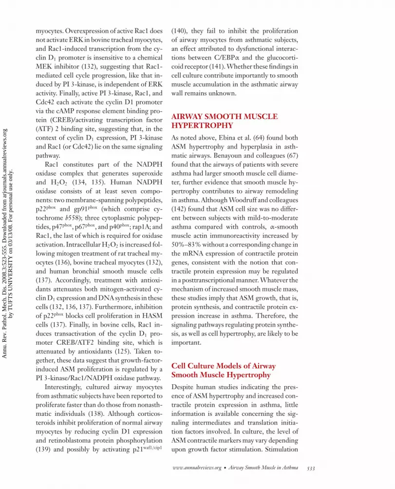
Airway remodeling may be defined as achange in airway structure arising from an in-flux of inflammatory cells and their mediators,which induces a cycle of airway wall damageand repair. The end result may be reconsti-tution of the airway wall, abnormal prolifera-tion and/or hypertrophy of airway cells, or ab-normal expression of extracellular matrix andmuscle proteins. Below we describe the struc-tural changes in the airways of patients withasthma (Figure 1), and follow with a discus-sion of potential functional significance.
Epithelium
Although the subject of this review is ASM,note that epithelial damage may provide theinitial stimulus for airway remodeling. Injuryto epithelial cell monolayers results in in-creased release of proliferative and fibrogenicgrowth factors, including fibroblast growthfactor, insulin-like growth factor, PDGF, andTGF-β (42, 43). In coculture models, epithe-lial injury induces myofibroblast proliferationand collagen gene expression (42). Togetherthese data suggest that epithelial injury mayinduce the release of soluble factors that, inturn, activate myofibroblasts in the underly-ing subepithelial layer, leading to subepithelialthickening.
Subepithelium
The true basement membrane of the airwayepithelium, or basal lamina, is composed pre-dominantly of collagen IV and laminin (44),
GG
MM
EBMBM
EpiEpi
EpiEpi
BMBM
M
M
E
G
G
MM
EBM
Epi
Epi
BM
M
M
E
G
Figure 1Typical histological appearance of central airway walls from an asthmatic(top panel ) and normal (bottom panel ) individual. In the asthmatic airway,there is epithelial (Epi) injury and thickening of the apparent subepithelialbasement membrane (BM), which actually includes both the true BM andsubepithelial fibrosis. In addition, excessive accumulation of airway smoothmuscle (M) is apparent, in addition to mucus gland (G) hypertrophy. Elastin(E) is found in both the asthmatic and normal airway wall. Originalmagnification is 100X. Images provided by Dr. Aliya N. Husain.
and it does not appear to be abnormal in asth-matic airways (45). However, it has long beenrecognized that in patients with asthma, thereis thickening of the lamina reticularis, whichin normal individuals is constituted by a loosearray of collagen fibrils that underlie the truebasement membrane. Increased deposition of
www.annualreviews.org • Airway Smooth Muscle in Asthma 527
Ann
u. R
ev. P
atho
l. M
ech.
Dis
. 200
8.3:
523-
555.
Dow
nloa
ded
from
arj
ourn
als.
annu
alre
view
s.or
gby
TU
FTS
UN
IVE
RSI
TY
on
03/1
3/08
. For
per
sona
l use
onl
y.

ANRV335-PM03-20 ARI 9 December 2007 21:8
Myofibroblast: afibroblast that hasdeveloped some ofthe characteristics ofsmooth muscle (e.g.,myofilaments)
Laminin: a family ofglycoproteins thatpromote celladhesion; an integralpart of the structureof the basementmembrane in almostevery animal tissue
collagens III, IV, V, and VII, as well as fi-bronectin, laminin chains α1, α2, and β2, andtenascin, has been found in asthmatic airways(44, 46, 47). On the basis of the protein com-position of the subepithelial deposits, whichdiffers from that of the true basement mem-brane, and the correlation between the depthof the subepithelial collagen layer and myofi-broblast number, Roche and colleagues con-cluded that the airway myofibroblast is re-sponsible for subepithelial thickening (44, 48).Vignola et al. (49) confirmed the correlation.In addition, antigen challenge of atopic asth-matics increases myofibroblast number (50),consistent with the notion that this cell playsa role in the asthmatic inflammatory and heal-ing responses.
The source of subepithelial myofibrob-lasts has until recently been assumed to beeither the deep subepithelial or ASM layer.However, recent data suggest that these cellsmay arise from the circulating fibrocytes,cells with the unique characteristic of ex-pressing hemopoietic stem cell antigen CD34.Schmidt and colleagues (51) showed the pres-ence of CD34-, collagen I–, and α-smoothmuscle-actin-positive cells in the subepithe-lium of patients with allergic asthma. Further-more, they demonstrated in a mouse model ofasthma that circulating fibrocytes are indeedrecruited into the bronchial tissue followingallergen exposure, and differentiate into my-ofibroblasts. Finally, activated mobile fibrob-lasts have been found in the bronchoalveolarlavage fluid of patients with mild asthma (52),consistent with the notion that circulating fi-brocytes contribute to subepithelial thicken-ing in patients with asthma.
It is well known that the airways of patientswith asthma are infiltrated with various typesof inflammatory cells including eosinophils,T lymphocytes, mast cells, and neutrophils(53). These cells are capable of elaboratinggrowth factors and cytokines, which increasethe synthesis of extracellular matrix proteinsby myofibroblasts. In asthmatic subjects, thedegree of subepithelial fibrosis correlates withthe amount of eosinophil-associated TGF-β
mRNA (54). TGF-β is increased in the air-ways of severe asthmatics with airway remod-eling, compared with patients with less-severedisease (54–57), suggesting that TGF-β,which stimulates fibroblasts to synthesize andsecrete proteins of the extracellular matrix,may play a key role in the remodeling process.
Airway Smooth Muscle
Substantial evidence demonstrates the coex-istence of airway hyperresponsiveness and ex-cess smooth muscle mass in patients with fa-tal asthma (58–63). However, these studiesdid not reliably measure the local mecha-nism of muscular thickening, that is, hyper-plasia versus hypertrophy. In 1993, Ebina andcolleagues (64) examined the airways of pa-tients with fatal asthma using a combinationof the dissector method and a serial sectioningtechnique. Two subgroups of asthmatic air-ways were found: one in which smooth musclemass was increased only in the central bronchi(Type I) and another in which smooth musclethickness was increased throughout the air-way tree (Type II). In Type I, smooth musclehyperplasia was responsible for central ASMthickening, whereas in Type II, cellular hyper-trophy was evident over the entire length ofthe airway. Thus, it appeared that both hyper-plasia and hypertrophy play a role. This reportis consistent with others suggesting the exis-tence of different asthma phenotypes (65).
Because of the obstacle of obtainingbronchial biopsies that include the full thick-ness of ASM, there are few data on the smoothmuscle hypertrophy/hyperplasia in patientswith nonfatal asthma. Carroll et al. (66) foundthat the ASM area of the larger membra-nous bronchioles was significantly greater inboth fatal and nonfatal cases of asthma, com-pared with control cases. They did not de-termine the mechanism of increased ASMmass, however. More recently, Benayoun et al.(67) found that the airways of patients withsevere asthma had larger smooth musclecell diameter than control subjects, patientswith mild asthma, or patients with chronic
528 Hershenson et al.
Ann
u. R
ev. P
atho
l. M
ech.
Dis
. 200
8.3:
523-
555.
Dow
nloa
ded
from
arj
ourn
als.
annu
alre
view
s.or
gby
TU
FTS
UN
IVE
RSI
TY
on
03/1
3/08
. For
per
sona
l use
onl
y.

ANRV335-PM03-20 ARI 9 December 2007 21:8
obstructive pulmonary disease. Expression ofmyosin light chain kinase was also increased.Furthermore, there was no evidence of ASMcell proliferation, as evidenced by the lack ofstaining for Ki67, a nuclear marker of cell cy-cle traversal. Together, these data suggest notonly that ASM mass is increased in patientswith severe but nonfatal asthma, but that ASMhypertrophy, rather than proliferation, may bethe cellular mechanism of increased smoothmass in these patients.
Most recently, Woodruff and colleagues(68) examined airway structure in patientswith mild-to-moderate asthma. They foundthat ASM cell number was nearly twofoldhigher in subjects with mild-to-moderateasthma, whereas there was no increase in cellsize between groups. These data suggest thatthe mechanism of increased smooth musclemass may vary according to severity. Also,while α-smooth muscle actin immunoreactiv-ity increased by 50%–83% in these patients,the mRNA expression of contractile proteingenes was not increased, consistent with thenotion that contractile protein expression maybe regulated in a posttranscriptional manner(see below).
The Woodruff study is important becauseit is the first to examine the molecular mecha-nisms by which contractile protein expressionis increased in asthma. Although the cellu-lar mechanism of ASM accumulation, hyper-trophy or hyperplasia, may vary in individualpatients, increased cell growth (i.e., size) andcontractile protein expression occur in bothcases. (Mammalian cells must undergo cell en-largement prior to mitosis.) In the case of hy-pertrophy, cells do not traverse the cell cy-cle, whereas in proliferation, mitosis occurs.Thus, the biochemical mechanisms underly-ing hypertrophy and hyperplasia may overlap.Therefore, mechanisms of ASM cell growthand contractile protein expression should bean active area of investigation.
Note that TGF-β, which is increased inthe airways of severe asthmatics with airwayremodeling compared with patients with aless-severe disease (54–57), may hold antimi-
togenic (69–71) as well as promitogenic (72)effects on ASM. We speculate that TGF-βmay promote ASM hypertrophy by increasingASM cell size and contractile protein expres-sion while simultaneously preventing S-phasetraversal.
Adventitia
In normal airways, the adventitia consistsof fibroblasts, connective tissue, and bloodvessels. Two groups (66, 73) have noted anincrease in the thickness of the airway wallexternal to the smooth muscle. Such thicken-ing may uncouple the airways from the lungparenchyma, reducing the static and dynamicstresses that oppose smooth muscle shorten-ing, thereby increasing airway narrowing.
Potential Functional Significance
Airway remodeling could exaggerate airwaynarrowing and increase airway responsivenessby (a) thickening of the airway wall inter-nal to the smooth muscle, thereby increas-ing the luminal obstruction generated by agiven degree of smooth muscle shortening;(b) increasing the amount of smooth muscle,thereby increasing shortening; and/or (c) re-ducing the load on the smooth muscle, ei-ther by increasing the compliance of the air-way wall or by reducing airway-parenchymalinterdependence. The possibility also exists,however, that airway remodeling representsa protective mechanism against excessive air-way narrowing.
First, let us address the functional sig-nificance of subepithelial fibrosis. Thicken-ing of the airway wall interior to the smoothmuscle might increase luminal narrowing forany given level of muscle shortening. How-ever, on the basis of a mathematical modelthat assesses the effects of airway wall thick-ening on airway resistance (74), subepithe-lial thickening is unlikely to increase maxi-mal airway resistance to the extent observedin patients with asthma. Subepithelial fibro-sis might also resist airway stretching during
www.annualreviews.org • Airway Smooth Muscle in Asthma 529
Ann
u. R
ev. P
atho
l. M
ech.
Dis
. 200
8.3:
523-
555.
Dow
nloa
ded
from
arj
ourn
als.
annu
alre
view
s.or
gby
TU
FTS
UN
IVE
RSI
TY
on
03/1
3/08
. For
per
sona
l use
onl
y.

ANRV335-PM03-20 ARI 9 December 2007 21:8
inspiration, increasing bronchoconstrictor re-sponsiveness (see below). Alternatively, it ispossible that subepithelial fibrosis might in-crease tensile stiffness and resistance to de-formation of the airway wall, opposing the ef-fect of smooth muscle contraction on airwaynarrowing.
To understand the functional significanceof subepithelial fibrosis, many investigatorshave correlated the magnitude of subepithe-lial thickening with the severity of airwaydisease. Such a correlation might provideevidence that subepithelial thickening playsa pathogenetic role in the development ormaintenance of airway narrowing. Mostbut not all of these studies have shown asignificant correlation between subepithelialthickness and the severity of disease, as mea-sured by a clinical scoring system, PC20 orFEV1 (44, 45, 75–77). At least two additionalstudies have shown that inhaled steroidtreatment reduces subepithelial thickness andtype III collagen deposition, concurrent withreduced airway inflammation and airway re-sponsiveness (78, 79)—further evidence thatsubepithelial thickness plays a pathogeneticrole in the development of airway obstructionin patients with asthma. However, morerecent data suggest that subepithelial fibrosislimits, rather than enhances, airway narrow-ing. First, a recent study examining airwaywall thickness by high-resolution computer-ized tomography showed, for the first time,that airway reactivity (an index of airway nar-rowing) was inversely correlated with airwaywall thickness, which is in part constitutedby subepithelial thickening (80). Second,Benayoun and colleagues (67), in a studyof severe asthmatics, showed that bronchialbiopsy subepithelial thickness was inverselycorrelated with asthma severity, again sug-gesting that this structural component ofremodeling limits airway narrowing.
A more convincing case may be madefor the hypothesis that abnormal ASMaccumulation increases airway narrowingand hyperresponsiveness. First, the afore-mentioned mathematical model predicted
that increased smooth muscle mass wouldlikely significantly increase maximal airwayresistance (74). Second, in the study by Be-nayoun and colleagues (67), asthma severitycorrelated with ASM mass, suggesting thatthis structural component of remodelingincreases airway responsiveness.
Two studies found no correlation betweenasthma duration and subepithelial thickness(44, 76). However, reports showing a corre-lation between asthma duration and the de-gree of irreversible airway obstruction, as as-sessed by postbronchodilator FEV1 (81, 82),led workers in the field to speculate thatairway remodeling is a cause of fixed air-way obstruction. Although this is a temptinghypothesis, no studies have specifically exam-ined airway structure in patients with irre-versible airway obstruction. The partial res-olution of subepithelial thickening after onlysix weeks of steroid treatment (78), combinedwith the presence of subepithelial thickeningin patients with mild asthma (44, 54, 75–77),suggests that subepithelial thickening is notconfined to patients with irreversible reduc-tions in airway function. No data are availableconcerning the possible role of the ASM layerin fixed airway obstruction.
ABNORMAL AIRWAY SMOOTHMUSCLE PROLIFERATION
Substantial evidence demonstrates the coex-istence of airway hyperresponsiveness and ex-cess smooth muscle mass in patients with fa-tal and nonfatal asthma (58–63, 66). Thereis much less information regarding the localmechanism of muscular thickening, that is,hyperplasia versus hypertrophy (64, 67, 68).However, as noted above, it is perhaps un-necessary and even misleading to focus onthis distinction, as both processes require in-creased cell growth (i.e., size) and contractileprotein expression. (Mammalian cells mustundergo cell enlargement prior to mitosis.)Indeed, it is useful to think of abnormal ASMaccumulation as a two-step process. First,the cell must be stimulated to increase size
530 Hershenson et al.
Ann
u. R
ev. P
atho
l. M
ech.
Dis
. 200
8.3:
523-
555.
Dow
nloa
ded
from
arj
ourn
als.
annu
alre
view
s.or
gby
TU
FTS
UN
IVE
RSI
TY
on
03/1
3/08
. For
per
sona
l use
onl
y.

ANRV335-PM03-20 ARI 9 December 2007 21:8
and contractile protein expression. Second,the cell must decide whether to traverse thecell cycle, leading to proliferation, or exit thecell cycle, leading to hypertrophy. Neverthe-less, because the preponderance of work in-vestigating the mechanisms of increased ASMmass has focused on proliferation, we coverthese topics separately.
Stimuli for Airway Smooth MuscleProliferation
Cultured ASM cells proliferate in responseto a vast number of stimuli, including pep-tide growth factors ligating receptor tyrosinekinases and bronchoconstrictor substancesassociated with G protein–coupled seven-transmembrane receptors. The former in-clude PDGF and EGF (83–86), whereas thelatter include histamine, thrombin, endothe-lin, and tryptase (87–95). Consistent with thenotion that peptide growth factors may induceabnormal ASM proliferation in asthma, EGFand PDGF have each been found in asth-matic airways (49, 96, 97). EGF receptor ex-pression is also increased in asthmatic airways(96). Increased levels of bronchoconstric-tor substances in asthma are well docu-mented. The response of cultured ASM cellsto growth factor activation may vary fromspecies to species. For example, histamineinduces HASM proliferation (87, 98) butdoes not induce proliferation in bovine cells(83). Nonetheless, the signaling pathways anddownstream transcription factor targets in-duced by growth factor stimulation may besurprisingly stable across species (see below).
Signaling Pathways RegulatingAirway Smooth Muscle Proliferation:The Extracellular Signal-RegulatedKinase Pathway
The mitogen-activated protein (MAP) kinasesare a superfamily consisting of three fami-lies of cytoplasmic serine/threonine kinasesthat participate in the transfer of growth-and differentiation-promoting signals to the
Eukaryotic species:single- ormultiple-celledorganisms thatcontain nuclei andorganelles
cell nucleus. They share a common activa-tion mechanism involving the phosphoryla-tion of tyrosine and threonine residues in aThr-X-Tyr motif in their activation loop. Onthe basis of the residue between the threonineand tyrosine, the MAP kinases can be dividedinto three families: ERKs (Thr-Glu-Tyr), Junamino terminal kinases (Thr-Pro-Tyr), andp38s (Thr-Gly-Tyr). Each MAP kinase is ac-tivated by successive activation of a MAP ki-nase kinase kinase and a MAP kinase kinase.Activation of ERK is required for DNA syn-thesis in bovine, rat, and HASM (92, 99–101),consistent with the fact that the MAP kinasecascades are highly conserved in eukaryoticspecies, including mammals, Drosophila, ne-matodes, and yeast (102–108).
The canonical path to ERK activationcomprises the growth factor receptor bindingprotein Grb2, the nucleotide exchange fac-tor Son of sevenless (Sos), the 21-kD GTPaseRas, the 74-kD cytosolic serine/threonine ki-nase Raf-1, and the 45-kD dual function ki-nase MAP kinase/ERK kinase (MEK) 1. Grb2is found in a stable complex with the nu-cleotide exchange factor Sos. Activation ofreceptor tyrosine kinases induces the forma-tion of phosphotyrosine residues, which serveas docking positions for downstream signaltransduction molecules containing Src ho-mology 2 domains, including Grb2 and PI3-kinase (see below). Docking of Grb2 tophosphotyrosine causes Sos to bind to andactivate Ras. Ras then escorts Raf-1 to thecell membrane, resulting in Raf-1 activation(109). Raf-1 phosphorylates MEK1 on Ser218
and Ser222 (110). MEK1 phosphorylates tyro-sine and threonine residues in the ERK acti-vation loop.
Several studies have demonstrated thatRas, Raf-1, and MEK are required for ERKactivation in airway myocytes. Microinjectionof the antipan Ras neutralizing antibody in-hibits DNA synthesis in HASM cells (111),and overexpression of a dominant-negativeform of H-Ras inhibits PDGF-induced ERKactivation in bovine cells (112). Overexpres-sion of a kinase-dead mutant of Raf-1 inhibits
www.annualreviews.org • Airway Smooth Muscle in Asthma 531
Ann
u. R
ev. P
atho
l. M
ech.
Dis
. 200
8.3:
523-
555.
Dow
nloa
ded
from
arj
ourn
als.
annu
alre
view
s.or
gby
TU
FTS
UN
IVE
RSI
TY
on
03/1
3/08
. For
per
sona
l use
onl
y.

ANRV335-PM03-20 ARI 9 December 2007 21:8
endothelin-mediated ERK activation in rattracheal myocytes (94). Inhibition of MEK1attenuates ERK activation and DNA synthe-sis in bovine, rat, and HASM cells (99–101,113).
Once activated, ERK translocates to thecell nucleus, where it regulates gene expres-sion by phosphorylating transcription factortargets. Although the precise downstream tar-gets of ERK in ASM cells are not known, ithas been shown in bovine ASM that, as inother cell types (106, 114, 115), ERK is an up-stream activator of transcription from the cy-clin D1 promoter (116). Finally, bronchoalve-olar lavage fluid from asthmatic airways wasshown to increase the ERK activation, cyclinD1 protein abundance, [3H]-thymidine incor-poration, and cell number of cultured HASMcells (117). Thus, the ERK pathway appearsto constitute an important regulator of entryinto the cell cycle and G1 progression in ASM.
Activation of the ERK pathway in NIH3T3 cells, although sufficient for cyclin D1 ex-pression, is inadequate for three additional G1
cell cycle events, namely, maximal phosphory-lation of the retinoblastoma protein, degrada-tion of the cyclin-dependent kinase inhibitorp27, and expression of cyclin A (118). More-over, Ras, but not ERK, is necessary forgrowth-factor-mediated breakdown of p27 inIIC9 fibroblasts (119). Lastly, overexpressionof cyclin D1 is insufficient for DNA synthesis(120, 121). Together these reports imply thatthe ERK/cyclin D1 pathway is insufficient forS-phase traversal and that other pathways arerequired for proliferation. It is in this contextthat we now address the PI 3-kinase pathway.
Signaling Pathways RegulatingAirway Smooth Muscle Proliferation:Phosphatidylinositol 3-KinasePathway
As noted above, receptor tyrosine kinases pos-sess tyrosine domains that, upon phosphory-lation, may interact with PI 3-kinase, a fam-ily of heterodimeric lipid kinases. Membersof the PI 3-kinase family are divided into
three classes according to their structure andin vitro substrate specificity, as reviewed inReference 122. Class 1A PI 3-kinases α, β,and γ are tightly bound with a regulatorysubunit (p85α, p85β, or p55γ) that containsSrc homology 2 domains with an affinity forphosphotyrosine residues in pYxxM motifsfound in growth factor receptors, their sub-strates, and adaptor proteins. The major sub-strate for the class I PI 3-kinases in vivoappears to be PI(4,5)P2, and the major prod-uct PI(3,4,5)P3 (123). When activated, classIA PI 3-kinases regulate cellular functions byrecruiting PI(3,4,5)P3-binding proteins to theplasma membrane. Most of these proteins,the prototype of which is the serine threo-nine kinase Akt, bind to 3,4-phosphorylatedPIs through a pleckstrin homology (PH) do-main. Guanine nucleotide exchange factors,the upstream activators of GTPases, also con-tain a PH domain (124).
The activation of PI 3-kinase and its re-quirement for ASM proliferation have beenwell studied. Peptide growth factors activatePI 3-kinase in human (69) and bovine ASMcells (125). Chemical inhibitors of PI 3-kinaseinhibit ASM transcription from the cyclinD1 promoter, as well as cyclin D1 proteinexpression (125) and DNA synthesis (125–127). Constitutive activation of PI 3-kinasein bovine tracheal myocytes is sufficient forcyclin D1 promoter activity but does not in-duce ERK activation (125), implying that PI3-kinase signaling occurs independently ofERK. Likewise, in HASM cells, inhibitors ofPI 3-kinase had no effect on ERK activation(127).
An important downstream target of PI 3-kinase appears to be the 21-kD Rho GTPaseRac1. The Rho family GTPases (Rho A–C,Rac1 and 2, and Cdc42) influence cell cycleprogression via their regulation of the actincytoskeleton and via interactions with mul-tiple target proteins. Abundant evidence ex-ists that the Rac1 signaling pathway is reg-ulated by PI 3-kinase (128–131). Rac1 (132)and Cdc42, but not RhoA (133), are requiredfor cyclin D1 expression in bovine tracheal
532 Hershenson et al.
Ann
u. R
ev. P
atho
l. M
ech.
Dis
. 200
8.3:
523-
555.
Dow
nloa
ded
from
arj
ourn
als.
annu
alre
view
s.or
gby
TU
FTS
UN
IVE
RSI
TY
on
03/1
3/08
. For
per
sona
l use
onl
y.
ANRV335-PM03-20 ARI 9 December 2007 21:8
myocytes. Overexpression of active Rac1 doesnot activate ERK in bovine tracheal myocytes,and Rac1-induced transcription from the cy-clin D1 promoter is insensitive to a chemicalMEK inhibitor (132), suggesting that Rac1-mediated cell cycle progression, like that in-duced by PI 3-kinase, is independent of ERKactivity. Finally, active PI 3-kinase, Rac1, andCdc42 each activate the cyclin D1 promotervia the cAMP response element binding pro-tein (CREB)/activating transcription factor(ATF) 2 binding site, suggesting that, in thecontext of cyclin D1 expression, PI 3-kinaseand Rac1 (or Cdc42) lie on the same signalingpathway.
Rac1 constitutes part of the NADPHoxidase complex that generates superoxideand H2O2 (134, 135). Human NADPHoxidase consists of at least seven compo-nents: two membrane-spanning polypeptides,p22phox and gp91phox (which comprise cy-tochrome b558); three cytoplasmic polypep-tides, p47phox, p67phox, and p40phox; rap1A; andRac1, the last of which is required for oxidaseactivation. Intracellular H2O2 is increased fol-lowing mitogen treatment of rat tracheal my-ocytes (136), bovine tracheal myocytes (132),and human bronchial smooth muscle cells(137). Accordingly, treatment with antioxi-dants attenuates both mitogen-activated cy-clin D1 expression and DNA synthesis in thesecells (132, 136, 137). Furthermore, inhibitionof p22phox blocks cell proliferation in HASMcells (137). Finally, in bovine cells, Rac1 in-duces transactivation of the cyclin D1 pro-moter CREB/ATF2 binding site, which isattenuated by antioxidants (125). Taken to-gether, these data suggest that growth-factor-induced ASM proliferation is regulated by aPI 3-kinase/Rac1/NADPH oxidase pathway.
Interestingly, cultured airway myocytesfrom asthmatic subjects have been reported toproliferate faster than do those from nonasth-matic individuals (138). Although corticos-teroids inhibit proliferation of normal airwaymyocytes by reducing cyclin D1 expressionand retinoblastoma protein phosphorylation(139) and possibly by activating p21waf1/cip1
(140), they fail to inhibit the proliferationof airway myocytes from asthmatic subjects,an effect attributed to dysfunctional interac-tions between C/EBPα and the glucocorti-coid receptor (141). Whether these findings incell culture contribute importantly to smoothmuscle accumulation in the asthmatic airwaywall remains unknown.
AIRWAY SMOOTH MUSCLEHYPERTROPHY
As noted above, Ebina et al. (64) found bothASM hypertrophy and hyperplasia in asth-matic airways. Benayoun and colleagues (67)found that the airways of patients with severeasthma had larger smooth muscle cell diame-ter, further evidence that smooth muscle hy-pertrophy contributes to airway remodelingin asthma. Although Woodruff and colleagues(142) found that ASM cell size was no differ-ent between subjects with mild-to-moderateasthma compared with controls, α-smoothmuscle actin immunoreactivity increased by50%–83% without a corresponding change inthe mRNA expression of contractile proteingenes, consistent with the notion that con-tractile protein expression may be regulatedin a posttranscriptional manner. Whatever themechanism of increased smooth muscle mass,these studies imply that ASM growth, that is,protein synthesis, and contractile protein ex-pression increase in asthma. Therefore, thesignaling pathways regulating protein synthe-sis, as well as cell hypertrophy, are likely to beimportant.
Cell Culture Models of AirwaySmooth Muscle Hypertrophy
Despite human studies indicating the pres-ence of ASM hypertrophy and increased con-tractile protein expression in asthma, littleinformation is available concerning the sig-naling intermediates and translation initia-tion factors involved. In culture, the level ofASM contractile markers may vary dependingupon growth factor stimulation. Stimulation
www.annualreviews.org • Airway Smooth Muscle in Asthma 533
Ann
u. R
ev. P
atho
l. M
ech.
Dis
. 200
8.3:
523-
555.
Dow
nloa
ded
from
arj
ourn
als.
annu
alre
view
s.or
gby
TU
FTS
UN
IVE
RSI
TY
on
03/1
3/08
. For
per
sona
l use
onl
y.

ANRV335-PM03-20 ARI 9 December 2007 21:8
of postconfluent cultures with either serum orgrowth factors has been shown to markedlyrepress the expression of contractile proteinssuch as α-smooth muscle actin and MLCK(143–146). In rat aortic smooth muscle cells,PDGF reduces α-actin expression by acti-vation of the PI 3-kinase pathway, which inturn localizes serum response factor (SRF) tothe cytoplasm, inhibiting binding to specificα-smooth muscle actin promoter elements(147).
The loss of ASM contractile protein ex-pression can be reversed when cells are cul-tured in a serum-free medium. Long-term(10–12 days) serum deprivation of confluentcanine tracheal myocytes induces the forma-tion of large myocytes and increases contrac-tile protein expression (148–150). However,serum deprivation paradoxically reduces tran-scription of smooth-muscle-encoding genesby redistributing SRF out of the nucleus (150).Also, transcriptional activity of the smooth-muscle-specific SM22 and smooth musclemyosin heavy chain (smMHC) promoters fallsby over 80% in seven-day serum-deprivedcultures, and there is no apparent differencein SM22 promoter activity between cells inthese cultures expressing SM22 protein andthose that do not. Accordingly, SM22 andsmMHC mRNA levels peak near confluencein serum-fed conditions, whereas the abun-dance of the proteins they encode is less than15% of the highest levels reached in serum-free culture (148). Taken together, these datasuggest that accumulation of smooth musclecontractile proteins is likely to be regulated ina posttranscriptional manner.
In a second model of ASM hypertrophy,clonal cell lines of human bronchial smoothmuscle origin were generated by retrovi-ral transduction with a temperature-sensitiveSimian virus 40 large tumor antigen (151).Simian virus 40 large tumor binds and in-activates the tumor suppressor protein p53.Thus, at the permissive temperature of 33◦C,cells demonstrate increased proliferative po-tential. However, a shift to the nonpermissivetemperature of 39◦C, with subsequent degra-
dation of large tumor and release of p53, in-duces expression of the cyclin-dependent ki-nase inhibitors p21Cip/Waf and p57Kip2, leadingto cell cycle arrest in mid-G1 of the cell cycle.Temperature shift also increases cell size andprotein expression of α-smooth muscle actinand of MLCK and SM22, paralleling changesobserved in the airways of patients with se-vere asthma (67). Because serum-induced cellgrowth may continue even when the cell cycleis blocked (152), ASM hypertrophy in this sys-tem likely results from continued cell growthin the absence of cell division and prolifera-tion. Although contractile protein abundanceis increased in this model, α-smooth muscleactin and MLCK mRNA are not elevated,suggesting that changes in contractile pro-tein expression, as in the first model, are reg-ulated in a posttranscriptional manner andlikely under translational control. These find-ings are also consistent with Woodruff’s re-sults (142) demonstrating increased α-smoothmuscle actin protein expression in mild asth-matics without an apparent change in mRNAexpression. The observed posttranscriptionalcontrol of ASM contractile protein expressionis also consistent with earlier findings in car-diac (153–155), skeletal (156–159), and vascu-lar hypertrophy (160). Finally, it has recentlybeen shown that TGF-β induces hypertrophyof cultured HASMs via both transcriptionaland posttranscriptional mechanisms (161). Inthis regard, TGF-β enhances the accumu-lation of smooth muscle proteins with con-comitant disruption of the nuclear associa-tion of SRF with its transcriptional inhibitorSmad7, a negative modulator of TGF-βsignaling (161a).
Regulation of Translational Initiation
Regulation of protein synthesis is achievedprimarily by the phosphorylation of variouseukaryotic translation initiation factors (eIFs).Within the overall process of mRNA trans-lation (which includes initiation, elongation,and termination), control seems to be exertedmainly at the initiation stage. There are two
534 Hershenson et al.
Ann
u. R
ev. P
atho
l. M
ech.
Dis
. 200
8.3:
523-
555.
Dow
nloa
ded
from
arj
ourn
als.
annu
alre
view
s.or
gby
TU
FTS
UN
IVE
RSI
TY
on
03/1
3/08
. For
per
sona
l use
onl
y.

ANRV335-PM03-20 ARI 9 December 2007 21:8
highly regulated steps in the translation initia-tion pathway: (a) recognition of the cap struc-ture at the 5′ end of the mRNA by the eIF4group of factors (collectively termed eIF4F),and (b) the binding of initiator methionyltRNA to the 40S ribosomal subunit to formthe 43S pre-initiation complex, mediated byGTP-bound eIF2.
The mRNA is prepared for ribosome bind-ing by the eIF4 group of initiation factors.Translation of the majority of eukaryoticmRNAs is initiated through a 7-methyl-guanosine cap structure at the 5′ end ofmRNA. The cap is recognized and clampedby the 24-kD eIF4E. The scaffolding pro-tein eIF4G binds to and stabilizes eIF4E andpoly-A binding protein, which in turn asso-ciates with the poly-A tail at the 3′ end ofthe mRNA. eIF4G also binds with eIF4A,an RNA helicase that serves to unwind sec-ondary mRNA structure. In addition to in-teracting with eIF4G, eIF4E associates withinhibitory proteins termed 4E-binding pro-teins (4E-BPs). 4E-BP1 undergoes phospho-rylation at multiple sites, which results in itsrelease from eIF4E, thereby increasing theavailability of eIF4E for binding to eIF4G,eIF4F complex formation, and cap-dependenttranslation. Typically, 4E-BP is phosphory-lated by mammalian target of rapamycin(mTOR) (162, 163), although other 4E-BPkinases exist. eIF4E itself is phosphorylatedat Ser209 by MAP-kinase-signal-integratingkinase (MNK) 1 and MNK2. MNK1 isactivated upon treatment with agents that ac-tivate the ERK and p38 MAP kinases includ-ing growth factors and anisomycin, whereasMNK2 has high basal activity (164, 165).
Note that whereas eIF2 activation reg-ulates overall translational activity (below),eIF4E and its partners regulate the trans-lation of specific mRNAs. Regions of hair-pin loops and other stable intermolec-ular secondary structures within the 5′
untranslated region impede the initiationof protein synthesis. Translation of mRNAswith regions of stable secondary structurewithin the 5′ untranslated region is particu-
larly dependent upon eIF4F assembly (166,167).
In contrast to the above cap-dependentmechanisms of translational control, twoother mechanisms of translational controlexist. Concurrent with the preparation ofmRNA, the pre-initiation complex must beformed. eIF2, a multimer consisting of α, β,and γ subunits, functions to recruit methionyltRNA and conduct it as a tRNA-eIF2-GTPternary complex to the 40S ribosomal subunit,to form the 43S pre-initiation complex. eIF2GTP loading is determined by the activityof eIF2B, a guanine nucleotide exchange fac-tor. eIF2Bε phosphorylation by glycogen syn-thase kinase (GSK)-3β inhibits its GDP/GTPexchange activity, thereby limiting binding ofmethionyl tRNA to the 40S ribosomal sub-unit. However, phosphorylation of GSK-3β
by the serine/threonine kinase Akt inacti-vates it, leading to eIF2B dephosphorylationand activation, and a general enhancement oftranslation initiation independent of the 7-methylguanosine mRNA cap (168).
Finally, the translation of mRNAs with 5′
terminal oligopyrimidine (TOP) tracts, manyof which encode elongation factors and ri-bosomal proteins involved in mRNA transla-tion, is upregulated by phosphorylation of theS6 ribosomal protein. S6 ribosomal protein isphosphorylated by p70 ribosomal S6 kinase.In contrast to the above pathways that regu-late the efficiency of translation, this pathwayregulates the translational capacity by increas-ing the synthesis of ribosomes.
Signaling molecules controlling cell sizewere first identified in Drosophila. Loss or inhi-bition of PI 3-kinase (169), Akt (170), mTOR(171), or S6 kinase (172) each decreased cellsize. These results have been confirmed inmammalian systems.
Characteristics of the PI 3-kinase fam-ily have been described above. To reiter-ate, class IA PI 3-kinases recruit PI(3,4,5)P3-binding proteins to the plasma membrane viatheir PH domains (124), including Akt andphosphoinositide-dependent protein kinase-1 (PDK-1). The three Akt genes encode a
www.annualreviews.org • Airway Smooth Muscle in Asthma 535
Ann
u. R
ev. P
atho
l. M
ech.
Dis
. 200
8.3:
523-
555.
Dow
nloa
ded
from
arj
ourn
als.
annu
alre
view
s.or
gby
TU
FTS
UN
IVE
RSI
TY
on
03/1
3/08
. For
per
sona
l use
onl
y.

ANRV335-PM03-20 ARI 9 December 2007 21:8
57-kD serine/threonine kinase involved incell survival, proliferation, and growth. Aktactivation requires membrane localizationand phosphorylation of Thr308 and Ser473.Thr308 is phosphorylated by PDK-1 (173).Until recently, the precise Ser473 kinase(PDK-2) was unknown (see below). Targetsof Akt relevant to translational control in-clude mTOR and GSK-3β. mTOR is a 290-kD protein similar in structure to phospho-inositide kinases. Rapamycin, when boundto FK506-binding protein, inhibits the activ-ity of mTOR. mTOR phosphorylates 4E-BP(163), releasing it from eIF4E. mTOR ac-tivation thereby increases the availability ofeIF4E for eIF4F complex formation and cap-dependent translation. mTOR also phospho-rylates S6 kinase, a mitogen- and amino acid–sensitive serine/threonine kinase. As notedabove, S6 phosphorylation increases transla-tion of mRNAs with 5′ TOP tracts, many ofwhich are involved in mRNA-translation-likeelongation factors and ribosomal proteins.Thus, mTOR may regulate cap-dependentand cap-independent pathways involved inmRNA translation.
As phosphorylation of S6 kinase is sen-sitive to inhibition by both rapamycin andchemical inhibitors of PI 3-kinase, researchersoften place PI 3-kinase, mTOR, and S6 ki-nase pathways in a linear signaling pathway.Such a linear scheme is too simplistic, how-ever, as a rapamycin-resistant mutant of S6kinase is still sensitive to inhibition by the PI3-kinase inhibitor wortmannin (174). Thus,mTOR and PI 3-kinase signals to S6 kinasecan be dissociated. S6 kinase may also bephosphorylated by PDK-1 (175, 176), pro-viding a mechanism for mTOR-independent,PI 3-kinase-dependent activation. Similarly,mTOR-independent mechanisms of 4E-BPphosphorylation may exist. It was recentlyshown that class 1A PI 3-kinases may functionas 4E-BP1 kinases (177). Also, ERK phospho-rylates 4E-BP1 (178).
Recent studies have elucidated the roleof two signaling molecules linking Akt andmTOR in the regulation of cell size. Data
from Drosophila first showed that tuberoussclerosis complex (TSC) 1 (also referredto as hamartin) and TSC2 (tuberin) neg-atively regulate cell size (179–181). Laterstudies in mammalian cells established thatTSC2 is phosphorylated and inhibited byAkt and suppresses mTOR signaling (182–185). In LAMD-SM pulmonary lymphan-gioleiomyomatosis-derived cells, TSC2 alsoinhibits S6 kinase (186). In HEK293 cells,TSC2 functions as a GTPase activatingprotein to inactivate Rheb (187, 188), aGTPase previously shown to regulate cellsize in yeast (189). Rheb is required for Ser2448
phosphorylation and activation of mTOR.Rheb and mTOR do not associate, suggestingthat Rheb activates mTOR indirectly (188).Thus, PI 3-kinase may positively regulatecell size via the successive activation of Akt,inactivation of TSC2, activation of Rheb, andactivation of mTOR.
More recently, the mTOR story has be-come even more intricate: It was discoveredthat mTOR exists in two distinct multiproteincomplexes, one rapamycin-sensitive (mTORcomplex 1) and one rapamycin-insensitive(mTOR complex 2) (190). mTOR complex1 includes mTOR and Raptor; mTOR com-plex 2 comprises mTOR, Rictor, and mam-malian stress-activated protein kinase inter-acting protein. Furthermore, mTOR complex2 appears to be identical to the proposed Aktkinase, PDK-2, which phosphorylates serine473 on Akt (191, 192). Thus, Akt acts as anupstream activator of mTOR for TOR com-plex 1, and Akt is also a target of mTOR viaTOR complex 2. TOR complex 2 may reg-ulate Akt specificity and permit high-level PI3-kinase/Akt signaling (193).
GSK-3β is a constitutively active ser-ine/threonine kinase that phosphorylatesmultiple substrates including eIF2Bε, cyclinD1, and p21 (168, 194–196). Phosphorylationby Akt inactivates GSK-3β, leading to eIF2Bdephosphorylation and activation, as well asa general enhancement of 43S pre-initiationcomplex formation (168). The phosphoryla-tion of GSK-3β by the PI 3-kinase effector
536 Hershenson et al.
Ann
u. R
ev. P
atho
l. M
ech.
Dis
. 200
8.3:
523-
555.
Dow
nloa
ded
from
arj
ourn
als.
annu
alre
view
s.or
gby
TU
FTS
UN
IVE
RSI
TY
on
03/1
3/08
. For
per
sona
l use
onl
y.

ANRV335-PM03-20 ARI 9 December 2007 21:8
Akt
TSC2
Rheb
mTOR
4E-BP/eIF4E
eIF4E
eIF4AeIF4GPABP
Enhanced5'-cap-dependentmRNA translation
initiation
PDK-1
p70 S6 kinase
S6
Enhanced5'-TOP-containingmRNA translation
initiation
GSK-3β
eIF2B eIF2B-P
Enhancedgeneral
mRNA translationinitiation
eIF4F
TORC1
TORC2PDK-2
Figure 2A central role for Akt in the regulation of translation initiation. Akt regulates the initiation of mRNAtranslation in three ways, as described in the text. TOP, terminal oligopyrimidine; PDK-1,phosphoinositide-dependent protein kinase-1; TSC, tuberous sclerosis complex; GSK, glycogensynthase kinase; eIF, eukaryotic translation initiation factor; mTOR, mammalian target of rapamycin;4E-BP, 4E-binding protein.
Akt indicates that PI 3-kinase may regulatemRNA translation via three distinct mech-anisms, as shown in Figure 2: (a) regula-tion of cap-dependent mRNAs via activationof the Akt/TSC2/Rheb/mTOR/4E-BP path-way, (b) regulation of 5′ TOP tract-containingmRNAs via activation of S6 kinase (througheither mTOR or PDK-1), and (c) a gen-eral enhancement of translation initiationvia activation of the Akt/GSK-3β/eIF2Bpathway.
Despite human studies indicating thepresence of ASM hypertrophy and increasedcontractile protein expression in asthma, littleinformation is available concerning the sig-naling intermediates and translation initiationfactors involved. In confluent serum-deprivedcanine tracheal myocyte cultures, PI 3-kinaseand S6 kinase activities are increased fiveand two days after serum deprivation, re-spectively, and immunohistochemical studiesshow selective phosphorylation of Akt and S6kinase in elongated cells expressing smMHCfive to seven days after serum deprivation(148). LY294002 and rapamycin blocked
S6 kinase phosphorylation and phenotypicchange, implying that PI 3-kinase, mTOR,and S6 kinase are responsible for ASM hyper-trophy and contractile protein accumulation.However, the precise requirement of S6 ki-nase for ASM hypertrophy was not assessed.More recently, it was shown in culturedHASM cells that serum-deprivation-inducedand TGF-β-induced hypertrophy are eachdependent upon 4E-BP phosphorylation andeIF4E release (161, 197).
Potential Role of ADAM33
ADAM (a disintegrin and metalloproteinase) 33was the first asthma gene identified by po-sitional cloning (198). An association be-tween ADAM33 and asthma has been veri-fied in several (199) but not all asthma cohorts(200, 201). ADAM33 is highly expressed inbronchial and vascular smooth muscle (198,202). Members of the ADAM family of met-alloproteinases have the capacity to release cy-tokines and growth factors from their pre-cursors, suggesting that ADAM33 may be
www.annualreviews.org • Airway Smooth Muscle in Asthma 537
Ann
u. R
ev. P
atho
l. M
ech.
Dis
. 200
8.3:
523-
555.
Dow
nloa
ded
from
arj
ourn
als.
annu
alre
view
s.or
gby
TU
FTS
UN
IVE
RSI
TY
on
03/1
3/08
. For
per
sona
l use
onl
y.

ANRV335-PM03-20 ARI 9 December 2007 21:8
involved in the remodeling process. More re-cently, however, it has been shown that humanairway fibroblasts contain a large number ofalternatively spliced forms of ADAM33, andthat more than 95% of the transcripts do notencode a functional metalloproteinase (203).However, a synthetic ADAM12-S minigenehas been shown to induce myogenesis in em-bryonal rhabdomyosarcoma cells both in cul-ture and in nude mouse tumors (204). It istherefore conceivable that ADAM33 plays arole in the abnormal accumulation of ASMobserved in asthma. The precise contributionof ADAM33 to ASM remodeling, if any, re-mains to be established.
CONTRACTILE DYSFUNCTIONOF AIRWAY SMOOTH MUSCLEIN ASTHMA
As noted above, bronchoconstrictor hyperre-sponsiveness is a cardinal pathophysiologicalfeature of asthma, and bronchoconstrictor re-sponsiveness testing is often sought to provideadditional evidence for or against a potentialdiagnosis that remains uncertain after theevaluation of history, physical examination,and routine pulmonary function evaluation.Testing is most often performed by having thepatient inhale aerosols that contain increasingconcentrations of a bronchoconstrictor agent(usually methacholine or histamine) andmeasuring the severity of airflow obstructionthat ensues. Normal individuals developbronchoconstriction only in response torelatively large doses of bronchoconstrictoragents, and even then modest maximal airflowobstruction is seen. In contrast, asthmaticpeople exhibit airway narrowing at even smalldoses of constrictor agonist, and quite severeairflow obstruction can develop as the dose ofconstrictor is increased. The acute airflow ob-struction that develops during such bronchialprovocation testing is clearly dominated bysmooth muscle contraction, as evidenced byits rapid onset and its rapid reversal withinhaled β2-adrenergic agonists. Further-more, these features of bronchoconstriction
intentionally elicited in the pulmonary func-tion laboratory often mimic those of airflowobstruction that occur during naturallytriggered asthma attacks. Together, thesephenomena suggested that bronchoconstric-tor hyperresponsiveness in asthma must stemfrom some disorder of contractile activationin ASM that imparts an increased sensitivityand greater maximal force generation thatparallel the abnormalities in airflow obstruc-tion versus bronchoconstrictor dose-responsecurves generated by asthmatics during bron-choprovocation testing. It was thereforesomewhat surprising that the bulk of evi-dence appears to speak against this possibility.
Force Generation by AsthmaticAirway Smooth Muscle
Death from asthma is relatively rare (e.g.,there are less than 6000 deaths per year fromasthma in the United States), and so ASMfrom asthmatics (from autopsy specimens,donor lungs that could not be transplanted,or lung resections for clinical indication) hasrarely been available for in vitro physiolog-ical evaluation.1 Not surprisingly, therefore,the entire body of research studying asth-matic ASM strips is relatively small. In mostof these studies, ASM strips from normal orasthmatic lungs were dissected free of epithe-lium and most connective tissues, then sus-pended in organ baths and isometric (with-out shortening) force generation by similarsized strips determined in response to in-creasing concentrations of constrictor ago-nists. Although a few studies demonstrate en-hanced constrictor responses to commonlyused provocative agents (e.g., 205) and thereare some agonists to which asthmatic mus-cle strips respond uniquely, such as adeno-sine (206), the most common finding is thatmuscle strips of equivalent cross section from
1Recently, endobronchial biopsy specimens obtained fromasthmatic volunteers have provided very small quantitiesof airway smooth muscle, from which individual cells havebeen isolated and studied (see below).
538 Hershenson et al.
Ann
u. R
ev. P
atho
l. M
ech.
Dis
. 200
8.3:
523-
555.
Dow
nloa
ded
from
arj
ourn
als.
annu
alre
view
s.or
gby
TU
FTS
UN
IVE
RSI
TY
on
03/1
3/08
. For
per
sona
l use
onl
y.

ANRV335-PM03-20 ARI 9 December 2007 21:8
normal or asthmatic subjects exhibit similardose-response relationships to methacholineor histamine (e.g., 207, 208). Granted, asnoted above, the increased mass of musclein asthmatics’ airways might intuitively beexpected, and has been quantitatively pre-dicted, to enhance force generation and am-plify airway narrowing (74), and it remainsconceivable that hypertrophy and hyperplasialeading to increased airway muscle mass rep-resent key mechanisms of airway constrictorhyperresponsiveness. Curiously, to date therehas been no animal model that directly teststhis possibility, although correlative stud-ies have certainly supported this possibility(209–213).
That excised ASM from asthmatics maynot develop increased isometric force is alsointriguing, in light of in vitro studies inwhich exposure to a number of cytokinesfound in asthmatic airways, or to serumfrom an asthmatic subject, was found to aug-ment isometric constrictor responses (24, 214,215). These effects appear to stem in partfrom augmented calcium responses: TNF-α, IL-1β, and IL-13 all increase ASM ex-pression of CD38 (216, 217), a cell sur-face protein that catalyzes the synthesisof cyclic ADP-ribose, which increases cal-cium release from intracellular stores throughmultiple mechanisms in myocytes stimu-lated with G protein–coupled receptor ago-nists. Another mechanism by which cytokinesmight potentially increase force generationis through activation of the RhoA/Rho ki-nase pathway (218). Rho kinase is knownto potentiate smooth muscle contractile re-sponses by phosphorylating and inhibitingthe myosin-binding subunit of myosin phos-phatase, which ordinarily dephosphorylatesthe regulatory 20-kD myosin regulatory lightchain 20, whose phosphorylation (chieflyby the calcium/calmodulin-activated myosinlight chain kinase) activates contraction. Thegreater myosin light chain 20 phosphoryla-tion that attends myosin phosphatase inhi-bition appears as calcium sensitization, andthis pathway has been implicated in animal
models of allergen-induced bronchoconstric-tor hyperresponsiveness (219–221). Perhapsremoval from the asthmatic airway milieurestores apparently normal force-generatingcapacity to asthmatic airway muscle. Un-fortunately, it has not been possible to as-sess force generation by asthmatic ASM invivo, within the intact airway. However, in-dividual smooth muscle cells isolated fromendobronchial biopsies of asthmatics’ air-ways have revealed increased velocity and ex-tent of shortening compared with myocytesfrom normal airways (222). Conceivably,this aspect of contractile dysfunction mightcontribute to asthmatic airway constrictorhyperresponsiveness (223).
Impaired Relaxation of AsthmaticSmooth Muscle
Another salient pathophysiological feature ofasthmatic ASM is its impaired relaxation in re-sponse to β2-adrenergic agonists, as demon-strated both in vivo (224) and in vitro (205).β2-adrenergic agonists bind to the cell surfaceβ2-adrenergic receptor (β2AR), which cou-ples to the stimulatory Gs G protein. Upon re-ceptor stimulation, the Gαs subunit promotesrelaxation of contracted myocytes by directlyactivating K+ channels and also by stimulatingadenylate cyclase to generate cAMP, which inturn activates PKA, which in turn effects re-laxation through multiple mechanisms. Thebronchodilator effects of β2-adrenergic ago-nists are limited in part through β2AR phos-phorylation by PKA and by specific G proteinreceptor kinases, which result in the bindingof arrestins, uncoupling of G protein bindingto the receptor, and termination of β2AR sig-naling. Phosphorylated receptors are also in-ternalized and degraded, and in the long termβ2-agonist exposure also reduces β2AR by re-ducing β2AR mRNA stability and abundance.Common haplotypes in the β2AR gene leadsto variant β2AR proteins that have differingpropensities for receptor desensitization, butthe representation of these variants is simi-lar among asthmatic and normal individuals,
www.annualreviews.org • Airway Smooth Muscle in Asthma 539
Ann
u. R
ev. P
atho
l. M
ech.
Dis
. 200
8.3:
523-
555.
Dow
nloa
ded
from
arj
ourn
als.
annu
alre
view
s.or
gby
TU
FTS
UN
IVE
RSI
TY
on
03/1
3/08
. For
per
sona
l use
onl
y.

ANRV335-PM03-20 ARI 9 December 2007 21:8
and so it is unlikely that genetics explainsthe broadly impaired relaxation of asthmaticASM.
Instead, cytokine exposure probably ac-counts for this phenomenon. Shore and col-leagues have convincingly shown that IL-1β
acts through an ERK- and p38-dependentmechanism to stimulate COX-2 expressionand consequent prostaglandin E2 releasefrom cultured HASM (224a–224d). Theprostaglandin E2 then acts in an autocrinefashion to stimulate Gs-coupled EP2 recep-tors, resulting in cAMP elevation, PKA ac-tivation, and heterologous desensitization ofβ2AR. In other species, IL-1β and/or TNF-α can also antagonize β-adrenergic relaxationby enhancing Gi-coupled receptor signaling,thereby promoting Gαi-mediated inhibitionof adenylate cyclase and reduced cAMP for-mation (224e–224g). Shore also demonstratedthat IL-13, but not IL-4, also induces β2ARdesensitization through an ERK-dependentmechanism (224h). The IL-13 effect dependsstrongly upon the haplotype at amino acids478 and 551 of IL-4Rα (one of two chainsthat comprise the signaling IL-13 receptor)(224i). Although genetic variation at this lo-cus is linked to asthma and atopy (225), itis uncertain if modulation of IL-13-mediatedβ2AR desensitization represents the molecu-lar mechanism.
Force-Fluctuation-InducedRelengthening
Another salient physiological difference be-tween normal and asthmatic individuals is thatdeep breathing results in greater protectionagainst bronchoconstriction and more long-lasting and perhaps more effective reversal ofinduced bronchoconstriction in normals thanin asthmatics. The effects of deep breathingare thought to stem from the stretching im-posed on ASM during the deep breath. As thelung parenchyma inflates during inhalation,it pulls radially outward on the airway adven-tia (to which it is attached), thereby impos-ing a circumferential stress within the airway
wall. During tidal breathing, each circum-ferentially oriented airway myocyte shouldexperience periodic force fluctuations thatwax and wane with each breath. Fredbergand colleagues (226) established that applica-tion of such force fluctuations to contractedASM result in its relengthening, even in thepresence of continued contractile stimulation.Critically, the extent of force-fluctuation-induced relengthening is physiologically reg-ulated, for manipulation of p38 or ERK1/2signaling modulates this phenomenon (M.L.Dowell, O.J. Lakser, T.L. Lavoie, N.O. Dulin,M.B. Hershenson, et al., unpublished obser-vations). Recent results from our own labora-tory implicate actin filament length as anotherkey determinant of force-fluctuation-inducedrelengthening (227). Certainly, it is easy toimagine that the inflammatory environmentof the asthmatic airway wall could modulatep38 or ERK1/2 signaling, or stimulate al-terations in actin filament length (which issubject to complex regulation, as reviewed inReference 228). However, whether abnormal-ities in force-fluctuation-induced relengthen-ing contribute importantly to the pathogene-sis of asthma remains to be determined.
SUMMARY
In this review, we see that ASM is far fromnormal in asthma, and that its likely contri-butions to asthma pathogenesis range frommodulation of immune function and inflam-mation to participation in the most dominantaspects of airway remodeling, to dysregulatedcontraction, relaxation, and relengthening inresponse to applied stress fluctuations evenwhile still contracted. All these potential con-tributions suggest that eliminating smoothmuscle from the airways might improve thecourse of the disease. Indeed, a bronchoscopicapproach employing radiofrequency ablationof ASM has been developed and recent re-sults suggest that it may have a therapeuticbenefit for asthma (229, 230). If this (or an-other) approach can actually eliminate airwaymuscle throughout most of the airway tree,
540 Hershenson et al.
Ann
u. R
ev. P
atho
l. M
ech.
Dis
. 200
8.3:
523-
555.
Dow
nloa
ded
from
arj
ourn
als.
annu
alre
view
s.or
gby
TU
FTS
UN
IVE
RSI
TY
on
03/1
3/08
. For
per
sona
l use
onl
y.

ANRV335-PM03-20 ARI 9 December 2007 21:8
it will be most interesting to learn whetherthis strategy positively influences the clinical
course substantially, or indeed the pathogen-esis, of asthma.
SUMMARY POINTS
1. ASM is abnormal in asthma and contributes to asthma pathogenesis in a multifacetedway.
2. ASM can express a number of substances that may participate in asthma pathogenesis.Eotaxin, COX-2, and TGF-β are examples of such molecules that may contribute toasthmatic inflammation.
3. ASM mass is increased in patients with asthma. This increase in mass may be secondaryto increased proliferation, increased hypertrophy, or a combination of both.
4. Both contraction and relaxation of ASM are deranged in asthma.
5. ASM may be a useful target for novel asthma therapies.
FUTURE ISSUES
1. What are the long-term effects of destroying the ASM? Will we ultimately learn thatsmooth muscle is essential to the airway?
2. Does ASM provide a quantitatively important contribution to asthmatic airwayinflammation?
3. Are abnormalities in force-fluctuation-induced relengthening important to the patho-genesis of asthma?
DISCLOSURE STATEMENT
The authors are not aware of any biases that might be perceived as affecting the objectivity ofthis review.
ACKNOWLEDGMENTS
The authors are supported by National Institute of Health grant numbers AI56352, HL79398,HL56399, and HL79339.
LITERATURE CITED
1. Moyer CF, Reinisch CL. 1984. The role of vascular smooth muscle cells in experimentalautoimmune vasculitis. I. The initiation of delayed type hypersensitivity angiitis. Am. J.Pathol. 117:380–90
2. Libby P, Ordovas JM, Birinyi LK, Auger KR, Dinarello CA. 1986. Inducible interleukin-1 gene expression in human vascular smooth muscle cells. J. Clin. Invest. 78:1432–38
3. Ghaffar O, Hamid Q, Renzi PM, Allakhverdi Z, Molet S, et al. 1999. Constitutive andcytokine-stimulated expression of eotaxin by human airway smooth muscle cells. Am. J.Respir. Crit. Care Med. 159:1933–42
www.annualreviews.org • Airway Smooth Muscle in Asthma 541
Ann
u. R
ev. P
atho
l. M
ech.
Dis
. 200
8.3:
523-
555.
Dow
nloa
ded
from
arj
ourn
als.
annu
alre
view
s.or
gby
TU
FTS
UN
IVE
RSI
TY
on
03/1
3/08
. For
per
sona
l use
onl
y.

ANRV335-PM03-20 ARI 9 December 2007 21:8
4. Wills-Karp M, Luyimbazi J, Xu X, Schofield B, Neben TY, et al. 1998. Interleukin-13:central mediator of allergic asthma. Science 282:2258–61
5. Grunig G, Warnock M, Wakil AE, Venkayya R, Brombacher F, et al. 1998. Requirementfor IL-13 independently of IL-4 in experimental asthma. Science 282:2261–63
6. Kuperman DA, Huang X, Koth LL, Chang GH, Dolganov GM, et al. 2002. Directeffects of interleukin-13 on epithelial cells cause airway hyperreactivity and mucus over-production in asthma. Nat. Med. 8:885–89
7. Li L, Xia Y, Nguyen A, Lai YH, Feng L, et al. 1999. Effects of Th2 cytokines onchemokine expression in the lung: IL-13 potently induces eotaxin expression by airwayepithelial cells. J. Immunol. 162:2477–87
8. Zhu Z, Homer RJ, Wang Z, Chen Q, Geba GP, et al. 1999. Pulmonary expression ofinterleukin-13 causes inflammation, mucus hypersecretion, subepithelial fibrosis, phys-iologic abnormalities, and eotaxin production. J. Clin. Invest. 103:779–88
9. Baraldo S, Faffe DS, Moore PE, Whitehead T, McKenna M, et al. 2003. Interleukin-9influences chemokine release in airway smooth muscle: role of ERK. Am. J. Physiol. LungCell Mol. Physiol. 284:L1093–102
10. Gounni AS, Hamid Q, Rahman SM, Hoeck J, Yang J, Shan L. 2004. IL-9-mediated in-duction of eotaxin1/CCL11 in human airway smooth muscle cells. J. Immunol. 173:2771–79
11. Jarai G, Sukkar M, Garrett S, Duroudier N, Westwick J, et al. 2004. Effects ofinterleukin-1β, interleukin-13 and transforming growth factor-β on gene expressionin human airway smooth muscle using gene microarrays. Eur. J. Pharmacol. 497:255–65
12. Moore PE, Church TL, Chism DD, Panettieri RA Jr, Shore SA. 2002. IL-13 and IL-4 cause eotaxin release in human airway smooth muscle cells: a role for ERK. Am. J.Physiol. Lung Cell Mol. Physiol. 282:L847–53
13. Nie M, Corbett L, Knox AJ, Pang L. 2005. Differential regulation of chemokine expres-sion by peroxisome proliferator-activated receptor γ agonists: interactions with gluco-corticoids and β2-agonists. J. Biol. Chem. 280:2550–61
14. Peng Q, Matsuda T, Hirst SJ. 2004. Signaling pathways regulating interleukin-13-stimulated chemokine release from airway smooth muscle. Am. J. Respir. Crit. Care Med.169:596–603
15. Zuyderduyn S, Hiemstra PS, Rabe KF. 2004. TGF-β differentially regulates TH2cytokine-induced eotaxin and eotaxin-3 release by human airway smooth muscle cells.J. Allergy Clin. Immunol. 114:791–98
16. Berkman N, Krishnan VL, Gilbey T, Newton R, O’Connor B, et al. 1996. Expressionof RANTES mRNA and protein in airways of patients with mild asthma. Am J. Respir.Crit. Care Med. 154:1804–11
17. Sukkar MB, Issa R, Xie S, Oltmanns U, Newton R, Chung KF. 2004.Fractalkine/CX3CL1 production by human airway smooth muscle cells: induction byIFN-γ and TNF-α and regulation by TGF-β and corticosteroids. Am. J. Physiol. LungCell Mol. Physiol. 287:L1230–40
18. Brightling CE, Bradding P, Symon FA, Holgate ST, Wardlaw AJ, Pavord ID. 2002.Mast-cell infiltration of airway smooth muscle in asthma. N. Engl. J. Med. 346:1699–705
19. Hakonarson H, Maskeri N, Carter C, Chuang S, Grunstein MM. 1999. Autocrineinteraction between IL-5 and IL-1β mediates altered responsiveness of atopic asthmaticsensitized airway smooth muscle. J. Clin. Invest. 104:657–67
542 Hershenson et al.
Ann
u. R
ev. P
atho
l. M
ech.
Dis
. 200
8.3:
523-
555.
Dow
nloa
ded
from
arj
ourn
als.
annu
alre
view
s.or
gby
TU
FTS
UN
IVE
RSI
TY
on
03/1
3/08
. For
per
sona
l use
onl
y.

ANRV335-PM03-20 ARI 9 December 2007 21:8
20. Whelan R, Kim C, Chen M, Leiter J, Grunstein MM, Hakonarson H. 2004. Role andregulation of interleukin-1 molecules in pro-asthmatic sensitised airway smooth muscle.Eur. Respir. J. 24:559–67
21. Saunders MA, Mitchell JA, Seldon PM, Yacoub MH, Barnes PJ, et al. 1997. Release ofgranulocyte-macrophage colony stimulating factor by human cultured airway smoothmuscle cells: suppression by dexamethasone. Br. J. Pharmacol. 120:545–46
22. Elias JA, Wu Y, Zheng T, Panettieri R. 1997. Cytokine- and virus-stimulated air-way smooth muscle cells produce IL-11 and other IL-6-type cytokines. Am. J. Physiol.273:L648–55
23. Grunstein MM, Hakonarson H, Leiter J, Chen M, Whelan R, et al. 2001. Autocrinesignaling by IL-10 mediates altered responsiveness of atopic sensitized airway smoothmuscle. Am. J. Physiol. Lung Cell Mol. Physiol. 281:L1130–37
24. Grunstein MM, Hakonarson H, Leiter J, Chen M, Whelan R, et al. 2002. IL-13-dependent autocrine signaling mediates altered responsiveness of IgE-sensitized airwaysmooth muscle. Am. J. Physiol. Lung Cell Mol. Physiol. 282:L520–28
24a. Gounni AS, Wellemans V, Yang J, Bellesort F, Kassiri K, et al. 2005. Human airwaysmooth muscle cells express the high affinity receptor for IgE (FcεRI): a critical roleof Fc epsilon RI in human airway smooth muscle cell function. J. Immunol. 175:2613–21
24b. Gounni AS. 2006. The high-affinity IgE receptor (FcεRI): a critical regulator of airwaysmooth muscle cells? Am. J. Physiol. Lung Cell Mol. Physiol. 291:L312–21
24c. Hakonarson H, Carter C, Kim C, Grunstein MM. 1999. Altered expression and actionof the low-affinity IgE receptor FcεRII (CD23) in asthmatic airway smooth muscle.J. Allergy Clin. Immunol. 104:575–84
25. Panettieri RA Jr, Lazaar AL, Pure E, Albelda SM. 1995. Activation of cAMP-dependentpathways in human airway smooth muscle cells inhibits TNF-α-induced ICAM-1 andVCAM-1 expression and T lymphocyte adhesion. J. Immunol. 154:2358–65
26. Lazaar AL, Albelda SM, Pilewski JM, Brennan B, Pure E, Panettieri RA Jr. 1994. Tlymphocytes adhere to airway smooth muscle cells via integrins and CD44 and inducesmooth muscle cell DNA synthesis. J. Exp. Med. 180:807–16
27. Syed F, Panettieri RA Jr, Tliba O, Huang C, Li K, et al. 2005. The effect of IL-13and IL-13R130Q, a naturally occurring IL-13 polymorphism, on the gene expressionof human airway smooth muscle cells. Respir. Res. 6:9
28. Wang CC, Lin WN, Lee CW, Lin CC, Luo SF, et al. 2005. Involvement of p42/p44MAPK, p38 MAPK, JNK, and NF-κB in IL-1β-induced VCAM-1 expression in hu-man tracheal smooth muscle cells. Am. J. Physiol. Lung Cell Mol. Physiol. 288:L227–37
29. Hughes JM, Arthur CA, Baracho S, Carlin SM, Hawker KM, et al. 2000. Humaneosinophil-airway smooth muscle cell interactions. Mediators Inflamm. 9:93–99
30. Lazaar AL, Krymskaya VP, Das SK. 2001. VCAM-1 activates phosphatidylinositol 3-kinase and induces p120Cbl phosphorylation in human airway smooth muscle cells.J. Immunol. 166:155–61
31. Tliba O, Tliba S, Da Huang C, Hoffman RK, DeLong P, et al. 2003. Tumor necrosisfactor α modulates airway smooth muscle function via the autocrine action of interferonβ. J. Biol. Chem. 278:50615–23
32. McKay S, de Jongste JC, Saxena PR, Sharma HS. 1998. Angiotensin II induces hyper-trophy of human airway smooth muscle cells: expression of transcription factors andtransforming growth factor-β1. Am. J. Respir. Cell Mol. Biol. 18:823–33
www.annualreviews.org • Airway Smooth Muscle in Asthma 543
Ann
u. R
ev. P
atho
l. M
ech.
Dis
. 200
8.3:
523-
555.
Dow
nloa
ded
from
arj
ourn
als.
annu
alre
view
s.or
gby
TU
FTS
UN
IVE
RSI
TY
on
03/1
3/08
. For
per
sona
l use
onl
y.

ANRV335-PM03-20 ARI 9 December 2007 21:8
33. De S, Zelazny ET, Souhrada JF, Souhrada M. 1993. Interleukin-1 β stimulates theproliferation of cultured airway smooth muscle cells via platelet-derived growth factor.Am. J. Respir. Cell Mol. Biol. 9:645–51
34. Wen FQ, Liu X, Manda W, Terasaki Y, Kobayashi T, et al. 2003. TH2 cytokine-enhanced and TGF-β-enhanced vascular endothelial growth factor production by cul-tured human airway smooth muscle cells is attenuated by IFN-γ and corticosteroids.J. Allergy Clin. Immunol. 111:1307–18
35. Kazi AS, Lotfi S, Goncharova EA, Tliba O, Amrani Y, et al. 2004. Vascular endothelialgrowth factor-induced secretion of fibronectin is ERK dependent. Am. J. Physiol. LungCell Mol. Physiol. 286:L539–45
36. Knox AJ, Corbett L, Stocks J, Holland E, Zhu YM, Pang L. 2001. Human airway smoothmuscle cells secrete vascular endothelial growth factor: up-regulation by bradykinin viaa protein kinase C and prostanoid-dependent mechanism. FASEB J. 15:2480–88
37. Huber HL, Koessler KK. 1922. The pathology of bronchial asthma. Arch. Intern. Med.30:689–760
38. Kountz WB, Alexander HC. 1928. Death from bronchial asthma. Arch. Pathol. Lab. Med.5:1003–19
39. Goldie RG, Spina D, Henry PJ, Lulich KM, Paterson JW. 1986. In vitro responsivenessof human asthmatic bronchus to carbachol, histamine, β-adrenoceptor agonists andtheophylline. Br. J. Clin. Pharmacol. 22:669–76
40. Whicker SD, Armour CL, Black JL. 1988. Responsiveness of bronchial smooth musclefrom asthmatic patients to relaxant and contractile agonists. Pulmonary Pharmacol. 1:25–31
41. Bai TR. 1991. Abnormalities in airway smooth muscle in fatal asthma. A comparisonbetween trachea and bronchus. Am. Rev. Respir. Dis. 143:441–43
42. Zhang S, Smartt H, Holgate ST, Roche WR. 1999. Growth factors secreted by bronchialepithelial cells control myofibroblast proliferation: an in vitro coculture model of airwayremodeling in asthma. Lab. Invest. 79:395–405
43. Howat WJ, Holgate ST, Lackie PM. 2002. TGF-β isoform release and activation dur-ing in vitro bronchial epithelial wound repair. Am. J. Physiol. Lung Cell Mol. Physiol.282:L115–23
44. Roche WR, Williams JH, Beasley R, Holgate ST. 1989. Subepithelial fibrosis in thebronchi of asthmatics. Lancet 1:520–24
45. Jeffery PK, Wardlaw AJ, Nelson FC, Collins JV, Kay AB. 1989. Bronchial biopsies inasthma: an ultrastructural, quantitiative study and correlation with hyperreactivity. Am.Rev. Respir. Dis. 140:1745–53
46. Altraja A, Laitinen A, Virtanen I, Kampe M, Simonsson BG, et al. 1996. Expression oflaminins in the airways in various types of asthmatic patients: a morphometric study.Am. J. Respir. Cell Mol. Biol. 15:482–88
47. Laitinen LA, Laitinen A, Altraja A, Virtanen I, Kampe M, et al. 1996. Bronchial biopsyfindings in intermittent or “early” asthma. J. Allergy Clin. Immunol. 98:S3–6
48. Brewster CE, Howarth PH, Djukanovic R, Wilson J, Holgate ST, Roche WR. 1990.Myofibroblasts and subepithelial fibrosis in bronchial asthma. Am. J. Respir. Cell Mol.Biol. 3:507–11
49. Vignola AM, Chanez P, Chiappara G, Merendino A, Pace E, et al. 1997. Transforminggrowth factor-β expression in mucosal biopsies in asthma and chronic bronchitis. Am.J. Respir. Crit. Care Med. 156:591–99
544 Hershenson et al.
Ann
u. R
ev. P
atho
l. M
ech.
Dis
. 200
8.3:
523-
555.
Dow
nloa
ded
from
arj
ourn
als.
annu
alre
view
s.or
gby
TU
FTS
UN
IVE
RSI
TY
on
03/1
3/08
. For
per
sona
l use
onl
y.

ANRV335-PM03-20 ARI 9 December 2007 21:8
50. Gizycki MJ, Adelroth E, Rogers AV, O’Byrne PM, Jeffery PK. 1997. Myofibroblastinvolvement in the allergen-induced late response in mild atopic asthma. Am. J. Respir.Cell Mol. Biol. 16:664–73
51. Schmidt M, Sun G, Stacey MA, Mori L, Mattoli S. 2003. Identification of circulatingfibrocytes as precursors of bronchial myofibroblasts in asthma. J. Immunol. 171:380–89
52. Larsen K, Tufvesson E, Malmstrom J, Morgelin M, Wildt M, et al. 2004. Presence ofactivated mobile fibroblasts in bronchoalveolar lavage from patients with mild asthma.Am. J. Respir. Crit. Care Med. 170:1049–56
53. Ollerenshaw SL, Woolcock AJ. 1992. Characteristics of the inflammation in biopsiesfrom large airways of subjects with asthma and subjects with chronic airflow limitation.Am. Rev. Respir. Dis. 145:922–27
54. Minshall EM, Leung DYM, Matin RJ, Song YL, Cameron L, et al. 1997. Eosinophil-associated TGF-β1 mRNA expression and airways fibrosis in bronchial asthma. Am. J.Respir. Cell Mol. Biol. 17:326–33
55. Ohno I, Nitta Y, Yamauchi K, Hoshi H, Honma M, et al. 1996. Transforming growthfactor β 1 (TGF β1) gene expression by eosinophils in asthmatic airway inflammation.Am. J. Respir. Cell Mol. Biol. 15:404–9
56. Tillie-Leblond I, Pugin J, Marquette C-H, Lamblin C, Saulnier F, et al. 1999. Bal-ance between proinflammatory cytokines and their inhibitors in bronchial lavage frompatients with status asthmaticus. Am. J. Respir. Crit. Care Med. 159:487–94
57. Nomura A, Uchida Y, Sakamoto T, Ishii Y, Masuyama K, et al. 2002. Increases incollagen type I collagen synthesis in asthma: the role of eosinophils and transforminggrowth factor-β. Clin. Exp. Allergy 32:860–65
58. Dunhill MS, Massarella GR, Anderson JA. 1971. A comparison of the quantitativeanatomy of the bronchi in normal subjects, in status asthmaticus, in chronic bronchitisand in emphysema. Thorax 24:176
59. Takizawa T, Thurlbeck WM. 1971. Muscle and mucous gland size in the major bronchiof patients with chronic bronchitis, asthma, and asthmatic bronchitis. Am. Rev. Respir.Dis. 104:331–36
60. Heard BE, Hossain S. 1973. Hyperplasia of bronchial smooth muscle in asthma.J. Pathol. 110:319–32
61. Sobonya RE. 1984. Quantitative structural alterations in long-standing allergic asthma.Am. Rev. Respirat. Dis. 130:289–92
62. James AL, Pare PD, Hogg JC. 1989. The mechanics of airway narrowing in asthma.Am. Rev. Respir. Dis. 139:242–46
63. Saetta M, Di Stefano A, Rosina C, Thiene G, Fabbri LM. 1991. Quantitative structuralanalysis of peripheral airways and arteries in sudden fatal asthma. Am. Rev. Respir. Dis.143:138–43
64. Ebina M, Takahashi T, Chiba T, Motomiya M. 1993. Cellular hypertrophy and hy-perplasia of airway smooth muscles underlying bronchial asthma. A 3-D morphometricstudy. Am. Rev. Respir. Dis. 148:720–26
65. Wenzel SE, Schwartz LB, Langmack EL, Halliday JL, Trudeau JB, et al. 1999. Evidencethat severe asthma can be divided pathologically into two inflammatory subtypes withdistinct physiologic and clinical characteristics. Am. J. Respir. Crit. Care Med. 160:1001–8
66. Carroll N, Elliot J, Morton A, James A. 1993. The structure of large and small airwaysin nonfatal and fatal asthma. Am. Rev. Respir. Dis. 147:405–10
67. Benayoun L, Druilhe A, Dombret MC, Aubier M, Pretolani M. 2003. Airway struc-tural alterations selectively associated with severe asthma. Am J. Respir. Crit. Care Med.167:1360–68
www.annualreviews.org • Airway Smooth Muscle in Asthma 545
Ann
u. R
ev. P
atho
l. M
ech.
Dis
. 200
8.3:
523-
555.
Dow
nloa
ded
from
arj
ourn
als.
annu
alre
view
s.or
gby
TU
FTS
UN
IVE
RSI
TY
on
03/1
3/08
. For
per
sona
l use
onl
y.

ANRV335-PM03-20 ARI 9 December 2007 21:8
68. Woodruff PG, Dolganov GM, Ferrando RE, Donnelly S, Hays SR, et al. 2004. Hyper-plasia of smooth muscle in mild to moderate asthma without changes in cell size or geneexpression. Am J. Respir. Crit. Care Med. 169:1001–6
69. Krymskaya VP, Hoffman R, Eszterhas A, Ciocca V, Panettieri PA. 1997. TGF-β1 mod-ulates EGF-stimulated phosphatidylinositol 3-kinase activity in human airway smoothmuscle cells. Am. J. Physiol. Lung Cell. Mol. Physiol. 273:L1220–L27
70. Cohen MD, Ciocca V, Panettieri RA Jr. 1997. TGF-β 1 modulates human airwaysmooth-muscle cell proliferation induced by mitogens. Am. J. Respir. Cell Mol. Biol.16:85–90
71. Puddicombe SM, Torres-Lozano C, Richter A, Bucchieri F, Lordan JL, et al. 2003.Increased expression of p21waf cyclin-dependent kinase inhibitor in asthmatic bronchialepithelium. Am. J. Respir. Cell Mol. Biol. 28:61–68
72. Sturrock A, Huecksteadt TP, Norman K, Sanders KA, Murphy TM, et al. 2007. Nox4mediates TGF-β1-induced retinoblastoma protein phosphorylation, proliferation andhypertrophy in human airway smooth muscle cells. Am. J. Physiol. Lung Cell Mol. Physiol.292:L1543–55
73. Kuwano K, Bosken CH, Pare PD, Bai TR, Wiggs BR, Hogg JC. 1993. Small airwaysdimensions in asthma and in chronic obstructive pulmonary disease. Am. Rev. Respir.Dis. 148:1220–25
74. Lambert RK, Wiggs BR, Kuwano K, Hogg JC, Pare PD. 1993. Functional significanceof increased airway smooth muscle in asthma and COPD. J. Appl. Physiol. 74:2771–81
75. Cho SH, Seo JY, Choi DC, Yoon HJ, Cho YJ, et al. 1996. Pathological changes accordingto the severity of asthma. Clin. Exp. Allergy 26:1210–19
76. Chetta A, Foresi A, Del Donno M, Bertorelli G, Pesci A, Olivieri D. 1997. Airwayremodeling is a distinctive feature of asthma and is related to severity of disease. Chest111:852–57
77. Boulet L-P, Laviolette M, Turcotte H, Cartier A, Dugas M, et al. 1997. Bronchial subep-itheial fibrosis correlates with airway responsiveness to methacholine. Chest 112:45–52
78. Olivieri D, Chetta A, DD M, Bertorelli G, Casalina A, et al. 1997. Effect of short-termtreatment with low-dose inhaled fluticasone proprionate on airway inflammation andremodelling in mild asthma: a placebo controlled study. Am. J. Respir. Crit. Care Med.155:1864–71
79. Trigg CJ, Manolitsas ND, Wang J, Calderon MA, McAulay A, et al. 1994. Placebo-controlled immunopathologic study of four months of inhaled corticosteroids in asthma.Am. J. Respir. Crit. Care Med. 150:17–22
80. Niimi A, Matsumoto H, Takemura M, Ueda T, Chin K, Mishima M. 2003. Relationshipof airway wall thickness to airway sensitivity and airway reactivity in asthma. Am. J. Respir.Crit. Care Med. 168:983–88
81. Brown PJ, Greville HW, Finucane KE. 1984. Asthma and irreversible airflow obstruc-tion. Thorax 39:131–36
82. Braman SS, Kaemmerlen JT, Davis SM. 1991. Asthma in the elderly: a comparisonbetween patients with recently acquired and long-standing disease. Am. Rev. Respir. Dis.143:336–40
83. Kelleher MD, Abe MK, Chao TS, Jain M, Green JM, et al. 1995. Role of MAP kinaseactivation in bovine tracheal smooth muscle mitogenesis. Am. J. Physiol. 268:L894–901
84. Hirst SJ, Barnes PJ, Twort CH. 1996. PDGF isoform-induced proliferation and receptorexpression in human cultured airway smooth muscle cells. Am. J. Physiol. 270:L415–28
546 Hershenson et al.
Ann
u. R
ev. P
atho
l. M
ech.
Dis
. 200
8.3:
523-
555.
Dow
nloa
ded
from
arj
ourn
als.
annu
alre
view
s.or
gby
TU
FTS
UN
IVE
RSI
TY
on
03/1
3/08
. For
per
sona
l use
onl
y.

ANRV335-PM03-20 ARI 9 December 2007 21:8
85. Hirst SJ, Barnes PJ, Twort CH. 1992. Quantifying proliferation of cultured human andrabbit airway smooth muscle cells in response to serum and platelet-derived growthfactor. Am. J. Respir. Cell Mol. Biol. 7:574–81
86. Krymskaya VP, Hoffman R, Eszterhas A, Kane S, Ciocca V, Panettieri RA Jr. 1999.EGF activates ErbB-2 and stimulates phosphatidylinositol 3-kinase in human airwaysmooth muscle cells. Am. J. Physiol. Lung Cell. Mol. Physiol. 276:L246–55
87. Panettieri RA, Yadvish PA, Kelly AM, Rubinstein NA, Kotlikoff MI. 1990. Histaminestimulates proliferation of airway smooth muscle and induces c-fos expression. Am. J.Physiol. 259:L365–71
88. Stewart AG, Grigoriadis G, Harris T. 1994. Mitogenic actions of endothelin-1 andepidermal growth factor in cultured airway smooth muscle. Clin. Exp. Pharmacol. Physiol.21:277–85
89. Panettieri RA Jr, Hall IP, Maki CS, Murray RK. 1995. α-Thrombin increases cytosoliccalcium and induces human airway smooth muscle cell proliferation. Am. J. Respir. CellMol. Biol. 13:205–16
90. Malarkey K, Chilvers ER, Lawson MF, Plevin R. 1995. Stimulation by endothelin-1 ofmitogen-activated protein kinases and DNA synthesis in bovine tracheal smooth musclecells. Br. J. Pharmacol. 116:2267–73
91. Shapiro PS, Evans JN, Davis RJ, Posada JA. 1996. The seven-transmembrane-spanningreceptors for endothelin and thrombin cause proliferation of airway smooth musclecells and activation of the extracellular regulated kinase and c-Jun NH2-terminal kinasegroups of mitogen-activated protein kinases. J. Biol. Chem. 271:5750–54
92. Whelchel A, Evans J, Posada J. 1997. Inhibition of ERK activation attenuates endothelin-stimulated airway smooth muscle cell proliferation. Am. J. Respir. Cell Mol. Biol. 16:589–96
93. Walker TR, Moore SM, Lawson MF, Panettieri RAJ, Chilvers ER. 1998. Platelet-derived growth factor-BB and thrombin activate phosphoinositide 3-kinase and proteinkinase B: role in mediating airway smooth muscle proliferation. Mol. Pharmacol. 54:1007–15
94. Vichi P, Whelchel A, Knot H, Nelson M, Kolch W, Posada J. 1999. Endothelin-stimulated ERK activation in airway smooth-muscle cells requires calcium influx andRaf activation. Am. J. Respir. Cell Mol. Biol. 20:99–105
95. Brown JK, Jones CA, Rooney LA, Caughey GH, Hall IP. 2002. Tryptase’s potent mito-genic effects in human airway smooth muscle cells are via nonproteolytic actions. Am.J. Physiol. Lung Cell Mol. Physiol. 282:L197–206
96. Amishima M, Munakata M, Nasuhara Y, Sato A, Takahashi T, et al. 1998. Expressionof epidermal growth factor and epidermal growth factor receptor immunoreactivity inthe asthmatic human airway. Am. J. Respir. Crit. Care Med. 157:1907–12
97. Ohno I, Nitta Y, Yamauchi K, Hoshi H, Honma M, et al. 1995. Eosinophils as a poten-tial source of platelet-derived growth factor B-chain (PDGF-B) in nasal polyposis andbronchial asthma. Am. J. Respir. Cell Mol. Biol. 13:639–47
98. Maruno K, Absood A, Said SI. 1995. VIP inhibits basal and histamine-stimulated pro-liferation of human airway smooth muscle cells. Am. J. Physiol. 268:L1047–51
99. Karpova AY, Abe MK, Li J, Liu P, Rhee JM, et al. 1997. MEK1 is required forPDGF-induced ERK activation and DNA synthesis in tracheal myocytes. Am. J. Physiol.272:L558–65
100. Lew DB, Dempsey BK, Zhao Y, Muthalif M, Fatima S, Malik KU. 1999. β-hexosaminidase-induced activation of p44/42 mitogen-activated protein kinase is
www.annualreviews.org • Airway Smooth Muscle in Asthma 547
Ann
u. R
ev. P
atho
l. M
ech.
Dis
. 200
8.3:
523-
555.
Dow
nloa
ded
from
arj
ourn
als.
annu
alre
view
s.or
gby
TU
FTS
UN
IVE
RSI
TY
on
03/1
3/08
. For
per
sona
l use
onl
y.

ANRV335-PM03-20 ARI 9 December 2007 21:8
dependent on p21Ras and protein kinase C and mediates bovine airway smooth-muscleproliferation. Am. J. Respir. Cell Mol. Biol. 21:111–18
101. Orsini MJ, Krymskaya VP, Eszterhas AJ, Benovic JL, Panettieri RA, Penn RB. 1999.MAPK superfamily activation in human airway smooth muscle: mitogenesis requiresprolonged p42/p44 activation. Am. J. Physiol. 277:L479–88
102. Herskowitz I. 1995. MAP kinase pathways in yeast: for mating and more. Cell 80:187–97103. Treisman R. 1996. Regulation of transcription by MAP kinase cascades. Curr. Opin. Cell
Biol. 8:205–15104. Waskiewicz AJ, Cooper JA. 1995. Mitogen and stress response pathways: MAP kinase
cascades and phosphatase regulation in mammals and yeast. Curr. Opin. Cell. Biol. 7:798–805
105. Watanabe N, Madaule P, Reid T, Ishizaki T, Watanabe G, et al. 1997. p140mDia, amammalian homolog of Drosophila diaphanous, is a target protein for Rho small Gtpaseand is a ligand for profilin. EMBO J. 16:3044–56
106. Watanabe G, Howe A, Lee R, Albanese C, Shu I, et al. 1996. Induction of cyclin D1 bysimian virus 40 small T tumor antigen. Proc. Natl. Acad. Sci. USA 93:12861–66
107. Edgar BA, Lehner CF. 1996. Developmental control of cell cycle regulators: a fly’sperspective. Science 274:1646–52
108. Elledge SJ. 1996. Cell cycle checkpoints: preventing an identity crisis. Science 274:1664–72
109. Stokoe D, Macdonald SG, Cadwallader K, Symons M, Hancock JF. 1994. Activation ofRaf as a result of recruitment to the plasma membrane. Science 264:1413–14
110. Yan M, Templeton DJ. 1994. Identification of 2 serine residues of MEK-1 that aredifferentially phosphorylated during activation by raf and MEK kinase. J. Biol. Chem.269:19067–73
111. Ammit AJ, Kane SA, Panettieri RAJ. 1999. Activation of K-p21ras and N-p21ras, butnot H-p21ras, is necessary for mitogen-induced airway smooth muscle proliferation.Am. J. Respir. Cell. Mol. Biol. 21:719–27
112. Page K, Li J, Hershenson MB. 1999. Platelet-derived growth factor stimulation ofmitogen-activated protein kinases and cyclin D1 promoter activity in cultured airwaysmooth-muscle cells. Role of Ras. Am. J. Respir. Cell Mol. Biol. 20:1294–302
113. Whelchel A, Evans J, Posada J. 1997. Inhibition of ERK activation attenuates endothelin-stimulated airway smooth muscle prolifeation. Am. J. Respir. Cell Mol. Biol. 16:589–96
114. Albanese C, Johnson J, Watanabe G, Eklund N, Vu D, et al. 1995. Transforming p21rasmutants and c-Ets-2 activate the cyclin D1 promoter through distinguishable regions.J. Biol. Chem. 270:23589–97
115. Lavoie J, L’Allemain G, Brunet A, Muller R, Pouyssegur J. 1996. Cyclin D1 expressionis regulated positively by p42/p44MAPK and negatively by p38/HOGMAPK pathway. J.Biol. Chem. 271:20608–16
116. Ramakrishnan M, Musa NL, Li J, Liu P, Pestell RG, Hershenson MB. 1998. Catalyticactivation of extracellular signal-regulated kinases induces cyclin D1 expression in airwaysmooth muscle. Am. J. Respir. Cell Mol. Biol. 18:736–40
117. Naureckas ET, Ndukwu IM, Halayko AJ, Maxwell C, Hershenson MB, Solway J. 1999.Bronchoalveolar lavage fluid from asthmatic subjects is mitogenic for human airwaysmooth muscle. Am. J. Respir. Crit. Care Med. 160:2062–66
118. Cheng M, Sexl V, Sherr CJ, Roussel MF. 1998. Assembly of cyclin D-dependent kinaseand titration of p27Kip1 regulated by mitogen-activated protein kinase kinase (MEK1).Proc. Natl. Acad. Sci. USA 95:1091–96
548 Hershenson et al.
Ann
u. R
ev. P
atho
l. M
ech.
Dis
. 200
8.3:
523-
555.
Dow
nloa
ded
from
arj
ourn
als.
annu
alre
view
s.or
gby
TU
FTS
UN
IVE
RSI
TY
on
03/1
3/08
. For
per
sona
l use
onl
y.

ANRV335-PM03-20 ARI 9 December 2007 21:8
119. Weber JD, Hu W, Jefcoat SC Jr, Raben DM, Baldassare JJ. 1997. Ras-stimulated extra-cellular signal-related kinase 1 and RhoA activities coordinate platelet-derived growthfactor-induced G1 progression through the independent regulation of cyclin D1 andp27. J. Biol. Chem. 272:32966–71
120. Quelle DE, Ashmun RA, Shurtleff SA, Kato JY, Bar-Sagi D, et al. 1993. Overexpressionof mouse D-type cyclins accelerates G1 phase in rodent fibroblasts. Genes Dev. 7:1559–71
121. Resnitzky D, Gossen M, Bujard H, Reed SI. 1994. Acceleration of the G1/S phasetransition by expression of cyclins D1 and E with an inducible system. Mol. Cell. Biol.14:1669–79
122. Wymann MP, Zvelebil M, Laffargue M. 2003. Phosphoinositide 3-kinase signalling—which way to target? Trends Pharmacol. Sci. 24:366–76
123. Carpenter CL, Cantley LC. 1990. Phosphoinositide kinases. Biochemistry 29:11147–56124. Lemmon MA, Ferguson KM, O’Brien R, Sigler PB, Schlessinger J. 1995. Specific and
high-affinity binding of inositol phosphates to an isolated pleckstrin homology domain.Proc. Natl. Acad. Sci. USA 92:10472–76
125. Page K, Li J, Wang Y, Kartha S, Pestell RG, Hershenson MB. 2000. Regulation of cyclinD1 expression and DNA synthesis by phosphatidylinositol 3-kinase in airway smoothmuscle cells. Am. J. Respir. Cell Mol. Biol. 23:436–43
126. Scott PH, Belham CM, al-Hafidh J, Chilvers ER, Peacock AJ, et al. 1996. A regulatoryrole for cAMP in phosphatidylinositol 3-kinase/p70 ribosomal S6 kinase-mediated DNAsynthesis in platelet-derived-growth-factor-stimulated bovine airway smooth-musclecells. Biochem. J. 318:965–71
127. Krymskaya VP, Penn RB, Orsini MJ, Scott PH, Plevin RJ, et al. 1999. Phosphatidylinos-itol 3-kinase mediates mitogen-induced human airway smooth muscle cell proliferation.Am. J. Physiol. Lung Cell. Mol. Physiol. 277:L65–78
128. Hawkins PT, Eguinoa A, Qiu R-G, Stokoe D, Cooke FT, et al. 1995. PDGF stimulatesan increase in GTP-Rac via activation of phosphoinositide 3-kinase. Curr. Biol. 5:393–403
129. Hall A. 1998. Rho GTPases and the actin cytoskelton. Science 279:509–14130. Wennstrom S, Hawkins P, Cooke F, Hara K, Yonesawa K, et al. 1994. Activation of
phosphoinositide 3-kinase is required for PDGF-stimulated membrane ruffling. Curr.Biol. 4:385–93
131. Reif K, Nobes CD, Thomas G, Hall A, Cantrell DA. 1996. Phosphatidylinositol 3-kinase signals activate a selective subset of Rac/Rho-dependent effector pathways. Curr.Biol. 6:1445–55
132. Page K, Li J, Hodge JA, Liu PT, Vanden Hoek TL, et al. 1999. Characterization of aRac1 signaling pathway to cyclin D1 expression in airway smooth muscle cells. J. Biol.Chem. 274:22065–71
133. Bauerfeld CP, Hershenson MB, Page K. 2001. Cdc42, but not RhoA, regulates cyclin D1expression in bovine tracheal myocytes. Am. J. Physiol. Lung Cell Mol. Physiol. 280:L974–82
134. Abo A, Pick E, Hall A, Totty N, Teahan CG, Segal AW. 1991. Activation of the NADPHoxidase involves the small GTP-binding protein p21rac1. Nature 353:668–70
135. Abo A, Boyhan A, West I, Thrasher AJ, Segal AW. 1992. Reconstitution of neutrophilNADPH oxidase activity in the cell-free system by four components: p67-phox, p47-phox, p21rac1, and cytochrome b-245. J. Biol. Chem. 267:16767–70
136. Brar SS, Kennedy TP, Whorton AR, Murphy TM, Chitano P, Hoidal JR. 1999. Re-quirement for reactive oxygen species in serum-induced and platelet-derived growthfactor-induced growth of airway smooth muscle. J. Biol. Chem. 274:20017–26
www.annualreviews.org • Airway Smooth Muscle in Asthma 549
Ann
u. R
ev. P
atho
l. M
ech.
Dis
. 200
8.3:
523-
555.
Dow
nloa
ded
from
arj
ourn
als.
annu
alre
view
s.or
gby
TU
FTS
UN
IVE
RSI
TY
on
03/1
3/08
. For
per
sona
l use
onl
y.
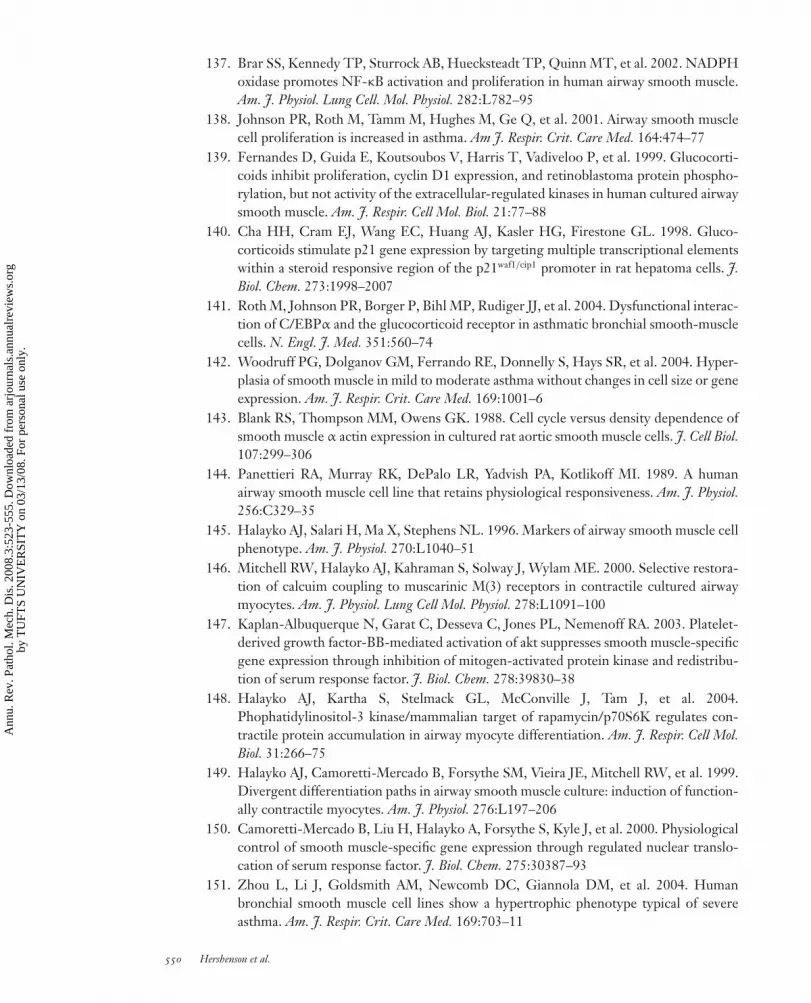
ANRV335-PM03-20 ARI 9 December 2007 21:8
137. Brar SS, Kennedy TP, Sturrock AB, Huecksteadt TP, Quinn MT, et al. 2002. NADPHoxidase promotes NF-κB activation and proliferation in human airway smooth muscle.Am. J. Physiol. Lung Cell. Mol. Physiol. 282:L782–95
138. Johnson PR, Roth M, Tamm M, Hughes M, Ge Q, et al. 2001. Airway smooth musclecell proliferation is increased in asthma. Am J. Respir. Crit. Care Med. 164:474–77
139. Fernandes D, Guida E, Koutsoubos V, Harris T, Vadiveloo P, et al. 1999. Glucocorti-coids inhibit proliferation, cyclin D1 expression, and retinoblastoma protein phospho-rylation, but not activity of the extracellular-regulated kinases in human cultured airwaysmooth muscle. Am. J. Respir. Cell Mol. Biol. 21:77–88
140. Cha HH, Cram EJ, Wang EC, Huang AJ, Kasler HG, Firestone GL. 1998. Gluco-corticoids stimulate p21 gene expression by targeting multiple transcriptional elementswithin a steroid responsive region of the p21waf1/cip1 promoter in rat hepatoma cells. J.Biol. Chem. 273:1998–2007
141. Roth M, Johnson PR, Borger P, Bihl MP, Rudiger JJ, et al. 2004. Dysfunctional interac-tion of C/EBPα and the glucocorticoid receptor in asthmatic bronchial smooth-musclecells. N. Engl. J. Med. 351:560–74
142. Woodruff PG, Dolganov GM, Ferrando RE, Donnelly S, Hays SR, et al. 2004. Hyper-plasia of smooth muscle in mild to moderate asthma without changes in cell size or geneexpression. Am. J. Respir. Crit. Care Med. 169:1001–6
143. Blank RS, Thompson MM, Owens GK. 1988. Cell cycle versus density dependence ofsmooth muscle α actin expression in cultured rat aortic smooth muscle cells. J. Cell Biol.107:299–306
144. Panettieri RA, Murray RK, DePalo LR, Yadvish PA, Kotlikoff MI. 1989. A humanairway smooth muscle cell line that retains physiological responsiveness. Am. J. Physiol.256:C329–35
145. Halayko AJ, Salari H, Ma X, Stephens NL. 1996. Markers of airway smooth muscle cellphenotype. Am. J. Physiol. 270:L1040–51
146. Mitchell RW, Halayko AJ, Kahraman S, Solway J, Wylam ME. 2000. Selective restora-tion of calcuim coupling to muscarinic M(3) receptors in contractile cultured airwaymyocytes. Am. J. Physiol. Lung Cell Mol. Physiol. 278:L1091–100
147. Kaplan-Albuquerque N, Garat C, Desseva C, Jones PL, Nemenoff RA. 2003. Platelet-derived growth factor-BB-mediated activation of akt suppresses smooth muscle-specificgene expression through inhibition of mitogen-activated protein kinase and redistribu-tion of serum response factor. J. Biol. Chem. 278:39830–38
148. Halayko AJ, Kartha S, Stelmack GL, McConville J, Tam J, et al. 2004.Phophatidylinositol-3 kinase/mammalian target of rapamycin/p70S6K regulates con-tractile protein accumulation in airway myocyte differentiation. Am. J. Respir. Cell Mol.Biol. 31:266–75
149. Halayko AJ, Camoretti-Mercado B, Forsythe SM, Vieira JE, Mitchell RW, et al. 1999.Divergent differentiation paths in airway smooth muscle culture: induction of function-ally contractile myocytes. Am. J. Physiol. 276:L197–206
150. Camoretti-Mercado B, Liu H, Halayko A, Forsythe S, Kyle J, et al. 2000. Physiologicalcontrol of smooth muscle-specific gene expression through regulated nuclear translo-cation of serum response factor. J. Biol. Chem. 275:30387–93
151. Zhou L, Li J, Goldsmith AM, Newcomb DC, Giannola DM, et al. 2004. Humanbronchial smooth muscle cell lines show a hypertrophic phenotype typical of severeasthma. Am. J. Respir. Crit. Care Med. 169:703–11
550 Hershenson et al.
Ann
u. R
ev. P
atho
l. M
ech.
Dis
. 200
8.3:
523-
555.
Dow
nloa
ded
from
arj
ourn
als.
annu
alre
view
s.or
gby
TU
FTS
UN
IVE
RSI
TY
on
03/1
3/08
. For
per
sona
l use
onl
y.

ANRV335-PM03-20 ARI 9 December 2007 21:8
152. Fingar DC, Salama S, Tsou C, Harlow E, Blenis J. 2002. Mammalian cell size is con-trolled by mTOR and its downstream targets S6K1 and 4EBP1/eIF4E. Genes Dev.16:1472–87
153. Ivester CT, Tuxworth WJ, Cooper G IV, McDermott PJ. 1995. Contraction acceleratesmyosin heavy chain synthesis rates in adult cardiocytes by an increase in the rate oftranslational initiation. J. Biol. Chem. 270:21950–57
154. Nikcevic G, Heidkamp MC, Perhonen M, Russell B. 1999. Mechanical activity in heartregulates translation of α-myosin heavy chain mRNA but not its localization. Am. J.Physiol. Heart Circ. Physiol. 276:H2013–19
155. Morgan HE, Gordon EE, Kira Y, Chua BHL, Russo LA, et al. 1987. Biochemicalmechanisms of cardiac hypertrophy. Annu. Rev. Physiol. 49:533–43
156. Sartorelli V, Fulco M. 2004. Molecular and cellular determinants of skeletal muscleatrophy and hypertrophy. Sci. STKE 244:re11
157. Glass DJ. 2003. Molecular mechanisms modulating muscle mass. Trends Mol. Med.9:344–50
158. Hesketh JE, Campbell GP, Lobley GE, Maltin CA, Acamovic F, Palmer RM. 1992.Stimulation of actin and myosin synthesis in rat gastrocnemius muscle by clenbuterol;evidence for translational control. Comp. Biochem. Physiol. C 102:23–27
159. Welle S, Bhatt K, Thornton CA. 1999. Stimulation of myofibrillar synthesis by exerciseis mediated by more efficient translation of mRNA. J. Appl. Physiol. 86:1220–25
160. Geisterfer A, Peach M, Owens G. 1988. Angiotensin II induces hypertrophy, not hy-perplasia, of cultured rat aortic smooth muscle cells. Circ. Res. 62:749–56
161. Goldsmith AM, Bentley JK, Zhou L, Jia Y, Bitar KN, et al. 2006. Transforming growthfactor-β induces airway smooth muscle hypertrophy. Am. J. Respir. Cell Mol. Biol. 34:247–54
161a. Camoretti-Mercado B, Fernandes DJ, Dewundara S, Churchill J, Ma L, et al. 2006.Inhibition of transforming growth factor β-enhanced serum response factor-dependenttranscription by SMAD7. J. Biol. Chem. 281:20383–92
162. Beretta L, Gingras AC, Svitkin YV, Hall MN, Sonenberg N. 1996. Rapamycin blocksthe phosphorylation of 4E-BP1 and inhibits cap-dependent initiation of translation.EMBO J. 15:658–64
163. Brunn GJ, Hudson CC, Sekuli A, Williams JM, Hosoi H, et al. 1997. Phosphorylationof the translational repressor PHAS-I by the mammalian target of rapamycin. Science277:99–101
164. Waskiewicz AJ, Flynn A, Proud CG, Cooper JA. 1997. Mitogen-activated protein kinasesactivate the serine/threonine kinases Mnk1 and Mnk2. EMBO J. 16:1909–20
165. Scheper GC, Morrice NA, Kleijn M, Proud CG. 2001. The mitogen-activated proteinkinase signal-integrating kinase Mnk2 is a eukaryotic initiation factor 4E kinase withhigh levels of basal activity in mammalian cells. Mol. Cell. Biol. 21:743–54
166. Sonenberg N, Gingras AC. 1998. The mRNA 5′ cap-binding protein eIF4E and controlof cell growth. Curr. Opin. Cell Biol. 10:268–75
167. Svitkin YV, Pause A, Haghighat A, Pyronnet S, Witherell G, et al. 2001. The require-ment for eukaryotic initiation factor 4A (elF4A) in translation is in direct proportion tothe degree of mRNA 5′ secondary structure. RNA 7:382–94
168. Welsh GI, Miller CM, Loughlin AJ, Price NT, Proud CG. 1998. Regulation of eu-karyotic initiation factor eIF2B: glycogen synthase kinase-3 phosphorylates a conservedserine which undergoes dephosphorylation in response to insulin. FEBS Lett. 421:125–30
www.annualreviews.org • Airway Smooth Muscle in Asthma 551
Ann
u. R
ev. P
atho
l. M
ech.
Dis
. 200
8.3:
523-
555.
Dow
nloa
ded
from
arj
ourn
als.
annu
alre
view
s.or
gby
TU
FTS
UN
IVE
RSI
TY
on
03/1
3/08
. For
per
sona
l use
onl
y.

ANRV335-PM03-20 ARI 9 December 2007 21:8
169. Leevers SJ, Weinkove D, MacDougall LK, Hafen E, Waterfield MD. 1996. TheDrosophila phosphoinositide 3-kinase Dp110 promotes cell growth. EMBO J. 15:6584–94
170. Verdu J, Buratovich MA, Wilder EL, Birnbaum MJ. 1999. Cell-autonomous regulationof cell and organ growth in Drosophila by Akt/PKB. Nat. Cell Biol. 1:500–6
171. Zhang H, Stallock JP, Ng JC, Reinhard C, Neufeld TP. 2000. Regulation of cellulargrowth by the Drosophila target of rapamycin dTOR. Genes Dev. 14:2712–24
172. Montagne J, Stewart MJ, Stocker H, Hafen E, Kozma SC, Thomas G. 1999. DrosophilaS6 kinase: a regulator of cell size. Science 285:2126–29
173. Stephens L, Anderson K, Stokoe D, Erdjument-Bromage H, Painter GF, et al.1998. Protein Kinase B kinases that mediate phosphatidylinositol 3,4,5-trisphosphate-dependent activation of protein kinase B. Science 279:710–14
174. Cheatham L, Monfar M, Chou M, Blenis J. 1995. Structural and functional analysis ofpp70S6k. Proc. Natl. Acad. Sci. USA 92:11696–700
175. Toker A, Newton AC. 2000. Cellular signaling: pivoting around PDK-1. Cell 103:185–88176. Vanhaesebroeck B, Alessi DR. 2000. The PI3K-PDK1 connection: more than just a
road to PKB. Biochem. J. 346:561–76177. Foukas LC, Shepherd PR. 2004. eIF4E binding protein 1 and H-Ras are novel substrates
for the protein kinase activity of class-I phosphoinositide 3-kinase. Biochem. Biophys. Res.Commun. 319:541–49
178. Herbert TP, Tee AR, Proud CG. 2002. The extracellular signal-regulated kinase path-way regulates the phosphorylation of 4E-BP1 at multiple sites. J. Biol. Chem. 277:11591–96
179. Gao X, Pan D. 2001. TSC1 and TSC2 tumor suppressors antagonize insulin signalingin cell growth. Genes Dev. 15:1383–92
180. Potter CJ, Huang H, Xu T. 2001. Drosophila Tsc1 functions with Tsc2 to antagonize in-sulin signaling in regulating cell growth, cell proliferation, and organ size. Cell 105:357–68
181. Tapon N, Ito N, Dickson BJ, Treisman JE, Hariharan IK. 2001. The Drosophila tuberoussclerosis complex gene homologs restrict cell growth and cell proliferation. Cell 105:345–55
182. Inoki K, Li Y, Zhu T, Wu J, Guan KL. 2002. TSC2 is phosphorylated and inhibited byAkt and suppresses mTOR signalling. Nat. Cell Biol. 4:648–57
183. Gao X, Zhang Y, Arrazola P, Hino O, Kobayashi T, et al. 2002. Tsc tumour suppressorproteins antagonize amino-acid-TOR signalling. Nat. Cell Biol. 4:699–704
184. Manning BD, Tee AR, Logsdon MN, Blenis J, Cantley LC. 2002. Identification of thetuberous sclerosis complex-2 tumor suppressor gene product tuberin as a target of thephosphoinositide 3-kinase/akt pathway. Mol. Cell. Biol. 10:151–62
185. Tee AR, Fingar DC, Manning BD, Kwiatkowski DJ, Cantley LC, Blenis J. 2002. Tuber-ous sclerosis complex-1 and -2 gene products function together to inhibit mammaliantarget of rapamycin (mTOR)-mediated downstream signaling. Proc. Natl. Acad. Sci. USA99:13571–76
186. Goncharova EA, Goncharov DA, Eszterhas A, Hunter DS, Glassberg MK, et al. 2002.Tuberin regulates p70 S6 kinase activation and ribosomal protein S6 phosphorylation.A role for the TSC2 tumor suppressor gene in pulmonary lymphangioleiomyomatosis(LAM). J. Biol. Chem. 277:30958–67
187. Tee AR, Manning BD, Roux PP, Cantley LC, Blenis J. 2003. Tuberous sclerosis complexgene products, Tuberin and Hamartin, control mTOR signaling by acting as a GTPase-activating protein complex toward Rheb. Curr. Biol. 13:1259–68
552 Hershenson et al.
Ann
u. R
ev. P
atho
l. M
ech.
Dis
. 200
8.3:
523-
555.
Dow
nloa
ded
from
arj
ourn
als.
annu
alre
view
s.or
gby
TU
FTS
UN
IVE
RSI
TY
on
03/1
3/08
. For
per
sona
l use
onl
y.

ANRV335-PM03-20 ARI 9 December 2007 21:8
188. Inoki K, Li Y, Xu T, Guan KL. 2002. Rheb GTPase is a direct target of TSC2 GAPactivity and regulates mTOR signaling. Genes Dev. 17:1829–34
189. Mach KE, Furge KA, Albright CF. 2000. Loss of Rhb1, a Rheb-Related GTPase infission yeast, causes growth arrest with a terminal phenotype similar to that caused bynitrogen starvation. Genetics 155:611–22
190. Jacinto E, Loewith R, Schmidt A, Lin S, Ruegg MA, et al. 2004. Mammalian TORcomplex 2 controls the actin cytoskeleton and is rapamycin insensitive. Nat. Cell Biol.6:1122–28
191. Jacinto E, Facchinetti V, Liu D, Soto N, Wei S, et al. 2006. SIN1/MIP1 maintains rictor-mTOR complex integrity and regulates Akt phosphorylation and substrate specificity.Cell 127:125–37
192. Yang Q, Inoki K, Ikenoue T, Guan K-L. 2006. Identification of Sin1 as an essentialTORC2 component required for complex formation and kinase activity. Genes Dev.20:2820–32
193. Hietakangas V, Cohen SM. 2007. Re-evaluating AKT regulation: role of TOR complex2 in tissue growth. Genes Dev. 21:632–37
194. Alt JR, Cleveland JL, Hannink M, Diehl JA. 2000. Phosphorylation-dependent regula-tion of cyclin D1 nuclear export and cyclin D1-dependent cellular transformation. GenesDev. 14:3102–14
195. Diehl JA, Cheng M, Roussel MF, Sherr CJ. 1998. Glycogen synthase kinase-3β regulatescyclin D1 proteolysis and subcellular localization. Genes. Dev. 12:3499–5511
196. Rossig L, Badorff C, Holzmann Y, Zeiher AM, Dimmeler S. 2002. Glycogen synthasekinase-3 couples AKT-dependent signaling to the regulation of p21Cip1 degradation.J. Biol. Chem. 277:9684–89
197. Zhou L, Goldsmith AM, Bentley JK, Jia Y, Rodriguez ML, et al. 2005. 4E-bindingprotein phosphorylation and eukaryotic initiation factor-4E release are required forairway smooth muscle hypertrophy. Am. J. Respir. Cell Mol. Biol. 33:195–202
198. Van Eerdewegh P, Little RD, Dupuis J, Del Mastro RG, Falls K, et al. 2002. Associationof the ADAM33 gene with asthma and bronchial hyperresponsiveness. Nature 418:426–30
199. Howard TD, Postma DS, Jongepier H, Moore WC, Koppelman GH, et al. 2003. Asso-ciation of a disintegrin and metalloprotease 33 (ADAM33) gene with asthma in ethnicallydiverse populations. J. Allergy Clin. Immunol. 112:717–22
200. Lind DL, Choudhry S, Ung N, Ziv E, Avila PC, et al. 2003. ADAM33 is not associatedwith asthma in Puerto Rican or Mexican populations. Am. J. Respir. Crit. Care Med.168:1312–16
201. Raby BA, Silverman EK, Kwiatkowski DJ, Lange C, Lazarus R, Weiss ST. 2004.ADAM33 polymorphisms and phenotype associations in childhood asthma. J. AllergyClin. Immunol. 113:1071–78
202. Umland SP, Garlisi CG, Shah H, Wan Y, Zou J, et al. 2003. Human ADAM33 messengerRNA expression profile and post-transcriptional regulation. Am. J. Respir. Cell Mol. Biol.29:571–82
203. Powell RM, Wicks J, Holloway JW, Holgate ST, Davies DE. 2004. The splicing andfate of ADAM33 transcripts in primary human airways fibroblasts. Am. J. Respir. CellMol. Biol. 31:13–21
204. Gilpin BJ, Loechel F, Mattei M-G, Engvall E, Albrechtsen R, Wewer UM. 1998. Anovel, secreted form of human ADAM 12 (meltrin α) provokes myogenesis in vivo.J. Biol. Chem. 273:157–66
www.annualreviews.org • Airway Smooth Muscle in Asthma 553
Ann
u. R
ev. P
atho
l. M
ech.
Dis
. 200
8.3:
523-
555.
Dow
nloa
ded
from
arj
ourn
als.
annu
alre
view
s.or
gby
TU
FTS
UN
IVE
RSI
TY
on
03/1
3/08
. For
per
sona
l use
onl
y.

ANRV335-PM03-20 ARI 9 December 2007 21:8
205. Bai TR. 1990. Abnormalities in airway smooth muscle in fatal asthma. Am. Rev. Respir.Dis. 141:552–57
206. Joos GF, O’Connor B, Anderson SD, Chung F, Cockcroft DW, et al. 2003. Indirectairway challenges. Eur. Respir. J. 21:1050–68
207. Black JL. 1996. Role of airway smooth muscle. Am J. Respir. Crit. Care Med. 153:S2–4208. Thomson NC. 1987. In vivo versus in vitro human airway responsiveness to different
pharmacologic stimuli. Am. Rev. Respir. Dis. 136:S58–62209. Hershenson MB, Aghili S, Punjabi N, Hernandez C, Ray DW, et al. 1992. Hyperoxia-
induced airway hyperresponsiveness and remodeling in immature rats. Am. J. Physiol.262:L263–69
210. Hershenson MB, Kelleher MD, Naureckas ET, Abe MK, Rubinstein VJ, et al. 1994.Hyperoxia increases airway cell S-phase traversal in immature rats in vivo. Am. J. Respir.Cell Mol. Biol. 11:296–303
211. Hershenson MB, Abe MK, Kelleher MD, Naureckas ET, Garland A, et al. 1994. Re-covery of airway structure and function after hyperoxic exposure in immature rats. AmJ. Respir. Crit. Care Med. 149:1663–69
212. Hershenson MB, Wylam ME, Punjabi N, Umans JG, Schumacker PT, et al. 1994. Ex-posure of immature rats to hyperoxia increases tracheal smooth muscle stress generationin vitro. J. Appl. Physiol. 76:743–49
213. Hershenson MB, Garland A, Kelleher MD, Zimmermann A, Hernandez C, Solway J.1992. Hyperoxia-induced airway remodeling in immature rats. Correlation with airwayresponsiveness. Am. Rev. Respir. Dis. 146:1294–300
214. Hakonarson H, Herrick DJ, Serrano PG, Grunstein MM. 1997. Autocrine role of inter-leukin 1β in altered responsiveness of atopic asthmatic sensitized airway smooth muscle.J. Clin. Invest 99:117–24
215. Hakonarson H, Halapi E, Whelan R, Gulcher J, Stefansson K, Grunstein MM. 2001.Association between IL-1β/TNF-α-induced glucocorticoid-sensitive changes in multi-ple gene expression and altered responsiveness in airway smooth muscle. Am. J. Respir.Cell Mol. Biol. 25:761–71
216. Deshpande DA, Dogan S, Walseth TF, Miller SM, Amrani Y, et al. 2004. Modulation ofcalcium signaling by interleukin-13 in human airway smooth muscle: role of CD38/cyclicadenosine diphosphate ribose pathway. Am. J. Respir. Cell Mol. Biol. 31:36–42
217. Deshpande DA, Walseth TF, Panettieri RA, Kannan MS. 2003. CD38/cyclicADP-ribose-mediated Ca2+ signaling contributes to airway smooth muscle hyper-responsiveness. FASEB J. 17:452–54
218. Hunter I, Cobban HJ, Vandenabeele P, MacEwan DJ, Nixon GF. 2003. Tumor necrosisfactor-α-induced activation of RhoA in airway smooth muscle cells: role in the Ca2+
sensitization of myosin light chain20 phosphorylation. Mol. Pharmacol. 63:714–21219. Chiba Y, Misawa M. 2004. The role of RhoA-mediated Ca2+ sensitization of bronchial
smooth muscle contraction in airway hyperresponsiveness. J. Smooth Muscle Res. 40:155–67
220. Hashimoto K, Peebles RS Jr, Sheller JR, Jarzecka K, Furlong J, et al. 2002. Suppressionof airway hyperresponsiveness induced by ovalbumin sensitisation and RSV infectionwith Y-27632, a Rho kinase inhibitor. Thorax 57:524–27
221. Chiba Y, Sakai H, Misawa M. 2001. Augmented acetylcholine-induced translocation ofRhoA in bronchial smooth muscle from antigen-induced airway hyperresponsive rats.Br. J. Pharmacol. 133:886–90
554 Hershenson et al.
Ann
u. R
ev. P
atho
l. M
ech.
Dis
. 200
8.3:
523-
555.
Dow
nloa
ded
from
arj
ourn
als.
annu
alre
view
s.or
gby
TU
FTS
UN
IVE
RSI
TY
on
03/1
3/08
. For
per
sona
l use
onl
y.

ANRV335-PM03-20 ARI 9 December 2007 21:8
222. Ma X, Cheng Z, Kong H, Wang Y, Unruh H, et al. 2002. Changes in biophysical andbiochemical properties of single bronchial smooth muscle cells from asthmatic subjects.Am. J. Physiol. Lung Cell Mol. Physiol. 283:L1181–89
223. Solway J, Fredberg JJ. 1997. Perhaps airway smooth muscle dysfunction contributes toasthmatic bronchial hyperresponsiveness after all. Am. J. Respir. Cell Mol. Biol. 17:144–46
224. Barnes PJ, Pride NB. 1983. Dose-response curves to inhaled β-adrenoceptor agonistsin normal and asthmatic subjects. Br. J. Clin. Pharmacol. 15:677–82
224a. Shore SA, Laporte J, Hall IP, Hardy E, Panettieri RA Jr. 1997. Effect of IL-1β onresponses of cultured human airway smooth muscle cells to bronchodilator agonists.Am. J. Respir. Cell. Mol. Biol. 16:702–12
224b. Lahiri T, Moore PE, Baraldo S, Whitehead TR, McKenna MD, et al. 2002. Effect ofIL-1β on CRE-dependent gene expression in human airway smooth muscle cells. Am.J. Physiol. Lung. Cell. Mol. Physiol. 283:L1239–46
224c. Laporte JD, Moore PE, Abraham JH, Maksym GN, Fabry B, et al. 1999. Role of ERKMAP kinases in responses of cultured human airway smooth muscle cells to IL-1β. Am.J. Physiol. 277:L943–51
224d. Laporte JD, Moore PE, Panettieri RA, Moeller W, Heyder J, Shore SA. 1998.Prostanoids mediate IL-1β-induced β-adrenergic hyporesponsiveness in human airwaysmooth muscle cells. Am. J. Physiol. 275:L491–501
224e. Hakonarson H, Herrick DJ, Serrano PG, Grunstein MM. 1996. Mechanism of cytokine-induced modulation of β-adrenoceptor responsiveness in airway smooth muscle. J. Clin.Invest. 97:2593–600
224f. Hakonarson H, Herrick DJ, Grunstein MM. 1995. Mechanism of impaired β-adrenoceptor responsiveness in atopic sensitized airway smooth muscle. Am. J. Physiol.269:L645–52
224g. Koto H, Mak JC, Haddad EB, Xu WB, Salmon M, et al. 1996 Mechanisms of impairedβ-adrenoceptor-induced airway relaxation by interleukin-1β in vivo in the rat. J. Clin.Invest. 98:1780–87
224h. Laporte JC, Moore PE, Baraldo S, Jouvin MH, Church TL, et al. 2001. Direct effectsof interleukin-13 on signaling pathways for physiological responses in cultured humanairway smooth muscle cells. Am. J. Respir. Crit. Care Med. 164:141–48
224i. Shore SA, Moore PE. 2003. Regulation of β-adrenergic responses in airway smoothmuscle. Respir. Physiol. Neurobiol. 137:179–95
225. Ober C, Leavitt SA, Tsalenko A, Howard TD, Hoki DM, et al. 2000. Variation in theinterleukin 4-receptor α gene confers susceptibility to asthma and atopy in ethnicallydiverse populations. Am. J. Hum. Genet. 66:517–26
226. Lakser OJ, Lindeman RP, Fredberg JJ. 2002. Inhibition of the p38 MAP kinase pathwaydestabilizes smooth muscle length during physiological loading. Am. J. Physiol. Lung CellMol. Physiol. 282:L1117–21
227. Dowell ML, Lakser OJ, Gerthoffer WT, Fredberg JJ, Stelmack GL, et al. 2005. La-trunculin B increases force fluctuation-induced relengthening of ACh-contracted, iso-tonically shortened canine tracheal smooth muscle. J. Appl. Physiol. 98:489–97
228. Dulin NO, Fernandes DJ, Dowell M, Bellam S, McConville J, et al. 2003. What evidenceimplicates airway smooth muscle in the cause of BHR? Clin. Rev. Allergy Immunol. 24:73–84
229. Cox G, Miller JD, McWilliams A, Fitzgerald JM, Lam S. 2006. Bronchial thermoplastyfor asthma. Am J. Respir. Crit. Care Med. 173:965–69
230. Cox G, Thomson N, Sperb-Rubin A, Niven R, Corris PA, et al. 2007. Asthma controlduring the year after bronchial thermoplasty. New Engl. J. Med. 356:1327–37
www.annualreviews.org • Airway Smooth Muscle in Asthma 555
Ann
u. R
ev. P
atho
l. M
ech.
Dis
. 200
8.3:
523-
555.
Dow
nloa
ded
from
arj
ourn
als.
annu
alre
view
s.or
gby
TU
FTS
UN
IVE
RSI
TY
on
03/1
3/08
. For
per
sona
l use
onl
y.

AR335-FM ARI 10 December 2007 18:20
Annual Reviewof Pathology:Mechanismsof Disease
Volume 3, 2008Contents
The Relevance of Research on Red Cell Membranes to theUnderstanding of Complex Human Disease: A Personal PerspectiveVincent T. Marchesi � � � � � � � � � � � � � � � � � � � � � � � � � � � � � � � � � � � � � � � � � � � � � � � � � � � � � � � � � � � � � � � � � � � � � � � �1
Molecular Mechanisms of Prion PathogenesisAdriano Aguzzi, Christina Sigurdson, and Mathias Heikenwalder � � � � � � � � � � � � � � � � � � � � 11
The Aging BrainBruce A. Yankner, Tao Lu, and Patrick Loerch � � � � � � � � � � � � � � � � � � � � � � � � � � � � � � � � � � � � � � � � � � 41
Gene Expression Profiling of Breast CancerMaggie C.U. Cheang, Matt van de Rijn, and Torsten O. Nielsen � � � � � � � � � � � � � � � � � � � � � � 67
The Inflammatory Response to Cell DeathKenneth L. Rock and Hajime Kono � � � � � � � � � � � � � � � � � � � � � � � � � � � � � � � � � � � � � � � � � � � � � � � � � � � � � � � 99
Molecular Biology and Pathogenesis of Viral MyocarditisMitra Esfandiarei and Bruce M. McManus � � � � � � � � � � � � � � � � � � � � � � � � � � � � � � � � � � � � � � � � � � � �127
Pancreatic CancerAnirban Maitra and Ralph H. Hruban � � � � � � � � � � � � � � � � � � � � � � � � � � � � � � � � � � � � � � � � � � � � � � � � �157
Kidney Transplantation: Mechanisms of Rejection and AcceptanceLynn D. Cornell, R. Neal Smith, and Robert B. Colvin � � � � � � � � � � � � � � � � � � � � � � � � � � � � � � �189
Metastatic Cancer CellMarina Bacac and Ivan Stamenkovic � � � � � � � � � � � � � � � � � � � � � � � � � � � � � � � � � � � � � � � � � � � � � � � � � � �221
Pathogenesis of Thrombotic MicroangiopathiesX. Long Zheng and J. Evan Sadler � � � � � � � � � � � � � � � � � � � � � � � � � � � � � � � � � � � � � � � � � � � � � � � � � � � � �249
Anti-Inflammatory and Proresolving Lipid MediatorsCharles N. Serhan, Stephanie Yacoubian, and Rong Yang � � � � � � � � � � � � � � � � � � � � � � � � � � � � �279
Modeling Morphogenesis and Oncogenesis in Three-DimensionalBreast Epithelial CulturesChristy Hebner, Valerie M. Weaver, and Jayanta Debnath � � � � � � � � � � � � � � � � � � � � � � � � � � � �313
v
Ann
u. R
ev. P
atho
l. M
ech.
Dis
. 200
8.3:
523-
555.
Dow
nloa
ded
from
arj
ourn
als.
annu
alre
view
s.or
gby
TU
FTS
UN
IVE
RSI
TY
on
03/1
3/08
. For
per
sona
l use
onl
y.

AR335-FM ARI 10 December 2007 18:20
The Origins of Medulloblastoma SubtypesRichard J. Gilbertson and David W. Ellison � � � � � � � � � � � � � � � � � � � � � � � � � � � � � � � � � � � � � � � � � � � �341
Molecular Biology and Pathology of LymphangiogenesisTerhi Karpanen and Kari Alitalo � � � � � � � � � � � � � � � � � � � � � � � � � � � � � � � � � � � � � � � � � � � � � � � � � � � � � � � �367
Endoplasmic Reticulum Stress in Disease PathogenesisJonathan H. Lin, Peter Walter, and T.S. Benedict Yen � � � � � � � � � � � � � � � � � � � � � � � � � � � � � � � �399
Autophagy: Basic Principles and Relevance to DiseaseMondira Kundu and Craig B. Thompson � � � � � � � � � � � � � � � � � � � � � � � � � � � � � � � � � � � � � � � � � � � � � � �427
The Osteoclast: Friend or Foe?Deborah V. Novack and Steven L. Teitelbaum � � � � � � � � � � � � � � � � � � � � � � � � � � � � � � � � � � � � � � � � �457
Applications of Proteomics to Lab DiagnosisRaghothama Chaerkady and Akhilesh Pandey � � � � � � � � � � � � � � � � � � � � � � � � � � � � � � � � � � � � � � � � �485
The Pathology of Influenza Virus InfectionsJeffrey K. Taubenberger and David M. Morens � � � � � � � � � � � � � � � � � � � � � � � � � � � � � � � � � � � � � � � �499
Airway Smooth Muscle in AsthmaMarc B. Hershenson, Melanie Brown, Blanca Camoretti-Mercado,and Julian Solway � � � � � � � � � � � � � � � � � � � � � � � � � � � � � � � � � � � � � � � � � � � � � � � � � � � � � � � � � � � � � � � � � � � � � � � �523
Molecular Pathobiology of Gastrointestinal Stromal SarcomasChristopher L. Corless and Michael C. Heinrich � � � � � � � � � � � � � � � � � � � � � � � � � � � � � � � � � � � � � � �557
Notch Signaling in LeukemiaJon C. Aster, Warren S. Pear, and Stephen C. Blacklow � � � � � � � � � � � � � � � � � � � � � � � � � � � � � � �587
The Role of Hypoxia in Vascular Injury and RepairTony E. Walshe and Patricia A. D’Amore � � � � � � � � � � � � � � � � � � � � � � � � � � � � � � � � � � � � � � � � � � � � � �615
Indexes
Cumulative Index of Contributing Authors, Volumes 1–3 � � � � � � � � � � � � � � � � � � � � � � � � � � �645
Cumulative Index of Chapter Titles, Volumes 1–3 � � � � � � � � � � � � � � � � � � � � � � � � � � � � � � � � � � � �647
Errata
An online log of corrections to Annual Review of Pathology: Mechanisms of Diseasearticles may be found at http://pathol.annualreviews.org
vi Contents
Ann
u. R
ev. P
atho
l. M
ech.
Dis
. 200
8.3:
523-
555.
Dow
nloa
ded
from
arj
ourn
als.
annu
alre
view
s.or
gby
TU
FTS
UN
IVE
RSI
TY
on
03/1
3/08
. For
per
sona
l use
onl
y.


