Embed Size (px)
Citation preview

Alexandra Elsing
Regulation of HSF2 and its function in mitosis
Alexandra Elsing | Regulation of H
SF2 and its function in mitosis | 2014

Regulation of HSF2 and its function in mitosis
Alexandra Elsing
Department of Biosciences, Åbo Akademi University Turku Centre for Biotechnology, University of Turku and Åbo Akademi University
Turku Doctoral Programme of Biomedical Sciences
2014

From the department of Biosciences, Åbo Akademi University, Turku Centre for Biotechnology, University of Turku and Åbo Akademi University, and Turku Doctoral Programme of Biomedical Sciences Supervised*by*Professor Lea Sistonen, PhD Department of Biosciences, Åbo Akademi University Turku Centre for Biotechnology, University of Turku and Åbo Akademi University Turku, Finland Reviewed*by*Docent Carina Holmberg-Still, PhD Research Programs Unit, Translational Cancer Biology Institute of Biomedicine University of Helsinki Helsinki, Finland Docent Anna-Liisa Levonen, MD, PhD A.I.Virtanen Institute for Molecular Sciences University of Eastern Finland Kuopio, Finland Opponent*Professor Harm H. Kampinga, PhD Faculty of Medical Sciences, University of Groningen Groningen, the Netherlands Author’s*adress*Åbo Akademi University Department of Biosciences Artillerigatan 6 20520 Åbo Finland e-mail: [email protected] © Alexandra Elsing ISBN: 978-952-12-3105-6 ISBN digital version: 978-952-12-3106-3 https://www.doria.fi/handle/10024/98908 Painosalama Oy– Åbo, Finland 2014

*
To my family, friends and Tequila


ABSTRACT*The cell is continuously subjected to various forms of external and intrinsic protein-damaging stresses, including hyperthermia, pathophysiological states, as well as cell differentiation and proliferation. Protein-damaging stresses result in denaturation and improper folding of proteins, leading to the formation of toxic aggregates that are detrimental for various pathological conditions, including Alzheimer’s and Huntington’s diseases. In order to maintain protein homeostasis, cells have developed different cytoprotective mechanisms, one of which is the evolutionary well-conserved heat shock response. The heat shock response results in the expression of heat shock proteins (Hsps), which act as molecular chaperones that bind to misfolded proteins, facilitate their refolding and prevent the formation of protein aggregates. Stress-induced expression of Hsps is mediated by a family of transcription factors, the heat shock factors, HSFs. Of the four HSFs found in vertebrates, HSF1-4, HSF1 is the major stress-responsive factor that is required for the induction of the heat shock response. HSF2 cannot alone induce Hsps, but modulates the heat shock response by forming heterotrimers with HSF1. HSFs are not only involved in the heat shock response, but they have also been found to have a function in development, neurodegenerative disorders, cancer, and longevity. Therefore, insight into how HSFs are regulated is important for the understanding of both normal physiological and disease processes.
The activity of HSF1 is mainly regulated by intricate post-translational modifications, whereas the activity of HSF2 is concentration-dependent. However, there is only limited understanding of how the abundance of HSF2 is regulated. This study describes two different means of how HSF2 levels are regulated. In the first study it was shown that microRNA miR-18, a member of the miR-17~92 cluster, directly regulates Hsf2 mRNA stability and thus protein levels. HSF2 has earlier been shown to play a profound role in the regulation of male germ cell maturation during the spermatogenesis. The effect on miR-18 on HSF2 was examined in
vivo by transfecting intact seminiferous tubules, and it was found that inhibition of miR-18 resulted in increased HSF2 levels and modified expression of the HSF2 targets Ssty2 and Speer4a.
HSF2 has earlier been reported to modulate the heat shock response by forming heterotrimers with HSF1. In the second study, it was shown that HSF2 is cleared off the Hsp70 promoter and degraded by the ubiquitin-proteasome pathway upon acute stress. By silencing components of the anaphase promoting complex/cyclosome (APC/C), including the co-activators Cdc20 and Cdh1, it was shown that APC/C mediates the heat-induced ubiquitylation of HSF2. Furthermore, down-regulation of Cdc20 was shown to alter the expression of heat shock-responsive genes.
Next, we studied if APC/C-Cdc20, which controls cell cycle progression, also regulates HSF2 during the cell cycle. We found that both HSF2 mRNA and protein levels decreased during mitosis in several but not all human cell lines, indicating that HSF2 has a function in mitotic cells. Interestingly, although transcription is globally repressed during mitosis, mainly due to the displacement of RNA polymerase II and transcription factors, including HSF1, from the mitotic chromatin, HSF2 is capable of binding DNA during mitosis. Thus, during mitosis the heat shock response is impaired, leaving mitotic cells vulnerable to proteotoxic stress. However, in HSF2-deficient mitotic cells the Hsp70 promoter is accessible to both HSF1 and RNA polymerase II, allowing for stress-inducible Hsp expression to occur. As a consequence HSF2-deficient mitotic cells have a survival advantage upon acute heat stress. The results, presented in this thesis contribute to the understanding of the regulatory mechanisms of HSF2 and its function in the heat shock response in both interphase and mitotic cells. Keywords: anaphase promoting complex/cyclosome, heat shock factor, heat shock proteins, heat shock response, microRNA, mitosis, spermatogenesis, transcriptional regulation
*

* *

SAMMANFATTNING*(Swedish*abstract)*Cellen utsätts konstant för olika former av proteinskadande stress, som kan resultera i denaturering och felaktig veckning av protein, vilket kan leda till bildandet av toxiska aggregat, som bland annat bidrar till uppkomsten av neurodegenerativa sjukdomar t.ex. Alzheimers sjukdom. För att upprätthålla proteinhomeostasen har cellen utvecklat olika försvarsmekanismer, exempelvis värmechockresponsen, som leder till att värmechockproteiner (Hsps, eng. heat shock proteins) uttrycks i cellen. Hsps fungerar som molekylära chaperoner som binder till felveckade proteiner och förhindrar bildandet av proteinaggregat. Den stress-inducerade expression av Hsps kontrolleras av värmechockfaktorer (HSFs, eng. heat shock factors). Av de fyra HSFs som återfinns hos ryggradsdjur, HSF1-4, så är HSF1 den huvudsakliga stress-känsliga faktorn som krävs för induktionen av värmechockresponsen och Hsps. HSF2 kan inte ensam inducera Hsps, men kan påverka genuttrycket genom att bilda heterotrimerer tillsammans med HSF1. HSFs är inte enbart involverade i värmechockresponsen, utan de spelar också en roll under utvecklingen, i neurodegenerativa sjukdomar, i cancer och i regleringen av livslängden. Därför är det viktigt att förstå hur HSFs regleras i både normala fysiologiska processer och sjukdomstillstånd.
HSF1s aktivitet kontrolleras främst genom post-translationella modifieringar, medan HSF2s aktivitet beror på dess koncentration i cellen. Denna studie beskriver två olika sätt med vilka HSF2-nivåerna kan regleras. I den första studien visades det att microRNA miR-18 kontrollerar mRNA stabiliteten och således protein nivåerna av HSF2. Eftersom HSF2 tidigare påvisats ha en betydelse för regleringen av sädescellernas utveckling studerades effekten av miR-18 på HSF2 in vivo genom transfektion av intakta sädeskanaler och det påvisades att inhiberingen av miR-18 ledde till ökade HSF2 nivåer och förändrad expression av HSF2s målgener Ssty2 och Speer4a.
I den andra studien undersöktes regleringen av HSF2 under
värmechockresponsen och det påvisades att HSF2 tas bort från Hsp70 promotorn och degraderas efter akut stress. Genom att inhibera komponenter av ubikvitin E3-ligaset anafasfrämjande komplex/cyklosom (APC/C, eng. anaphase promoting complex/cyclosome), inklusive dess kofaktorer Cdc20 och Cdh1, kunde det påvisas att APC/C medierar värme-inducerad ubikvitinering av HSF2. Det visade sig dessutom att nedreglering av Cdc20 påverkade expressionen av gener som induceras i respons till värmechock.
I den sista studien undersökte vi om APC/C-Cdc20, som har visats kontrollera cell cykelns progression, också kan påverka nivåerna av HSF2 under cellcykelns förlopp. Det visade sig att både mRNA- och proteinnivåer av HSF2 minskade under mitosen i vissa, men inte alla, cellinjer som undersöktes. Detta indikerar att HSF2 har en funktion i mitotiska celler. Under mitosen är genexpressionen inhiberad, främst på grund av att RNA polymeras II och transkriptionsfaktorer, inklusive HSF1, inte kan binda till det mitotiska kromatinet. I och med att transkription inte sker under mitosen, kan värmechockresponsen inte induceras och således är mitotiska celler mycket känsliga för stress. Intressant nog visade det sig att HSF2 kan binda till kromatinet även i mitotiska celler och att bindningen av HSF2 till DNA bidrar till att genexpressionen inhiberas. Således kan både HSF1 och RNA polymeras II binda till Hsp70 promotorn och inducera dess expression även under mitosen i de celler som har lägre HSF2-nivåer. Följaktligen har mitotiska celler med lägre HSF2-nivåer en överlevnadsfördel gentemot celler där HSF2 nivåerna inte minskar under mitosen. Resultaten som presenteras i denna avhandling bidrar till förståelsen om hur HSF2 regleras och dess funktion i värmechockresponsen under både interfas och mitos. Nyckelord: anafasfrämjande komplex/cyklosom, microRNA, mitos, spermatogenes, transkriptionell reglering, värmechockfaktorer, värmechockprotein, värmechockrespons.

** *

Table of Contents
1
TABLE*OF*CONTENTS*
LIST%OF%ORIGINAL%PUBLICATIONS%.................................................................................................................%3%
ABBREVIATIONS%..................................................................................................................................................%4%
INTRODUCTION%....................................................................................................................................................%6%
REVIEW%OF%THE%LITERATURE%.........................................................................................................................%7%
The%eukaryotic%cell%cycle%..........................................................................................................................%7%
1.1! Mitosis%........................................................................................................................................................................%8!1.1.1! Chromatin!is!condensed!during!prophase!...............................................................................................................!9!1.1.2! Centrosomes!are!important!for!spindle!assembly!.............................................................................................!10!1.1.3! Nuclear!envelope!breakdown!and!spindle!formation!in!prometaphase!..................................................!10!1.1.4! BipolarlyBattached!sister!chromatids!are!separated!during!anaphase!....................................................!11!1.1.5! Telophase!and!cytokinesis!result!in!two!newly!formed!daughter!cells!...................................................!11!
1.2! Cyclins%and%cyclinMdependent%kinases%–%the%motors%of%cell%cycle%progression%................................%11!1.3! E3%ubiquitin%ligases%in%cell%cycle%control%......................................................................................................%12!1.3.1! The!APC/C!controls!metaphase!to!anaphase!transition!..................................................................................!13!1.3.2! Ubiquitylation!and!the!pathway!to!proteasomal!degradation!.....................................................................!16!1.3.3! The!26S!proteasome!and!proteasomal!degradation!.........................................................................................!18!
1.4! Inactivation%of%APC/C%by%the%spindle%assembly%checkpoint%protects%cells%against%aneuploidy% 19!
Regulation%of%gene%expression%............................................................................................................%21%
2.1! Regulation%of%transcription%at%the%level%of%chromatin%.............................................................................%22!2.1.1! DNA!regulatory!elements!assist!in!controlling!transcriptional!initiation!...............................................!22!2.1.2! DNA!is!organized!into!chromatin!that!facilitates!transcriptional!regulation!.........................................!23!
2.2! Initiation%of%transcription%.................................................................................................................................%24!2.3! RNAPII%and%the%elongation%phase%of%transcription%...................................................................................%26!2.4! Translation%–%from%mRNA%to%a%nascent%polypeptide%chain%....................................................................%27!2.5! PostMtranscriptional%regulation%of%gene%expression%................................................................................%27!2.6! nonMcoding%RNAs%as%regulators%of%gene%expression%.................................................................................%28!2.6.1! MicroRNAs!regulate!gene!expression!postBtranscriptionally!.......................................................................!28!2.6.2! miRNA!biogenesis!............................................................................................................................................................!29!2.6.3! miRNA!mechanisms!of!action!.....................................................................................................................................!31!2.6.4! Regulation!of!miRNA!biogenesis!and!activity!......................................................................................................!32!2.6.5! Biological!functions!of!miRNA!....................................................................................................................................!32!2.6.6! The!miRB17~92!cluster!.................................................................................................................................................!34!
Transcriptional%regulation%during%mitosis%.....................................................................................%37%
3.1! The%chromatin%environment%in%mitotic%cells%..............................................................................................%37!3.1.1! Changes!in!histone!modifications!in!mitosis!........................................................................................................!38!3.1.2! The!role!of!histone!H3!phosphorylation!in!mitosis!...........................................................................................!38!
3.2! Transcription%and%translation%are%periodically%regulated%in%a%cell%cycleMdependent%manner%..%40!3.3! Eukaryotic%transcription%is%repressed%during%mitosis%............................................................................%41!3.4! How%is%mitotic%transcriptional%repression%executed?%.............................................................................%41!3.4.1! Chromatin!condensation!has!only!a!minor!contribution!to!mitotic!transcriptional!repression!...!41!3.4.2! Phosphorylation!of!the!RNAPII!transcriptional!machinery!represses!transcription!.........................!42!3.4.3! Many!transcription!factors!are!phosphorylated!and!thus!repressed!in!mitosis!...................................!44!3.4.4! Displacement!of!chromatin!modifyers!from!mitotic!chromatin!..................................................................!44!
3.5! Mitotic%transcription%is%preserved%on%specific%genes%...............................................................................%44!3.6! Mitotic%bookmarking%..........................................................................................................................................%44!
Transcriptional%regulation%by%heat%shock%factors%–%in%the%stress%response%and%beyond%.%47%
4.1! The%heat%shock%response%...................................................................................................................................%47!4.1.1! Heat!shock!proteins!act!as!molecular!chaperones!facilitating!protein!folding!.....................................!48!
4.2! The%heat%shock%factor%family%............................................................................................................................%49!4.2.1! Functional!domains!of!the!HSFs!................................................................................................................................!50!
4.3! HSF1%–%the%main%stressMresponsive%factor%in%mammals%..........................................................................%50!4.3.1! Regulation!of!HSF1!by!postBtranslational!modifications!................................................................................!52!4.3.2! StressBinduced!HSF1Bmediated!expression!of!Hsp70!.......................................................................................!53!4.3.3! Functions!of!HSF1!beyond!the!acute!heat!shock!response!............................................................................!54!
4.4! HSF2%.........................................................................................................................................................................%55!4.4.1! HSF2!activity!is!regulated!by!its!concentration!in!the!cell!.............................................................................!57!

Table of Contents
2
4.4.2! Sumoylation!of!HSF2!.......................................................................................................................................................!58!4.4.3! HSF2!modulates!the!heat!shock!response!through!formation!of!HSF1BHSF2!heterotrimers!..........!58!4.4.4! The!role!of!HSF2!in!early!development,!aging!and!physiological!processes!...........................................!59!4.4.5! HSF2!in!spermatogenesis!..............................................................................................................................................!60!
4.5! HSFs%as%regulators%of%cell%cycle%progression%and%heatMinduced%cell%cycle%arrest%...........................%63!4.5.1! Heat!shock!causes!cell!cycle!arrest!............................................................................................................................!63!4.5.2! Proteotoxic!stress!is!detrimental!to!mitotic!cells!................................................................................................!63!4.5.3! The!heat!shock!response!in!mitotic!cells!................................................................................................................!64!
OUTLINE%AND%AIMS%OF%THE%THESIS%...........................................................................................................%65%
EXPERIMENTAL%PROCEDURES%.....................................................................................................................%66%
RESULTS%AND%DISCUSSION%............................................................................................................................%71%
1% PostMtranscriptional%regulation%of%HSF2%through%miRM18%(I)%....................................................%71%
1.1! Identification%of%the%miRM18%target%site%on%Hsf2%(I)%..................................................................................%71!1.2! Inverse%correlation%between%miRM18%and%HSF2%expression%in%spermatogenesis%(I)%....................%72!1.3! Inhibition%of%miRM18%in%spermatocytes%affects%expression%of%HSF2%and%its%target%genes%............%74!
2% HSF2%is%postMtranslationally%modified%by%ubiquitylation%in%response%to%heat%stress%(II)%76%
2.1! HSF2%is%a%shortMlived%protein%...........................................................................................................................%76!2.2! HSF2%is%ubiquitylated%in%response%to%thermal%stress%...............................................................................%76!2.3! HSF2%is%modified%by%K11M%and%K48Mlinked%polyubiquitin%.......................................................................%77!2.4! APC/CMmediated%ubiquitylation%of%HSF2%......................................................................................................%78!2.5! Silencing%of%APC/C%increases%HSF2%stability%...............................................................................................%78!2.6! HSF2%is%rapidly%cleared%from%the%Hsp70%promoter%upon%heat%shock%.................................................%80!2.7! Cdc20%modulates%the%transcription%of%heat%shock%genes%........................................................................%81!
3% Cell%cycleMdependent%regulation%of%the%HSFs%(III,%unpublished)%..............................................%83%
3.1! HSF2%protein%levels%decrease%in%mitotic%cells%.............................................................................................%83!3.2! Mitotic%cells%display%reduced%Hsf2%mRNA%levels%........................................................................................%84!3.3! HSF1%is%hyperphosphorylated%during%mitosis%...........................................................................................%86!
4% Regulation%of%the%mitotic%heat%shock%response%by%HSF2%(III)%....................................................%88%
4.1! HSF2%represses%the%heat%shock%response%in%mitotic%cells%.......................................................................%88!4.2! HSF2%interferes%with%the%stressMinducible%DNAMbinding%capacity%of%HSF1%.......................................%88!4.3! HSF2%regulates%the%H3S10P%occupancy%of%the%heatMinducible%Hsp%promoters%................................%89!
5% The%impact%of%HSF2%on%mitotic%progression%and%cell%proliferation%(III,%unpublished)%.....%90%
5.1! Reduced%HSF2%levels%increase%cell%survival%in%response%to%acute%stress%...........................................%90!5.2! Cell%lines%with%decreased%mitotic%HSF2%levels%show%increased%Hsp70%induction%during%mitosis
% 91!
6% MultiMlevel%regulation%of%HSF2%levels%determines%HSF2%activity%(I,%II,%III)%............................%92%
IMPLICATIONS%AND%FUTURE%RESEARCH%DIRECTIONS%.........................................................................%94%
CONCLUDING%REMARKS%..................................................................................................................................%95%
ACKNOWLEDGEMENTS%...................................................................................................................................%96%
REFERENCES%.......................................................................................................................................................%99%
ORIGINAL%PUBLICATIONS……………………………………………………………………………………………...….130

List of Original Publications
3
LIST*OF*ORIGINAL*PUBLICATIONS* This thesis is based on the following original publications and a manuscript, which are referred to in the text by their Roman numerals (I-III). In addition, unpublished results are included.
I. Björk JK*, Sandqvist A*, Elsing AN, Kotaja N, Sistonen L (2010) mir-18, a member of Oncomir-1, targets heat shock transcription factor 2 in spermatogenesis. Development 137, 3177-3184. doi:10.1242/dev.050955
II. Ahlskog JK, Björk JK*, Elsing AN*, Aspelin C, Kallio M, Roos-Mattjus P, Sistonen L (2010) APC/C participates in the acute response to protein-damaging stress. Mol. Cell Biol., 30, 5608-5620. doi:10.1128/MCB.01506-09
III. Elsing AN, Aspelin C*, Björk JK*, Bergman HA, Himanen SV, Kallio MJ, Roos-Mattjus P, Sistonen L (2014) Expression of HSF2 decreases in mitosis to enable stress-inducible transcription and cell survival. Manuscript in press in Journal of Cell Biology. doi:10.1083/jcb.201402002
*equal contribution Original publications have been reprinted with the kind permission of the copyright holders.

Abbreviations
4
ABBREVIATIONS* β-TrCP β-transducin repeat–containing
protein γ-TuRC γ-tubulin ring complex ac acetylation AD Transactivation domain APC/C Anaphase-promoting
complex/cyclosome ARE adenylate- and uridylate-rich
element ATM Ataxia telangiectasia mutated ATO Arsenic trioxide ATP Adenosine 5´-triphosphate BRD Bromodomain containing protein BRE TFIIB recognition element Bub Budding uninhibited by
benzimidazole CaMKII Calcium/calmodulin-dependent
kinase Cdc Cell division cycle Cdh1 Cdc20 homolog 1 CDK Cyclin-dependent kinase CENP Centromere protein CHD Chromodomain-helicase DNA-
binding protein ChIP Chromatin immunoprecipitation ChIP-seq Chromatin immunoprecipitation,
combined with sequencing CIN Chromosomal instability CKI Cyclin-dependent kinase inhibitor CTD C-terminal domain D-box Destruction box DBD DNA-binding domain DCE Downstream core element DDX5 DEAD box RNA helicases p68 DDX17 DEAD box RNA helicases p72 DGCR8 DiGeorge syndrome critical region
gene 8 DPE Downstream promoter element DSIF DRB-sensitivity-inducing factor DUB Deubiquitylase eIF Eukaryotic translation initiation
factor Emi1 Early mitotic inhibitor 1 EMSA Electrophoretic mobility shift
assay FACS Fluorescence-activated cell sorting Fbw7 F-box WD40 repeat–containing
protein 7 G1 Gap 1 phase of the cell cycle G2 Gap 2 phase of the cell cycle
GFP Green fluorescent protein GTF General transcription factor GTP Guanosine triphosphate H Histone H3 S10P Histone H3 serine 10
phosphorylation HAT Histone acetyltransferase HDAC Histone deacetylases hnRNP Heterogeneous nuclear
ribonucleoproteins HR-A/B Heptad repeat A/B HSE Heat shock element HSF Heat shock transcription factor Hsp Heat shock protein HSR Heat shock response KMT Lysine methyltransferases KO Knock out Id1 Inhibitor of DNA binding 1 Inr Initiator element IRES Internal ribosome entry site ISWI Imitation switch kb Kilobase M Mitosis phase of the cell cycle Mad Mitotic-arrest deficient me Methylation MEF Mouse embryonic fibroblast MK2 MAPK- activated protein kinase 2 MLL Mixed lineage leukemia MNase micrococcal nuclease miRNA MicroRNA MPS1 Multipolar spindle-1 MSCI Meiotic sex chromatid inactivation mRNA Messenger RNA MTE Motif ten element ncRNA Non-coding RNA NEF Nucleotide exchange factor Nek2A Never in mitosis (NIMA)-related
kinase 2A NELF Negative elongation factor NFκB nuclear factor κ B nSB Nuclear stress body nt Nucleotide PACT Interferon-inducible double-
stranded RNA-dependent protein kinase activator A
PABP1 Polyadenylate-binding protein 1 PcG Polycomb protein PGC Primordial germ cells PI Propidium iodide

Abbreviations
5
PIC Transcription preinitiation complex
PKC Protein kinase C Plk Polo-like kinase P-TEFb Positive transcription elongation
factor b PTM Post-translational modification Prm Protamines RBP RNA-binding protein RD Regulatory domain RING Really interesting new gene RISC RNA-induced silencing complex RNA Ribonucleic acid RNAi RNA interference RNAPII RNA polymerase II RPA Replication protein A RPN Regulatory particle non-ATPase RPT Regulatory particle triple A
protein rRNA Ribosomal RNA RT-PCR Reverse transcription polymerase
chain reaction S Synthesis phase of the cell cycle SCF Skp1-Cullin 1-F-box protein SAC Spindle assembly checkpoint
SEM Standard error of mean shRNA Small hairpin RNA siRNA Short interfering RNA snRNP Small nuclear ribonuclear particle SPEER Sperm-associated glutamate rich Ssty Spermiogenesis-specific transcript
of the Y 2 SUMO Small ubiquitin-like modifier SWI/SNF Switching-defective-sucrose non-
fermenting TAF TBP-associated factors TBP TATA-box binding protein TFII Transcription factor for RNA
polymerase II transcribed genes T-GIST Transfection of germ cells in intact
seminiferous tubules TP Transition protein TRBP TAR RNA-binding protein TSP1 Thrombospondin 1 TSS Transcription start site UBD Ubiquitin-binding domain UIM Ubiquitin-interacting motif UTR Untranslated region WT Wild type

Introduction
6
INTRODUCTION* Cells are the building blocks of life, and organisms consist of many different cell types that build up tissues and organs. All cells in an organism are derived from a single cell, which propagates through a series of events called the cell cycle. Every cell cycle ends with the division of the cell and its genetic material, into two daughter cells. The cell cycle, and all other functions in a cell, depends on proteins, and thus the viability of the cell, and the whole organism depend on the proper function of the proteins. An increase in the number of misfolded proteins, e.g. as a consequence of increased intracellular temperature, results in the induction of heat shock proteins (Hsps). Hsps are molecular chaperones that bind to misfolded proteins and facilitate their refolding and prevent their aggregation. The expression of Hsps is mediated by heat shock transcription factors, HSFs. Mammals have a family of four HSFs, HSF1-4, which together, and separately, regulate the expression of Hsps and other proteins. HSF1 is the master regulator of Hsps, but recent advances have found that HSF2 modulate the expression of Hsps by forming heterotrimers with HSF1. The activity of HSF1 is mainly regulated by post-translational modifications, whereas HSF2 regulation is mainly dependent on its protein levels in the cell. In this thesis it was examined how the amount of HSF2 is regulated during the differentiation of the male germ cell, and in response to heat shock. It was established that during spermatogenesis HSF2 levels fluctuate, and that they inversely correlate with the microRNA, miR-18. In a series of experiments it was found that the 3’UTR of Hsf2 mRNA was directly targeted by miR-18, which regulates Hsf2 mRNA stability. Furthermore, by utilizing a novel method, transfection of germ cells in intact
seminiferous tubules, it was found that miR-18 controls the transactivation capacity of HSF2 in spermatogenesis. Furthermore, the regulation of HSF2 in a second system, in the heat shock response, was examined. In response to heat the rate of HSF2 turnover is further increased, and HSF2 is subjected to proteasomal degradation. Ubiquitylation of HSF2 is mediated by the E3 ubiquitin ligase anaphase promoting complex/cyclosome (APC/C). Intriguingly, Cdc20, the co-activator of APC/C, and the proteasome were found to be recruited to the Hsp70 promoter upon heat shock, and Cdc20 contributes to the transcriptional regulation of heat shock responsive genes. In a final study, the cell cycle-dependent regulation of HSF2 was studied, and it was found that HSF2 levels decrease during mitosis in several, but not all, cell lines. Mitotic HSF2 levels are controlled both at the level of mRNA and protein. During mitosis a majority of the proteins involved in transcription, including RNA polymerase II and sequence-specific transcription factors such as HSF1, are displaced from the chromatin, resulting in the repression of general transcription. Intriguingly, in cells where HSF2 levels were decreased, heat-inducible expression of Hsp70 was detected. In mitotic cells where HSF2 was down-regulated HSF2 and HSF1 and RNA polymerase II (RNAPII) bound to the Hsp70 promoter. Thus, HSF2 was found to be a repressor of the heat shock response in mitotic cells. Taken together, the results presented in this thesis provide new insight into the regulation of HSF2 protein levels and stability in spermatogenesis and in response to thermal stress. Furthermore, it investigates how HSF2 contributes to the regulation of the repression of the heat shock response in mitotic cells.

Review of the Literature – The Cell Cycle
7
REVIEW*OF*THE*LITERATURE* 1. The*eukaryotic*cell*cycle*
“Omnis cellula e cellula”. In 1858, Rudolf Virchow stated the famous cell doctrine “Where a cell arises, there must be a previous cell, just as animals can only arise from animals and plants from plants”. What he meant was that a new cell can only be generated by the division of a previously existing cell. Each and every one of us started from a single fertilized egg, and now we are complex organisms with approximately 1013 cells in our bodies. The growth and reproduction of all organisms depend on the faithful duplication of their genomic DNA and the even distribution of the duplicated DNA to the daughter cells. This sequence of events, which ends in the identical replication of a cell, is called the cell cycle. In unicellular organisms the cell cycle, with the subsequent division of the cell, reproduces the whole organism. Multicellular organisms depend on the cell cycle for the development from a fertilized cell to a full-grown organism with multiple different tissues and organs, and for the repair of damaged tissues. Furthermore, tissues, such as the skin and intestine, need to be continuously renewed. The cell cycle must be coordinated so that in multicellular organisms the cell cycle is only initiated when new cells are needed. Cell cycling and on-going or halted division is strongly associated with cellular and organismal aging. Moreover, the regulation of the cell cycle has proven to be important in various human diseases and pathological processes, especially tumor formation where the balance between cell division and apoptosis is deregulated (Behl!and!Ziegler,!2013). Each cell cycle ends with cell division, and for division to take place five events need to occur. The cell has to receive a reproductive signal, the cell has to grow, DNA has to be duplicated
and segregated between the daughter cells, and finally the separation of the daughter cells, cytokinesis, has to occur. The typical cell cycle is thus divided into four phases: gap 1 (G1), synthesis (S), gap 2 (G2), and mitosis (M) phase (Figure 1). The first three phases are collectively called interphase. The G1 phase, which begins immediately after cell division, is the primary growth phase where proteins are synthesised and new organelles are formed. Upon receiving a signal initiating reproduction, the cell enters S phase, where the nuclear DNA is replicated. In the succeeding G2 phase, the cell usually continues growing, the machinery for cell division is assembled, and the fidelity of the DNA synthesis is controlled. Finally, in M phase, sister chromatids are separated and distributed to the two daughter cells, which are formed by cytokinesis. The length of the cell cycle varies depending on cell and organ type, developmental stage, and physiological conditions (Behl!and!Ziegler,!2013). Progression through the cell cycle is regulated by three specific checkpoints. The restriction point, between G1 and S phase, regulates the entry into S phase and the onset of the cell cycle. The G2/M or DNA-damage checkpoint, which senses DNA replication status and damage, regulates entry into mitosis. The spindle assembly checkpoint (SAC) prevents the separation of sister chromatids until each chromatid is properly attached to the spindle apparatus. The cell cycle and its checkpoints are molecularly controlled by cyclin-dependent kinases (CDKs). The activity of cell cycle kinases and other mitotic regulators are actively and tightly regulated for timely progression through the cell cycle. This is achieved by ubiquitin-mediated proteolysis of the cell cycle checkpoint proteins (Peters,!2006).

Review of the Literature – The Cell Cycle
8
Figure* 1.* The* eukaryotic* cell* cycle* and* its* checkpoints.* The$eukaryotic$ cell$ cycle$ can$be$divided$ into$ four$phases:$ G1,$ S,$ G2$ and$ mitosis.$ During$ G1$ phase$ the$ cell$ grows$ and$ undergoes$ metabolic$ changes.$ The$restriction$point$commits$the$cell$for$division,$and$is$regulated$by$both$internal$and$external$cues.$In$S$phase$the$DNA$and$the$centrosomes$are$duplicated.$In$G2$the$cell$continues$growing,$and$the$DNA$is$checked$for$errors.$During$mitosis$the$duplicated$DNA$is$divided$between$the$two$daughter$cells.$The$SAC$controls$that$the$chromosomes$are$equally$divided.$Mitosis$ends$with$cytokinesis$when$the$cytoplasma$is$divided.$ 1.1 Mitosis%
The major event taking place during mitosis is the precise segregation of the sister chromatids, which were generated through DNA duplication during the S phase. With the assistance of the mitotic spindle the sister chromatids are separated to the opposite poles of the cell, which then divides through cytokinesis. Mitosis consists of five distinct phases: prophase, prometaphase, metaphase, anaphase, and telophase (Figure 2). During prophase the chromosomes start to condense to form visible structures, and the microtubule spindle starts to form between the separated centrosomes. Prometaphase starts abruptly when the nuclear envelope breaks down. This enables the microtubules to attach to the chromosomes, and chromosomes are moved towards the metaphase plate. In metaphase, all
chromosomes are aligned at the metaphase plate and the microtubules from opposite poles attach to the kinetochores of sister chromatids. When all sister chromatids are bipolarly attached, the anaphase starts with the separation of the sister chromatids, and the chromosomes are moved to opposite poles of the cell by the action of shortening microtubules and motor proteins. During telophase the spindle starts to decondense upon the arrival of the chromosomes at opposite poles. The nuclear envelope reassembles around the chromosomes marking the end of mitosis. Cytokinesis starts by the contraction of the contractile actin-myosin ring and at the end two new daughter cells are formed and the cells enter G1 phase (Morgan, 2007).

Review of the Literature – The Cell Cycle
9
Figure*2.*Schematic*presentation*of*the*phases*in*mitosis.*Mitosis$is$divided$into$prophase,$prometaphase,$metaphase,$ anaphase$ and$ telophase.$ During$ prophase$ the$ duplicated$ DNA$ starts$ to$ condense,$ the$centrosomes$separate,$and$the$nuclear$envelope$breaks$down.$ In$prometaphase$the$microtubule$spindle$grows$ and$ attach$ to$ the$ kinetochores$ of$ chromosomes,$ resulting$ in$ their$ movements$ towards$ the$metaphase$ plate.$ In$metaphase$ the$ chromosomes$ are$ organized$ into$ the$metaphase$ plate,$ and$ the$ cell$checks$for$bipolar$attachment$of$the$sister$chromatids.$When$the$SAC$is$satisfied$at$the$onset$of$anaphase,$the$chromosomes$are$moved$apart$by$the$microtubule$spindle,$and$the$cell$starts$to$elongate.$In$telophase$the$nuclear$envelope$is$reformed$around$the$chromosomes$that$start$to$decondense,$and$a$contractile$ring$consisting$of$actin$filaments$is$formed$around$the$cell.$During$cytokinesis$the$cytoplasma$of$the$daughter$cells$are$split.$
1.1.1 Chromatin% is% condensed% during%
prophase%%
At the end of S phase the duplicated sister chromatids exist as a long, intertwined DNA-protein mass. The separation of the sister chromatids in this state would most certainly lead to DNA breakage. Thus, for accurate separation of the genetic material, mitotic DNA needs to be condensed into chromosomes, consisting of two sister chromatids. This supercoiling of DNA starts in early prophase when cyclin A-CDK1 activates condensin. When the nuclear envelope starts to break down and cyclin B1-CDK1 that resides in the cytoplasma get access to the nucleus, condensation is accelerated. Condensin, which exists as two different complexes, mediates the condensation of DNA in an ATP-dependent manner (Hirano, 2005). The initiation of chromosome condensation is also suggested
to be dependent on the phosphorylation of histone H3 serine 10 (H3S10P) by Aurora kinase B (Crosio et al., 2002; Van Hooser et al., 1998). This phosphorylation is thought to recruit condensin complexes (Giet and Glover, 2001; Nowak and Corces, 2004). The cohesion between the sister chromatids is particularly important for the process of sister-chromatid separation as it governs both chromosome movement in prometaphase and sister chromatid segregation at the onset of anaphase. The sister-chromatid cohesion is established when the DNA is replicated during S phase by two mechanisms, DNA catenation and cohesin complexes. The cohesin complex, consisting of four subunits (Smc1, Smc3, Scc1, and Scc3), associates with the chromosome at distinct sites along the arm of the chromosomes. At the beginning of mitosis topoisomerase II has almost removed

Review of the Literature – The Cell Cycle
10
all catenation and also some of the cohesion complexes along the chromosome arms are lost triggered by Polo-like kinase 1 (Plk1) and Aurora B kinase. Thus, in preparation for the chromosome segregation, the sister chromatids remain attached only at the centromere region where the spindle microtubules attach (Musacchio and Salmon, 2007; Zachariae, 1999). 1.1.2 Centrosomes% are% important% for%
spindle%assembly%
Another crucial event in early mitosis is the separation and migration of the two centrosomes, which are pivotal for the organization of the bipolar microtubule spindle, start to separate and migrate to opposite sides of the nucleus. The centrosomes are important in that they regulate the number, polarity and distribution of microtubules, thus affecting not only chromosome segregation and plane of cytokinesis, but also cell shape, polarity, adhesion and motility, as well as the intracellular transport and positioning of organelles. The replication cycle of the centrosomes needs to be strictly regulated as the number of centrosomes determines the number of spindle poles, and excess centrosomes frequently cause multipolar spindles. Cells can obtain extra centrosomes by various mechanisms including cell fusion, failure in completion of cytokinesis, and the deregulation of centrosome biogenesis, i.e. the overduplication of centrosomes or the de novo assembly of extra centrosomes. Centrosome amplification can cause mitotic problems, e.g. chromosome missegregation and lagging chromosomes, in a two-step mechanism, first by the formation of a multipolar spindle and then by the resolution of this aberrant mitotic configuration into a bipolar spindle with aberrant kinetochore microtubule attachments. A failure of centrosomes to separate during prophase results in monopolar asters, and both multipolar spindles and monopolar asters cause chromosome missegregation (Nigg, 2002; Vitre and Cleveland, 2012).
1.1.3 Nuclear% envelope% breakdown%
and% spindle% formation% in%
prometaphase%
The breakdown of the nuclear envelope defines the transition from prophase to prometaphase. A combination of several events leads to the breakdown of the nuclear envelope. An early event is the CDK1-mediated phosphorylation of the nuclear pore complex, which triggers its disassembly and dissociation from the nuclear membrane. Cyclin B-CDK1, together with protein kinase C (PKC), also phosphorylate the nuclear lamins, causing the disassembly of the lamin filaments. Finally, the growing mitotic spindle with the dynein motor proteins is involved in the breakdown of the nuclear envelope through the separation-process of the centrosomes. The dyneins that are attached to the nuclear membrane move towards the centrosomes, separating them, which in turn creates tension on the nuclear envelope that contributes to its breakdown (Burke and Ellenberg, 2002; Smoyer and Jaspersen, 2014). During prometaphase the condensed chromosomes are moved towards the metaphase plate. The movement of the chromosomes is executed by microtubules. These radiate from microtubule-organizing centers, centrosomes, at opposite poles of the cell, and then bind to the kinetochore of the chromosomes. When the nuclear envelope breaks down the cytoplasmic microtubules ascertain the chromosomes. Several microtubule-organizing mechanisms cooperate during mitotic spindle assembly. Microtubules emanate from the centrosome and in a cycle of continuous growing and shrinking, called dynamic instability, the microtubules search for the chromosomes. A gradient of Ran guanosine triphosphate (RanGTP), established along the chromosomes, assists in this stabilization by attracting astral microtubules and directing them towards the kinetochores. Furthermore, kinetochores themselves can nucleate microtubules thus inducing polymerization and growth of a microtubule bundle that is able to associate with the astral microtubules originating from the centrosomes. Together with the movement created by the
Review of the Literature – The Cell Cycle
11
polymerization and depolymerization the motor proteins dynein and kinesin move the chromosomes towards the metaphase plate (Kaláb and Heald, 2008; O'Connell and Khodjakov, 2007; O'Connell et al., 2009). 1.1.4 BipolarlyMattached% sister%
chromatids% are% separated%
during%anaphase%
The bi-oriented attachment of the kinetochores results in tension caused by the forces pulling the kinetochores poleward. The tension is generated by the force of microtubule flux, i.e. when the microtubules are dismantled at their minus ends, a force is generated that pulls the microtubules and the attached chromosomes towards the pole (Cross and McAinsh, 2014). This tension between the sister kinetochores stabilize the kinetochore-microtubule connection. Incorrect attachments are weak and easily reversed, which is in part promoted by Aurora B-dependent phosphorylation of kinetochore components, such as Ndc80/Hec1 and KNL1, resulting in a reduced affinity for microtubules. Several different mechanisms for Aurora B action on the kinetochores have been proposed. The mechanism with most support proposes that the tension-sensing ability of Aurora B depends on its localization relative to its substrates at the outer kinetochore. The force exerted on bi-oriented kinetochores separates Aurora B at the inner centromere from its outer kinetochore substrates, rendering Aurora B less able to phosphorylate these substrates, especially at the outer kinetochore (Lampson and Cheeseman, 2011). Upon the establishment of bi-oriented attached kinetochores, the SAC is switched off, resulting in both the inactivation of cyclin B-CDK1 and degradation of the inhibitor-protein securin. Interestingly, securin is first needed for priming separase for activation but then securin remains bound to separase inhibiting separase activity until securin is degraded. Separase is also kept inactive by CDK1-dependent phosphorylation. Therefore, the inhibition of CDK1 enables dephosphorylation of separase, contributing
to its activation. The active separase cleaves the condensin subunit, Ssc1, leading to dismantling of the cohesin complex that has held sister chromatids together (Nasmyth, 2002). The dismantling of the cohesin complex results in the separation of the sister chromatids, and their movement towards the poles, which occurs during anaphase. The major forces that move the chromosomes toward the spindle pole are the microtubule flux in combination with the depolymerization of microtubules. Furthermore, elongation of the cell contributes to chromosome segregation (Cross and McAinsh, 2014). 1.1.5 Telophase% and% cytokinesis%
result% in% two% newly% formed%
daughter%cells%
During telophase the major event is the disassembly of the spindle, including detaching of kinetochores from microtubules and a decrease in microtubule dynamics. The dephosphorylation of the target proteins of CDK1 and other mitotic kinases, as well as the activation of anaphase-promoting complex/cyclosome-Cdh1 (APC/C-Cdh1), promote spindle disassembly and chromosome decondensation (Nigg, 2001; Sullivan and Morgan, 2007). The dephosphorylation of CDK1 targets is important for the formation of the new nuclear envelope around the chromosomes at each pole. Ran-GTPase, which is associated with the chromosomes, aids in nuclear envelope assembly by recruiting nuclear pore complex components and nuclear membrane vesicles to the chromosomes (Schooley et al., 2012). 1.2 Cyclins% and% cyclinMdependent%
kinases% –% the% motors% of% cell%
cycle%progression%%
The duplication and equal division of cellular components, especially the genetic material, needs to be accomplished with utmost precision and reliability over many generations. The chromosomes can be duplicated only once, and this needs to

Review of the Literature – The Cell Cycle
12
happen before the chromosomes are distributed between the daughter cells. Thus, it is understandable that the orderly progression of the cell cycle, as well as the metabolic and synthetic processes in each phase need to be strictly controlled. The vast amount of proteins involved in the cell cycle are post-translationally regulated by CDKs, whose catalytic activity in turn is governed by cyclins and CDK inhibitor proteins (CKIs). CDKs are serine/threonine kinases that exhibit their function by transferring a phosphate group from ATP to their target proteins, thus altering their activity. The CDKs are dependent on the association of cyclins, which control kinase activity and substrate specificity (Duronio 2013). In yeast, a single CDK, together with different cyclins, drive cell cycle progression (Nurse 1981). In humans, nine different CDKs (CDK1-9) and eight types of cyclins (cyclin A-H) have been described of which cyclins A-E and CDK1, -2, -4, and -6 are directly involved in cell cycle regulation. Different cyclins are produced in each phase of the cell cycle, resulting in the formation of specific cyclin-CDK complexes. The kinase activity of the CDK/cyclin complexes is further controlled by a plethora of CKIs, which halt cell cycle progression under unfavourable conditions (Malumbres and Barbacid, 2009). Each cyclin-CDK complex promotes the activation of the next in the sequence. The concentrations of CDK proteins are constant
throughout the cell cycle. The cyclical oscillations in CDK activity, hence the orderly cell cycle progression, is enabled by the cyclical synthesis and destruction of cyclins (Figure 3). The periodic expression of cyclins is accomplished by the cell cycle-dependent activation of the transcription factors E2F and FoxM1 and two families of E3 ubiquitin ligases mediate the oscillating proteolysis of the cyclins. The APC/C operates from onset of anaphase until the end of G1 phase, and Skp1-Cullin 1-F-box protein (SCF) complex functions from late G1 to early M phase (Bassermann et al., 2014; Nakayama and Nakayama, 2006). At onset of mitosis, cyclin A together with CDK1 or CDK2 is active until it is degraded by APC/C-Cdc20. CDK1-cyclin B is considered the major mitotic kinase targeting among others Cdc20 (Ma and Poon, 2011; Ubersax et al., 2003). 1.3 E3% ubiquitin% ligases% in% cell%
cycle%control%
The accurate progression through mitosis, and other cell cycle phases, rely on periodic fluctuations in the activity of key cell cycle proteins. These fluctuations are in part mediated by the precise and timely ubiquitylation of key proteins by the ubiquitin E3 ligases, SCF and APC/C. Both APC/C and SCF are structurally similar members of the cullin-RING E3 ligases. They both contain a really interesting new gene (RING)-finger
Figure*3.* The* periodic* expression* of* cyclins* and* APC/C* activity.* The$periodic$ fluctuations$ in$ the$cyclin$levels$determine$the$function$of$the$CDKs$and$are$thus$important$for$cell$cycle$progression.$The$activity$of$APC/C$ is$ also$ regulated$ in$ the$ cell$ cycle$ and$ aids$ in$ the$ control$ of$ cyclin$ levels.$ P$ =$ prophase,$ M$ =$metaphase,$A$=$anaphase,$T$=$telophase.$Modified$from$(Morgan,$2007;$Peters,$2006).*

Review of the Literature – The Cell Cycle
13
domain, which is responsible for the interaction with the E2 conjugating enzyme. The SCF complexes play a profound role in cell cycle regulation, including controlling initiation of DNA replication and entry into mitosis, by ubiquitylating phosphorylated CKIs, G1-S phase cyclins, and mitotic inhibitors. SCF is also involved in preventing the already replicated origins from becoming relicensed, and in regulating centrosome replication (Teixeira and Reed, 2013; Vitre and Cleveland, 2012). SCF ligases consist of three constant subunits, S phase kinase-associated protein 1 (Skp1), Cul1, and Rbx1, in addition to a variable F-box protein, such as Skp2, F-box WD40 repeat–containing protein 7 (Fbw7), and β-transducin repeat–containing protein (β-TrCP), which recruit the substrates to the complex. Most of the F-box proteins recognize specifically phosphorylated target sequences, phosphodegrons, on the substrate (Teixeira and Reed, 2013). 1.3.1 The% APC/C% controls% metaphase%
to%anaphase%transition%%
APC/C is necessary for the progression through mitosis. Without APC/C the cell is unable to separate its sister chromatids in anaphase, exit from mitosis, divide into two daughter cells, and is incapable of initiating the steps that are necessary for DNA replication later in S phase. APC/C functions
by compiling polyubiquitin chains on target proteins, which leads to destruction of these proteins by the 26S proteasome (Peters, 2006). 1.3.1.1 The%structure%of%APC/C%
APC/C is a multi-subunit complex comprising of more than a dozen subunits in animal cells (Figure 4). The function of APC/C requires the help of three cofactors: the ubiquitin-activating (E1) enzyme, a ubiquitin-conjugating (E2) enzyme and a co-activator protein, either Cdc20 or Cdh1. APC/C is organized into two main subcomplexes that are held together by APC1. One subcomplex contains the catalytic subunits: the RING-finger protein APC11, Doc1/APC10 that is important for both substrate recognition and extending the ubiquitin-chain on a substrate, and the cullin-like protein APC2. APC11 interacts with the E2 enzymes, and APC2 serves as a scaffold for the interaction of the subcomplex with APC1 (Peters 2006, Thornton 2006). The other subcomplex consists of several subunits containing tetratricopeptide (TRP) motifs, which are phosphorylated during mitosis in order to activate APC/C. Several of these phosphorylation sites are targeted by mitotic cyclin–CDK and Plk1, of which the cyclin–CDK sites are the most important for activating the APC/C. Interestingly, both Plk1 and the cyclins are targets of APC/C, pointing
* *
Figure*4.*Structure*of*the*anaphase*promoting*complex/cyclosome*(APC/C)*and*its*coVactivators*and*inhibitors*when*the*SAC*is*active*(on)*or*inactive*(off).$APC/C$consists$of$several$subunits.$When$SAC$is$active$ the$ mitotic$ checkpoint$ complex$ (MCC,$ black)$ represses$ APC/C$ activity$ by$ hindering$ Cdc20$substrate$ binding$ and$ by$ autoTubiquitylation$ of$ Cdc20.$ APC11$ is$ a$ RINGTfinger$ protein$ that$ interacts$with$the$E2$enzyme$and$APC2.$Doc1,$together$with$the$coTactivators$Cdc20$or$Cdh1,$interact$with$the$DTbox$or$KEN$box$of$the$substrate$and$confers$specificity$to$the$substrate$recognition.$APC2$funcions$as$a$ scaffold$ between$ the$ two$ APC/C$ subcomplexes.$ The$ coTactivators$ bind$ to$ the$ complex$ through$Cdc27/APC3$and$APC2.$When$SAC$ is$ satisfied$ the$coTactivator$Cdc20$can$ recruit$ the$ substate$ (green)$and$induce$its$ubiquitylation.*Modified$from$(Izawa$and$Pines,$2011;$Peters,$2006).*

Review of the Literature – The Cell Cycle
14
out the importance of feedback regulation in the cell cycle (Peters, 2006; Pines, 2011). Substrate recognition is achieved by the collaborative action of the Doc1/APC10 subunit together with one of the two WD40 protein co-activators, Cdc20 or Cdh1. Substrate specificity is achieved by alternating interactions between APC/C and Cdc20 or Cdh1. These proteins are specifically expressed in a cell cycle phase-dependent way, and for further regulation they are post-translationally modified. A third WD40 protein, Ama1, is active only in meiosis. The WD40 proteins bind to APC/C subunits containing TRP motifs through a conserved isoleucine-arginine (IR) dipeptide motif at their C-terminus and through a C-box element (Pines, 2006). 1.3.1.2 Cyclic%regulation%of%APC/C%
APC/C, together with its subunit Cdc20, becomes a key player in promoting mitotic progression from metaphase onwards. At the end of anaphase Cdc20 is degraded and Cdh1 becomes the substrate-recognizing subunit of APC/C. APC/C-Cdh1 remains active from the end of mitosis throughout G1 phase. When the cell re-enters mitosis following another cell cycle, the inhibitory mechanisms repressing APC/C during S and G2 phase must be removed (Bassermann et al., 2014). Although Cdc20 levels begin to accumulate during S phase, APC/C does not become fully activated. Inhibition of Cdc20, which is carried out by the early mitotic inhibitor 1 (Emi1), enables the build-up of cyclins A and B. Emi1 represses APC/C-Cdc20 activity during S and G2 phase by inhibiting Cdc20 substrate binding, a similar mechanism of action is used by BubR1 during SAC activation in mitosis. In early mitosis Plk1 and cyclin B-CDK1 phosphorylate Emi1, leading to its ubiquitylation by SCFβ–TrCP and subsequent proteasomal degradation. At this point the activity of SAC takes over the control of APC/C-Cdc20 regulation(Teixeira and Reed, 2013). Despite the multiple mechanisms keeping APC/C-Cdc20 under control at the beginning of mitosis, its activity is not
completely inhibited and a small fraction remains active even when SAC is operating. This subpopulation targets cyclin A and NIMA-related kinase 2A (Nek2A) for degradation in early mitosis as well as sustains cyclin B-CDK1 activity during prometaphase by targeting p21 for degradation (Bassermann et al., 2014). It is unclear how APC/C activity can remain resistant to checkpoint activation, but possible mechanisms include the Cdc20-independent recruitment of Nek2A to APC/C and the increased affinity of cyclin A for Cdc20 that competes with, and overcomes the MCC binding. In both cases Cdc20 is still needed for the ubiquitylaion of the proteins (Bassermann et al., 2014; Di Fiore and Pines, 2010; Hayes et al., 2006). Moreover, Cdc20 binds different sites of APC/C depending on the activity of SAC. When the SAC is satisfied Cdc20, requires both APC3/Cdc27 and APC8 to bind and activate the APC/C, but only APC8 is required when the SAC is active. Furthermore, Doc1/APC10 is essential for the destruction of cyclin B1 and securin, but not cyclin A (Izawa and Pines, 2011). Upon SAC inactivation APC/C-Cdc20 is phosphorylated by cyclin B-CDK1, increasing the activity of APC/C, and leading to the degradation of cyclin B and securin. Degradation of securin results in the activation of the cysteine protease, separase, that cleaves the cohesin subunit Scc1, allowing for sister chromatid separation. In late mitosis APC/C-Cdc20 is inactivated by APC/C-Cdh1 degradation of Cdc20 and decreases Cdc20 expression (Teixeira and Reed, 2013). In late mitosis and until end of G1, APC/C is active together with another substrate recognising co-activator, Cdh1. The role of APC/C-Cdh1 during G1 phase is to keep DNA-replication factors, cyclin-CDKs, and other mitotic kinases, such as the Aurora kinases and Plk1, inactive. Furthermore, by ubiquitylating the SCF F-box protein Skp2, the CKIs p21Cip1 and p27Kip1 can accumulate during G1, thus preventing both premature entry into S phase and allowing for the assembly of prereplication complexes at origins in preparation for DNA replication. Upon S phase initiation until late mitosis, APC/C-

Review of the Literature – The Cell Cycle
15
Cdh1 is kept inactive through phosphorylation by CDK and association with Emi1. Interestingly, the same cyclin B-CDK1 that activates APC/C-Cdc20 in mitosis inhibits Cdh1-binding to APC/C. APC/C-Cdh1 is inactivated both by Cdh1 autoubiquitylation and ubiquitylation by SCF, as well as autoubiquitylation and degradation of its E2 ubiquitin-conjugating enzyme UbcH10. At the end of mitosis, when APC/C-Cdc20 inhibits cyclin B-CDK1 activity, Cdh1 becomes active through dephosphorylation by Cdc14 phosphatase (Teixeira 2013). Moreover, the availability of the E2 enzymes employed by APC/C-Cdc20, UBCH10 and UBE2S, is also regulated in the cell. Deviant accumulation in UBCH10 has been shown to lead to premature APC/C activation in mitosis and inaccurate sister chromatid separation (Mocciaro and Rape, 2012). 1.3.1.3 Substrate% recognition% and%
ubiquitylation%of%target%proteins%by%
APC/C%
APC/C mainly targets proteins containing two distinct sequence elements called the destruction-box (D-box) and KEN-box. The D-box motif, RxxLxxxN/D/E, and the minimal D-box, RxxL, are recognized by both APC/C-Cdc20 and APC/C-Cdh1. The KEN-box is constituted by amino acids lysine-glutamic acid-aspargine and is preferentially recognized by APC/C-Cdh1. APC/C-Cdc20 recognizes the D-box only in specific, unstructured regions, whereas APCCdh1 is able to identify the D-box in a broader context, and the recognition of the KEN-box is highly context-dependent (Peters, 2006; Pines, 2006). The majority of APC/C ubiquitylation sites are predicted to be in unstructured regions, and it has been proposed that phosphorylation of residues adjacent of the degron can promote ubiquitylation (Min et al., 2013). It has been shown that the binding of Cdh1 to the degron of the substrate protein is mediated through the C-terminal WD40 domain of Cdh1. In mitosis APC/C is able to interact with substrate proteins also in the absence of Cdc20, and the Doc1/APC10 subunit has been indicated in the substrate binding (Peters, 2006; Pines, 2006).
In ubiquitylating its substrate proteins APC/C, like all E3 enzymes, use ubiquitin residues that first have been activated by E1 and then transferred to E2 conjugating enzymes. APC/C employs the E2 enzymes UBCH10 and UBE2S for ubiquitylation reactions. UBCH10 catalyses chain initiation-dependent on stretches of conserved and positively charged substrate residues that are referred to as initiation motifs. Mutation of the initiation motif does not interfere with substrate binding to APC/C but with the degradation (Mocciaro and Rape, 2012) Target proteins of APC/C include cyclin B, which is important for mitotic exit, securin that mediates the separation of the sister chromatids at the onset of anaphase, and geminin, an inhibitor of DNA replication(Pines and Clute, 1999; Teixeira and Reed, 2013). Even though the role of APC/C in the control of cell cycle progression is the best characterized one, other functions for APC/C are emerging. The first evidence was the TGFβ-induced APC/C-Cdh1-mediated degradation of SnoN, the negative regulator of TGFβ signalling (Stroschein et al., 2001). Interestingly, APC/C has been shown to regulate differentiation of post-mitotic neurons and is important for memory formation (Kuczera et al., 2011; Pick et al., 2013). In neurons APC/C-Cdh1 actively destabilizes cyclin B1 and the glycolytic enzyme 6-phosphofructo-2-kinase/fructose-2,6-bisphosphatase-3, thereby preventing the abnormal re-entry of post-mitotic neurons into the cell cycle and maintaining the reduced antioxidant status of the neurons (Almeida, 2012). APC/C-Cdh1 represses axonal growth in postmitotic granule neurons (Konishi et al., 2004). Intriguingly, APC/C-Cdc20 has been shown to have a positive impact on dendrite formation and maintenance by regulating inhibitor of DNA binding 1 (Id1) stability. Id1 prevents the DNA-binding of helix-loop-helix-containing transcription factors by forming dimers with them (Kim et al., 2009a). APC/C-Cdc20 also mediates downregulation of the transcription factor NeuroD2, thus stimulating presynaptic axonal differentiation (Yang et al., 2009). In addition to NeuroD2 and Id1 only a few transcription factors have been reported
Review of the Literature – The Cell Cycle
16
to be targeted by APC/C. APC/C targets FoxM1, AML1/RUNX1, and HOXC10 for degradation in a cell cycle-dependent manner (Biggs et al., 2006; Gabellini et al., 2003; Park et al., 2008). Interestingly, APC/C has also been shown to regulate Rad17 in response to genotoxic stress (Zhang et al., 2010). Together these studies suggest that APC/C not only regulates proteins involved in cell cycle regulation but also transcription factors in a cell cycle-dependent manner and neuronal determinants in the post-mitotic neurons. The function of APC/C outside the cell cycle still needs to be further elucidated. 1.3.2 Ubiquitylation% and% the% pathway%
to%proteasomal%degradation%
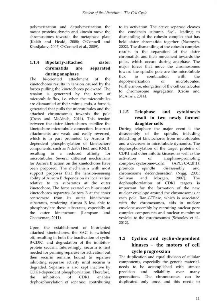
Ubiquitin-mediated proteolysis is carried out by the ubiquitin-proteasome system (UPS). In humans, 650 distinct E1, E2, and E3 enzymes and about 100 deubiqyitylases are involved in
the ubiquitylation process (Bhoj and Chen, 2009; Nijman et al., 2005). This system mediates modification of target substrates with multiple ubiquitin molecules (Teixeira and Reed, 2013). Ubiquitin is a small, highly conserved, protein consisting of 76 amino acids that is encoded by four genes (UBC, UBB, UBA52 and UBA80). These are transcribed and translated as linear fusions with multiple copies of ubiquitin forming precursor proteins. The polyubiquitin precursor is cleaved by endopeptidases to obtain free ubiquitin moieties that can be conjugated to substrates (Catic and Ploegh, 2005). Conjugation of ubiquitin to the substrate occurs in a stepwise-process (Figure 5). First, the ubiquitin molecule is linked to an ubiquitin-activating enzyme (E1) in an ATP-dependent manner. Next, the activated ubiquitin is transferred to the ubiquitin-conjugating enzyme E2, which in the case of APC/C are UBCH5 and UBCH10 (Peters,
Figure* 5.* The* enzymatic* cascade* of* ubiquitinVproteasome* pathway.*Ubiquitin$ (U)$ is$ activated$ and$covalently$ bound$ through$ a$ thioester$ bond$ to$ the$ ubiquitinTactivating$ (E1)$ enzyme$ in$ an$ ATPTdependent$reaction.$Next,$it$is$transferred$to$the$E2$conjugating$enzyme.$The$E2$interacts$with$the$E3$ubiquitinTligase$that$promotes$the$transfer$of$the$ubiquitin$from$the$E2$to$the$substrate.$The$ubiquitin$forms$a$covalent$ isopeptide$bond$between$ the$ubiquitin$and$ the$ target$ lysine$on$ the$substrate.$The$majority$of$the$E3s$are$either$RING/RINGTlike$or$HECT$ubiquitin$ligases.$The,$at$least$four$residue$long,$ubiquitin$chain$ is$recognized$by$the$26S$proteasome$which$degrades$the$substrate$protein.$Modified$from$(Ravid$and$Hochstrasser,$2008).*

Review of the Literature – The Cell Cycle
17
2006). Together with the substrate-recognizing E3 ubiquitin ligase, the E2 enzyme conjugates the C-terminus of the ubiquitin to a specific lysine residue on the target protein. In addition, specialized E3-like enzymes, E4, can catalyse chain extension (Teixeira and Reed, 2013). The E3 ligase, together with the substrate, determines what kind of ubiquitin-chain should be formed. A single E2 can interact with several different E3 ligases, and additional cofactors can affect the specificity or catalytic activity of the E2. The E2 plays a pivotal role in determing the outcome of the ubiqutylation as it influences the selection of the correct modifier, ubiquitin, and a suitable E3, as well as regulates processivity of the ubiquitin chain formation (Ye and Rape, 2009). Furthermore, the E2 conjugating
enzymes control the type of ubiquitin modification that will be added to the substrate, e.g. monoubiquitylation, multimonoubiquitylation (a single ubiquitin conjugated to multiple lysines on the substrate), or polyubiquitylation. Ubiquitin chain initiation and elongation are often performed by different E2s. To form distinct polyubiquitin chains, the next ubiquitin can be conjugated to one of seven lysine residues (K6, K11, K27, K29, K33, K48 and K63), or to the amino-terminus on the previous ubiquitin (Figure 6). The various chain topologies are structurally divergent and define the fate of the ubiquitylated protein. Depending on the topology of the chain, the protein can become targeted for proteasome-dependent proteolysis, or it can modulate the function, structure, assembly and localization of the protein (Flick and Kaiser, 2012; Ye and Rape,
Figure* 6.* Ubiquitin* chain* topology* dictates* the* outcome* of* ubiquitylation.* Ubiquitin$ chains$ of$different$ topologies$ have$ distinct$ functional$ consequences.$ A$ substrate$ (green)$ can$ be$modified$ by$monoT$ or$ polyubiquitin.$ The$ polyubiquitin$ chains$ are$ formed$ by$ the$ addition$ of$ another$ ubiquitin,$which$is$linked$to$the$previous$one$through$one$of$its$seven$residues.$These$form$specific$chains$that$are$recognized$different$proteins$and$thus$have$specific$outcomes.$Adapted$from$(Mocciaro$and$Rape,$2012;$Ye$and$Rape,$2009).*

Review of the Literature – The Cell Cycle
18
2009). The K48-linked chains were originally described as the signal that targets substrates for proteasomal degradation, whereas nonclassical linkage chains, such as K63-, K11-, or M1-linked chains, were associated with DNA repair regulation, cell-cycle progression, innate immunity, and inflammation (Ye and Rape, 2009). This is, however, not the whole truth as e.g. the K11-chains generated by APC/C and a mixture of K48-, K63-, and K11-linked chains on yeast cyclin B can target the substrates for destruction (Ikeda et al., 2010; Mocciaro and Rape, 2012). The most prominent polyubiquitin chains found on cell cycle regulators that are recognized and degraded by the 26S proteasome are the Lys11- and Lys48-linked chains formed by APC/C and SCF, respectively (Teixeira & Reed 2013).
Ubiquitin has been observed on proteins involved in a majority of the cellular processes, including DNA replication, DNA damage response, cell cycle regulation, chromatin organization, chromatin remodelling, apoptosis, transcription, and protein folding (Wagner et al., 2011). Ubiquitin signals are read and processed by ubiquitin-binding domains (UBDs), which specifically detect monoubiquitylation or different types of polyubiquitylation chains. For example, the proteasome receptor protein Rpn13 has a plextrin receptor for ubiquitin (Pru) that preferentially interacts with K48-linked diubiquitin, another UBD is the tandem ubiquitin-interacting motifs (UIMs) which can be found e.g. in the proteasome receptor S5a and in Rap80 (Ikeda et al., 2010). Furthermore,
ubiquitylation is also regulated by deubiquitylases (DUBs) (Nijman et al., 2005). Since several different post-translational modifications (PTMs) can be attached to a specific lysine, crosstalk between different modifications has been observed. These modifications have distinct outcomes, for example, monoubiquitylation of proliferating cell nuclear antigen promotes DNA repair, whereas sumoylation of the same lysine blocks sister chromatid recombination (Ye and Rape, 2009). Similarly, up to 30% of the lysines that have been found to be acetylated have also been shown to be ubiquitylated (Wagner et al., 2011). It has been suggested that acetylation prevents ubiquitin-dependent proteasomal degradation of proteins (Caron et al., 2005), pointing to intricate regulation of protein stability by the two modifications. 1.3.3 The% 26S% proteasome% and%
proteasomal%degradation%
The amount of proteins in the cell is determined by the rate of their synthesis and degradation. A majority of proteins are degraded in the 26S proteasome in an ATP-dependent manner. The proteasome is an enormous, 2.5 megadalton complex, which recognizes proteins that are specifically ubiquitylated (see chapter 1.3.2). The 26S proteasome consists of approximately 33 different proteins and comprises two subcomplexes, the 20S core particle and the 19S regulatory particle (Figure 7). Often two regulatory subcomplexes cap either end of the core particle. The proteolytic activity resides in the 20S core particle, which consists of four
Figure*7.*The*26S*proteasome*consists*of*the*20S* core* particle* flanked* by* the* 19S*regulatory* particles.* The$ 20S$ core$ contains$heptameric$ rings$ of$ αT$ and$ βTsubunits.$ Three$of$the$the$βTsubunits$have$proteolytic$activity.$The$ 19S$ regulatory$ particle$ can$ be$ divided$ in$the$ base$ (blue)$ and$ the$ lid$ (green).$ The$ base$contains$ e.g.$ RPN10$ and$ 13,$which$ recognize$the$ubiquitin$chain.$Modified$from$(Weissman$et$al.,$2011).$

Review of the Literature – The Cell Cycle
19
seven-subunit rings made up of α- and β-subunits. The β-subunits contain the catalytic activity, whereas the two flanking α-rings ensure that only unfolded polypeptides can enter the proteolytic chamber. The 19S regulatory particle consists of a base and a lid structure. The lid recognizes specific ubiquitin chains. The ATPases in the base unfolds the substrate protein and interact with the α-ring of the core particle, thus promoting the entry of the unfolded protein into the catalytic chamber (Weissman et al., 2011). The function of the 26S proteasome is mainly regulated by altering the composition of the complex, and certain subunits can be displaced in an inducible manner. Proteins that can associate with the proteasome include proteasome activators, ubiquitin rexeptors, as well as DUBs and E3 ligases, which can remodel the ubiquitin chain on the substrate, thus affecting its susceptibility for degradation. The proteasome recognizes substrates tagged with a chain of at least four ubiquitin molecules, or multiple monoubiquitins. The ubiquitin-chain is recognized by the ubiquitin receptors in the regulatory particle, usually RPN10 and RPN13. The proteasome then initiates degradation at an unstructured region in the substrate, and the ATPase motors pulls the substrate into the degradation channel and unfolds the substrate. The ubiquitin tag is cleaved off by the action of proteins in the 19S lid, and then recycled. The proteasome moves along the polypeptide chain and cuts the substrate sequentially into smaller peptides. By using this mechanism of sequential degradation, the proteasome can remodel protein complexes by only degrading the specific subunit at which it first initiates degradation (Weissman et al., 2011).
1.4 Inactivation% of% APC/C% by% the%
spindle% assembly% checkpoint%
protects% cells% against%
aneuploidy%
During mitosis the SAC operates to maintain genome stability by delaying the onset of
anaphase until all chromosomes are stably attached to the microtubule spindle and accurate chromosome segregation can be guaranteed (Figure 8). This requires that all chromosomes are correctly attached to the microtubule-spindle through their kinetochores, i.e. each kinetochore is held by microtubules from opposite poles. Unattached or incorrectly attached kinetochores activate the SAC, thus halting progression of mitosis. In essence, the SAC prolongs prometaphase by preventing the degradation of cyclin B and securin until all chromosomes are bi-polarly attached to the spindle microtubules in the metaphase. The SAC operates through a downstream target, the E3 ubiquitin ligase APC/C. APC/C, together with its subunit Cdc20, initiates anaphase by ubiquitylating cyclin B and securin, the degradation of which is required for the progression of mitosis (Musacchio, 2011). Thus, de novo cyclin B transcription and active translation are also needed to sustain an active SAC (Mena et al., 2010). In the absence of a working SAC, cells progress through anaphase prematurely with unattached or improperly attached chromosomes, usually leading to apoptosis of the cell. Sometimes, however, the resulting aneuploidy culminates in cellular transformation, especially if accompanied by the loss of function of genomic gatekeepers (Musacchio, 2011). In the presence of improperly attached kinetochores, SAC is activated through Mad2 binding to Cdc20, which enables the association between Cdc20 and BubR1 (Han 2013). This association of Mad2 and BubR1 with Cdc20 occurs at the kinetochores, which serve as catalytic platforms to accelerate the formation of the MCC. The kinases Bub1, MPS1 and Aurora B promote the recruitment of SAC proteins to the kinetochores. The kinetochore also serves as a sensor of correct bi-oriented attachment, where Aurora B assist in correcting improper attachments (Musacchio and Salmon, 2007). In the presence of incorrectly attached microtubules MCC, consisting of Cdc20 together with the SAC proteins BubR1, Mad2 and Mad3, is bound to APC/C. BubR1 inhibits the substrate recruitment of APC/C-Cdc20 as a

Review of the Literature – The Cell Cycle
20
pseudosubstrate, thus obstructing degron-recognition sites on Cdc20. By changing the interaction between Cdc20 and APC/C, BubR1 disrupts the bipartite D-box recognition site that is formed between Cdc20 and APC10 (Chao et al., 2012; Lara-Gonzalez et al., 2011). This means that even though the APC/C could be catalytically active, the absence of substrate recognition ultimately leads to the inhibition of the activity of the complex (Lara-Gonzalez et al., 2011). Instead APC/C, through APC15, autoubiquitylates Cdc20 leading to its degradation (Foster and Morgan, 2012; Mansfeld et al., 2011; Nilsson et al., 2008; Uzunova et al., 2012). The binding of p31Comet to the MCC complex also contributes to the continuous ubiquitylation of Cdc20 (Varetti et al., 2011). Furthermore, yet another step of regulation of Cdc20 has been reported, the deubiquitylation of Cdc20, mediated by
ubiquitin-specific protease 44 (UPS44), serves to prevent excessive ubiquitylation of Cdc20 and MCC disassembly and is thus required to sustain the SAC (Stegmeier et al., 2007). Degradation of Cdc20 mediates the constant turnover of Cdc20 and MCC on the APC/C, which preserves the possibility of SAC to respond to the attachment state of kinetochores (Mansfeld 2012). Consequently, the precise regulation of Cdc20 is important since it has been shown that cells devoid of Cdc20 cannot undergo mitosis, or undergo mitosis at a much slower pace, and removing Cdc20 during anaphase reactivates the SAC (Chow et al., 2011; Musacchio and Salmon, 2007). Overexpression of Cdc20, or a Cdc20 that cannot be ubiquitylated, overrides the SAC (Nilsson et al., 2008), which has been explained by that the MCC cannot contain the excess Cdc20. The continuous synthezitation and degradation of Cdc20 is thus essential
Figure* 8.! Principle* of* SAC* activation* and* deVactivation.* During$ prometaphase,$ in$ the$ presence$ of$unattached$kinetochores,$formation$of$the$mitotic$checkpoint$(MCC)$complex$is$catalysed.$The$MCC$is$composed$of$BubR1,$Bub3,$Mad2$and$Cdc20.$The$MCC$blocks$APC/C$substrate$recognition,$leading$to$inhibition$of$APC/C$activity.$ In$metaphase$when$the$kinetochores$are$bipolarly$attached$to$the$poles$generation$ of$ MCC$ ceases,$ allowing$ Cdc20$ to$ activate$ APC/C,$ resulting$ in$ the$ ubiquitylation$ and$degradation$ of$ cyclin$ B$ and$ securin.$ Degradation$ of$ securin$ renders$ separase$ catalytically$ active.$Separase$cleaves$the$cohesion$protein$Scc1$and$sister$chromatids$are$separated.$Cyclin$B$degradation$inactivates$ CDK1$ and$ the$ mitotic$ phosphorylation$ on$ several$ regulatory$ proteins$ is$ removed$ by$phosphatases,$resulting$in$the$progression$of$mitosis.$Modified$from$(LaraTGonzalez$et$al.,$2012).*

Review of the Literature – The Cell Cycle
21
both to maintain an active SAC, and also for enabling its timely and rapid switching off. SAC inactivation and cell cycle progression is in part due to the microtubule-kinetochore attachment but also by tension. Upon bi-oriented attachment of the kinetochore the centromeric chromatin is stretched, thus increasing kinetochore-to-kinetochore distance and tension. When the microtubules are attached to the kinetochore, the motorprotein dynein strips Mad1, Mad2, Mps1, CENP-F and other proteins from the kinetochore. The removal of Mps1 from the kinetochore has been suggested to be essential for avoiding SAC reactivation (Musacchio and Salmon, 2007). 2 Regulation*of*gene*expression**The genes hold the code for synthesis of the proteins that are needed for the building and functioning of a cell. Genes are encoded by a basic alphabet consisting of only four nucleobases: guanine (G), adenine (A), thymine (T), and cytosine (C). Together with a backbone, consisting of sugar and phosphate, these nucleobases form the double stranded helices that make up DNA, and thus the genetic instructions for the development and operation of every organism, from bacteria to humans. The DNA is inherited across generations, and in humans the DNA is divided on 46 chromosomes, 23 from each parent. With the exception of some mutations, every diploid cell in a given individual has the same set of chromosomes. Although every somatic cell contains the same set of genes, individual cell types need to express specific subsets of genes for their proper function. Moreover, in response to environmental changes the cell needs to rapidly adjust its transcriptional program to accommodate these (Maston et al., 2006). It is therefore substantial that gene expression is tightly controlled. Different transcriptional-regulatory programs are orchestrated by the
concerted action of factors that control transcription initiation and elongation, and the processing, transport, translation, and stability of messenger RNA (mRNA) (Figure 9) (Lundberg et al., 2010; Maston et al., 2006; Schwanhäusser et al., 2011; Vogel et al., 2010). The activity and function of the protein produced can then be further regulated by its localization, stability and by PTMs. Transcription of genes into RNA is catalyzed by RNA polymerases (RNAP). The RNAPs are multi-subunit protein complexes that faithfully generate an RNA copy of the coding part of the gene using DNA as a template. RNAPI is involved in transcribing 18S and 28S ribosomal RNA (rRNA) and RNAPIII transcribes 5S rRNA, tRNAs and some small RNAs. Protein-encoding genes are transcribed by RNAPII in eukaryotes. General transcription factors (GTFs), sequence-specific activator proteins, and coactivators contribute to RNAPII function and transcription. The GTFs include proteins such as Mediator, TFIIA, TFIIB, TFIID, TFIIE, TFIIF, and TFIIH, which assemble at the core promoter in an ordered fashion to form the transcription preinitiation complex (PIC) and direct RNAPII to the transcription start site (TSS). Mediator acts as a connection between sequence-specific transcription factors and the PIC (Thomas and Chiang, 2006). The direct connection between RNAPII and Mediator has been shown to be required for expression of most RNAPII transcribed genes in vivo (Soutourina et al., 2011). Since the assembly of PIC on the core promoter is only sufficient to initiate basal levels of mRNA transcription, the activators, e.g. HSF1, are needed. The activators usually bind to short specific DNA sequences upstream of the core promoter and stimulate transcription by modulating chromatin structure, by recruiting chromatin modifiers, by increasing the formation of PIC, or by promoting initiation, elongation or reinitiation of transcription (Maston et al., 2006; Thomas and Chiang, 2006).

Review of the Literature – Gene Expression
22
2.1 Regulation% of% transcription% at%
the%level%of%chromatin%
2.1.1 DNA% regulatory% elements% assist%
in% controlling% transcriptional%
initiation%
Gene expression is managed by several DNA regulatory elements that impact the rate of transcription and the positioning of the transcriptional machinery. The core promoter is the minimal proportion of the promoter needed for initiating transcription. The core promoter is usually located ~35 base pairs (bp) up- or downstreams of the TSS. The best-known core promoter element is the TATA-box, consisting of an AT-rich sequence located ~27 bp upstream of the TSS, but several other core promoter elements exist (Figure 10). To increase possibilities for transcriptional regulation, the promoters are highly diverse and utilize different variants and combinations of core elements, or no core promoter elements at all. The core promoter elements are usually associated with genes where focused transcription initiation occurs
e.g. regulated genes (Juven-Gershon and Kadonaga, 2010).
In addition to the core promoter, other regions of DNA, such as the proximal promoter and distal regulatory elements, contribute to transcriptional control by providing combinatorial control (Figure 10). These regulatory elements do not only affect transcription initiation but also transcription elongation. The proximal promoter is the region immediately upstream of the core promoter, which contains multiple binding sites for activators (Maston et al., 2006; Ong and Corces, 2011). One such binding site is the heat shock element (HSE), consisting of inverted repeats of the pentameric sequence nGAAn, which is accessible to heat shock factors (HSFs) (Amin et al., 1988).
%
Figure* 9.* A* schematic* picture* of* the* regulatory* steps* of* gene* expression* and* the* function* of* the*protein* product.*Gene$expression$ and$protein$ activity$ can$be$ regulated$ at$ several$ steps$ (blue$ text):$epigenetic$ marking$ and$ chromatin$ remodelling,$ transcription,$ RNA$ processing,$ mRNA$ stability$ and$export,$mRNA$translation,$protein$folding,$localization,$modification$and$degradation.$
Review of the Literature – Gene Expression
23
2.1.2 DNA%is%organized%into%chromatin%
that% facilitates% transcriptional%
regulation%
To provide means for further regulation of transcription, replication and repair, but above all to help organizing and compacting the DNA, the DNA exists as chromatin in complex with proteins called histones. The estimated length of the DNA in one human cell is about 2 meters, calling for a mechanism of efficient DNA compaction (Orphanides and Reinberg, 2000). For packaging into the nucleus the DNA is wrapped around a histone octamer consisting of four core histones that exist in pairs (H2A, H2B, H3 and H4). Additional linker histones, H1, and proteins that interact with the nucleosomes bring on further compaction of the DNA (Bell et al., 2011). This compaction of the chromatin, albeit indispensable, forms an arduous structural barrier for the transcription machinery. Chromatin packing affects all stages of transcription from activator binding and PIC formation to elongation. To interact with genomic regulatory elements, transcription factors must induce the reorganization of local nucleosome structures
at the transcribed loci. The assembly and the compaction of chromatin are regulated by multiple means, including DNA modifications (eg cytosine methylation and demethylation), PTMs of histones, the incorporation of histone variants, ATP-dependent chromatin remodelling and non-coding RNA (ncRNA)-mediated pathways (Li et al., 2007).
Five families of ATP-dependent chromatin-remodelling complexes have been found: SWI/SNF, imitation switch (ISWI), mi2/NuRD, chromodomain-helicase DNA-binding protein (CHD), and INO80/SWR1 complexes. These utilize ATP hydrolysis to exchange histones from the nucleosomes, to reposition or to evict nucleosomes. The SWI/SNF and INO80 families tend to increase transcription, whereas the mi2/NuRD and ISWI families usually regulate transcription negatively. Since the chromatin remodelling complexes are not capable of targeting specific DNA-sequences, they are recruited by sequence-specific regulators that bind specific histone modifications (Chen and Dent, 2013; Reid et al., 2009; Venters and Pugh, 2009).
*
Figure*10.*Typical*regulatory*elements* in*eukaryotic*gene*expression.*The$bestTknown$core$promoter$element$ is$ the$ TATATbox,$ consisting$ of$ an$ATTrich$ sequence$ located$ ~27$ bp$ upstream$of$ the$ TSS,$ but$several$ other$ core$ promoter$ elements$ exist,$ including$ initiator$ element$ (Inr)$ and$ X$ core$ promoter$element$1$ (XCPE1)$ localized$around$ the$TSS,$ the$TFIIB$ recognition$elements$ (BRE)$ that$ are$positioned$upstream$ of$ the$ TSS,$ and$ downstream$ promoter$ element$ (DPE),$ motif$ ten$ element$ (MTE)$ and$downstream$core$element$ (DCE)$ that$are$ situated$downstream$of$TSS.$The$distal$ regulatory$elements$include$ locus$ control$ regions$ (LCR),$ enhancers,$ silencers$ and$ insulators.$ The$ enhancers$ and$ silencers$have$ sites$ for$ binding$ multiple$ transcription$ factors$ and$ they$ function$ in$ activating$ and$ repressing$transcription,$ respectively.$ Insulators$operate$by$blocking$genes$ from$being$affected$by$ the$regulatory$elements$ of$ neighbouring$ genes.$ The$ LCR$ consists$ of$ multiple$ transcription$ regulatory$ elements$ that$function$together$to$provide$proper$expression$regulation$to$a$cluster$of$genes.$Modified$from$(JuvenTGershon$and$Kadonaga,$2010;$Maston$et$al.,$2006).$$

Review of the Literature – Gene Expression
24
2.1.2.1 Histone% modifications% and% their%
effect%on%transcription%
Dynamic changes of chromatin achieved by covalent modifications of histones, including phosphorylation, acetylation, methylation, sumoylation and ubiquitination, play a key role in regulating gene expression (Figure 11, also see Table 2 in Section 3.1.1). The reversible histone modifications are added and removed by kinases, histone acetyltransferases (HATs), histone deacetylases (HDACs), lysine methyltransferases (KMTs), lysine demethylases (KDMs), ubiquitylation enzymes and deubiquitylases (DUBs) (Kouzarides, 2007; Li et al., 2007; Taverna et al., 2007).
The histone modifications have a direct impact on the biophysical properties of nucleosomes and can function to recruit or exclude proteins from chromatin depending on the composition of the PTMs. Specific combinations of covalent modifications are associated with a given transcriptional state. Actively transcribed genes contain nucleosomes with modifications that are hallmarks of both initiation and elongation. Genome-wide analyses show that a combination of hyperacetylated H4, H3 lysine 9 acetylation (H3K9ac), H3K14ac and H3 lysine 4 methylation (H3K4me) correlates with genes that have an associated RNAPII. The elongation-associated modifications,
H3K36me3 or H3K79me2, are found at actively transcribed genes. In contrast, low levels of acetylation as well as histone H3 methylation of residues 9 and 27 (H3K9me and H3K27me) are associated with repressed or inactive genes (Guenther et al., 2007; Li et al., 2007; Reid et al., 2009).
The modifications recruit various proteins that bind via specific domains. Acetylation is recognized by bromodomains (BRD), which are found in many proteins including HATs and chromatin remodelling complexes. Phosphorylation is recognized by a domain within 14-3-3 proteins that act as an adaptor between the histone and another phosphoprotein. The chromo-like domains of the Royal family (chromo, tudor, MBT) and the plant homeodomain (PHD) domains recognize specific methylation states (Reid et al., 2009). 2.2 Initiation%of%transcription%
The first step of transcription initiation is the recognition of the core promoter, the assembly of PIC onto the core promoter, and the positioning of the RNAPII at the correct TSS (Figure 12)(Thomas and Chiang, 2006). The formation of the PIC is initiated by TFIID, consisting of the TATA-binding protein (TBP) and multiple other TBP-associated factors (TAFs). With the assistance of TFIIs and TAFs, TFIID binds to the TATA box through TBP.
Figure*11.*An*example*of*histone*tail*modifications*associated*with*active*chromatin.*These$histone$modifications$are$enriched$in$normal$growth$conditions$at$the$promoters$that$are$bound$by$HSF1$upon$heat$stress$according$to$Guertin,$et$al.,$2010.$

Review of the Literature – Gene Expression
25
TFIID does not only bind promoters containing TATA, but it has been shown to function at most of the RNAPII promoters, independently of the core promoter structure. The TAFs assist transcription by binding promoter elements, by integrating signals from multiple activators and by recognizing histone marks. Upon binding to the TATA box TFIID induces bending of the template DNA, thus nucleating the binding of the remaining GTFs. The action of the GTFs TFIIE, TFIIF and TFIIH mediate the localized unwinding of DNA and the direction of RNAPII to the TSS. The composition and build up of the transcriptional machinery can vary, and it is
suggested that the promoter sequence affects this, thus contributing to further transcriptional regulation (Goodrich and Tjian, 2010; Juven-Gershon and Kadonaga, 2010; Maston et al., 2006). In vivo experiments have shown that the transcription machinery is recruited as a complex containing both RNAPII, GTFs and additional factors (Prasanth et al., 2003; Thomas and Chiang, 2006).
After the formation of the PIC onto the TSS and the transcription of the first few nucleotides of mRNA the initially transcribing complex must undergo gross rearrangements
Figure*12.! A*model*of*transcription.*The$binding$of$sequenceTspecific$transcription$factors$(HSF$and$TF)$to$the$proximal$promoter$and$enhancer$leads$to$the$recruitment$of$chromatin$remodeling$enzymes$and$the$assembly$of$PIC.$PIC$consists$of$the$DNA$template,$hypophosphorylated$RNAPII$and$the$GTFs$TFIIA,$TFIIB,$ TFIIC,$ TFIID,$ TFIIE,$ TFIIH.$ TFIID$with$ its$ subunit$ TBP$ nucleates$ the$ assembly$ of$ PIC$ at$ the$ core$promoter.$The$other$components$of$PIC$catalyze$the$localized$melting$of$DNA$and$sliding$of$RNAPII$to$the$ TSS.$ RNAPII$ pausing$ occurs$ shortly$ after$ transcription$ initiation,$ and$ involves$ the$ negative$elongation$ factors$NELF$ and$DSIF.$During$ pausing$ the$ nascent$ transcript$ is$ capped$ at$ the$ 5´end.$ The$paused$RNAPII$is$released$by$the$action$of$PTTEFb,$which$phosphorylates$DSIF$and$dissociates$the$DSIFTNELF$complex.$PTTEFb$is$recruited$by$Mediator$and$by$the$capped$nascent$mRNA,$as$well$as$by$other$factors.$ Upon$ elongation$ DSIF$ turns$ into$ an$ elongation$ factor,$ the$ elongation$ factors$ TFIIS$ and$ TFIIF$associate$with$RNAPII,$and$the$CTD$of$RNAPII$is$phosphorylated$on$Ser2.$The$phosphorylation$serves$as$a$ platform$ for$ binding$ of$ RNA$ processing$ factors$ and$ further$ chromatin$ remodelling$ factors.$Termination$of$transcription$leads$to$the$dephosphorylation$of$the$CTD.*

Review of the Literature – Gene Expression
26
of its GTFs. For example, TFIIB that in the beginning helps stabilizing short RNA transcripts is displaced, thus committing RNAPII to promoter escape and stabilizing the transcription complex. This exchange of factors is in part orchestrated by the phosphorylation of the C-terminal fomain (CTD) of RNAPII (Nechaev and Adelman, 2011).
The RNAPII subunit Rbp1 contains a flexible CTD that is extensively modified by PTMs, mainly phosphorylation, during transcription. The phosphorylation pattern of the CTD varies depending on the stage of transcription and on the specific promoter. In humans, the CTD consists of 52 repeats of the conserved Y1S2P3T4S5P6S7 sequence. At the promoter the CTD is hypophosphorylated, and it is thought that this is required for PIC formation. Upon transition from preinitiation to elongation the CTD is extensively phosphorylated on Ser5 by the TFIIH component CDK7, leading to the release of RNAPII from Mediator (Heidemann et al., 2013). 2.3 RNAPII% and% the% elongation%
phase%of%transcription%
Traditionally the recruitment of RNAPII to the core promoter was considered the main regulatory step in transcription. This view has, however, been challenged by the notion that RNAPII is bound to the core promoter of approximately 30% of the metazoan genes, even ones that are not actively transcribed (Adelman and Lis, 2012; Core et al., 2008; Guenther et al., 2007; Lee et al., 2008a; Muse et al., 2007; Rahl et al., 2010; Zeitlinger et al., 2007). This promoter proximal pausing of RNAPII was first discovered on the Hsp70 promoter in D. melanogaster, where studies revealed that transcriptionally engaged RNAPII accumulated just downstream of the promoter and was associated with 20–60-nt-long nascent RNA (Gilmour and Lis, 1985; 1986; Rasmussen and Lis, 1993; Rougvie and Lis, 1988).
The paused RNAPII is maintained by the actions of negative elongation factor (NELF), DRB-sensitivity-inducing factor (DSIF) and
Gdown1. Gdown1 contributes to stalling RNAPII by inhibiting the association of the elongation factor TFIIF and termination factor TTF2 to RNAPII (Cheng et al., 2012; Guo and Price, 2013; Mullen Davis et al., 2014). Pausing is relieved by the action of the CDK, positive transcription elongation factor b (P-TEFb), and the histone chaperone FACT. P-TEFb is recruited to the promoters by various factors, including BRD4, the MED26 component of Mediator complex, and transcription factors such as c-Myc and nuclear factor κ B (NFκB). BRD4 can mediate the release of the inhibitory 7SK small nuclear ribonuclear particle (7SK snRNP) from P-TEFb, resulting in activation of P-TEFB (Barboric et al., 2001; Eberhardy and Farnham, 2002; Jang et al., 2005; Rahl et al., 2010; Takahashi et al., 2011; Zhou et al., 2012). During promoter pausing, 5’ capping of the nascent transcript functions as a checkpoint for productive elongation. The capping enzyme is recruited to RNAPII via DSIF and phosphorylation of S5 of the CTD, and it provides an additional platform for P-TEFB loading (Kwak and Lis, 2013; Mandal et al., 2004).
P-TEFb phosphorylates the DSIF-NELF complex, leading to its dissociation, which results in the release of NELF from RNAPII, and the transformation of DSIF into a positive elongation factor (Adelman and Lis, 2012; Wada et al., 2000; 1998). Upon the release of NELF, other elongation factors, such as TFIIS and TFIIF, which facilitate the release of the paused RNAPII into productive elongation, are able to associate with RNAPII. Elongation is obstructed by chromatin, thus the presence of chromatin modifying factors is required. Upon the transition to elongation mode, CDK8 is thought to phosphorylate Ser2 on the CTD, creating a platform for the recruitment of chromatin modifiers (Kwak and Lis, 2013). The RNAPII elongation complex does not only extend the nascent mRNA chain to produce the full-length pre-mRNA, it also serves as a central coordinator of post-transcriptional processes that control proper expression of a gene. The recruitment of RNA processing factors in an orderly manner is regulated by the sequential phosphorylation

Review of the Literature – Gene Expression
27
of the CTD. For example, CDK7-mediated phosphorylation of Ser5 creates a binding site for capping enzymes, and phosphorylation of Ser2 upon elongation recruits other RNA-processing factors. After termination the CTD needs to be dephosphorylated to allow RNAPII to enter the next round of transcription. Several phosphatases that are associated with the TFIIs regulate this event. The phosphorylation and dephosphorylation of the RNAPII CTD thus allows for coordinated regulation of transcription elongation, 5’ capping, splicing, and 3’ processing (Heidemann et al., 2013; Zhou et al., 2012). 2.4 Translation% –% from%mRNA% to% a%
nascent%polypeptide%chain%
Translation is the part of gene expression when ribosome complexes decode the mRNA that was produced as a result of transcription. The product of translation is a chain of amino acids, which will fold into the active protein. Translation can be divided into three stages: initiation, elongation and termination of which initiation is the most regulated step. During initiation the 80S ribosome complex is assembled and then positioned at the translation start site of the mRNA, usually the AUG codon closest to the ribosome-binding site. Next the peptide chain is elongated and during termination the newly synthesized protein is released and the ribosomal subunits are dissociated from the mRNA.
Translational initiation requires at least nine eukaryotic transcription factors (eIFs) that assist in the assembly of the 80S ribosome and the recognition of the right start codon. Initiation starts when the eukaryotic translation initiation factor (eIF), eIF4F, recognizes the 5’-cap of the mRNA and unwinds the secondary structures to enable binding of the 43S complex. eIF4F and eIF3 facilitate the recruitment of the 43S ribosomal subunit. The 43S complex scans the mRNA in a 5’-to-3’ direction until it encounters an AUG codon in an optimal context. When the Met-tRNAMet base pairs with the correct AUG start-codon it switches the scanning complex to a closed conformation by displacing eIF1 that is
essential in maintaining the fidelity of transcription by dissociating the complex from suboptimal codons. Hydrolysis of the eIF2-bound GTP commits the ribosome to a specific start codon. The final step of the 80S ribosome formation is the joining of the 60S subunit and the simultaneous dissociation of eIFs is mediated by the ribosome-dependent GTPase eIF5B (Jackson et al., 2010). During elongation amino acids are added to the growing amino acid chain in a step-wise fashion. The cycle is repeated until the ribosome encounters a stop-codon and the translation is terminated. Subsequently the subunits of the 80S are dissociated and cycled back for initiation of another round of mRNA translation (Graille and Séraphin, 2012; Voorhees and Ramakrishnan, 2013).
Translation is a cyclic process, in which initiation and elongation is followed by termination and subsequent recycling of the 80S ribosome to a new initiation complex. The efficacy of this is cyclic process is assisted by the circularization of the mRNA. mRNA circularization stabilizes the interaction of the eIF4E with the cap, leading to an increased rate of translation initiation. This process is facilitated by the eIF4G, which in addition to assisting in recruitment of the 43S ribosome, also interacts with the polyadenylate-binding protein 1 (PABP1). The ability of eIF4G to interact simultaneously with eIF4E at the 5’ end and PABP1 at the 3’ end brings the two ends of the mRNA in close proximity (Brook et al., 2012; Kahvejian et al., 2005). Not all translation of mRNA is initiated in a cap-dependent manner. The internal ribosome entry site (IRES) provides an internal ribosome-binding site, thus bypassing the requirement for the cap. Consequently, IRESs function independently of eIF4E (Hellen and Sarnow, 2001; Johannes and Sarnow, 1998; Kolupaeva et al., 1998). 2.5 PostMtranscriptional%
regulation%of%gene%expression%
The concentration of proteins in the cell reflects the integration of regulation at several levels, from mRNA production, stability and translation to protein degradation. Gene

Review of the Literature – Gene Expression
28
expression can be controlled post-transcriptionally on the level of processing, export, localization, turnover and translation of mRNAs. Global translational regulation that affects most mRNA transcripts usually occurs by changes in the phosphorylation state of the eIFs or by adjusting the number of available ribosomes. In contrast, the transcript-specific regulation, which modulates the translation of a distinct group of mRNAs, is mediated by various mechanisms. Translational regulation is especially important under conditions that require sudden and precise changes in protein levels, including the cellular response to stress and apoptosis, the regulation of cell growth and its coordination with cell division, and during differentiation and development (Mata et al., 2005). Stress that results in global repression of translation is often accompanied by an increase in translation of specific proteins that are required for cell survival (Richter et al., 2010). A study by Fan et al. reveals that the altered abundance of approximately 50% of the transcripts affected by various stresses, including heat shock and UV radiation, was dependent on the mRNA stability rather than the expression (Fan et al., 2002). 2.6 NonMcoding%RNAs%as%regulators%
of%gene%expression%
In the human genome, and across metazoans, only approximately 20 000 protein coding genes are found, which constitutes less than 1.5% of the whole human genome. However, still the vast majority of the genome is transcribed. For many years it was assumed that untranslated RNA molecules serve no function, but in the past 20 years scientists have argued that these transcripts have a regulatory role (Mattick, 2013). Interestingly, even though the number of protein-coding RNAs is approximately the same among all metazoans, the amount of non-coding genomic sequences positively correlates with the developmental complexity of the organism, further increasing the importance of the non-coding RNAs. In mammals non-coding sequences constitute more than 98% of the genome (Liu et al., 2013). The repertoire of
the non-coding RNAs (ncRNA) differs between cells, and they are differentially expressed during differentiation, development and in the cell cycle (Dinger et al., 2008; Kitagawa et al., 2013; Mercer et al., 2008; Zhou et al., 2009). Furthermore, many of these ncRNAs are either sequentially or structurally conserved among species, indicating that these are important for the function of the cell (Mercer et al., 2013). Despite the fairly recent discovery of the ncRNAs many intriguing functions have already been unveiled. They control gene expression at all levels from transcription and RNA processing to translation. They also have functions in protecting genomes from foreign nucleic acids. The ncRNAs can based on their length be divided into long non-coding RNAs (lncRNAs) and small non-coding RNAs (sncRNAs). The sncRNAs include microRNA (miRNA), Piwi-interacting RNA (piRNA), small interfering RNA (siRNA) and small nucleolar RNA (snoRNA). The small RNAs can roughly be divided into two groups: small RNAs that are involved in gene silencing and those that are not. The group involved in gene silencing through the RNA interference (RNAi) pathway includes miRNA, piRNA and various types of siRNAs. These all share the same characteristic features; they are between 20 and 30 nt long, they have 5´phosphate groups and 3´hydroxyl termini and they all associate with Argonaute family proteins. The function of the small RNA is determinded by the combination of a specific small RNA with a specific Argonaute (Ago) protein (Czech and Hannon, 2011). 2.6.1 MicroRNAs% regulate% gene%
expression% postM
transcriptionally%
MiRNAs function as powerful post-transcriptional regulators of gene expression. The first miRNA, lin-4, was discovered in C. elegans where it regulates the temporal development of the worm (Lee et al., 1993; Wightman et al., 1993). Lin-4 appeared to be specific for worms, and it was only with the discovery of the second miRNA, let-7, that was found in a wide range of animal species

Review of the Literature – Gene Expression
29
(Pasquinelli et al., 2000), that it became clear that miRNAs are widespread regulators of gene expression in the animal kingdom. MiRNAs play an important role also in plant development and metabolism (Huntzinger and Izaurralde, 2011). The amount of miRNAs has expanded through evolution, and more complex animals have a more intricate repertoire of miRNAs (Berezikov, 2011). To date, more than 1872 miRNA precursors and 2578 mature miRNAs have been identified in humans (miRBase release 20)(Kozomara and Griffiths-Jones, 2013). Because of the nature of the interaction between the miRNA and its target mRNA, a single miRNA can target multiple mRNAs simultaneously (Baek et al., 2008). Moreover, many miRNAs and RNA-binding proteins (RBP) can together target a single mRNA. This makes the miRNAs well suited for co-ordinating or fine-tuning the expression of proteins in the cell (Peter, 2010; Wu et al., 2010a). In a characterization of different tissues and individual cell lines, an average of 70 miRNAs were expressed in each sample (Landgraf et al., 2007), pointing to the complexity of the systemNAs regulate the activity of a majority of the protein-coding genes and are thus involved in everything from cell differentiation, metabolism and apoptosis to tumorigenesis (Krol et al., 2010). 2.6.2 miRNA%biogenesis%%
MiRNAs are single-stranded RNAs with a length of approximately 22 nt. As only hairpins are recognized and processed by Drosha and Dicer, a prerequisite for miRNA biogenesis is the correct secondary structure of the pri- and pre-miRNA. The genes encoding miRNAs are found to mainly be located in introns and but some are also localized to exons of protein-coding genes or in pseudogenes and transposons (Berezikov, 2011). MiRNAs are frequently transcribed as clusters from a polycistronic transcription unit, and about 50% of the mammalian miRNA loci are found in close proximity to other miRNAs. Furthermore, the majority of the miRNA genes have multiple isoforms, paralogues, generated by gene duplication. Paralogues often have identical 5´seed
sequences (nt 2-7 from the 5´end) and more variable 3´sequences (Kim et al., 2009b). The transcription of most miRNAs is mediated by RNAPII, but a few miRNAs, those associated with Alu repeats, are transcribed by RNAPIII (Borchert et al., 2006; Lee et al., 2004). The primary transcripts, pri-miRNA, are several kb long and contains many stem loop structures that are recognized by the Microprocessor complex. The Microprocessor complex consists of nuclear RNase III-type protein Drosha that cleaves the transcript and its cofactor DiGeorge syndrome critical region gene 8 (DGCR8) that recognizes the stem-loop structure (Han et al., 2004; Kim et al., 2009b; Lee et al., 2003). A number of other Microprocessor cofactors have also been found, including DEAD box RNA helicases p68 (DDX5) and p72 (DDX17), as well as heterogeneous nuclear ribonucleoproteins (hnRNPs)(Gregory et al., 2004). The Microprocessor complex cleaves the pri-miRNA, thus releasing an approximately 70 nt long hairpin-like transcript, called pre-miRNA (Lee et al., 2003). pre-miRNA is transported out of the nucleus by the co-ordinated action of Exportin-5, which recognizes the hairpin-structure, and a Ran-GTP (Yi et al., 2003). !In the cytoplasm, Dicer, another nuclear RNase III-type protein, processes the pre-miRNA further by cleaving near the loop of the hairpin, thus releasing ~22 nt long imperfectly double-stranded miRNAs with 2-nt single-stranded 3’ overhangs at both ends. Dicer collaborates with two proteins TAR RNA-binding protein (TRBP) and Interferon-inducible double-stranded RNA-dependent protein kinase activator A (PACT) that are thought to advance formation of the RNA-induced silencing complex (RISC). Following, or simultaneously with, the cleavage of pre-miRNA by Dicer, the miRNA/miRNA* duplex must be loaded onto the Ago protein, to form the RISC complex. Upon loading maturation occurs, leading to the release of the passenger strand (miRNA*) of the duplex miRNA, leaving only the guide strand (miRNA) in the RISC complex. The maturation depends on the Dicer, the structure of the miRNA/miRNA* duplex, its

Review of the Literature – Gene Expression
30
terminal nucleotides and thermodynamic properties and the Ago protein (Czech and Hannon, 2011; Kim et al., 2009b). Generally the strand with the less-stably base-paired 5´ end becomes the guide strand, that guides the mature RISC to target mRNAs (Khvorova et al., 2003). Dicer, TRBP and PACT, together with a Hsc70-Hsp90 complex, contribute to the loading of miRNA/miRNA* into the pocket of Ago and to the subsequent release of miRNA* from Ago in a process that requires ATP (Figure 13)(Czech and Hannon, 2011; Iwasaki et al., 2010; Johnston et al., 2010). *
In addition to the pre-miRNAs processed by Drosha and Dicer, several unconventional miRNAs using alternative maturation strategies have been found. For example, mirtrons have been reported in both flies and mammals. Mirtrons bypass the Drosha-mediated processing and instead use the splicing machinery to produce pre-miRNAs. Mirtrons are very short introns that use the splicing machinery to produce pre-miRNAs. The introns are first excised from the mRNA, debranched, trimmed and refolded into short stem–loop structures that mimic pre-miRNAs, thus bypassing Drosha-mediated processing. Mirtrons are then exported from the nucleus
*
Figure*13.*miRNA*biogenesis.*Canonical$pathway:$The$nascent$priTmicroRNA$(priTmiRNA)$transcripts$are$first$processed$into$~70Tnucleotide$preTmiRNAs$by$Drosha$inside$the$nucleus.$Mirtron$pathway:$Lariat$introns$ spliced$ from$preTmRNA$are$debranced$by$ lariat$ debranching$enzyme$ (Ldbr),$ after$which$ they$either$directly$fold$into$preTmiRNA$hairpins$or$undergo$trimming$of$the$tails.$3’$tails$are$trimmed$by$the$RNA$ exosome,$ while$ the$ enzymes$ responsible$ for$ 5’$ trimming$ are$ not$ known.$ Both$ pathways:$ PreTmiRNAs$are$transported$to$the$cytoplasm$by$Exportin$5$and$processed$into$miRNA:miRNA*$duplexes$by$Dicer.$Dicer$also$processes$long$dsRNA$molecules$into$small$interfering$RNA$(siRNA)$duplexes.$Only$one$strand$ of$ the$ miRNA:miRNA*$ duplex$ or$ the$ siRNA$ duplex$ is$ preferentially$ assembled$ into$ the$ RNATinduced$silencing$complex$ (RISC),$which$subsequently$acts$on$ its$ target$by$ translational$ repression$or$mRNA$cleavage,$depending,$at$ least$ in$part,$on$ the$ level$of$complementarity$between$ the$small$RNA$and$its$target.$ORF,$open$reading$frame.$(Suzuki$and$Miyazono,$2011).$

Review of the Literature – Gene Expression
31
by Exportin-5 and processed into mature miRNAs by Dicer (Berezikov et al., 2007; Okamura et al., 2007; Ruby et al., 2007). Another pathway, utilized by miR-451, bypasses Dicer processing and the short stem-loop pre-miR-452 is cleaved by Ago2 instead, before further processing (Cifuentes et al., 2010; Suzuki and Miyazono, 2011). 2.6.3 miRNA%mechanisms%of%action%
Generally, miRNAs have two primary modes of action, they inhibit protein synthesis either by repressing translation or by causing deadenylation and subsequent degradation of mRNA targets (Fabian et al., 2010). The fate of the targeted mRNA depends on two things, the complementarity in base-pairing between the miRNA and the catalytic activity of the proteins within the RISC complex. By utilizing Watson-Crick base-pairing miRNAs interact with their target mRNA. The base-pairing is usually imperfect, except for in the seed region, which comprises nt 2-8 from the 5´end. The 3´end does not need to be complementary, however, it stabilizes the miRNA-mRNA interaction, thus contributing to the regulation. Mismatches that prevent endonucleolytic cleavage of mRNA by the Ago occur frequently at nt 10-12. Usually the miRNA targets the 3’ UTR of the mRNA, but other target sites have also been found. For the most part, multiple target sites on the mRNA, either for the same or different miRNAs, are required for effective repression (Bartel, 2009). The exact modes of action that miRNAs use to exert translational repression are unclear, but several different mechanisms have been proposed, including inhibition of translation initiation and elongation, co-translational mRNA degradation, and premature termination of translation. At the initiation step, the RISC complex has been suggested to repress translation by competing with eIF4E binding to the cap, by deadenylation of the 5’ end thus eventually preventing mRNA circularization, or by hindering ribosome 60S-association (Chendrimada et al., 2007; Eulalio et al., 2009; Filipowicz et al., 2008; Kiriakidou et al., 2007). Inhibition of initiation is thought
to be more common than restriction of elongation, where RISC is thought to slow down elongation or to terminate translation prematurely by causing premature ribosome dissociation (Filipowicz et al., 2008; Huntzinger and Izaurralde, 2011; Petersen et al., 2006). Most of the repression is, however, thought to occur by destabilization of the target mRNA (Baek et al., 2008; Behm-Ansmant et al., 2006; Hendrickson et al., 2009; Huntzinger and Izaurralde, 2011; Lim et al., 2005). Albeit miRNAs have been shown to mediate endonucleolytic cleavage of perfectly complementary target mRNAs, this is not a common mechanism in animal cells, since the majority of the targets are not fully complementary (Yekta et al., 2004). Instead, degradation of the target mRNA occurs through the 5’-to-3’ mRNA decay pathway, where the mRNA is first deadenylated, which triggers decappapping and finally digestion by the 5’-to3’ exonuclease XRN1. Huntzinger and co-workers (2011) propose a model for miRNA-mediated mRNA decay that is dependent on the miRNA, Ago, GW182, PABPC, deadenylases (CAF1, CCR4 and NOT complex) and DCP decapping enzymes. According to the model Ago bridges the interaction between miRNA and the RISC complex component GW182, which is required for miRNA-mediated silencing. GW182 interacts with PABPC that normally is located at the 5’ poly(A) tail of the target mRNA, thus triggering deadenylation of the target mRNA. The PABPC-GW182 interaction could contribute to silencing by competing with eIF4G binding, by reducing the affinity of PABPC for the poly(A) tail, or by recruiting other proteins that degrade the mRNA. Inhibition of eIF4G-PABPC or poly(A)-PABPC binding counteracts mRNA circularization and thereby ribosome recycling as well as exposes the target mRNA to decay enzymes (Huntzinger and Izaurralde, 2011). In addition to these above proposed mechanisms of mRNA degradation it has recently been shown that miRISC can recruit decapping enzymes to the mRNA (Nishihara et al., 2013). The deadenylation of the target leads to storage of the translationally repressed

Review of the Literature – Gene Expression
32
deadenylated mRNA in the P-bodies, or upon decapping ultimately to its degradation by the 5’-to-3’ exonuclease (Huntzinger and Izaurralde, 2011). Traditionally miRNAs have been considered translational repressors. Some data has, however, shown that miRNAs under certain conditions have the potential to activate mRNA translation. During amino acid starvation miR-10a relieves the translational repression of the ribosome mRNAs, and upon serum starvation-induced cell cycle arrest some miRNAs can up-regulate translation of the same target genes that they repress in cycling cells (Vasudevan and Steitz, 2007; Vasudevan et al., 2007; Ørom et al., 2008). 2.6.4 Regulation%of%miRNA%biogenesis%
and%activity%
As with mRNAs, miRNAs are regulated on multiple levels: transcription, processing, stability, and localization. Transcription is regulated by TFs, enhancers, silencing elements and chromatin modifications (Sato et al., 2011). Several activators and repressors regulate miRNA processing through either protein–protein or protein–RNA interactions. Regulation of processing can occur both via regulating processing activity of Drosha or Dicer by e.g. post-translational modification of the RNases, by affecting the microprocessor cofactors or by RNPs binding to the specific pri-miRNA or pre-miRNA. Also editing of the RNA transcript itself regulates miRNA activity (Krol et al., 2010; Siomi and Siomi, 2010). MiRNA stability and turnover are tightly regulated. The stability is in part regulated by the miRNA sequence itself, especially by modifications at the 3´end, as well as by exonuclease proteins, including XRN1, RRP41, and PNPaseold-35 (Bail et al., 2010; Krol et al., 2010; Rüegger and Großhans, 2012).
Another cell cycle and stress-regulated step of miRNA function is the miRISC and mRNA degradation machinery, the protein components of which can be regulated by various PTMs. An example is the phosphorylation of DCP family members during both stress conditions and mitosis,
which leads to stabilization of a subset of mRNA, suggesting that DCP phosphorylation inhibits decapping (Aizer et al., 2013; Yoon et al., 2010). The miRISC-target mRNA interaction can also be inhibited by the binding of HuR protein to the target mRNA (Bhattacharyya et al., 2006; Kundu et al., 2012). Furthermore, to ensure proper regulation of miRNA activity, a majority of the miRNAs, miRNA-processing factors and miRISC components are tightly controlled in various feedback regulatory mechanisms (Siomi and Siomi, 2010). 2.6.5 Biological%functions%of%miRNA%
Because of the capability of miRNA to co-ordinately target multiple mRNAs and the possibility of a single mRNA to be targeted by numerous different miRNAs, miRNAs have the capacity to precisely control complex regulatory networks (Sass et al., 2011). MiRNA-mediated regulation is predicted to control many cellular events, and the precise and timely expression of miRNAs in a cell- and tissue-dependent manner during embryogenesis suggests that miRNAs are involved in the development and function of the organisms (Stefani and Slack, 2008). 2.6.5.1 miRNA%in%spermatogenesis%
The spatiotemporal regulation of gene expression is extremely important during spermatogenesis. Post-transcriptional regulation is particularly important due to the transcriptional silencing that occurs during later stages of spermatogenesis (Yadav and Kotaja, 2014). Large-scale transcription of genes occurs in two phases during spermatogenesis: in the pachytene spermatocytes before the cells become quiescent during the process of meiosis, and post-meiotically before nuclear silencing. Most of the transcripts produced during these transcriptional bursts are then stored for translation later in spermatogenesis. Interestingly, increased miRNA levels coincide with both of these transcriptional bursts, emphasizing the potential role of miRNAs in the post-transcriptional regulation

Review of the Literature – Gene Expression
33
of genes during spermatogenesis (McIver et al., 2012a). During spermatogenesis, miRNAs, Dicer and Ago2 are co-localized in the chromatoid body, which is a fibrous structure in the cytoplasm of spermatocyte and spermatides (Kotaja et al., 2006). Germ-cell specific disruption of Dicer at different stages of development has demonstrated that miRNA biogenesis is essential at various phases of germ cell development and spermatogenesis. During embryogenesis, Dicer disruption causes decreased proliferation and problems in the colonization of primordial germ cells (PGCs) into the gonads, leading to a substantial decrease in spermatogonia cells in Dicer knock-out mice. Intriguingly, a testis-specific Dicer knock-out showed decrease in spermatogonia proliferation, and only some of the seminiferous tubules had active spermatogenesis. Thus, these mice were fertile at young age (4 months), but became sterile after 8 months (Hayashi et al., 2008; Maatouk et al., 2008). The mature spermatozoa found in the epididymis were few and had pronounced morphological abnormalities, with abnormal mitochondria and chromatin organization (Korhonen et al., 2011; Maatouk et al., 2008). Thus, the Dicer knockout (KO) spermatozoa had pronounced motility defects (Maatouk et al., 2008). Interestingly, a similar Ago2 testis-specific knock-out did not generate the same phenotype, suggesting that miRNAs regulate gene expression together with another Ago protein (Hayashi et al., 2008). Another evidence for the importance of miRNA-mediated regulation during spermatogenesis are the high miRNA expression levels in PGCs, germ cells and germ stem cells (McIver et al., 2012a; 2012b). The miR-221~222 cluster as well as miR-146 and miR-21, which are expressed in undifferentiated spermatogonia are suggested to maintain stem cell capacity and inhibit differentiation of the spermatogonia (Huszar and Payne, 2013; Niu et al., 2011; Yang et al., 2013). The miR-17~92 cluster, together with its paralog miR-106b~25, are also highly expressed in PGC cells and spermatogonia, where they are thought to promote cell
cycling (Hayashi et al., 2008; Tong et al., 2012). Deletion of the miR-17~92 cluster in mouse results in small testes, mild defects in spermatogenesis, and a decrease in sperm in the epididymis. Furthermore, deletion of miR-17~92 leads to increased expression of miR-106b~25, pointing to functional co-operation (Tong et al., 2012). MiR-34c is not only expressed in spermatogonia but also in spermatocytes and round spermatids where it is involved in enhancing the germinal phenotype. The miR-449 cluster is highly expressed upon meiotic initiation. Together with miR-34c, miR-449 targets the E2F transcription factor that regulates cell cycle progression. Later in spermatogenesis, histones are replaced with transition proteins (TP) and protamines (Prm), which is important for the correct compaction of DNA in the elongated spermatids. Before this stage, TP2 and Prm2 mRNAs are targeted by the testis-specific miR-469 that represses TP2 and Prm2 protein expression in pachytene spermatocytes and round spermatids, thus preventing early chromatid reorganization. Also miR-122a targets TP2 (Yadav and Kotaja, 2014). Taken together these data show that Dicer and specific miRNAs are vital for early male germ cell proliferation and late spermatogenesis. 2.6.5.2 miRNA%as%cell%cycle%regulators%
Many miRNAs studied so far in the cell cycle seem to have an effect on cell cycle-entry and the restriction point by regulating fast-decaying cyclins (Bueno and Malumbres, 2011). Other miRNAs regulate proliferation by inducing cell cycle arrest (Lal et al., 2009). Only a few studies have identified miRNAs that could potentially affect mitosis. These include miR-125b, miR-24 and let-7 that decrease the expression of cyclin A or cyclin B, which drive entry and progression through mitosis. Three other miRNAs, miR195, miR-516-3p and miR-128a, have been shown to down-regulate Wee1, one of the kinases that negatively regulates the cyclin B-CDK1 complex at the G2/M transition. Down-regulation of Wee1 activity could lead to early mitotic entry (Bueno and Malumbres, 2011). Hypoxia-induced expression of miR-210 was

Review of the Literature – Gene Expression
34
found lead to a decrease in the protein levels of mitosis-related genes, including Cdc25B, Plk1 and Bub1B. An increase in miR-210 was further shown to correlate with disturbance in progression through mitosis and aberrant mitosis (He et al., 2013). Overexpression of the miR-17~92 cluster results in increased proliferation and elevated levels of cell cycle regulatory proteins like CDK2, cyclin D2, c-Myc and CREB, whereas p15, Smad2 and Rb expression decreased. These proteins are mainly involved in regulating S phase entry (Attar et al., 2012). Many of the above mentioned miRNAs are found to be de-regulated in cancer where they affect cell cycle progression, but studies examining cell cycle-regulated miRNA expression are scarce. Since miRNAs are regulated in a similar manner as mRNAs, their expression should be regulated throughout the cell cycle. A study examining the expression of miRNAs by using a microarray after synchronization by double thymidine block, found that 25 miRNAs are differentially expressed in a cell cycle-dependent manner in HeLa cells. Among these the levels of let-7 family members as well as miR-34a, were found to be increased at G2/M transition and the miR-34b/c and miR-221 family members were increased during S phase (Zhou et al., 2009). Also miRNA clusters let-7a-d, let-7i, miR-15b-16-2, and miR-106b-25 have been found to be induced during G1/S transition (Bueno et al., 2010). The cell cycle-dependent concentration of miRNAs is not only regulated by expression of the miRNAs, but also by their stability. In a study by Hwang and co-workers, miR-29b was shown to be imported into the nucleus and degraded rapidly in interphase cells, whereas it was more stable in mitosis (Hwang et al., 2007). MiRNAs are only active in association with Ago, so expression alone cannot tell about the miRNA activity. A study examining miRNAs co-immunoprecipitated with Ago2 in unsynchronized and mitotic HeLa cells found increased association of let-7 family members with Ago2 in mitosis, whereas miR-21 association was decreased (Hausser et al., 2013; Kishore et al., 2013).
Taken together, evidence for miRNAs as cell cycle regulators is accumulating, especially in causing cell cycle arrest in response to DNA damage. In agreement with the important role for the cell cycle, multiple miRNAs have the same targets, pointing to a very precise control of these cell cycle regulators. Furthermore, some evidence exist that miRNAs are regulated in a cell cycle-dependent manner. Interestingly, a few important transcription factors can induce the expression of multiple miRNA clusters, thus fine-tuning cell cycle control. 2.6.5.3 The% effect% of% miRNAs% in% disease% is%
enhanced%upon%stress%
When miRNAs were detected they were thought to mainly function in development. Since the discovery of miRNAs in humans, hundreds of expression profiling studies have shown that tumors exhibit dysregulated miRNA expression patterns, with both loss and amplification of miRNAs, relative to corresponding normal tissue. Several pieces of evidence from different transgenic mouse models suggest that specific miRNAs function as tumor suppressors and oncogenes in various tissues (Kota et al., 2009; Ma et al., 2010; Mendell and Olson, 2012; Trang et al., 2011). However, the transgenic deletion of most of the miRNAs does not show any phenotype unless the animal or the tissue is subjected to stress, implying that the effect of miRNA is enhanced under stress conditions. Cancer cells are under constant stress induced by DNA damage, aneuploidy, protein misfolding and the accumulation of reactive oxygen species. This results in an addiction of the cells to stress-induced signalling pathways, which can be strongly modulated by the miRNAs (Luo et al., 2009; Mendell and Olson, 2012). Evidence of miRNAs as mediators of strong positive and negative feedback circuits promoting tumor progression also exist (Mendell and Olson, 2012). 2.6.6 The%miRM17~92%cluster%
Many miRNAs are transcribed as clusters, where one pri-miRNA is processed into

Review of the Literature – Gene Expression
35
several mature miRNAs (Kim 2009, Stefani & Slack 2008). One such polycistronic cluster, which is conserved among vertebrates, is the miR-17~92. The 800 nt long miR-17~92 cluster is located in the non-protein coding gene MIR17HG (miR-17/92 cluster host gene or C13orf25) on human chromosome 13 (chromosome 14 in mouse) and comprises six miRNAs: miR-17, miR-18a, miR-19a, miR-20a, miR-19b-1 and miR-92a-1 (Mendell, 2008; Ota et al., 2004). The cluster has been duplicated during evolution, and both mouse and human genomes contain two paralogues of the main cluster, miR-106b~25 and miR-106~363-106b~25 containing three miRNAs is located in an intron of the MCM7 gene on chromosome 7 (7q22.1), whereas the miR-106a~363 cluster is located on the X chromosome (Xq26.2) and comprises six miRNAs (Figure 14)(Tanzer and Stadler, 2004). The miR-17~92 and miR-106~25 clusters are abundantly expressed in a wide range of tissues, but miR-106~363 is expressed at extremely low levels in normal tissues and cells (Mendell, 2008).
Figure*14.*Organization*of*the*human*miRV17~92*cluster*and*its*paralogs.*The$boxes$with$the$same$colors$ indicate$ miRNAs$ belonging$ to$ the$ same$family$ based$ on$ the$ identical$ seed$ sequences$(Mendell,$2008).$
Transcription of the miR-17~92 cluster is regulated by various transcription factors. The first factor to be found to regulate miR-17~92 was the oncogene c-Myc that up-regulates its expression (O'Donnell et al., 2005). E2F1 and E2F3 are two other regulators that bind to the promoter of miR-17~92. Intriguingly, miR-17~92 itself targets the E2F transcription factors, thus forming an auto-regulatory loop
that aids in the control of G1 to S phase progression (O'Donnell et al., 2005; Sylvestre et al., 2007; Woods et al., 2007). The miR-17~92 is repressed by p53, which in turn is controlled by miR-25 in response to c-Myc or E2F regulation, forming yet another loop of regulatory control (Yan et al., 2009). In cells containing p53, miR-17~92 expression will thus result in apoptosis, whereas it in cells deficient of p53 will result in increased proliferation (Zeitels and Mendell, 2013). The ENCODE project found another 33 transcription factors, including Bcl3, that regulate both miR-17~92 and miR-106~25 (Mogilyansky and Rigoutsos, 2013). Despite the discovery of all these transcription factors that control miR-17~92, the regulation of the cluster is still highly enigmatic. Another layer of complexity is added by the post-transcriptional regulation of the individual miRNAs, as is the case for miR-18. 2.6.6.1 MiRH18%
MiR-18 exists as two isoforms, miR-18a and miR-18b (Figure 15). The two miRNAs only differ from each other with a single nucleotide, situated in the 3’-region of the sequence. Thus the seed region in the 5´-end is identical between the two isoforms, suggesting that they can target the same mRNAs. MiR-18a is transcribed from the miR-17~92 cluster, whereas miR-18b resides in the paralog miR-106a~363 cluster located on the X chromosome. The expression of these paralogues is different, and the miR-17~92 cluster, including miR18a, is more redundantly expressed in the organism (Mendell, 2008; Ventura et al., 2008). Although miR-17~92, as well as other clusters, are expressed as clusters of several miRNAs, the levels of the individual member miRNAs vary within the cluster (Hayashita et al., 2005; Jevnaker et al., 2011; Landais et al., 2007). MiR-18a, for example, is specifically bound by the RNA binding protein hnRNP A1. hnRNP A1 associates with the loop of the pre-miR-18a thus reshaping the stem-loop structure which eventually results in more efficient processing by Drosha. This leads to increased control and enhanced processing of miR-18a compared to

Review of the Literature – Gene Expression
36
the other cluster members (Guil and Cáceres, 2007; Michlewski et al., 2008).
Figure*15.*The*sequence*of*miRV18a*and*miRV18b.*The$ seed$ region$ is$ in$ blue,$ the$ one$ base$ that$differs$is$underlined$(Mendell,$2008).$
Thorough analysis of the function of miR-18 is still missing to date, but some information on miR-18 expression does exist. During mouse embryogenesis miR-18a levels fluctuate, indicating a developmental function for miR-18a (Mineno et al., 2006). In the whole embryo the miR-18a levels increase from embryonic day E8.5 to E11.5 (Jevnaker et al., 2011). Another study showed that the miR-18 family members, miR-18a and b, are crucial for the formation and segregation of the germ layers during embryogenesis in both X. laevis and M. musculus. At this stage miR-18 represses Smad2, rendering the cells less sensitive to Nodal signalling, thus targeting blastomeres to become either ectoderm or mesoderm (Colas 2012). In several tissues, including skin, kidney and heart, miR-18a expression is highest during embryogenesis, and markedly decreased in postnatal tissues (Jevnaker et al., 2011). MiR-18 levels are generally low in adult tissues, the highest levels of miR-18 expression being detected in kidney and lung (Boggs et al., 2007; Sempere et al., 2004). During brain development the relative miR-18a levels decrease (Miska et al., 2004; Podolska et al., 2011). A reduction in miR-18 is also observed during post-natal growth in various brain tissues, except in the hippocampus, where expression fluctuates. In cultured neuronal cells miR-18 has been shown to decrease glucocorticoid receptor levels (Uchida et al., 2008; Vreugdenhil et al., 2009). Since the researchers found an inverse correlation of miR-18 and glucocorticoid receptor in the paraventricular nucleus region
of the brain in two rat strains with intrinsic differences in reactivity to acute stress, it has been suggested that miR-18 would increase the stress sensitivity of the organism (Uchida et al., 2008). However, the levels of miR-18 that are needed to down-regulate glucocorticoid receptor protein in vitro exceeds by far the amount found in the brain, leaving the role of miR-18 in stress-susceptibility still open (Vreugdenhil and Berezikov, 2010; Vreugdenhil et al., 2009).
Interestingly, although miR-18 expression in tissues decreases during development, it again increases during carcinogenesis, indicating that miR-18 would counteract cell differentiation and aid proliferation. Increased miR-18a expression has been found in numerous cancers, including B-cell chronic lymphocytic leukemia, B-cell lymphoma, diffuse large B-cell lymphoma, bladder cancer, breast cancer, colorectal cancer, head and neck cancer, hepatocellular carcinoma, nasopharyngeal cancer, osteosarcoma, pancreatic cancer, renal cancer, and urothelial cancer (Leivonen et al., 2009; Mogilyansky and Rigoutsos, 2013; Motoyama et al., 2009; Murakami et al., 2013; Song et al., 2014; Wu et al., 2013; Xu et al., 2014; Yu et al., 2013). Only some targets of miR-18 in cancer have been validated (Table 1). For example, in colorectal cancer, where Myc induces expression of miR-17~92, miR-18 promotes angiogenesis and tumor growth by directly repressing the antiangiogenic protein thrombospondin 1 (TSP1) (Dews et al., 2006). Another study found that miR-18a inhibits DNA damage repair in colorectal cancer by directly targeting one of the major kinases, ataxia telangiectasia mutated (ATM). Consequently, treating HT-29 cells with a double stranded break-inducing agent, etoposide, impaired the restoration of DNA damage in cells were miR-18 was overexpressed. This resulted in decreased proliferation and increased apoptosis (Wu et al., 2013). The suppression of the DNA damage repair mechanism induced by increased miR-18a expression could possibly serve a catalyzing role in the formation of colorectal cancer. In agreement, up-regulation
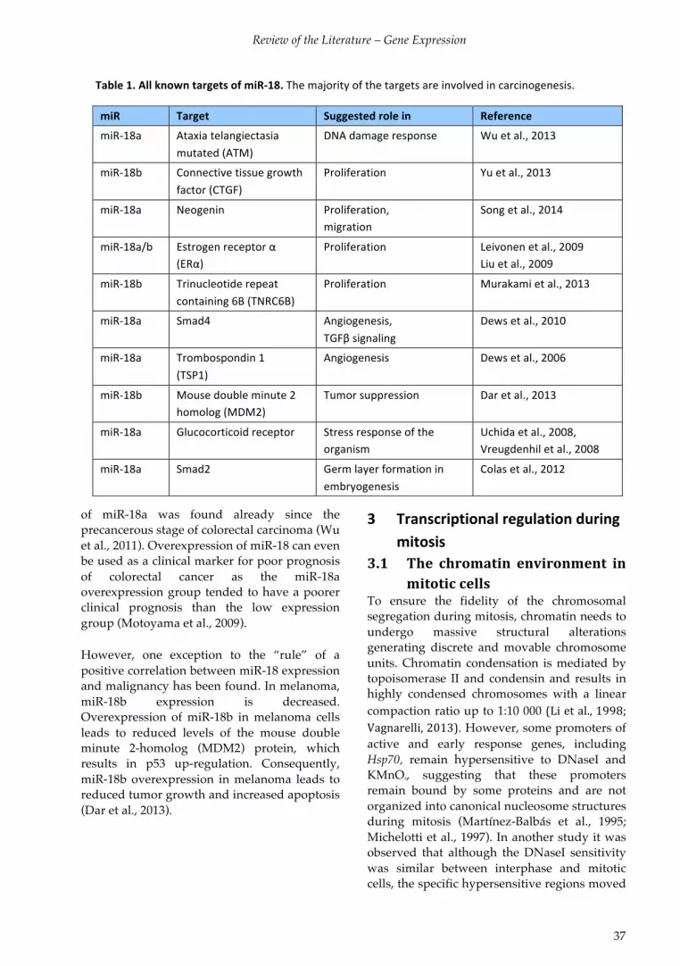
Review of the Literature – Gene Expression
37
*Table*1.*All*known*targets*of*miRV18.*The$majority$of$the$targets$are$involved$in$carcinogenesis.
of miR-18a was found already since the precancerous stage of colorectal carcinoma (Wu et al., 2011). Overexpression of miR-18 can even be used as a clinical marker for poor prognosis of colorectal cancer as the miR-18a overexpression group tended to have a poorer clinical prognosis than the low expression group (Motoyama et al., 2009).
However, one exception to the “rule” of a positive correlation between miR-18 expression and malignancy has been found. In melanoma, miR-18b expression is decreased. Overexpression of miR-18b in melanoma cells leads to reduced levels of the mouse double minute 2-homolog (MDM2) protein, which results in p53 up-regulation. Consequently, miR-18b overexpression in melanoma leads to reduced tumor growth and increased apoptosis (Dar et al., 2013).
3 Transcriptional*regulation*during*mitosis*
3.1 The% chromatin% environment% in%
mitotic%cells%%
To ensure the fidelity of the chromosomal segregation during mitosis, chromatin needs to undergo massive structural alterations generating discrete and movable chromosome units. Chromatin condensation is mediated by topoisomerase II and condensin and results in highly condensed chromosomes with a linear compaction ratio up to 1:10 000 (Li!et!al.,!1998;!Vagnarelli,!2013). However, some promoters of active and early response genes, including Hsp70, remain hypersensitive to DNaseI and KMnO4, suggesting that these promoters remain bound by some proteins and are not organized into canonical nucleosome structures during mitosis (Martínez-Balbás et al., 1995; Michelotti et al., 1997). In another study it was observed that although the DNaseI sensitivity was similar between interphase and mitotic cells, the specific hypersensitive regions moved
miR*$ Target$ Suggested*role*in* Reference$
miRT18a$ Ataxia$telangiectasia$
mutated$(ATM)$
DNA$damage$response$ Wu$et$al.,$2013$
miRT18b$ Connective$tissue$growth$
factor$(CTGF)$
Proliferation$ Yu$et$al.,$2013$
miRT18a$ Neogenin$ Proliferation,$$
migration$
Song$et$al.,$2014$
miRT18a/b$
$
Estrogen$receptor$α$
(ERα)$
Proliferation$ Leivonen$et$al.,$2009$
Liu$et$al.,$2009$
miRT18b$ Trinucleotide$repeat$
containing$6B$(TNRC6B)$
Proliferation$ Murakami$et$al.,$2013$
miRT18a$ Smad4$ Angiogenesis,$$
TGFβ$signaling$
Dews$et$al.,$2010$
miRT18a$ Trombospondin$1$
(TSP1)$
Angiogenesis$ Dews$et$al.,$2006$
miRT18b$ Mouse$double$minute$2$
homolog$(MDM2)$
Tumor$suppression$ Dar$et$al.,$2013$
miRT18a$ Glucocorticoid$receptor$ Stress$response$of$the$
organism$
Uchida$et$al.,$2008,$
Vreugdenhil$et$al.,$2008$
miRT18a$ Smad2$ Germ$layer$formation$in$
embryogenesis$
Colas$et$al.,$2012$

Review of the Literature – Transcription during Mitosis
38
during mitosis (Kuo et al., 1982). Even when the DNA structure remains open in mitosis, the majority of genes are transcriptionally silenced and RNAPII and RNAPIII as well as many of the sequence-specific transcription factors are displaced (Christova and Oelgeschläger, 2001; Martínez-Balbás et al., 1995)25. Interestingly, most of these factors, including HSF1, retain their in vitro DNA-binding capacity, but they cannot access the DNA (Martínez-Balbás et al., 1995; Vihervaara et al., 2013), suggesting that changes in the chromatin environment, in chromatin-protein or protein-protein interaction take place during mitosis. These changes occur through phosphorylation of transcription factors but they also involve chromatin modifications that displace histone-bound proteins. RNAPI and some transcription factors retain their dynamic association dynamic association/dissociation with the transcriptionally silent mitotic DNA (Chen et al., 2005). 3.1.1 Changes%in%histone%modifications%
in%mitosis%
During mitosis histone modifications play critical roles in chromosome segregation, transcription silencing, epigenetic inheritance, and re-establishing cellular programs during division (Table 2)(Wang and Higgins, 2013). Histone acetylation, which is associated with active transcription in interphase, decrease globally in mitosis, whereas gene promoters with active transcription at the onset of mitosis retain acetylation (Bonenfant et al., 2007; Dey et al., 2009; Kouskouti and Talianidis, 2005; Kruhlak et al., 2001; McManus et al., 2006; Valls et al., 2005; Zhao et al., 2011). Ubiquitylation of histones H2A and H2B, involved in transcription repression and activation, respectively, also decrease in mitotic cells (Wang and Higgins, 2013). Monomethylation increase throughout the genome in mitosis, whereas di- and trimethylation of histone H3K4 (H3K4me2 and H3K4me3), which are associated with active or poised genes, remain largely unchanged upon entry into mitosis (Bonenfant et al., 2007; Kelly et al., 2010; Kouskouti and Talianidis, 2005; McManus et al., 2006; Valls et al., 2005). The repressive
histone methylation marks, H3K9me2, H3K9me3 and H3K27me3, are also retained in mitosis (Kelly et al., 2010; McManus et al., 2006). As a result of PRSet7 activity the histone H4 methylation mark, H4K20me1, starts to increase in S phase and reaches its peak in metaphase. Disruption of PRSet7 or H4K20me1 has been suggested to result in the inability of cells to progress past G2, global chromosome condensation failure, aberrant centrosome amplification, and substantial DNA damage (Houston et al., 2008). Another study showed no effect of H4K20me1 modification on cell cycle progression (Pesavento et al., 2008). 3.1.2 The% role% of% histone% H3%
phosphorylation%in%mitosis%%
Phosphorylation of histone H3 has emerged as an important modification in transcriptional activation, in chromosome condensation, and in protein displacement during mitosis. When cells enter mitosis histone H3 is extensively phosphorylated both on serine 10 (H3S10P), as well as on threonine 3 (H3T3P), threonine 11 (H3T11P) and serine 28 (H3S28P). Aurora B kinase-mediated phosphorylation of H3 serine 10 coincides with chromatin condensation, and it has been suggested to contribute to chromatin compaction (Crosio et al., 2002; Hsu et al., 2000; Kouzarides, 2007). In the ciliate protozoa Tetrahymena a S10A mutation resulted in abnormal chromosome condensation, demonstrating that H3S10P is required for chromatin condensation. Similarly, the H3S10A mutation or the disruption of Aurora B homolog, Ipl1, in S. cerevisiae compromised chromosome condensation (Neurohr et al., 2011). However, another study done in HeLa and CHO cells showed that Aurora B was required for initiation, but not maintenance of chromatin condensation, and that H3S10P alone was not sufficient for condensation (Van Hooser et al., 1998). Taken together, these data show that the function of H3 phosphorylation in condensation is still not fully understood.

Review of the Literature – Transcription during Mitosis
39
Histone* Enzyme*writer*
Change*in*mitosis* Suggested*function*in*mitosis*and*function*in*transcription*(italics)*
H2AT120P$ Bub1$ Increase$ Shugosin$recruitment,$CPC$localization$and$
chromatin$structure$modulation.$
H2AK119Ub$ PRC1,$
BRCA1$
Decrease$ Prevents$Aurora$B$binding$and$H3S10P$until$
mitosis?$Repression.*H2BK120Ub$ $ Decrease$ Set1$methyltransferase$recruitment.$Activation.*H3T3P$ Haspin$ Increase$ Survivin$binding,$TFIID$displacement$
H3K4me2/3$ Set1,$MLL$ Maintained$ Bookmarking$active$genes,$CENPTA$loading?$
Activation*of*transcription*initiation.*H3K4ac$ $ Generally$
decreased$
Counteracting$H3K4me2$and$Shugosin$binding$at$
centromeres?$
H3K9me2/3$ Suv39H1/2,$
G9a$
Maintained$ Bookmarking$heterochromatin$and$inactive$
genes?$Repression.*H3K9ac$ $ Decreased$ Hinders$Aurora$B$H3S10P$until$mitosis?$
Activation*of*transcription*initiation,*when*together*with*H3K14ac.*
H3S10P$ Aurora$B$ Increase$ Displacement$of$HP1,$ASF/SF2,$SRp20,$
chromosome$condensation?$Activation*when*together*with*hyperacetylated*histones.*
H3T11P$ $ Increase$ ?$
H3K14ac$ p300,$ PCAF,$
TIP60$
Maintained$ HP1$displacement?$Activation*of*transcription*initiation*when*together*with*H3K9ac.*
H3K27me3$ PRC2$ Generally$
decreased,$
maintained$on$
bookmarked$genes$
Bookmarking$PcG$genes?$Repression.*
H3S28P$ Aurora$B$ Increase$ PRC1$displacement?$
H3K34me2/3$ Set2$ Maintained$ CENPTA$loading?$
H3K36me$ ?$ Decreased$ ?$in$mitosis.$Represses*internal*initiation,*increases*elongation.*
H4S1P$ ?$ Increase$ ?$
H4K5ac$ HAT1,$p300,$
TIP60$
Decreased,$except$
at$bookmarked$
promoters$
Brd4$binding,$bookmarking.$Activation.*
H4K20me1$ PRset7$ Increase$in$G2$ Condensin$II$binding.$Silencing*transcription.*H1.4S27P$ Aurora$ B,$
CDKs$
Increase$ H1$mobility?$HP1$displacement?$
H3.3S31P$ $ Increase$ Unknown$
Table*2.*Histone*modifications*with* functional* relevance* in*mitosis.*Modified$ from$(Berger,$2007;$Bonenfant$et$al.,$2007;$Li$et$al.,$2007;$Wang$and$Higgins,$2013).$
Review of the Literature – Transcription during Mitosis
40
In contrast, H3S10P has been suggested to activate the immediate-early response genes, c-Jun and c-Fos. These promoters are also hyperacetylated, indicating that phosphorylation and acetylation function together in activating the genes (Barratt et al., 1994; Crosio et al., 2003; Dyson et al., 2005; Mahadevan et al., 1991). H3S10P has also been shown to increase on the Hsp70 promoter in HSF1-dependent manner upon heat shock in D. melanogaster. In D. melanogaster phosphorylation of H3 seems to correspond to mammalian histone H4 acetylation, which correlates with active transcription in mammalian cells. Histone H4 acetylation is not affected by heat in D. melanogaster (Nowak and Corces, 2000; Nowak et al., 2003; Thomson et al., 2004). The role of H3S10P in the heat shock response in mammalian cells is less clear; one study reports that it increases upon heat shock in interphase cells (Valls et al., 2005), whereas other studies show no or a reversed effect (Dyson et al., 2005; Thomson et al., 2004). Instead, arsenic, which activates HSF1, increases H3S10P on the Hsp70 promoter (Thomson et al., 2004). Changes in histone modifications can help displacing proteins from the chromatin. Histone H3 phosphorylation has been implicated in a phospho-methyl switch that displaces heterochromatin protein 1 (HP1), splicing factors SRp20 and ASF/SF2, polycomb proteins (PcGs), as well as the TFIID subunit TAF3 from chromatin (Fischle et al., 2005; Fonseca et al., 2012; Hirota et al., 2005; Loomis et al., 2009; Varier et al., 2010). 3.2 Transcription% and% translation%
are% periodically% regulated% in% a%
cell%cycleMdependent%manner%
Gene regulation during cell cycle progression is an elaborately choreographed process. The flawless temporal control of gene expression in distinct phases of the cell cycle maintains essential checkpoints that ensure the precise completion of chromosome duplication and the accurate segregation of the chromosomes to the daughter cells. Thus, it is not surprising that many mRNAs are transcriptionally activated in
a cell-cycle phase-dependent manner. In human primary fibroblasts, HeLa and U2OS cells more than 700, 850 and 1870 genes, respectively, have been found to be transcriptionally regulated. A comparison between normal and cancer cells reveal that 60% of the cell cycle regulated genes are common between HeLa and primary foreskin fibroblasts, the rest are differentially expressed. The periodically expressed genes include those participating in cell cycle progression (Bar-Joseph et al., 2008; Cho et al., 2001; Grant et al., 2013; Whitfield et al., 2002). Two distinct waves of gene expression take place during the cell cycle. Early cell cycle gene expression occurs during G1/S to produce proteins required for DNA replication, while late cell cycle gene expression begins during G2 and prepares the cell for mitosis (Whitfield et al., 2002). A plethora of different transcription factors have been shown to regulate the cell cycle-dependent gene expression. Examples are p53 and E2F1 that control G1/S transition, FOXM1 and B-Myb that regulate G2/M transition, and the DREAM complex (dimerization partner, RB-like, E2F and multi-vulval class B) that represses transcription in quiescent cells (Dynlacht, 1997; Grant et al., 2013; Sadasivam et al., 2012; Sadasivam and DeCaprio, 2013). The activity of these transcription factors, in turn, is mainly controlled by various cell cycle kinases, including CDKs and Plk1 (Fairley et al., 2012). In addition to transcription being regulated during the cell cycle, translation is also controlled. By employing ribosome profiling, it was found that translation of functionally related sets of mRNAs are co-ordinately regulated in a cell cycle-dependent manner. The regulation was most prominent in the G1 and S phases. Among the translationally controlled mRNA networks were transcripts encoding proteins required for metabolism, nuclear transport, and DNA repair. Several cell cycle regulators, such as Plk1 and Bub1, as well as proteins responsible for maintaining genomic integrity and organizing the higher-order structure of chromosomes are also translationally regulated. Interestingly the mRNA encoding the chaperone HSPA1A was shown to be translated to a higher extent in S

Review of the Literature – Transcription during Mitosis
41
phase than in mitosis, whereas more HSPA1B mRNA is translated in mitosis than in G1 phase (Stumpf et al., 2013). Taken together, these data suggest that translational control is a suitable mechanism for fine-tuning gene expression during dynamic processes such as cell-cycle progression. 3.3 %Eukaryotic% transcription% is%
repressed%during%mitosis%
In interphase, genes are actively transcribed into RNA at a high rate, but in mitosis transcription abruptly ceases. Experiments done at the beginning of the 1960s show that the incorporation of labelled RNA in the nucleus begins to decline at early prophase and from mid-prophase, when the chromatin becomes condensed, until mid-telophase, almost all RNA synthesis is seized (Johnson and Holland, 1965; Konrad, 1963; Prescott and Bender, 1962; Taylor, 1960). Transcription by all three RNAPs is repressed (Fink and Turnock, 1977; Johnston et al., 1987), whereas transcription of mitochondrial RNA as well as Cyclin B, needed for mitotic progression, have been suggested to continue (Fan! and! Penman,!1970a;!Mena!et!al.,!2010;!Sciortino!et!al.,!2001). Not only transcription but also pre-mRNA processing is inhibited in mitosis when the pre-mRNA processing factors become dispersed throughout the cytoplasm (Prasanth et al., 2003). In accordance with the repressed RNA synthesis, protein synthesis also decreases during mitosis to the rate of 25-30% of that in interphase cells (Fan and Penman, 1970a; Konrad, 1963; Le Breton et al., 2005; Prescott and Bender, 1962; Salb and Marcus, 1965; Stumpf et al., 2013; Taylor, 1960). Interestingly, even though the rate of RNA synthesis is drastically decreased during prophase, a temporary increase in protein synthesis is observed in some cell types simultaneously (Konrad, 1963), indicating that translation of mitosis-specific mRNAs occurs. For example, Cyclin B transcripts are polyadenylated and translated at a greater rate during M-phase compared to interphase (Le Breton et al., 2005). The protein translation is not only reduced
because of the diminished accessibility of mRNAs, but also the initiation and elongation of protein translation is decreased (Fan! and!Penman,! 1970b). The decrease in translation initiation is owing to the switch from cap-dependent to cap-independent translation of mRNAs with specific IRES (Pyronnet et al., 2001; 2000; Qin and Sarnow, 2004). This change is mediated by inhibiting the cap-recognizing factors, eIF4E and eIF4B by hypophosphorylation, thus leading to the sequestering of the factors by 14-3-3σ (Pyronnet et al., 2001; Wilker et al., 2007). Interestingly, if this translation switch is abrogated, cells undergo an aberrant mitosis resulting in binucleate cells (Wilker et al., 2007). Shortening of the poly(A) tail is another mechanism that is suggested to repress translation of specific transcripts during mitosis. In mitosis the activity of poly(A) polymerase is repressed because of phosphorylation by Cyclin B-CDK1, resulting in a reduction in poly(A) RNA (Colgan et al., 1996; Le Breton et al., 2005). 3.4 How% is% mitotic% transcriptional%
repression%executed?%
Although mitotic transcriptional repression was discovered more than 50 years ago, the exact mechanism behind this repression still remains unclear. It has been speculated that repression could depend on: the condensation of chromosomes, on other modifications of the nucleosome that would limit the accessibility of the transcriptional machinery, or on direct inhibition of transcription factors or the transcriptional machinery by e.g. PTMs. The most likely cause is a combination of these mechanisms. 3.4.1 Chromatin%condensation%has%only%
a% minor% contribution% to% mitotic%
transcriptional%repression%
The first hypothesis put forward on how transcription is silenced in mitosis was transcriptional repression by condensation of the interphase chromatin into mitotic chromosomes. Condensation of chromatin would limit the accessibility of the DNA to

Review of the Literature – Transcription during Mitosis
42
transcription factors and transcription machinery. Since the transcriptional repression coincides with the condensation of the chromosomes, this hypothesis got some support (Johnson and Holland, 1965; Prescott and Bender, 1962). However, subsequently both in vitro transcription studies and in situ hybridization studies have shown that chromatin condensation alone is not enough to inhibit transcription. In Xenopus laevis egg extracts the inhibition of topoisomerase II, which facilitates chromatin condensation both in vivo and in vitro, did not prevent mitotic repression of transcription. The addition of excessive quantities of non-specific DNA, which inhibits nucleosome formation, was not able to de-repress mitotic transcription. These results indicated that neither the nucleosome formation, nor the binding of a general repressive factor was required to mediate mitotic repression in vitro (Hartl et al., 1993). Evidence confirming that chromatin condensation itself does not inhibit transcription came when it was shown that no transcription occurred in mitotic cells infected with nucleosome-free virus DNA, showing that transcriptional repression occurs in the absence of nucleosomal condensation (Spencer et al., 2000). In mitosis, the chromatin is also moved around the cell. The relocalization of a transcriptionally active locus from the interior of the nucleus to the nuclear envelope, causes silencing, which could contribute to the repression of transcription observed in mitosis (Reddy et al., 2008). 3.4.2 Phosphorylation% of% the% RNAPII%
transcriptional% machinery%
represses%transcription%
During mitosis, RNAPII as well as most of the transcription initiation factors and sequence specific transcription factors are excluded from the chromatin (Chen et al., 2002; Christova and Oelgeschläger, 2001; Parsons and Spencer, 1997). The displacement of RNAPII from mitotic chromatin and transcription inhibition coincide with increased hyperphosphorylation of the RNAP II CTD (Akoulitchev and Reinberg, 1998; Dirks and Snaar, 1999; Parsons and Spencer, 1997). Although
hyperphosphorylation of RNAPII is needed for transcriptional elongation, it has been shown that hyperphosphorylated RNAPII cannot assemble in transcriptional initiation complexes. Therefore, hyperphosphorylation of unengaged RNAPII results in inactivation of the complex (Egloff and Murphy, 2008). In a reconstituted system purified Cyclin B-CDK1 is enough to repress RNAPII-mediated transcription. Cyclin B-CDK1 phosphorylates the CTD of RNAPII in the PIC, thus disrupting the complex in vitro (Gebara et al., 1997; Leresche et al., 1996; Zawel et al., 1993). Another site on the CTD is phosphorylated by CDK7, a kinase that is associated with TFIIH, and phosphorylation of this site activates transcription (Gebara et al., 1997; Oelgeschl ger, 2002). During mitosis the Cyclin H-CDK7 kinase activity is inhibited by CDK1 phosphorylation (Akoulitchev and Reinberg, 1998; Long et al., 1998), presumably contributing to the inactivation of RNAPII. Not all of the DNA-bound proteins are displaced from mitotic chromosomes, as e.g. TFIIB, TFIID and TBP together with TAFs were found to associate, albeit to a lesser extent, with active gene promoters, including Hsp70, during mitosis (Chen et al., 2002; Christova and Oelgeschläger, 2001; Xing et al., 2008). Although bound to DNA, the ability of TFIID to direct activator-dependent transcription is repressed. This repression is mediated by mitosis-specific phosphorylation of TBP and TFIID-specific TAFs (Leresche et al., 1996; Segil et al., 1996). Taken together, even though human TFIID–promoter complexes associate with some active promoters, they are unable to stabilize fully assembled transcription initiation complexes during mitosis, and paused promoter-proximal RNAP II is displaced (Christova and Oelgeschläger, 2001).
Abortion of ongoing transcription contributes to transcriptional repression in mitotic cells. The transcription elongation complex P-TEFb is inhibited through phosphorylation by Plk1, resulting in destabilization of elongation (Jiang et al., 2013). Also the transcription termination factor TTF2 represses transcriptional elongation during mitosis. TTF2 levels rise in the cytoplasm during S and G2 phases, and upon mitosis TTF2 translocates into the nucleus

Review of the Literature – Transcription during Mitosis
43
where it terminates RNAPII transcriptional elongation, leading to the displacement of RNAPII from DNA. The Gdown1 subunit of RNAPII stabilizes promoter-proximal paused RNAPII and inhibits termination by TTF2. Gdown1 that is phosphorylated during mitosis shows reduced affinity with RNAPII, thus enabling TTF2-dependent termination of transcription (Guo et al., 2014).
As summarized in Figure 16, regulation of RNAPII-mediated transcription during mitosis is due to several factors. The decreased promoter occupancy of basal and gene-specific transcription factors inhibits the recruitment of RNAPII. The transcription initiation factors that remain associated with the promoter are phosphorylated which reduces their capacity to
nucleate the assembly of RNAPII and the PIC at the promoter. Furthermore, RNAPII itself is phosphorylated on the CTD, which inhibits initiation of transcription. Finally, by the action of the transcription termination factor transcription elongation, already engaged RNAPIIs are aborted. Mitotic phosphorylation of components of the transcription machinery that represses transcription and re-activation of transcription upon subsequent dephosphorylation occurring from telophase onwards is similar to the phosphorylation and dephosphorylation of other important factors that regulate progression through mitosis. These events seem to be regulated by common kinases, including CDK1 and Plk1.
Figure* 16.* Transcription* is* repressed* in* mitotic* cells.*Mitotic$ repression$ of$ transcription$ is$ due$ to$several$factors.$The$chromatin$environment$changes$in$mitosis,$and$nucleosomes$can$slide,$thus$hiding$transcription$ factor$ binding$ sites$ in$ mitosis.$ However,$ some$ proteins,$ including$ the$ TFIID$ and$ some$bookmarking$proteins,$are$retained$at$the$DNA.$Several$sequenceTspecific$transcription$factors,$general$transcription$ factors$ (GTF),$ and$ chromatin$ remodelling$ factors$ are$ displaced$ from$mitotic$ chromatin$due$ to$mitosisTspecific$ phosphorylation.$ The$ assembly$ of$ the$ preTinitiation$ complex$ (PIC)$ is$ inhibited$due$ to$ hyperphosphorylation$ of$ the$ CTD$ of$ RNAPII,$ phosphorylation$ of$ TFIID$ and$ displaced$ GTFs.$Elongation$is$aborted$due$to$phosphorylation$of$Gdown1$which$results$in$early$termination.$

Review of the Literature – Transcription during Mitosis
44
3.4.3 Many% transcription% factors% are%
phosphorylated% and% thus%
repressed%in%mitosis%%
Although inactivation of the basic transcriptional machinery probably is sufficient to turn off the transcriptional activity in the cell, several other mitotic inactivation mechanisms exist for specific transcription factors, e.g. displacement from chromatin and phosphorylation. At onset of mitosis many transcription factors, including Oct-1, Oct-2, c-Fos, E2F1, Bcl-6, Sp1, Sp3, GATA-1, c-Myc, YY1 and HSF1 are displaced from mitotic chromatin (Boyd et al., 2003; He and Davie, 2006; Martínez-Balbás et al., 1995; Rizkallah and Hurt, 2009; Xin et al., 2007). Many of these transcription factors have been shown to be phosphorylated during mitosis, including the general RNAPII factor Sp1 and the oncoproteins Myc and Myb (Lüscher and Eisenman, 1992; Martínez-Balbás et al., 1995). For some protein families a simultaneous inactivation of all proteins by a common mechanism has been proposed. The POU domain-containing transcription factors Oct-1 and GHF1 are phosphorylated upon entry into mitosis by protein kinase A (PKA)(Caelles et al., 1995; Segil et al., 1991), and the C2H2 zinc finger proteins are phosphorylated in their linker domains (Rizkallah et al., 2011). As the phosphorylation co-insides with displacement from the chromatin, it has been suggested to contribute to the reduced DNA-binding capacity. In addition to transcriptional activators, negative regulators of gene expression also dissociate from chromatin during mitosis (Egli et al., 2008; Nuthall et al., 2002). 3.4.4 Displacement% of% chromatin%
modifyers% from% mitotic%
chromatin%
Similarly to RNAPII and the transcription factors, the chromatin remodelling factors SWI/SNF complex members, lysine acetyltransferases (KATs) and histone deacetylases (HDACs) are dispersed around the cell during mitosis (Kouskouti and Talianidis, 2005; Kruhlak et al., 2001; Muchardt et al., 1996)(Kruhlak et al., 2001; Kouskouti and
Talianidis, 2005; Muchardt 1996). The spatial reorganization of the KATs and HDACs in the cell renders them unable to acetylate or deacetylate the chromatin, although they are fully active in vitro (Kruhlak 2001). However, some contrasting reports exist on the acetyltransferase p300, suggesting it could remain associated with specific loci throughout mitosis (Zaidi et al., 2003). What is the mechanism behind this relocalization? Intriguingly, the displacement of some of these factors, including SWI/SNF, HMGN proteins and HDACs, correlate with their mitosis-dependent phosphorylation (Egli et al., 2008; Escargueil and Larsen, 2007; Khan et al., 2013; Muchardt et al., 1996; Prymakowska-Bosak et al., 2001; Sif et al., 1998). 3.5 Mitotic% transcription% is%
preserved%on%specific%genes%
Strikingly, active transcription of the centromeric �-satellite, mediated by RNAPII and its associated transcription factors, CTDP1 and SSRP1, during mitosis have been shown to be required for CENP-C binding to the centromere. Inhibition of RNAPII activity during mitosis is sufficient to cause chromosomal missegregation (Chan et al., 2012). Active, albeit reduced, transcription has also been detected in lymphoblastoid cells, and exogenous CIITA protein expression can rescue transcriptions almost to the levels in interphase cells (Arampatzi et al., 2013). Together with experiments showing active transcription of Plk1 and cyclin B during mitosis (Kelly et al., 2010; Mena et al., 2010; Sciortino et al., 2001), this data suggests that RNAPII transcription is regulated both temporally and spatially, and that the activity of specific transcription factors is important for RNAPII repression during mitosis. 3.6 Mitotic%bookmarking%
Interestingly, several studies have revealed that genes active before mitosis are marked and rapidly reactivated at the onset of G1 phase, whereas previously inactive genes remain silent. The transcription machinery is quickly assembled onto these active sites and resume

Review of the Literature – Transcription during Mitosis
45
transcription immediately after cell division (Prasanth et al., 2003; Zhao et al., 2011). A fundamental biological question is how the transcription factors and machinery find these loci so that gene expression patterns are retained after mitosis to ensure the phenotype of progeny cells. The existence of a “cell memory” consisting of epigenetic information has been suggested to guide cells in re-establishing transcription complexes on only previously active genes. It has been demonstrated that DNA methylation, histone modifications, selective utilization of histone variants, inheritable RNA molecules, as well as specific DNA-bound proteins serve as epigenetic marks. In contrast to most proteins that are displaced from mitotic chromatin during mitosis, some proteins explicitly associate with the chromosomes in mitosis. This phenomenon is referred to as mitotic bookmarking, and it is thought to maintain specific transcription patterns through
generations of cells (Egli et al., 2008; Kadauke and Blobel, 2013; Zaidi et al., 2014). Bookmarking is executed by various proteins including components of the basal transcription machinery, activating and repressing transcription factors, chromatin remodelling factors as well as lineage-specific transcription factors (Figure 17, Table 3). Interestingly, these factors, e.g. TBP, remains bound to some specific promoters but is displaced from most of the chromatin (Blobel et al., 2009; Komura et al., 2007; Segil et al., 1996; Varier et al., 2010). Most of the proteins that remain DNA bound during mitosis exhibit sequence-specific binding and they function as a scaffold to attract multi-protein complexes to the genes. These regulatory complexes often contain chromatin-remodelling factors that keep the DNA accessible (Arampatzi et al., 2013; Xing et al., 2005; 2008; Young et al., 2007b). In fact, many bookmarked genes show DNaseI and KMnO4 hypersensitivity (Bostock et al., 1976; Martínez-Balbás et al., 1995; Michelotti et al., 1997; Yan et al., 2006; Zaidi et al., 2014). For example, TBP and HSF2 have been suggested to bind to the histone H4 and Hsp70 promoter, respectively, and recruit PP2A that dephosphorylates condensin, subsequently resulting in the inhibition of chromatin condensation (Xing et al., 2005; 2008), thus HSF2 and TBP would serve as scaffolds where DNA remodelling factors can assemble.
Figure* 17.* Epigenetic* mechanisms* conferring*bookmarking* of* genes* on* the* mitotic*chromosome.* Genes$ are$ marked$ for$ early$activation$ in$ G1$ phase$ through$ changes$ in$ the$nucleosome$ composition$ or$ histone$modifications$(e.g.$ H4K5ac$ and$ H3K4me3).$ Some$ bookmarking$occurs$ through$ the$ binding$ of$ sequence$ specific$transcription$ factors$ and$ their$ recruitment$ of$chromatin$ modifying$ complexes.$ Modified$ from$(Zaidi$et$al.,$2014).*

Review of the Literature – Transcription during Mitosis
46
The specific histone occupancy and histone modifications at a specific promoter contribute to bookmarking (for histone modifications involved in bookmarking, see Table 2)(Wang and Higgins, 2013). Emerging evidence points to an enrichment in histone variants H2A.Z and H3.3 at bookmarked genes (Kelly and Jones, 2011; Kelly et al., 2010; Ng and Gurdon, 2008). In an elegant experiment, Kelly and coworkers have shown that a shift of the -1 nucleosome causes blocking of the TSS by the nucleosome, thus preventing transcription. The maintenance of a specific set of histone marks, H2A.Z, H3K4me3, and H3S10P, allows the gene to be quickly reactivated following mitotic exit. This nucleosome sliding, which is in part
thought to be dependent on the sequence of the promoter, does not occur on methylated genes, which are silenced, or on genes that are active in mitosis (Kelly and Jones, 2011; Kelly et al., 2010). Taken together, these observations indicate that the incorporation of selected histone variants and the retention of specific PTMs contribute to bookmarking of active genes during mitosis. However, the transcription factors, histone variants and PTMs are not retained on all active genes, suggesting that some additional, unknown mechanisms contribute to choosing which genes are bookmarked.
Protein* Bookmarked*gene*
Function* Reference*
TBP$ Histone*H4,*etc* Recruits$PP2A,$
decondenses$chromatin.$
Chen$et$al.,$2002;$Christova$&$
Oelgeschläger,$2002;$Segil$et$al.,$
1996;$Xing$et$al.,$2008$
GATA1$ Runx1,*Zfpm1,*Nfe2,*etc*
Hematopoesis.$ Kadauke$et$al.,$2012$
Runx2$ Smad4,*GADD45A,*rRNA*genes*
Alters$histone$
modifications.$$
Lineage$specificity.$
Young$et$al.,$2007;$2007$$
HSF2$ Hsp70,*Hsp27,*Hsp90,*cJFos,*MLL,*etc*
Decondenses$chromatin.$
Stress$response.$
Xing$et$al.,$2005;$Vihervaara$et$
al.,$2013;$Wilkerson$et$al.,$2007$
FoxA1$AFP,$etc*
Liver$differentiation,$etc.$ Caravaca$et$al.,$2013;$Zaret$&$
Carroll,$2011$
Foxl1$ T$ Chromatin$remodelling.$ Yan$et$al.,$2006$
MLL$ MYC,*PABPC1,*PPIA,*RPL41,*etc*
Recruitment$of$
transcriptional$regulators.$
Blobel$et$al.,$2009$
MCETCIITAT
GTM$complex$
DRA,*MHCII*genes*
Chromatin$remodelling.$ Arampatzi$et$al.,$2013$
RBPJ$Hes1,*Tcerg1*
Interaction$with$CTCF.$ Lake$et$al.,$2014$
CTCF$ Igf2R,*H19$locus$ LongTrange$chromosomal$
interactions.$
Burke$et$al.,$2005$
PSC$ Flanking$Hox$gene$clusters.$
Nucleate$rebinding$of$PcG$
proteins.$
(in$D.*melanogaster)$
Follmer$et$al.,$2012$
Brd4$ Housekeeping$
genes$
Chromatin$remodelling.$ Dey$et$al.,$2011$
Table*3.*Proteins*involved*in*bookmarking*of*mitotic*genes,*their*targets*and*function.*

Review of the Literature – Transcriptional Regulation by Heat Shock Factors
47
4 Transcriptional*regulation*by*heat*shock*factors*–*in*the*stress*response*and*beyond*
Cells are exposed to constant changes in the environment, such as increases in oxidative stress, changes in nutrient supply, temperature shifts, or imbalances in osmolarity, which can jeopardize cell viability, thus causing stress. Moreover, stress is also caused by cell intrinsic perils including spontaneous DNA damage, aneuploidy, or even normal processes e.g. cell proliferation and differentiation. Acute stress puts cells at risk, and a rapid adaptation of the cell to environmental or intrinsic changes is crucial for cell survival. The adaptive response depends on the organism’s current physiological state. Multicellular organisms can buffer extracellular alterations to minimize the intracellular effect, but also these cells have to adapt to cope with sudden extracellular changes (de Nadal et al., 2011). 4.1 The%heat%shock%response%
Heat shock and other forms of proteotoxic stress, result in massive changes in the cell. The cytoskeleton is disrupted, organelles and
proteins are relocalized, and the intracellular transport processes break down. To protect against all these changes, cell cycle arrest occurs. Depending on the duration and severity of the heat stress, the accumulation of defects can result in the death of the cell (Richter et al., 2010). The heat shock response (HSR) is a conserved transcriptional program induced in response to proteotoxic stress (Figure 18). During the HSR transcription of genes encoding heat shock proteins (Hsps) is induced. The Hsps act as molecular chaperones that bind to the misfolded proteins and prevent them from aggregating and assist their refolding. During the HSR the majority of the transcription is shut down, and only stress-responsive genes such as Hsps, ubiquitin and cytoskeletal proteins are transcribed (Morimoto, 1998; Richter et al., 2010). The Alu RNA, transcribed from short interspersed elements, has been shown repress transcription during the HSR. Alu RNA blocks transcription by binding RNAPII (Mariner et al., 2008). Intriguingly, not only the transcription of the stress-induced genes is elevated, but they are also more efficiently translated (Preiss et al., 2003). RNA splicing is also interrupted by heat shock and is rescued by Hsp synthesis (Yost and Lindquist, 1986). All these evidence point
%
Figure* 18.* The* heat* shock* response* is* induced* by* several* different* stressors.*Upon$ heat$ shock$ HSFs$(HSF1$red,$HSF2$green)$are$activated$by$accumulation$in$the$nucleus,$trimerization$and$postTtranslational$modifications.$The$HSFs$ $ induce$ the$expression$of$Hsps,$which$aid$ in$ refolding$of$misfolded$proteins$or$keep$them$from$forming$toxic$aggregates.$Modified$from$(Morimoto,$1998).*

Review of the Literature – Transcriptional Regulation by Heat Shock Factors
48
to a very tightly regulated gene expression. Intriguingly, the HSR is not only regulated on the cellular level, but has been shown to be regulated cell non-autonomously by specific sensory neurons in less complex organisms such as C. elegans. These pathways have also been indicated in regulating the life span of the organism (Beverly et al., 2011; Lee and Kenyon, 2009; Prahlad et al., 2008). Upon aging, the HSR is impaired in C. elegans (Ben-Zvi et al., 2009; Taylor and Dillin, 2011)(Ben-Zvi 2010, Taylor 2013). However, a recent study did not find the same tissue wide age-related decline in the HSR in mice. The only significant reduction was found in the heart (Carnemolla et al., 2014), and not in the brain as one would expect considering the importance of the neurons on the HSR in less complex organisms. 4.1.1 Heat% shock% proteins% act% as%
molecular% chaperones%
facilitating%protein%folding%
The generation of a functional protein is dependent on proper transcription, RNA processing, translation, protein folding, location, post-translational modification, and assembly into complexes (Figure 9). To maintain protein homeostasis (proteostasis) and prevent the production of misfolded, malfunctional proteins that would endanger the function of the cell, every step of protein biogenesis is tightly regulated. Functional proteins need to be folded into a native structure that maintains conformational flexibility. The amino acid sequence contains all information needed to achieve the specific three-dimensional structure. However, because of the many intermediate states of the protein, protein folding is inefficient and needs to be aided by various molecular chaperones. Hence, the folding step is fundamental for the function of the protein and to maintain proteostasis in the cell (Gidalevitz et al., 2011; Hartl and Hayer-Hartl, 2002). This is emphasized by the many diseases where misfolded or aggregated proteins are involved. Molecular chaperones, including the Hsps, have been shown to suppress the aggregation formation by inhibiting the accumulation of toxic folds. The
members of the Hsp family bind to exposed hydrophobic residues and unstructured regions of the protein backbone and hold the peptides in non-native intermediate states until they can be properly folded (Kakkar et al., 2014). Molecular chaperones have numerous roles in protein biogenesis: they facilitate protein folding, prevent deleterious intermolecular interactions, and regulate a multitude of cellular processes that employ protein conformation dynamics (Gidalevitz et al., 2011). The Hsp super family members are grouped into six classes according to molecular weight, Hsp100 (HSPH), Hsp90 (HSPC), Hsp70 (HSPA), the chaperonins Hsp60 (HSPD), Hsp40 (DNAJB), and small Hsps (HSPB). The ability to facilitate de novo folding of proteins is a property of the Hsp70 (HSPA) and Hsp60 (HSPD) proteins. Reflecting their function in the folding of native proteins some Hsps, for example the 70-kDa heat shock cognate protein Hsc70 (HSPA8), are constitutively expressed. Hsc70/HSPA8 is involved in co-translational folding and protein translocation across intracellular membranes. Upon proteotoxic stress the expression of many Hsps, such as Hsp70i (HSPA1A/B), Hsp40 (DNAJB1), and the small Hsp Hsp27 (HSPB1), is markedly increased leading to protection against misfolded proteins and protein aggregates (Hartl and Hayer-Hartl, 2002; Kampinga et al., 2008). The Hsp70s always act in concert with DNAJ proteins, and in many cases an additional protein, the nucleotide exchange factor (NEF) is also required. The balance between Hsp70s and its cofactors in the cell is crucial for the proper folding cycle. The co-operation of the Hsp70 proteins with DNAJ proteins provides diversity to the system. The cytosolic Hsp70 has an N-terminal ATP-binding domain with ATPase activity and a C-terminal peptide-binding domain, which binds to a short hydrophobic amino acid segment of client proteins. The Hsp70 cycles between ATP- and ADP-bound states, which interact with the

Review of the Literature – Transcriptional Regulation by Heat Shock Factors
49
substrate in dramatically different ways. Binding of ATP triggers structural changes that open the site for substrate binding. With the help of the DNAJ co-chaperones that deliver the substrate and stimulate ATP hydrolysis the substrate is captured. When the client has obtained the correctly folded state, the binding of a NEF, Bcl2-associated atganogene-1 (Bag1) or Hsp70-binding protein 1 (Hspbp1), to the chaperone mediates the release of the client. This cycle is repeated until the client protein is properly folded (Kampinga and Craig, 2010). In addition to protecting against protein aggregates, Hsps have also been indicated in cancer where they protect the cells against the oncogenesis-associated cellular stresses. The cancer cell is constantly subjected to stresses in the form of DNA damage, chromosome instability (CIN), as well as metabolic and oxidative stress, resulting in non-oncogene addiction (Luo et al., 2009). As a result of aneuploidy and the frequent mutations, the balance between proteins is altered and the amount of misfolded proteins is increased, thus putting pressure on chaperone pathways (Oromendia et al., 2012; Sheltzer et al., 2012). As a consequence many tumors exhibit constitutive expression of Hsps (HWANG et al., 2003; Kaur and Ralhan, 1995; Pick et al., 2007). By functioning as biochemical buffers
the Hsps allow mutant proteins to retain function while permitting cancer cells to tolerate the imbalanced signalling, thus enabling malignant transformation and rapid somatic evolution (Whitesell and Lindquist, 2005). 4.2 The%heat%shock%factor%family%
The rapid induction of Hsps is regulated by the heat shock transcription factors (HSFs). In invertebrates, such as yeast, nematode and fruitfly, a single HSF has been found. Mammals have a family of four HSFs, HSF1-4 (Figure 19). HSF1 is the main stress-responsive factor and the mammalian counterpart of the single HSFs in lower organisms. The function of HSF2 in the HSR is less well understood but evidence from genome wide studies show that also HSF2 binds to DNA in a heat-inducible manner. The HSF2 KO mice reveal a developmental function for HSF2 in corticogenesis and gametogenesis, mainly spermatogenesis. Like HSF2, another HSF-family member, HSF4, is mainly involved in the cell growth and differentiation, especially in the development of sensory organs, such as the lens and the olfactory epithelium (Åkerfelt et al., 2010a). The newest member of the HSF family, the murine HSF3, also activates non-classical heat shock genes upon thermal stress.
Figure* 19.* The* HSF* family* members.* A$ conserved$ DNATbinding$ domain$ (DBD)$ and$ oligomerization$domain$ (HRTA/B)$ are$ found$ in$ all$ HSFs.$ The$ HSFs,$ except$ HSF4,$ have$ an$ additional$ CTterminal$ HRTC$domain$that$inhibits$the$activity$of$the$protein$by$interacting$with$the$HRTA/B.$Modified$from$(Åkerfelt$et$al.,$2010a).*

Review of the Literature – Transcriptional Regulation by Heat Shock Factors
50
However, in humans only an Hsf3 pseudogene has been detected (Fujimoto et al., 2010). 4.2.1 Functional%domains%of%the%HSFs%
Similarly to other transcription factors, the HSFs consist of distinct functional domains, including the DNA-binding (DBD), oligomerization domain, and activation domain (AD) (Figure 19). These domains have been best characterized in HSF1, but all members of the HSF family share functional domains that exhibit various degrees of conservation. The best-preserved domain, the N-terminal DBD that consists of a winged helix-turn-helix motif, is found in all HSFs (Wu, 1995). The DBD forms a compact globular structure consisting of three α-helices and an anti-parallel β-sheet with four strands (Harrison 1994). The third helix contacts the major groove of the DNA (Damberger et al., 1994; Harrison et al., 1994; Kroeger et al., 1993; Wu, 1995). An interesting feature of the HSF1 DBD is the loop that is located between β-strands 3 and 4. This loop, which does not exist in HSF2, enhances cooperative binding of HSFs to extended HSEs by generating a protein-protein interface between adjacent subunits of the HSF1 trimer and to other proteins, e.g. replication protein A (RPA)(Ahn et al., 2001; Fujimoto et al., 2012; Littlefield and Nelson, 1999). Monomeric HSF1 has little affinity for the heat shock element (HSE) (Kim et al., 1994). Stress induces trimerization of the HSFs, thus greatly increasing the affinity of HSF for HSE-binding (Westwood and Wu, 1993; Westwood et al., 1991). Trimerization is mediated by arrays of hydrophobic heptad repeats (HR-A and HR-B), located directly C-terminal of the DBD, and they form a tripled-stranded coiled coil structure (Sorger and Nelson, 1989). Some HSFs contain an additional third array of hydrophobic repeats (HR-C), which is suggested to suppress spontaneous trimerization through the formation of an intramolecular coiled coil between HR-C and HR-A/B (Rabindran et al., 1994; Zuo et al., 1994). Mammalian HSF4 and yeast HSF lack this HR-C and consequently they exist as
constitutive trimers (Nakai et al., 1997; Sorger and Nelson, 1989). The transactivation domains are the least conserved and least structured domains of the HSFs. The HSF1 AD facilitates transcriptional activation of target genes and regulates the magnitude of HSF1 activation. HSF1 AD1 and AD2 recruit initiation and elongation factors, as well as chromatin remodelling complexes, and the deletion of AD1 severely hampers HSF1 transactivation capacity. The AD of HSF1 is controlled by a regulatory domain (RD). The RD has intrinsic stress-sensor capacity and is also extensively modified by various PTMs (Anckar and Sistonen, 2011; Corey et al., 2003; Green et al., 1995; Newton et al., 1996; Sullivan et al., 2001). The AD of HSF2 is a weak stress-induced regulator that has been suggested to be surrounded by RDs (Ahn and Thiele, 2003; Yoshima et al., 1998; Zhu and Mivechi, 1999). A conserved short amino acid stretch in the RD of the HSFs has been found to impede oligomerization and the recognition of discontinuous HSEs (Ota et al., 2013). Taken together, the N-terminal DBD recognizes the HSE, the HR-A/B facilitates oligomerization, the AD recruits co-activators and mediates transactivation, whereas the RD controls AD activity and is regulated by PTMs. Since the post-translational signature of the HSF1 RD seems to regulate transcription it has been suggested that it would also control various transcriptional programs. 4.3 HSF1% –% the% main% stressM
responsive%factor%in%mammals%
In the stress response the mammalian HSF1 is the functional counterpart of the single HSF in S.cerevisiae and D.melanogaster. Through its critical role in inducing Hsps, the mammalian HSF1 is fundamental for the acute response to proteotoxic stress, for development of thermotolerance, and in preserving cell integrity during stress. The stress-responsive function of HSF1 cannot be substituted by another mammalian HSF (McMillan et al., 1998; Xiao et al., 1999). Due to its essential function in the HSR, HSF1 is the best-characterized member of the HSF family. The activity of

Review of the Literature – Transcriptional Regulation by Heat Shock Factors
51
HSF1, which is constitutively expressed in most tissues and cell types, is regulated through PTMs, protein-protein interactions and its subcellular localization (Anckar and Sistonen, 2011; Fiorenza et al., 1995). In its inactive, repressed state HSF1 exists mainly as a monomer, shuttling between the cytoplasma and the cell nucleus (Mercier et al., 1999; Sarge et al., 1993; Vujanac et al., 2005). In response to proteotoxic stress, HSF1 is rapidly activated through a multistep process involving trimerization, nuclear accumulation and extensive post-translational modification resulting in an increased affinity for DNA and transactivation capacity (Figure 20)(Anckar and Sistonen, 2011; Morimoto, 1998). HSF1 responds to a vast variety of stressors, including environmental, developmental and pathological stimuli. In addition to Hsps, HSF1 also regulates a myriad of other genes, both in non-stressed conditions and in response prolonged stress caused by pathological states and in acute stress (Mendillo et al., 2012; Trinklein et al., 2004; Vihervaara et al., 2013). Recent studies have highlighted the importance of HSF1 in various cellular and pathological processes, from the acute stress response to cancer, neurodegenerative disorders and regulation of life span. During non-stress conditions, HSF1 shuttles between the nucleus and the cytoplasm, but upon heat shock the export of HSF1 from the nucleus is inhibited, leading to an accumulation of HSF1 in the nucleus (Mercier et al., 1999; Vujanac et al., 2005). The first step
of the HSF1 activation cycle is the oligomerization of the repressed monomer to a trimer capable of binding DNA with high affinity (Wu, 1995). It has been suggested that an excess of chaperones keep HSF1 inactive in non-stressed cells through the direct association between the HSF1 monomer and Hsp90. In response to proteotoxic stress, HSF1 is released from the chaperones as they are sequestered to the denatured proteins, allowing the trimerization of HSF1. Upon refolding of the denatured proteins, Hsps are dispensed leading to their association with, and inhibition of HSF1, thus forming a negative feedback loop (Abravaya et al., 1992; Ali et al., 1998; Anckar and Sistonen, 2011; DiDomenico et al., 1982; Zou et al., 1998). Moreover, during the attenuation phase of heat shock response the Hsp90-FKBP52-p23 complex supresses trimeric HSF1 by binding to the RD (Ali et al., 1998; Bharadwaj et al., 1999; Guo et al., 2001). HSF1 also interacts with Hsp70 and its co-chaperone Hsp40 upon increased Hsp70 levels in response to stress. This interaction does not prevent the DNA-binding of HSF1 but the trans-activating capacity of HSF1 (Anckar and Sistonen, 2011; Rabindran et al., 1994). Stress-sensitivity of HSF1 is not only regulated by external factors, such as Hsps, but the HSF1 monomers can be converted to trimers also in vitro in response to certain forms of stress stimuli, i.e. heat or oxidative stress (Goodson and Sarge, 1995; Larson et al., 1995; Mosser et al., 1990; Zhong et al., 1998).
Figure* 20.* The* activation* cycle* of*HSF1.$In$its$inactive$state$HSF1$exists$as$ a$ monomer$ that$ is$ negatively$regulated$ by$ various$ means,$including$ the$ binding$ of$ Hsps.$ Upon$stress,$ HSF1$ is$ converted$ to$ a$ DNATbound$ trimer$ and$ sumoylated$ and$phosphorylated.$ Attenuation$ of$ the$transcription$ is$ mediated$ through$negative$feedback$from$Hsps$and$the$DNATbinding$ activity$ is$ inhibited$ by$acetylation$ by$ p300.$ SIRT1$ removes$acetyl$and$helps$keeping$HSF1$active.$Acetylation$ is$ also$ suggested$ to$protect$ from$ ubiquitylation$ and$degradation$ of$ HSF1.$Modified$ from$(Åkerfelt$et$al.,$2010a).$*

Review of the Literature – Transcriptional Regulation by Heat Shock Factors
52
4.3.1 Regulation% of% HSF1% by% postM
translational%modifications%
Upon proteotoxic stress HSF1 is converted from a monomer into trimer with a high DNA-binding capacity. However, the trimerization and DNA-binding of HSF1 is not enough to activate transcription. This is evident upon the treatment of cells with sodium salicylate that induces binding of HSF1 to Hsp promoters but does not activate transcription of these genes (Jurivich 1992, 1995, Petesch 2008). These results indicate that another regulatory step is needed for HSF1 to induce transcription. HSF1 undergoes extensive post-translational modification, both in its inactive and in its active DNA-bound form. Upon heat shock, after HSF1 has trimerized, HSF1 is extensively phosphorylated on serine residues, especially within the RD (Cotto 1996, Kline 1997). HSF1 has been shown to be phosphorylated on 21 residues, a majority of these, 15, reside in the RD of the protein (Figure 21). A comprehensive study conducted with mass spectrometry revealed 12 phosphorylated sites in heat-treated cells. These sites are: S121, S230, S292, S303, S307, S314, S319, S326, S344, S363, S419, and S444. By doing mutagenesis, it was shown that S326 affects heat-induced trans-activating capacity of HSF1 of HSF1 upon heat shock, as Hsp70 expression was markedly reduced, possibly by recruiting the co-activator Daxx (Boellmann et al., 2004; Guettouche et al., 2005). Mutation of the other sites did not result in any detectable effect on Hsp70 expression, suggesting that specific phosphorylation of the rest of the sites are not required for the Hsp-
induction (Guettouche et al., 2005). Another phosphorylation event that has been shown to increase Hsp70 induction upon heat shock is the phosphorylation of S230 by calcium/calmodulin-dependent kinase (CaMKII) (Holmberg et al., 2001). Phosphorylation of S320 by PKA and of S419 by Plk1 have both been suggested to enable HSF1 translocation into the nucleus upon thermal stress (Kim et al., 2005; Murshid et al., 2010). Although HSF1 is hyperphosphorylated upon heat shock, it has been shown that most of the phosphorylation events actually repress the transcriptional activity of HSF1 (Kline and Morimoto, 1997; Knauf et al., 1996). Phosphorylation on S121 by MAPK- activated protein kinase 2 (MK2) is suggested to inhibit HSF1 transactivation capacity by promoting Hsp90 binding to HSF1 (Wang et al., 2006). Phosphorylation of S303, S307 and S363 occurs both in unstressed and heat-treated cells. In unstressed cells, these phosphorylation events assist in keeping HSF1 inactive (Chu et al., 1998; Dai et al., 2000; Kline and Morimoto, 1997). Phosphorylation of S303 is thought to inhibit trimer formation in the unstressed cells (Batista-Nascimento et al., 2011). Accordingly, mutation of S303 results in the constitutive activation of Hsp70 expression (Batista-Nascimento et al., 2011; Kline and Morimoto, 1997). The suppressive effect of S303/S307 can be reversed by heat shock (Kline and Morimoto, 1997; Knauf et al., 1996). In addition to phosphorylation, HSF1 is also
Figure*21.*Sites*of*postVtranslational*modification*on*HSF1.*A$majority$of$the$phosphorylation$sites$are$situated$ in$ the$ regulatory$ domain$ (RD),$ whereas$ the$ acetylation$ sites$ are$mainly$ found$ in$ the$ DNATbinding$domain$(DBD)$and$oligomerization$domain$(HRTA/B).$Sites$marked$with$green$are$stimulatory,$red$ are$ inhibitory.$ K298$ can$ be$ both$ acetylated$ and$ sumoylated,$ and$ K62$ has$ been$ shown$ to$ be$ubiquitylated.*

Review of the Literature – Transcriptional Regulation by Heat Shock Factors
53
marked by the small ubiquitin like modifier (SUMO) in response to stress. Intriguingly, phosphorylation of S303 is a pre-requisite for sumoylation of K298. Since sumoylation of K298 represses transcription, this could be an additional mechanism by which phosphorylation by S303 inhibits HSF1 transactivation capacity. Upon severe stress, HSF1 is desumoylated, thus possibly functioning as a stress-sensitive barrier that restrains HSF1 activity upon moderate stress (Hietakangas et al., 2006). In a recent study it was found that HSF1 is associated with SUMO2 during the recovery phase of heat shock (Fujimoto et al., 2012). HSF1 is also acetylated in a stress-inducible manner (Figure 21). The level of acetylation gradually increases and reaches its peak upon attenuation of the HSR. A mass-spectrometric analysis revealed nine acetylation sites, and most of them are found in the DBD or HR-A/B of HSF1, suggesting that they affect DNA recognition, oligomerization and localization of HSF1. Of these sites, acetylation of K80 was shown to reduce DNA-binding capacity of HSF1 by interfering with the interaction between HSF1 and the DNA. It was suggested that attenuation occurs when SIRT1, the deacetylase that maintains HSF1 activity, is inactivated by various mechanisms (Anckar and Sistonen, 2011; Westerheide et al., 2009). A recent study confirmed that HSF1 is subjected to acetylation upon heat shock. Acetylation of K118 and K208 by p300 was suggested to regulate HSF1 stability by protecting HSF1 against ubiquitylation and subsequent degradation in the 26S proteasome, and thus attenuation of the HSR (Raychaudhuri et al., 2014). In a genome wide study HSF1 has been shown to be ubiquitylated on K62 in response to DNA-damage stress (Povlsen et al., 2012) Taken together, based on the knowledge from the PTMs of HSF1 that have accumulated so far one can depict a scenario where HSF1 is kept transcriptionally silenced, or directed to transcription of other targets than Hsps, by basal phosphorylation and sumoylation. Upon heat shock, additional sites are phosphorylated, thus reversing the action of the repressive phosphorylations, resulting in activation of
HSF1. Upon heat shock acetylation is also induced, which is important for both stabilizing HSF1 and for attenuation of transcription. 4.3.2 StressMinduced% HSF1Mmediated%
expression%of%Hsp70%%
The stress-inducible Hsp70 expression has served as a model for regulated transcription for decades. To find factors that contribute to HSF1 DNA-bining, Fujimoto and coworkers conducted co-immunoprecipitation of HSF1 with subsequent mass spectrometry analysis. They found that several proteins involved in e.g. DNA repair and RNA splicing, affect Hsp70 expression upon heat shock. One such protein was RPA and it was shown that in unstressed cells some HSF1, together with RPA, access the proximal HSE. The HSF1-RPA complex recruited the SWI/SNF family member BRG1 and the histone chaperone FACT that mediates exchange of H2A/H2B histone dimers from the nucleosome. Thus HSF1-RPA would help establishing a nucleosome free region where the PIC can assemble. The histones are also marked by active chromatin marks (Fujimoto et al., 2012). The rapid induction of Hsp70 expression is aided by a nucleosome-free promoter and by transcriptionally engaged promoter-proximally paused RNAPII (Core et al., 2008; Rougvie and Lis, 1988). As described in chapter 2.3, the paused RNAPII is maintained by the actions of DSIF, NELF and Gdown1, and relieved by the action of P-TEFb. Upon heat shock HSF1 is recruited to both the proximal and distal HSE. Although required for HSF1 DNA binding in non-stressed cells, RPA is not necessary for the heat-induced DNA-binding (Fujimoto et al., 2012). Upon heat shock P-TEFb is recruited to the Hsp70 promoter in an HSF1-dependent manner. P-TEFb transforms the paused RNAPII into an actively elongating complex (Lis et al., 2000; Ni et al., 2004). Also the recruitment of SWI/SNF family member BRG1 to the Hsp70 promoter is mediated by HSF1. It has been shown that mutation of the HSF1 AD prevents BRG1 recruitment and subsequent chromatin remodelling, and Hsp70

Review of the Literature – Transcriptional Regulation by Heat Shock Factors
54
transcription (Brown 1998, Corey 2003, Sullivan 2001). Nucleosome removal across the whole Hsp70 gene is further facilitated by the poly(ADP)-ribose polymerase 1 (PARP-1) that is recruited upon heat shock (Martin 2009, Ouararhni 2006). In addition, in S. cerevisiae and D. melanogaster, Mediator has been shown to be recruited to the Hsp70 promoter upon heat shock through the direct interaction between HSF and specific Mediator subunits (Kim and Gross, 2013; Park et al., 2001). 4.3.3 Functions% of% HSF1% beyond% the%
acute%heat%shock%response%
In yeast, the sole HSF is not only needed for the HSR but it is essential for normal growth (Gallo et al., 1993; Sorger and Pelham, 1988). In contrast to yeast HSF, the HSF in D. melanogaster is dispensable for general cell growth and viability, but it is an important developmental factor as it is fundamental for early larval development and oogenesis. The transcriptional programme in development is distinct from the HSR and is not mediated by inducible Hsps (Jedlicka et al., 1997). These results indicate that HSF has other target genes that are important for development (Figure 22). Similarly, in mice, deletion of HSF1 results in several physiological defects even though the basal Hsp expression is not affected by HSF1-deficiency, suggesting that other HSF1-regulated genes are involved. In mice, HSF1-depletion causes prenatal lethality by affecting the placental cell layers and post-natal growth retardation (Xiao et al., 1999). The female HSF1 KO mice are infertile as the fertilized oocytes of female HSF1 KO mice do not develop past the zygote stage (Bierkamp et al., 2010; Christians et al., 2000; Metchat et al., 2009), suggesting that HSF1 is an important maternal factor fundamental for post-fertilization development. In the male reproductive system HSF1 have a function together with HSF2, see sections 4.4.3. and 4.4.5.2. Cancer cells display high levels and activity of HSF1 (Chuma et al., 2014; Mendillo et al., 2012; Meng et al., 2010; Santagata et al., 2011). In cancer, HSF1 does not work as an oncogene,
but supports malignancy as it promotes non-oncogene addiction, survival, and proliferation in the presence of diverse malignancy-associated stressors including aneuploidy and mutations. Thus HSF1 facilitates tumorigenesis of cancer cells by enabling their adaptation to hostile conditions (Dai et al., 2007; Gabai et al., 2012; Solimini et al., 2007). In breast cancer HSF1 has a distinct transcriptional program regulating cell cycle, signaling, metabolism, adhesion ribosome biogenesis and translation that supports oncogenic processes. HSF1 also regulates Hsps in cancer, however, many of the Hsps are uniquely regulated in malignant cells compared to heat-treated cells (Dai et al., 2007; Mendillo et al., 2012). Consequently, increased HSF1 is associated with decreased breast cancer survival (Santagata et al., 2011). Some studies have also shown that HSF1 overexpression results in resistance against some cancer therapeutics (Vydra et al., 2013). Interestingly, the cancer specific transcriptional programme mediated by HSF1 can be inactivated by targeting translation initiation, which reduces HSF1 DNA-binding capacity (Santagata et al., 2013). Translational inhibition could be utilized to counteract HSF1 activity and thereby the cancer-specific transcriptional program. HSF1-deficient mice exhibit normal brain development until E18.5 (Xiao et al., 1999), but adult mice have markedly enlarged lateral ventricles and reduced white matter, suggesting that HSF1 is critical for the perinatal or post-natal development of the brain (Santos and Saraiva, 2004). The periventricular regions in the brain of the HSF1 KO mice exhibit myelin loss that accompanies astrogliosis. The neurons have been shown to be deficient in degrading ubiquitylated proteins, which ultimately results in aggregate accumulation and neurodegeneration (Homma et al., 2007; Santos and Saraiva, 2004). Recently a function for HSFs has emerged in the aging organisms, where it acts as a protector against neurodegenerative diseases and other diseases associated with protein aggregates. This was first detected
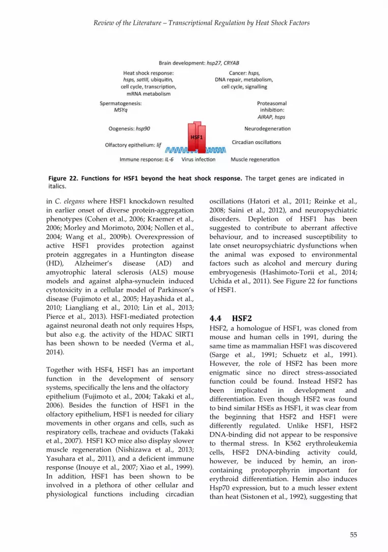
Review of the Literature – Transcriptional Regulation by Heat Shock Factors
55
in C. elegans where HSF1 knockdown resulted in earlier onset of diverse protein-aggregation phenotypes (Cohen et al., 2006; Kraemer et al., 2006; Morley and Morimoto, 2004; Nollen et al., 2004; Wang et al., 2009b). Overexpression of active HSF1 provides protection against protein aggregates in a Huntington disease (HD), Alzheimer’s disease (AD) and amyotrophic lateral sclerosis (ALS) mouse models and against alpha-synuclein induced cytotoxicity in a cellular model of Parkinson’s disease (Fujimoto et al., 2005; Hayashida et al., 2010; Liangliang et al., 2010; Lin et al., 2013; Pierce et al., 2013). HSF1-mediated protection against neuronal death not only requires Hsps, but also e.g. the activity of the HDAC SIRT1 has been shown to be needed (Verma et al., 2014). Together with HSF4, HSF1 has an important function in the development of sensory systems, specifically the lens and the olfactory epithelium (Fujimoto et al., 2004; Takaki et al., 2006). Besides the function of HSF1 in the olfactory epithelium, HSF1 is needed for ciliary movements in other organs and cells, such as respiratory cells, tracheae and oviducts (Takaki et al., 2007). HSF1 KO mice also display slower muscle regeneration (Nishizawa et al., 2013; Yasuhara et al., 2011), and a deficient immune response (Inouye et al., 2007; Xiao et al., 1999). In addition, HSF1 has been shown to be involved in a plethora of other cellular and physiological functions including circadian
oscillations (Hatori et al., 2011; Reinke et al., 2008; Saini et al., 2012), and neuropsychiatric disorders. Depletion of HSF1 has been suggested to contribute to aberrant affective behaviour, and to increased susceptibility to late onset neuropsychiatric dysfunctions when the animal was exposed to environmental factors such as alcohol and mercury during embryogenesis (Hashimoto-Torii et al., 2014; Uchida et al., 2011). See Figure 22 for functions of HSF1. 4.4 HSF2%%
HSF2, a homologue of HSF1, was cloned from mouse and human cells in 1991, during the same time as mammalian HSF1 was discovered (Sarge et al., 1991; Schuetz et al., 1991). However, the role of HSF2 has been more enigmatic since no direct stress-associated function could be found. Instead HSF2 has been implicated in development and differentiation. Even though HSF2 was found to bind similar HSEs as HSF1, it was clear from the beginning that HSF2 and HSF1 were differently regulated. Unlike HSF1, HSF2 DNA-binding did not appear to be responsive to thermal stress. In K562 erythroleukemia cells, HSF2 DNA-binding activity could, however, be induced by hemin, an iron-containing protoporphyrin important for erythroid differentiation. Hemin also induces Hsp70 expression, but to a much lesser extent than heat (Sistonen et al., 1992), suggesting that
Figure* 22.* Functions* for* HSF1* beyond* the* heat* shock* response.* The$ target$ genes$ are$ indicated$ in$italics.*

Review of the Literature – Transcriptional Regulation by Heat Shock Factors
56
HSF2 could have a function in processes related to development and differentiation. HSF2 could not substitute for the single HSF in yeast in stress-conditions, but human and murine HSF2 were the only vertebrate HSFs that could functionally substitute for the only HSF in yeast during normal growth conditions. This was attributed to the constitutive trimerization of HSF2 in yeast (Liu et al., 1997), but perhaps it could also relate to the specific targets or co-activators of the two HSFs. These early observations reflect some of the differences and similarities observed in the function and structure between the two proteins. The overall amino acid sequences are only 35% identical, but both the DBD and oligomerization domains are highly conserved, with a 70% similarity within the amino acid sequences of the DBDs (Pirkkala et al., 2001; Sandqvist et al., 2009). In contrast, the RD and AD of the two HSFs have few similarities. The HSF2 ADs are placed in the C-terminal region, scattered between negative RDs. The mechanism behind the restriction of the AD by the RDs is unknown, but the RDs of HSF2 seem to be regulated by neither heat shock nor hemin treatment (Yoshima et al., 1998; Zhu and Mivechi, 1999). The transactivation potential of the HSF2 ADs are much weaker than that of the two HSF1 ADs, but the HSF2 ADs are sufficient for restoring the expression of Hsp genes in HSF1-deficient cells (Ahn and Thiele, 2003). Despite their unique target genes, both HSFs have been found to bind to an analogous HSE, consisting of inverted nGAAn pentamers (Sarge et al., 1991; Vihervaara et al., 2013). However, the two HSFs seem to favor HSEs with slightly different architecture. It has been reported that HSF1 occupies HSEs containing four or five copies of the nGAAn repeat, whereas HSF2 favors shorter, only one to three pentamers long HSEs, indicating that fewer HSF2 trimers are DNA-bound (Kroeger and Morimoto, 1994; Kroeger et al., 1993; Sistonen et al., 1992; 1994). Indeed, it was later shown that two trimers of HSF1 were able to occupy an HSE consisting of four continuous nGAAn repeats, whereas only one HSF2 trimer was bound. HSF2 bound discontinuous nGAAn
repeats slightly better than HSF1. This discrepancy in DNA-binding properties resulted in differently regulated target genes depending on the type of HSE present on the genes (Yamamoto et al., 2009). This diversity in DNA-binding could explain why HSF2, but not HSF1, can access the HSEs in compacted mitotic chromatin, as HSF2 needs a shorter exposed stretch of DNA to be able to bind (Vihervaara et al., 2013). The different DNA-binding capabilities of HSF1 and HSF2 are thought to be associated with dissimilarities in the ability for co-operative DNA-binding. For HSF1 it has been shown that DNA-binding of one HSF1 trimer facilitates the binding of the second trimer, however the same has not been observed with HSF2 (Kroeger and Morimoto, 1994; Xiao et al., 1991). The HSF2 protein exists as two isoforms, α and β. These isoforms are produced by alternative splicing of exon 11, rendering HSF2-β deficient in an 18-amino acid region situated C-terminal to the HR-C domain (Fiorenza et al., 1995; Goodson and Sarge, 1995). The isoforms seem to generally be expressed in equal amounts but upon certain stresses, e.g. proteasomal inhibition, splicing is regulated (Lecomte et al., 2013). The isoforms are also differentially expressed in some organs and in testis the expression of the isoforms seems to be developmentally regulated. As the expression of the HSF2-β splice-variant remains stable throughout development, HSF2-α expression increases during post-natal development, and HSF2-α becomes the predominant isoform in the adult mouse testis. In the adult mouse, HSF2-β is the dominant isoform in heart and brain (Goodson et al., 1995). The tissue-specific function of the isoforms remains elusive, but some of the effects seem to depend on the ratio between α and β. Thus, the impact of HSF2 on transcription can be modulated by changing the ratio between the isoforms. Of the two isoforms, HSF2-α is considered a stronger transcriptional activator, which positively modulates the HSF1-induced Hsp expression (Goodson et al., 1995; He et al., 2003). HSF2-α is also involved in the differentiation of K562 erythroleukemia cells to erythrocytes (Leppä et al., 1997). The overexpression of HSF2-β, on the

Review of the Literature – Transcriptional Regulation by Heat Shock Factors
57
other hand, results in the inhibition of hemin-induced erythroid differentiation, Hsp expression and c-Jun N-terminal kinase induction (Hietakangas et al., 2001; Lecomte et al., 2013; Leppä et al., 1997), suggesting that HSF2-β would have a repressive effect on HSF1 transactivation. Together these results demonstrate that the specific regulation of the isoforms is important for the proper function of HSF2 in a specific context. 4.4.1 HSF2% activity% is% regulated% by% its%
concentration%in%the%cell%
In its inactive form HSF2 exists primarily as a dimer, residing in both the nucleus and cytoplasm (Sarge et al., 1993), the subcellular localization of HSF2 is suggested to be directed by an NLS that is unmasked upon activation (Sheldon and Kingston, 1993). Similarly to HSF1, the active DNA-binding form of HSF2 is trimeric (Mathew et al., 1998; Sistonen et al., 1994). In contrast to HSF1, the activity of which is regulated by the RD and PTMs such as phosphorylation, HSF2 has not been shown to be phosphorylated in cycling cells. Neither does HSF2 have an RD that would influence the transcriptional activity in response to heat shock or hemin-treatment (Yoshima et al., 1998; Zhu and Mivechi, 1999). Instead, the concentration of HSF2 in the cell plays a role for the conversion of HSF2 to an active DNA-binding state. Sarge and coworkers showed that by overexpressing HSF2, HSF2 was able to transactivate an Hsp70-luciferase construct, to an even higher degree than heat shock (Sarge et al., 1993). An increase in HSF2 levels is thought to result in its trimerization and thus DNA-binding capacity. Accordingly, HSF2 has constitutive in vitro DNA-binding activity in cells and tissues, e.g. testis, pre-implantation blastocysts,
developing heart and mouse embryonal carcinoma cells, where HSF2 levels are high (Eriksson et al., 2000; Fiorenza et al., 1995; Mezger et al., 1994; Murphy et al., 1994; Rallu et al., 1997; Sarge et al., 1994). Furthermore, hemin treatment or proteasome inhibition by MG132 or bortezomib results in increased HSF2 levels, with a corresponding increase in transcription of Hsp70. The accumulation of HSF2 protein is dependent on an increase in Hsf2 mRNA stability as well as in protein synthesis and stability (Mathew et al., 1998; Pirkkala et al., 1999; Rossi et al., 2014). It has been envisioned that changes in the amount of HSF2 in the cell contributes to its target specificity (Sandqvist et al., 2009). HSF2 is a labile protein with a half-life of only 60 min (Mathew 1998). Cullin3, a subunit of the Cullin3-RING E3 ligase has been suggested to interact with PEST sequences (PEST1 aa 241-309, PEST2 aa 479-499) on HSF2 and thus directing HSF2 for proteasomal degradation (Xing et al., 2010). Mass spectrometric analyses have found that HSF2 is ubiquitylated on lysines K51, K54, and K210 in untreated cells (Figure 23). This ubiquitylation is not changed by proteasomal inhibition by MG132 (Wagner 2011). Another study revealed that upon proteasomal and translational inhibition by bortezomib and cyclohexamide lysines K151 and K420 are marked by ubiquitin. The K420 is still ubiquitylated upon inhibition with MLN2924, a NEDD8 activating enzyme inhibitor, suggesting that this site is not a cullin-RING ubiquitin ligase substrate, whereas the K151 could be modified by the cullin-RING ubiquitin ligase (Kim et al., 2011b). HSF2 also appears to be ubiquitylated upon heat shock as it is found in an insoluble fraction. However, HSF2 remains soluble upon heat shock in thermotolerant cells, suggesting that increased Hsp levels protect HSF2 from ubiquitylation (Mathew et al., 2001). Taken together,
Figure*23.*PostVtranslational*modifications*of*HSF2.*HSF2$has$been$shown$to$be$ubiquitylated$(U)$on$five$sites$by$mass$spectrometry,$and$sumoylated$(S)$on$two$sites.$

Review of the Literature – Transcriptional Regulation by Heat Shock Factors
58
ubiquitylation contributes to the regulation of HSF2 activity by controlling the levels of HSF2 in the cell. 4.4.2 Sumoylation%of%HSF2%
In addition to ubiquitylation, HSF2 has also been shown to be sumoylated. In a yeast two-hybrid screen HSF2 was shown to interact with the SUMO-conjugating enzyme Ubc9 (Goodson et al., 2001). The target lysine, K82, is located within the loop of the DBD that controls paralog-specific DNA-binding (Ahn and Thiele, 2003; Anckar et al., 2006; Goodson et al., 2001). Another minor sumoylation site was found on K139 (Anckar et al., 2006). Contradicting results on the effect of sumoylation of K82 on the DNA-binding activity have been reported. Earlier studies reported that sumoylation converted inactive HSF2 to its active form (Goodson et al., 2001; Hilgarth et al., 2004). Later studies, on the other hand, showed that sumoylation represses DNA-binding activity without affecting oligomerization (Anckar et al., 2006; Tateishi et al., 2009). It has been suggested that HSF1 DNA-binding could be repression by SUMO could be mediated by sterical or electrostatic interference. Tateishi and coworkers demonstrated that a longer stretch of DNA flanking the HSE negatively affects occupation of sumoylated HSF2, implying that the interaction between SUMO and DNA would inhibit HSF2 DNA-binding activity (Tateishi et al., 2009). Sumoylation of HSF2 has been proposed to increase during mitosis when HSF2 is bound to the Hsp70 promoter (Xing et al., 2005). Perhaps the changes in the chromatin environment that occurs during mitosis would enable DNA-binding of sumoylated HSF2, if the flanking DNA covered by the nucleosome. However, previous data point to only small changes, if any, of the chromatin organization of the Hsp70 promoter (Martínez-Balbás et al., 1995; Valls et al., 2005).
4.4.3 HSF2% modulates% the% heat% shock%
response% through% formation% of%
HSF1MHSF2%heterotrimers%%
The two HSFs, HSF1 and HSF2, show high sequence similarity of the DBD and recognize a similar HSE, thus it is conceivable that HSF1 and HSF2 could have shared targets (Kroeger and Morimoto, 1994; Kroeger et al., 1993; Sistonen et al., 1992; 1994; Vihervaara et al., 2013; Yamamoto et al., 2009). If this would be the case during development, it would be expected that the HSF1 and HSF2 knockout mice would show similar phenotypes. Indeed, HSF1 and HSF2 knockout mice have comparable phenotypes in the testis, with vacuolarization of the seminifereous tubules and sperm head abnormlaties. ChIP-chip studies revealed that HSF1 and HSF2 have shared targets on the male specific region of the mouse Y chromosome long arm (MSYq) (Åkerfelt et al., 2008; 2010b). These results suggest that in the testis HSF1 and HSF2 act together to ensure proper sperm maturation, consequently the double knockout results in male sterility (Wang et al., 2004). Furthermore, HSF1 and HSF2 also co-operate in the brain as myelin loss and astroglisosis found in the brain of HSF1 knockouts is aggravated in the absence of HSF2 (Homma et al., 2007). Having established that HSF1 and HSF2 act in concert in the brain and during spermatogenesis, the question remains if they also function together in other tissues or processes. A ChIP study conducted in K562 cells revealed that both HSF1 and HSF2 occupy the same Hsp promoters. Even though HSF2 binding was only induced by hemin treatment (Trinklein et al., 2004), this study showed that HSF1 and HSF2 share the same target genes. By using RNAi interference (RNAi) to mediate HSF2 down-regulation it became evident that, albeit incapable of alone inducing the HSR, HSF2 can modulate the HSR. In heat-treated cells depletion of HSF2 resulted in decreased Hsp70 and Hsp25 induction, suggesting that HSF2 can function as a positive modulator of expression of some Hsps. However, HSF2 has a negative effect on the expression of Hsp40 and Hsp110, pointing to a target-specific effect of HSF2 (Östling et al., 2007). A recent genome-

Review of the Literature – Transcriptional Regulation by Heat Shock Factors
59
wide study utilizing the ChIP-seq technique to characterize HSF1 and HSF2 binding sites in unstressed and heat-treated cycling and mitotic cells, found that upon heat shock in cycling cells a vast majority of the occupied loci are shared targets of HSF1 and HSF2. Upon heat shock both HSF1 and HSF2 are bound to genes involved in the stress response, i.e. chaperones, cell cycle, condensed chromatin, promoter binding, translation, ubiquitin, transcriptional repression, transport and membrane depolarization (Vihervaara et al., 2013). It has been shown that HSF2 is activated at a lower temperature, at which it induces αB-crystallin, and accordingly HSF2-deficient cells are more sensitive to sustained mild fever-like heat shock (Shinkawa et al., 2011). Depletion of HSF2 was shown to reduce the threshold for HSF1 activation, resulting in increased Hsp expression upon mild heat shock (Shinkawa et al., 2011)(Paslaru 2003, Shinkawa 2011). Upon thermal stress the localization of HSF2 is regulated. In response to heat, HSF2, together with HSF1, is translocated into subnuclear structures, the nuclear stress bodies (nSBs), formed predominantly at the 9q12 locus in human cells. In the nSBs the HSFs induce the expression of satellite III transcripts (satIII) (Alastalo et al., 2003; Jolly et al., 1997; Sandqvist et al., 2009; Sheldon and Kingston, 1993). The precise biological function of the nSBs is still unknown, but it has been suggested that the nSBs could function as RNA-processing factories where stress-specific processing of transcripts occur (Sandqvist and Sistonen, 2004). In another study both HSF1 and HSF2 were detected on the clusterin promoter, which contains only a single HSE, suggesting that HSF1 and HSF2 could form heterotrimers (Loison et al., 2006). The high sequence similarity between the oligomerization domains of HSF1 and HSF2, would indeed enable heterotrimerization (Sandqvist et al., 2009). Heterotrimerization is further supported by the observation that the deletion of HSF2 HR-A/B diminishes the interaction with HSF1 (Alastalo et al., 2003). Furthermore, structural modelling and fluorescence resonance energy transfer microscopy showed that HSF1 and HSF2 indeed could form DNA-bound
heterotrimers (Sandqvist et al., 2009). It was suggested that the different combinations of HSF1 and HSF2 could contribute to the transcriptional regulation of their shared targets. 4.4.4 The% role% of% HSF2% in% early%
development,% aging% and%
physiological%processes%
Compared to the evenly expressed HSF1, the concentration of HSF2 varies both spatially and temporarily, suggesting that HSF2 would have distinct functions in development (Abane and Mezger, 2010; Fiorenza et al., 1995; Rallu et al., 1997). Studies on HSF2 knockout (HSF2 KO) mice have contributed to our understanding of the function of HSF2 in the development. To date, three independent groups have developed HSF2 KO mice. In none of these mice any apparent morphological abnormalities could be detected (Kallio et al., 2002; McMillan et al., 2002; Wang et al., 2003). Two of the groups found various phenotypic defects, affecting mainly corticogenesis and gametogenesis (Kallio et al., 2002; Wang et al., 2003). In female mice the depletion of HSF2 was associated with reduced fertility and increased prenatal lethality of the embryos. The decreased fertility was associated with meiotic defects resulting in fewer ovarian follicles and more abnormal eggs (Kallio et al., 2002; Wang et al., 2003). These abnormalities could, however, not be detected in the third mouse model (McMillan et al., 2002). This inconsistency has been attributed to the different genetic background of the animals. HSF2 was found to have in vitro DNA-bindning activity in the blastocyst stage of the preimplantation development (Mezger et al., 1994). During post-implantation HSF2 expression increased throughout the embryo and peaked at E9.75, after which it starts to decrease (Min et al., 2000; Rallu et al., 1997). The raised HSF2 levels correlated with increased DNA-binding activity. However, corresponding expression of Hsp genes was not detected, and the target genes during embryogenesis remain unkown. HSF2 concentration in the cells seem to be controlled

Review of the Literature – Transcriptional Regulation by Heat Shock Factors
60
mainly through changes in transcription or mRNA levels during development (Rallu et al., 1997). Although HSF2 expression increases progressively throughout the embryo in early development the expression becomes restricted to the central nervous system during the second half of gestation (Rallu et al., 1997). In the nervous system, HSF2 is most prominently expressed in the ventricular layer of the neural tube containing dividing progenitor cells and in post-mitotic neurons in the cortical plate (Chang et al., 2006; Rallu et al., 1997). During post-natal development, HSF2 localization changes from nuclear to cytoplasmic and the overall levels were found to decrease in the neurons (Brown and Rush, 1999). The adult brain of HSF2 KO mice exhibited reduced size of the cortex, hippocampus and striatum, as well as enlarged ventricles (Kallio et al., 2002; Wang et al., 2003). In HSF2 KO mice the neurons in the superficial layers of the cerebral cortex are disorganized, which is suggested to be due to aberrant radial migration of the neurons that is regulated by HSF2 (Chang et al., 2006), suggesting that HSF2 is involved in cortical layering. So far only one direct target of HSF2, p35, has been found in the brain, however, the pronounced phenotype in the brain of HSF2-deficient mice suggest that other signalling pathways are affected as well. HSF2 has not only been implied in the developing brain but also in the aging brain. In the Huntington’s disease mouse model, loss of HSF2 function increases the accumulation of aggregated polyglutamine proteins (polyQ) in
the striatum. The disruption of both Hsf2 alleles shortens lifespan by 36%. HSF2 activates �B-crystallin expression upon sustained mild heat shock. Overexpression of � B-crystallin in HSF2 KO MEFs partially prevented the accumulation of polyQ aggregation and misfolded cellular proteins (Shinkawa et al., 2011), suggesting that HSF2 regulates proteostasis in neurons in part by controlling the expression of �B-crystallin. In addition to its role in the central nervous system, HSF2 has also been shown to have a function in some diseases. In the intestine increased expression HSF2 has been associated with ulcerative colitis and HSF2 is proposed to be a novel molecular marker of the disease (Miao et al., 2014). In the brain cancer glioma Hsf2 mRNA levels where shown to be significantly increased in low-grade, but not high-grade, glioma (Mustafa et al., 2010). The plethora of target genes and processes that HSF2 is involved in either alone or together with HSF1 (Figure 24) data underline the importance of the precise regulation of HSF2 in both normal development and disease. 4.4.5 HSF2%in%spermatogenesis%
4.4.5.1 A% short% introduction% to%
spermatogenesis%
The first cell in every new animal arises from the fusion of a male and a female germ cell. Thus, all genetic and epigenetic information in an individual organism is derived from these two germ cells. This makes the complex process through which germ cells are produced
$
Figure*24.*HSF2*is*involved*in*a*wide*range*of*biological*processes*either*alone*or*together*with*HSF1.*Examples$of$targets$genes$are$indicated$in$italics.$

Review of the Literature – Transcriptional Regulation by Heat Shock Factors
61
extremely important, and both spermatogenesis and oogenesis are strictly regulated. The primordial germ cells (PGCs) arise shortly after implantation of the embryo. During gastrulation the PGCs rapidly proliferate while they migrate towards the gonadal ridge, where the PGCs become gonocytes surrounded by Sertoli cell precursors and peritubular cells. The gonocytes divide rapidly through mitosis before they arrest in G0 phase until after birth. During this time the cells undergo epigenetic reprogramming where all somatic epigenetic marks and new sex-specific DNA methylation patterns are established (Yadav and Kotaja, 2014). Spermatogenesis is a continuous process in which three principal phases can be detected: spermatogonia renewal and proliferation, meiosis and spermiogenesis (Figure 25). Shortly after birth the spermatogonial stem cells return to the cell cycle and start proliferating through mitosis. Depending on the signals from the Sertoli cells the spermatogonial stem cells divide for either self-
renewal or for the formation of daughter cells. The daughter cells undergo another division to become differentiating spermatogonia, which mature into type B spermatogonia, before entering the meiotic phase and becoming spermatocytes. The type B spermatogonia divide through mitosis and form two preleptotene spermatocytes. Preleptotene spermatocytes then go through meiosis I, forming leptotene, zygotene, pachytene and diplotene spermatocytes. DNA synthesis occurs in the preleptotene stage, after which chromosome condense and are paired in the zygotene spermatocyte. The pairing of homologous chromosomes leads to the formation of the synaptonemal complex in the pachytene cells. In the pachytene stage, homologous recombination occurs and the cells exhibit an increase in RNA content. In the diplotene cells the synaptonemal complex is separated and the chromosomes are spread out in the cell. As a result, small secondary spermatocytes containing only half the chromosomes (2n) are produced. The secondary spermatocytes go through the rest of meiosis without replicating their DNA, thus
Figure*25.*Spermatogenesis.*The$seminiferous$tubules$are$organized$in$cyclic$stages$$(ITXII)$so$that$each$crosssection$ of$ the$ tubule$ contains$ defined$ groups$ of$ germ$ cell$ types$ at$ particular$ developmental$phases$(Martianov$et$al.,$2005).$*
Review of the Literature – Transcriptional Regulation by Heat Shock Factors
62
forming small haploid round spermatids (Hess, 1998; Hess and Franca, 2009; McIver et al., 2012b). The post-meiotic haploid round spermatids undergo a differentiation process called spermiogenesis. Spermiogenesis involves four phases, and results in dramatic morphological changes, including elongation through flagellum formation as well as chromatin condensation where protamines replace histones. The first Golgi stage involves the formation of the proacrosomal vesicle. During the second stage, capping, the proacrosomal vesicle interacts with the nuclear envelope and covers the nuclear surface. In the next acrosomal phase, the vesicles migrate and the nucleus is highly condensed by histone replacement. The final maturation phase results in the removal and phagocytosis of the residual cytoplasm. The resulting spermatozoa are released into the lumen of the seminiferous tubules and travel to the epididymis where the final maturation occurs (Hess and Franca, 2009). 4.4.5.2 HSF2% as% a% regulator% of% gene%
expression%in%spermatogenesis%
From early on, it was apparent that HSF2 expression was high in testis (Fiorenza et al., 1995; Sarge et al., 1994). During rodent spermatogenesis, HSF2 displays stage-specific expression with high levels in pachytene spermatocytes (stages I-IV) and round spermatids (V-VII) (Alastalo et al., 1998; Sarge et al., 1994). A constitutively active DNA-bound form of HSF2 was detected in testis (Sarge et al., 1994), but was not found in subsequent studies (Abane and Mezger, 2010; Alastalo et al., 1998). These data suggest that HSF2 plays a role as regulator of stage-specific gene expression during spermatogenesis. In accordance, HSF2 deficiency results in abnormal spermatogenesis. HSF2 knockout mice display increased apoptosis in late pachytene spermatocytes, possibly due to the observed disorganization of the synaptonemal complex that disturbs meiosis (Kallio et al., 2002). In HSF2 KO mice, the size of the testis is reduced and the seminiferous tubules are
disrupted and vacuolarized, and the amount of abnormal and mature sperm is increased and reduced, respectively. The male HSF2 KO mice remain fertile (Åkerfelt et al., 2008; Kallio et al., 2002; Wang et al., 2003), whereas disruption of both HSF1 and HSF2 results in complete absence of spermatozoa (Wang et al., 2004), pointing to compensatory mechanisms between HSF1 and HSF2 in the testis.
These early studies on HSF2 in spermatogenesis were not able to identify HSF2-specific target genes in the testis. It was only by using a ChIP-chip approach that HSF2 targets were identified. Of the 546 putative targets detected in testis a striking accumulation of HSF2 occupancy was found on the MSYq. This otherwise gene-poor region harbors a few families of multicopy genes, of which HSF2 targets include spermiogenesis specific transcript on the Y (Ssty2), Scyp3 like Y-linked (Sly) and Similar to Ssty2. These show decreased expression in HSF2 KO testis. On the contrary, expression of the Sly paralogue found on the X chromosome, Scyp3 like X-linked (Slx), is increased in HSF2 KO testis (Åkerfelt et al., 2008). The MSYq genes are thought to affect chromatin compaction in the sperm head (Ellis et al., 2005; Touré et al., 2005). Sly has been shown to repress sex chromosomes post-meiotically, and Sly depletion results in reduced repressive marks and severe impairment of sperm differentiation (Abane and Mezger, 2010). In agreement, depletion of HSF2 in testis results in atypical levels of chromatin packing proteins, including transition proteins and protamines and increased chromatin fragmentation in the mature sperm (Åkerfelt et al., 2008). By studying testis from HSF1 knockout it was found that HSF1 occupies the promoters of the same multicopy genes and results in abnormal sperm (Åkerfelt et al., 2010b), suggesting that HSF1 and HSF2 synergistically regulate the multicopy genes. In humans, mutations of Hsf2 have been found in infertile men with idiopathic azoospermia, but missense mutations were detected in less than 1 % of the cases (Mou et al., 2013).
In addition to the MSYq genes, HSF2 was also found to target genes on autosomal

Review of the Literature – Transcriptional Regulation by Heat Shock Factors
63
chromosomes in the testis. Among these targets HSF2 was verified to bind sperm-associated glutamate-rich 4 (Speer4) which is expressed in germ cells during spermatocyte-spermatid transition. Other targets include HSPA8 (Hsc70) and ferritin mitochondrial (ftmt). Intriguingly, binding of HSF2 to these genes occurs only in testis and could not be detected in other tissues such as brain and muscle (Åkerfelt et al., 2008), suggesting that the DNA sequence alone is not enough to control tissue-specific gene expression. 4.5 HSFs% as% regulators% of% cell% cycle%
progression% and% heatMinduced%
cell%cycle%arrest%
4.5.1 Heat% shock% causes% cell% cycle%
arrest%
In 1960, Bergan observed that when mitotic eggs of the fish Trichogaster trichopterus were subjected to heat shock mitosis temporarily halted. Depending on the severity of the heat shock the eggs were then able to resume mitosis right after removal from heat or, in case of a more severe heat shock, after the mitotic apparatus was re-established. Some cells died as a result of the heat insult (Bergan, 1960). Although it has been known for more than 50 years that heat shock stalls cell cycle progression, the mechanism behind this arrest remains elusive. Depending on the severity of the stress and the cell cycle phase during which the heat shock occurs, the outcome is different (Maldonado-Codina et al., 1993). In human cells acute exposure to heat leads to a transient arrest both at the G1/S and G2/M borders (Nitta et al., 1997). The G1/S arrest has been shown to be dependent on p53-mediated induction of the CDK inhibitor p21 and on other regulatory proteins. In addition, severe heat stress has been shown to reduce the level of cell cycle regulatory molecules, including cyclin D1, CDK and phosphorylated Rb, resulting in G1 arrest. Upon the induction of Hsps normal cell cycle resumes (Fuse et al., 1996; Kühl and Rensing, 2000; Kühl et al., 2000; Nitta et al., 1997).
Different studies have shown that HSF1 knock-out cells accumulate in G2-M phase during thermal stress, whereas wild type cells are able to overcome the cell cycle arrest induced by heat (Luft et al., 2001; Nakai and Ishikawa, 2001). This effect could partly be dependent on the expression of Hsp90α (HSPC). Hsp90 is markedly reduced under normal growth conditions in avian cells lacking the heat-inducible HSF1 and HSF3. The reduction in Hsp90 caused both Plk1- and CDK1-instablity, resulting in G2/M arrest, but also G1 arrest, upon mild heat shock (de Cárcer, 2004; Nakai and Ishikawa, 2001). HSF1 has a similar function in oocyte meiosis, where it increases Hsp90 expression and thus stabilizes CDK1 (Metchat et al., 2009). Taken together, it has been shown that HSF1 is an important regulator of cell cycle progression in response to heat. 4.5.2 Proteotoxic% stress% is%detrimental%
to%mitotic%cells%
In contrast to interphase cells, mitotic cells are very sensitive to heat, in part due to repressed expression of chaperones (Martínez-Balbás et al., 1995). In mitotic D. melanogaster cells, heat shock causes the disruption of the mitotic spindle and strongly affects the structure of the microtubule-organizing centre, the centrosome (Debec and Marcaillou, 1997). The centrosomes are important for proper segregation of DNA, cell shape, motility and division. Centrosome aberrations can give rise to chromosomal instability (CIN) (Nigg, 2002). The centrosomes have been shown to be sensitive to various stresses. In CHO cells, centrosomes split in the presence of DNA damage, resulting in multiple spindle poles and eventually aneuploidy or mitotic catastrophe (Hut et al., 2003). In human and D.melanogaster interphase cells thermal stress causes electron dense material to accumulate around the centrioles, pointing to denaturation of the proteins in the pericentriolar mass (Barrau et al., 1978; Debec and Marcaillou, 1997). Heat shock can disrupt the centrosome or induce centrosomal amplification, leading to disturbances in microtubule nucleation and to the disorganization of the mitotic spindle in the

Review of the Literature – Transcriptional Regulation by Heat Shock Factors
64
subsequent mitosis. Heat-induced spindle disturbances result in chromosomal missegregation and a several fold increase in chromosomal instability. A prolonged or more severe heat shock causes centrosomes to disappear and the cells to undergo apoptosis (Barrau et al., 1978; Debec and Marcaillou, 1997; Gupta and Srinivas, 2008; Nakahata et al., 2002; Vidair et al., 1993). Several HSPA and HSPC class molecular chaperones have been identified at the centrosome. Hsp70 associates with centrosomes or microtubule-organizing centers in various species, from green algae and dinoflagellates to humans (Bloch and Johnson, 1995; de Cárcer, 2004; Perret et al., 1995). Hsc70 and TCP-1 exist in centrosomes throughout the cell cycle, especially in dividing cells (Brown et al., 1996; Rattner, 1991). Upon heat shock the centrosome is fragmented and additional Hsp70 is recruited to the centrosome (Brown et al., 1996; Hut et al., 2005; Rattner, 1991; Vidair et al., 1993). Similarly, in the presence of unfolded proteins or arsenic trioxide (ATO), Hsp70 is recruited to the centrosome, and interestingly, also the proteasome is recruited to the centrosomes upon proteotoxic stress (Chen et al., 2014; Wigley et al., 1999). During the recovery from heat shock there is a strong correlation between increased Hsp70 centrosomal staining and the re-appearance of microtubules (Brown et al., 1996; Hut et al., 2005; Rattner, 1991; Vidair et al., 1993). By inhibiting Hsp70 with an antibody, centrosome assembly after heat shock can be blocked, whereas Hsp70 expression prior to heating or thermotolerance accelerates recovery of centrosome function after heat shock (Brown et al., 1996; Hut et al., 2005; Vidair et al., 1993). In the ATO-treated cells, Plk1-mediated phosphorylation is needed for Hsp70 to localize at the centrosome and phosphorylated Hsp70 increases microtubule stability (Chen et al., 2014). In addition to being required for centrosome integrity in mitosis Hsp70 has also been shown to bind to microtubules in yeast (Makhnevych and Houry, 2013). Taken together, these data strongly supports a function for Hsp70 in stabilizing centrosomes, especially in response to stress. The importance of Hsp70 in cell cycle regulation is further
underlined by the finding that Hsp70 depletion in cells cause G2/M arrest (Daugaard et al., 2005). Since centrosome function is fundamental for cell cycle progression, it is possible that the depletion of Hsp70 from the centrosomes causes arrest. 4.5.3 The% heat% shock% response% in%
mitotic%cells%
From the above-presented data the importance for Hsp70 in protecting cells against proteotoxic damage during mitosis is indisputable. However, during mitosis, Hsp70 expression, along with the majority of transcription, is repressed (Martínez-Balbás et al., 1995). In contrast to the majority of genes where the chromatin environment changes during mitosis and the DNA is compacted into tight chromatin, the chromatin environment at the Hsp70 promoter remains largely unchanged. Upon entry into mitosis, both di- and trimethylation of histone H3 K4 remained stable, H3 acetylation remained stable or increased slightly, whereas histone H4 acetylation decreased. Phosphorylation of histone H3 serine 10 was the only modification that showed a dramatic increase (Valls et al., 2005; Xin et al., 2007). Acetylation of histone H3 and H4, as well as H3 K4 methylation are marks of active chromatin and have been implicated in gene bookmarking during mitosis (Li et al., 2007; Wang and Higgins, 2013). As shown by DNaseI and KMnO4 treatments the Hsp70 promoter remains open and accessible to proteins during mitosis (Martínez-Balbás et al., 1995; Michelotti et al., 1997). Although the chromatin remains open, paused RNAPII, HSF1 and other transcription factors are displaced from the Hsp70 promoter, but some proteins remain bound (Christova and Oelgeschläger, 2001; Martínez-Balbás et al., 1995). The TFIIB and the TFIID subunit TAFII100, as well as HSF2, remained bound to the promoter in mitosis (Christova and Oelgeschläger, 2001; Vihervaara et al., 2013; Xing et al., 2005). The DNA-bound TBP has been suggested to interact with condensin and mediate its dephosphorylation by recruiting protein phosphatase 2A (PP2A). This leads to
Outline and Aims of the Thesis
65
the inactivation of condensin and to locally accessible chromatin (Xing et al., 2008). However, even though the promoter is open, mitosis-specific phosphorylation of TBP and the TAFs result in inhibition of their activator-dependent functions, i.e. PIC formation and transcription activation (Segil et al., 1996). The reason why HSF1 is displaced is unknown. Intriguingly, the stress-inducible in vitro DNA-binding capacity is maintained, whereas its binding to chromatin is markedly reduced (Borrelli et al., 1996; Martínez-Balbás et al., 1995; Vihervaara et al., 2013). Thus, the inhibition of HSF1-binding is not due to its intrinsic DNA-binding capacity but to some other factors. One possibility is that the bound HSF2 repells HSF1, or that some property of the mitotic chromatin or HSF1 inhibits HSF1 binding. Although the promoter remains accessible to some transcription factors due to displacement of HSF1 and RNAPII and inhibition of PIC formation, the Hsp70 promoter is transcriptionally inactive in mitosis. This results in repression of stress-induced Hsp transcription in mitotic cells (Borrelli et al., 1996; Martínez-Balbás et al., 1995). Intriguingly, although no Hsps were induced, thermotolerance was still developed, resulting in increased survival upon another thermal insult during mitosis (Borrelli et al., 1996). Whether this is due to the association of the chaperones with important cellular structures, such as the centrosome, or due to some other mechanism is unknown. Since HSF2 remains bound to the Hsp70 promoter, it has been suggested that it bookmarks the gene for immediate early activation in G1 phase. Similarly to TBP, HSF2 has been proposed to recruit PP2A and inhibit local condensin activation (Xing et al., 2005). If HSF2 and TBP have a similar mechanism of action or if one of the factors recruits the other one that execute condensing-inhibition has not been determined. In addition to Hsp70, HSF2 is also bound to Hsp27, Hsp60, Hsp90, and c-fos promoters during mitosis (Vihervaara et al., 2013; Wilkerson et al., 2007). Furthermore, HSF2 binds to more than 500 target loci upon heat shock in mitosis, of these only 67 are shared targets of the interphase HSF2
(Vihervaara et al., 2013), suggesting that HSF2 has a distinct transcriptional program in mitotic cells. Perhaps HSF2 functions as an epigenetic marker. Intriguingly, HSF1 acquires in vitro DNA-binding capacity upon entry into G1 phase, independently of stress (Bruce et al., 1999). Also HSF4b exhibits cell cycle-dependent DNA-binding. Upon entry into G1 phase, HSF4b associates with the SWI/SNF Brg1 and HSF4b exhibits increased binding to the Hsp promoters, where it induces Hsp expression, especially in the absence of HSF1 and HSF2 (Tu et al., 2006). Whether HSF1 in fact binds to DNA in vivo upon G1-entry is not known. These observations raise an intriguing possibility that HSF1 could access the genes bookmarked by HSF2 upon entry into mitosis and induce expression of these genes. OUTLINE*AND*AIMS*OF*THE*THESIS*
Although HSF2 was identified more than 20 years ago, the function and regulation of HSF2 to this day remains enigmatic. Previous to this study it was established that HSF2 activity is regulated by its concentrations in the cells, however, the mechanism for this regulation was not known. The general aim of this study was to gain deeper knowledge on the regulation of HSF2 and how the fluctuation of HSF2 levels affects cell fate. Since most of the study was done in mitotic cells, this study also served to elucidate the function for this stress-regulated transcription factor in the cell cycle and how heat shock affects cell cycle progression. In short, the specific aims of this study were:
• To determine the mechanisms regulating expression and activity of HSF2
• To establish the regulation of HSF2 in response to heat shock and cell cycle phase
• To elucidate the function of HSF2 in regulating the heat shock response in mitotic cells

Experimental Procedures
66
EXPERIMENTAL*PROCEDURES*
More detailed information on the experimental procedures is available in the original publications. Animals Male HSF2 knockout (KO) and wild-type (WT) mice, described earlier (Kallio et al., 2002) were used to study spermatogenesis (I). Cell lines Name* Publication*Human$HeLa$cervical$carcinoma$cells$ II,$III$
Human$HEK293$embryonic$kidney$cells$ I,$II$
Human$K562$chronic$myelogenous$leukemia$ II,$III,$unpublished$
Human$MCF7$breast$adenocarcinoma$cells$ I,$III$
Human$MDATMBT231$breast$adenocarcinoma$cells$ III$
Human$WI38$diploid$lung$fibroblasts$ III$
Hsf2+/+*immortalized$mouse$embryonic$fibroblasts$(MEFs)$ III$
Hsf2J/J**immortalized$mouse$embryonic$fibroblasts$(MEFs)$ III$
Mouse$GCT1$spg$spermatogonia$cells$ I$
Rat$ST15A$cerebellar$cells$ I$ Plasmids Name*(manufacturer)* Publication*pcDNA3.1$(Invitrogen)$ I,$II,$III$
pD40THis/V5TcTMyc$(Yeh$et$al.,$2004)$ I$
pEGFPTC1$(Clontech)$ I$
pMIRTREPORT$vector$(Ambion)$containing$3’$UTR$of$HSF2,$
constructed$by$PCR$using$primers$$
(5’TCATCCACTAGTTCCCCAGGAAGTGGACTTTACT3’,$
5’TCATCCAAGCTTGGAGAAAAATGGCCATTTGAATCCT3’)$
I$
pRLTSV40$(Clontech)$ I$
Glutathione$STtransferase$(GST)–ubiquitin,$gift$from$J.$Palvimo$$ II$
MycTtagged$ubiquitin$(Morris$and$Solomon,$2004)$ II$
MycTtagged$HSF1$(Holmberg$et$al.,$2001)$ II$
MycTtagged$HSF2$(Alastalo$et$al.,$2003)$ II$
FlagTtagged$HSF2$(Pirkkala$et$al.,$2000)$ II$
Green$fluorescent$proteinTtagged$Cdc20$(Kallio$et$al.,$1998)$ II$
MycTtagged$Cdc20$(Pfleger$et$al.,$2001)$ II$
MycTtagged$Cdh1$(Pfleger$et$al.,$2001)$ II$
HSF2$shRNA$(Östling$et$al.,$2007)$ III$
Scrambled$shRNA$(Östling$et$al.,$2007)$ III$

Experimental Procedures
67
Antibodies Target*(manufacturer)* Application** Publication*
alpha2$proteasome$(Biomol$International,$Inc)$ ChIP$ II$
APC2$(BD$Pharmingen)$ WB$ II$
betaTactin$(Sigma)$ WB$ I$
betaTtubulin$(Sigma)$ WB$ III$
Cdc20$(Bethyl$Laboratories,$Inc)$ IP,$WB$ II,$unpublished$
Cdc27$(BD$Biosciences)$ IP,$WB$ II,$III,$unpublished$
Cdh1$(Thermo$Scientific)$ WB$ II$
GFP$(Clontech)$ WB$ II$
Histone$H3$(Abcam)$ ChIP,$WB$ III$
Histone$H3$phospho$S10$(Abcam)$ ChIP,$WB$ III$
Hsc70$(Stressgen)$ WB$ I,$II,$III,$unpublished$
HSF1$(Holmberg$et*al.,$2001)$ WB$ II,$III$
HSF1$(Enzo/Stressgen)$ ChIP,$IP,$WB$ III,$unpublished$
HSF2$(Östling$et*al.,$2007)$ ChIP,$IP$ II,$III,$unpublished$
HSF2$(Sarge$et*al.,*1993)$ WB$ I,$II$
HSF2$(Upstate)$ WB$ III$
Hsp25$(Enzo)$ WB$ III$
Hsp27$(Enzo)$ WB$ III$
Hsp70$(Enzo/Stressgen)$ WB$ II,$III$
Myc$(Cell$Signaling$Technology)$ IP,$WB$ II$
RNA$polymerase$II$(Covance)$ ChIP$ III$
Ubiquitin$FK2$(Biomol$International,$Inc)$ WB$ II$
V5Ttag$(AbD$Serotec)$ WB$ I$ siRNA, and microRNA mimics and inhibitors Name*(manufacturer)* Publication*APC2$siRNA,$mixture$of$four$specific$siRNAs$(GeneSolution$siRNA,$Qiagen)$ II$
Cdc20$siRNA,$mixture$of$four$specific$siRNAs$(GeneSolution$siRNA,$Qiagen)$ II$
Cdc27$siRNA,$mixture$of$four$specific$siRNAs$(GeneSolution$siRNA,$Qiagen)$ II$
Cdh1$siRNA,$mixture$of$four$specific$siRNAs$(GeneSolution$siRNA,$
Qiagen)reverse$transcription$PCR$
II$
miRIDIAN$miRNA$mimics$(Dharmacon)$ I$

Experimental Procedures
68
Methods Name* Publication*Cell$culture$ I,$II,$III,$unpublished$
Cell$synchronization$ II,$III,$unpublished$
Cell$viability$assay$through$counting$ III$
Chromatin$immunoprecipitation$(ChIP)$ II,$III,$unpublished$
Densitometry$quantification$ I,$II,$III,$unpublished$
Electrophoretic$mobility$shift$assay$ III$
Flow$cytometry$ I,$II,$III,$unpublished$
FluorescenceTactivated$cell$sorting$of$transfected$spermatocytes$ I$
Image$analysis$ I,$III,$unpublished$
Immunofluorescence$$ I$
Immunoprecipitation$ II,$III$
In*situ$hybridization$ I$
In*vitro*pulldown$assay$ II$
In*vitro$ubiquitylation$assay$ II$
Micrococcal$nuclease$assay$ III$
Microscopy$ I,$III,$unpublished$
MTS$cell$viability$assay$ III$
Propidium$iodide$staining$ I,$II,$III,$unpublished$
Quantitative$reverse$transcription$PCR$ I,$II,$III,$unpublished$
SDSTPAGE$and$immunoblotting$ I,$II,$III,$unpublished$
Squash$preparation$ I$
Statistical$analysis$ I,$II,$III,$unpublished$
Time$lapse$imaging$ III,$unpublished$
Transient$transfections$ I,$II,$III,$unpublished$
Transfection$of$germ$cells$in$intact$seminiferous$tubules$(TTGIST)$ I$
Ubiquitylation$assay$ II,$unpublished$

Experimental Procedures
69
Reagents Name*(manufacturer)* Application* Publication*ABsolute$QPCR$ROX$Mix$(Thermo$Scientific)$ qPCR$ I,$II$
CellTiter$96$Aqueous$One$Solution$
(Promega)$
MTS$cell$viability$assay$ III$
Cycloheximide$(SigmaTAldrich)$ Inhibit$protein$synthesis$ II$
DigoxigeninTlabeled$LNA$probe$(Exicon)$ miRNA$detection$in$in*situ*hybridization$
I$
DualTLuciferase$Reporter$Assay$System$
(Promega)$
Luciferase$assay$ I$
DRAQ5$(Biostatus$Limited)$ Nuclear$staining$ III,$unpublished$
FITCTlabelled$miRCURY$LNA$knockdown$
oligonucleotides$(Exiqon)$
miRNA$inhibition$ I$
High$Capacity$cDNA$Reverse$Transcription$
Kit$(Applied$biosystems)$
cDNA$synthesis$ I,$II,$unpublished$
iScript™$cDNA$Synthesis$Kit$(BioRad)$ cDNA$synthesis$ III$
Kapa$probe$fast$ABI$prism$(Kapa$biosystems)$ qPCR$ III$
Lipofectamine$2000$(Invitrogen)$ Transfection$ I,$II$
Lipofectamine$RNAi$Max$(Invitrogen)$ Transfection$$ I,$II$
MG132$(Peptide$Insititute)$ Proteasome$inhibition$ II$
Micrococcal$nuclease$(New$England$Biolabs)$ Nuclear$accessibility$assay$ III$
miRIDIAN$miRNA$mimics$(Dharmacon)$ miRNA$mimics$ I$
mirPremier$(SigmaTAldrich)$ microRNA$Isolation$Kit$
$
I,$unpublished$
Moloney$Murine$Leukemia$Virus$RNase$H(–)$
(Promega)$
Reverse$transcription$of$RNA$to$
cDNA$
I,$II$
Nocodazole$(Fluka)$ Cell$synchronization$into$
prometaphase$by$microtubule$
destabilization$
II,$III,$
unpublished$
pMIRTREPORT$bTgal$(Ambion)$ Luciferase$assay$ I$
Propidium$iodide$(SigmaTAldrich)$ DNA$labelling$for$flow$cytometry$ I,$III$
Protein$G$sepharose$(GE$healthcare)$ Immunoprecipitation$ II,$III,$
unpublished$
RNase$(SigmaTAldrich)$ RNA$cleavage$ I,$III$
RNeasy$Kit$(Qiagen)$ RNA$isolation$ I,$II,$III$
RQ1$DNase$(Promega)$ DNase$treatment$$ I,$II,$III$
TaqMan$miRNA$Assays$(Applied$biosystems)$ qPCR$detection$of$miRNAs$ I,$unpublished$
Thymidine$(SigmaTAldrich)$ Cell$synchronization$into$S$phase$by$
inhibition$of$DNA$synthesis$
III$

Experimental Procedures
70
Additional methods Mass spectrometry K562 cells were synchronized into S phase by a double thymidine block, released into normal growth medium for 6 h, after which nocodazole was added for 4 h to obtain mitotic cells. Mitotic and unsynchronized cells were washed twice in growth medium before either left untreated or subjected to a 30 min heat shock at 42°C. HSF1 was immunoprecipitated according to a protocol described in (Alastalo et al., 2003), with an additional wash containing 300 nM NaCl. Samples were run on an 8% SDS-PAGE, gels were stained with Coomassie and destained with methanol and acetic acid, and the band corresponding to the molecular weight of HSF1 was cut out.
Phosphorylation sites were identified by a method described earlier (Imanishi et al., 2007). After in-gel digestion with trypsin, proteins were enriched by TiO2 affinity chromatography and liquid chromatography-tandem mass spectrometry (LC-MS/MS) was performed. The protein abundance was not constant, therefore peptide abundance was normalized for phosphorylation analysis. A Mascot database search was performed against SwissProt (Homo Sapiens). Label-free quantification was performed utilizing Progenesis LC-MS 4.0 (Nonlinear Dynamics).

Results and Discussion – Regulation of HSF2 by miR-18
71
RESULTS*AND*DISCUSSION*
Ever since its discovery HSF2 has been considered the “little brother” of HSF1. The function of HSF2 could not be easily related to the HSR and it was an enigmatic protein whose expression and function was difficult to comprehend. It was early known that HSF2 and HSF1 share the same target sequence, nGAAn, but that they were differently regulated in response to stress. Recent studies suggest that HSF2 is an important modulator of the HSR in cells, and of proteostasis in protein folding diseases, e.g. Huntington’s disease. Furthermore, HSF2 has a function during the development in both corticogenesis and spermatogenesis. During development HSF2 levels were shown to be tightly regulated, and subsequently it was shown that HSF2 activity depends on its levels in the cell. However, the mechanism by which concentration of HSF2 is controlled is not known. 1 PostVtranscriptional*regulation*of*
HSF2*through*miRV18*(I)*For proper function, the HSF2 concentrations need to be tightly regulated both spatially and temporally. During developmental processes, or in response to hemin-induced differentiation of K562 cells, elevated HSF2 levels in the cell result in an increased Hsp70 expression and DNA-binding capacity of HSF2 (Sandqvist et al., 2009; Sistonen et al., 1992; 1994). Similarly, HSF2 levels vary between tissues and developmental stages (Fiorenza et al., 1995; Rallu et al., 1997). A mechanism for quick and precise regulation of protein levels is via post-translational repression by miRNAs. The predicted regulatory targets of mammalian miRNAs have been shown to be enriched for genes involved in transcriptional regulation, making HSF2 a suitable target for miRNAs (Shalgi et al., 2007). To examine if HSF2 can be targeted by miRNAs, we used miRNA target prediction programs TargetScan, miRanda, PicTar and miRBase (Betel et al., 2008; Griffiths-Jones et al., 2008; Krek et al., 2005; Lewis et al., 2003), and
identified several miRNAs that could target HSF2. Of these we decided to further investigate miR-18a, miR-18b, miR-182, miR-185, miR-464, miR-494, and miR-495. We based the choice on different aspects, including how well the target site and miRNA were conserved. We utilized mimics of the miRNAs and transfected them into different cell lines to study if any of the miRNAs had an effect on HSF2 protein levels, and only miR-18a and miR-18b were shown to have an impact (I Figure 2A, unpublished results). Although miR-494 had been predicted to target HSF2 in silico, it is not surprising that it did not do so in the cells, as some miRNAs cannot alone regulate transcription of a specific target. It is possible that miR-494 together with other miRNAs might have an effect. Furthermore, the miRNA target prediction is based on a very short sequence, therefore many neutral targets are predicted (Bartel, 2009). The two miRNAs, miR-18a and miR-18b that were found to affect HSF2 are paralogs with only one base difference in the sequence (Mendell, 2008; Tanzer and Stadler, 2004). They are located in the miR-17~92 and the miR-106a~363 clusters (Figure 14). Since miR-106a~363 expression is very low or undetectable in most tissues (Ventura et al., 2008), we decided to use the miR-18a paralogue, hereafter referred to as miR-18, in the further experiments. We transfected increasing amounts of miR-18 mimics and a negative control into GC-spg1 spermatogonia cells, and found that both HSF protein and mRNA levels decreased in a concentration-dependent manner (I, Figure 2B, C). Since Hsf2 mRNA also decreased correspondingly, it suggests that miR-18 not only inhibits HSF2 translation but also destabilizes the Hsf2 mRNA, which is the predicted mode of action of most miRNAs (Huntzinger and Izaurralde, 2011). 1.1 Identification% of% the% miRM18%
target%site%on%Hsf2%(I)%
To study if miR-18 has a direct effect on HSF2 we set out to identify the putative target site. By aligning the sequences, a potential target

Results and Discussion – Regulation of HSF2 by miR-18
72
site for miR-18 at positions 112-134 of Hsf2 3’-UTR was found (I, Figure 3A, upper panel). The putative target site displays a perfect Watson-Crick base pairing in the 5´seed region, which as discussed earlier (chapter 2.6.3) is important for miR targeting. The seed region is at nt 2-7 of miR-18, and is thus a 7mer-m8 site, one of the sites most associated with a decrease in target transcripts. Furthermore, this region of the Hsf2 3’-UTR has several characteristics that have been reported to increase efficacy of silencing. It is situated in an AU-rich region, which increases accessibility, and it is placed far enough from the stop-codon, but not in the centre of the UTR (Grimson et al., 2007). The site on the UTR is well conserved between various species (I, Figure 3A, lower panel), indicating that it has a function for the organism. To examine whether the putative site actually is a target site of miR-18, we generated reporter constructs with a 258-nt stretch of the Hsf2 3’-UTR downstream of a luciferase gene (I, Figure 3B). In an experiment where ST15A cells were co-transfected with miR-mimics and the reporter construct, only the miR-18 mimic decreased luciferase activity, whereas the negative construct or miR-494 mimic did not (I, Figure 3C). Furthermore, the luciferase activity of a construct holding a mutated the 7mer-m8 site was not any longer repressed by miR-18 (I, Figure 3D), providing evidence that miR-18 mimics indeed target the Hsf2 transcript by binding to nt 112-134 of the 3’UTR, thereby mediating repression of the HSF2 protein. Since the first experiments utilized miR-18 mimics, we wanted to study whether endogenous miR-18 also is able to decrease HSF2 expression. It has earlier been shown that c-Myc directly increases transcription of the miR-17~92 cluster, including miR-18 (Dews et al., 2006; O'Donnell et al., 2005). As detected by qRT-PCR, the miR-18 levels increased relative to control upon transfection of MCF7 cells with exogeneous c-Myc (I, Figure 4C). Similarly to when the cells were transfected with miR-18 mimics, a c-Myc-mediated increase in endogenous miR-18 also resulted in decreased HSF2 protein and mRNA levels in both MCF7 (I, Figure 4A, B, D) and NT2 cells (data not
shown). To confirm that the repressive effect of c-Myc is mediated via miR-18, we utilized the reporter constructs bearing wild type and mutated 3’-UTR of HSF2. Upon co-expression of c-Myc, luciferase activity from a construct bearing the wild type Hsf2 3’UTR was decreased when compared to cells transfected with an empty vector. The reporter construct with the mutated miR-18 binding site was not repressed by c-Myc overexpression (I, Figure 4E). This data demonstrates that HSF2 is a target of endogeneous miR-18, and also provides evidence for a c-Myc-mediated regulation of HSF2 through miR-18. 1.2 Inverse% correlation% between%
miRM18%and%HSF2%expression%in%
spermatogenesis%(I)%
From the above-presented data it is evident that miR-18 is capable of targeting HSF2, but whether this regulation has a physiological role is not known. To investigate if miR-18 regulates HSF2 expression and activity in the organism, we measured miR-18 and Hsf2 mRNA steady-state levels in different murine organs, including liver, brain, thymus, heart, lung, spleen, testis, ovary, kidney, and whole embryo. Expression of miR-18 was especially high in testis, thymus, and midterm embryos (I, Figure 1A). Interestingly, we found an inverse correlation of Hsf2 and miR-18 in most tissues, except the brain and testis (data not shown). Intriguingly, these are the two organs where it previously has been shown that HSF2 is tightly regulated and where the clearest HSF2 KO phenotype has been detected (Kallio 2003, Wang 2003). In testis, HSF2 is involved in spermatogenesis, and the size of the testis in HSF2 KO mice is reduced with fewer normal mature spermatids (Åkerfelt et al., 2008; Kallio et al., 2002; Wang et al., 2003). A closer examination of HSF2 expression during spermatogenesis reveals variations in HSF2 levels at different stages, pointing to a tight stage-specific regulation of HSF2 (I, Figure 5)(Alastalo et al., 1998; Sarge et al., 1994). Similarly, as shown by in situ hybridization miR-18 is expressed in a cell- and stage-dependent fashion in the seminiferous tubules (I, Figures 1B, 5A, B).

Results and Discussion – Regulation of HSF2 by miR-18
73
To examine whether there is a physiological link between miR-18 and HSF2, we closely monitored their pattern of expression by miR-18 in situ hybridization and HSF2 immunohistochemistry. We utilized consecutive cryosections of the testis so that comparisons of the specific expression could be made. In each cryosection, a specific subset of germ cells in different phases of spermatogenesis are found, and the precise stage can be determined by the appearance of the DAPI-stained nuclei. We investigated all the 12 developmental stages that make up the epithelial cycle of spermatogenesis in mouse (I, Figure 5B) (Kotaja et al., 2004). Intriguingly, we found that HSF2 and miR-18 displayed a mutually exclusive expression pattern during spermatogenesis (I, Figures 5A, B). HSF2 expression was high in the outer layer consisting mainly of spermatogonial stem cells that are continuously renewed and proliferating (I, Figure 5A). When the spermatogonia developed into spermatocytes through meiosis HSF2 levels decreased, whereas miR-18 expression increased. HSF2 levels started to slowly increase from the pachytene stage, and from the round spermatids forward HSF2 expression was high and stayed expressed through the elongation phase. Conversely, miR-18 expression was high in the spermatocytes, from the pre-leptotene stage of meiosis until the cells became round spermatids. In the spermatogonia and during spermiogenesis of the spermatids, the expression was low (III, Figure 5A). If the spermatogenesis would be divided into three phases: spermatogonia renewal, meiosis and spermiogenesis, our results suggest that HSF2 expression is repressed by miR-18 during meiosis. Previous data show that HSF2 expression fluctuates during different stages of spermatogenesis in mouse and rat, with high levels in the pachytene spermatocytes and spermatids (Alastalo et al., 1998; Sarge et al., 1994). Similarly, we detected high levels of HSF2 in the round and elongating spermatids. However, in pachytene spermatocytes we only found comparably low, albeit detectable, levels of HSF2. This discrepancy could be due to the
use of a different antibody and to species differences as the studies have been conducted in two different mice strains with different background and in rat. The expression of miR-18 has not previously been studied in testis, but another member of the miR-17~92 cluster, pri-miR-17, is expressed in corresponding stages in human seminiferous tubules. The pri-miR-17 expression is lowest in spermatogonia, increases during spermatocyte maturation and it peaks in pachytene spermatocytes. A similar complementary expression of pri-miR-17 and its target E2F1 was observed in the human testis as we see between miR-18 and HSF2 in mouse testis (Novotny et al., 2007). The well-known miR-17~92 activator, c-Myc, is highly expressed in late type B spermatogonia and during early prophase of meiosis in the preleptotene spermatocytes(Wolfes et al., 1989), possibly explaining the miR-18 expression pattern that increases during meiotic divisions of spermatocytes. Interestingly, germ-cell specific knockout of the miR-17~92 cluster resulted in an analogous phenotype of the testis as the HSF2 knockout mice, with occasional empty tubules, significantly reduced testis weight, and number of mature sperm (Kallio et al., 2002; Tong et al., 2012; Wang et al., 2003). However, as opposed to that observed in HSF2 KO testes, no major changes in the morphology of the mature sperm were found in the miR-17~92 knockdown (Åkerfelt et al., 2008; Tong et al., 2012). In the miR-17~92 knockout testis expression of the miR-106b~25 cluster increases, pointing to a compensatory mechanism between the two paralogs (Tong et al., 2012). However, even with upregulation of miR-106b~25, the phenotype cannot be abrogated. Interestingly, only the miR-19 and miR-18 family members are not included in the miR-106b~25 cluster, suggesting that they might be important in the regulation of spermatogenesis. Furthermore, the striking similarity in the testis phenotype between the miR-17~92 and HSF2 knockouts, together with our data imply that miR-18-mediated control of HSF2 activity is imperative for spermatogenesis.

Results and Discussion – Regulation of HSF2 by miR-18
74
1.3 Inhibition% of% miRM18% in%
spermatocytes% affects%
expression% of% HSF2% and% its%
target%genes%
To study whether miR-18-mediated regulation of HSF2 indeed affected HSF2 activity and function during spermatogenesis, we designed a new method that enabled us to alter the activity of miR-18 in vivo. Since spermatogenesis is an intricate process depending on the interaction between the different cells in the seminiferous tubules (La Salle et al., 2009; Sassone-Corsi, 2002), cell lines mimicking sperm cell development do not exist. Thus, we developed a new method named transfection of germ cells in intact seminiferous tubules (T-GIST, I, Supplementary Figure S3), in which spematogenic cells could be manipulated in cell culture conditions. In this method seminiferous tubules are isolated from adult testis. Based on the transilluminating pattern of the tubule (Kotaja et al., 2004), specific stages are dissected using scissors. The pieces of tubules are placed in normal growth medium, where the cells due to their intact environment, can survive for an extended period of time. The short stretches of tubules can be treated by for example liposome-mediated transfection and later the effect of the treatment can be elucidated using different methods (I, Supplementary Figure S3). By utilizing the T-GIST method we isolated stage IX, and transfected them with FITC-tagged miRNA inhibitors. Stage IX was chosen because of several reasons. A good penetration of the transfection agent was observed at this stage, possible due to the release of the mature spermatids in the previous stage (Russell et al., 1993). It has also been previously shown that rat pachytene spermatocytes can complete meiotic division in in vitro cultured tubules (Toppari and Parvinen, 1985). Most importantly, HSF2 and mi-R18 are co-expressed in the pachytene spermatocytes and the low expression of HSF2 in the pachytene spermatocytes points to HSF2 being a target of miR-18 in these cells. By inhibiting miR-18 in these specific cells we wanted to probe whether
we were able to increase HSF2 expression. HSF2 expression was examined by squash preparation of the transfected tubules (Kotaja et al., 2004; Toppari and Parvinen, 1985). Immunostainings of HSF2 showed that in pachytene spermatocytes containing the FITC-tagged miR-18 inhibitor, HSF2 levels increased considerably compared to both untransfected cells (I, Figure 6A, upper panel, small arrow) and to cells transfected with a non-specific scrambled inhibitor (I, Figure 6A, lower panel). These results clearly establish HSF2 as a miR-18 target during mouse spermatogenesis. For quantification of HSF2 expression we made use of flow cytometry to sort cells based on the FITC-tag, cell size and DNA content. In germ cells the DNA content varies so that spermatogonia are diploid (2C), spermatocytes are tetraploid (4C), and spermatids are haploid (1C) (Toppari et al., 1985). From the transfected spermatocytes that were collected by flow cytometry, we isolated RNA and measured levels of Hsf2 mRNA. In accordance with the data from immunohistochemistry, a modest but consistent increase in Hsf2 mRNA was observed in the spermatocytes where miR-18 was inhibited compared to the scrambled control (unpublished data). Since miRNAs have been shown to regulate protein levels by either translational repression or by promoting degradation of mRNA, the more pronounced effect of miR-18 inhibition on HSF2 protein than mRNA could be an effect of the mode of action of the miRNA (Fabian et al., 2010). Even though studies conducted with cell lines have shown that up to 84% of the regulation by miRNAs is through mRNA destabilization (Guo et al., 2010), the mechanism of action of specific miRNAs has previously been shown to vary between somatic cells and germ cells in Zebra fish (Mishima et al., 2006). This could explain the discrepancy between our data from human MCF7 and rat ST15A cells versus mouse spermatocytes. A small effect on mRNA could, however, also translate into a stronger impact on protein levels depending on cell-specific protein stability. Several studies have shown that mRNA and protein expression do not perfectly correlate {(Lundberg et al., 2010; Schwanhäusser et al., 2011).

Results and Discussion – Regulation of HSF2 by miR-18
75
Finally, the consequence of miR-18 mediated regulation of HSF2 in spermatogenesis was characterized. We decided to measure the effect of miR-18 inhibition on Speer4a, and the multicopy gene Ssty2, in spermatocytes collected by flow cytometry. These two genes have previously been shown to be targeted by HSF2 in spermatogenesis (Åkerfelt et al., 2008). Since miR-18 inhibition resulted in a pronounced reduction of Speer4a and Ssty2 mRNA (I, Figure 6B), we can conclude that miR-18 indeed has an effect on HSF2 transcription of downstream targets. According to our data, HSF2 represses expression of Speer4a and Ssty2 in spermatocytes. Whereas HSF2 seems to have the opposite effect on Ssty2 in whole cell testis lysates when Ssty2 levels are normalized to the round spermatid-specific Acrv1. The discrepancy could be due to normalization against different genes, i.e. Gapdh and Acrv1. Another possibility is that HSF2 has a gene- and stage-specific function as a repressor and activator, respectively, so that
it acts as a repressor of meiotic sex chromosomes. Meiotic sex chromosome inactivation (MSCI) is the process of transcriptional silencing of the sex chromosomes that occurs in pachytene spermatocytes, and it has been shown to be essential for male fertility. MSCI is mediated by large-scale chromatin remodelling of the sex chromosomes, rendering the chromosomes inaccessible to the transcriptional machinery. Repression of MSCI activates the pachytene checkpoint and apoptosis (Cloutier and Turner, 2010; Royo et al., 2010). Ssty2 is repressed in pachytene spermatocytes and only expressed later during spermiogenesis (Royo et al., 2010). It is thus possible that HSF2 restrains the expression of Ssty2 and other similar genes, thus contributing to MSCI (Figure 26). This could help explain the HSF2 knockout phenotype in testis, where pachytene arrest results in apoptosis and reduced mature sperm (Kallio et al., 2002).
Figure*26.* Inverse*correlation*between*HSF2* levels*and*miRV18*expression* in*spermatogenesis.$HSF2$expression$ is$ high$ in$ spermatogonia$ and$ during$ spermiogenesis,$ whereas$ miRT18$ expression$ is$ high$during$meiosis.$ In$pachytene$ spermatocytes,$HSF2$ functions$as$a$ repressor$of$Ssty2$ and$Speer4a$ thus$possibly$contributing$to$MSCI,$which$renders$sex$chromosomes$inactive$in$a$compact$structure,$the$sex$body.$In$round$spermatids,$some$genes$are$relieved$from$MSCI$and$HSF2$turns$into$an$activator$of$Ssty2$expression.$

Results and Discussion – Regulation of HSF2 by APC/C
76
2 HSF2*is*postVtranslationally*modified*by*ubiquitylation*in*response*to*heat*stress*(II)*
2.1 HSF2%is%a%shortMlived%protein%
In the first study we establish that HSF2 concentrations are tightly regulated during the spermatogenesis by miR-18. Next, we wanted to study how HSF2 is regulated in the heat shock response, during which HSF2 has been shown to translocate into the nSBs, form heteritrimers with HSF1, bind to the promoters of the Hsps and modulate their expression (Alastalo et al., 2003; Östling et al., 2007; Sandqvist et al., 2009). First, by treating HEK293 cells with cycloheximide that inhibits protein translation, we determined the turnover of the HSFs in the absence of thermal stress. We concluded that the half-life of HSF2 in the absence of stress is between 1 and 2 h (II, Figure 1C). This is in agreement with an earlier report, where the half-life of HSF2 was predicted to be approximately 1 h (Mathew et al., 1998). Next, we set out to examine HSF2 protein levels in the course of a heat shock. We collected HEK293 cells after 0, 15, 30, 45 and 60 min of heat shock and lysed them in Laemmli sample buffer to detect both soluble and insoluble HSF2. We found that HSF2 protein levels decrease rapidly upon heat shock, with nearly 40% less protein already after 30 min (II, Figure 1B), suggesting that the turnover is increased upons heat shock compared to non-stress conditions (II, Figure 1C). This is in line with several previous studies showing that soluble HSF2 decreases after heat shock (Mathew et al., 2001; Murphy et al., 1994; Sarge et al., 1993). A study measuring the absolute concentration of HSF2 in untreated and heat-treated cells established that the number of HSF2 molecules decrease upon heat shock in both human and murine cells, whereas the number of HSF1 molecules remains stable (Sarge et al., 1993). This stress-dependent decrease in HSF2 protein levels implies that HSF2 is a labile protein that is subjected to active degradation upon heat shock. These results show that HSF2 is a labile protein, whose degradation increase even further upon
heat shock. Similarly to HSF2, many transcription factors tend to have unstable mRNA and protein (Schwanhäusser et al., 2011) and their function has been shown to be regulated by their turnover (Haupt et al., 1997; Kim and Maniatis, 1996; Lo and Massagué, 1999; Maxwell et al., 1999). In contrast to HSF2, HSF1 is a more stable protein in normal growth conditions. The amount of HSF1 was not significantly reduced even after 6 h of treatment with cycloheximide (II, Figure 1C). This is in agreement with a recent study where no differences in HSF1 levels after up to 4 h cycloheximide treatment was detected in normal growth conditions. HSF1 destabilization occurred only when p300 was inhibited (Raychaudhuri et al., 2014), suggesting that HSF1 stability is controlled by p300, which is in consonance with the notion that HSF1 activity is regulated by PTMs (Anckar and Sistonen, 2011). 2.2 HSF2% is% ubiquitylated% in%
response%to%thermal%stress%
Unstable proteins are often degraded by the ubiquitin-proteasome pathway (Ciechanover, 2005; Finley et al., 1984). Earlier reports have shown that HSF2 is stabilized by inhibition of the proteasome with MG132, suggesting that HSF2 could be subjected to degradation in the proteoasome (Mathew et al., 1998). To examine whether HSF1 and HSF2 were targeted for ubiquitin-mediated degradation, HEK293 cells were treated with MG132. Inhibition of the proteasome results in the accumulation of ubiquitylated proteins, which can be detected by immunoprecipitation and immunoblotting. We immunoprecipitated both HSF1 and HSF2, and were not able to detect any ubiquitylation of HSF1 in untreated cells, but some ubiquitylation was observed upon proteasome inhibition (II, Figure 2A). Proteasome inhibition results not only in build-up of ubiqutylated proteins but also in the accumulation of misfolded proteins. This triggers the HSR, including the activation of HSF1 and binding of HSF1 and HSF2 to the Hsp70 promoter (Holmberg et al., 2000; Mathew et al., 1998; 2001; Pirkkala et al., 2000; Rossi et al., 2014).
Results and Discussion – Regulation of HSF2 by APC/C
77
Our results show hyperphosphorylation of HSF1 upon proteasome inhibition, pointing to HSF1 activation, but no evident increase in HSF1 levels (II, Figure 2A). In agreement with our data, HSF1 ubiquitylation and proteasomal degradation have been suggested to occur only during the attenuation of the HSR in response to proteotoxic stress (Raychaudhuri et al., 2014). In contrary to HSF1, HSF2 is ubiquitylated during normal growth conditions. The ubiquitylation is noticeably increased upon MG132 treatment, indicating that HSF2 is degraded by the proteasome (II, Figure 2A). Furthermore, HSF2 protein levels increase upon proteasomal inhibition, underlining the importance of the proteasome for HSF2 degradation (II, Figure 2A) (Pirkkala et al., 2001; Rossi et al., 2014). Interestingly, increased HSF2 stability and thus increased concentration, results in the activation of HSF2 DNA-binding activity (Rossi et al., 2014), suggesting that the proteasomal degradation is essential for regulating HSF2 activity. Since HSF2 protein decreased upon heat shock, we investigated whether HSF2 is subjected to ubiquitylation in response to thermal stress. Indeed, HSF2 was stress-inducibly ubiquitylated in HEK293 cells. The ubiquitylation occurred rapidly; it was detectable already after 10 min at 42°C andpeaked at 40 min of heat shock (II, Figure 2B), correlating with the decrease in HSF2 protein levels upon heat shock. The dynamic kinetics of ubiquitylation implies that HSF2 ubiquitylation is actively induced in response to heat shock and abrogated upon prolonged exposure. This dynamic regulation could be achieved by various mechanisms. An enzyme in the ubiquitylation pathway, e.g. the E3 ligase, could be activated by hyperthermia, and subsequently deactivated during prolonged exposure. Another option for deactivation is that the concentration of HSF2 is under a certain threshold, thus limiting its ubiquitylation or that only a specific pool, e.g. the DNA-bound HSF2, can be ubiquitylated.
2.3 HSF2% is% modified% by% K11M% and%
K48Mlinked%polyubiquitin%
Proteins can be modified by polyubiquitin chains with specific topologies, depending on to which of the seven lysines the new ubiquitin is conjugated. The specific topology of the polyubiquitin chain is recognized by proteins containing distinct ubiquitin interacting motifs, or ubiquitin-associated domains, thus triggering different consequences of the ubiquitylation. For example, K11- and K48-linked chains target proteins for degradation in the 26S proteasome, whereas K63-linked chains are involved in cell signalling pathways and DNA repair (Figure 6)(Mocciaro and Rape, 2012; Pickart and Fushman, 2004; Ye and Rape, 2009). The decrease in HSF2 protein level upon heat shock, and increase upon proteasomal inhibition, indicates that HSF2 is ubiquitylated by either K11 or K48 chains that typically target the protein for proteasomal degradation. By utilizing mutants of ubiquitin where specific lysines (K) were converted to arginines (R) that inhibit formation of chains with specific topologies, we explored the topology of the chain formed on HSF2. We transfected K11R, K48R and K63R ubiquitin mutants into HEK293 cells, subjected the cells to heat shock and analyzed HSF2 ubiquitylation. Polyubiquitin chain-formation was diminished in cells transfected with either K11R or K48R, compared to cells transfected with wild type ubiquitin or the K63R mutant (II, Figure 2C), suggesting that both K11- and K48-linked chains can be assembled onto HSF2 upon heat stress. This result can be explained either by a mixed linkage chain, where one chain is formed through both K11- and K48-linkages, or that several separate chains are conjugated to distinct acceptor-lysines on HSF2. As we look at a population of HSF2, it is also possible that different HSF2 molecules are targeted by specific E2 ubiquitin conjugating enzymes. Several different ubiquitin E3 ligases together with their E2 have been shown to mediate K48 linkage formation. Among them are the SCF-Cdc34 complex and cullin 3 (Jin et al., 2009; Petroski and Deshaies, 2005). Cullin 3 has been suggested to recognize PEST sequences on HSF2, thus mediating the ubiquitylation and

Results and Discussion – Regulation of HSF2 by APC/C
78
degradation of HSF2 in unstressed cells (Xing et al., 2010). Since we detected K48 chains upon heat shock, it is possible that Cullin 3 is involved in stress-dependent degradation of HSF2. However, when this study was initiated this was not known, so we decided to focus on another E3 ligase, APC/C. APC/C has been established as the E3 ligase that, together with the E2s UbcH10/Ube2C and Ube2S, promotes K11 chain formation on target proteins recognized by APC/C. The formation of K11 chains by APC/C is rapid and efficiently recognized by the proteasome (Garnett et al., 2009; Jin et al., 2008; Matsumoto et al., 2010; Wickliffe et al., 2011; Williamson et al., 2009; Wu et al., 2010b). APC/C is the major source of K11-linked chains (Matsumoto et al., 2010). Interestingly, APC/C together with another more promiscuous E2 UbcH5, has also been shown to form K48- and K63-linked chains in vitro (Kirkpatrick et al., 2006), although this E2 targets cell cycle regulators less efficiently in vivo (Jin et al., 2008). However, since HSF2 is targeted for degradation upon stress it is possible that APC/C utilized a different E2 outside its cell cycle-dependent function, and thus APC/C could have assembled K48 linked chains as well. Taken together, these data suggest that APC/C could be the E3 ligase that mediates the rapid degradation of HSF2 upon thermal stress. 2.4 APC/CMmediated% ubiquitylation%
of%HSF2%
To investigate whether APC/C affects HSF2 protein stability we overexpressed Cdc20, a co-activator of APC/C, in HEK293 cells. We found that overexpression of Cdc20 resulted in reduced amounts of HSF2 in the cell. In fact, the HSF2 levels were similar to those of heat-treated cells (II, Figure 3A). This result suggests that raised Cdc20 levels resulted in increased recognition of HSF2 by APC/C. Consequently, overexpression of Cdc20 resulted in a dose-dependent ubiquitylation of HSF2 (data not shown). Since HSF2 ubiquitylation is increased upon heat shock, we examined whether HSF2 interacts with APC/C in a heat stress-dependent manner. Co-immunoprecipitation of Cdc27, Cdc20 or Cdh1, reveals that HSF2
interacts with APC/C in normal growth conditions, and that this interaction is increased upon heat shock, especially for Cdc27 and Cdc20 (II, Figure 3B). Since, HSF1 previously has been suggested to interact with Cdc20 (Lee et al., 2007; 2008b), we also assessed interaction between HSF1 and Cdc20. However, no interaction between HSF1 and Cdc20 was detected (II, Figure 3B, left panel), indicating that the association of HSF2 with Cdc20 was not mediated through HSF1 that forms heterotrimers with HSF2. In addition, the in vitro pull-down assay supports the notion that HSF2 interacts with Cdc20 and Cdh1 directly (II, Figure 3C). 2.5 Silencing% of% APC/C% increases%
HSF2%stability%
To confirm that HSF2 stability is dependent on APC/C, we disrupted the function of APC/C by depleting the co-activators Cdc20, Cdh1 or other APC/C subunits by specific siRNAs. HSF2 half-life was examined upon cyclohexamide treatment in HEK293 cells. Compared to cells transfected with an unspecific scrambled control, siRNA down-regulatioon of either Cdc20 or Cdh1 resulted in a slightly prolonged turnover of HSF2 (II, Figure 4A, left panels). This data is in agreement with the observation that HSF2 can interact with both co-activators (II, Figure 3B), and it is feasible to assume that down-regulation of one co-activator can be compensated by the other co-activator. Consequently, down-regulation of both Cdc20 and Cdh1 resulted in a pronounced stabilization of HSF2 (II, Figure 4A, upper right panel). Similarly, down-regulation of Cdc27, which bridges the interaction between the co-activators and the APC/C complex, led to increased HSF2 half-life (II, Figure 4A, lower right panel). These data indicate that APC/C contributes to HSF2 degradation in the absence of stress. Down-regulation of APC/C components could not completely stabilize HSF2, which could be due to the incomplete inhibition of APC/C activity or to the presence of other ubiquitin-ligases that target HSF2, for example cullin 3 (Xing et al., 2010).

Results and Discussion – Regulation of HSF2 by APC/C
79
To investigate whether APC/C has a function in heat-induced degradation of HSF2, we subjected HEK293 cells, where Cdc20 alone, or together with Cdh1, was down-regulated, to a time course of 0, 30, 45 and 60 min of heat shock. Silencing of Cdc20 alone resulted in stabilization of HSF2 early in the time course when compared to the scrambled transfected cells. However, silencing of both Cdc20 and Cdh1 almost completely abrogated the heat-induced decrease in HSF2 levels (II, Figure 5). Heat stress-induced ubiquitylation of HSF2 was also diminished in cells depleted of both Cdc20 and Cdh1 (II, Figure 4B). In addition, knock-down of Cdc27 or the scaffolding subunit APC2 resulted in impaired ubiquitulation of HSF2 upon heat shock (II, Figure 4C). These results show that Cdc20 and Cdh1 are required for hyperthermia-induced HSF2 degradation. These results suggest that heat-inducible ubiquitylation of HSF2 is mediated by APC/C, which is in contrast to the majority of previously reported functions for the APC/C as a cell cycle-dependently regulated E3 ligase that controls the progression of the cell cycle. Interestingly, evidence for a function of APC/C outside the cell cycle is accumulating, e.g. APC/C has been shown to be active in post-mitotic neurons (Kim et al., 2009a; Konishi et al., 2004; Kuczera et al., 2011), and in unsynchronized cells where it affects the motility by targeting a Rho GTPase activating protein (Naoe et al., 2010). Thus, the notion that our experiments have been performed in cycling cells is not necessarily an argument against the involvement of APC/C in the heat-induced ubiquitylation of HSF2. Furthermore, an in vitro ubiquitylation assay showed that APC/C isolated from heat-treated HeLa cells, is as competent as mitotic APC/C to mediate ubiquitylation of in vitro translated HSF2 (II, Figure 3D). This implies that APC/C is not only active in mitosis, but also in response to thermal stimuli. However, studying the levels of other APC/C substrates in response to heat shock in K562 cells, reveals that both cyclin A and cyclin B are stable (Figure 27), suggesting that the APC/C activity is substrate specific in response to stress.
It is tempting to speculate that thermal stress, perhaps also other proteotoxic stress, activates APC/C. The activity of APC/C is regulated by several means, e.g. degradation, phosphorylation, and binding of inhibitors to the co-activators Cdc20 and Cdh1. Furthermore, APC/C has also been shown to interact with both different E2 enzymes, and other activators. For example, in meiosis it has been shown that specific activators modulate the function of APC/C to enable it to execute its meiosis-specific role (Pesin and Orr-Weaver, 2008). Therefore, it is possible that APC/C interacts with different activators or E2 enzymes, e.g. Ubc5h, in response to distinct environmental stimuli. In S. cerevisiae it has been found that the heat-induced expression of the small heat shock protein Cdc26 binds to the APC6 subunit, thus stabilizing the APC/C complex (Araki et al., 1992; Wang et al., 2009a; Zachariae et al., 1996). A similar mechanism could function in mammalian cells. Furthermore, other stimulus-specific activation of APC/C has also been found. In cycling cells, TGFβ stimulation has been shown to activate APC/C-Cdh1, and enhance its ubiquitylation of SnoN and other APC/C substrates, including cyclin B. In this case APC/C-Cdh1 forms a complex with Smad3 that regulates the degradation of SnoN (Stroschein et al., 2001; Wan et al., 2001). Upon UV radiation of cycling
*
Figure*27.*The*APC/C*substrates*cyclin*A*and*cyclin*B* are* stable* upon* heat* stress* in* all* cell* cycle*phases,*whereas*Cdc20*levels*decrease*upon*heat*shock.$K562$cells$synchronized$into$mitosis$(M),$G1$phase,$S$phase$and$G2$phase$or$left$unsynchronized$(U)$before$subjected$to$heat$shock$(30$min$at$42°C,$1$ h$ at$ 37°C).$ Immunoblot$ was$ performed$ with$indicated$antibodies.$

Results and Discussion – Regulation of HSF2 by APC/C
80
cells APC/C-Cdh1 ubiquitylates Rad17 during the recovery phase, thus enabling the re-entry into the cell cycle after genotoxic stress (Zhang et al., 2010). These data suggest that APC/C can also be activated in a cell cycle-independent manner in response to different stress stimuli. 2.6 HSF2% is% rapidly% cleared% from%
the%Hsp70% promoter% upon% heat%
shock%%
Upon heat shock HSF2 and HSF1 bind as heterotrimers to the inducible Hsp promoters and, both alone and separately, to numerous other promoters (Östling et al., 2007; Sandqvist et al., 2009; Vihervaara et al., 2013). To investigate how HSF2 degradation affects DNA-binding activity of HSF2, we utilized chromatin immunoprecipitation (ChIP). We found that the DNA occupancy of HSF1 was induced after a 15-min heat shock, and remained stable throughout the time course (II, Figure 1A). HSF2 on the other hand was bound to the Hsp70 promoter already in non-stressed K562 cells, and upon heat shock an increased occupation was detected (II, Figure 1A). However, after 30 min of heat shock the levels of DNA-bound HSF2 had started to decline, and after 45 min only 60% remained bound. This coincided with the heat-induced ubiquitylation and decrease in total HSF2 levels (II, Figures 1B, 2B). These results are in agreement with earlier observations where heat stress resulted in reduced DNA-binding activity of the constitutively bound HSF2 (Mathew et al., 2001; Murphy et al., 1994; Sarge et al., 1991). To study the role of Cdc20 in HSF2 promoter clearance we performed ChIP in heat-treated HeLa cells where Cdc20 was down-regulated by siRNA and compared them to the scrambled control transfected cells. As expected, the down-regulation of Cdc20 resulted in a slight increase in HSF2 protein levels (II, Figure 6A, right panel). Remarkably, depletion of Cdc20 totally abolished the dynamics of HSF2 DNA-binding (II, Figure 6A, left panel). In the scrambled control cells, prominent heat-induced occupancy of HSF2 of
the Hsp70 promoter was observed and HSF2 DNA-binding was reduced after 45 min. In contrast, in the Cdc20-depleted cells HSF2 occupancy of the Hsp70 promoter did not significantly increase and HSF2 remained bound throughout the time course. From these results it is evident that Cdc20 affects the dynamics of HSF2 DNA-occupancy in response to stress, both the transient promoter occupancy and the elimination of HSF2 from the promoter. Since depletion of Cdc20 altered the heat-induced kinetics of HSF2 DNA-binding and Cdc20 was involved in HSF2 ubiquitylation, we analyzed whether Cdc20 was present at the Hsp70 promoter. Interestingly, Cdc20 was recruited to the Hsp70 promoter instantly in response to thermal stress, and it remained at the promoter throughout the time course of 45 min (II, Figure 6B), suggesting that HSF2 ubiquitylation occurs at the promoter or in its vicinity. To investigate whether proteasomal degradation also occurs in the vicinity of the promoter we performed ChIP with an antibody against the α2 subunit of the 20S proteasome. We found that a 30-min heat shock induced the association of the proteasomal subunit with the Hsp70 promoter (II, Figure 6C). In agreement with our data, the proteasome has been shown to accumulate in the nucleus in response to heat, where it degrades nuclear proteins and plays a role in the attenuation of the HSR by degrading HSF1 (Raychaudhuri et al., 2014). In addition to its function in the attenuation of the HSR, the proteasome also has other roles in regulating transcription (Collins and Tansey, 2006). In S.cerevisiae, the proteasome has been shown to be involved in the transcription elongation by recruiting the co-activator complex SAGA and mediating its interaction with transcription factors (Ferdous et al., 2001; Lee et al., 2005). Ubiquitylation is needed for the activation of several transcription factors in yeast, including Vp16, Gcn4 and Gal4, and in some cases proteolysis is required for activation (Lipford et al., 2005; Muratani et al., 2005; Salghetti et al., 2001). In mammals, proteasome subunits occupy entire genes, both promoters and coding regions (Szutorisz et al.,

Results and Discussion – Regulation of HSF2 by APC/C
81
2006; Zhang et al., 2006), implicating a role for the proteasome in transcription initiation, elongation and termination. Three different mechanisms of ubiquitin-mediated regulation of transcription have been proposed: degradation of the ubiquitylated activator could stimulate PIC recruitment and release, ubiquitin-mediated activator recycling from the promoter to enable cyclic re-association of transcription factors, or the swapping of a repressor for an activator by repressor degradation. By linking the rate of transcription to the rate of ubiquitylation, transcription can be fine-tuned (Collins and Tansey, 2006; Lipford and Deshaies, 2003; Rochette-Egly, 2005). 2.7 Cdc20% modulates% the%
transcription% of% heat% shock%
genes%
To understand the transcriptional impact of Cdc20-mediated ubiquitylation and removal of HSF2 from the Hsp70 promoter, we exposed cells transfected with Cdc20 or Scrambled control siRNA to a 60-min heat shock and analyzed the expression of HSF2 target genes. Down-regulation of Cdc20 resulted in a slight increase in Hsp70 mRNA expression in response to heat shock compared to the scrambled transfected HeLa cells (II, Figure 7A) and mouse embryonic fibroblasts (MEFs, data not shown). To examine whether the regulation of HSF2 by Cdc20 has a more general function, we analyzed satIII transcripts, which are known to be regulated by HSF2 together with HSF1 (Sandqvist et al., 2009). In contrast to the more pronounced induction of Hsp70 transcription, Cdc20 down-regulation resulted in reduced expression of satIII transcripts (II, Figure 7B). The opposite effect of Cdc20 on Hsp70 and satIII implies that the effect of Cdc20 on transcription is target-specific. This is most likely due to the effect of HSF2 on the expression of these proteins. Induction of Hsp70 upon stress is reduced in HSF2 KO MEFs (Östling et al., 2007), whereas silencing of HSF2 increases heat-induced satIII expression (Sandqvist et al., 2009). These results suggest that Cdc20 regulates the transcription of heat shock-responsive genes through affecting the
HSF2 dynamics on the gene promoters upon thermal stress. It is noteworthy that whether the promoter-bound HSF2 specifically is targeted for degradation cannot be deducted from our experiments. To understand whether it is the DNA-bound pool of HSF2 that is ubiquitylated one could use ectopic expression of HSF2 mutants that cannot bind DNA. If DNA-bound HSF2 would be targeted, some of it would be in complex with HSF1. HSF1, especially in its DNA-bound form, has been shown to stabilize HSF2 (Östling et al., 2007; Pirkkala et al., 2000). These data suggest that only HSF2 homotrimers would be degraded. It has previously been shown that HSF1 is required for HSF2 occupancy of the Hsp70 promoter (Östling et al., 2007; Sandqvist et al., 2009), but it is not known whether Cdc20 is also required for the recruitment of additional HSF2 to the promoter. The diminished initial occupancy of HSF2 on the Hsp70 promoter in response to heat in the absence of Cdc20 suggests that this could be the case. Another option is that Cdc20 affects HSF1-mediated recruitment of HSF2. However, although it was previously reported that HSF1 and Cdc20 interact, we were not able to detect any interaction (II, Figure 3B). A third option is that HSF2 recruits Cdc20 to the promoter that mediates ubiquitylation of another protein, which needs to be cleared for efficient binding of HSF2 and perhaps also HSF1. To examine whether Cdc20 affects HSF2 DNA-binding one could impair the interaction between HSF2 and Cdc20. Substrate identification by Cdc20 and Cdh1 is promoted by a recognition motif found in the majority of the targets. One such motif is the D-box motif (RxxLxxN), which can be recognized by both Cdc20 and Cdh1 (Peters, 2006). HSF2 has two minimal D-box sequences (RxxL), one in the oligomerization domain (aa 198-201), and the other one further downstream (aa 307-310). In an in vitro immunoprecipitation assay, the association between the in vitro translated Cdc20 and the HSF2 D-box mutants was impaired, but the interaction was not affected in vivo in HEK293 cells or HSF2 KO MEFs (Johanna Ahlskog, unpublished data). The discrepancy between

Results and Discussion – Regulation of HSF2 by APC/C
82
the in vitro and in vivo data could be due to other bound proteins, or to other PTMs that repress ubiquitylation. It is also possible that additional recognition motifs might be present in the HSF2 sequence. These aspects need to be further studied before we will be able to conclude whether Cdc20 is involved in the recruitment of HSF2 to DNA. Another possibility is that ubiquitylation itself could be required for HSF2 recruitment, but since the peak in DNA-binding (II, Figure 1A) occurs before the peak in ubiquitylation (II, Figure 2B), ubiquitylation is probably not needed. The heat-induced sites of HSF2 ubiquitylation have not been established but genome-wide studies have found that HSF2 is ubiquitylated at K51, K54, K151, K210 and K420 (Kim et al., 2011b; Wagner et al., 2011). These sites would provide a good starting point in the search for heat-
induced ubiquitylation sites. Interestingly, two of these sites have been shown to be acetylation sites and acetylation has been shown to precede ubiquitylation upon heat shock (Johanna Ahlskog, Heidi Bergman, Lea Sistonen, personal communication). Acetylation could prevent ubiquitylation (Caron et al., 2005). It is, thus, possible that acetylation of HSF2 upon heat shock enables increased DNA-binding and modulates the subsequent clearance of HSF2 from the promoter depending on the stress stimuli. Together these data point to an intricate regulation of HSF2 levels in the cell. From the accumulated data, one can envision a scenario where promoter-bound HSF2 keeps the promoter accessible in the absence of stress. This is in line with the proposal by the Sarge
Figure*28.*A*revised*model*of*the*HSF*transactivation*cycle.*Under$nonTstress$conditions,$some$HSF2$is$bound$to$the$target$promoter,$whereas$HSF1$is$repressed$by$proteinTprotein$interactions.$Upon$stress$HSF1$and$more$HSF2$are$ recruited$ to$ the$ the$DNA,$where$HSF2$participates$ in$ the$ initiation$of$ gene$transcription.$ Depending$ on$ the$ severity$ of$ the$ stress,$ HSF2$ remains$ bound$ to$ the$ promoter,$ or$ is$displaced$by$ubiquitinTmediated$degradation.$ In$ response$ to$ severe$ acute$proteotoxic$ stress,$HSF2$ is$ubiquitylated$ by$ APC/C$ and$ subsequently$ degraded.$ This$ enables$ more$ HSF1,$ which$ is$ a$ strong$transactivator$ of$ Hsps,$ to$ bind$ to$ the$ target$ promoter.$ HSF1$ phosphorylation$ pattern$ is$ changed$ for$maximal$transcriptional$activity.$Subsequently,$HSF1$is$marked$with$acetylTresidues$by$p300.$Depending$on$the$site$of$acetylation,$it$either$prevents$HSF1$from$being$degraded$or$mediates$attenuation$of$the$HSR.$The$SIRT1$deacetylase$prevents$premature$attenuation.*

Results and Discussion – Role and Regulation of HSFs in the Cell Cycle
83
group, which suggests that HSF2 acts as a bookmarker of several Hsp promoters and other early response genes (Wilkerson et al., 2007; Xing et al., 2005). Upon heat stress, additional HSF2, together with HSF1, is recruited to the promoter to initiate active transcription. In case of severe the stress, APC/C-Cdc20 and the proteasome are also recruited to the promoter. By ubiquitylating HSF2, APC/C-Cdc20 promotes the turnover of HSF2, and the subsequent removal of HSF2 from the promoter. The loss of HSF2 affects the composition of promoter-bound trimers so that HSF1, which has a stronger transactivation capacity, becomes the more dominant HSF at the promoter , resulting in a more pronounced induction of Hsps. In this way, APC/C-Cdc20 can modulate the intensity of the HSR (Figure 28). Interestingly, upon sustained mild fever-like heat shock (40°C), HSF2 remains stable and provides protection against misfolded proteins. However, HSF2 is not able to induce Hsp70 expression effectively at this temperature, but it induces the non-classical heat inducible gene β-crystallin. In contrast to what has been observed at higher temperatures, HSF2 down-regulation at this lower temperature results in increased Hsp expression (Shinkawa et al., 2011). This effect could be due to the increased stability of HSF2 at the Hsp70 promoter upon mild stress, and thus fewer of the strong transactivating HSF1 trimers can bind to Hsp70. The reason why HSF2 is more stable is not known, but it could be due to other PTMs, e.g. acetylation, which represses ubiquitylation, or due to factors affecting the activity of the APC/C. Taken together, the APC/C-mediated degradation of HSF2 could modulate the HSR and adapt it to the severity of the proteotoxic stimuli. 3 Cell*cycleVdependent*regulation*
of*the*HSFs*(III,*unpublished)*3.1 HSF2%protein%levels%decrease%in%
mitotic%cells%
It has previously been shown that APC/C-Cdc20 is mainly active in mitosis, where it controls the destabilization of proteins such as cyclin B and securin after the SAC has been
satisfied. APC/C is also active at onset of mitosis, before the SAC is satisfied, when it mediates the degradation of cyclin A (Di Fiore and Pines, 2010; Izawa and Pines, 2011; Pines, 2011). Interestingly, we found that overexpression of Cdc20, mimicking the situation in mitosis where Cdc20 levels are increased, reduced HSF2 levels (II, Figure 3A). As APC/C is involved in the degradation of HSF2 upon heat shock, we wanted to study how the increased APC/C-Cdc20 activity in mitosis affects HSF2 concentrations during the cell cycle. We synchronized K562 cells into G1, S, G2 and M phases, and measured HSF2 protein levels by immunoblotting. We found that the amount of HSF2 decreased markedly in mitotic cells compared to the other phases (III, Figure 1A). The cells were synchronized into mitosis by a thymidine-nocodazole block, which activates the SAC. Since APC/C is active at the onset of mitosis when it ubiquitylates cyclin A, it is possible that APC/C ubiquitylates HSF2 during mitosis in a SAC-independent manner. To study if HSF2 and APC/C interact in mitosis, we performed co-immunoprecipitation between components of the APC/C machinery and HSF2. We found that Cdc20 interacts with HSF2 in untreated mitotic cells and that this interaction was enhanced upon heat shock (Figure 29A). Similarly, the Cdc27-HSF2 interaction was increased upon heat shock, but as opposed to Cdc20, only little Cdc27–HSF2 interaction was detected in the untreated cells (Figure 29B). An in vitro ubiquitylation assay showed that HSF2 can be ubiquitylated by APC/C both in mitosis and upon heat shock (II, Figure 3D). To investigate whether HSF2 is ubiquitylated in mitosis in vivo, we performed an ubiquitylation assay in K562 cells, where both HSF2 and ubiquitin were overexpressed. We found slightly more ubiquitylation in untreated mitotic cells compared to unsynchronized cells. Upon heat shock, HSF2 ubiquitylation increased in both unsynchronized and mitotic cells, and the increase was more pronounced in the mitotic cells (Figure 29C). HSF2 protein levels decreased correspondingly in mitosis and further upon heat shock (III, Figure 1C). These data suggest that APC/C ubiquitylates HSF2 in mitotic cells, in particular in response

Results and Discussion – Role and Regulation of HSFs in the Cell Cycle
84
to heat shock. To examine the impact of APC/C on HSF2 protein levels, we down-regulated Cdc20 or Cdc27 using siRNA. We found that the mitosis-dependent decrease in HSF2 levels could not be completely rescued (data not shown), indicating that HSF2 levels decrease in untreated mitotic cells by other means as well. The heat shock-dependent decrease in HSF2 levels, on the other hand, seemed to be largely dependent on APC/C in both mitotic and unsynchronized cells (data not shown), which corresponds to the results from publication II. *3.2 Mitotic% cells% display% reduced%
Hsf2%mRNA%levels%%
In addition to regulation of protein stability, the amount of protein is adjusted on the level of transcription and mRNA stability. To examine whether Hsf2 mRNA levels changed during mitosis we compared the Hsf2 mRNA levels in mitotic K562 cells to unsynchronized cells, and found a 40% reduction in Hsf2
mRNA in mitotic cells (III, Figure 1B). This is in agreement with previous genome-wide studies where it has been shown that Hsf2 mRNA levels are regulated in a cell cycle-dependent manner. In HeLa and U2OS cells Hsf2 mRNA decreases in mitosis and increases upon entering G1 phase and reaches its peak in S phase (Grant et al., 2013; Whitfield et al., 2002). Interestingly, Hsf2 mRNA levels were not regulated during the cell cycle of primary foreskin fibroblasts and HaCaT cells, which are non-tumorigenic immortalized skin keratinocytes (Bar-Joseph et al., 2008; Grant et al., 2013; Peña-Diaz et al., 2013). These data suggest that HSF2 protein levels are regulated during mitosis in a cell line specific manner. We found a similar cell line-specific regulation of mitotic HSF2 protein levels. HeLa and MCF7 cells showed decreased HSF2 protein levels in mitosis (III, Figure 6A, B), whereas MDA-MB-231 and WI38 cells had stable mitotic HSF2 protein levels (III, Figure 6C, D). We measured Hsf2 mRNA levels by qPCR in MCF7 and MDA-MB-231 cells and found corresponding changes in mRNA levels as in the protein levels
A
C
*
B
Figure* 29.* HSF2* interaction* increases* with* APC/C* upon* heat* shock.* (A,* B)* K562$ cells,$ either$unsynchronized$(U)$or$synchronized$into$mitosis$(M)$by$a$thymidine$nocodazole$block,$or$into$S$phase$(S)$by$double$thymidine$block,$were$left$untreated$(C)$or$treated$with$a$heat$shock$(HS,$30$min$at$42°C).$Lysates$were$ immunoprecipitated$with$antiTHSF2$ (A),$ antiTCdc20$ (B)$ antibodies,$or$normal$ rabbit$ IgG$(NS).$The$amounts$of$coTprecipitated$HSF2$(A)$and$Cdc27$(B)$were$analyzed$by$immunoblotting.$Whole$cell$ lysates$ (input)$were$analyzed$with$ indicated$antibodies.$C)*K562$cells$were$transfected$with$wildTtype$HSF2$and$HATtagged$ubiquitin$(UbiTHA)$before$they$were$left$unsynchronized$(U)$or$synchronized$into$mitosis$ (M)$and$either$ left$untreated$(C),$ treated$with$heat$shock$(HS,$30$min$at$42°C),$or$ left$to$recover$3$h$at$37°C$ (HS+R).$HSF2$was$ immunoprecipitated$with$antiTHSF2$antibody$ from$ the$ lysates,$and$the$samples$were$analyzed$by$immunoblotting$with$an$HA$antibody.$Input$samples$were$analyzed$with$indicated$antibodies.$*$denotes$ectopic$HSF2,$<$denotes$endogeneous$HSF2.*
C HS C HS CU M U
HSF2HSF2Hsc70
IP: Cdc20 NS
IPInput
C HS C HS CU M U
HSF2HSF2Hsc70
IP: Cdc20 NS
IPInput
C HS
C HS
C
U MU
HSF2
HSF2 + Ubi-HAHSF2
IP:HSF2
HS+
R
HS+
R
InputHSF2
Hsc70
HA
*<

Results and Discussion – Role and Regulation of HSFs in the Cell Cycle
85
(III, Figure 6A,C, right panels). To further pinpoint when HSF2 levels start to decrease, we synchronized K562 cells by double thymidine block into early S phase and collected cells every hour until a majority reached G1 phase. We found that HSF2 levels start to decrease in late G2 phase, the lowest levels are detected in mitosis and in G1 phase the levels increase again (III, Figure 1A and data not shown). Together, our results indicate that some cell lines exhibit a regulatory mechanism that reduces HSF2 levels in preparation for mitosis, by both decreasing the mRNA and protein levels of HSF2. HSF2 protein levels are in part regulated by APC/C, but the factor that controls mRNA remains to be identified. Since we have shown that miR-18 controls HSF2 translation in meiotic spermatocytes, we decided to measure miR-18 levels during mitosis. We found that miR-18 levels are significantly increased in mitotic HeLa cells (Figure 30A). The increase in miR-18 levels correlates negatively with the decrease in HSF2 levels observed in HeLa cells. In MDA-MB-231 cells, the miR-18 levels slightly decreased during mitosis (Figure 30B), correlating with the stable HSF2 levels observed in these cells. MiR-18, or other miR-17~92 members, are not among the miRNAs that have previously been shown to be expressed in a cell cycle-dependent manner (Bueno et al., 2010; Zhou et al., 2009). However, the miR-17~92 cluster is positively regulated by both c-Myc and E2F transcription factors, which are active at the G1/S transition (Dews et al., 2006; Müller and Helin, 2000; O'Donnell et al., 2005; Sylvestre et al., 2007; Woods et al., 2007), as well as by p53, which represses mir-17~92 expression through an intricate feedback system (Zeitels and Mendell, 2013). It is thus tempting to hypothesize that the c-Myc- or E2F-mediated increase in miR-17~92 expression could result in an increase in miR-18 during later phases of the cell cycle, which in turn would affect HSF2 levels in late G2 phase and during mitosis. The levels and functions of miRNAs are not only controlled by their expression but also by their stability and interaction with the Ago proteins. The processing of miR-18 to mature miRNA has previously been shown to be differently
regulated by the RNA-binding protein hnRNP A1 (Guil and Cáceres, 2007; Michlewski et al., 2008). The hnRNP A1 protein has been reported to be regulated in a cell cycle-dependent manner in specific cell lines (He et al., 2005), thus miR-18 processing could be regulated in a cell cycle dependent manner in specific cells. Furthermore, for other miRNAs it has been shown that their stability is increased during mitosis or that their association with Ago increases (Hausser et al., 2013; Hwang et al., 2007). Therefore, it is possible that miR-18 could contribute to the cell cycle-dependent regulation of HSF2. To establish if miR-18 controls HSF2 levels, specific miR-18 inhibitors could be utilized. They should stabilize HSF2 levels in mitosis in HeLa cells if miR-18 is the culprit.
Figure* 30.* MiRV18* levels* are* increased* in* mitotic*HeLa* (A),* and* reduced* in* mitotic* MDAVMBV231*cells* (B).* qRTT$ PCR$ analyzis$ of$ miRT18$ in$unsynchronized$(U)$cells$and$cells$synchronized$into$mitosis$ (M)$ by$ a$ thymidineTnocodazole$ block$ and$collected$ by$ mitotic$ shakeoff.$ The$ relative$expression$was$calculated$by$first$normalizing$miRT18$ to$ RNU44$ and$ then$ comparing$ miRT18$transcripts$ in$ mitotic$ cells$ with$ the$ control$ cells,$which$was$ arbitrarily$ set$ to$ value$ 1,$ n$ =$ 3.$ Values$represent$mean$+$SEM.*
Our results do not fully explain why some cells decrease their HSF2 levels during mitosis, whereas others do not. If increased miR-18 levels would contribute to the decrease in HSF2, c-Myc should be active in those cells, but not in the ones with stable HSF2. Accordingly, most of the cells that have been shown to display stable HSF2 by others and us, do not contain increased c-Myc levels, but MDA-MB-231 cells have. However, since p53 and various
A
B
Rel
ativ
e m
iR-1
8 ex
pres
sion
U M0.0
0.5
1.0
1.5
2.0
2.5 p = .03
Rel
ativ
e m
iR-1
8 ex
pres
sionU M
0.0
0.5
1.0
1.5
p = .02

Results and Discussion – Role and Regulation of HSFs in the Cell Cycle
86
other transcription factors and proteins contributes to the control of the miR-17~92 cluster, it is possible that an intricate regulatory loop is involved. It is also worth noting that in K562 cells, where Hsf2 mRNA levels also decline, no differences in miR-18 levels were observed (data not shown), pointing to additional regulatory mechanisms of Hsf2 mRNA stability than miR-18. Other miRNAs could be involved, or HSF2 could be transcriptionally regulated. Thus, the transcriptional regulation of HSF2 would be important to examine in addition to the post-transcriptional regulation.
3.3 HSF1% is% hyperphosphorylated%
during%mitosis%%
During the course of our studies, we noticed that the SDS-PAGE migration pattern of HSF1 from untreated mitotic cells, resembled that of heat-treated cells (III, Figures 1A, 2B). Nocodazole causes microtubule-depolymerisation, which is a stress for the cell. To establish whether the changed migration pattern was due to stress, we synchronized cells by three different methods and observed that HSF1 migration was altered in all methods. Thus, the difference in the migration
A
B
C
D
Figure* 31.* HSF1* is* hyperphosphorylated* in* mitosis.* A)* HeLa$ cells$ were$ left$ unsynchronized$ (U)$ or$synchronized$into$mitosis$by$mitotic$shakeoff$of$cycling$cells$(M),$of$cells$released$from$thymidine$for$8$h$ (T)$ or$ of$ cells$ synchronized$ by$ a$ thymidineTnocodazole$ block$ (N).$ HeatTtreated$ cells$ served$ as$ a$positive$ control$ of$ changes$ in$ HSF1$ migration.$ Immunoblotting$ was$ performed$ with$ the$ indicated$antibodies.$B)*Unsynchronized$(U)$control$(C)$and$heatTtreated$(HS,$30$min$at$42°C)$K562$cells,$as$well$as$cell$synchronized$into$mitosis$by$a$thymidineTnocodazole$block$were$lysed$and$immunoprecipitated$with$an$antibody$against$HSF1.$The$precipitate$was$treatedwith$calf$intestinal$phosphatase$(CIP)$alone$or$together$with$βTglycerophosphate$(βTGP)$to$compete$with$the$CIP.$The$samples$were$run$on$an$SDSTPAGE$and$ immunoblotted$with$HSF1.$C)* Immunoblotting$of$HSF1$ in$ lysates$ from$unsynchronized$ (U)$and$mitotic$(M)$MCF7,$K562,$Hek293,$WI38,$and$HaCaT$cell$ lines.$Hsc70$serves$as$a$loading$control$ in$panels$ AVC.$ D)$ Schematic$ presentation$ of$ the$ previously$ known$ phosphorylation$ sites$ (above)$ and$mitosisTspecific$ phosphorylation$ sites$ as$ found$ by$mass$ spectrometry$ of$mitotic$ K562$ cells.$ The$ blue$arrows$indicate$increase/decrease$in$mitosis,$the$yellow$arrow$indicates$that$phosphorylation$increases$upon$heat$shock$in$unsynchronized$cells$and$even$further$in$mitotic$cells.$

Results and Discussion – Role and Regulation of HSFs in the Cell Cycle
87
pattern was not due to the synchronization protocol (Figure 31A). The more slowly migrating HSF1 has earlier been shown to consist of hyperphosphorylated HSF1 (Anckar and Sistonen, 2011; Sarge et al., 1993). To examine whether HSF1 is hyperphoshorylated during mitosis we synchronized K562 cells into mitosis by a thymidine-nocodazole block and subjected the immunoprecipitated HSF1 to phosphatase treatment, alone or combined with �-glycerophosphate that competes with the phosphatase (Figure 31B). Since phosphatase alone, but not together with �-glycerophosphate, altered the migration pattern of HSF1 on the SDS-PAGE, we concluded that HSF1 indeed is hyperphosphorylated in mitotic cells. This hyperphosphorylation can be detected during mitosis in all human lines studied (Figure 31C). To further investigate the mitosis-specific phosphorylation of HSF1 we conducted mass spectrometry analyzis of untreated and heat-treated (30 min at 42°C) K562 cells synchronized into mitosis by a thymidine-nocodazole block (Figure 31D). Among the sites where phosphorylation increases during mitosis is S121, which previously has been shown to inhibit transactivation of HSF1 by promoting Hsp90 binding (Wang et al., 2006). Other sites which increased in mitosis, compared to unsynchronized cells, included T369, which has been found to be phosphorylated in a cell cycle-dependent manner in a genome-wide study (Olsen et al., 2010), and S344 that previously has been shown in heat-treated cells (Guettouche et al., 2005). The sites S307 and S363, of which S363 is repressive (Chu et al., 1998; Dai et al., 2000), were phosphorylated to the same extent in mitotic and unsynchronized cells. The phosphorylation of another repressive site, S303, was shown to decrease in our study. It has, however, previously been shown to be phosphorylated to a greater extent during mitosis in another study (Olsen et al., 2010). In addition, phosphorylation of S216, S218 and T369 increases upon heat shock, especially in mitotic cells. Phosphorylation of S216 has previously been proposed to be involved in inhibiting mitotic progression through its
interaction with Cdc20 (Lee et al., 2007; 2008b), and it is possible that the heat-induced phosphorylation of S216 would contribute to the cell cycle arrest observed upon heat shock. However, we have not been able to detect any interaction of HSF1 with Cdc20, neither in unsynchronized (II, Figure 3B), nor in mitotic cells (data not shown). The S218 is a new phosphorylation site that has not been reported before. Interestingly, both S216 and S218 are in the 215DpSGpSAHS221 sequence, which resembles the phosphodegron DpSGX2-4pS recognized by E3 ligase SCFβ-TrCP. SCFβ-TrCP has earlier been suggested to ubiquitylate HSF1 at mitotic exit (Lee et al., 2008b). To examine whether phosphorylation of S216 and S218 affects HSF1 stability one could utilize serine to alanine mutants of these specific sites. Compared to the number of amino acid residues that have been observed to be phosphorylated upon heat shock, the sites that increased in mitotic cells are few compared to the pronounced difference in the migration pattern. However, it is possible that some sites could not be detected due to low amount of peptides. Upon heat shock, HSF1 hyperphosphorylation occurs simultaneously as the transactivation capacity increases. To investigate whether a similar increase in DNA-binding activity is observed in mitotic cells, we conducted an EMSA. However, the in vitro DNA-binding capacity of the HSFs does not increase in untreated mitotic cells (III, Figure 3B). On the contrary, a ChIP assay revealed even less Hsp70 promoter occupancy of HSF1 upon heat shock compared to unsynchronized cells (III, Figure 3C). This is in agreement with a genome-wide study showing that HSF1 DNA-binding is drastically reduced in mitosis (Vihervaara et al., 2013). Interestingly, HSF1 still has in vitro DNA-binding capacity upon heat shock (III, Figure 3B)(Martínez-Balbás et al., 1995; Vihervaara et al., 2013). This suggests that HSF1 DNA-binding capacity is hampered by other factors. Mitosis-specific phosphorylation has previously been shown to displace transcription factors from mitotic chromatin. Phosphorylated factors include the general transcription factor Sp1, the proto-oncogene c-

Results and Discussion – Role and Regulation of HSFs in the Cell Cycle
88
Myc, the POU domain-containing factors Oct-1 and GHF1 as well as the zinc-finger proteins from mitotic chromatin (Caelles et al., 1995; Dovat, 2002; Lüscher and Eisenman, 1992; Martínez-Balbás et al., 1995; Rizkallah et al., 2011; Segil et al., 1991). The mitosis-specific phosphorylation of HSF1 could thus contribute to the eviction of HSF1 from chromatin, but the direct mechanism remains unknown. One possibility could be changes in the interplay with interacting factors, such as HSF2 or Hsp90, which inhibits HSF1 (Abravaya et al., 1991; Ali et al., 1998; Anckar and Sistonen, 2011). HSF2 has been shown to form heterotrimers with HSF1 upon thermal stress (Sandqvist et al., 2009). Co-immunoprecipitation revealed that HSF1 and HSF2 interacted in response to stress in both unsynchronized and mitotic cells (III, Figure 3A). Another alternative for the displacement of phosphorylated HSF1 from the chromatin is that phosphorylation impedes the interaction between HSF1 and the DNA, which could be studied by utilizing a HSF1 construct that cannot be phosphorylated and perform experiments such as ChIP. 4 Regulation*of*the*mitotic*heat*
shock*response*by*HSF2*(III)*4.1 HSF2% represses% the% heat% shock%
response%in%mitotic%cells%
Since HSF2 concentration in the cell is tightly regulated during mitosis, we hypothesized that HSF2 plays a role in mitosis and wanted to investigate its function. HSF2 has previously been suggested to mediate the bookmarking of early response genes during mitosis (Wilkerson et al., 2007; Xing et al., 2005). In line with this, we recently reported that HSF2 occupies striking 545 target loci upon heat shock in mitotic cells, whereas HSF1 only binds to 35 loci during mitosis. This is a marked reduction from the 1242 HSF1 target loci in heat-treated unsynchronized cells (Vihervaara et al., 2013). Thus, we set out to study the HSR in mitotic cells. We focused on Hsp70 as a marker for the HSR since it is one of the most well studied heat-responsive genes and it is a shared target
for both HSF1 and HSF2 in mitosis (Vihervaara et al., 2013). We investigated the impact of HSF2 on the HSR in mitotic cells, using HSF2 WT and HSF2 KO immortalized MEFs that were either left unsynchronized or synchronized into mitosis. We found that unsynchronized MEFs responded to heat shock by increasing expression of the inducible Hsps, Hsp70 and Hsp25. In mitosis, the stress-inducible expression of these Hsps was repressed in the wild-type cells, and when cells proceeded to G1 phase, the heat-induced expression of the Hsps was restored (III, Figure 2A). This is in agreement with previous reports showing that stress-inducible expression of Hsp70 is inhibited during mitosis along with the majority of transcription (Konrad, 1963; Martínez-Balbás et al., 1995; Prescott and Bender, 1962). Remarkably, in heat-treated HSF2 KO MEFs, both Hsp70 and Hsp25 were prominently increased during mitosis (III, Figure 2A). To confirm that these findings not only were specific for immortalized MEFs, we assessed the Hsp70 induction in mitotic primary MEFs. We found that in HSF2 knockout MEFs, Hsp70 was more abundantly induced than in HSF2 wild-type MEFs (data not shown). A similar, although less pronounced, effect was seen in K562 cells where HSF2 was down-regulated by shRNA. In mitotic cells, down-regulation of HSF2 resulted in more induced Hsp70 mRNA and protein levels upon heat shock, than in the Scrambled control cells (III, Figure 2B, C). These results propose that HSF2, which positively modulates Hsp70 and Hsp25 expression in cycling cells (Östling et al., 2007), is a repressor of the HSR during mitosis. 4.2 HSF2%interferes%with%the%stressM
inducible%DNAMbinding%capacity%
of%HSF1%
HSF1 is the major stress-responsive factor that has been shown to be absolutely required for the stress-inducible expression of Hsps in both genetic and biochemical studies (Ahn and Thiele, 2003; McMillan et al., 1998; Pirkkala et al., 2000). However, during mitosis, HSF1 is

Results and Discussion – Role and Regulation of HSFs in the Cell Cycle
89
hyperphosphorylated and displaced from mitotic chromatin (Figure 31; III, Figure 3C)(Martínez-Balbás et al., 1995; Vihervaara et al., 2013). Since the depletion of HSF2 can rescue Hsp70 expression during mitosis, we wanted to investigate if HSF2 affects the DNA-binding activity of HSF1. As expected the down-regulation of HSF2 did not affect the in vitro DNA-binding capability of the HSFs (III, Figure 3B). However, HSF2 down-regulation resulted in an increased occupancy of HSF1 to the Hsp70 promoter upon heat shock in mitotic cells. Intriguingly, an inverse correlation between Hsf2 mRNA levels and HSF1 binding was observed, suggesting that HSF2 inhibits HSF1 binding to the Hsp70 promoter. To understand the mechanism behind this repression of HSF1 binding, we examined whether HSF2 could possibly inhibit HSF1 binding by steric hindrance. A ChIP analyzis revealed that HSF2 binding to the Hsp70 promoter is increased during mitosis in untreated cells (III, Figure 3D), which is in line with a previous study showing that HSF2 occupies the promoter during mitosis (Xing et al., 2005). The down-regulation of HSF2 could enable HSF1-binding by removing HSF2, thus rendering the promoter accessible to HSF1. How is HSF2 able to access mitotic DNA, when HSF1 is not? It is possible that the recuirement of a shorter HSE for HSF2 (Kroeger and Morimoto, 1994), enables stronger HSF2 binding, whereas the binding of HSF1, which prefers a more extended HSE, is weaker if other HSEs are hidden by e.g. nucleosomes. Furthermore, because HSF1 is hyperphosphorylated during mitosis, another possibility is that the HSF trimers formed in mitotic cells differ from those formed in interphase so that they contain more of the transcriptionally weak HSF2 molecules, or that some other factors restrict the co-operative binding of HSF1. Since down-regulation of HSF2 increases HSF1 DNA-binding, it indicates that the ratio between the factors is important.
4.3 HSF2% regulates% the% H3S10P%
occupancy%of%the%heatMinducible%
Hsp%promoters%
At the onset of mitosis, the chromatin environment undergoes dramatic changes. The chromatin is condensed, histone modifications are altered, and paused RNAPII, together with transcription factors and chromatin modifiers, are displaced from the chromatin (Chen et al., 2005; Christova and Oelgeschläger, 2001; Martínez-Balbás et al., 1995; Sif et al., 1998). A similar, although even more dramatic, change in chromatin state occurs during spermatogenesis. HSF2 has been shown to be required for correct chromatin compaction in the spermatids (Åkerfelt et al., 2008). To examine whether HSF2 affects the chromatin environment in mitotic cells, we studied histone modifications. Phosphorylation of histone H3 (H3S10P) is associated with a repressive chromatin state and has been suggested to be involved in both chromatin condensation and bookmarking of early response genes (Crosio et al., 2003; Hsu et al., 2000; Kouzarides, 2007; Sawicka and Seiser, 2012). In agreement with a previous study, the levels of H3S10P increased on the Hsp70 promoter during mitosis in untreated cells (III, Figure 4A, upper panel)(Valls et al., 2005). Down-regulation of HSF2 resulted in a decrease in H3S10P occupancy of the promoter, whereas the total histone H3 levels remained stable. In response to heat shock, the levels of H3S10P were further reduced in cells where HSF2 was down-regulated, whereas no decrease was observed in scrambled control cells (III, Figure 4A, upper panel). The total levels of H3S10P were not affected by HSF2 down-regulation (III, Figure 4A, lower panel). To further examine the chromatin structure and DNA accessibility we utilized a micrococcal nuclease (MNase) assay. In mitotic K562 cells depleted of HSF2, an enhanced MNase resistance of the Hsp70 promoter, but not coding region, was detected (Figure 4B). This result indicates that proteins are bound to the Hsp70 promoter in the absence of HSF2. One such protein that increases MNase resistance is RNAPII (Weber et al., 2010). However, during mitosis RNAPII, which in

Results and Discussion – Role and Regulation of HSFs in the Cell Cycle
90
interphase cells is associated with the Hsp70 promoter as a paused polymerase, is displaced along with factors from the general transcription machinery (Adelman and Lis, 2012; Christova and Oelgeschläger, 2001; Parsons and Spencer, 1997). In accordance, ChIP analyzis revealed that RNAPII is removed from the mitotic Hsp70 promoter. However, upon HSF2 down-regulation, RNAPII binding to the Hsp70 promoter in mitotic cells was restored (III, Figure 4C). Together our results suggest that HSF2 contributes to changes in the chromatin environment or components of the transcriptional machinery to prevent HSF1 and RNAPII from binding and transactivating the Hsp70 promoter during mitosis. However, the precise mechanism remains unknown. It has previously been shown that histone H3S10P functions as a switch in displacing heterochromatin protein 1 (HP1) from mitotic chromatin (Hirota et al., 2005). HP1, which is normally associated with repressed heterochromatin, has been reported to be required for Hsp70 induction in both human and D. melanogaster cells. In human cells, HP1�, together with the histone variant H3.3, are recruited to the Hsp70 promoter upon heat stress and promote an increase in H3K4me3, H3Ac and H4Ac modifications (Kim et al., 2011a). Another similar switch, the phosphorylation of an adjacent threonine residue (H3T3P), has been shown to inhibit the DNA-binding of TFIID in mitotic cells, thereby inhibiting transcription (Varier et al., 2010). The small, but consistent, decrease in histone H3S10P occupancy of both the Hsp70 and Hsp27 promoter in mitotic cells, where HSF2 was down-regulated (III, Figure 4A and data not shown), could thus provide the cell with a similar switch that serves to increase the accessibility the promoter to HSF1 and RNAPII (III, Figures 3C, 4C). Another possible mechanism is the recruitment of other proteins that could affect the transcriptional machinery. During mitosis, the Hsp70 promoter is open and HSF2 and TBP remain bound to the Hsp70 promoter (Christova and Oelgeschläger, 2001; Martínez-
Balbás et al., 1995; Michelotti et al., 1997; Vihervaara et al., 2013; Xing et al., 2005), whereas other transcription factors and the transcriptional machinery are displaced by a mechanism likely involving phosphorylation. HSF2 and TBP have been suggested to recruit proteins that modulate the chromatin (Xing et al., 2005; 2008). It is thus possible that HSF2 might recruit additional proteins that could contribute to the displacement of RNAPII, or HSF2 could mediate the displacement of the transcription factors by occupying the promoter. Another possibility is that HSF2 could affect nucleosome sliding, which on other genes has been shown to block the TSS during mitosis, and thus prevent binding of transcription factors (Kelly et al., 2010). 5 The*impact*of*HSF2*on*mitotic*
progression*and*cell*proliferation*(III,*unpublished)*
5.1 Reduced% HSF2% levels% increase%
cell% survival% in% response% to%
acute%stress%
What is the functional impact of Hsp70 expression during mitosis? Mitotic cells are extremely vulnerable to proteotoxic stress. This vulnerability has in part been ascribed to the diminished Hsp70 induction (Martínez-Balbás et al., 1995), and it has previously been shown that an increase in Hsp70 levels prior to mitosis can protect cells from heat-induced mitotic abnormalities (Hut et al., 2005). Thermal stress impairs one of the key players in mitosis, the microtubule organizing centers, the centrosomes (Debec and Marcaillou, 1997; Hut et al., 2005; Nakahata et al., 2002). Hsp70 protects the centrosomes against heat-induced structural changes (Hut et al., 2005). In addition, in yeast, Hsp70 is essential for proper spindle assembly by chaperoning the microtubules (Makhnevych et al., 2012). Since HSF2-deficient cells were capable of inducing Hsp70 during mitosis, we hypothesized that they would have an advantage over cells with HSF2, and therefore display survival advantage. To test this hypothesis, we subjected mitotic K562 cells where HSF2 was

Results and Discussion – Role and Regulation of HSFs in the Cell Cycle
91
down-regulated to heat shock. Indeed, more viable cells were found in the pool where HSF2 was depleted compared to the Scrambled control, both 4 and 24 h after the heat shock treatment (III, Figure 5A). Similar results were obtained when studying HeLa cells by using an MTS assay, which measures cell metabolism (III, Figure 5B). To further understand the mechanism underlying increased survival in the absence of HSF2, we utilized time-lapse microscopy to monitor the progression of HSF2 WT and KO MEFs through mitosis, in the presence or absence of thermal stress. Time-lapse microscopy revealed that heat-treated Hsf2 knockout cells progressed through mitosis faster than wild-type cells (III, Figures 5C, D). This delay in mitotic exit observed in the wild-type cells, could be due to increased mitotic errors as a result of abnormal spindle organization. Furthermore, a delayed exit from mitosis can also be the cause of chromosomal instability and apoptosis (Orth et al., 2012; Sotillo et al., 2007). To investigate whether the prolonged mitosis observed in wild-type cells subjected to heat shock was associated with increased mitotic errors, we examined the observed mitoses closer to detect cells with defects in chromosome segregation. We found that HSF2 knockout MEFs displayed fewer mitotic errors than their wild-type counterpart in response to acute stress (III, Figure 5E). Taken together, these results suggest that decreased mitotic HSF2 levels provide protection against heat-induced mitotic defects and prolonged mitosis. 5.2 Cell% lines% with% decreased%
mitotic% HSF2% levels% show%
increased% Hsp70% induction%
during%mitosis%%
A decline in HSF2 levels during mitosis promotes cell survival and protects against delayed mitotic exit and mitotic errors upon acute thermal stress. To determine whether the reduction in mitotic HSF2 levels observed in some cell lines gives them a survival advantage in response to heat stress we first investigated
their Hsp70 induction and monitored their progression through mitosis in the presence of stress. To examine how the decreased HSF2 levels altered stress-induced Hsp70 expression we subjected unsynchronized cells and cells synchronized into mitosis to heat shock. In agreement with our hypothesis, the Hsp70 induction was similar in both unsynchronized and mitotic MCF7 and HeLa cells, which exhibit a decrease in mitotic HSF2 levels (III, Figures 6A, B, E). Thermal stress, in the form of a heat shock pulse or 30 min at 42°C, did not increase the length of mitosis in the HeLa and MCF7 cells (III, Figure 6F). In contrast, in MDA-MB-231 and WI38 cells where HSF2 remained stable during mitosis, the Hsp70 induction was repressed during mitosis (III, Figures 6C, D, E) and heat shock markedly increased the length of mitosis (III, Figure 6F). Taken together, our results show that a decline in HSF2 levels promotes cell survival in mitosis. Some cells, including K562, HeLa and MCF7, have acquired a mechanism to do so, which enables them to express Hsp70 in response to proteotoxic stress. This is possibly a great advantage as some cells, cancer cells in particular, are constantly subjected to proteotoxic stress (Luo et al., 2009; Richter et al., 2010; Solimini et al., 2007). The notion that HSF2 represses HSF1-dependent Hsp70 expression in mitosis, which results in an increase in mitotic errors, could give new insights into the function of HSF2 in malignant transformation. Mitotic defects frequently cause CIN. Low levels of CIN promote tumourigenesis, whereas high levels of CIN repress tumour formation, as cells containing a lot of CIN undergo apoptosis or G1 arrest (Orth et al., 2012; Pfau and Amon, 2012; Uetake and Sluder, 2010; Weaver et al., 2007). Cells with only modest mitotic problems survive and propagate CIN (Schvartzman et al., 2010; Weaver et al., 2007). We found that the mitotic problems upon heat shock were usually more severe in the HSF2 WT MEFs and more frequently resulted in apoptosis or mitotic catastrophe. In HSF2 KO MEFs, however, the observed errors more frequently consisted of lagging chromosomes and decondensing of the

Results and Discussion – Role and Regulation of HSFs in the Cell Cycle
92
DNA without division (Figure 32). The cells that exhibited DNA decondensation in most cases proceeded to divide the chromosomes later. However, this could produce small errors, which could later be propagated. Thus, loss of HSF2 during mitosis might protect against excessive mitotic problems that lead to cell death, and instead result in the accumulation of minor mitotic problems, which in the end could compromise the cell and organism more by contributing to malignant transformation of cells. These results showing that HSF2-deficient cells continue through mitosis despite of minor errors, such as lagging chromosomes, indicate that HSF2 could have an impact on the SAC. To study this one would need to examine the effect of HSF2 on the activity of APC/C activity. In our studies we have found that Cdc20 levels decrease approximately 40% immediately upon heat shock in mitosis (Figure 27). It has previously been shown that depletion of Cdc20 activates the SAC (Chow et al., 2011; Musacchio and Salmon, 2007), therefore the heat-inducible decrease in Cdc20 could facilitate inhibition of cell cycle progression. Interestingly, in K562 cells where HSF2 is down-regulated, Cdc20 levels are stable upon heat shock (data not shown), which might contribute to the progression through mitosis. It remains to be established how the SAC is regulated in response to heat shock and how HSF2, and possibly the Hsp70 induction, affects the regulation.
Although the interplay between HSF2 and HSF1 in mitosis is not fully understood, our results demonstrate that down-regulation of HSF2 enables HSF1-mediated induction of Hsp70. Whether this effect occurs only in response to thermal stress, or whether it is a more general stress effect is not known. It is, however, tempting to speculate that HSF2 could affect the HSF1 transcriptional program and cell survival during mitosis in the fast proliferating cancer cells. Accumulating evidence shows that HSF1 has a key regulatory role in promoting cancer progression (Ciocca et al., 2013; Dai et al., 2007; Mendillo et al., 2012; Santagata et al., 2011; 2013). In breast cancer it has been found that HSF1 has a specific transcriptional program, which is distinct from that in acute stress (Mendillo et al., 2012). As the proteotoxic environment poses a chronic challenge for the tumour cells, it is possible that HSF2 plays a major role in these cells as well. The precise regulation of HSF2 levels could be vital for cell survival in response to prolonged stress. In future studies it would thus be important to study the regulation of HSF2 in mitosis in a panel of different cell lines. Although some indications of the regulation of HSF2 have been shown in this thesis, the question what it is that regulates mitotic HSF2, and why does it only occur in specific cells, remains to be answered. 6 MultiVlevel*regulation*of*HSF2*
levels*determines*HSF2*activity*(I,*II,*III)*
In contrast to HSF1 that is regulated by several PTMs, including phosphorylation, sumoylation and acetylation (Anckar and Sistonen, 2011), the regulation of HSF2 has been more difficult to understand. Several observations have indicated that HSF2 is regulated by its concentration in the cell. HSF2 levels fluctuate during development and differentiation and high HSF2 levels correlate with increased DNA-binding capacity (Murphy et al., 1994; Rallu et al., 1997; Sarge et al., 1994; Sistonen et al., 1992). We show that HSF2 is a labile protein that is precisely regulated during germ cell differentiation, the HSR, and in the cell cycle (I,
Figure*32.*HeatVinduced*errors*more*frequently*result* in* apoptosis* and*mitotic* catastrophe* in*wild*type*MEFs*(HSF2*WT)*and*minor*errors*are*more* common* in* Hsf2%/%MEFs* (HSF2* KO).*Unsynchronized$MEFs$were$stained$with$DRAQ5$and$ subjected$ to$ a$ 15Tmin$ heat$ shock$ at$ 43°C$and$ the$ cells$ undergoing$ mitosis$ were$monitored$using$timeTlapse$imaging$(n$=$2).$
WT KO0
20406080
100120
% m
itosi
s
NormalApoptosisMitotic catastropheMicronucleiDecondensed
HSF2

Results and Discussion – Role and Regulation of HSFs in the Cell Cycle
93
II, III), adding HSF2 to the group of transcription factors whose activity is regulated by their abundance and turnover (Haupt et al., 1997; Kim and Maniatis, 1996; Lo and Massagué, 1999; Maxwell et al., 1999; Schwanhäusser et al., 2011). During spermatogenesis, HSF2 translation is controlled by miR-18 (I). Similarly, HSF2 regulation occurs on the transcriptional or translational level during the cell cycle (III), and miR-18 could play a role in the cell cycle-dependent regulation as well (unpublished data). In spermatogenesis, HSF2 levels are decreased during meiosis and they start to slowly increase during late pachytene stage (I). Interestingly, HSF2 seems to act as a repressor of Speer4a and Ssty2 at this stage when MSCI occurs (I), and in round spermatids when HSF2 levels are high, HSF2 promotes Ssty2 expression (Åkerfelt et al., 2008). This indicates not only a gene-specific regulation of transcriptional control by HSF2, but also a stage- or phase-specific control, as HSF2 can act both as an activator and repressor of the same gene. A similar effect was observed during the cell cycle: in interphase cells HSF2 positively modulates Hsp70 and Hsp25 expression (Östling et al., 2007), whereas HSF2 during mitosis, in the presence of the repressed
chromatin environment, acts as an inhibitor of Hsp70 and Hsp25 (III). Examples of other transcription factors acting as both repressors and activators of the same gene are p53, Sp3, NFY, nuclear receptors and the circadian complex CLOCK/BMAL1 (Kondratov et al., 2006; Marks et al., 2003; Peng and Jahroudi, 2002; Valin and Gill, 2007). The dual functions of transcription factors can depend on different stimuli e.g. in the form of specific PTMs or co-activators/repressors, or on the context, e.g. the chromatin state or the specific cis-regulatory domain (Boyle and Després, 2010). In the studies presented here we have not determined if the binding site differs, but in a recent ChIP-seq study no noticeable differences were found in HSF2 target sequences between cycling and mitotic cells (Vihervaara et al., 2013). Since the chromatin environment differs dramatically in both the pachytene spermatocytes and mitotic cells (I, III), the affinity of HSF2 for DNA could be affected although the binding site does not change. It remains to be determined how, and through what the chromatin environment affects HSF2 activity. Neither do we know in what form HSF2 binds to the promoters during mitosis, is it as a trimer or a dimer, and what proteins does it interact with.
*
*
*
*
*
*
Figure*33.*A*model*for*the*HSF2Vmediated*regulation*of*transcription*in*mitosis.*In$mitotic$cells$where$HSF2$ is$ stably$ expressed,$ HSF2$ occupies$ the$ promoter$ of$ Hsp70$ thus$ affecting$ the$ chromatin$environment$and$preventing$HSF1$and$RNAPII$from$binding.$These$events$repress$heatTinducible$Hsp70$expression$ and$ leads$ to$ mitotic$ problems$ or$ cell$ death.$ In$ cells$ where$ HSF2$ levels$ are$ dramatically$decreased,$ e.g.$ by$ destabilizing$ its$mRNA$ and$ protein,$ HSF1$ and$ RNAPII$ access$ the$Hsp70$ promoter,$resulting$in$increased$Hsp70$levels$and$ultimaterly$cell$survival.$$

Implications and Future Research Directions
94
IMPLICATIONS*AND*FUTURE*RESEARCH*DIRECTIONS*HSF1 has been considered the main stress responsive factor whereas HSF2 only has been shown to have a minor function in the heat shock response. Results obtained from this thesis however suggests that HSF2 could have a more important function in the regulation of proteostasis in the cell. The maintenance of proteostasis is of importance in different diesease states, including tumorigenesis and neurodegeneration, where proteostasis is impaired due to the accumulation of misfolded proteins and protein aggregates. An interesting difference between these two diseases is the role and function of the HSFs. In neurodegenerative diseases, increased activity of the HSFs protects the organism in that it increases the expression of chaperones and other proteins that help keep proteins from forming toxic aggregates. In cancer on the other hand, HSF1 supports malignant transformation by regulating a variety of different networks, including proliferation, protein synthesis and metabolism. Furthermore, HSF1 contributes to the non-oncogene addiction seen in many cancers. Many cancer therapeutics targeting HSF1 and its down-stream regulators have been considered. However, systemic distribution of such a drug could be detrimental through its effect on protein-folding diseases. When neurons are post-mitotic cells that do not divide, tumour cells divide frequently. As a matter of fact many of the current treatment options for cancer that exist today target mitosis. In the light of this background our finding that HSF2 is specifically regulated during mitosis and that reduced HSF2 levels provides survival advantage to the cells is very
interesting. Intriguingly, all of the cell lines except one where HSF2 has been observed to be stable during mitosis are non-cancerous cells. Thus, by targeting the mechanisms that decreases HSF2 during mitosis, it could perhaps be possible to target tumour cells specifically. By increasing mitotic HSF2 levels and expose cells to proteotoxic stress one could possibly induce apoptosis in the dividing cells. In our studies we were not able to conclusively determine how HSF2 levels are regulated during mitosis, but we found some interesting data that point to the involvement of miR-18. It would thus be important to further investigate how exactly HSF2 decreases in the specific cells. Furthermore, while we have only been studying how the levels of HSF2 affects the outcome of proteotoxic stress it would be important to also study other forms of stress, e.g. genotoxic stress. In addition, we found evidence that HSF2 is involved in the repression of transcription in mitotic cells. However, the mechanism is still unknown. It would be interesting to study closer by which means the decrease in HSF2 levels enables transcription. Previous studies have shown that phosphorylation of transcription factors and the transcriptional machinery play an important role in the repression. We also found that HSF1 is hyperphosphorylated in mitosis, however the function of this is not understood. Could HSF2 recruit proteins that affect the phosphorylation of these transcriptional regulators, or does HSF2 itself repel these proteins? These are some of the questions that remain unanswered.

Concluding Remarks
95
CONCLUDING*REMARKS*When this work was initiated, little was known about the role and regulation of HSF2, and the majority of the studies were performed in unsynchronized cells. Data pointing to a function of HSF2 concentration in regulating its activity had started to accumulate, however, how HSF2 levels were regulated was largely unknown. In this thesis, it is shown that HSF2 protein levels are regulated both at the level of mRNA stability by miR-18, and at the level of protein degradation by APC/C. It was previously known that HSF2 plays a pivotal regulatory role during spermatogenesis, where it together with HSF1 affects the maturation of sperm. We utilized this system to study how the concentration of HSF2 is controlled and found that a miRNA, miR-18, directly regulates HSF2 by binding to its 3’UTR and destabilizing its mRNA. This is the first time miRNA-mediated regulation of any HSF has been described. It also provides insights into how miRNAs help controlling spermatogenesis, as we show how the regulation of HSF2 by miRNA affects the expression of genes important for sperm maturation. Furthermore, it demonstrates how the regulation of HSF2 levels affect HSF2 activity. This is also shown in the second study, where we examined the regulation of HSF2 in response to heat shock and we show how HSF2 is dynamically regulated at the Hsp70 promoter. Upon acute heat shock, HSF2 is quickly recruited to the DNA where it likely is involved in the
initiation of transcription. Shortly after its reqruitment to the promoter, HSF2 becomes ubiquitylated by APC/C, which subsequently results in proteasomal degradation of HSF2. Surprisingly, depletion of Cdc20, the co-activator of APC/C alters the expression of heat responsive genes. This likely occurs through its regulation of the precise dynamics of HSF2 at the promoter. The third study investigates the role and regulation of HSF2 during specific phases of the cell cycle, especially in mitosis. We find that HSF2 declines in some, but not all, mitotic cells, thus providing a survival advantage upon proteotoxic stress as the cells with reduced HSF2 levels are protected against mitotic errors. The HSF2 decline enables Hsp expression in the otherwise transcriptionally silenced mitotic phase by making the chromatin accessible to HSF1 and RNAPII. This study establishes a function of HSF2 in the repression of transcription in mitosis. In conclusion, this thesis provides valuable insights into the regulation of HSF2 in spermatogenesis, in response to thermal stress, and at different phases of the cell cycle (Figure 34). It gives new understandings on how HSF2 contributes to the regulation of the HSR in both cycling and mitotic cells. Furthermore, it facilitates the understanding of how transcriptional repression is mediated in mitosis.
Figure*34.*A*model*of*how*the*regulation*of*HSF2*by*various*means*affects*HSF2Vmediated*transcription*in*development* and* different* phases* of* the* cell* cycle.$ During$ spermatogenesis$miRT18$ strictly$ regulates$ the$amount$ of$ HSF2$ (green),$ enabling$ the$ timely$ expression$ of$ genes$ that$ are$ important$ for$ male$ germ$ cell$maturation.$Upon$heat$shock$in$interphase$cells$HSF2$activity$is$regulated$by$sumoylation$and$ubiquitylation.$Acute$heat$ stress$ results$ in$ the$ recruitment$of$APC/C$and$ the$proteasome$ to$ the$Hsp70$ promoter$ and$ the$subsequent$ degradation$ of$ HSF2.$ This$ serves$ to$modulate$ the$ HSR.$ In$mitosis$ HSF2$ inhibits$ heatTinducible$transcription,$but$in$cells$where$HSF2$levels$are$decreased,$heatTinducible$Hsp$expression$can$occur.$Adapted$from$(Björk$and$Sistonen,$2010).*

Acknowledgements
96
ACKNOWLEDGEMENTS*
This work was carried out at the Department of Biosciences, Åbo Akademi University, and at the Centre for Biotechnology Turku (CBT), University of Turku and Åbo Akademi University. I would like to express my gratitude to the Professors Peter Slotte, Erik Bonsdorff and Kid Törnquist at the Department of Biosciences, and to Professor Riita Lahesmaa at CBT, for providing excellent research facilities and a stimulating atmosphere. I would like to extend my gratitude to the technical and secretarial staff at the Department of Biosciences, CBT, and the Cell Imaging Core for their invaluable and friendly assistance. The Turku Doctoral Programme of Biomedical Sciences (TuBS) and Professor Olli Lassila are thanked for the academic support, financial support, and most of all, for the social support in the form of like-minded students. It has been great to be a part of TuBS and the TuBS student committee! My childhood dream was to do research and become a Ph.D. I owe deep gratitude to Professor Lea Sistonen that this dream now has become true. Your never-ending enthusiasm for science and for always expecting outstanding work is inspiring. Thank you for giving me the opportunity to be a part of your lab and for believing in me, and in this project. You have encouraged me and pushed me forward, and given me the freedom to try my own wings as a scientist. I am also forever grateful to you that you made it possible to both do a Ph.D., and to pursue my studies in Psychology, even though it took a few extra years. I also wish to warmly thank Professor John Eriksson for all the support and for all the events with the Eriksson laboratory. It has been a great opportunity to keep up with more diverse research subjects. I wish to express my profound gratitude to the official reviewers of the dissertation, Docent Carina Holmberg-Still and Docent Anna-Liisa Levonen. They both provided constructive and valuable feedback, which has improved this dissertation, and I am very grateful for their effort. I would also like to give many thanks to Marko Kallio and Diana Toivola in my thesis
advisory committee for all the invaluable advise and constructive criticism a young aspiring researcher needs on the way. The annual meetings have been inspiring! I am deeply grateful to Professor Harm H Kampinga for agreeing to be my opponent, and for that one paper that has been a great reference for all the work I have done on mitosis. This work has been a collaborative effort. I wish to express my sincere gratitude to my co-authors Johanna Ahlskog, Camilla Aspelin, Heidi Bergman, Samu Himanen, Marko Kallio, Noora Kotaja, and Anton Sandqvist. I want to especially thank Johanna Björk and Pia Roos-Mattjus, who have not only been my co-authors, but also my supervisors when I started off in the Sistonen lab. I consider myself extremely lucky to have been surrounded by fantastic research fellows. To all current and past members of the Sistonen-Eriksson labs that I have shared the lab with, and to everyone that I have shared the office with, thank you. Knowing that all of you are in the lab has made it so much more fun to come to work every day. Thank you for all the help, discussions and lab outings! A special thanks goes to Camilla Aspelin, Joanna Pylvänäinen and Petra Vainio for the lively discussions during lunch, to Jenny Joutsen for all the discussions about lab and life, to the office girls Marika Sjöqvist, Julia Misiorek and Terhi Helenius for non-scientific discussions, to the TLBs Karoliina Rautoma and Terhi Maula for sharing the first exiting moments in the lab, to Anniina Vihervaara who has followed a parallel path, to Claire Hyder for all the whatsapp discussions, and to Julia Lindqvist who shares the love for a good glass of wine. Also thanks to Gunilla Henriksson, Beata Paziewska and Helena Saarento for making things in the lab run so smoothly and to all former students of mine! I also want to acknowledge the first lab that I was a member of, the Alexandre Arcaro lab in Switzerland. Thank you for teaching me valuable things about research, for example how to choose the right controls. I especially want to thank Ana Guerreiro for taking me

Acknowledgements
97
under your wings when I took the first wobbling steps into the wonderful world of science. There is yet another lab, the DA lab, that I would like to thank for adopting me as a special lab party member. Doing a Ph.D. is not always easy. When experiments fail no matter how hard you try and you just feel like you cannot do this anymore that is when your friends are there to help you keep going. I am extremely lucky to be surrounded by a bunch of great people that I consider my friends. To my fabulous Flodhästar (Lottan, Henna, Nina, Jessica, Hanna, Lottis, Johanna, Malena, Becca, Julia, Pamela) thank you for always being there and for your understanding when I could not always attend all of our gatherings because of something I had to do in the lab (even though you probably didn’t understand much of what I was doing). Chilling (and eating) with you is like coming home, every single time, no matter how long it is since we last time met. I would especially like to thank Pamela, the co-author of my first “scientific book”on frogs, and the mother of one of my two lovely goddaughters Sienna. Martina, we have had a blast during all of these years, and spent many, many hours on the phone discussing all weird hypotheses on life and love. During the past years you have also been the source of a continuous flow of film clips starring Malvina, which always make me laugh, thank you! One of the first people I met when I walked into Gadolina 12 years ago is Cami, and you and me have followed a similar path ever since. Thank you for all the pizza and movie nights! Thank you Chia for all the nights on messenger, Cess and Eki for the long walks, and Olga and Maja for the all the girl’s nights out. A few years ago I was also lucky enough to start a new journey into the wonderful world of Psychology. Thank you everyone from the Psyk10 crew, Thomas, Steffi, Annika, Joanna, Linda, and Daniel, who have made it so enjoyable! I have loved every moment and it has been great to have something else to focus on in between all the research! A special thanks to Lena, Thomas and Steffi who know exactly when you need someone to talk to. All of my friends, also those not mentioned here, I am so happy to have all of you great people in my life and proud of
calling you my friends! Thank you! Tack! Kiitos! Danke! I could never have done this without you! I also have a wonderful family that supports me and understand that research is not only a normal job, it's a way of life. I want to thank my parents Anne and Bernhard for always supporting me in everything I do, and for putting up with my scientific endeavours ever since I the first time decided to do “experiments” in my first ”lab” in the caravan. I also want to thank my brothers Markus and Andreas, and sister Mikaela who always bore with the annoying little sister with a spicy temperament. Especially Andreas, you have always challenged me, and I think that if it would not be for you trying to avoid me, I would not so quickly have learned how to come up with new ways around problems. Finally, I am grateful to Arafath, my partner in crime. Thank you for being there with me through all the long nights, for holding me when I cannot do it myself anymore, for cheering me on when I go into working mode, for being patient when I have yet another dispute with the mac, for not thinking I am totally crazy when I think that HSFs are super exciting, for all long, insightful discussions on HSFs and RAS, and most of all, for being you. You mean the world to me! This work would never have been completed without the financial support of various foundations, which I gratefully acknowledge here: Turku Doctoral Programme of Biomedical Sciences, the Swedish Cultural Foundation, the Finnish Cultural Foundation, the Research Institute of the Åbo Akademi Foundation, the K. Albin Johanson Foundation, the Medical Research Foundation Liv och Hälsa r.f., the Cancer Foundation of Southwestern Finland, the Ida Montinin Foundation, the Magnus Ehrnrooth Foundation, the Oskar Öflund Foundation, the Maud Kuistila Foundation, makarna Agneta och Carl-Erik Olins Minnesfond, and Tor, Joe och Pentti Borgs Minnesfond. Turku, August, 2014 Alexandra Elsing

Acknowledgements
98
“Man får välja själv och ta saker som de är, är det jobbigt att tiden går och man blir äldre, eller kul att vara med? Och få ro då och då bland alla tentor och poäng, det är så lätt att gå på det är så svårt att stanna upp och gå tillbaka igen, som ni vet! Den vinner som är trägen, den förlorar som ger upp drömmarna och vindarna hjälps åt Och så fort man tittar neråt så vänder dom hitåt och sjunger: Vart tog den där elden vägen?”
Lars Winnerbäck - Elden *

References
99
REFERENCES*
Abane, R., and Mezger, V. (2010). Roles of heat shock factors in gametogenesis and development. Febs J. 277, 4150–4172.
Abravaya, K., Myers, M.P., Murphy, S.P., and Morimoto, R.I. (1992). The human heat shock protein hsp70 interacts with HSF, the transcription factor that regulates heat shock gene expression. Genes Dev. 6, 1153–1164.
Abravaya, K., Phillips, B., and Morimoto, R.I. (1991). Attenuation of the heat shock response in HeLa cells is mediated by the release of bound heat shock transcription factor and is modulated by changes in growth and in heat shock temperatures. Genes Dev. 5, 2117–2127.
Adelman, K., and Lis, J.T. (2012). Promoter-proximal pausing of RNA polymerase II: emerging roles in metazoans. Nat. Rev. Genet. 13, 720–731.
Ahn, S.G., Liu, P.C., Klyachko, K., Morimoto, R.I., and Thiele, D.J. (2001). The loop domain of heat shock transcription factor 1 dictates DNA-binding specificity and responses to heat stress. Genes Dev. 15, 2134–2145.
Ahn, S.-G., and Thiele, D.J. (2003). Redox regulation of mammalian heat shock factor 1 is essential for Hsp gene activation and protection from stress. Genes Dev. 17, 516–528.
Aizer, A., Kafri, P., Kalo, A., and Shav-Tal, Y. (2013). The P body protein Dcp1a is hyper-phosphorylated during mitosis. PLoS ONE 8, e49783.
Akoulitchev, S., and Reinberg, D. (1998). The molecular mechanism of mitotic inhibition of TFIIH is mediated by phosphorylation of CDK7. Genes Dev. 12, 3541–3550.
Alastalo, T.P., Hellesuo, M., Sandqvist, A., Hietakangas, V., Kallio, M., and Sistonen, L. (2003). Formation of nuclear stress granules involves HSF2 and coincides with the nucleolar localization of Hsp70. J. Cell Sci. 116, 3557–3570.
Alastalo, T.P., Lönnström, M., Leppä, S., Kaarniranta, K., Pelto-Huikko, M., Sistonen, L., and Parvinen, M. (1998). Stage-specific expression and cellular localization of the heat shock factor 2 isoforms in the rat seminiferous epithelium. Exp. Cell Res. 240, 16–27.
Ali, A., Bharadwaj, S., O'Carroll, R., and Ovsenek, N. (1998). HSP90 interacts with and regulates the activity of heat shock factor 1 in Xenopus oocytes. Mol. Cell. Biol. 18, 4949–4960.
Almeida, A. (2012). Regulation of APC/C-Cdh1 and Its Function in Neuronal Survival. Mol. Neurobiol. 46, 547.
Amin, J., Ananthan, J., and Voellmy, R. (1988). Key features of heat shock regulatory elements. Mol. Cell. Biol. 8, 3761–3769.
Anckar, J., and Sistonen, L. (2011). Regulation of HSF1 function in the heat stress response: implications in aging and disease. Annu. Rev. Biochem. 80, 1089–1115.
Anckar, J., Hietakangas, V., Denessiouk, K., Thiele, D.J., Johnson, M.S., and Sistonen, L. (2006). Inhibition of DNA binding by differential sumoylation of heat shock factors. Mol. Cell. Biol. 26, 955–964.
Araki, H., Awane, K., Ogawa, N., and Oshima, Y. (1992). The CDC26 gene of Saccharomyces cerevisiae is required for cell growth only at high temperature. Molec. Gen. Genet. 231, 329–331.
Arampatzi, P., Gialitakis, M., Makatounakis, T., and Papamatheakis, J. (2013). Gene-specific factors determine mitotic expression and bookmarking via alternate regulatory elements. 41, 2202–2215.
Attar, M., Arefian, E., Nabiuni, M., Adegani, F.J., Bakhtiari, S.H.A., Karimi, Z., Barzegar, M., and Soleimani, M. (2012). MicroRNA 17–92 expressed by a transposone-based vector changes expression level of cell-cycle-related genes. Cell Biol. Intl 36, 1005–1012.
Åkerfelt, M., Henriksson, E., Laiho, A., Vihervaara, A., Rautoma, K., Kotaja, N., and Sistonen, L. (2008). Promoter ChIP-chip analysis in mouse testis reveals Y chromosome occupancy by HSF2. Proc. Natl. Acad. Sci. U.S.a. 105, 11224–11229.
Åkerfelt, M., Morimoto, R.I., and Sistonen, L. (2010a). Heat shock factors: integrators of cell stress, development and lifespan. Nat. Rev. Mol. Cell Biol. 11, 545–555.
Åkerfelt, M., Vihervaara, A., Laiho, A., Conter, A., Christians, E.S., Sistonen, L., and Henriksson, E. (2010b). Heat shock transcription factor 1 localizes to sex chromatin during meiotic repression. J. Biol. Chem. 285, 34469–34476.
Baek, D., Villén, J., Shin, C., Camargo, F.D., Gygi, S.P., and Bartel, D.P. (2008). The impact of microRNAs on protein output. Nature 455, 64–71.
Bail, S., Swerdel, M., Liu, H., Jiao, X., Goff, L.A., Hart, R.P., and Kiledjian, M. (2010). Differential regulation of microRNA stability. Rna 16, 1032–1039.
Bar-Joseph, Z., Siegfried, Z., Brandeis, M., Brors, B., Lu, Y., Eils, R., Dynlacht, B.D., and Simon, I. (2008). Genome-wide transcriptional analysis of the human cell cycle identifies genes differentially regulated in normal and cancer cells. Proc. Natl. Acad. Sci. U.S.a. 105, 955–960.
Barboric, M., Nissen, R.M., Kanazawa, S., Jabrane-Ferrat, N., and Peterlin, B.M. (2001). NF-kappaB binds P-TEFb to stimulate transcriptional elongation by RNA polymerase II. Mol. Cell 8, 327–337.

References
100
Barratt, M.J., Hazzalin, C.A., Cano, E., and Mahadevan, L.C. (1994). Mitogen-stimulated phosphorylation of histone H3 is targeted to a small hyperacetylation-sensitive fraction. Proc. Natl. Acad. Sci. U.S.a. 91, 4781–4785.
Barrau, M.D., Blackburn, G.R., and Dewey, W.C. (1978). Effects of heat on the centrosomes of Chinese hamster ovary cells. Cancer Res. 38, 2290–2294.
Bartel, D.P. (2009). MicroRNAs: Target Recognition and Regulatory Functions. Cell 136, 215–233.
Bassermann, F., Eichner, R., and Pagano, M. (2014). The ubiquitin proteasome system - implications for cell cycle control and the targeted treatment of cancer. Biochim. Biophys. Acta 1843, 150–162.
Batista-Nascimento, L., Neef, D.W., Liu, P.C.C., Rodrigues-Pousada, C., and Thiele, D.J. (2011). Deciphering human heat shock transcription factor 1 regulation via post-translational modification in yeast. PLoS ONE 6, e15976.
Behl, C., and Ziegler, C. (2013). Cell Cycle: The Life Cycle of a Cell. In Link.Springer.com, (Berlin, Heidelberg: Springer Berlin Heidelberg), pp. 9–19.
Behm-Ansmant, I., Rehwinkel, J., Doerks, T., Stark, A., Bork, P., and Izaurralde, E. (2006). mRNA degradation by miRNAs and GW182 requires both CCR4:NOT deadenylase and DCP1:DCP2 decapping complexes. Genes Dev. 20, 1885–1898.
Bell, O., Tiwari, V.K., Thomä, N.H., and Schübeler, D. (2011). Determinants and dynamics of genome accessibility. Nat. Rev. Genet. 12, 554–564.
Ben-Zvi, A., Miller, E.A., and Morimoto, R.I. (2009). Collapse of proteostasis represents an early molecular event in Caenorhabditis elegans aging. Proc. Natl. Acad. Sci. U.S.a. 106, 14914–14919.
Berezikov, E. (2011). Evolution of microRNA diversity and regulation in animals. Nat. Rev. Genet. 12, 846–860.
Berezikov, E., Chung, W.-J., Willis, J., Cuppen, E., and Lai, E.C. (2007). Mammalian mirtron genes. Mol. Cell 28, 328–336.
Bergan, P. (1960). Blocking of mitosis by heat shock. Nature 186, 905–906.
Berger, S.L. (2007). The complex language of chromatin regulation during transcription. Nature 447, 407–412.
Betel, D., Wilson, M., Gabow, A., and Marks, D.S. (2008). The microRNA. org resource: targets and expression. Nucleic Acids ….
Beverly, M., Anbil, S., and Sengupta, P. (2011). Degeneracy and neuromodulation among thermosensory neurons contribute to robust thermosensory behaviors in Caenorhabditis elegans. J. Neurosci. 31, 11718–11727.
Bharadwaj, S., Ali, A., and Ovsenek, N. (1999). Multiple components of the HSP90 chaperone complex function in regulation of heat shock factor 1 In vivo. Mol. Cell. Biol. 19, 8033–8041.
Bhattacharyya, S.N., Habermacher, R., Martine, U., Closs, E.I., and Filipowicz, W. (2006). Relief of microRNA-mediated translational repression in human cells subjected to stress. Cell 125, 1111–1124.
Bhoj, V.G., and Chen, Z.J. (2009). Ubiquitylation in innate and adaptive immunity. Nature 458, 430–437.
Bierkamp, C., Luxey, M., Metchat, A., Audouard, C., Dumollard, R., and Christians, E. (2010). Lack of maternal Heat Shock Factor 1 results in multiple cellular and developmental defects, including mitochondrial damage and altered redox homeostasis, and leads to reduced survival of mammalian oocytes and embryos. Dev. Biol. 339, 338–353.
Biggs, J.R., Peterson, L.F., Zhang, Y., Kraft, A.S., and Zhang, D.E. (2006). AML1/RUNX1 phosphorylation by cyclin-dependent kinases regulates the degradation of AML1/RUNX1 by the anaphase-promoting complex. Mol. Cell. Biol. 26, 7420–7429.
Björk, J.K., and Sistonen, L. (2010). Regulation of the members of the mammalian heat shock factor family. Febs J. 277, 4126–4139.
Blobel, G.A., Kadauke, S., Wang, E., Lau, A.W., Zuber, J., Chou, M.M., and Vakoc, C.R. (2009). A reconfigured pattern of MLL occupancy within mitotic chromatin promotes rapid transcriptional reactivation following mitotic exit. Mol. Cell 36, 970–983.
Bloch, M.A., and Johnson, K.A. (1995). Identification of a molecular chaperone in the eukaryotic flagellum and its localization to the site of microtubule assembly. J. Cell Sci. 108 ( Pt 11), 3541–3545.
Boehm, A.K., Saunders, A., Werner, J., and Lis, J.T. (2003). Transcription factor and polymerase recruitment, modification, and movement on dhsp70 in vivo in the minutes following heat shock. Mol. Cell. Biol. 23, 7628–7637.
Boellmann, F., Guettouche, T., Guo, Y., Fenna, M., Mnayer, L., and Voellmy, R. (2004). DAXX interacts with heat shock factor 1 during stress activation and enhances its transcriptional activity. Proc. Natl. Acad. Sci. U.S.a. 101, 4100–4105.
Boggs, R.M., Moody, J.A., Long, C.R., Tsai, K.L., and Murphy, K.E. (2007). Identification, amplification and characterization of miR-17-92 from canine tissue. Gene 404, 25–30.
Bonenfant, D., Towbin, H., Coulot, M., Schindler, P., Mueller, D.R., and van Oostrum, J. (2007). Analysis of dynamic changes in post-translational modifications of human histones during cell cycle by mass spectrometry. Mol. Cell Proteomics 6, 1917–1932.
Borchert, G.M., Lanier, W., and Davidson, B.L. (2006). RNA polymerase III transcribes human microRNAs. Nat. Struct. Mol. Biol. 13, 1097–1101.
Borrelli, M.J., Stafford, D.M., Karczewski, L.A., Rausch, C.M., Lee, Y.J., and Corry, P.M. (1996). Thermotolerance expression in mitotic CHO cells without increased translation of heat shock proteins. J. Cell. Physiol. 169, 420–428.
References
101
Bostock, C.J., Christie, S., and Hatch, F.T. (1976). Accessibility of DNA in condensed chromatin to nuclease digestion. Nature 262, 516–519.
Boyd, D.C., Pombo, A., and Murphy, S. (2003). Interaction of proteins with promoter elements of the human U2 snRNA genes in vivo. Gene 315, 103–112.
Boyle, P., and Després, C. (2010). Dual-function transcription factors and their entourage: unique and unifying themes governing two pathogenesis-related genes. Plant Signal Behav 5, 629–634.
Brook, M., McCracken, L., Reddington, J.P., Lu, Z.-L., Morrice, N.A., and Gray, N.K. (2012). The multifunctional poly(A)-binding protein (PABP) 1 is subject to extensive dynamic post-translational modification, which molecular modelling suggests plays an important role in co-ordinating its activities. Biochem. J. 441, 803–812.
Brown, C.R., Hong-Brown, L.Q., Doxsey, S.J., and Welch, W.J. (1996). Molecular chaperones and the centrosome. A role for HSP 73 in centrosomal repair following heat shock treatment. J. Biol. Chem. 271, 833–840.
Brown, I.R., and Rush, S.J. (1999). Cellular localization of the heat shock transcription factors HSF1 and HSF2 in the rat brain during postnatal development and following hyperthermia. Brain Res. 821, 333–340.
Bruce, J.L., Chen, C., Xie, Y., Zhong, R., Wang, Y.Q., Stevenson, M.A., and Calderwood, S.K. (1999). Activation of heat shock transcription factor 1 to a DNA binding form during the G(1)phase of the cell cycle. Cell Stress Chaperon. 4, 36–45.
Bueno, M.J., Gómez de Cedrón, M., Laresgoiti, U., Fernández-Piqueras, J., Zubiaga, A.M., and Malumbres, M. (2010). Multiple E2F-induced microRNAs prevent replicative stress in response to mitogenic signaling. Mol. Cell. Biol. 30, 2983–2995.
Bueno, M.J., and Malumbres, M. (2011). MicroRNAs and the cell cycle. Biochim. Biophys. Acta 1812, 592–601.
Burke, B., and Ellenberg, J. (2002). Remodelling the walls of the nucleus. Nat. Rev. Mol. Cell Biol. 3, 487–497.
Caelles, C., Hennemann, H., and Karin, M. (1995). M-phase-specific phosphorylation of the POU transcription factor GHF-1 by a cell cycle-regulated protein kinase inhibits DNA binding. Mol. Cell. Biol. 15, 6694–6701.
Caravaca, J.M., Donahue, G., Becker, J.S., He, X., Vinson, C., and Zaret, K.S. (2013). Bookmarking by specific and nonspecific binding of FoxA1 pioneer factor to mitotic chromosomes. Genes Dev. 27, 251–260.
Holmberg, C.I., Illman, S. A., Kallio, M., Mikahilov, A., Sistonen, L. (2000). Formation of nuclear HSF1 granules varies depending on stress stimuli. Cell Stress Chaperon. 5, 219–228.
Carnemolla, A., Labbadia, J.P., Lazell, H., Neueder, A., Moussaoui, S., and Bates, G.P. (2014). Contesting the dogma of an age-related heat shock response impairment: implications for cardiac-specific age-related disorders. Hum. Mol. Gen.
Caron, C., Boyault, C., and Khochbin, S. (2005). Regulatory cross-talk between lysine acetylation and ubiquitination: role in the control of protein stability. Bioessays 27, 408–415.
Catic, A., and Ploegh, H.L. (2005). Ubiquitin–conserved protein or selfish gene? Trends Biochem. Sci. 30, 600–604.
Chan, F.L., Marshall, O.J., Saffery, R., Kim, B.W., Earle, E., Choo, K.H.A., and Wong, L.H. (2012). Active transcription and essential role of RNA polymerase II at the centromere during mitosis. Proc. Natl. Acad. Sci. U.S.a. 109, 1979–1984.
Chang, Y., Östling, P., Åkerfelt, M., Trouillet, D., Rallu, M., Gitton, Y., Fatimy, El, R., Fardeau, V., Le Crom, S., Morange, M., et al. (2006). Role of heat-shock factor 2 in cerebral cortex formation and as a regulator of p35 expression. Genes Dev. 20, 836–847.
Chao, W.C.H., Kulkarni, K., Zhang, Z., Kong, E.H., and Barford, D. (2012). Structure of the mitotic checkpoint complex. Nature 484, 208–213.
Chen, D., Hinkley, C.S., and Henry, R.W. (2002). TBP dynamics in living human cells: constitutive association of TBP with mitotic chromosomes. Mol. Biol. Cell 13, 276–284.
Chen, D., Dundr, M., Wang, C., Leung, A., Lamond, A., Misteli, T., and Huang, S. (2005). Condensed mitotic chromatin is accessible to transcription factors and chromatin structural proteins. J. Cell Biol. 168, 41–54.
Chen, T., and Dent, S.Y.R. (2013). Chromatin modifiers and remodellers: regulators of cellular differentiation. Nat. Rev. Genet. 15, 93–106.
Chen, Y.-J., Lai, K.-C., Kuo, H.-H., Chow, L.-P., Yih, L.-H., and Lee, T.-C. (2014). HSP70 colocalizes with PLK1 at the centrosome and disturbs spindle dynamics in cells arrested in mitosis by arsenic trioxide. Arch. Toxicol. 1–13.
Chendrimada, T.P., Finn, K.J., Ji, X., Baillat, D., Gregory, R.I., Liebhaber, S.A., Pasquinelli, A.E., and Shiekhattar, R. (2007). MicroRNA silencing through RISC recruitment of eIF6. Nature 447, 823–828.
Cheng, B., Li, T., Rahl, P.B., Adamson, T.E., Loudas, N.B., Guo, J., Varzavand, K., Cooper, J.J., Hu, X., Gnatt, A., et al. (2012). Functional association of Gdown1 with RNA polymerase II poised on human genes. Mol. Cell 45, 38–50.
Cho, R.J., Huang, M., Campbell, M.J., Dong, H., Steinmetz, L., Sapinoso, L., Hampton, G., Elledge, S.J., Davis, R.W., and Lockhart, D.J. (2001). Transcriptional regulation and function during the human cell cycle. Nat. Genet. 27, 48–54.

References
102
Chow, J.P.H., Poon, R.Y.C., and Ma, H.T. (2011). Inhibitory phosphorylation of cyclin-dependent kinase 1 as a compensatory mechanism for mitosis exit. Mol. Cell. Biol. 31, 1478–1491.
Christians, E., Davis, A.A., Thomas, S.D., and Benjamin, I.J. (2000). Maternal effect of Hsf1 on reproductive success. Nature 407, 693–694.
Christova, R., and Oelgeschläger, T. (2001). Association of human TFIID–promoter complexes with silenced mitotic chromatin in vivo. Nat. Cell Biol. 4, 79–82.
Chu, B., Zhong, R., Soncin, F., Stevenson, M.A., and Calderwood, S.K. (1998). Transcriptional activity of heat shock factor 1 at 37 degrees C is repressed through phosphorylation on two distinct serine residues by glycogen synthase kinase 3 and protein kinases Calpha and Czeta. J. Biol. Chem. 273, 18640–18646.
Chuma, M., Sakamoto, N., Nakai, A., Hige, S., Nakanishi, M., Natsuizaka, M., Suda, G., Sho, T., Hatanaka, K., Matsuno, Y., et al. (2014). Heat shock factor 1 accelerates hepatocellular carcinoma development by activating nuclear factor-κB/mitogen-activated protein kinase. Carcinogenesis 35, 272–281.
Ciechanover, A. (2005). Proteolysis: from the lysosome to ubiquitin and the proteasome. Nat. Rev. Mol. Cell Biol. 6, 79–87.
Cifuentes, D., Xue, H., Taylor, D.W., Patnode, H., Mishima, Y., Cheloufi, S., Ma, E., Mane, S., Hannon, G.J., Lawson, N.D., et al. (2010). A novel miRNA processing pathway independent of Dicer requires Argonaute2 catalytic activity. Science 328, 1694–1698.
Ciocca, D.R., Arrigo, A.P., and Calderwood, S.K. (2013). Heat shock proteins and heat shock factor 1 in carcinogenesis and tumor development: an update. Arch. Toxicol. 87, 19–48.
Cloutier, J.M., and Turner, J.M.A. (2010). Meiotic sex chromosome inactivation. Curr. Biol. 20, R962–R963.
Cohen, E., Bieschke, J., Perciavalle, R.M., Kelly, J.W., and Dillin, A. (2006). Opposing activities protect against age-onset proteotoxicity. Science 313, 1604–1610.
Colgan, D.F., Murthy, K.G.K., Prives, C., and Manley, J.L. (1996). Cell-cycle related regulation of poly(A) polymerase by phosphorylation. Nature 384, 282–285.
Collins, G.A., and Tansey, W.P. (2006). The proteasome: a utility tool for transcription? Curr. Opin. Gen. Dev. 16, 197–202.
Core, L.J., Waterfall, J.J., and Lis, J.T. (2008). Nascent RNA sequencing reveals widespread pausing and divergent initiation at human promoters. Science 322, 1845–1848.
Corey, L.L., Weirich, C.S., Benjamin, I.J., and Kingston, R.E. (2003). Localized recruitment of a chromatin-remodeling activity by an activator in vivo drives transcriptional elongation. Genes Dev. 17, 1392–1401.
Crosio, C., Fimia, G.M., Loury, R., Kimura, M., Okano, Y., Zhou, H., Sen, S., Allis, C.D., and Sassone-Corsi, P. (2002). Mitotic phosphorylation of histone H3: spatio-temporal regulation by mammalian Aurora kinases. Mol. Cell. Biol. 22, 874–885.
Crosio, C., Heitz, E., Allis, C.D., Borrelli, E., and Sassone-Corsi, P. (2003). Chromatin remodeling and neuronal response: multiple signaling pathways induce specific histone H3 modifications and early gene expression in hippocampal neurons. J. Cell Sci. 116, 4905–4914.
Cross, R.A., and McAinsh, A. (2014). Prime movers: the mechanochemistry of mitotic kinesins. Nat. Rev. Mol. Cell Biol. 15, 257–271.
Czech, B., and Hannon, G.J. (2011). Small RNA sorting: matchmaking for Argonautes. Nat. Rev. Genet. 12, 19–31.
Dai, C., Whitesell, L., Rogers, A.B., and Lindquist, S. (2007). Heat shock factor 1 is a powerful multifaceted modifier of carcinogenesis. Cell 130, 1005–1018.
Dai, R., Frejtag, W., He, B., Zhang, Y., and Mivechi, N.F. (2000). c-Jun NH2-terminal kinase targeting and phosphorylation of heat shock factor-1 suppress its transcriptional activity. J. Biol. Chem. 275, 18210–18218.
Damberger, F.F., Pelton, J.G., Harrison, C.J., Nelson, H.C., and Wemmer, D.E. (1994). Solution structure of the DNA-binding domain of the heat shock transcription factor determined by multidimensional heteronuclear magnetic resonance spectroscopy. Protein Sci. 3, 1806–1821.
Dar, A.A., Majid, S., Rittsteuer, C., de Semir, D., Bezrookove, V., Tong, S., Nosrati, M., Sagebiel, R., Miller, J.R., and Kashani-Sabet, M. (2013). The role of miR-18b in MDM2-p53 pathway signaling and melanoma progression. J. Natl. Cancer Inst. 105, 433–442.
Daugaard, M., Jäättelä, M., and Rohde, M. (2005). Hsp70-2 is required for tumor cell growth and survival. Cell Cycle 4, 877–880.
de Cárcer, G. (2004). Heat shock protein 90 regulates the metaphase-anaphase transition in a polo-like kinase-dependent manner. Cancer Res. 64, 5106–5112.
de Nadal, E., Ammerer, G., and Posas, F. (2011). Controlling gene expression in response to stress. Nat. Rev. Genet. 12, 833–845.
Debec, A., and Marcaillou, C. (1997). Structural alterations of the mitotic apparatus induced by the heat shock response in Drosophila cells. Biol. Cell 89, 67–78.

References
103
Dews, M., Fox, J.L., Hultine, S., Sundaram, P., Wang, W., Liu, Y.Y., Furth, E., Enders, G.H., El-Deiry, W., Schelter, J.M., et al. (2010). The myc-miR-17~92 axis blunts TGF{beta} signaling and production of multiple TGF{beta}-dependent antiangiogenic factors. Cancer Res. 70, 8233–8246.
Dews, M., Homayouni, A., Yu, D., Murphy, D., Sevignani, C., Wentzel, E., Furth, E.E., Lee, W.M., Enders, G.H., Mendell, J.T., et al. (2006). Augmentation of tumor angiogenesis by a Myc-activated microRNA cluster. Nat. Genet. 38, 1060–1065.
Dey, A., Ellenberg, J., Farina, A., Coleman, A.E., Maruyama, T., Sciortino, S., Lippincott-Schwartz, J., and Ozato, K. (2000). A bromodomain protein, MCAP, associates with mitotic chromosomes and affects G2-to-M transition. Mol. Cell. Biol. 20, 6537–6549.
Dey, A., Nishiyama, A., Karpova, T., McNally, J., and Ozato, K. (2009). Brd4 marks select genes on mitotic chromatin and directs postmitotic transcription. Mol. Biol. Cell 20, 4899–4909.
Di Fiore, B., and Pines, J. (2010). How cyclin A destruction escapes the spindle assembly checkpoint. J. Cell Biol. 190, 501–509.
DiDomenico, B.J., Bugaisky, G.E., and Lindquist, S. (1982). The heat shock response is self-regulated at both the transcriptional and posttranscriptional levels. Cell 31, 593–603.
Dinger, M.E., Amaral, P.P., Mercer, T.R., Pang, K.C., Bruce, S.J., Gardiner, B.B., Askarian-Amiri, M.E., Ru, K., Soldà, G., Simons, C., et al. (2008). Long noncoding RNAs in mouse embryonic stem cell pluripotency and differentiation. Genome Research 18, 1433–1445.
Dirks, R.W., and Snaar, S. (1999). Dynamics of RNA polymerase II localization during the cell cycle. Histochem. Cell Biol. 111, 405–410.
Dovat, S. (2002). A common mechanism for mitotic inactivation of C2H2 zinc finger DNA-binding domains. Genes Dev. 16, 2985–2990.
Dynlacht, B.D. (1997). Regulation of transcription by proteins that control the cell cycle. Nature 389, 149–152.
Dyson, M.H., Thomson, S., and Mahadevan, L.C. (2005). Heat shock, histone H3 phosphorylation and the cell cycle. Cell Cycle 4, 13–17.
Eberhardy, S.R., and Farnham, P.J. (2002). Myc recruits P-TEFb to mediate the final step in the transcriptional activation of the cad promoter. J. Biol. Chem. 277, 40156–40162.
Egli, D., Birkhoff, G., and Eggan, K. (2008). Mediators of reprogramming: transcription factors and transitions through mitosis. Nat. Rev. Mol. Cell Biol. 9, 505–516.
Egloff, S., and Murphy, S. (2008). Cracking the RNA polymerase II CTD code. Trends in Genetics 24, 280–288.
Ellis, P.J.I., Clemente, E.J., Ball, P., Touré, A., Ferguson, L., Turner, J.M.A., Loveland, K.L., Affara, N.A., and Burgoyne, P.S. (2005). Deletions on mouse Yq lead to upregulation of multiple X- and Y-linked transcripts in spermatids. Hum. Mol. Gen. 14, 2705–2715.
Eriksson, M., Jokinen, E., Sistonen, L., and Leppä, S. (2000). Heat shock factor 2 is activated during mouse heart development. Int. J. Dev. Biol. 44, 471–477.
Escargueil, A.E., and Larsen, A.K. (2007). Mitosis-specific MPM-2 phosphorylation of DNA topoisomerase IIα is regulated directly by protein phosphatase 2A. Biochem. J. 403, 235.
Eulalio, A., Huntzinger, E., Nishihara, T., Rehwinkel, J., Fauser, M., and Izaurralde, E. (2009). Deadenylation is a widespread effect of miRNA regulation. Rna 15, 21–32.
Fabian, M.R., Sonenberg, N., and Filipowicz, W. (2010). Regulation of mRNA translation and stability by microRNAs. Annu. Rev. Biochem. 79, 351–379.
Fairley, J.A., Mitchell, L.E., Berg, T., Kenneth, N.S., Schubert, von, C., Silljé, H.H.W., Medema, R.H., Nigg, E.A., and White, R.J. (2012). Direct regulation of tRNA and 5S rRNA gene transcription by Polo-like kinase 1. Mol. Cell 45, 541–552.
Fan, H., and Penman, S. (1970a). Mitochondrial RNA synthesis during mitosis. Science 168, 135–138.
Fan, H., and Penman, S. (1970b). Regulation of protein synthesis in mammalian cells. J. Mol. Biol. 50, 655–670.
Fan, J., Yang, X., Wang, W., Wood, W.H., Becker, K.G., and Gorospe, M. (2002). Global analysis of stress-regulated mRNA turnover by using cDNA arrays. Proc. Natl. Acad. Sci. U.S.a. 99, 10611–10616.
Ferdous, A., Gonzalez, F., Sun, L., Kodadek, T., and Johnston, S.A. (2001). The 19S regulatory particle of the proteasome is required for efficient transcription elongation by RNA polymerase II. Mol. Cell 7, 981–991.
Filipowicz, W., Bhattacharyya, S.N., and Sonenberg, N. (2008). Mechanisms of post-transcriptional regulation by microRNAs: are the answers in sight? Nat. Rev. Genet. 2008, 102–114.
Fink, K., and Turnock, G. (1977). Synthesis of transfer RNA during the synchronous nuclear division cycle in Physarum polycephalum. Eur. J. Biochem. 80, 93–96.
Finley, D., Ciechanover, A., and Varshavsky, A. (1984). Thermolability of ubiquitin-activating enzyme from the mammalian cell cycle mutant ts85. Cell 37, 43–55.
Fiorenza, M.T., Farkas, T., Dissing, M., Kolding, D., and Zimarino, V. (1995). Complex expression of murine heat shock transcription factors. Nucleic Acids Res. 23, 467–474.

References
104
Fischle, W., Tseng, B.S., Dormann, H.L., Ueberheide, B.M., Garcia, B.A., Shabanowitz, J., Hunt, D.F., Funabiki, H., and Allis, C.D. (2005). Regulation of HP1–chromatin binding by histone H3 methylation and phosphorylation. Nature 438, 1116–1122.
Flick, K., and Kaiser, P. (2012). Protein degradation and the stress response. Semin. Cell Dev. Biol. 23, 515–522.
Follmer, N.E., Wani, A.H., and Francis, N.J. (2012). A polycomb group protein is retained at specific sites on chromatin in mitosis. PLoS Genet. 8, e1003135.
Fonseca, J.P., Steffen, P.A., Müller, S., Lu, J., Sawicka, A., Seiser, C., and Ringrose, L. (2012). In vivo Polycomb kinetics and mitotic chromatin binding distinguish stem cells from differentiated cells. Genes Dev. 26, 857–871.
Foster, S.A., and Morgan, D.O. (2012). The APC/C subunit Mnd2/Apc15 promotes Cdc20 autoubiquitination and spindle assembly checkpoint inactivation. Mol. Cell 47, 921–932.
Fujimoto, M., Hayashida, N., Katoh, T., Oshima, K., Shinkawa, T., Prakasam, R., Tan, K., Inouye, S., Takii, R., and Nakai, A. (2010). A novel mouse HSF3 has the potential to activate nonclassical heat-shock genes during heat shock. Mol. Biol. Cell 21, 106–116.
Fujimoto, M., Izu, H., Seki, K., Fukuda, K., Nishida, T., Yamada, S.-I., Kato, K., Yonemura, S., Inouye, S., and Nakai, A. (2004). HSF4 is required for normal cell growth and differentiation during mouse lens development. Embo J. 23, 4297–4306.
Fujimoto, M., Takaki, E., Hayashi, T., Kitaura, Y., Tanaka, Y., Inouye, S., and Nakai, A. (2005). Active HSF1 significantly suppresses polyglutamine aggregate formation in cellular and mouse models. J. Biol. Chem. 280, 34908–34916.
Fujimoto, M., Takaki, E., Takii, R., Tan, K., Prakasam, R., Hayashida, N., Iemura, S.-I., Natsume, T., and Nakai, A. (2012). RPA assists HSF1 access to nucleosomal DNA by recruiting histone chaperone FACT. Mol. Cell 48, 182–194.
Fuse, T., Yamada, K., Asai, K., Kato, T., and Nakanishi, M. (1996). Heat shock-mediated cell cycle arrest is accompanied by induction of p21 CKI. Biochem. Biophys. Res. Commun. 225, 759–763.
Gabai, V.L., Meng, L., Kim, G., Mills, T.A., Benjamin, I.J., and Sherman, M.Y. (2012). Heat shock transcription factor Hsf1 is involved in tumor progression via regulation of hypoxia-inducible factor 1 and RNA-binding protein HuR. Mol. Cell. Biol. 32, 929–940.
Gabellini, D., Colaluca, I.N., Vodermaier, H.C., Biamonti, G., Giacca, M., Falaschi, A., Riva, S., and Peverali, F.A. (2003). Early mitotic degradation of the homeoprotein HOXC10 is potentially linked to cell cycle progression. Embo J. 22, 3715–3724.
Gallo, G.J., Prentice, H., and Kingston, R.E. (1993). Heat shock factor is required for growth at normal temperatures in the fission yeast Schizosaccharomyces pombe. Mol. Cell. Biol. 13, 749–761.
Garnett, M.J., Mansfeld, J., Godwin, C., Matsusaka, T., Wu, J., Russell, P., Pines, J., and Venkitaraman, A.R. (2009). UBE2S elongates ubiquitin chains on APC/C substrates to promote mitotic exit. Nat. Cell Biol. 11, 1363–1369.
Gebara, M.M., Sayre, M.H., and Corden, J.L. (1997). Phosphorylation of the carboxy-terminal repeat domain in RNA polymerase II by cyclin-dependent kinases is sufficient to inhibit transcription. J. Cell. Biochem. 64, 390–402.
Gidalevitz, T., Prahlad, V., and Morimoto, R.I. (2011). The stress of protein misfolding: from single cells to multicellular organisms. Cold Spring Harb. Perspect. Biol. 3, a009704–a009704.
Giet, R., and Glover, D.M. (2001). Drosophila aurora B kinase is required for histone H3 phosphorylation and condensin recruitment during chromosome condensation and to organize the central spindle during cytokinesis. J. Cell Biol. 152, 669–682.
Gilmour, D.S., and Lis, J.T. (1985). In vivo interactions of RNA polymerase II with genes of Drosophila melanogaster. Mol. Cell. Biol. 5, 2009–2018.
Gilmour, D.S., and Lis, J.T. (1986). RNA polymerase II interacts with the promoter region of the noninduced hsp70 gene in Drosophila melanogaster cells. Mol. Cell. Biol. 6, 3984–3989.
Goodrich, J.A., and Tjian, R. (2010). Unexpected roles for core promoter recognition factors in cell-type-specific transcription and gene regulation. Nat. Rev. Genet. 11, 549–558.
Goodson, M.L., and Sarge, K.D. (1995). Heat-inducible DNA binding of purified heat shock transcription factor 1. J. Biol. Chem. 270, 2447–2450.
Goodson, M.L., Hong, Y., Rogers, R., Matunis, M.J., Park-Sarge, O.K., and Sarge, K.D. (2001). Sumo-1 modification regulates the DNA binding activity of heat shock transcription factor 2, a promyelocytic leukemia nuclear body associated transcription factor. J. Biol. Chem. 276, 18513–18518.
Goodson, M.L., Park-Sarge, O.K., and Sarge, K.D. (1995). Tissue-dependent expression of heat shock factor 2 isoforms with distinct transcriptional activities. Mol. Cell. Biol. 15, 5288–5293.
Graille, M., and Séraphin, B. (2012). Surveillance pathways rescuing eukaryotic ribosomes lost in translation. Nat. Rev. Mol. Cell Biol. 13, 727–735.
Grant, G.D., Brooks, L., Zhang, X., Mahoney, J.M., Martyanov, V., Wood, T.A., Sherlock, G., Cheng, C., and Whitfield, M.L. (2013). Identification of cell cycle-regulated genes periodically expressed in U2OS cells and their regulation by FOXM1 and E2F transcription factors. Mol. Biol. Cell 24, 3634–3650.

References
105
Green, M., Schuetz, T.J., Sullivan, E.K., and Kingston, R.E. (1995). A heat shock-responsive domain of human HSF1 that regulates transcription activation domain function. Mol. Cell. Biol. 15, 3354–3362.
Gregory, R.I., Yan, K.-P., Amuthan, G., Chendrimada, T., Doratotaj, B., Cooch, N., and Shiekhattar, R. (2004). The Microprocessor complex mediates the genesis of microRNAs. Nature 432, 235–240.
Griffiths-Jones, S., Saini, H.K., van Dongen, S., and Enright, A.J. (2008). miRBase: tools for microRNA genomics. Nucleic Acids Res. 36, D154–D158.
Grimson, A., Farh, K.K.-H., Johnston, W.K., Garrett-Engele, P., Lim, L.P., and Bartel, D.P. (2007). MicroRNA targeting specificity in mammals: determinants beyond seed pairing. Mol. Cell 27, 91–105.
Guenther, M.G., Levine, S.S., Boyer, L.A., Jaenisch, R., and Young, R.A. (2007). A chromatin landmark and transcription initiation at most promoters in human cells. Cell 130, 77–88.
Guettouche, T., Boellmann, F., Lane, W.S., and Voellmy, R. (2005). Analysis of phosphorylation of human heat shock factor 1 in cells experiencing a stress. BMC Biochem. 6, 4.
Guil, S., and Cáceres, J.F. (2007). The multifunctional RNA-binding protein hnRNP A1 is required for processing of miR-18a. Nat. Struct. Mol. Biol. 14, 591–596.
Guo, H., Ingolia, N.T., Weissman, J.S., and Bartel, D.P. (2010). Mammalian microRNAs predominantly act to decrease target mRNA levels. Nature 466, 835–840.
Guo, J., and Price, D.H. (2013). RNA polymerase II transcription elongation control. Chem. Rev. 113, 8583–8603.
Guo, J., Turek, M.E., and Price, D.H. (2014). Regulation of RNA polymerase II termination by phosphorylation of Gdown1. J. Biol. Chem. 289, 12657–12665.
Guo, Y., Guettouche, T., Fenna, M., Boellmann, F., Pratt, W.B., Toft, D.O., Smith, D.F., and Voellmy, R. (2001). Evidence for a mechanism of repression of heat shock factor 1 transcriptional activity by a multichaperone complex. J. Biol. Chem. 276, 45791–45799.
Gupta, R.K., and Srinivas, U.K. (2008). Heat shock induces chromosomal instability in near-tetraploid embryonal carcinoma cells. Cancer Biol. Ther. 7, 1471–1480.
Han, J., Lee, Y., Yeom, K.-H., Kim, Y.-K., Jin, H., and Kim, V.N. (2004). The Drosha-DGCR8 complex in primary microRNA processing. Genes Dev. 18, 3016–3027.
Harrison, C.J., Bohm, A.A., and Nelson, H.C. (1994). Crystal structure of the DNA binding domain of the heat shock transcription factor. Science 263, 224–227.
Hartl, F.U., and Hayer-Hartl, M. (2002). Molecular chaperones in the cytosol: from nascent chain to folded protein. Science 295, 1852–1858.
Hartl, P., Gottesfeld, J., and Forbes, D.J. (1993). Mitotic repression of transcription in vitro. J. Cell Biol. 120, 613–624.
Hashimoto-Torii, K., Torii, M., Fujimoto, M., Nakai, A., Fatimy, El, R., Mezger, V., Ju, M.J., Ishii, S., Chao, S.-H., Brennand, K.J., et al. (2014). Roles of heat shock factor 1 in neuronal response to fetal environmental risks and its relevance to brain disorders. Neuron 82, 560–572.
Hatori, M., Hirota, T., Iitsuka, M., Kurabayashi, N., Haraguchi, S., Kokame, K., Sato, R., Nakai, A., Miyata, T., Tsutsui, K., et al. (2011). Light-dependent and circadian clock-regulated activation of sterol regulatory element-binding protein, X-box-binding protein 1, and heat shock factor pathways. Proc. Natl. Acad. Sci. U.S.a. 108, 4864–4869.
Haupt, Y., Maya, R., Kazaz, A., and Oren, M. (1997). Mdm2 promotes the rapid degradation of p53. Nature 387, 296–299.
Hausser, J., Syed, A.P., Selevsek, N., van Nimwegen, E., Jaskiewicz, L., Aebersold, R., and Zavolan, M. (2013). Timescales and bottlenecks in miRNA-dependent gene regulation. Mol. Syst. Biol. 9, 711.
Hayashi, K., Chuva de Sousa Lopes, S.M., Kaneda, M., Tang, F., Hajkova, P., Lao, K., O'Carroll, D., Das, P.P., Tarakhovsky, A., Miska, E.A., et al. (2008). MicroRNA biogenesis is required for mouse primordial germ cell development and spermatogenesis. PLoS ONE 3, e1738.
Hayashida, N., Fujimoto, M., Tan, K., Prakasam, R., Shinkawa, T., Li, L., Ichikawa, H., Takii, R., and Nakai, A. (2010). Heat shock factor 1 ameliorates proteotoxicity in cooperation with the transcription factor NFAT. Embo J. 29, 3459–3469
Hayashita, Y., Osada, H., Tatematsu, Y., Yamada, H., Yanagisawa, K., Tomida, S., Yatabe, Y., Kawahara, K., Sekido, Y., and Takahashi, T. (2005). A polycistronic microRNA cluster, miR-17-92, is overexpressed in human lung cancers and enhances cell proliferation. Cancer Res. 65, 9628–9632.
Hayes, M.J., Kimata, Y., Wattam, S.L., Lindon, C., Mao, G., Yamano, H., and Fry, A.M. (2006). Early mitotic degradation of Nek2A depends on Cdc20-independent interaction with the APC/C. Nat. Cell Biol. 8, 607–614.
He, H., Soncin, F., Grammatikakis, N., and Li, Y. (2003). Elevated expression of heat shock factor (HSF) 2a stimulates HSF1-induced transcription during stress. J. Biol. Chem. 278, 35465–35475.
He, J., Wu, J., Xu, N., Xie, W., Li, M., Li, J., Jiang, Y., Yang, B.B., and Zhang, Y. (2013). MiR-210 disturbs mitotic progression through regulating a group of mitosis-related genes. Nucleic Acids Res. 41, 498–508.
He, S., and Davie, J.R. (2006). Sp1 and Sp3 foci distribution throughout mitosis. J. Cell Sci. 119, 1063–1070.

References
106
He, Y., Brown, M.A., Rothnagel, J.A., Saunders, N.A., and Smith, R. (2005). Roles of heterogeneous nuclear ribonucleoproteins A and B in cell proliferation. J. Cell Sci. 118, 3173–3183.
Heidemann, M., Hintermair, C., Voß, K., and Eick, D. (2013). Dynamic phosphorylation patterns of RNA polymerase II CTD during transcription. Biochim. Biophys. Acta 1829, 55–62.
Hellen, C.U., and Sarnow, P. (2001). Internal ribosome entry sites in eukaryotic mRNA molecules. Genes Dev. 15, 1593–1612.
Hendrickson, D.G., Hogan, D.J., McCullough, H.L., Myers, J.W., Herschlag, D., Ferrell, J.E., and Brown, P.O. (2009). Concordant regulation of translation and mRNA abundance for hundreds of targets of a human microRNA. PLoS Biol. 7, e1000238.
Hess, R.A. (1998). Spermatogenesis, overview. In Encyclopedia of Reproduction, E. Knobil, and J.D. Neill, eds. (San Diego: Academic press), pp. 539–545.
Hess, R.A., and Franca, L.R. (2009). Spermatogenesis and Cycle of the Seminiferous Epithelium. In Advances in Experimental Medicine and Biology, (New York, NY: Springer New York), pp. 1–15.
Hietakangas, V., Anckar, J., Blomster, H.A., Fujimoto, M., Palvimo, J.J., Nakai, A., and Sistonen, L. (2006). PDSM, a motif for phosphorylation-dependent SUMO modification. Proc. Natl. Acad. Sci. U.S.a. 103, 45–50.
Hietakangas, V., Elo, I., Rosenström, H., Coffey, E.T., Kyriakis, J.M., Eriksson, J.E., and Sistonen, L. (2001). Activation of the MKK4-JNK pathway during erythroid differentiation of K562 cells is inhibited by the heat shock factor 2-β isoform. FEBS Letters 505, 168–172.
Hilgarth, R.S., Murphy, L.A., O'Connor, C.M., Clark, J.A., Park-Sarge, O.-K., and Sarge, K.D. (2004). Identification of Xenopus heat shock transcription factor-2: conserved role of sumoylation in regulating deoxyribonucleic acid-binding activity of heat shock transcription factor-2 proteins. Cell Stress Chaperon. 9, 214–220.
Hirano, T. (2005). Condensins: organizing and segregating the genome. Curr. Biol. 15, R265–R275.
Hirota, T., Lipp, J.J., Toh, B.-H., and Peters, J.-M. (2005). Histone H3 serine%10 phosphorylation by Aurora B causes HP1 dissociation from heterochromatin. Nature 438, 1176–1180.
Holmberg, C.I., Hietakangas, V., Mikhailov, A., Rantanen, J.O., Kallio, M., Meinander, A., Hellman, J., Morrice, N., MacKintosh, C., Morimoto, R.I., et al. (2001). Phosphorylation of serine 230 promotes inducible transcriptional activity of heat shock factor 1. Embo J. 20, 3800–3810.
Homma, S., Jin, X., Wang, G., Tu, N., Min, J., Yanasak, N., and Mivechi, N.F. (2007). Demyelination, astrogliosis, and accumulation of ubiquitinated proteins, hallmarks of CNS disease in hsf1-deficient mice. J. Neurosci. 27, 7974–7986.
Houston, S.I., McManus, K.J., Adams, M.M., Sims, J.K., Carpenter, P.B., Hendzel, M.J., and Rice, J.C. (2008). Catalytic function of the PR-Set7 histone H4 lysine 20 monomethyltransferase is essential for mitotic entry and genomic stability. J. Biol. Chem. 283, 19478–19488.
Hsu, J.-Y., Sun, Z.-W., Li, X., Reuben, M., Tatchell, K., Bishop, D.K., Grushcow, J.M., Brame, C.J., Caldwell, J.A., Hunt, D.F., et al. (2000). Mitotic phosphorylation of histone H3 is governed by Ipl1/Aurora kinase and Glc7/PP1 phosphatase in budding yeast and nematodes. Cell 102, 279–291.
Huntzinger, E., and Izaurralde, E. (2011). Gene silencing by microRNAs: contributions of translational repression and mRNA decay. Nat. Rev. Genet. 12, 99–110.
Huszar, J.M., and Payne, C.J. (2013). MicroRNA 146 (Mir146) modulates spermatogonial differentiation by retinoic acid in mice. Biol. Reprod. 88, 15.
Hut, H.M.J., Kampinga, H.H., and Sibon, O.C.M. (2005). Hsp70 protects mitotic cells against heat-induced centrosome damage and division abnormalities. Mol. Biol. Cell 16, 3776–3785.
Hut, H.M.J., Lemstra, W., Blaauw, E.H., Van Cappellen, G.W.A., Kampinga, H.H., and Sibon, O.C.M. (2003). Centrosomes split in the presence of impaired DNA integrity during mitosis. Mol. Biol. Cell 14, 1993–2004.
Hwang, H.W., Wentzel, E.A., and Mendell, J.T. (2007). A hexanucleotide element directs microRNA nuclear import. Science 315, 97–100.
Hwang, T.S., Han, H.S., Choi, H.K., Lee, Y.J., Kim, Y.-J., Han, M.-Y., and Park, Y.-M. (2003). Differential, stage‐dependent expression of Hsp70, Hsp110 and Bcl‐2 in colorectal cancer. Journal of Gastroenterology and Hepatology 18, 690–700.
Ikeda, F., Crosetto, N., and Dikic, I. (2010). What determines the specificity and outcomes of ubiquitin signaling? Cell 143, 677–681.
Imanishi, S.Y., Kochin, V., and Eriksson, J.E. (2007). Optimization of phosphopeptide elution conditions in immobilized Fe(III) affinity chromatography. Proteomics 7, 174–176.
Inouye, S., Fujimoto, M., Nakamura, T., Takaki, E., Hayashida, N., Hai, T., and Nakai, A. (2007). Heat shock transcription factor 1 opens chromatin structure of interleukin-6 promoter to facilitate binding of an activator or a repressor. J. Biol. Chem. 282, 33210–33217.
Iwasaki, S., Kobayashi, M., Yoda, M., Sakaguchi, Y., Katsuma, S., Suzuki, T., and Tomari, Y. (2010). Hsc70/Hsp90 chaperone machinery mediates ATP-dependent RISC loading of small RNA duplexes. Mol. Cell 39, 292–299.
Izawa, D., and Pines, J. (2011). How APC/C–Cdc20 changes its substrate specificity in mitosis. Nat. Cell Biol. 13, 223–233.

References
107
Jackson, R.J., Hellen, C.U.T., and Pestova, T.V. (2010). The mechanism of eukaryotic translation initiation and principles of its regulation. Nat. Rev. Mol. Cell Biol. 11, 113–127.
Jang, M.K., Mochizuki, K., Zhou, M., Jeong, H.-S., Brady, J.N., and Ozato, K. (2005). The bromodomain protein Brd4 is a positive regulatory component of P-TEFb and stimulates RNA polymerase II-dependent transcription. Mol. Cell 19, 523–534.
Jedlicka, P., Mortin, M.A., and Wu, C. (1997). Multiple functions of Drosophila heat shock transcription factor in vivo. Embo J. 16, 2452–2462.
Jevnaker, A.-M., Khuu, C., Kjøle, E., Bryne, M., and Osmundsen, H. (2011). Expression of members of the miRNA17-92 cluster during development and in carcinogenesis. J. Cell. Physiol. 226, 2257–2266.
Jiang, L., Huang, Y., Deng, M., Liu, T., Lai, W., and Ye, X. (2013). Polo-like kinase 1 inhibits the activity of positive transcription elongation factor of RNA Pol II b (P-TEFb). PLoS ONE 8, e72289.
Jin, L., Williamson, A., Banerjee, S., Philipp, I., and Rape, M. (2008). Mechanism of ubiquitin-chain formation by the human anaphase-promoting complex. Cell 133, 653–665.
Jin, Z., Li, Y., Pitti, R., Lawrence, D., Pham, V.C., Lill, J.R., and Ashkenazi, A. (2009). Cullin3-based polyubiquitination and p62-dependent aggregation of caspase-8 mediate extrinsic apoptosis signaling. Cell 137, 721–735.
Johannes, G., and Sarnow, P. (1998). Cap-independent polysomal association of natural mRNAs encoding c-myc, BiP, and eIF4G conferred by internal ribosome entry sites. Rna 4, 1500–1513.
Johnson, T.C., and Holland, J.J. (1965). Ribonucleic acid and protein synthesis in mitotic HeLa cells. J. Cell Biol. 27, 565–574.
Johnston, L.H., White, J.H., Johnson, A.L., Lucchini, G., and Plevani, P. (1987). The yeast DNA polymerase I transcript is regulated in both the mitotic cell cycle and in meiosis and is also induced after DNA damage. Nucleic Acids Res. 15, 5017–5030.
Johnston, M., Geoffroy, M.-C., Sobala, A., Hay, R., and Hutvagner, G. (2010). HSP90 protein stabilizes unloaded argonaute complexes and microscopic P-bodies in human cells. Mol. Biol. Cell 21, 1462–1469.
Jolly, C., Morimoto, R., Robert-Nicoud, M., and Vourc'h, C. (1997). HSF1 transcription factor concentrates in nuclear foci during heat shock: relationship with transcription sites. J. Cell Sci. 110 ( Pt 23), 2935–2941.
Juven-Gershon, T., and Kadonaga, J.T. (2010). Regulation of gene expression via the core promoter and the basal transcriptional machinery. Dev. Biol. 339, 225–229.
Kadauke, S., and Blobel, G.A. (2013). Mitotic bookmarking by transcription factors. Epigenetics Chromatin 6, 6.
Kahvejian, A., Svitkin, Y.V., Sukarieh, R., M'Boutchou, M.-N., and Sonenberg, N. (2005). Mammalian poly(A)-binding protein is a eukaryotic translation initiation factor, which acts via multiple mechanisms. Genes Dev. 19, 104–113.
Kakkar, V., Meister-Broekema, M., Minoia, M., Carra, S., and Kampinga, H.H. (2014). Barcoding heat shock proteins to human diseases: looking beyond the heat shock response. Dis. Model Mech. 7, 421–434.
Kaláb, P., and Heald, R. (2008). The RanGTP gradient–a GPS for the mitotic spindle. J. Cell Sci. 121, 1577–1586.
Kallio, M., Weinstein, J., Daum, J.R., Burke, D.J., and Gorbsky, G.J. (1998). Mammalian p55CDC mediates association of the spindle checkpoint protein Mad2 with the cyclosome/anaphase-promoting complex, and is involved in regulating anaphase onset and late mitotic events. J. Cell Biol. 141, 1393–1406.
Kallio, M., Chang, Y., Manuel, M., Alastalo, T.-P., Rallu, M., Gitton, Y., Pirkkala, L., Loones, M.-T., Paslaru, L., and Larney, S. (2002). Brain abnormalities, defective meiotic chromosome synapsis and female subfertility in HSF2 null mice. Embo J. 21, 2591–2601.
Kampinga, H.H., and Craig, E.A. (2010). The HSP70 chaperone machinery: J proteins as drivers of functional specificity. Nat. Rev. Mol. Cell Biol. 11, 579–592.
Kampinga, H.H., Hageman, J., Vos, M.J., Kubota, H., Tanguay, R.M., Bruford, E.A., Cheetham, M.E., Chen, B., and Hightower, L.E. (2008). Guidelines for the nomenclature of the human heat shock proteins. Cell Stress Chaperon. 14, 105–111.
Kaur, J., and Ralhan, R. (1995). Differential expression of 70–kDa heat shock-protein in human oral tumorigenesis. Int. J. Cancer 63, 774–779.
Kelly, T.K., and Jones, P.A. (2011). Role of nucleosomes in mitotic bookmarking. Cell Cycle 10, 370–371.
Kelly, T.K., Miranda, T.B., Liang, G., Berman, B.P., Lin, J.C., Tanay, A., and Jones, P.A. (2010). H2A.Z maintenance during mitosis reveals nucleosome shifting on mitotically silenced genes. Mol. Cell 39, 901–911.
Khan, A.A., Penny, L.A., Yuzefpolskiy, Y., Sarkar, S., and Kalia, V. (2013). MicroRNA-17 92 regulates effector and memory CD8 T-cell fates by modulating proliferation in response to infections. Blood 121, 4473–4483.
Khvorova, A., Reynolds, A., and Jayasena, S.D. (2003). Functional siRNAs and miRNAs exhibit strand bias. Cell 115, 209–216.
Kim, A.H., Puram, S.V., Bilimoria, P.M., Ikeuchi, Y., Keough, S., Wong, M., Rowitch, D., and Bonni, A. (2009a). A centrosomal Cdc20-APC pathway controls dendrite morphogenesis in postmitotic neurons. Cell 136, 322–336.

References
108
Kim, H., Heo, K., Choi, J., Kim, K., and An, W. (2011a). Histone variant H3.3 stimulates HSP70 transcription through cooperation with HP1. Nucleic Acids Res. 39, 8329–8341.
Kim, S.-A., Yoon, J.-H., Lee, S.-H., and Ahn, S.-G. (2005). Polo-like kinase 1 phosphorylates heat shock transcription factor 1 and mediates its nuclear translocation during heat stress. J. Biol. Chem. 280, 12653–12657.
Kim, S.-J., Tsukiyama, T., Lewis, M.S., and Wu, C. (1994). Interaction of the DNA-binding domain of Drosophila heat shock factor with its cognate DNA site: A thermodynamic analysis using analytical ultracentrifugation. Protein Sci. 3, 1040–1051.
Kim, S., and Gross, D.S. (2013). Mediator recruitment to heat shock genes requires dual HSF1 activation domains and mediator tail subunits Med15 and Med16. J. Biol. Chem. 288, 12197–12213.
Kim, T.-K., and Maniatis, T. (1996). Regulation of interferon-gamma-activated STAT1 by the ubiquitin-proteasome pathway. Science 273, 1717–1719.
Kim, V.N., Han, J., and Siomi, M.C. (2009b). Biogenesis of small RNAs in animals. Nat. Rev. Mol. Cell Biol. 10, 126–139.
Kim, W., Bennett, E.J., Huttlin, E.L., Guo, A., Li, J., Possemato, A., Sowa, M.E., Rad, R., Rush, J., Comb, M.J., et al. (2011b). Systematic and quantitative assessment of the ubiquitin-modified proteome. Mol. Cell 44, 325–340.
Kiriakidou, M., Tan, G.S., Lamprinaki, S., De Planell-Saguer, M., Nelson, P.T., and Mourelatos, Z. (2007). An mRNA m7G cap binding-like motif within human Ago2 represses translation. Cell 129, 1141–1151.
Kirkpatrick, D.S., Hathaway, N.A., Hanna, J., Elsasser, S., Rush, J., Finley, D., King, R.W., and Gygi, S.P. (2006). Quantitative analysis of in vitro ubiquitinated cyclin B1 reveals complex chain topology. Nat. Cell Biol. 8, 700–710.
Kishore, S., Gruber, A.R., Jedlinski, D.J., Syed, A.P., Jorjani, H., and Zavolan, M. (2013). Insights into snoRNA biogenesis and processing from PAR-CLIP of snoRNA core proteins and small RNA sequencing. Genome Biol. 14, R45.
Kitagawa, M., Kitagawa, K., Kotake, Y., Niida, H., and Ohhata, T. (2013). Cell cycle regulation by long non-coding RNAs. Cell. Mol. Life Sci. 70, 4785–4794.
Kline, M.P., and Morimoto, R.I. (1997). Repression of the heat shock factor 1 transcriptional activation domain is modulated by constitutive phosphorylation. Mol. Cell. Biol. 17, 2107–2115.
Knauf, U., Newton, E.M., Kyriakis, J., and Kingston, R.E. (1996). Repression of human heat shock factor 1 activity at control temperature by phosphorylation. Genes Dev. 10, 2782–2793.
Kolupaeva, V.G., Pestova, T.V., Hellen, C.U., and Shatsky, I.N. (1998). Translation eukaryotic initiation factor 4G recognizes a specific structural element
within the internal ribosome entry site of encephalomyocarditis virus RNA. J. Biol. Chem. 273, 18599–18604.
Komura, J.-I., Ikehata, H., and Ono, T. (2007). Chromatin fine structure of the c-MYC insulator element/DNase I-hypersensitive site I is not preserved during mitosis. Proc. Natl. Acad. Sci. U.S.a. 104, 15741–15746.
Kondratov, R.V., Shamanna, R.K., Kondratova, A.A., Gorbacheva, V.Y., and Antoch, M.P. (2006). Dual role of the CLOCK/BMAL1 circadian complex in transcriptional regulation. Faseb J. 20, 530–532.
Konishi, Y., Stegmüller, J., Matsuda, T., Bonni, S., and Bonni, A. (2004). Cdh1-APC controls axonal growth and patterning in the mammalian brain. Science 303, 1026–1030.
Konrad, C.G. (1963). Protein synthesis and rna synthesis during mitosis in animal cells. J. Cell Biol. 19, 267–277.
Korhonen, H.M., Meikar, O., Yadav, R.P., Papaioannou, M.D., Romero, Y., Da Ros, M., Herrera, P.L., Toppari, J., Nef, S., and Kotaja, N. (2011). Dicer is required for haploid male germ cell differentiation in mice. PLoS ONE 6, e24821.
Kota, J., Chivukula, R.R., O'Donnell, K.A., Wentzel, E.A., Montgomery, C.L., Hwang, H.-W., Chang, T.-C., Vivekanandan, P., Torbenson, M., Clark, K.R., et al. (2009). Therapeutic microRNA delivery suppresses tumorigenesis in a murine liver cancer model. Cell 137, 1005–1017.
Kotaja, N., Bhattacharyya, S.N., Jaskiewicz, L., Kimmins, S., Parvinen, M., Filipowicz, W., and Sassone-Corsi, P. (2006). The chromatoid body of male germ cells: similarity with processing bodies and presence of Dicer and microRNA pathway components. Proc. Natl. Acad. Sci. U.S.a. 103, 2647–2652.
Kotaja, N., Kimmins, S., Brancorsini, S., Hentsch, D., Vonesch, J.-L., Davidson, I., Parvinen, M., and Sassone-Corsi, P. (2004). Preparation, isolation and characterization of stage-specific spermatogenic cells for cellular and molecular analysis. Nat. Meth. 1, 249–254.
Kouskouti, A., and Talianidis, I. (2005). Histone modifications defining active genes persist after transcriptional and mitotic inactivation. Embo J. 24, 347–357.
Kouzarides, T. (2007). Chromatin modifications and their function. Cell 128, 693–705.
Kozomara, A., and Griffiths-Jones, S. (2013). miRBase: annotating high confidence microRNAs using deep sequencing data. Nucleic Acids Res. 42, D68–D73.
Kraemer, B.C., Burgess, J.K., Chen, J.H., Thomas, J.H., and Schellenberg, G.D. (2006). Molecular pathways that influence human tau-induced pathology in Caenorhabditis elegans. Hum. Mol. Gen. 15, 1483–1496.

References
109
Krek, A., Grün, D., Poy, M.N., Wolf, R., Rosenberg, L., Epstein, E.J., MacMenamin, P., da Piedade, I., Gunsalus, K.C., Stoffel, M., et al. (2005). Combinatorial microRNA target predictions. Nat. Genet. 37, 495–500.
Kroeger, P.E., and Morimoto, R.I. (1994). Selection of new HSF1 and HSF2 DNA-binding sites reveals difference in trimer cooperativity. Mol. Cell. Biol. 14, 7592–7603.
Kroeger, P.E., Sarge, K.D., and Morimoto, R.I. (1993). Mouse heat shock transcription factors 1 and 2 prefer a trimeric binding site but interact differently with the HSP70 heat shock element. Mol. Cell. Biol. 13, 3370–3383.
Krol, J., Loedige, I., and Filipowicz, W. (2010). The widespread regulation of microRNA biogenesis, function and decay. Nat. Rev. Genet. 11, 597–610.
Kruhlak, M.J., Hendzel, M.J., Fischle, W., Bertos, N.R., Hameed, S., Yang, X.J., Verdin, E., and Bazett-Jones, D.P. (2001). Regulation of global acetylation in mitosis through loss of histone acetyltransferases and deacetylases from chromatin. J. Biol. Chem. 276, 38307–38319.
Kuczera, T., Stilling, R.M., Hsia, H.-E., Bahari-Javan, S., Irniger, S., Nasmyth, K., Sananbenesi, F., and Fischer, A. (2011). The anaphase promoting complex is required for memory function in mice. Learn. Mem. 18, 49–57.
Kundu, P., Fabian, M.R., Sonenberg, N., Bhattacharyya, S.N., and Filipowicz, W. (2012). HuR protein attenuates miRNA-mediated repression by promoting miRISC dissociation from the target RNA. Nucleic Acids Res. 40, 5088–5100.
Kuo, M.T., Iyer, B., and Schwarz, R.J. (1982). Condensation of chromatin into chromosomes preserves an open configuration but alters the DNase I hypersensitive cleavage sites of the transcribed gene. Nucleic Acids Res. 10, 4565–4579.
Kühl, N.M., and Rensing, L. (2000). Heat shock effects on cell cycle progression. Cell. Mol. Life Sci. 57, 450–463.
Kühl, N.M., Kunz, J., and Rensing, L. (2000). Heat shock-induced arrests in different cell cycle phases of rat C6-glioma cells are attenuated in heat shock-primed thermotolerant cells. Cell Prolif. 33, 147–166.
Kwak, H., and Lis, J.T. (2013). Control of transcriptional elongation. Annu. Rev. Genet. 47, 483–508.
La Salle, S., Sun, F., and Handel, M.A. (2009). Isolation and short-term culture of mouse spermatocytes for analysis of meiosis. Methods Mol. Biol. 558, 279–297.
Lal, A., Navarro, F., Maher, C.A., Maliszewski, L.E., Yan, N., O'Day, E., Chowdhury, D., Dykxhoorn, D.M., Tsai, P., Hofmann, O., et al. (2009). MiR-24 inhibits cell proliferation by targeting E2F2, MYC, and other cell-cycle genes via binding to “seedless” 3′UTR microRNA recognition elements. Mol. Cell 35, 610–625.
Lampson, M.A., and Cheeseman, I.M. (2011). Sensing centromere tension: Aurora B and the regulation of kinetochore function. Trends Cell Biol. 21, 133–140.
Landais, S., Landry, S., Legault, P., and Rassart, E. (2007). Oncogenic potential of the miR-106-363 cluster and its implication in human T-cell leukemia. Cancer Res. 67, 5699–5707.
Landgraf, P., Rusu, M., Sheridan, R., Sewer, A., Iovino, N., Aravin, A., Pfeffer, S., Rice, A., Kamphorst, A.O., Landthaler, M., et al. (2007). A mammalian microRNA expression atlas based on small RNA library sequencing. Cell 129, 1401–1414.
Lara-Gonzalez, P., Scott, M.I.F., Diez, M., Sen, O., and Taylor, S.S. (2011). BubR1 blocks substrate recruitment to the APC/C in a KEN-box-dependent manner. J. Cell Sci. 124, 4332–4345.
Lara-Gonzalez, P., Westhorpe, F.G., and Taylor, S.S. (2012). The spindle assembly checkpoint. Curr. Biol. 22, R966–R980.
Larson, J.S., Schuetz, T.J., and Kingston, R.E. (1995). In vitro activation of purified human heat shock factor by heat. Biochemistry 34, 1902–1911.
Le Breton, M., Cormier, P., Bellé, R., Mulner-Lorillon, O., and Morales, J. (2005). Translational control during mitosis. Biochimie 87, 805–811.
Lecomte, S., Reverdy, L., Le Quément, C., Le Masson, F., Amon, A., le Goff, P., Michel, D., Christians, E., and le Dréan, Y. (2013). Unraveling complex interplay between heat shock factor 1 and 2 splicing isoforms. PLoS ONE 8, e56085.
Lee, C., Li, X., Hechmer, A., Eisen, M., Biggin, M.D., Venters, B.J., Jiang, C., Li, J., Pugh, B.F., and Gilmour, D.S. (2008a). NELF and GAGA factor are linked to promoter-proximal pausing at many genes in Drosophila. Mol. Cell. Biol. 28, 3290–3300.
Lee, D., Ezhkova, E., Li, B., Pattenden, S.G., Tansey, W.P., and Workman, J.L. (2005). The proteasome regulatory particle alters the SAGA coactivator to enhance its interactions with transcriptional activators. Cell 123, 423–436.
Lee, R.C., Feinbaum, R.L., and Ambros, V. (1993). The C. elegans heterochronic gene lin-4 encodes small RNAs with antisense complementarity to lin-14. Cell 75, 843–854.
Lee, S.-J., and Kenyon, C. (2009). Regulation of the longevity response to temperature by thermosensory neurons in Caenorhabditis elegans. Curr. Biol. 19, 715–722.

References
110
Lee, Y.J., Lee, H.J., Lee, J.S., Jeoung, D., Kang, C.M., Bae, S., Lee, S.J., Kwon, S.H., Kang, D., and Lee, Y.S. (2007). A novel function for HSF1-induced mitotic exit failure and genomic instability through direct interaction between HSF1 and Cdc20. Oncogene 27, 2999–3009.
Lee, Y.-J., Kim, E.-H., Lee, J.S., Jeoung, D., Bae, S., Kwon, S.H., and Lee, Y.-S. (2008b). HSF1 as a mitotic regulator: phosphorylation of HSF1 by Plk1 is essential for mitotic progression. Cancer Res. 68, 7550–7560.
Lee, Y., Ahn, C., Han, J., Choi, H., Kim, J., Yim, J., Lee, J., Provost, P., Rådmark, O., Kim, S., et al. (2003). The nuclear RNase III Drosha initiates microRNA processing. Nature 425, 415–419.
Lee, Y., Kim, M., Han, J., Yeom, K.-H., Lee, S., Baek, S.H., and Kim, V.N. (2004). MicroRNA genes are transcribed by RNA polymerase II. Embo J. 23, 4051–4060.
Leivonen, S.-K., Mäkelä, R., Ostling, P., Kohonen, P., Haapa-Paananen, S., Kleivi, K., Enerly, E., Aakula, A., Hellström, K., Sahlberg, N., et al. (2009). Protein lysate microarray analysis to identify microRNAs regulating estrogen receptor signaling in breast cancer cell lines. Oncogene 28, 3926–3936.
Leppä, S., Pirkkala, L., Saarento, H., Sarge, K.D., and Sistonen, L. (1997). Overexpression of HSF2-beta inhibits hemin-induced heat shock gene expression and erythroid differentiation in K562 cells. J. Biol. Chem. 272, 15293–15298.
Leresche, A., Wolf, V.J., and Gottesfeld, J.M. (1996). Repression of RNA polymerase II and III transcription during M phase of the cell cycle. Exp. Cell Res. 229, 282–288.
Lewis, B.P., Shih, I.-H., Jones-Rhoades, M.W., Bartel, D.P., and Burge, C.B. (2003). Prediction of mammalian microRNA targets. Cell 115, 787–798.
Li, B., Carey, M., and Workman, J.L. (2007). The role of chromatin during transcription. Cell 128, 707–719.
Li, G., Sudlow, G., and Belmont, A.S. (1998). Interphase cell cycle dynamics of a late-replicating, heterochromatic homogeneously staining region: precise choreography of condensation/decondensation and nuclear positioning. J. Cell Biol. 140, 975–989.
Liangliang, X., Yonghui, H., Shunmei, E., Shoufang, G., Wei, Z., and Jiangying, Z. (2010). Dominant-positive HSF1 decreases alpha-synuclein level and alpha-synuclein-induced toxicity. Mol. Biol. Rep. 37, 1875–1881.
Lim, L.P., Lau, N.C., Garrett-Engele, P., Grimson, A., Schelter, J.M., Castle, J., Bartel, D.P., Linsley, P.S., and Johnson, J.M. (2005). Microarray analysis shows that some microRNAs downregulate large numbers of target mRNAs. Nature 433, 769–773.
Lin, P.-Y., Simon, S.M., Koh, W.K., Folorunso, O., Umbaugh, C.S., and Pierce, A. (2013). Heat shock factor 1 over-expression protects against exposure of hydrophobic residues on mutant SOD1 and early mortality in a mouse model of amyotrophic lateral sclerosis. Mol. Neurodegener. 8, 43.
Lipford, J.R., and Deshaies, R.J. (2003). Diverse roles for ubiquitin-dependent proteolysis in transcriptional activation. Nat. Cell Biol. 5, 845–850.
Lipford, J.R., Smith, G.T., Chi, Y., and Deshaies, R.J. (2005). A putative stimulatory role for activator turnover in gene expression. Nat. Cell Biol. 438, 113–116.
Lis, J.T., Mason, P., Peng, J., Price, D.H., and Werner, J. (2000). P-TEFb kinase recruitment and function at heat shock loci. Genes Dev. 14, 792–803.
Littlefield, O., and Nelson, H.C. (1999). A new use for the 'wing' of the “winged” helix-turn-helix motif in the HSF-DNA cocrystal. Nat. Struct. Biol. 6, 464–470.
Liu, G., Mattick, J.S., and Taft, R.J. (2013). A meta-analysis of the genomic and transcriptomic composition of complex life. Cell Cycle 12, 2061–2072.
Liu, W.-H., Yeh, S.-H., Lu, C.-C., Yu, S.-L., Chen, H.-Y., Lin, C.-Y., Chen, D.-S., and Chen, P.-J. (2009). MicroRNA-18a prevents estrogen receptor-alpha expression, promoting proliferation of hepatocellular carcinoma cells. Gastroenterology 136, 683–693.
Liu, X.D., Liu, P.C., Santoro, N., and Thiele, D.J. (1997). Conservation of a stress response: human heat shock transcription factors functionally substitute for yeast HSF. Embo J. 16, 6466–6477.
Lo, R.S., and Massagué, J. (1999). Ubiquitin-dependent degradation of TGF-beta-activated smad2. Nat. Cell Biol. 1, 472–478.
Loison, F., Debure, L., Nizard, P., le Goff, P., Michel, D., and le Dréan, Y. (2006). Up-regulation of the clusterin gene after proteotoxic stress: implication of HSF1–HSF2 heterocomplexes. Biochem. J. 395, 223–231.
Long, J.J., Leresche, A., Kriwacki, R.W., and Gottesfeld, J.M. (1998). Repression of TFIIH transcriptional activity and TFIIH-associated cdk7 kinase activity at mitosis. Mol. Cell. Biol. 18, 1467–1476.
Loomis, R.J., Naoe, Y., Parker, J.B., Savic, V., Bozovsky, M.R., Macfarlan, T., Manley, J.L., and Chakravarti, D. (2009). Chromatin binding of SRp20 and ASF/SF2 and dissociation from mitotic chromosomes is modulated by histone H3 serine 10 phosphorylation. Mol. Cell 33, 450–461.
Luft, J.C., Benjamin, I.J., Mestril, R., and Dix, D.J. (2001). Heat shock factor 1-mediated thermotolerance prevents cell death and results in G2/M cell cycle arrest. Cell Stress Chaperon. 6, 326–336.

References
111
Lundberg, E., Fagerberg, L., Klevebring, D., Matic, I., Geiger, T., Cox, J., Älgenäs, C., Lundeberg, J., Mann, M., and Uhlen, M. (2010). Defining the transcriptome and proteome in three functionally different human cell lines. Mol. Syst. Biol. 6, 450.
Luo, J., Solimini, N.L., and Elledge, S.J. (2009). Principles of cancer therapy: oncogene and non-oncogene addiction. Cell 136, 823–837.
Lüscher, B., and Eisenman, R.N. (1992). Mitosis-specific phosphorylation of the nuclear oncoproteins Myc and Myb. J. Cell Biol. 118, 775–784.
Ma, H.T., and Poon, R.Y.C. (2011). How protein kinases co-ordinate mitosis in animal cells. Biochem. J. 435, 17–31.
Ma, L., Reinhardt, F., Pan, E., Soutschek, J., Bhat, B., Marcusson, E.G., Teruya-Feldstein, J., Bell, G.W., and Weinberg, R.A. (2010). Therapeutic silencing of miR-10b inhibits metastasis in a mouse mammary tumor model. Nat. Biotechnol. 28, 341–347.
Maatouk, D.M., Loveland, K.L., McManus, M.T., Moore, K., and Harfe, B.D. (2008). Dicer1 is required for differentiation of the mouse male germline. Biol. Reprod. 79, 696–703.
Mahadevan, L.C., Willis, A.C., and Barratt, M.J. (1991). Rapid histone H3 phosphorylation in response to growth factors, phorbol esters, okadaic acid, and protein synthesis inhibitors. Cell 65, 775–783.
Makhnevych, T., and Houry, W.A. (2013). The control of spindle length by Hsp70 and Hsp110 molecular chaperones. FEBS Letters 587, 1067–1072.
Makhnevych, T., Wong, P., Pogoutse, O., Vizeacoumar, F.J., Greenblatt, J.F., Emili, A., and Houry, W.A. (2012). Hsp110 is required for spindle length control. J. Cell Biol. 198, 623–636.
Maldonado-Codina, G., Llamazares, S., and Glover, D.M. (1993). Heat shock results in cell cycle delay and synchronisation of mitotic domains in cellularised Drosophila melanogaster embryos. J. Cell Sci. 105 ( Pt 3), 711–720.
Malumbres, M., and Barbacid, M. (2009). Cell cycle, CDKs and cancer: a changing paradigm. Nat. Rev. Cancer 9, 153–166.
Mandal, S.S., Chu, C., Wada, T., Handa, H., Shatkin, A.J., and Reinberg, D. (2004). Functional interactions of RNA-capping enzyme with factors that positively and negatively regulate promoter escape by RNA polymerase II. Proc. Natl. Acad. Sci. U.S.a. 101, 7572–7577.
Mansfeld, J., Collin, P., Collins, M.O., Choudhary, J.S., and Pines, J. (2011). APC15 drives the turnover of MCC-CDC20 to make the spindle assembly checkpoint responsive to kinetochore attachment. Nat. Cell Biol. 13, 1234–1243.
Mariner, P.D., Walters, R.D., Espinoza, C.A., Drullinger, L.F., Wagner, S.D., Kugel, J.F., and Goodrich, J.A. (2008). Human Alu RNA is a modular transacting repressor of mRNA transcription during heat shock. Mol. Cell 29, 499–509.
Marks, J., Saifudeen, Z., Dipp, S., and El-Dahr, S.S. (2003). Two functionally divergent p53-responsive elements in the rat bradykinin B2 receptor promoter. J. Biol. Chem. 278, 34158–34166.
Martianov, I., Brancorsini, S., Catena, R., Gansmuller, A., Kotaja, N., Parvinen, M., Sassone-Corsi, P., and Davidson, I. (2005). Polar nuclear localization of H1T2, a histone H1 variant, required for spermatid elongation and DNA condensation during spermiogenesis. Proc. Natl. Acad. Sci. U.S.a. 102, 2808–2813.
Martínez-Balbás, M.A., Dey, A., Rabindran, S.K., Ozato, K., and Wu, C. (1995). Displacement of sequence-specific transcription factors from mitotic chromatin. Cell 83, 29–38.
Maston, G.A., Evans, S.K., and Green, M.R. (2006). Transcriptional regulatory elements in the human genome. Annu. Rev. Genomics Hum. Genet. 7, 29–59.
Mata, J., Marguerat, S., and Bähler, J. (2005). Post-transcriptional control of gene expression: a genome-wide perspective. Trends Biochem. Sci. 30, 506–514.
Mathew, A., Mathur, S.K., and Morimoto, R.I. (1998). Heat shock response and protein degradation: regulation of HSF2 by the ubiquitin-proteasome pathway. Mol. Cell. Biol. 18, 5091–5098.
Mathew, A., Mathur, S.K., Jolly, C., Fox, S.G., Kim, S., and Morimoto, R.I. (2001). Stress-specific activation and repression of heat shock factors 1 and 2. Mol. Cell. Biol. 21, 7163–7171.
Matsumoto, M.L., Wickliffe, K.E., Dong, K.C., Yu, C., Bosanac, I., Bustos, D., Phu, L., Kirkpatrick, D.S., Hymowitz, S.G., Rape, M., et al. (2010). K11-linked polyubiquitination in cell cycle control revealed by a K11 linkage-specific antibody. Mol. Cell 39, 477–484.
Mattick, J.S. (2013). Probing the phenomics of noncoding RNA. Elife 2, e01968.
Maxwell, P.H., Wiesener, M.S., Chang, G.W., Clifford, S.C., Vaux, E.C., Cockman, M.E., Wykoff, C.C., Pugh, C.W., Maher, E.R., and Ratcliffe, P.J. (1999). The tumour suppressor protein VHL targets hypoxia-inducible factors for oxygen-dependent proteolysis. Nature 399, 271–275.
McIver, S.C., Roman, S.D., Nixon, B., and McLaughlin, E.A. (2012a). miRNA and mammalian male germ cells. Hum. Reprod. Update 18, 44–59.
McIver, S.C., Stanger, S.J., Santarelli, D.M., Roman, S.D., Nixon, B., and McLaughlin, E.A. (2012b). A unique combination of male germ cell miRNAs coordinates gonocyte differentiation. PLoS ONE 7, e35553.
References
112
McManus, K.J., Biron, V.L., Heit, R., Underhill, D.A., and Hendzel, M.J. (2006). Dynamic changes in histone H3 lysine 9 methylations: identification of a mitosis-specific function for dynamic methylation in chromosome congression and segregation. J. Biol. Chem. 281, 8888–8897.
McMillan, D.R., Xiao, X., Shao, L., Graves, K., and Benjamin, I.J. (1998). Targeted disruption of heat shock transcription factor 1 abolishes thermotolerance and protection against heat-inducible apoptosis. J. Biol. Chem. 273, 7523–7528.
McMillan, D.R., Christians, E., Forster, M., Xiao, X., Connell, P., Plumier, J.-C., Zuo, X., Richardson, J., Morgan, S., and Benjamin, I.J. (2002). Heat shock transcription factor 2 is not essential for embryonic development, fertility, or adult cognitive and psychomotor function in mice. Mol. Cell. Biol. 22, 8005–8014.
Mena, A.L., Lam, E.W.-F., and Chatterjee, S. (2010). Sustained spindle-assembly checkpoint response requires de novo transcription and translation of cyclin B1. PLoS ONE 5.
Mendell, J.T. (2008). miRiad roles for the miR-17-92 cluster in development and disease. Cell 133, 217–222.
Mendell, J.T., and Olson, E.N. (2012). MicroRNAs in stress signaling and human disease. Cell 148, 1172–1187.
Mendillo, M.L., Santagata, S., Koeva, M., Bell, G.W., Hu, R., Tamimi, R.M., Fraenkel, E., Ince, T.A., Whitesell, L., and Lindquist, S. (2012). HSF1 drives a transcriptional program distinct from heat shock to support highly malignant human cancers. Cell 150, 549–562.
Meng, L., Gabai, V.L., and Sherman, M.Y. (2010). Heat-shock transcription factor HSF1 has a critical role in human epidermal growth factor receptor-2-induced cellular transformation and tumorigenesis. Oncogene 29, 5204–5213.
Mercer, T.R., Dinger, M.E., Sunkin, S.M., Mehler, M.F., and Mattick, J.S. (2008). Specific expression of long noncoding RNAs in the mouse brain. Proc. Natl. Acad. Sci. U.S.a. 105, 716–721.
Mercer, T.R., Mercer, T.R., Mattick, J.S., and Mattick, J.S. (2013). Structure and function of long noncoding RNAs in epigenetic regulation. Nat. Struct. Mol. Biol. 20, 300–307.
Mercier, P.A., Winegarden, N.A., and Westwood, J.T. (1999). Human heat shock factor 1 is predominantly a nuclear protein before and after heat stress. J. Cell Sci. 112 ( Pt 16), 2765–2774.
Metchat, A., Åkerfelt, M., Bierkamp, C., Delsinne, V., Sistonen, L., Alexandre, H., and Christians, E.S. (2009). Mammalian heat shock factor 1 is essential for oocyte meiosis and directly regulates Hsp90alpha expression. J. Biol. Chem. 284, 9521–9528.
Mezger, V., Rallu, M., Morimoto, R.I., Morange, M., and Renard, J.P. (1994). Heat shock factor 2-like activity in mouse blastocysts. Dev. Biol. 166, 819–822.
Miao, J., Niu, J., Wang, K., Xiao, Y., Du, Y., Zhou, L., Duan, L., Li, S., Yang, G., Chen, L., et al. (2014). Heat shock factor 2 levels are associated with the severity of ulcerative colitis. PLoS ONE 9, e88822.
Michelotti, E.F., Sanford, S., and Levens, D. (1997). Marking of active genes on mitotic chromosomes. Nature 388, 895–899.
Michlewski, G., Guil, S., Semple, C.A., and Cáceres, J.F. (2008). Posttranscriptional regulation of miRNAs harboring conserved terminal loops. Mol. Cell 32, 383–393.
Min, J.N., Han, M.Y., Lee, S.S., Kim, K.J., and Park, Y.M. (2000). Regulation of rat heat shock factor 2 expression during the early organogenic phase of embryogenesis. Biochim. Biophys. Acta 1494, 256–262.
Min, M., Mayor, U., and Lindon, C. (2013). Ubiquitination site preferences in anaphase promoting complex/cyclosome (APC/C) substrates. Open Biol. 3, 130097.
Mineno, J., Okamoto, S., Ando, T., Sato, M., Chono, H., Izu, H., Takayama, M., Asada, K., Mirochnitchenko, O., Inouye, M., et al. (2006). The expression profile of microRNAs in mouse embryos. Nucleic Acids Res. 34, 1765–1771.
Mishima, Y., Giraldez, A.J., Takeda, Y., Fujiwara, T., Sakamoto, H., Schier, A.F., and Inoue, K. (2006). Differential regulation of germline mRNAs in soma and germ cells by zebrafish miR-430. Curr. Biol. 16, 2135–2142.
Miska, E.A., Alvarez-Saavedra, E., Townsend, M., Yoshii, A., Sestan, N., Rakic, P., Constantine-Paton, M., and Horvitz, H.R. (2004). Microarray analysis of microRNA expression in the developing mammalian brain. Genome Biol. 5, R68.
Mocciaro, A., and Rape, M. (2012). Emerging regulatory mechanisms in ubiquitin-dependent cell cycle control. J. Cell Sci. 125, 255–263.
Mogilyansky, E., and Rigoutsos, I. (2013). The miR-17/92 cluster: a comprehensive update on its genomics, genetics, functions and increasingly important and numerous roles in health and disease. Cell Death Differ. 20, 1603–1614.
Morano, K.A., Santoro, N., Koch, K.A., and Thiele, D.J. (1999). A trans-activation domain in yeast heat shock transcription factor is essential for cell cycle progression during stress. Mol. Cell. Biol. 19, 402–411.
Morgan, D.O. (2007). The Cell Cycle: Principles of Control (New Science Press).
Morimoto, R.I. (1998). Regulation of the heat shock transcriptional response: cross talk between a family of heat shock factors, molecular chaperones, and negative regulators. Genes Dev. 12, 3788–3796.

References
113
Morley, J.F., and Morimoto, R.I. (2004). Regulation of longevity in Caenorhabditis elegans by heat shock factor and molecular chaperones. Mol. Biol. Cell 15, 657–664.
Morris, J.R., and Solomon, E. (2004). BRCA1 : BARD1 induces the formation of conjugated ubiquitin structures, dependent on K6 of ubiquitin, in cells during DNA replication and repair. Hum. Mol. Gen. 13, 807–817.
Mosser, D.D., Kotzbauer, P.T., Sarge, K.D., and Morimoto, R.I. (1990). In vitro activation of heat shock transcription factor DNA-binding by calcium and biochemical conditions that affect protein conformation. Proc. Natl. Acad. Sci. U.S.a. 87, 3748–3752.
Motoyama, K., Inoue, H., Takatsuno, Y., Tanaka, F., Mimori, K., Uetake, H., Sugihara, K., and Mori, M. (2009). Over- and under-expressed microRNAs in human colorectal cancer. Int. J. Oncol. 34, 1069–1075.
Mou, L., Wang, Y., Li, H., Huang, Y., Jiang, T., Huang, W., Li, Z., Chen, J., Xie, J., Liu, Y., et al. (2013). A dominant-negative mutation of HSF2 associated with idiopathic azoospermia. Hum. Genet. 132, 159–165.
Muchardt, C., Reyes, J.C., Bourachot, B., Leguoy, E., and Yaniv, M. (1996). The hbrm and BRG-1 proteins, components of the human SNF/SWI complex, are phosphorylated and excluded from the condensed chromosomes during mitosis. Embo J. 15, 3394–3402.
Mullen Davis, M.A., Guo, J., Price, D.H., and Luse, D.S. (2014). Functional Interactions of the RNA Polymerase II-interacting Proteins Gdown1 and TFIIF. J. Biol. Chem. 289, 11143–11152.
Murakami, Y., Tamori, A., Itami, S., Tanahashi, T., Toyoda, H., Tanaka, M., Wu, W., Brojigin, N., Kaneoka, Y., Maeda, A., et al. (2013). The expression level of miR-18b in hepatocellular carcinoma is associated with the grade of malignancy and prognosis. BMC Cancer 13, 99.
Muratani, M., Kung, C., Shokat, K.M., and Tansey, W.P. (2005). The F box protein Dsg1/Mdm30 is a transcriptional coactivator that stimulates Gal4 turnover and cotranscriptional mRNA processing. Cell 120, 887–899.
Murphy, S.P., Gorzowski, J.J., Sarge, K.D., and Phillips, B. (1994). Characterization of constitutive HSF2 DNA-binding activity in mouse embryonal carcinoma cells. Mol. Cell. Biol. 14, 5309–5317.
Murshid, A., Chou, S.-D., Prince, T., Zhang, Y., Bharti, A., and Calderwood, S.K. (2010). Protein kinase A binds and activates heat shock factor 1. PLoS ONE 5, e13830.
Musacchio, A. (2011). Spindle assembly checkpoint: the third decade. Phil. Trans. R. Soc. B 366, 3595–3604.
Musacchio, A., and Salmon, E.D. (2007). The spindle-assembly checkpoint in space and time. Nat. Rev. Mol. Cell Biol. 8, 379–393.
Muse, G.W., Gilchrist, D.A., Nechaev, S., Shah, R., Parker, J.S., Grissom, S.F., Zeitlinger, J., and Adelman, K. (2007). RNA polymerase is poised for activation across the genome. Nat. Genet. 39, 1507–1511.
Mustafa, D.A.M., Sieuwerts, A.M., Zheng, P.P., and Kros, J.M. (2010). Overexpression of Colligin 2 in Glioma Vasculature is Associated with Overexpression of Heat Shock Factor 2. Gene Regul Syst Bio 4, 103–107.
Müller, H., and Helin, K. (2000). The E2F transcription factors: key regulators of cell proliferation. Biochim. Biophys. Acta 1470, M1–M12.
Nakahata, K., Miyakoda, M., Suzuki, K., Kodama, S., and Watanabe, M. (2002). Heat shock induces centrosomal dysfunction, and causes non-apoptotic mitotic catastrophe in human tumour cells. Int. J. Hyperthermia 18, 332–343.
Nakai, A., Tanabe, M., Kawazoe, Y., Inazawa, J., Morimoto, R.I., and Nagata, K. (1997). HSF4, a new member of the human heat shock factor family which lacks properties of a transcriptional activator. Mol. Cell. Biol. 17, 469–481.
Nakai, A., and Ishikawa, T. (2001). Cell cycle transition under stress conditions controlled by vertebrate heat shock factors. Embo J. 20, 2885–2895.
Nakayama, K.I., and Nakayama, K. (2006). Ubiquitin ligases: cell-cycle control and cancer. Nat. Rev. Cancer 6, 369–381.
Naoe, H., Araki, K., Nagano, O., Kobayashi, Y., Ishizawa, J., Chiyoda, T., Shimizu, T., Yamamura, K.-I., Sasaki, Y., and Saya, H. (2010). The anaphase-promoting complex/cyclosome activator Cdh1 modulates Rho GTPase by targeting p190 RhoGAP for degradation. Mol. Cell. Biol. 30, 3994–4005.
Nasmyth, K. (2002). Segregating Sister Genomes: The Molecular Biology of Chromosome Separation. Science 297, 559–565.
Nechaev, S., and Adelman, K. (2011). Pol II waiting in the starting gates: Regulating the transition from transcription initiation into productive elongation. 1809, 34–45.
Neurohr, G., Naegeli, A., Titos, I., Theler, D., Greber, B., Díez, J., Gabaldón, T., Mendoza, M., and Barral, Y. (2011). A midzone-based ruler adjusts chromosome compaction to anaphase spindle length. Science 332, 465–468.
Newton, E.M., Knauf, U., Green, M., and Kingston, R.E. (1996). The regulatory domain of human heat shock factor 1 is sufficient to sense heat stress. Mol. Cell. Biol. 16, 839–846.
Ng, R.K., and Gurdon, J.B. (2008). Epigenetic memory of an active gene state depends on histone H3.3 incorporation into chromatin in the absence of transcription. Nat. Cell Biol. 10, 102–109.

References
114
Ni, Z., Schwartz, B.E., Werner, J., Suarez, J.-R., and Lis, J.T. (2004). Coordination of transcription, RNA processing, and surveillance by P-TEFb kinase on heat shock genes. Mol. Cell 13, 55–65.
Nigg, E.A. (2001). Mitotic kinases as regulators of cell division and its checkpoints. Nat. Rev. Mol. Cell Biol.
Nigg, E.A. (2002). Centrosome aberrations: cause or consequence of cancer progression? Nat. Rev. Cancer 2, 815–825.
Nijman, S.M.B., Luna-Vargas, M.P.A., Velds, A., Brummelkamp, T.R., Dirac, A.M.G., Sixma, T.K., and Bernards, R. (2005). A genomic and functional inventory of deubiquitinating enzymes. Cell 123, 773–786.
Nilsson, J., Yekezare, M., Minshull, J., and Pines, J. (2008). The APC/C maintains the spindle assembly checkpoint by targeting Cdc20 for destruction. Nat. Cell Biol. 10, 1411–1420.
Nishihara, T., Zekri, L., Braun, J.E., and Izaurralde, E. (2013). miRISC recruits decapping factors to miRNA targets to enhance their degradation. Nucleic Acids Res. 41, 8692–8705.
Nishizawa, S., Koya, T., Ohno, Y., Goto, A., Ikuita, A., Suzuki, M., Ohira, T., Egawa, T., Nakai, A., Sugiura, T., et al. (2013). Regeneration of injured skeletal muscle in heat shock transcription factor 1-null mice. Physiol. Rep. 1, e00071.
Nitta, M., Okamura, H., Aizawa, S., and Yamaizumi, M. (1997). Heat shock induces transient p53-dependent cell cycle arrest at G1/S. Oncogene 15, 561–568.
Niu, Z., Goodyear, S.M., Rao, S., Wu, X., Tobias, J.W., Avarbock, M.R., and Brinster, R.L. (2011). MicroRNA-21 regulates the self-renewal of mouse spermatogonial stem cells. Proc. Natl. Acad. Sci. U.S.a. 108, 12740–12745.
Nollen, E.A.A., Garcia, S.M., van Haaften, G., Kim, S., Chavez, A., Morimoto, R.I., and Plasterk, R.H.A. (2004). Genome-wide RNA interference screen identifies previously undescribed regulators of polyglutamine aggregation. Proc. Natl. Acad. Sci. U.S.a. 101, 6403–6408.
Novotny, G.W., Sonne, S.B., Nielsen, J.E., Jonstrup, S.P., Hansen, M.A., Skakkebaek, N.E., Meyts, E.R.-D., Kjems, J., and Leffers, H. (2007). Translational repression of E2F1 mRNA in carcinoma in situ and normal testis correlates with expression of the miR-17-92 cluster. Cell Death Differ. 14, 879–882.
Nowak, S.J., and Corces, V.G. (2000). Phosphorylation of histone H3 correlates with transcriptionally active loci. Genes Dev. 14, 3003–3013.
Nowak, S.J., and Corces, V.G. (2004). Phosphorylation of histone H3: a balancing act between chromosome condensation and transcriptional activation. Trends Genet. 20, 214–220.
Nowak, S.J., Pai, C.-Y., and Corces, V.G. (2003). Protein phosphatase 2A activity affects histone H3 phosphorylation and transcription in Drosophila melanogaster. Mol. Cell. Biol. 23, 6129–6138.
Nuthall, H.N., Joachim, K., Palaparti, A., and Stifani, S. (2002). A role for cell cycle-regulated phosphorylation in Groucho-mediated transcriptional repression. J. Biol. Chem. 277, 51049–51057.
O'Connell, C.B., and Khodjakov, A.L. (2007). Cooperative mechanisms of mitotic spindle formation. J. Cell Sci. 120, 1717–1722.
O'Connell, C.B., Lončarek, J., Kaláb, P., and Khodjakov, A. (2009). Relative contributions of chromatin and kinetochores to mitotic spindle assembly. J. Cell Biol. 187, 43–51.
O'Donnell, K.A., Wentzel, E.A., Zeller, K.I., Dang, C.V., and Mendell, J.T. (2005). c-Myc-regulated microRNAs modulate E2F1 expression. Nat. Cell Biol. 435, 839–843.
Oelgeschl ger, T. (2002). Regulation of RNA polymerase II activity by CTD phosphorylation and cell cycle control. J. Cell. Physiol. 190, 160–169.
Okamura, K., Hagen, J.W., Duan, H., Tyler, D.M., and Lai, E.C. (2007). The mirtron pathway generates microRNA-class regulatory RNAs in Drosophila. Cell 130, 89–100.
Olsen, J.V., Vermeulen, M., Santamaria, A., Kumar, C., Miller, M.L., Jensen, L.J., Gnad, F., Cox, J., Jensen, T.S., Nigg, E.A., et al. (2010). Quantitative phosphoproteomics reveals widespread full phosphorylation site occupancy during mitosis. Sci Signal 3, ra3.
Ong, C.-T., and Corces, V.G. (2011). Enhancer function: new insightsinto the regulation of tissue-specificgene expression. Nat. Rev. Genet. 12, 284–293.
Oromendia, A.B., Dodgson, S.E., and Amon, A. (2012). Aneuploidy causes proteotoxic stress in yeast. Genes Dev. 26, 2696–2708.
Orphanides, G., and Reinberg, D. (2000). RNA polymerase II elongation through chromatin. Nature 407, 471–475.
Orth, J.D., Loewer, A., Lahav, G., and Mitchison, T.J. (2012). Prolonged mitotic arrest triggers partial activation of apoptosis, resulting in DNA damage and p53 induction. Mol. Biol. Cell 23, 567–576.
Ota, A., Tagawa, H., Karnan, S., Tsuzuki, S., Karpas, A., Kira, S., Yoshida, Y., and Seto, M. (2004). Identification and characterization of a novel gene, C13orf25, as a target for 13q31-q32 amplification in malignant lymphoma. Cancer Res. 64, 3087–3095.
Ota, A., Enoki, Y., Yamamoto, N., Sawai, M., and Sakurai, H. (2013). Evolutionarily conserved domain of heat shock transcription factor negatively regulates oligomerization and DNA binding. Biochim. Biophys. Acta 1829, 930–936.

References
115
Östling, P., Björk, J.K., Roos-Mattjus, P., Mezger, V., and Sistonen, L. (2007). Heat shock factor 2 (HSF2) contributes to inducible expression of hsp genes through interplay with HSF1. J. Biol. Chem. 282, 7077–7086.
Park, H.J., Costa, R.H., Lau, L.F., Tyner, A.L., and Raychaudhuri, P. (2008). Anaphase-promoting complex/cyclosome-Cdh1-mediated proteolysis of the forkhead box M1 transcription factor is critical for regulated entry into S phase. Mol. Cell. Biol. 28, 5162–5171.
Park, J.M., Werner, J., Kim, J.M., Lis, J.T., and Kim, Y.J. (2001). Mediator, not holoenzyme, is directly recruited to the heat shock promoter by HSF upon heat shock. Mol. Cell 8, 9–19.
Parsons, G.G., and Spencer, C.A. (1997). Mitotic repression of RNA polymerase II transcription is accompanied by release of transcription elongation complexes. Mol. Cell. Biol. 17, 5791–5802.
Pasquinelli, A.E., Reinhart, B.J., Slack, F., Martindale, M.Q., Kuroda, M.I., Maller, B., Hayward, D.C., Ball, E.E., Degnan, B., Müller, P., et al. (2000). Conservation of the sequence and temporal expression of let-7 heterochronic regulatory RNA. Nature 408, 86–89.
Peng, Y., and Jahroudi, N. (2002). The NFY transcription factor functions as a repressor and activator of the von Willebrand factor promoter. Blood 99, 2408–2417.
Peña-Diaz, J., Hegre, S.A., Anderssen, E., Aas, P.A., Mjelle, R., Gilfillan, G.D., Lyle, R., Drablos, F., Krokan, H.E., and Saetrom, P. (2013). Transcription profiling during the cell cycle shows that a subset of Polycomb-targeted genes is upregulated during DNA replication. Nucleic Acids Res. 41, 2846–2856.
Perret, E., Moudjou, M., Geraud, M.L., Derancourt, J., Soyer-Gobillard, M.O., and Bornens, M. (1995). Identification of an HSP70-related protein associated with the centrosome from dinoflagellates to human cells. J. Cell Sci. 108 ( Pt 2), 711–725.
Pesavento, J.J., Yang, H., Kelleher, N.L., and Mizzen, C.A. (2008). Certain and progressive methylation of histone H4 at lysine 20 during the cell cycle. Mol. Cell. Biol. 28, 468–486.
Pesin, J.A., and Orr-Weaver, T.L. (2008). Regulation of APC/C activators in mitosis and meiosis. Annu. Rev. Cell Dev. Biol. 24, 475–499.
Peter, M.E. (2010). Targeting of mRNAs by multiple miRNAs: the next step. Oncogene 29, 2161–2164.
Peters, J.-M. (2006). The anaphase promoting complex/cyclosome: a machine designed to destroy. Nat. Rev. Mol. Cell Biol. 7, 644–656.
Petersen, C.P., Bordeleau, M.-E., Pelletier, J., and Sharp, P.A. (2006). Short RNAs repress translation after initiation in mammalian cells. Mol. Cell 21, 533–542.
Petroski, M.D., and Deshaies, R.J. (2005). Mechanism of lysine 48-linked ubiquitin-chain synthesis by the Cullin-RING ubiquitin-ligase complex SCF-Cdc34. Cell 123, 1107–1120.
Pfau, S.J., and Amon, A. (2012). Chromosomal instability and aneuploidy in cancer: from yeast to man. EMBO Rep. 13, 515–527.
Pfleger, C.M., Lee, E., and Kirschner, M.W. (2001). Substrate recognition by the Cdc20 and Cdh1 components of the anaphase-promoting complex. Genes Dev. 15, 2396–2407.
Pick, E., Kluger, Y., Giltnane, J.M., Moeder, C., Camp, R.L., Rimm, D.L., and Kluger, H.M. (2007). High HSP90 expression is associated with decreased survival in breast cancer. Cancer Res. 67, 2932–2937.
Pick, J.E., Wang, L., Mayfield, J.E., and Klann, E. (2013). Neuronal expression of the ubiquitin E3 ligase APC/C-Cdh1 during development is required for long-term potentiation, behavioral flexibility, and extinction. Neurobiol. Learn. Mem. 100, 25–31.
Pickart, C.M., and Fushman, D. (2004). Polyubiquitin chains: polymeric protein signals. Curr. Opin. Chem. Biol. 8, 610–616.
Pierce, A., Podlutskaya, N., Halloran, J.J., Hussong, S.A., Lin, P.-Y., Burbank, R., Hart, M.J., and Galvan, V. (2013). Over-expression of heat shock factor 1 phenocopies the effect of chronic inhibition of TOR by rapamycin and is sufficient to ameliorate Alzheimer's-like deficits in mice modeling the disease. J. Neurochem. 124, 880–893.
Pines, J. (2006). Mitosis: a matter of getting rid of the right protein at the right time. Trends Cell Biol. 16, 55–63.
Pines, J. (2011). Cubism and the cell cycle: the many faces of the APC/C. Nat. Rev. Mol. Cell Biol. 12, 427–438.
Pines, J., and Clute, P. (1999). Temporal and spatial control of cyclin B1 destruction in metaphase. Nat. Cell Biol. 1, 82–87.
Pirkkala, L., Alastalo, T.P., Nykänen, P., Seppa, L., and Sistonen, L. (1999). Differentiation lineage-specific expression of human heat shock transcription factor 2. Faseb J. 13, 1089–1098.
Pirkkala, L., Alastalo, T.P., Zuo, X., Benjamin, I.J., and Sistonen, L. (2000). Disruption of heat shock factor 1 reveals an essential role in the ubiquitin proteolytic pathway. Mol. Cell. Biol. 20, 2670–2675.
Pirkkala, L., Nykänen, P., and Sistonen, L. (2001). Roles of the heat shock transcription factors in regulation of the heat shock response and beyond. Faseb J. 15, 1118–1131.
Podolska, A., Kaczkowski, B., Kamp Busk, P., Søkilde, R., Litman, T., Fredholm, M., and Cirera, S. (2011). MicroRNA expression profiling of the porcine developing brain. PLoS ONE 6, e14494.

References
116
Povlsen, L.K., Beli, P., Wagner, S.A., Poulsen, S.L., Sylvestersen, K.B., Poulsen, J.W., Nielsen, M.L., Bekker-Jensen, S., Mailand, N., and Choudhary, C. (2012). Systems-wide analysis of ubiquitylation dynamics reveals a key role for PAF15 ubiquitylation in DNA-damage bypass. Nat. Cell Biol. 14, 1089–1098.
Prahlad, V., Cornelius, T., and Morimoto, R.I. (2008). Regulation of the cellular heat shock response in Caenorhabditis elegans by thermosensory neurons. Science 320, 811–814.
Prasanth, K.V., Sacco-Bubulya, P.A., Prasanth, S.G., and Spector, D.L. (2003). Sequential entry of components of the gene expression machinery into daughter nuclei. Mol. Biol. Cell 14, 1043–1057.
Preiss, T., Baron-Benhamou, J., Ansorge, W., and Hentze, M.W. (2003). Homodirectional changes in transcriptome composition and mRNA translation induced by rapamycin and heat shock. Nat. Struct. Biol. 10, 1039–1047.
Prescott, D.M., and Bender, M.A. (1962). Synthesis of RNA and protein during mitosis in mammalian tissue culture cells. Exp. Cell Res. 26, 260–268.
Prymakowska-Bosak, M., Misteli, T., Herrera, J.E., Shirakawa, H., Birger, Y., Garfield, S., and Bustin, M. (2001). Mitotic Phosphorylation Prevents the Binding of HMGN Proteins to Chromatin. Mol. Cell. Biol. 21, 5169–5178.
Pyronnet, S., Dostie, J., and Sonenberg, N. (2001). Suppression of cap-dependent translation in mitosis. Genes Dev. 15, 2083–2093.
Pyronnet, S., Pradayrol, L., and Sonenberg, N. (2000). A cell cycle-dependent internal ribosome entry site. Mol. Cell 5, 607–616.
Qin, X., and Sarnow, P. (2004). Preferential translation of internal ribosome entry site-containing mRNAs during the mitotic cycle in mammalian cells. J. Biol. Chem. 279, 13721–13728.
Rabindran, S.K., Wisniewski, J., Li, L., Li, G.C., and Wu, C. (1994). Interaction between heat shock factor and hsp70 is insufficient to suppress induction of DNA-binding activity in vivo. Mol. Cell. Biol. 14, 6552–6560.
Rahl, P.B., Lin, C.Y., Seila, A.C., Flynn, R.A., McCuine, S., Burge, C.B., Sharp, P.A., and Young, R.A. (2010). c-Myc regulates transcriptional pause release. Cell 141, 432–445.
Rallu, M., Loones, M.-T., Lallemand, Y., Morimoto, R., Morange, M., and Mezger, V. (1997). Function and regulation of heat shock factor 2 during mouse embryogenesis. Proc. Natl. Acad. Sci. U.S.a. 94, 2392–2397.
Rasmussen, E.B., and Lis, J.T. (1993). In vivo transcriptional pausing and cap formation on three Drosophila heat shock genes. Proc. Natl. Acad. Sci. U.S.a. 90, 7923–7927.
Rattner, J.B. (1991). hsp70 is localized to the centrosome of dividing HeLa cells. Exp. Cell Res. 195, 110–113.
Ravid, T., and Hochstrasser, M. (2008). Diversity of degradation signals in the ubiquitin-proteasome system. Nat. Rev. Mol. Cell Biol. 9, 679–690.
Raychaudhuri, S., Loew, C., Körner, R., Pinkert, S., Theis, M., Hayer-Hartl, M., Buchholz, F., and Hartl, F.U. (2014). Interplay of acetyltransferase EP300 and the proteasome system in regulating heat shock transcription factor 1. Cell 156, 975–985.
Reddy, K.L., Zullo, J.M., Bertolino, E., and Singh, H. (2008). Transcriptional repression mediated by repositioning of genes to the nuclear lamina. Nature 452, 243–247.
Reid, G., Gallais, R., and Métivier, R. (2009). Marking time: The dynamic role of chromatin and covalent modification in transcription. Int. J. Biochem. Cell Biol. 41, 155–163.
Reinke, H., Saini, C., Fleury-Olela, F., Dibner, C., Benjamin, I.J., and Schibler, U. (2008). Differential display of DNA-binding proteins reveals heat-shock factor 1 as a circadian transcription factor. Genes Dev. 22, 331–345.
Richter, K., Haslbeck, M., and Buchner, J. (2010). The heat shock response: life on the verge of death. Mol. Cell 40, 253–266.
Rizkallah, R., and Hurt, M.M. (2009). Regulation of the transcription factor YY1 in mitosis through phosphorylation of its DNA-binding domain. Mol. Biol. Cell 20, 4766–4776.
Rizkallah, R., Alexander, K.E., and Hurt, M.M. (2011). Global mitotic phosphorylation of C2H2 zinc finger protein linker peptides. Cell Cycle 10, 3327–3336.
Rochette-Egly, C. (2005). Dynamic combinatorial networks in nuclear receptor-mediated transcription. J. Biol. Chem. 280, 32565–32568.
Rosenberg, M., Fan, A.X., Lin, I.-J., Liang, S.Y., and Bungert, J. (2013). Cell-cycle specific association of transcription factors and RNA polymerase II with the human β-globin gene locus. J. Cell. Biochem. 114, 1997–2006.
Rossi, A., Riccio, A., Coccia, M., Trotta, E., La Frazia, S., and Santoro, M.G. (2014). The proteasome inhibitor bortezomib is a potent inducer of zinc finger AN1-type domain 2a gene expression: role of heat shock factor 1 (HSF1)-heat shock factor 2 (HSF2) heterocomplexes. J. Biol. Chem. 289, 12705–12715.
Rougvie, A.E., and Lis, J.T. (1988). The RNA polymerase II molecule at the 5' end of the uninduced hsp70 gene of D. melanogaster is transcriptionally engaged. Cell 54, 795–804.
Royo, H., Polikiewicz, G., Mahadevaiah, S.K., Prosser, H., Mitchell, M., Bradley, A., de Rooij, D.G., Burgoyne, P.S., and Turner, J.M.A. (2010). Evidence that meiotic sex chromosome inactivation is essential for male fertility. Curr. Biol. 20, 2117–2123.

References
117
Ruby, J.G., Jan, C.H., and Bartel, D.P. (2007). Intronic microRNA precursors that bypass Drosha processing. Nature 448, 83–86.
Russell, L.D., Ettlin, R.A., Hikim, A.P.S., and Clegg, E.D. (1993). Histological and histopathological evaluation of the testis. Int. J. Androl. 16, 83–83.
Rüegger, S., and Großhans, H. (2012). MicroRNA turnover: when, how, and why. Trends Biochem. Sci. 37, 436–446.
Sadasivam, S., Duan, S., and Decaprio, J.A. (2012). The MuvB complex sequentially recruits B-Myb and FoxM1 to promote mitotic gene expression. Genes Dev. 26, 474–489.
Sadasivam, S., and DeCaprio, J.A. (2013). The DREAM complex: master coordinator of cell cycle-dependent gene expression. Nat. Rev. Cancer 13, 585–595.
Saini, C., Morf, J., Stratmann, M., Gos, P., and Schibler, U. (2012). Simulated body temperature rhythms reveal the phase-shifting behavior and plasticity of mammalian circadian oscillators. Genes Dev. 26, 567–580.
Salb, J.M., and Marcus, P.I. (1965). Translational inhibition in mitotic HeLa cells. Proc. Natl. Acad. Sci. U.S.a. 54, 1353–1358.
Salghetti, S.E., Caudy, A.A., Chenoweth, J.G., and Tansey, W.P. (2001). Regulation of transcriptional activation domain function by ubiquitin. Science 293, 1651–1653.
Sandqvist, A., and Sistonen, L. (2004). Nuclear stress granules: the awakening of a sleeping beauty? J. Cell Biol. 164, 15–17.
Sandqvist, A., Björk, J.K., Åkerfelt, M., Chitikova, Z., Grichine, A., Vourc'h, C., Jolly, C., Salminen, T.A., Nymalm, Y., and Sistonen, L. (2009). Heterotrimerization of heat-shock factors 1 and 2 provides a transcriptional switch in response to distinct stimuli. Mol. Biol. Cell 20, 1340–1347.
Santagata, S., Hu, R., Lin, N.U., Mendillo, M.L., Collins, L.C., Hankinson, S.E., Schnitt, S.J., Whitesell, L., Tamimi, R.M., Lindquist, S., et al. (2011). High levels of nuclear heat-shock factor 1 (HSF1) are associated with poor prognosis in breast cancer. Proc. Natl. Acad. Sci. U.S.a. 108, 18378–18383.
Santagata, S., Mendillo, M.L., Tang, Y.-C., Subramanian, A., Perley, C.C., Roche, S.P., Wong, B., Narayan, R., Kwon, H., Koeva, M., et al. (2013). Tight coordination of protein translation and HSF1 activation supports the anabolic malignant state. Science 341, 1238303.
Santos, S.D., and Saraiva, M.J. (2004). Enlarged ventricles, astrogliosis and neurodegeneration in heat shock factor 1 null mouse brain. Neuroscience 126, 657–663.
Sarge, K.D., Murphy, S.P., and Morimoto, R.I. (1993). Activation of heat shock gene transcription by heat shock factor 1 involves oligomerization, acquisition of DNA-binding activity, and nuclear localization and can occur in the absence of stress. Mol. Cell. Biol. 13, 1392–1407.
Sarge, K.D., Park-Sarge, O.K., Kirby, J.D., Mayo, K.E., and Morimoto, R.I. (1994). Expression of heat shock factor 2 in mouse testis: potential role as a regulator of heat-shock protein gene expression during spermatogenesis. Biol. Reprod. 50, 1334–1343.
Sarge, K.D., Zimarino, V., Holm, K., Wu, C., and Morimoto, R.I. (1991). Cloning and characterization of two mouse heat shock factors with distinct inducible and constitutive DNA-binding ability. Genes Dev. 5, 1902–1911.
Sass, S., Dietmann, S., Burk, U.C., Brabletz, S., Lutter, D., Kowarsch, A., Mayer, K.F., Brabletz, T., Ruepp, A., Theis, F.J., et al. (2011). MicroRNAs coordinately regulate protein complexes. BMC Syst. Biol. 5, 136.
Sassone-Corsi, P. (2002). Unique chromatin remodeling and transcriptional regulation in spermatogenesis. Science 296, 2176–2178.
Sato, F., Tsuchiya, S., Meltzer, S.J., and Shimizu, K. (2011). MicroRNAs and epigenetics. Febs J. 278, 1598–1609.
Sawicka, A., and Seiser, C. (2012). Histone H3 phosphorylation - a versatile chromatin modification for different occasions. Biochimie 94, 2193–2201.
Schooley, A., Vollmer, B., and Antonin, W. (2012). Building a nuclear envelope at the end of mitosis: coordinating membrane reorganization, nuclear pore complex assembly, and chromatin de-condensation. Chromosoma 121, 539–554.
Schuetz, T.J., Gallo, G.J., and Sheldon, L. (1991). Isolation of a cDNA for HSF2: evidence for two heat shock factor genes in humans. Proc. Natl. Acad. Sci. U.S.a. 88, 6911–6915.
Schvartzman, J.-M., Sotillo, R., and Benezra, R. (2010). Mitotic chromosomal instability and cancer: mouse modelling of the human disease. Nat. Rev. Cancer 10, 102–115.
Schwanhäusser, B., Busse, D., Li, N., Dittmar, G., Schuchhardt, J., Wolf, J., Chen, W., and Selbach, M. (2011). Global quantification of mammalian gene expression control. Nature 473, 337–342.
Sciortino, S., Gurtner, A., Manni, I., Fontemaggi, G., Dey, A., Sacchi, A., Ozato, K., and Piaggio, G. (2001). The cyclin B1 gene is actively transcribed during mitosis in HeLa cells. EMBO Rep. 2, 1018–1023.
Segil, N., Guermah, M., Hoffmann, A., Roeder, R.G., and Heintz, N. (1996). Mitotic regulation of TFIID: inhibition of activator-dependent transcription and changes in subcellular localization. Genes Dev. 10, 2389–2400.

References
118
Segil, N., Roberts, S.B., and Heintz, N. (1991). Mitotic phosphorylation of the Oct-1 homeodomain and regulation of Oct-1 DNA binding activity. Science 254, 1814–1816.
Sempere, L.F., Freemantle, S., Pitha-Rowe, I., Moss, E., Dmitrovsky, E., and Ambros, V. (2004). Expression profiling of mammalian microRNAs uncovers a subset of brain-expressed microRNAs with possible roles in murine and human neuronal differentiation. Genome Biol. 5, R13.
Shalgi, R., Lieber, D., Oren, M., and Pilpel, Y. (2007). Global and local architecture of the mammalian microRNA-transcription factor regulatory network. PLoS Comput. Biol. 3, e131.
Sheldon, L.A., and Kingston, R.E. (1993). Hydrophobic coiled-coil domains regulate the subcellular localization of human heat shock factor 2. Genes Dev. 7, 1549–1558.
Sheltzer, J.M., Torres, E.M., Dunham, M.J., and Amon, A. (2012). Transcriptional consequences of aneuploidy. Proc. Natl. Acad. Sci. U.S.a. 109, 12644–12649.
Shinkawa, T., Tan, K., Fujimoto, M., Hayashida, N., Yamamoto, K., Takaki, E., Takii, R., Prakasam, R., Inouye, S., Mezger, V., et al. (2011). Heat shock factor 2 is required for maintaining proteostasis against febrile-range thermal stress and polyglutamine aggregation. Mol. Biol. Cell 22, 3571–3583.
Sif, S., Stukenberg, P.T., Kirschner, M.W., and Kingston, R.E. (1998). Mitotic inactivation of a human SWI/SNF chromatin remodeling complex. Genes Dev. 12, 2842–2851.
Siomi, H., and Siomi, M.C. (2010). Posttranscriptional regulation of microRNA biogenesis in animals. Mol. Cell 38, 323–332.
Sistonen, L., Sarge, K.D., and Morimoto, R.I. (1994). Human heat shock factors 1 and 2 are differentially activated and can synergistically induce hsp70 gene transcription. Mol. Cell. Biol. 14, 2087–2099.
Sistonen, L., Sarge, K.D., Phillips, B., Abravaya, K., and Morimoto, R.I. (1992). Activation of heat shock factor 2 during hemin-induced differentiation of human erythroleukemia cells. Mol. Cell. Biol. 12, 4104–4111.
Smoyer, C.J., and Jaspersen, S.L. (2014). Breaking down the wall: the nuclear envelope during mitosis. Curr. Opin. Cell Biol. 26, 1–9.
Solimini, N.L., Luo, J., and Elledge, S.J. (2007). Non-oncogene addiction and the stress phenotype of cancer cells. Cell 130, 986–988.
Song, Y., Wang, P., Zhao, W., Yao, Y., Liu, X., Ma, J., Xue, Y., and Liu, Y. (2014). MiR-18a regulates the proliferation, migration and invasion of human glioblastoma cell by targeting neogenin. Exp. Cell Res. 324, 54–64.
Sorger, P.K., and Nelson, H.C. (1989). Trimerization of a yeast transcriptional activator via a coiled-coil motif. Cell 59, 807–813.
Sorger, P.K., and Pelham, H.R. (1988). Yeast heat shock factor is an essential DNA-binding protein that exhibits temperature-dependent phosphorylation. Cell 54, 855–864.
Sotillo, R., Hernando, E., Díaz-Rodríguez, E., Teruya-Feldstein, J., Cordón-Cardo, C., Lowe, S.W., and Benezra, R. (2007). Mad2 overexpression promotes aneuploidy and tumorigenesis in mice. Cancer Cell 11, 9–23.
Soutourina, J., Wydau, S., Ambroise, Y., Boschiero, C., and Werner, M. (2011). Direct interaction of RNA polymerase II and Mediator required for transcription in vivo. Science 331, 1451–1454.
Spencer, C.A., Kruhlak, M.J., Jenkins, H.L., Sun, X., and Bazett-Jones, D.P. (2000). Mitotic transcription repression in vivo in the absence of nucleosomal chromatin condensation. J. Cell Biol. 150, 13–26.
Stefani, G., and Slack, F.J. (2008). Small non-coding RNAs in animal development. Nat. Rev. Mol. Cell Biol. 9, 219–230.
Stegmeier, F., Rape, M., Draviam, V.M., Nalepa, G., Sowa, M.E., Ang, X.L., McDonald, E.R., III, Li, M.Z., Hannon, G.J., Sorger, P.K., et al. (2007). Anaphase initiation is regulated by antagonistic ubiquitination and deubiquitination activities. Nature 446, 876–881.
Stroschein, S.L., Bonni, S., Wrana, J.L., and Luo, K. (2001). Smad3 recruits the anaphase-promoting complex for ubiquitination and degradation of SnoN. Genes Dev. 15, 2822–2836.
Stumpf, C.R., Moreno, M.V., Olshen, A.B., Taylor, B.S., and Ruggero, D. (2013). The translational landscape of the mammalian cell cycle. Mol. Cell 52, 574–582.
Sullivan, E.K., Weirich, C.S., Guyon, J.R., Sif, S., and Kingston, R.E. (2001). Transcriptional activation domains of human heat shock factor 1 recruit human SWI/SNF. Mol. Cell. Biol. 21, 5826–5837.
Sullivan, M., and Morgan, D.O. (2007). Finishing mitosis, one step at a time. Nat. Rev. Mol. Cell Biol. 8, 894–903.
Suzuki, H.I., and Miyazono, K. (2011). Emerging complexity of microRNA generation cascades. J. Biochem. 149, 15–25.
Sylvestre, Y., De Guire, V., Querido, E., Mukhopadhyay, U.K., Bourdeau, V., Major, F., Ferbeyre, G., and Chartrand, P. (2007). An E2F/miR-20a autoregulatory feedback loop. J. Biol. Chem. 282, 2135–2143.
Szutorisz, H., Georgiou, A., Tora, L., and Dillon, N. (2006). The proteasome restricts permissive transcription at tissue-specific gene loci in embryonic stem cells. Cell 127, 1375–1388.
Takahashi, H., Parmely, T.J., Sato, S., Tomomori-Sato, C., Banks, C.A.S., Kong, S.E., Szutorisz, H., Swanson, S.K., Martin-Brown, S., Washburn, M.P., et al. (2011). Human mediator subunit MED26 functions as a docking site for transcription elongation factors. Cell 146, 92–104.
References
119
Takaki, E., Fujimoto, M., Nakahari, T., Yonemura, S., Miyata, Y., Hayashida, N., Yamamoto, K., Vallee, R.B., Mikuriya, T., Sugahara, K., et al. (2007). Heat shock transcription factor 1 is required for maintenance of ciliary beating in mice. J. Biol. Chem. 282, 37285–37292.
Takaki, E., Fujimoto, M., Sugahara, K., Nakahari, T., Yonemura, S., Tanaka, Y., Hayashida, N., Inouye, S., Takemoto, T., Yamashita, H., et al. (2006). Maintenance of olfactory neurogenesis requires HSF1, a major heat shock transcription factor in mice. J. Biol. Chem. 281, 4931–4937.
Tanzer, A., and Stadler, P.F. (2004). Molecular evolution of a microRNA cluster. J. Mol. Biol. 339, 327–335.
Tateishi, Y., Ariyoshi, M., Igarashi, R., Hara, H., Mizuguchi, K., Seto, A., Nakai, A., Kokubo, T., Tochio, H., and Shirakawa, M. (2009). Molecular basis for SUMOylation-dependent regulation of DNA binding activity of heat shock factor 2. J. Biol. Chem. 284, 2435–2447.
Taverna, S.D., Li, H., Ruthenburg, A.J., Allis, C.D., and Patel, D.J. (2007). How chromatin-binding modules interpret histone modifications: lessons from professional pocket pickers. Nat. Struct. Mol. Biol. 14, 1025–1040.
Taylor, J.H. (1960). Nucleic acid synthesis in relation to the cell division cycle. Ann. N. Y. Acad. Sci. 90, 409–421.
Taylor, R.C., and Dillin, A. (2011). Aging as an event of proteostasis collapse. Cold Spring Harb. Perspect. Biol. 3.
Teixeira, L.K., and Reed, S.I. (2013). Ubiquitin ligases and cell cycle control. Annu. Rev. Biochem. 82, 387–414.
Thomas, M.C., and Chiang, C.-M. (2006). The general transcription machinery and general cofactors. Crit. Rev. Biochem. Mol. Biol. 41, 105–178.
Thomson, S., Hollis, A., Hazzalin, C.A., and Mahadevan, L.C. (2004). Distinct stimulus-specific histone modifications at hsp70 chromatin targeted by the transcription factor heat shock factor-1. Mol. Cell 15, 585–594.
Tong, M.-H., Mitchell, D.A., McGowan, S.D., Evanoff, R., and Griswold, M.D. (2012). Two miRNA clusters, Mir-17-92 (Mirc1) and Mir-106b-25 (Mirc3), are involved in the regulation of spermatogonial differentiation in mice. Biol. Reprod. 86, 72.
Toppari, J., and Parvinen, M. (1985). In vitro differentiation of rat seminiferous tubular segments from defined stages of the epithelial cycle morphologic and immunolocalization analysis. J. Androl. 6, 334–343.
Toppari, J., Eerola, E., and Parvinen, M. (1985). Flow cytometric DNA analysis of defined stages of rat seminiferous epithelial cycle during in vitro differentiation. J. Androl. 6, 325–333.
Touré, A., Clemente, E.J., Ellis, P., Mahadevaiah, S.K., Ojarikre, O.A., Ball, P.A.F., Reynard, L., Loveland, K.L., Burgoyne, P.S., and Affara, N.A. (2005). Identification of novel Y chromosome encoded transcripts by testis transcriptome analysis of mice with deletions of the Y chromosome long arm. Genome Biol. 6, R102.
Trang, P., Wiggins, J.F., Daige, C.L., Cho, C., Omotola, M., Brown, D., Weidhaas, J.B., Bader, A.G., and Slack, F.J. (2011). Systemic Delivery of Tumor Suppressor microRNA Mimics Using a Neutral Lipid Emulsion Inhibits Lung Tumors in Mice. Mol. Ther. 19, 1116–1122.
Trinklein, N.D., Murray, J.I., Hartman, S.J., Botstein, D., and Myers, R.M. (2004). The role of heat shock transcription factor 1 in the genome-wide regulation of the mammalian heat shock response. Mol. Biol. Cell 15, 1254–1261.
Tu, N., Hu, Y., and Mivechi, N.F. (2006). Heat shock transcription factor (Hsf)-4b recruits Brg1 during the G1 phase of the cell cycle and regulates the expression of heat shock proteins. J. Cell. Biochem. 98, 1528–1542.
Ubersax, J.A., Woodbury, E.L., Quang, P.N., Paraz, M., Blethrow, J.D., Shah, K., Shokat, K.M., and Morgan, D.O. (2003). Targets of the cyclin-dependent kinase Cdk1. Nature 425, 859–864.
Uchida, S., Hara, K., Kobayashi, A., Fujimoto, M., Otsuki, K., Yamagata, H., Hobara, T., Abe, N., Higuchi, F., Shibata, T., et al. (2011). Impaired hippocampal spinogenesis and neurogenesis and altered affective behavior in mice lacking heat shock factor 1. Proc. Natl. Acad. Sci. U.S.a. 108, 1681–1686.
Uchida, S., Nishida, A., Hara, K., Kamemoto, T., Suetsugi, M., Fujimoto, M., Watanuki, T., Wakabayashi, Y., Otsuki, K., McEwen, B.S., et al. (2008). Characterization of the vulnerability to repeated stress in Fischer 344 rats: possible involvement of microRNA-mediated down-regulation of the glucocorticoid receptor. Eur. J. Neurosci. 27, 2250–2261.
Uetake, Y., and Sluder, G. (2010). Prolonged prometaphase blocks daughter cell proliferation despite normal completion of mitosis. Curr. Biol. 20, 1666–1671.
Uzunova, K., Dye, B.T., Schutz, H., Ladurner, R., Petzold, G., Toyoda, Y., Jarvis, M.A., Brown, N.G., Poser, I., Novatchkova, M., et al. (2012). APC15 mediates CDC20 autoubiquitylation by APC/C(MCC) and disassembly of the mitotic checkpoint complex. 19, 1116–1123.
Vagnarelli, P. (2013). Chromatin Reorganization Through Mitosis. In Sciencedirect.com, (Elsevier), pp. 179–224.
Valin, A., and Gill, G. (2007). Regulation of the dual-function transcription factor Sp3 by SUMO. Biochem. Soc. Trans. 35, 1393–1396.

References
120
Valls, E., Sánchez-Molina, S., and Martínez-Balbás, M.A. (2005). Role of histone modifications in marking and activating genes through mitosis. J. Biol. Chem. 280, 42592–42600.
Van Hooser, A., Goodrich, D.W., Allis, C.D., Brinkley, B.R., and Mancini, M.A. (1998). Histone H3 phosphorylation is required for the initiation, but not maintenance, of mammalian chromosome condensation. J. Cell Sci. 111, 3497–3506.
Varetti, G., Guida, C., Santaguida, S., Chiroli, E., and Musacchio, A. (2011). Homeostatic control of mitotic arrest. Mol. Cell 44, 710–720.
Varier, R.A., Outchkourov, N.S., de Graaf, P., van Schaik, F.M., Ensing, H.J.L., Wang, F., Higgins, J.M., Kops, G.J., and Timmers, H.M. (2010). A phospho/methyl switch at histone H3 regulates TFIID association with mitotic chromosomes. Embo J. 29, 3967–3978.
Vasudevan, S., and Steitz, J.A. (2007). AU-rich-element-mediated upregulation of translation by FXR1 and Argonaute 2. Cell 128, 1105–1118.
Vasudevan, S., Tong, Y., and Steitz, J.A. (2007). Switching from repression to activation: microRNAs can up-regulate translation. Science 318, 1931–1934.
Venters, B.J., and Pugh, B.F. (2009). How eukaryotic genes are transcribed. Crit. Rev. Biochem. Mol. Biol. 44, 117–141.
Ventura, A., Young, A.G., Winslow, M.M., Lintault, L., Meissner, A., Erkeland, S.J., Newman, J., Bronson, R.T., Crowley, D., Stone, J.R., et al. (2008). Targeted deletion reveals essential and overlapping functions of the miR-17∼92 family of miRNA clusters. Cell 132, 875–886.
Verma, P., Pfister, J.A., Mallick, S., and D'Mello, S.R. (2014). HSF1 protects neurons through a novel trimerization- and HSP-independent mechanism. J. Neurosci. 34, 1599–1612.
Vidair, C.A., Doxsey, S.J., and Dewey, W.C. (1993). Heat shock alters centrosome organization leading to mitotic dysfunction and cell death. J. Cell. Physiol. 154, 443–455.
Vihervaara, A., Sergelius, C., Vasara, J., Blom, M.A.H., Elsing, A.N., Roos-Mattjus, P., and Sistonen, L. (2013). Transcriptional response to stress in the dynamic chromatin environment of cycling and mitotic cells. Proc. Natl. Acad. Sci. U.S.a. E3388–E3397.
Vitre, B.D., and Cleveland, D.W. (2012). Centrosomes, chromosome instability (CIN) and aneuploidy. Curr. Opin. Cell Biol. 24, 809–815.
Vogel, C., de Sousa Abreu, R., Ko, D., Le, S.-Y., Shapiro, B.A., Burns, S.C., Sandhu, D., Boutz, D.R., Marcotte, E.M., and Penalva, L.O. (2010). Sequence signatures and mRNA concentration can explain two-thirds of protein abundance variation in a human cell line. Mol. Syst. Biol. 6.
Voorhees, R.M., and Ramakrishnan, V. (2013). Structural basis of the translational elongation cycle. Annu. Rev. Biochem. 82, 203–236.
Vreugdenhil, E., and Berezikov, E. (2010). Fine-tuning the brain: MicroRNAs. Front. Neuroendocrinol. 31, 128–133.
Vreugdenhil, E., Verissimo, C.S.L., Mariman, R., Kamphorst, J.T., Barbosa, J.S., Zweers, T., Champagne, D.L., Schouten, T., Meijer, O.C., de Kloet, E.R., et al. (2009). MicroRNA 18 and 124a down-regulate the glucocorticoid receptor: implications for glucocorticoid responsiveness in the brain. Endocrinology 150, 2220–2228.
Vujanac, M., Fenaroli, A., and Zimarino, V. (2005). Constitutive nuclear import and stress-regulated nucleocytoplasmic shuttling of mammalian heat-shock factor 1. Traffic 6, 214–229.
Vydra, N., Toma, A., Glowala-Kosinska, M., Gogler-Piglowska, A., and Widlak, W. (2013). Overexpression of Heat Shock Transcription Factor 1 enhances the resistance of melanoma cells to doxorubicin and paclitaxel. BMC Cancer 13, 504.
Wada, T., Orphanides, G., Hasegawa, J., Kim, D.K., Shima, D., Yamaguchi, Y., Fukuda, A., Hisatake, K., Oh, S., Reinberg, D., et al. (2000). FACT relieves DSIF/NELF-mediated inhibition of transcriptional elongation and reveals functional differences between P-TEFb and TFIIH. Mol. Cell 5, 1067–1072.
Wada, T., Takagi, T., Yamaguchi, Y., Watanabe, D., and Handa, H. (1998). Evidence that P-TEFb alleviates the negative effect of DSIF on RNA polymerase II-dependent transcription in vitro. Embo J. 17, 7395–7403.
Wagner, S.A., Beli, P., Weinert, B.T., Nielsen, M.L., Cox, J., Mann, M., and Choudhary, C. (2011). A proteome-wide, quantitative survey of in vivo ubiquitylation sites reveals widespread regulatory roles. Mol. Cell Proteomics 10, M111.013284.
Wan, Y., Liu, X., and Kirschner, M.W. (2001). The anaphase-promoting complex mediates TGF-beta signaling by targeting SnoN for destruction. Mol. Cell 8, 1027–1039.
Wang, F., and Higgins, J.M.G. (2013). Histone modifications and mitosis: countermarks, landmarks, and bookmarks. Trends Cell Biol. 23, 175–184.
Wang, G., Ying, Z., Jin, X., Tu, N., Zhang, Y., Phillips, M., Moskophidis, D., and Mivechi, N.F. (2004). Essential requirement for both hsf1 and hsf2 transcriptional activity in spermatogenesis and male fertility. Genesis 38, 66–80.
Wang, G., Zhang, J., Moskophidis, D., and Mivechi, N.F. (2003). Targeted disruption of the heat shock transcription factor (hsf)-2 gene results in increased embryonic lethality, neuronal defects, and reduced spermatogenesis. Genesis 36, 48–61.

References
121
Wang, J., Dye, B.T., Rajashankar, K.R., Kurinov, I., and Schulman, B.A. (2009a). Insights into anaphase promoting complex TPR subdomain assembly from a CDC26-APC6 structure. Nat. Struct. Mol. Biol. 16, 987–989.
Wang, J., Farr, G.W., Hall, D.H., Li, F., Furtak, K., Dreier, L., and Horwich, A.L. (2009b). An ALS-linked mutant SOD1 produces a locomotor defect associated with aggregation and synaptic dysfunction when expressed in neurons of Caenorhabditis elegans. PLoS Genet. 5, e1000350.
Wang, X., Khaleque, M.A., Zhao, M.J., Zhong, R., Gaestel, M., and Calderwood, S.K. (2006). Phosphorylation of HSF1 by MAPK-activated protein kinase 2 on serine 121, inhibits transcriptional activity and promotes HSP90 binding. J. Biol. Chem. 281, 782–791.
Weaver, B.A.A., Silk, A.D., Montagna, C., Verdier-Pinard, P., and Cleveland, D.W. (2007). Aneuploidy acts both oncogenically and as a tumor suppressor. Cancer Cell 11, 25–36.
Weber, C.M., Henikoff, J.G., and Henikoff, S. (2010). H2A.Z nucleosomes enriched over active genes are homotypic. 17, 1500–1507.
Weissman, A.M., Shabek, N., Ciechanover, A. (2011). The predator becomes the prey: regulating the ubiquitin system by ubiquitylation and degradation. Nat. Rev. Mol. Cell Biol 12, 605-620.
Westerheide, S.D., Anckar, J., Stevens, S.M., Sistonen, L., and Morimoto, R.I. (2009). Stress-inducible regulation of heat shock factor 1 by the deacetylase SIRT1. Science 323, 1063–1066.
Westwood, J.T., and Wu, C. (1993). Activation of Drosophila heat shock factor: conformational change associated with a monomer-to-trimer transition. Mol. Cell. Biol. 13, 3481–3486.
Westwood, J.T., Clos, J., and Wu, C. (1991). Stress-induced oligomerization and chromosomal relocalization of heat-shock factor. Nature 353, 822–827.
Whitesell, L., and Lindquist, S.L. (2005). HSP90 and the chaperoning of cancer. Nat. Rev. Cancer 5, 761–772.
Whitfield, M.L., Sherlock, G., Saldanha, A.J., Murray, J.I., Ball, C.A., Alexander, K.E., Matese, J.C., Perou, C.M., Hurt, M.M., Brown, P.O., et al. (2002). Identification of genes periodically expressed in the human cell cycle and their expression in tumors. Mol. Biol. Cell 13, 1977–2000.
Wickliffe, K.E., Lorenz, S., Wemmer, D.E., Kuriyan, J., and Rape, M. (2011). The mechanism of linkage-specific ubiquitin chain elongation by a single-subunit E2. Cell 144, 769–781.
Wightman, B., Ha, I., and Ruvkun, G. (1993). Posttranscriptional regulation of the heterochronic gene lin-14 by lin-4 mediates temporal pattern formation in C. elegans. Cell 75, 855–862.
Wigley, W.C., Fabunmi, R.P., Lee, M.G., Marino, C.R., Muallem, S., DeMartino, G.N., and Thomas, P.J. (1999). Dynamic association of proteasomal machinery with the centrosome. J. Cell Biol. 145, 481–490.
Wilker, E.W., van Vugt, M.A.T.M., Artim, S.A., Huang, P.H., Petersen, C.P., Reinhardt, H.C., Feng, Y., Sharp, P.A., Sonenberg, N., White, F.M., et al. (2007). 14-3-3sigma controls mitotic translation to facilitate cytokinesis. Nature 446, 329–332.
Wilkerson, D.C., Skaggs, H.S., and Sarge, K.D. (2007). HSF2 binds to the Hsp90, Hsp27, and c-Fos promoters constitutively and modulates their expression. Cell Stress Chaperon. 12, 283–290.
Williamson, A., Wickliffe, K.E., Mellone, B.G., Song, L., Karpen, G.H., and Rape, M. (2009). Identification of a physiological E2 module for the human anaphase-promoting complex. Proc. Natl. Acad. Sci. U.S.a. 106, 18213–18218.
Wolfes, H., Kogawa, K., Millette, C., and Cooper, G. (1989). Specific expression of nuclear proto-oncogenes before entry into meiotic prophase of spermatogenesis. Science 245, 740–743.
Woods, K., Thomson, J.M., and Hammond, S.M. (2007). Direct regulation of an oncogenic micro-RNA cluster by E2F transcription factors. J. Biol. Chem. 282, 2130–2134.
Wu, C. (1995). Heat shock transcription factors: structure and regulation. Annu. Rev. Cell Dev. Biol. 11, 441–469.
Wu, C.-W., Dong, Y.-J., Liang, Q.-Y., He, X.-Q., Ng, S.S.M., Chan, F.K.L., Sung, J.J.Y., and Yu, J. (2013). MicroRNA-18a attenuates DNA damage repair through suppressing the expression of ataxia telangiectasia mutated in colorectal cancer. PLoS ONE 8, e57036.
Wu, C.-W., Dong, Y.-J., Ng, S., Leung, W.W., Wong, C.Y., Sung, J.J., Chan, F.K.L., and Yu, J. (2011). Identification of a panel of micrornas in stool as screening markers for colorectal cancer. Gastroenterology 140, S–73–S–73.
Wu, S., Huang, S., Ding, J., Zhao, Y., Liang, L., Liu, T., Zhan, R., and He, X. (2010a). Multiple microRNAs modulate p21Cip1/Waf1 expression by directly targeting its 3′ untranslated region. Oncogene 29, 2302–2308.
Wu, T., Merbl, Y., Huo, Y., Gallop, J.L., Tzur, A., and Kirschner, M.W. (2010b). UBE2S drives elongation of K11-linked ubiquitin chains by the anaphase-promoting complex. Proc. Natl. Acad. Sci. U.S.a. 107, 1355–1360.
Xiao, H., Perisic, O., and Lis, J.T. (1991). Cooperative binding of Drosophila heat shock factor to arrays of a conserved 5 bp unit. Cell 64, 585–593.

References
122
Xiao, X., Zuo, X., Davis, A.A., McMillan, D.R., Curry, B.B., Richardson, J.A., and Benjamin, I.J. (1999). HSF1 is required for extra-embryonic development, postnatal growth and protection during inflammatory responses in mice. Embo J. 18, 5943–5952.
Xin, L., Zhou, G.-L., Song, W., Wu, X.-S., Wei, G.-H., Hao, D.-L., Lv, X., Liu, D.-P., and Liang, C.-C. (2007). Exploring cellular memory molecules marking competent and active transcriptions. BMC Mol. Biol. 8, 31.
Xing, H., Wilkerson, D.C., Mayhew, C.N., Lubert, E.J., Skaggs, H.S., Goodson, M.L., Hong, Y., Park-Sarge, O.K., and Sarge, K.D. (2005). Mechanism of hsp70i Gene Bookmarking. Science 307, 421–423.
Xing, H., Hong, Y., and Sarge, K.D. (2010). PEST sequences mediate heat shock factor 2 turnover by interacting with the Cul3 subunit of the Cul3-RING ubiquitin ligase. Cell Stress Chaperon. 15, 301–308.
Xing, H., Vanderford, N.L., and Sarge, K.D. (2008). The TBP–PP2A mitotic complex bookmarks genes by preventing condensin action. Nat. Cell Biol. 10, 1318–1323.
Xu, X.-L., Jiang, Y.-H., Feng, J.-G., Su, D., Chen, P.-C., and Mao, W.-M. (2014). MicroRNA-17, microRNA-18a, and microRNA-19a are prognostic indicators in esophageal squamous cell carcinoma. Ann. Thorac. Surg. 97, 1037–1045.
Yadav, R.P., and Kotaja, N. (2014). Small RNAs in spermatogenesis. Mol. Cell. Endocrinol. 382, 498–508.
Yamamoto, N., Takemori, Y., Sakurai, M., Sugiyama, K., and Sakurai, H. (2009). Differential recognition of heat shock elements by members of the heat shock transcription factor family. Febs J. 276, 1962–1974.
Yan, H.-L., Xue, G., Mei, Q., Wang, Y.-Z., Ding, F.-X., Liu, M.-F., Lu, M.-H., Tang, Y., Yu, H.-Y., and Sun, S.-H. (2009). Repression of the miR-17-92 cluster by p53 has an important function in hypoxia-induced apoptosis. Embo J. 28, 2719–2732.
Yan, J., Xu, L., Crawford, G., Wang, Z., and Burgess, S.M. (2006). The forkhead transcription factor FoxI1 remains bound to condensed mitotic chromosomes and stably remodels chromatin structure. Mol. Cell. Biol. 26, 155–168.
Yang, Q.-E., Racicot, K.E., Kaucher, A.V., Oatley, M.J., and Oatley, J.M. (2013). MicroRNAs 221 and 222 regulate the undifferentiated state in mammalian male germ cells. Development 140, 280–290.
Yang, Y., Kim, A.H., Yamada, T., Wu, B., Bilimoria, P.M., Ikeuchi, Y., la Iglesia, de, N., Shen, J., and Bonni, A. (2009). A Cdc20-APC ubiquitin signaling pathway regulates presynaptic differentiation. Science 326, 575–578.
Yasuhara, K., Ohno, Y., Kojima, A., Uehara, K., Beppu, M., Sugiura, T., Fujimoto, M., Nakai, A., Ohira, Y., Yoshioka, T., et al. (2011). Absence of heat shock transcription factor 1 retards the regrowth of atrophied soleus muscle in mice. J. Appl. Physiol. 111, 1142–1149.
Ye, Y., and Rape, M. (2009). Building ubiquitin chains: E2 enzymes at work. Nat. Rev. Mol. Cell Biol. 10, 755–764.
Yeh, E., Cunningham, M., Arnold, H., Chasse, D., Monteith, T., Ivaldi, G., Hahn, W.C., Stukenberg, P.T., Shenolikar, S., Uchida, T., et al. (2004). A signalling pathway controlling c-Myc degradation that impacts oncogenic transformation of human cells. Nat. Cell Biol. 6, 308–318.
Yekta, S., Shih, I.-H., and Bartel, D.P. (2004). MicroRNA-directed cleavage of HOXB8 mRNA. Science 304, 594–596.
Yi, R., Qin, Y., Macara, I.G., and Cullen, B.R. (2003). Exportin-5 mediates the nuclear export of pre-microRNAs and short hairpin RNAs. Genes Dev. 17, 3011–3016.
Yoon, J.-H., Choi, E.-J., and Parker, R. (2010). Dcp2 phosphorylation by Ste20 modulates stress granule assembly and mRNA decay in Saccharomyces cerevisiae. J. Cell Biol. 189, 813–827.
Yoshima, T., Yura, T., and Yanagi, H. (1998). Function of the C-terminal transactivation domain of human heat shock factor 2 is modulated by the adjacent negative regulatory segment. Nucleic Acids Res. 26, 2580–2585.
Yost, H.J., and Lindquist, S. (1986). RNA splicing is interrupted by heat shock and is rescued by heat shock protein synthesis. Cell 45, 185–193.
Young, D.W., Hassan, M.Q., Yang, X.Q., Galindo, M., Javed, A., Zaidi, S.K., Furcinitti, P., Lapointe, D., Montecino, M., Lian, J.B., et al. (2007a). Mitotic retention of gene expression patterns by the cell fate-determining transcription factor Runx2. Proc. Natl. Acad. Sci. U.S.a. 104, 3189–3194.
Young, D.W., Hassan, M.Q., Pratap, J., Galindo, M., Zaidi, S.K., Lee, S.-H., Yang, X., Xie, R., Javed, A., Underwood, J.M., et al. (2007b). Mitotic occupancy and lineage-specific transcriptional control of rRNA genes by Runx2. Nature 445, 442–446.
Yu, X., Zhen, Y., Yang, H., Wang, H., Zhou, Y., Wang, E., Marincola, F.M., Mai, C., Chen, Y., Wei, H., et al. (2013). Loss of connective tissue growth factor as an unfavorable prognosis factor activates miR-18b by PI3K/AKT/C-Jun and C-Myc and promotes cell growth in nasopharyngeal carcinoma. Cell Death Dis. 4, e634.
Zachariae, W. (1999). Progression into and out of mitosis. Curr. Opin. Cell Biol. 11, 708–716.
Zachariae, W., Shin, T.H., Galova, M., Obermaier, B., and Nasmyth, K. (1996). Identification of subunits of the anaphase-promoting complex of Saccharomyces cerevisiae. Science 274, 1201–1204.
Zaidi, S.K., Grandy, R.A., Lopez-Camacho, C., Montecino, M., van Wijnen, A.J., Lian, J.B., Stein, J.L., and Stein, G.S. (2014). Bookmarking target genes in mitosis: a shared epigenetic trait of phenotypic transcription factors and oncogenes? Cancer Res. 74, 420–425.
References
123
Zaidi, S.K., Young, D.W., Pockwinse, S.M., Javed, A., Lian, J.B., Stein, J.L., van Wijnen, A.J., and Stein, G.S. (2003). Mitotic partitioning and selective reorganization of tissue-specific transcription factors in progeny cells. Proc. Natl. Acad. Sci. U.S.a. 100, 14852–14857.
Zawel, L., Lu, H., Cisek, L.J., Corden, J.L., and Reinberg, D. (1993). The cycling of RNA polymerase II during transcription. Cold Spring Harb. Symp. Quant. Biol. 58, 187–198.
Zeitels, L.R., and Mendell, J.T. (2013). When 19 is greater than 92. Elife 2, e01514.
Zeitlinger, J., Stark, A., Kellis, M., Hong, J.-W., Nechaev, S., Adelman, K., Levine, M., and Young, R.A. (2007). RNA polymerase stalling at developmental control genes in the Drosophila melanogaster embryo. Nat. Genet. 39, 1512–1516.
Zhang, H., Sun, L., Liang, J., Yu, W., Zhang, Y., Wang, Y., Chen, Y., Li, R., Sun, X., and Shang, Y. (2006). The catalytic subunit of the proteasome is engaged in the entire process of estrogen receptor-regulated transcription. Embo J. 25, 4223–4233.
Zhang, L., Park, C.-H., Wu, J., Kim, H., Liu, W., Fujita, T., Balasubramani, M., Schreiber, E.M., Wang, X.-F., and Wan, Y. (2010). Proteolysis of Rad17 by Cdh1/APC regulates checkpoint termination and recovery from genotoxic stress. Embo J. 29, 1726–1737.
Zhao, R., Nakamura, T., Fu, Y., Lazar, Z., and Spector, D.L. (2011). Gene bookmarking accelerates the kinetics of post-mitotic transcriptional re-activation. Nat. Cell Biol. 13, 1295–1304.
Zhong, M., Orosz, A., and Wu, C. (1998). Direct sensing of heat and oxidation by Drosophila heat shock transcription factor. Mol. Cell 2, 101–108.
Zhou, J.-Y., Ma, W.-L., Liang, S., Zeng, Y., Shi, R., Yu, H.-L., Xiao, W.-W., and Zheng, W.-L. (2009). Analysis of microRNA expression profiles during the cell cycle in synchronized HeLa cells. BMB Rep. 42, 593–598.
Zhou, Q., Li, T., and Price, D.H. (2012). RNA polymerase II elongation control. Annu. Rev. Biochem. 81, 119–143.
Zhu, Z., and Mivechi, N.F. (1999). Regulatory domain of human heat shock transcription factor-2 is not regulated by hemin or heat shock. J. Cell. Biochem. 73, 56–69.
Zou, J., Guo, Y., Guettouche, T., Smith, D.F., and Voellmy, R. (1998). Repression of heat shock transcription factor HSF1 activation by HSP90 (HSP90 complex) that forms a stress-sensitive complex with HSF1. Cell 94, 471–480.
Zuo, J., Baler, R., Dahl, G., and Voellmy, R. (1994). Activation of the DNA-binding ability of human heat shock transcription factor 1 may involve the transition from an intramolecular to an intermolecular triple-stranded coiled-coil structure. Mol. Cell. Biol. 14, 7557–7568.
Ørom, U.A., Nielsen, F.C., and Lund, A.H. (2008). MicroRNA-10a binds the 5′UTR of ribosomal protein mRNAs and enhances their translation. Mol. Cell 30, 460–471.

Alexandra Elsing
Regulation of HSF2 and its function in mitosis
Alexandra Elsing | Regulation of H
SF2 and its function in mitosis | 2014
Regulation of HSF2 and its function in mitosis This thesis describes how a stress-sensitive transcription factor, HSF2, is regulated in spermatogenesis, during the stress response, and in the cell cycle. This study shows how HSF2 concentration in the cell is control-led both at the level of mRNA and protein stability. HSF2 is a protein that helps cells cope with stress by affecting the expression of molecular chaperones, which shield other proteins from damage. However, during the sensitive mitotic phase, when chromosomes are divided between the daughter cells, HSF2 increases the vulnerability of the mitotic cells. One of the main findings is that the amount of HSF2 during the cell division, mitosis, is differently regulated depending on the cell type. Those cells that are able to decrease their HSF2 levels in mitosis have a survival ad-vantage since they are enabled to express chaperones from the otherwise transcriptionally repressed chromatin environment.
The authorAlexandra Elsing graduated from Lovisa Gymnasium in 2002. She received her M.Sc. in Cell Biology from Åbo Akademi Univer-sity in 2008, and has since 2009 been wor-king as a Ph.D. student in the laboratory of Professor Lea Sistonen at the Department of Biosciences at Åbo Akademi University.
ISBN 978-952-12-3105-6© Alexandra ElsingÅbo Akademi, Painosalama OyÅbo 2014
9 789521 231056