Embed Size (px)
Citation preview
Basic Studies
Altered protein expression and proteinnitration pattern during D-galactosamine-induced cell death in human hepatocytes:a proteomic analysis
Antonio Rodrıguez-Ariza1, Laura M.Lopez-Sanchez1, Raul Gonzalez1,Fernando J. Corrales2, Pedro Lopez3,Angel Bernardos4 and JordiMuntane1
1Liver Research Unit, Hospital Universitario
Reina Sofıa, Cordoba, Spain, 2Hepatology and
Gene Therapy Unit, Universidad de Navarra,
Pamplona, Spain, 3Surgery Department, Hospital
Universitario Reina Sofıa, Cordoba, Spain, 4HBP
Surgery and Transplant Unit, Hospital
Universitario Virgen del Rocıo, Sevilla, Spain
Rodrıguez-Ariza A, Lopez-Sanchez LM, Gonzalez R, Corrales FJ, Lopez P,Bernardos A, Muntane J. Altered protein expression and protein nitrationpattern during D-galactosamine-induced cell death in human hepatocytes: aproteomic analysis.Liver International 2005: 25: 1259–1269. r Blackwell Munksgaard 2005
Abstract: Background/Aims: Hepatic injury by D-galactosamine (D-GalN) isa suitable experimental model of hepatocellular injury. The induction ofoxidative and nitrosative stress participates during D-GalN-induced celldeath in cultured rat hepatocytes. This study aimed to identify proteinexpression changes during the induction of apoptosis and necrosis by D-GalNin cultured human hepatocytes. Methods: A proteomic approach was used toidentify the proteins involved and those altered by tyrosine nitration. A highdose of D-GalN (40mM) was used to induce apoptosis and necrosis inprimary culture of human hepatocytes. Cellular lysates prepared at differenttimes after addition of D-GalN were separated by two-dimensionalelectrophoresis. Gel spots with an altered expression and those matchingnitrotyrosine-immunopositive proteins were excised and analyzed by massspectrometry. Results: D-GalN treatment upregulated microsomalcytochrome b5, fatty acid binding protein and manganese superoxidedismutase, and enhanced annexin degradation.D-GalN increased tyrosine nitration of four cytosolic (Hsc70, Hsp70, annexinA4 and carbonyl reductase) and three mitochondrial (glycineamidinotransferase, ATP synthase b chain, and thiosulfate sulfurtransferase)proteins in human hepatocytes. Conclusions: The results provide evidencesthat oxidative stress and nitric oxide-derived reactive oxygen intermediatesinduce specific alterations in protein expression that may be critical for theinduction of apoptosis and necrosis by D-GalN in cultured humanhepatocytes.
Key words: apoptosis – D-galactosamine – he-
patocyte – necrosis – nitric oxide – nitrotyrosine
– oxidative stress
Antonio Rodrıguez-Ariza, Liver Research Unit,
Unidad de Investigacion, Hospital Universitario
Reina Sofıa, Avda Menendez Pidal s/n, 14004
Cordoba, Spain.
Tel: 134 957011070
Fax: 134 957010452
e-mail: antonio.rodriguez.exts@juntadeandalu-
cia.es
Received 1 March 2005,
accepted 8 May 2005
������������������������������������������������
������������������������������������������������
Hepatic injury induced by D-galactosamine(D-GalN) is a suitable experimental model ofhepatocellular injury. D-GalN is metabolized toUDP-hexosamines and UDP-N-acetylhexosaminesin hepatocytes. This effect depletes the intracellularpool of UTP (1) causing a transient arrest in thecellular transcription and protein synthesis thatalters hepatocellular function. However, the exactmolecular mechanisms responsible for D-GalN-induced apoptosis and/or necrosis in hepatocytesare not well understood. Diverse intracellular reg-ulatory signals affect the induction of apoptosisand necrosis (2). Nowadays, there is growingevidence that changes in intracellular redox state
appear to regulate critical biological responses.Reactive oxygen species influence signal transduc-tion and transcription factors, and play a centralrole in the pathophysiology of liver injury (3). Inthis sense, we (4) and others (5) have shown thatoxidative stress shifts cell death from apoptotic tonecrotic pathways in D-GalN-treated rat hepato-cytes. Mitochondria, as the most important cellularsources of free radicals, play an essential role in thedevelopment of cell death by apoptosis or necrosis.We have previously shown that nitric oxide (NO)
mediates apoptosis by D-GalN in primary cultureof rat hepatocytes (6, 7). NO has been recog-nized as a critical mediator in normal hepatocyte
Liver International 2005: 25: 1259–1269Printed in Denmark. All rights reserved
Copyright r Blackwell Munksgaard 2005
DOI: 10.1111/j.1478-3231.2005.01172.x
1259

function as well as in the development of liverdiseases (8, 9). In addition, NO participates innumerous biological processes, including bloodvessel relaxation (10, 11), neurotransmission (12)and host defense (13). Some chemical aspects ofthis molecule determine its free radical capacity tomodify protein targets that may mediate cellsignaling. NO generated by iNOS has been shownto exert a noxious effect in the initiation andprogression of cell death, specifically through thereaction with superoxide to form peroxynitrite(14). This reactive nitrogen species may modulateNO signaling functions and have direct cytotoxiceffects through protein tyrosine nitration (15).The purpose of the present study was the
analysis of apoptosis and necrosis induced by D-GalN using a proteomic approach. In particular,the study will identify the alteration of the ex-pression of specific proteins and the presence oftyrosine-nitrated proteins during the cell death byD-GalN in human cultured hepatocytes.
Methods
Materials
All reagents were from Sigma Chemical Co. (St.Louis, MO) unless otherwise stated. DME:Ham-F12 and William’s E culture mediums were ob-tained from Sigma Chemical Co. and Applichem(ApplichemGmbH, Darmstadt, Germany), respec-tively. Antibiotics–antimycotic solution and fetalbovine serum were from Life Technologies Inc.(Paisley, UK). The study protocols comply withthe Institution’s guidelines.
Preparation of primary human hepatocytes, cell cultureand sample preparation
Liver resection was obtained after written consentfrom four patients (two women, two men;61� 10 years old) submitted to surgical interven-tion for primary or secondary liver tumor. Cellisolation was carried out through ex vivo collage-nase perfusion as described by Ferrini et al. (16).Liver was first perfused with a non-recirculatingwashing solution I (20mM HEPES, 120mMNaCl, 5mMKCl, 0.5% glucose, 100 mM sorbitol,100 mM manitol, 100 mM GSH, 100U/ml penicil-lin, 100mg/ml streptomycin, 0.25mg/ml ampho-tericin B) pH 7.4 at a flow of 75ml/min in orderto remove blood cells. Afterwards, liver was per-fused with a non-recirculating chelating solution II(0.5mM EGTA, 58.4mM NaCl, 5.4mM KCl,0.44mM KH2PO4, 0.34mM NaHPO4, 25mMN-tris[hydroxymethyl]methylglycine, 100mM sor-bitol, 100mM manitol, 100mM GSH, 100U/mlpenicillin, 100mg/ml streptomycin, 0.25mg/ml am-
photericin B) pH 7.4 at a flow of 75ml/min. Liverwas further perfused with recirculating isolationsolution III (0.050% collagenase, 20mM HEPES,120mM NaCl, 5mM KCl, 0.7mM CaCl2, 0.5%glucose, 100mM sorbitol, 100mMmanitol, 100mMGSH, 100U/ml penicillin, 100mg/ml streptomycin,0.25mg/ml amphotericin B) pH 7.4 at a flow of75ml/min. Cell suspension was filtered throughnylon mesh (250mm) and washed three times at 50gfor 5min at 4 1C in supplemented culture medium.DEM:Ham-F12 and William’s E mediums (1:1)were supplemented with 26mM NaHCO3, 15mMHEPES, 0.292g/l glutamine, 50mg/l vitamin C,0.04mg/l dexamethasone, 2mg/l insulin, 200mg/lglucagon, 50mg/l transferrin, 4 ng/l ethanolamine).Cell viability was consistently 485%, as deter-mined by trypan blue exclusion. Hepatocytes(150 000 cells/cm2) were seeded in type I collagen-coated dishes (Iwaki, Gyouda, Japan) and culturedin culture medium containing 5% fetal calf serumfor 4h. Afterwards, the medium was removed andreplaced by fresh culture medium without fetalbovine serum. The study was initiated 24 h afterseeding of the cells to allow stabilization of theculture. A kinetic study (0–24 h) of cell damage byD-GalN (40mM) was carried out in culturedhepatocytes. The hepatocyte population, includ-ing the floating cells obtained from collectedculture medium, was treated with 1ml of lysisbuffer (urea 7M, thiourea 2M, 4% CHAPS, 1%DTT, 0.5% Pharmalyte 3–10). The sample wascentrifuged at 14 000g for 5min at 4 1C and thesupernatant was stored at � 80 1C before proteo-mic analysis. Culture medium was also stored at� 80 1C for the measurement of lactate dehydro-genase (LDH) release.
DNA fragmentation
The hepatocyte population, including the floatingcells obtained from collected culture medium, wastreated with 1ml of lysis buffer (100mMTris-HCl,5mM EDTA, 150mM NaCl and 0.5% sarkosyl),pH 8.0 at 4 1C for 10min. Samples were incubatedwith RNAse (50mg/ml) at 37 1C for 2 h and pro-teinase K (100mg/ml) at 48 1C for 45min. DNAwas obtained by phenol:chloroform:isoamyl al-cohol (25:24:1) (Sigma Chemical Co.) extractionand precipitated with cold isopropanol (1:1) at� 20 1C for 12 h. DNA was recovered by centri-fugation of the sample at 20 800g at 4 1C for10min. Thereafter, the precipitate was washedwith 70% ethanol, dried and re-suspended inTris-EDTA buffer (10mM Tris, 1mM EDTA)at pH 8.0. Samples (100mg DNA) were analyzedon 1.5% agarose gel with ethidium bromide(0.5 mg/ml).
1260
Rodrıguez-Ariza et al.
Measurement of LDH release
LDH was measured by modification of a colori-metric routine laboratory method (17). Briefly, avolume of culture medium or cell lysate rangingfrom 50 to 200 ml was incubated with 0.2mM b-NADH and 0.4mM pyruvic acid diluted in PBS,pH 7.4. LDH concentration in the sample wasproportional to the linear decrease in the absor-bance at 334 nm. LDH concentration was calcu-lated using a commercial standard.
Two-dimensional (2D) electrophoresis
IPG strips (17 cm, pH range 3–10, BioRad, Her-cules, CA) were passively rehydrated with 150mg(analytical gels) or 400–600mg (preparative gels)of protein lysate in 300 ml of rehydration buffer(7M urea, 2M thiourea, 4% CHAPS, 20mMDTT, 0.5% Triton X-100, 0.5% pharmalyte 3–10and 0.001% bromophenol blue) for 12 h. Isoelec-trofocusing was carried out at 20 1C, using aPROTEAN IEF system (BioRad). Focusing wasstarted with a conditioning step of 250V for15min, followed by a voltage ramping step to10 000V for 3 h, and a final focusing step of60 000Vh. Thereafter, the strips were soaked inequilibration solution (50mM Tris-HCl, pH 8.8,6M urea, 30% glycerol, 2% SDS, and 0.001%bromophenol blue) containing 7.5mg/ml DTTfor 15min and then in equilibration solutioncontaining 45mg/ml iodoacetamide for 15minlater. Second dimension was carried out in 12%polyacrylamide gels at 35mA/gel (PROTEAN XiCell, BioRad).
Gel staining and image analysis
The analytical gels were silver stained accordingto the procedure described by Rabilloud et al.(18). The preparative gels used for peptide identi-fication were stained with colloidal Coomassiestain (BioSafe, BioRad). Gel images were ob-tained using a GS-800 imaging densitometer(BioRad) and analyzed with the PDQuest 2Danalysis software (BioRad). To accurately com-pare spot quantities between gels, image spotquantities were normalized dividing the rawquantity of each spot in a gel by the total quantityof all the valid spots in that gel.
Protein identification
Protein spots of interest were manually excisedfrom preparative gels, transferred to Eppendorftubes and subjected to MS analysis.For the matrix-assisted laser desorption ioniza-
tion/time-of-flight mass spectrometry (MALDI-TOF-MS) analysis, the Coomasie-stained gel
specimens were destained with 50mM ammo-nium bicarbonate, 50% acetonitrile. Then, pro-teins were reduced with 10mM DTT in 100mMammonium bicarbonate and alkylated with55mM iodoacetamide in the same buffer. In-gelprotein digestion was performed with 6 ng/mltrypsin in 50mM ammonium bicarbonate, for5 h at 37 1C. The resulting peptides were extractedwith 1% formic acid, 2% acetonitrile. Finally,1.2ml sample were mixed with 1.2ml of a satu-rated solution of a-cyano-4-hydroxy-trans-cina-minic acid in 0.1% TFA and 50% acetonitrileand spotted into a MALDI target plate. Trypticdigests were then analyzed on a MALDI-TOF-GL-REF mass spectrometer (Waters, Milford,MA). Data processing was performed with Mas-sLynx 4 and database searching (SWISSPROT,TREMBL, ENSEMBL) to identify the proteinsof interest from their peptide fingerprint wasperformed with ProteinLynx Global Server 2(Waters).For the LC-MS/MS analysis, microcapillary
reversed phase LC was performed with aCapLCTM (Waters) capillary system. Reversedphase separation of tryptic digests were performedwith an Atlantis, C18, 3mm, 75mm� 10 cm NanoEaseTM-fused silica capillary column (Waters)equilibrated in 5% acetonitrile, 0.2% formicacid. After injection of 6ml of sample, the columnwas washed during 5min with the same buffer andthe peptides were eluted using a linear gradient of5–50% acetonitrile in 30min at a constant flowrate of 0.2ml/min. The column was coupled onlineto a Q-TOF Micro (Waters) using a PicoTipnanospray ionization source (Waters). The heatedcapillary temperature was 80 1C and the sprayvoltage was 1.8–2.2 kV. MS/MS data were col-lected in an automated data-dependent mode. Thethree most intense ions in each survey scan weresequentially fragmented by collision-induced dis-sociation using an isolation width of 2.5 and arelative collision energy of 35%. Data processingwas performed withMassLynx 4 and ProteinLynxGlobal Server 2 (Waters).
Western blot analysis
The samples (600mg protein) separated by 2Delectrophoresis were transferred to a nitrocellu-lose membrane and tyrosine nitrated proteinswere selectively detected using polyclonal antibo-dies raised against nitrotyrosine (Sigma ChemicalCo.). A secondary antibody-horseradish peroxi-dase conjugate (Santa Cruz Biotechnology Inc.,Santa Cruz, CA) and the ECL Advance detectionsystem (Amersham Biosciences, Uppsala, Swe-den) were used to visualize immunoreactive spots.
1261
Cell death in cultured human hepatocytes
Statistical analysis
Results are expressed as means with their corre-sponding standard errors. Comparisons were madeusing ANOVAwith least significant difference test.Statistical significance was set at P � 0.05.
Results
Induction of apoptosis and necrosis by D-GalN in humanhepatocytes
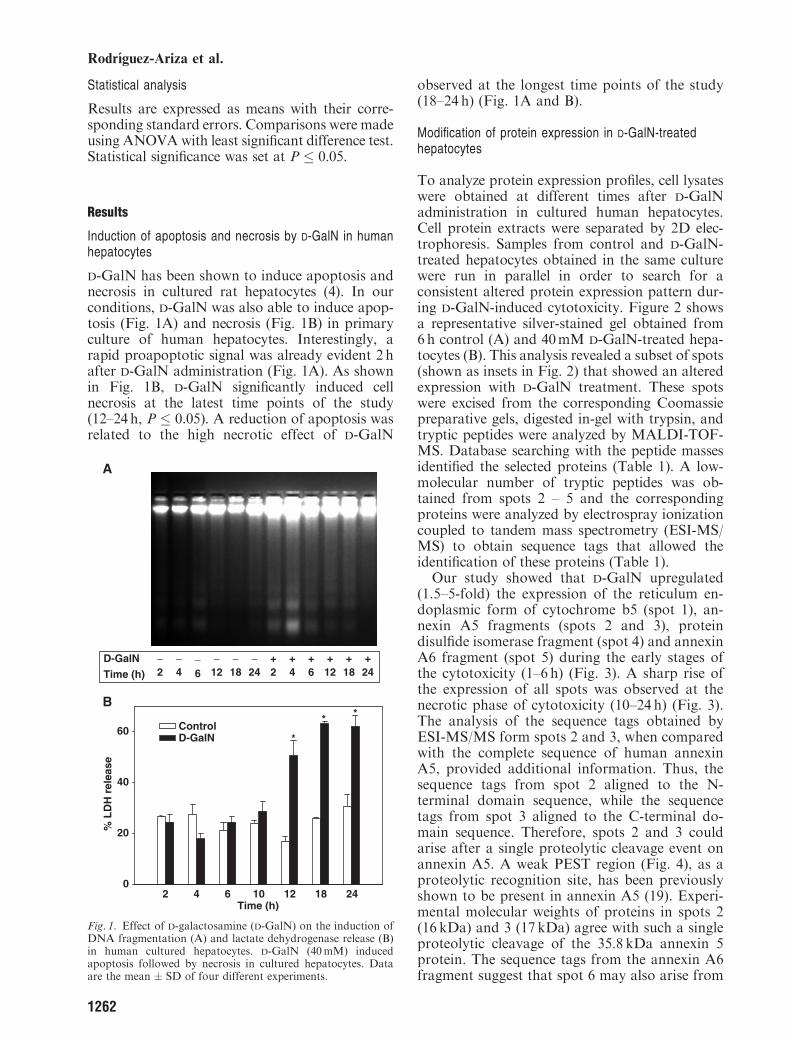
D-GalN has been shown to induce apoptosis andnecrosis in cultured rat hepatocytes (4). In ourconditions, D-GalN was also able to induce apop-tosis (Fig. 1A) and necrosis (Fig. 1B) in primaryculture of human hepatocytes. Interestingly, arapid proapoptotic signal was already evident 2 hafter D-GalN administration (Fig. 1A). As shownin Fig. 1B, D-GalN significantly induced cellnecrosis at the latest time points of the study(12–24h, P � 0.05). A reduction of apoptosis wasrelated to the high necrotic effect of D-GalN
observed at the longest time points of the study(18–24 h) (Fig. 1A and B).
Modification of protein expression in D-GalN-treatedhepatocytes
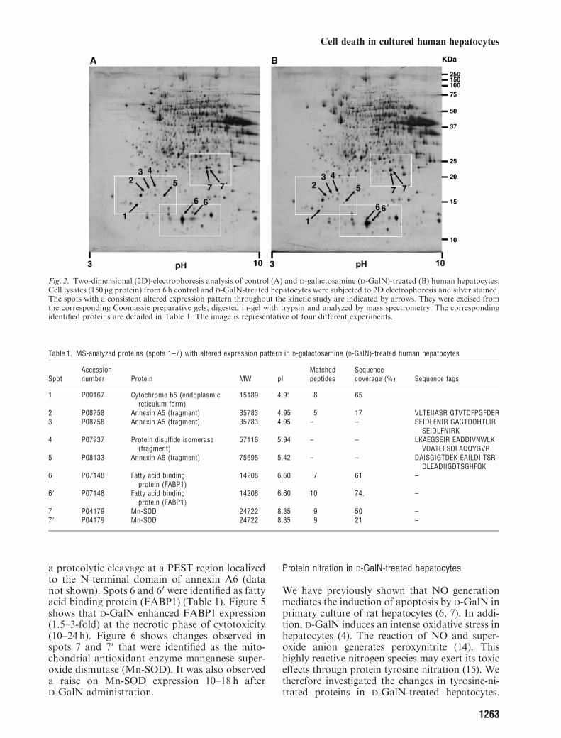
To analyze protein expression profiles, cell lysateswere obtained at different times after D-GalNadministration in cultured human hepatocytes.Cell protein extracts were separated by 2D elec-trophoresis. Samples from control and D-GalN-treated hepatocytes obtained in the same culturewere run in parallel in order to search for aconsistent altered protein expression pattern dur-ing D-GalN-induced cytotoxicity. Figure 2 showsa representative silver-stained gel obtained from6h control (A) and 40mM D-GalN-treated hepa-tocytes (B). This analysis revealed a subset of spots(shown as insets in Fig. 2) that showed an alteredexpression with D-GalN treatment. These spotswere excised from the corresponding Coomassiepreparative gels, digested in-gel with trypsin, andtryptic peptides were analyzed by MALDI-TOF-MS. Database searching with the peptide massesidentified the selected proteins (Table 1). A low-molecular number of tryptic peptides was ob-tained from spots 2 – 5 and the correspondingproteins were analyzed by electrospray ionizationcoupled to tandem mass spectrometry (ESI-MS/MS) to obtain sequence tags that allowed theidentification of these proteins (Table 1).
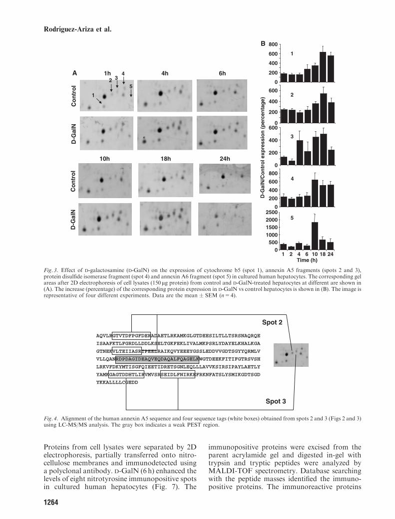
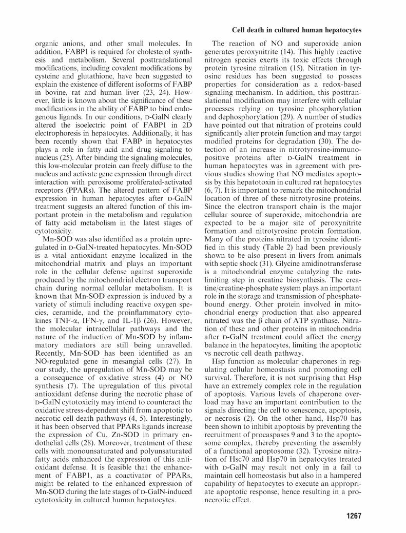
Our study showed that D-GalN upregulated(1.5–5-fold) the expression of the reticulum en-doplasmic form of cytochrome b5 (spot 1), an-nexin A5 fragments (spots 2 and 3), proteindisulfide isomerase fragment (spot 4) and annexinA6 fragment (spot 5) during the early stages ofthe cytotoxicity (1–6 h) (Fig. 3). A sharp rise ofthe expression of all spots was observed at thenecrotic phase of cytotoxicity (10–24 h) (Fig. 3).The analysis of the sequence tags obtained byESI-MS/MS form spots 2 and 3, when comparedwith the complete sequence of human annexinA5, provided additional information. Thus, thesequence tags from spot 2 aligned to the N-terminal domain sequence, while the sequencetags from spot 3 aligned to the C-terminal do-main sequence. Therefore, spots 2 and 3 couldarise after a single proteolytic cleavage event onannexin A5. A weak PEST region (Fig. 4), as aproteolytic recognition site, has been previouslyshown to be present in annexin A5 (19). Experi-mental molecular weights of proteins in spots 2(16 kDa) and 3 (17 kDa) agree with such a singleproteolytic cleavage of the 35.8 kDa annexin 5protein. The sequence tags from the annexin A6fragment suggest that spot 6 may also arise from
Time (h)D-GalN
24+
18+
12+
6+
4+
2+
24−
18−
12−
6−
4−
2−
A
Time (h)2 4 6 10 12 18 24
% L
DH
rel
ease
0
20
40
60 ControlD-GalN
B
*
* *
Fig. 1. Effect of D-galactosamine (D-GalN) on the induction ofDNA fragmentation (A) and lactate dehydrogenase release (B)in human cultured hepatocytes. D-GalN (40mM) inducedapoptosis followed by necrosis in cultured hepatocytes. Dataare the mean � SD of four different experiments.
1262
Rodrıguez-Ariza et al.
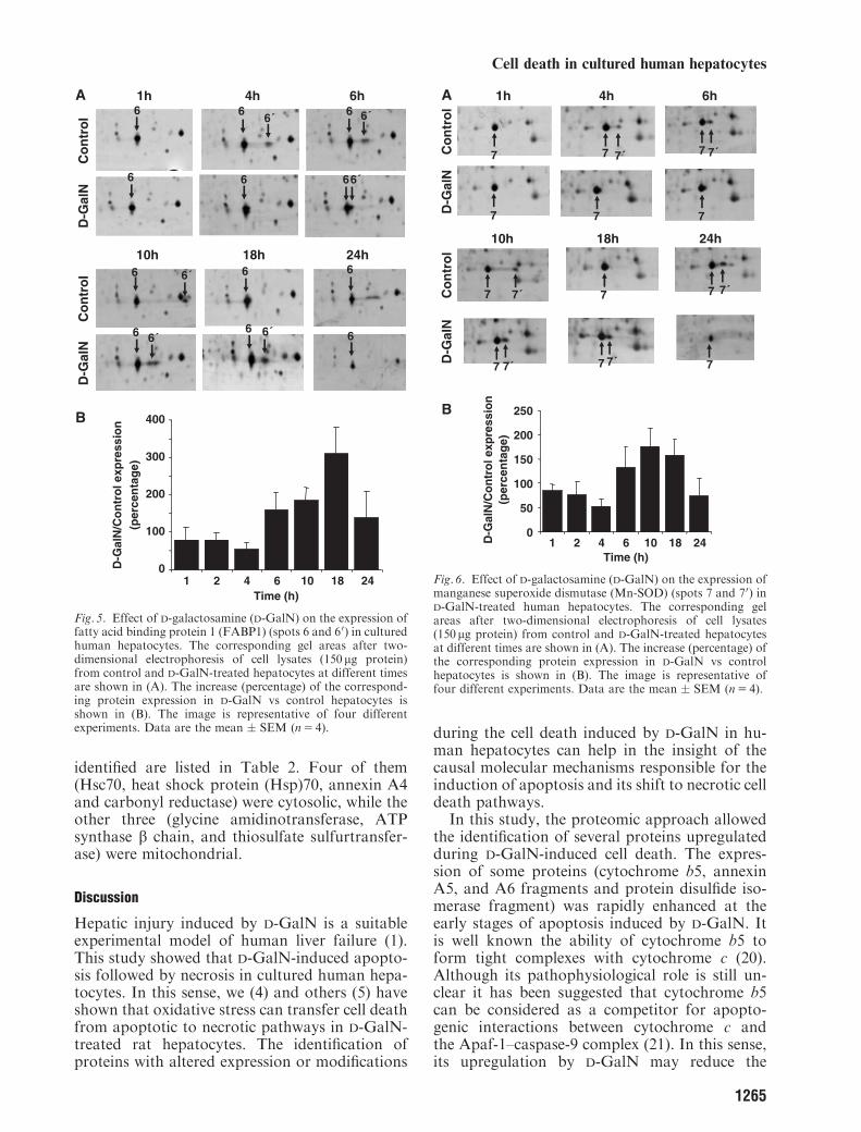
a proteolytic cleavage at a PEST region localizedto the N-terminal domain of annexin A6 (datanot shown). Spots 6 and 60 were identified as fattyacid binding protein (FABP1) (Table 1). Figure 5shows that D-GalN enhanced FABP1 expression(1.5–3-fold) at the necrotic phase of cytotoxicity(10–24 h). Figure 6 shows changes observed inspots 7 and 70 that were identified as the mito-chondrial antioxidant enzyme manganese super-oxide dismutase (Mn-SOD). It was also observeda raise on Mn-SOD expression 10–18 h afterD-GalN administration.
Protein nitration in D-GalN-treated hepatocytes
We have previously shown that NO generationmediates the induction of apoptosis by D-GalN inprimary culture of rat hepatocytes (6, 7). In addi-tion, D-GalN induces an intense oxidative stress inhepatocytes (4). The reaction of NO and super-oxide anion generates peroxynitrite (14). Thishighly reactive nitrogen species may exert its toxiceffects through protein tyrosine nitration (15). Wetherefore investigated the changes in tyrosine-ni-trated proteins in D-GalN-treated hepatocytes.
Fig. 2. Two-dimensional (2D)-electrophoresis analysis of control (A) and D-galactosamine (D-GalN)-treated (B) human hepatocytes.Cell lysates (150 mg protein) from 6h control and D-GalN-treated hepatocytes were subjected to 2D electrophoresis and silver stained.The spots with a consistent altered expression pattern throughout the kinetic study are indicated by arrows. They were excised fromthe corresponding Coomassie preparative gels, digested in-gel with trypsin and analyzed by mass spectrometry. The correspondingidentified proteins are detailed in Table 1. The image is representative of four different experiments.
Table 1. MS-analyzed proteins (spots 1–7) with altered expression pattern in D-galactosamine (D-GalN)-treated human hepatocytes
SpotAccessionnumber Protein MW pI
Matchedpeptides
Sequencecoverage (%) Sequence tags
1 P00167 Cytochrome b5 (endoplasmicreticulum form)
15189 4.91 8 65
2 P08758 Annexin A5 (fragment) 35783 4.95 5 17 VLTEIIASR GTVTDFPGFDER3 P08758 Annexin A5 (fragment) 35783 4.95 – – SEIDLFNIR GAGTDDHTLIR
SEIDLFNIRK4 P07237 Protein disulfide isomerase
(fragment)57116 5.94 – – LKAEGSEIR EADDIVNWLK
VDATEESDLAQQYGVR5 P08133 Annexin A6 (fragment) 75695 5.42 – – DAISGIGTDEK EAILDIITSR
DLEADIIGDTSGHFQK6 P07148 Fatty acid binding
protein (FABP1)14208 6.60 7 61 –
6 0 P07148 Fatty acid bindingprotein (FABP1)
14208 6.60 10 74. –
7 P04179 Mn-SOD 24722 8.35 9 50 –7 0 P04179 Mn-SOD 24722 8.35 9 21 –
1263
Cell death in cultured human hepatocytes
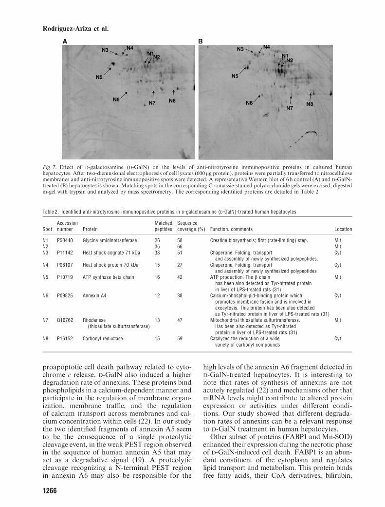
Proteins from cell lysates were separated by 2Delectrophoresis, partially transferred onto nitro-cellulose membranes and immunodetected usinga polyclonal antibody. D-GalN (6 h) enhanced thelevels of eight nitrotyrosine immunopositive spotsin cultured human hepatocytes (Fig. 7). The
immunopositive proteins were excised from theparent acrylamide gel and digested in-gel withtrypsin and tryptic peptides were analyzed byMALDI-TOF spectrometry. Database searchingwith the peptide masses identified the immuno-positive proteins. The immunoreactive proteins
10h 18h 24h
Co
ntr
ol
D-G
alN
1h 4h 6h
1
2 34
5
Co
ntr
ol
D-G
alN
A
D-G
alN
/Co
ntr
ol e
xpre
ssio
n (
per
cen
tag
e)Time (h)
1
2
0
200
400
600
3
4
5
B
0
200
400
600
800
0
200
400
600
0
200
400
600
800
0500
1000150020002500
1 2 4 6 10 18 24
Fig. 3. Effect of D-galactosamine (D-GalN) on the expression of cytochrome b5 (spot 1), annexin A5 fragments (spots 2 and 3),protein disulfide isomerase fragment (spot 4) and annexin A6 fragment (spot 5) in cultured human hepatocytes. The corresponding gelareas after 2D electrophoresis of cell lysates (150 mg protein) from control and D-GalN-treated hepatocytes at different are shown in(A). The increase (percentage) of the corresponding protein expression in D-GalN vs control hepatocytes is shown in (B). The image isrepresentative of four different experiments. Data are the mean � SEM (n5 4).
AQVLRGTVTDFPGFDERADAETLRKAMKGLGTDEESILTLLTSRSNAQRQE
ISAAFKTLFGRDLLDDLKSELTGKFEKLIVALMKPSRLYDAYELKHALKGA
GTNEKVLTEIIASRTPEELRAIKQVYEEEYGSSLEDDVVGDTSGYYQRMLV
VLLQANRDPDAGIDEAQVEQDAQALFQAGELKWGTDEEKFITIFGTRSVSH
LRKVFDKYMTISGFQIEETIDRETSGNLEQLLLAVVKSIRSIPAYLAETLY
YAMKGAGTDDHTLIRVMVSRSEIDLFNIRKEFRKNFATSLYSMIKGDTSGD
YKKALLLLCGEDD
Spot 2
Spot 3
Fig. 4. Alignment of the human annexin A5 sequence and four sequence tags (white boxes) obtained from spots 2 and 3 (Figs 2 and 3)using LC-MS/MS analysis. The gray box indicates a weak PEST region.
1264
Rodrıguez-Ariza et al.
identified are listed in Table 2. Four of them(Hsc70, heat shock protein (Hsp)70, annexin A4and carbonyl reductase) were cytosolic, while theother three (glycine amidinotransferase, ATPsynthase b chain, and thiosulfate sulfurtransfer-ase) were mitochondrial.
Discussion
Hepatic injury induced by D-GalN is a suitableexperimental model of human liver failure (1).This study showed that D-GalN-induced apopto-sis followed by necrosis in cultured human hepa-tocytes. In this sense, we (4) and others (5) haveshown that oxidative stress can transfer cell deathfrom apoptotic to necrotic pathways in D-GalN-treated rat hepatocytes. The identification ofproteins with altered expression or modifications
during the cell death induced by D-GalN in hu-man hepatocytes can help in the insight of thecausal molecular mechanisms responsible for theinduction of apoptosis and its shift to necrotic celldeath pathways.In this study, the proteomic approach allowed
the identification of several proteins upregulatedduring D-GalN-induced cell death. The expres-sion of some proteins (cytochrome b5, annexinA5, and A6 fragments and protein disulfide iso-merase fragment) was rapidly enhanced at theearly stages of apoptosis induced by D-GalN. Itis well known the ability of cytochrome b5 toform tight complexes with cytochrome c (20).Although its pathophysiological role is still un-clear it has been suggested that cytochrome b5can be considered as a competitor for apopto-genic interactions between cytochrome c andthe Apaf-1–caspase-9 complex (21). In this sense,its upregulation by D-GalN may reduce the
10h 18h 24h
1h 4h 6hC
on
tro
lD
-Gal
NC
on
tro
lD
-Gal
N
Time (h)
D-G
alN
/Co
ntr
ol e
xpre
ssio
n(p
erce
nta
ge)
A
B
6
6
6
6
6
6
6
6
6
6
6
6
6´ 6´
6´
6´
6´ 6´
0
100
200
300
400
1 2 4 6 10 18 24
Fig. 5. Effect of D-galactosamine (D-GalN) on the expression offatty acid binding protein 1 (FABP1) (spots 6 and 60) in culturedhuman hepatocytes. The corresponding gel areas after two-dimensional electrophoresis of cell lysates (150 mg protein)from control and D-GalN-treated hepatocytes at different timesare shown in (A). The increase (percentage) of the correspond-ing protein expression in D-GalN vs control hepatocytes isshown in (B). The image is representative of four differentexperiments. Data are the mean � SEM (n5 4).
10h 18h 24h
1h 4h 6h
Co
ntr
ol
D-G
alN
Co
ntr
ol
D-G
alN
Time (h)
D-G
alN
/Co
ntr
ol e
xpre
ssio
n(p
erce
nta
ge)
A
B
0
50
100
150
200
250
1 2 4 6 10 18 24
7 7´
7
7
7 7
7
7
7 7
7
7
7
7´
7´
7´ 7´
7´
Fig. 6. Effect of D-galactosamine (D-GalN) on the expression ofmanganese superoxide dismutase (Mn-SOD) (spots 7 and 70) inD-GalN-treated human hepatocytes. The corresponding gelareas after two-dimensional electrophoresis of cell lysates(150 mg protein) from control and D-GalN-treated hepatocytesat different times are shown in (A). The increase (percentage) ofthe corresponding protein expression in D-GalN vs controlhepatocytes is shown in (B). The image is representative offour different experiments. Data are the mean � SEM (n5 4).
1265
Cell death in cultured human hepatocytes
proapoptotic cell death pathway related to cyto-chrome c release. D-GalN also induced a higherdegradation rate of annexins. These proteins bindphospholipids in a calcium-dependent manner andparticipate in the regulation of membrane organ-ization, membrane traffic, and the regulationof calcium transport across membranes and cal-cium concentration within cells (22). In our studythe two identified fragments of annexin A5 seemto be the consequence of a single proteolyticcleavage event, in the weak PEST region observedin the sequence of human annexin A5 that mayact as a degradative signal (19). A proteolyticcleavage recognizing a N-terminal PEST regionin annexin A6 may also be responsible for the
high levels of the annexin A6 fragment detected inD-GalN-treated hepatocytes. It is interesting tonote that rates of synthesis of annexins are notacutely regulated (22) and mechanisms other thatmRNA levels might contribute to altered proteinexpression or activities under different condi-tions. Our study showed that different degrada-tion rates of annexins can be a relevant responseto D-GalN treatment in human hepatocytes.
Other subset of proteins (FABP1 and Mn-SOD)enhanced their expression during the necrotic phaseof D-GalN-induced cell death. FABP1 is an abun-dant constituent of the cytoplasm and regulateslipid transport and metabolism. This protein bindsfree fatty acids, their CoA derivatives, bilirubin,
Fig. 7. Effect of D-galactosamine (D-GalN) on the levels of anti-nitrotyrosine immunopositive proteins in cultured humanhepatocytes. After two-diemnsional electrophoresis of cell lysates (600mg protein), proteins were partially transferred to nitrocellulosemembranes and anti-nitrotyrosine inmunopositive spots were detected. A representative Western blot of 6 h control (A) and D-GalN-treated (B) hepatocytes is shown. Matching spots in the corresponding Coomassie-stained polyacrylamide gels were excised, digestedin-gel with trypsin and analyzed by mass spectrometry. The corresponding identified proteins are detailed in Table 2.
Table 2. Identified anti-nitrotyrosine immunopositive proteins in D-galactosamine (D-GalN)-treated human hepatocytes
SpotAccessionnumber Protein
Matchedpeptides
Sequencecoverage (%) Function. comments Location
N1 P50440 Glycine amidinotranferase 26 58 Creatine biosynthesis; first (rate-limiting) step. MitN2 35 66 MitN3 P11142 Heat shock cognate 71 kDa 33 51 Chaperone. Folding, transport
and assembly of newly synthesized polypeptides.Cyt
N4 P08107 Heat shock protein 70 kDa 15 27 Chaperone. Folding, transportand assembly of newly synthesized polypeptides
Cyt
N5 P10719 ATP synthase beta chain 16 42 ATP production. The b chainhas been also detected as Tyr-nitrated proteinin liver of LPS-treated rats (31)
Mit
N6 P09525 Annexin A4 12 38 Calcium/phospholipid-binding protein whichpromotes membrane fusion and is involved inexocytosis. This protein has been also detectedas Tyr-nitrated protein in liver of LPS-treated rats (31)
Cyt
N7 Q16762 Rhodanese(thiosulfate sulfurtransferase)
13 47 Mitochondrial thiosulfate sulfurtransferase.Has been also detected as Tyr-nitratedprotein in liver of LPS-treated rats (31)
Mit
N8 P16152 Carbonyl reductase 15 59 Catalyzes the reduction of a widevariety of carbonyl compounds
Cyt
1266
Rodrıguez-Ariza et al.
organic anions, and other small molecules. Inaddition, FABP1 is required for cholesterol synth-esis and metabolism. Several posttranslationalmodifications, including covalent modifications bycysteine and glutathione, have been suggested toexplain the existence of different isoforms of FABPin bovine, rat and human liver (23, 24). How-ever, little is known about the significance of thesemodifications in the ability of FABP to bind endo-genous ligands. In our conditions, D-GalN clearlyaltered the isoelectric point of FABP1 in 2Delectrophoresis in hepatocytes. Additionally, it hasbeen recently shown that FABP in hepatocytesplays a role in fatty acid and drug signaling tonucleus (25). After binding the signaling molecules,this low-molecular protein can freely diffuse to thenucleus and activate gene expression through directinteraction with peroxisome proliferated-activatedreceptors (PPARs). The altered pattern of FABPexpression in human hepatocytes after D-GalNtreatment suggests an altered function of this im-portant protein in the metabolism and regulationof fatty acid metabolism in the latest stages ofcytotoxicity.Mn-SOD was also identified as a protein upre-
gulated in D-GalN-treated hepatocytes. Mn-SODis a vital antioxidant enzyme localized in themitochondrial matrix and plays an importantrole in the cellular defense against superoxideproduced by the mitochondrial electron transportchain during normal cellular metabolism. It isknown that Mn-SOD expression is induced by avariety of stimuli including reactive oxygen spe-cies, ceramide, and the proinflammatory cyto-kines TNF-a, IFN-g, and IL-1b (26). However,the molecular intracellular pathways and thenature of the induction of Mn-SOD by inflam-matory mediators are still being unravelled.Recently, Mn-SOD has been identified as anNO-regulated gene in mesangial cells (27). Inour study, the upregulation of Mn-SOD may bea consequence of oxidative stress (4) or NOsynthesis (7). The upregulation of this pivotalantioxidant defense during the necrotic phase ofD-GalN cytotoxicity may intend to counteract theoxidative stress-dependent shift from apoptotic tonecrotic cell death pathways (4, 5). Interestingly,it has been observed that PPARs ligands increasethe expression of Cu, Zn-SOD in primary en-dothelial cells (28). Moreover, treatment of thesecells with monounsaturated and polyunsaturatedfatty acids enhanced the expression of this anti-oxidant defense. It is feasible that the enhance-ment of FABP1, as a coactivator of PPARs,might be related to the enhanced expression ofMn-SOD during the late stages of D-GalN-inducedcytotoxicity in cultured human hepatocytes.
The reaction of NO and superoxide aniongenerates peroxynitrite (14). This highly reactivenitrogen species exerts its toxic effects throughprotein tyrosine nitration (15). Nitration in tyr-osine residues has been suggested to possessproperties for consideration as a redox-basedsignaling mechanism. In addition, this posttran-slational modification may interfere with cellularprocesses relying on tyrosine phosphorylationand dephosphorylation (29). A number of studieshave pointed out that nitration of proteins couldsignificantly alter protein function and may targetmodified proteins for degradation (30). The de-tection of an increase in nitrotyrosine-immuno-positive proteins after D-GalN treatment inhuman hepatocytes was in agreement with pre-vious studies showing that NO mediates apopto-sis by this hepatotoxin in cultured rat hepatocytes(6, 7). It is important to remark the mitochondriallocation of three of these nitrotyrosine proteins.Since the electron transport chain is the majorcellular source of superoxide, mitochondria areexpected to be a major site of peroxynitriteformation and nitrotyrosine protein formation.Many of the proteins nitrated in tyrosine identi-fied in this study (Table 2) had been previouslyshown to be also present in livers from animalswith septic shock (31). Glycine amidinotransferaseis a mitochondrial enzyme catalyzing the rate-limiting step in creatine biosynthesis. The crea-tine/creatine-phosphate system plays an importantrole in the storage and transmission of phosphate-bound energy. Other protein involved in mito-chondrial energy production that also appearednitrated was the b chain of ATP synthase. Nitra-tion of these and other proteins in mitochondriaafter D-GalN treatment could affect the energybalance in the hepatocytes, limiting the apoptoticvs necrotic cell death pathway.Hsp function as molecular chaperones in reg-
ulating cellular homeostasis and promoting cellsurvival. Therefore, it is not surprising that Hsphave an extremely complex role in the regulationof apoptosis. Various levels of chaperone over-load may have an important contribution to thesignals directing the cell to senescence, apoptosis,or necrosis (2). On the other hand, Hsp70 hasbeen shown to inhibit apoptosis by preventing therecruitment of procaspases 9 and 3 to the apopto-some complex, thereby preventing the assemblyof a functional apoptosome (32). Tyrosine nitra-tion of Hsc70 and Hsp70 in hepatocytes treatedwith D-GalN may result not only in a fail tomaintain cell homeostasis but also in a hamperedcapability of hepatocytes to execute an appropri-ate apoptotic response, hence resulting in a pro-necrotic effect.
1267
Cell death in cultured human hepatocytes
In summary, the proteomic approach has beenproven to be a very useful tool for the identifica-tion of changes in protein expression and mod-ification in a model of hepatocellular injury. Theidentification of oxidative stress and NO-derivedreactive oxygen intermediates as critical contri-butors to protein modification and hepatocellularinjury, provides potential targets for therapeuticintervention. In the present study we showed thatthe increase of the selected protein expression isassociated with the induction of apoptosis ornecrosis during D-GalN-related cytotoxicity. Inaddition, the tyrosine nitration of proteins in-volved in cellular homeostasis and energy contentmight also influence the shift from an apoptotic tonecrotic cell death pathway by D-GalN in culturedhuman hepatocytes. The role of all these proteinsand their modifications in the initiation of apop-tosis and/or necrosis in hepatocytes after D-GalNtreatment are currently under investigation.
Acknowledgements
This work was supported by grants from the Programa dePromocion de la Investigacion en Salud del Ministerio deSanidad y Consumo (FIS CP03/00116 and PI04/1470).
References
1. Keppler D O, Pausch J, Decker K. Selective uridinetriphosphate deficiency induced by D-galactosamine in liverand reversed by pyrimidine nucleotide precursors. Effect onribonucleic acid synthesis. J Biol Chem 1974; 249: 211–6.
2. Sreedhar A S, Csermely P. Heat shock proteins in theregulation of apoptosis: new strategies in tumor therapy: acomprehensive review. Pharmacol Ther 2004; 101: 227–57.
3. Kaplowitz N, Tsukamoto H. Oxidative stress and liverdisease. In: Boyer J L, Ockner RK, eds. Progress in LiverDiseases, Vol. XIV. Philadelphia: W.B. Saunders Co., 1996;131–59.
4. Quintero A, Pedraza CA, Siendones E, Kamal Elsaid
A M, Colell A, Garcia-Ruiz C, et al. PGE1 protectionagainst apoptosis induced by D-galactosamine is not relatedto the modulation of intracellular free radical production inprimary culture of rat hepatocytes. Free Radic Res 2002; 36:345–55.
5. Samali A, Nordgren H, Zhivotovsky B, Peterson E,Orrenius S. A comparative study of apoptosis and necrosisin HepG2 cells: oxidant-induced caspase inactivation leadsto necrosis. Biochem Biophys Res Commun 1999; 255: 6–11.
6. Siendones E, Fouad D, Abou-Elella A M, Quintero
A, Barrera P, Muntane J. Role of nitric oxide in D-galactosamine-induced cell death and its protection byPGE1 in cultured hepatocytes. Nitric Oxide 2003; 8: 133–43.
7. Siendones E, Fouad D, Diaz-Guerra M J M, De la
Mata M, Bosca¤ L,Muntane J. PGE1 induced nitric oxidereduces apoptosis by D-galactosamine through attenuationof NF-KB and NOS-2 expression. Hepatology 2004; 40:1295–303.
8. Clemens M G. Nitric oxide in liver injury. Hepatology1999; 30: 1–5.
9. Rockey D C, Shah V. Nitric oxide biology and the liver:report of an AASLD research workshop. Hepatology 2004;39: 250–7.
10. Rees D D, Palmer R M, Moncada S. Role of endothe-lium-derived nitric oxide in the regulation of blood pressure.Proc Natl Acad Sci USA 1989; 86: 3375–8.
11. Moncada S, Rees D D, Schulz R, Palmer R M. Devel-opment and mechanism of a specific supersensitivity tonitrovasodilators after inhibition of vascular nitric oxidesynthesis in vivo. Proc Natl Acad Sci USA 1991; 88: 2166–70.
12. Knowles R G, Palacios M, Palmer R M, Moncada S.
Formation of nitric oxide from L-arginine in the centralnervous system: a transduction mechanism for stimulationof the soluble guanylate cyclase. Proc Natl Acad Sci USA1989; 86: 5159–62.
13. Stuehr D J, Gross S S, Sakuma I, Levi R, Nathan C F.
Activated murine macrophages secrete a metabolite ofarginine with the bioactivity of endothelium-derived relax-ing factor and the chemical reactivity of nitric oxide. J ExpMed 1989; 169: 1011–20.
14. Radi R, Beckman J S, Bush K M, Freeman B A.
Peroxynitrite oxidation of sulfhydryls. The cytotoxic poten-tial of superoxide and nitric oxide. J Biol Chem 1991; 266:4244–50.
15. Patel R P, McAndrew J, Sellak H, White C R, Jo H,Freeman B A, et al. Biological aspects of reactive nitrogenspecies. Biochim Biophys Acta 1989; 1411: 385–400.
16. Ferrini J B, Ourlin J C, Richard L, Fabre G, Maurel
P. Human hepatocyte culture. Methods Mol Biol 1998;107: 341–52.
17. Taffs R, Sitkovsky M. In vitro assays for mouse B and Tlymphocyte function. In: Coligan J E, Kruisbeek A M,Margulies D H, Shevach EM, StroberW, eds. CurrentProtocols in Immunology. New York: Greene Publishingand Wiley-Interscience, 1991; 1–8.
18. Rabilloud T, Carpentier G, Tarroux P. Improvementand simplification of low-background silver staining ofproteins by using sodium dithionite. Electrophoresis 1988;9: 288–91.
19. Barnes J A, Gomes AV. PEST sequences in calmodulin-binding proteins. Mol Cell Biochem 2002; 149–150: 17–27.
20. McLean M A, Sligar S G. Thermodynamic characteriza-tion of the interaction between cytochrome b5 and cyto-chrome c. Biochem Biophys Res Commun 1995; 215: 316–20.
21. Davydov D R. Microsomal monooxygenase in apoptosis:another target for cytochrome c signaling? Trends BiochemSci 2001; 26: 155–60.
22. Gerke V, Moss S E. Annexins: from structure to function.Physiol Rev 2002; 82: 331–71.
23. Dormann P, Borchers T, Korf U, Hojrup P, Roep-storff P, Spener F. Amino acid exchange and covalentmodification by cysteine and glutathione explain isoformsof fatty acid-binding protein occurring in bovine liver. J BiolChem 1993; 268: 16286–92.
24. Schroeder F, Jolly CA, Cho T H, Frolov A. Fatty acidbinding protein isoforms: structure and function. ChemPhys Lipids 1998; 92: 1–25.
25. Wolfrum C, Borrmann C M, Borchers T, Spener F.
Fatty acids and hypolipidemic drugs regulate peroxisomeproliferator-activated receptors alpha- and gamma-mediated gene expression via liver fatty acid binding pro-tein: a signaling path to the nucleus. Proc Natl Acad SciUSA 2001; 98: 2323–8.
26. Rogers R J, Monnier J M, Nick H S. Tumor necrosisfactor-alpha selectively induces MnSOD expression viamitochondria-to-nucleus signaling, whereas interleukin-1beta utilizes an alternative pathway. J Biol Chem 2001;276: 20419–27.
27. Keller T, Pleskova M, McDonald MC, ThiemermannC, Pfeilschifter J, Beck KF. Identification of manganesesuperoxide dismutase as a NO-regulated gene in rat
1268
Rodrıguez-Ariza et al.
glomerular mesangial cells by 2D gel electrophoresis. NitricOxide 2003; 9: 183–93.
28. Inoue I, Goto S, Matsunaga T, Nakajima T, Awata T,Hokari S, et al. The ligands/activators for peroxisomeproliferator-activated receptor alpha (PPARalpha) andPPARgamma increase Cu21, Zn21-superoxide dismutaseand decrease p22phox message expressions in primaryendothelial cells. Metabolism 2001; 50: 3–11.
29. Klotz LO, Schroeder P, Sies H. Peroxynitrite signaling:receptor tyrosine kinases and activation of stress-responsivepathways. Free Radic Biol Med 2002; 33: 737–43.
30. Ischiropoulos H. Biological selectivity and functionalaspects of protein tyrosine nitration. Biochem BiophysRes Commun 2003; 305: 776–83.
31. Aulak K S, Miyagi M, Yan L,West KA, Massillon D,Crabb J W, et al. Proteomic method identifies proteinsnitrated in vivo during inflammatory challenge. Proc NatlAcad Sci USA 2001; 98: 12056–61.
32. Beere HM, Wolf BB, Cain K, Mosser DD,Mahboubi
A, Kuwana T, et al. Heat-shock protein 70 inhibits apop-tosis by preventing recruitment of procaspase-9 to the Apaf-1 apoptosome. Nat Cell Biol 2000; 2: 469–75.
1269
Cell death in cultured human hepatocytes


