Embed Size (px)
Citation preview

Aminoflavone, a ligand of the Aryl Hydrocarbon Receptor (AhR),inhibits HIF-1α expression in an AhR-independent fashion
Erika Terzuoli1,2,*, Maura Puppo1,3,*, Annamaria Rapisarda1, Badarch Uranchimeg1, LiangCao4, Angelika M. Burger5, Marina Ziche2, and Giovanni Melillo1,61Tumor Hypoxia Laboratory, SAIC-Frederick, Inc., NCI at Frederick, Frederick, MD2Department of Molecular Biology, University of Siena, Siena, Italy3G. Gaslini Institute, Laboratory of Molecular Biology, Genoa, Italy4Molecular Targets Core, Genetics Branch, Center for Cancer Research, National Cancer Institute,Bethesda, MD5Barbara Ann Karmanos Cancer Institute, Detroit, MI
AbstractAminoflavone (AF), the active component of a novel anticancer agent (AFP464) in phase I clinicaltrials, is a ligand of the aryl hydrocarbon receptor (AhR). AhR dimerizes with HIF-1β/ARNT, whichis shared with HIF-1α, a transcription factor critical for the response of cells to oxygen deprivation.To address whether pharmacological activation of the AhR pathway might be a potential mechanismfor inhibition of HIF-1, we tested the effects of AF on HIF-1 expression. AF inhibited HIF-1αtranscriptional activity and protein accumulation in MCF-7 cells. However, inhibition of HIF-1α byAF was independent from a functional AhR pathway. Indeed, AF inhibited HIF-1α expression inAhR100 cells, in which the AhR pathway is functionally impaired, yet did not induce cytotoxicity,providing evidence that these effects are mediated by distinct signaling pathways. Moreover, AF wasinactive in MDA-MB-231 cells, yet inhibited HIF-1α in MDA-MB-231 cells transfected with theSULT1A1 gene. AF inhibited HIF-1α mRNA expression by approximately 50%. Notably,actinomycin-D completely abrogated the ability of AF to down-regulate HIF-1α mRNA, indicatingthat active transcription was required for the inhibition of HIF-1α expression. Finally, AF inhibitedHIF-1α protein accumulation and the expression of HIF-1-target genes in MCF-7 xenografts.
These results demonstrate that AF inhibits HIF-1α in an AhR-independent fashion and they unveiladditional activities of AF that may be relevant for its further clinical development.
KeywordsHIF-1; aminoflavone; hypoxia; AhR; mRNA transcription
IntroductionHypoxia, a decrease in oxygen levels, is a hallmark of solid tumors. The response ofmammalian cells to hypoxia is mediated, at least in part, by a family of transcription factors
Copyright © 2010 American Association for Cancer Research6To whom correspondence should be addressed at: Giovanni Melillo, M.D. DTP-Tumor Hypoxia Laboratory Bldg. 432, Room 218 SAIC– Frederick, Inc., NCI at Frederick Frederick, Maryland 21702 Tel: (301)-846-5050 Fax: (301)-846-6081 [email protected].*These two authors contributed equally to this work.
NIH Public AccessAuthor ManuscriptCancer Res. Author manuscript; available in PMC 2011 September 1.
Published in final edited form as:Cancer Res. 2010 September 1; 70(17): 6837–6848. doi:10.1158/0008-5472.CAN-10-1075.
NIH
-PA Author Manuscript
NIH
-PA Author Manuscript
NIH
-PA Author Manuscript

known as Hypoxia Inducible Factors (HIF) (1). HIF-1 is a heterodimer consisting of aconstitutively expressed β subunit and an oxygen regulated α subunit (2) which is ubiquitinatedand degraded under normoxic conditions (3). In contrast under hypoxic conditions the HIF-αsubunit is stabilized and translocates to the nucleus, where it dimerizes with HIF-1β (alsoknown as aryl hydrocarbon receptor nuclear translocator, ARNT) and activates transcriptionof genes involved in key steps of tumorigenesis, including angiogenesis, metabolism,proliferation, metastasis and differentiation (4). Overexpression of HIF-α has been reported inmore than 70% of human cancers (5–11) and is associated with poor patients prognosis, makingHIF-1 an attractive target for the development of novel cancer therapeutics (12).
Aminoflavone (NSC 686288) is the active component of a pro-drug (AFP464) in phase Iclinical trials. AFP464 is rapidly converted to AF, in plasma or tissue culture, by nonspecificplasma esterases. AF has a unique COMPARE pattern of cytotoxicity in the NCI60 (13,14),with activity only in a subset of cell lines, including MCF-7 breast cancer cells (15–18). Thesensitivity of cancer cell lines to AF has been associated with its ability to act as a ligand ofthe aryl hydrocarbon receptor (AhR), which upon dimerization with HIF-1β/ARNT activatestranscription by binding to the xenobiotic response element (XRE) in the promoters of targetgenes, including but not limited to cytochrome P450 1A1 (CYP1A1). Indeed, the presence ofa functional AhR pathway and the induction of CYP1A1 expression by AF appear to beessential for its anti-proliferative activity in MCF-7 cells (18–20).
Several studies have suggested the existence of a crosstalk between the AhR and HIF-1pathways (21–26). However, whether pharmacological activation of the AhR pathway may bea viable approach to inhibit HIF-1 remains poorly understood. We demonstrate that AF inhibitsHIF-1α expression, both in vitro and in MCF-7 xenografts, in an AhR-independent fashion.Notably, AF partially inhibited HIF-1α mRNA expression, yet almost completely blockedHIF-1α protein accumulation. The ability of actinomycin-D to completely revert inhibition ofHIF-1α mRNA expression by AF is consistent with the existence of a transcription-dependentpathway that may regulate HIF-1α mRNA expression and its translation.
Materials and methodsCell lines and reagents
Human cells lines were maintained in RPMI-1640 supplemented with L-glutamine and 5%heat-inactivated fetal bovine serum (Hyclone) and grown at 37°C in 5% CO2 and ambientoxygen (normoxia). Hypoxia was achieved in an Invivo2 400 hypoxic workstation (RuskinnTechnologies) delivering 1% oxygen in 5% CO2 at 37°C. AhR-deficient MCF-7 (AhR100)(27) were kindly provided by Dr. David Vistica (STB, NCI, Frederick, MD). MDA-MB-231stably transfected with a SULT1A1 cDNA, MDA/SULT1A1 (28), were kindly provided byDr David C Spink, (Wadsworth Center, Albany, NY). All cell lines were obtained fromDevelopmental Therapeutics Program (DTP) and were validated according to informationprovided on the DTP web site(http://dtp.nci.nih.gov/branches/btb/characterizationNCI60.html). Aminoflavone (AF, NSC686288) was from the Drug Synthesis and Chemistry Branch, DTP, NCI.
Sulforhodamine B assay (SRB)Cells, seeded into 96-well plates, were treated with AF for additional 72 hours and cell viabilitywas assessed as previously described (29).
Terzuoli et al. Page 2
Cancer Res. Author manuscript; available in PMC 2011 September 1.
NIH
-PA Author Manuscript
NIH
-PA Author Manuscript
NIH
-PA Author Manuscript
Immunoblot analysisTotal protein lysates were obtained as described previously (30). Antibodies used are:HIF-1α and p21 (BD- Transduction Laboratories), HIF-1β, SULT1A1, HIF-2α and AhR(Novus Biologicals), actin (Chemicon International), γH2AX (Cell Signaling, Inc.).
Real-Time PCRVEGF, HIF-1α, p21, CA9, LOX, actin and CYP1A1 expression was measured by real-timePCR as described previously (30). Primers and probes used are listed in Supplementary Table1. VEGF primers and probe were described previously (30). 18S rRNA was used as an internalcontrol.
HIF-1α Protein Translational AssayMCF-7, treated for 12 hours with AF (0.25 μM), were with 35S-methionine/cysteine (MPBiomedicals) as previously described (31). Total 35S incorporation was monitored by both sizefractionation of total lysates and TCA precipitation.
Luciferase activityMCF-7 were transfected using Effectene (Qiagen) with pGL2-TK-HRE, pGL3-control (30),NF-κB-Luc, AP-1-luc (Dr. Nancy Colburn, National Cancer Institute Frederick, MD) andXRE-Luc (by Dr. F.J. Gonzalez, NCI, NIH, Bethesda, MD). Efficiency of transfection wasassessed by co-transfection with pQTK-Renilla (Promega). Results are expressed as foldincrease of luciferase levels relative to normoxic untreated controls.
Transfection with siRNA for HIF-1α, SULT1A1 and AhRSiRNA targeting SULT1A1 (s5`-CAGAGGGAGTGTGCGAATCAA), AhR (5`-TTGGATTAAATTAGTTTGTGA), HIF-1 α (5`-AGGACAAGTCACCACAGGA) andnegative control (NC) were purchased from Qiagen.
Animal studiesStudies were conducted in an AAALAC-accredited facility with an approved animal protocol.MCF-7 (1 × 10⁷) were injected subcutaneously (s.c.) into the flank of female athymic nude(NCr/nu) mice (Animal Production Area, NCI-Frederick). Beta-estradiol cypionate (3 mg/kg)was administered intramuscularly every 7 days. Tumor size was determined by collectinglength and width measurements and calculating the tumor weight (mg) as [tumor length ×(tumor width)2]/2, where the tumor length is the longest dimension (mm) and the tumor widthis the narrowest dimension (mm). AF (saline/0.05% Tween 80) was dosed i.p. Five mice pergroup were treated daily for 4 days with AF (60 mg/kg) or vehicle control. When mice weresacrificed (day 4), tumors from each animal were harvested and used to analyze mRNA andprotein expression, as described previously (32).
Tissue HIF-1α assayTissue lysates were prepared as described previously (32) and used for the quantitativedetermination of HIF-1α using electrochemiluminescence assay (Meso-Scale). HIF-1αconcentration (pg/ml) was determined using a recombinant protein (R&D Systems) asstandard.
Statistical analysisResults are either representative or average of at least three independent experimentsperformed. Statistical analysis was performed using ANOVA test and t test (Prism, GraphPad).
Terzuoli et al. Page 3
Cancer Res. Author manuscript; available in PMC 2011 September 1.
NIH
-PA Author Manuscript
NIH
-PA Author Manuscript
NIH
-PA Author Manuscript

RESULTSAF inhibits HIF-1α transcriptional activity in MCF-7 breast cancer cells
To test whether AF inhibited HIF-1 activity, MCF-7 cells were transiently transfected eitherwith pGL2-TK-HRE, containing the luciferase reporter gene under control of 3 copies of ahypoxia response element (HRE) or with a control vector (pGL3 control). Hypoxia increasedHRE-dependent luciferase expression by 49-fold, relative to cells cultured under normoxicconditions (Figure 1A, left panel). AF inhibited luciferase expression in a dose-dependentmanner, but did not affect constitutive luciferase expression (Figure 1A, right panel) suggestingthat inhibition of luciferase by AF was HIF-1 dependent. AF also inhibited endogenous HIF-1transcriptional activity, as indicated by inhibition of hypoxic induction of VEGF (Figure 1B,left panel), CA9 and LOX mRNA expression (Figure 1B, right panel), similar to the effectsof siRNA targeting HIF-1α. In contrast, AF caused up to 40-fold increase of p21 mRNAexpression, under both normoxic and hypoxic conditions (Figure 1C, left panel), indicatingthat AF differentially affects gene expression in MCF-7 cells.
Notably, neither NF-κB-(Fig 1D, left panel) nor AP-1-dependent transcriptional activities(right panel) were inhibited by AF, further demonstrating that AF specifically inhibits HIF-1.
AF inhibits HIF-1α and HIF-2α protein accumulation in a cell-type dependent fashionTo address whether AF inhibited HIF-1α and HIF-2α protein accumulation, we tested 6 celllines from the NCI60 panel that were reported to be sensitive to AF. Hypoxia increasedHIF-1α protein expression in all the cell lines examined, while HIF-2α was induced todetectable levels in three cell lines (T47D, CAKI, UACC257). AF (0.5μM) inhibited HIF-1αprotein accumulation, albeit to different extent, in MCF-7, T47D, CAKI and OVCAR3. Incontrast, HIF-2α was slightly decreased by AF in T47D and CAKI cells but not in UACC257(Figure 2A), suggesting that AF inhibits HIF-1α and HIF-2α protein accumulation in a celltype specific fashion, although HIF-2α appears to be slightly less susceptible to AF inhibition,relative to HIF-1α.
Further experiments demonstrated that AF inhibited HIF-1α protein accumulation in MCF-7cells in a dose-dependent fashion, with approximately 80% decrease at 0.25 μM (Figure 2B).By contrast, HIF-1β was only inhibited by 15% and actin levels were not changed in thepresence of up to 1 μM of AF. Kinetic experiments demonstrated that AF (0.25 μM) causedlittle or no inhibition of HIF-1α protein at 8 hours, but completely abrogated its accumulationafter 12–24 hours of treatment (Figure 2C). AF also inhibited HIF-1α protein accumulationinduced by the iron chelator desferrioxamine (100 μM) (Figure 2D), suggesting that its effectsare not restricted to hypoxic signaling. By contrast, AF induced p21 protein accumulation inMCF-7 cells cultured under either normoxia (data not shown) or hypoxia (Figure 1C, rightpanel), consistent with the induction of p21 mRNA expression and demonstrating a differentialeffect on distinct target proteins.
A functional AhR pathway is not required for inhibition of HIF-1α expression by AFPrevious studies have indicated that AF is a ligand of AhR (18). Indeed, AF caused a 7- to 8-fold increase in XRE-dependent luciferase expression (Figure 3A) and induced up to 200-foldhigher levels of CYP1A1 mRNA expression in MCF-7 cells (Figure 3B), demonstrating thatAF was able to induce AhR-dependent transcriptional activity. TCDD, used as positive control,induced a 15-fold increase in XRE-dependent luciferase expression (Figure 3A) and up to1160-fold higher levels of CYP1A1 mRNA expression (Figure 3B), relative to untreated cells.
To address whether inhibition of HIF-1α by AF required a functional AhR pathway, we tookadvantage of AhR100 cells, MCF-7-derived cells that express low levels of AhR and are resistant
Terzuoli et al. Page 4
Cancer Res. Author manuscript; available in PMC 2011 September 1.
NIH
-PA Author Manuscript
NIH
-PA Author Manuscript
NIH
-PA Author Manuscript

to the cytotoxic effects of AF (18). Indeed, AF caused cytotoxicity in parental MCF-7 cells butnot in AhR100 cells, even at concentrations as high as 2 μM (Figure 3C). Consistent with afunctional impairment of the AhR pathway, TCDD induction of XRE-dependent luciferaseexpression was decreased by 75% in AhR100 cells, relative to parental MCF-7 cells, andinduction of CYP1A1 mRNA expression was decreased by 95% (Figure 3A–B). Moreimportantly, AF failed to induce XRE-dependent luciferase expression in AhR100 cells andonly modestly induced CYP1A1 mRNA expression (35% of the levels induced in wild typeMCF-7 cells) (Figure 3A–B). However, AF inhibited hypoxic induction of HIF-1transcriptional activity irrespective of a functional AhR, as demonstrated by inhibition of HRE-dependent luciferase expression in both MCF-7 and AhR100 cells (Figure 3D). Accordingly,AF also completely inhibited hypoxic induction of HIF-1α protein accumulation in AhR100
cells (Figure 3E), demonstrating that inhibition of HIF-1α expression by AF is independentfrom a functional AhR pathway.
Aminoflavone inhibits HIF-1α in MDA/SULT1A1, but not in MDA-MB-231 parental breastcancer cells
The restricted spectrum of AF activity in the NCI60 cells has been attributed to a requirementfor its intracellular activation (18) by pathway(s) yet to be completely elucidated. A potentialcorrelation between sensitivity to AF and expression of SULT1A1 has also been suggested,consistent with the ability of SULT1A1 to induce the formation of AF metabolites that mediateDNA damage (33). Indeed, AF exerted anti-proliferative effects in MDA/SULT1A1, but notin MDA-MB-231 parental cells (Figure 3C). However, both MDA-MB-231 and MDA/SULT1A1 cells express little or no AhR transcriptional activity, as indicated by minimal if anyinduction of XRE-dependent luciferase (Figure 3A) or CYP1A1 mRNA expression by eitherAF or TCDD (Figure 3B).
We then tested whether AF inhibited HIF-1 transcriptional activity in MDA-MB-231 andMDA-SULT1A1 cells. AF completely inhibited HIF-1-dependent luciferase expression inMDA/SULT1A1, but did not affect its expression in MDA-MB-231 parental cells,demonstrating that exogenous expression of SULT1A1 was sufficient to mediate AF-dependent inhibition of HIF-1 activity (Figure 3D). Accordingly, AF almost completelyinhibited hypoxic induction of HIF-1α protein in MDA/SULT1A1 cells, but not in MDA-MB-231 (Figure 3E and Supplementary Figure 1).
Next, we evaluated expression of HIF-1α in MCF-7 cells transfected with either negativecontrol (NC) siRNA or siRNA targeting SULT1A1 or AhR (Supplementary Figure 2A–B).AF inhibited hypoxic induction of HIF-1α protein by 75% in cells transfected with NC siRNAand by 50% in cells transfected with AhR siRNA, relative to hypoxia treated cells(Supplementary Figure 2B). In contrast, down-regulation of SULT1A1 almost completelyprevented inhibition of HIF-1α by AF (15% inhibition, compared to hypoxia) (SupplementaryFigure 2B), demonstrating that SULT1A1 expression was implicated in HIF-1α inhibition byAF and further supporting the conclusion that inhibition of HIF-1α by AF is independent froma functional AhR pathway.
Inhibition of HIF-1α by AF is independent from DNA damageInduction of DNA damage by AF, measured by phosphorylation of H2AX, paralleled resultsobtained in cytotoxicity assay. Indeed, AF induced significantly higher levels of γH2AX insensitive MCF-7 and MDA/SULT1A1 cells than in resistant AhR100 and MDA-MB-231 cells(Figure 3E). However, AF was equally able to inhibit hypoxic induction of HIF-1α protein inAhR100 and MDA/SULT1A1 cells, demonstrating a complete dissociation between inductionof DNA damage and HIF-1α inhibition (Figure 3E).
Terzuoli et al. Page 5
Cancer Res. Author manuscript; available in PMC 2011 September 1.
NIH
-PA Author Manuscript
NIH
-PA Author Manuscript
NIH
-PA Author Manuscript

We have previously demonstrated that agents that inhibit topoisomerase I or II may affectHIF-1α protein translation (31,34). Consistent with the finding that induction of DNA damageby AF does not appear to involve topoisomerases (35), AF was able to inhibit HIF-1α proteinexpression in cells transfected with siRNA targeting topo I or topo IIα (Supplementary Figure3B). Furthermore, AF inhibited hypoxic induction of HIF-1α protein accumulation in thepresence of aphidicolin, which blocks DNA polymerase and prevents DNA damage(Supplementary Figure 4), further demonstrating that AF inhibited HIF-1α by a DNA damageindependent pathway.
AF does not affect HIF-1α degradation, but decreases the rate of HIF-1α translationHIF-1α steady state is the result of a balance between protein translation and its degradation.To investigate whether AF affected HIF-1α degradation, we performed experiments in thepresence of inhibitors of either proteasome activity (MG-132) or prolyl hydroxylase enzymes(DMOG, a global inhibitor of 2-oxoglutarate-dependent dioxygenase enzymes), which causean accumulation of HIF-1α under normoxic conditions. As shown in Figure 4A, AF inhibitedHIF-1α protein accumulation regardless of inhibition of the proteasome or prolyl hydroxylases,suggesting that AF did not affect degradation of HIF-1α protein. Consistent with these results,AF also inhibited normoxic accumulation of HIF-2α in a sensitive VHL-deficient renal cancercell line (A498, which do not express HIF-1α), indicating that a functional VHL pathway,essential for normoxic degradation of the HIF-α subunit, is not required for the inhibition ofHIF-α by AF (Figure 4B). Finally, experiments conducted in the presence of cycloheximidefailed to demonstrated significant differences in HIF-1α protein half-life between cells culturedin the absence or presence of AF (half-life ~60 min) (Figure 4C). Taken together, these resultsdemonstrate that AF does not affect HIF-1α protein degradation.
We then assessed whether AF inhibited HIF-1α translation by evaluating the rate of HIF-1αprotein synthesis in normoxic MCF-7 cells in the absence or presence of AF. Cells were pulse-labeled with [35S] methionine for 60 minutes at which point HIF-1α was immuno-precipitatedand analyzed by PAGE and autoradiography. As shown in Figure 4D (left panel), AFsignificantly decreased 35S-labeled HIF-1α accumulation relative to untreated cells, suggestingthat it may affect its rate of translation. Importantly, under the same experimental conditionsAF did not significantly affect the synthesis of HIF-1β or that of total proteins (Figure 4D,right panel).
Active transcription is required for inhibition of HIF-1α mRNA expression by AFRegulation of HIF-1α translation under hypoxic conditions is still poorly understood and recentevidence suggests that levels of HIF-1α mRNA may be a crucial factor affecting the rate ofHIF-1α translation (36). AF, under hypoxic but not normoxic conditions, caused a 50%decrease of HIF-1α mRNA expression, relative to normoxic cells (p=0.005) (Figure 5A). Next,to address whether AF inhibited HIF-1α mRNA transcription, we measured the levels ofHIF-1α pre-mRNA. Unexpectedly, we found that hypoxia induced up to 5.8 fold higher levelsof HIF-1α pre-mRNA, relative to normoxic cells (Figure 5B). Notably, despite the increase ofHIF-1α pre-mRNA under hypoxic conditions, which was confirmed using different set ofprimers (data not shown), hypoxia only marginally affected the levels of HIF-1α mRNA,suggesting possible abnormalities in HIF-1α mRNA maturation and/or processing. AF did notaffect HIF-1α pre-mRNA expression under normoxic conditions, yet reduced the hypoxicinduction to levels 2–3-fold higher than those present in normoxic cells.
To further address the mechanism by which AF affected HIF-1α mRNA levels, we performedexperiments in the presence of actinomycin-D, an inhibitor of transcription. We found thathypoxic induction of HIF-1α pre-mRNA, both in the absence and presence of AF, wascompletely abrogated by addition of actinomycin-D (Figure 5D), demonstrating that it was
Terzuoli et al. Page 6
Cancer Res. Author manuscript; available in PMC 2011 September 1.
NIH
-PA Author Manuscript
NIH
-PA Author Manuscript
NIH
-PA Author Manuscript

dependent on active transcription. Surprisingly, we also found that inhibition of matureHIF-1α mRNA expression by AF, observed in hypoxic cells, was completely reversed byaddition of actinomycin-D, demonstrating that induction rather than inhibition of transcriptionis required for HIF-1α mRNA down-regulation in the presence of AF (Figure 5C). Consistentwith these results, addition of actinomycin-D also partially reversed the inhibitory effect of AFon HIF-1α protein accumulation (Supplementary Fig 5), suggesting that the effects onHIF-1α mRNA expression were causally related to the inhibition of HIF-1α protein.
Taken together, these results demonstrate that active transcription, in hypoxic cells treated withAF, is required for the inhibition of HIF-1α mRNA expression.
AF inhibits HIF-1α expression in MCF-7 xenograftsTo test whether AF was able to inhibit HIF-1α expression in tumor xenografts, MCF-7 cellswere implanted subcutaneously in female athymic nude mice. When tumors reachedapproximately 200 mg, mice (n=5/group) were randomized to receive either vehicle controlor AF (60mg/kg, ip) daily for four days. As shown in Figure 6A, AF exerted a cytostatic effecton tumor growth (p<0.01), relative to vehicle-treated mice. Notably, AF inhibited HIF-αprotein accumulation in tumor lysates by 90% (p<0.005), relative to vehicle treated mice(Figure 6B) and mRNA expression of the HIF-1-target genes VEGF, CA9 and PDK-1 byapproximately 70% (p<0.05, Figure 6C), demonstrating that AF inhibits HIF-1α expressionand activity in MCF-7 xenografts.
Because of the potentially confounding effects of tumor size on HIF-1α expression, we alsoconducted experiments in which MCF-7 tumors bearing animals were only treated for two dayswith either vehicle control or AF (120mg/kg, ip). As shown in Supplementary figure 6, AF didnot affect tumor growth under these conditions, yet it significantly inhibited HIF-1α proteinaccumulation and VEGF mRNA expression (Supplementary figure 6B–C, respectively),suggesting that inhibition of HIF-1α and HIF-1-target genes by AF is independent from itscytostatic/cytotoxic activity.
DiscussionHypoxia Inducible Factor 1 (HIF-1) has emerged over the last several years as an attractivetarget for the development of novel cancer therapeutics. We and others have identified smallmolecule inhibitors of HIF-1α that act by distinct mechanisms of action, including, but notlimited to, inhibition of HIF-1α translation (31,34,37,38), inhibition of HIF-1 DNA binding(39) or transcriptional activity (40–42), inhibition of protein-protein interaction (43), increasedprotein degradation (44,45), or inhibition of mitochondrial respiration (46). A number of HIF-1inhibitors identified at the National Cancer Institute share the property of inducing DNAdamage, including topotecan (30) and NSC 644221 (34), although DNA damage and HIF-1inhibition appear to be concomitant but independent events.
AF induces DNA damage and acts as a ligand of AhR. AF exerts anti-proliferative activity ina fairly limited number of human cancer cell lines and activation of AhR appears to be requiredfor its conversion to DNA damaging species, at least in part, by transcriptional activation ofCYP1A1 and SULT1A1, two XRE target genes (18). In this manuscript we provide evidencethat AF inhibits HIF-1α expression in MCF-7 cells in an AhR independent fashion, as indicatedby the following results: a) AF inhibited HIF-1α protein accumulation in both MCF-7 andAhR100 cells, in which the AhR pathway is functionally impaired; b) AF did not inhibitHIF-1α in the resistant MDA-MB-231 breast cancer cells, yet it did in MDA/SULT1A1, MDA-MB-231 cells transfected with SULT1A1and c) siRNA targeting SULT1A1 significantlyblocked the ability of AF to inhibit HIF-1α in MCF-7 cells (Supplementary Figure 2). Alongwith the ability of AF to exert anti-proliferative activity in MCF-7 and MDA-SULT1A1, but
Terzuoli et al. Page 7
Cancer Res. Author manuscript; available in PMC 2011 September 1.
NIH
-PA Author Manuscript
NIH
-PA Author Manuscript
NIH
-PA Author Manuscript

not in AhR100 or MDA-MB-231, these results suggest that a) inhibition of HIF-1α by AF isindependent from a functional AhR pathway and b) there is no correlation between cytotoxicityand HIF-1α inhibition. Indeed, AF was completely ineffective in inducing anti-proliferativeeffects in AhR100 cells yet it was able to inhibit HIF-1α expression. In addition, AF inducedsignificantly lower levels of γH2AX, a marker of DNA damage, in AhR100 and MDA-MB-231cells, which did not correlate with its ability to inhibit HIF-1α. These results not onlydemonstrate a dissociation between cytotoxicity and HIF-1 inhibition, but they also raise thepossibility that cancer cells found to be “resistant” to the cytotoxic effects of AF may besensitive to HIF-1α inhibition.
A significant number of small molecule inhibitors of HIF-1α identified thus far appears toaffect HIF-1 translation (31,34,37,38), yet regulation of HIF-1α translation under hypoxicconditions is still poorly understood. We were then intrigued by the fact that AF also appearedto inhibit HIF-1α synthesis. Further experiments demonstrated that AF inhibited HIF-1αmRNA expression by approximately 50%, under hypoxic, but not normoxic conditions. Themagnitude of HIF-1α mRNA inhibition was unlikely to account for the almost completeinhibition of HIF-1α protein accumulation, raising the possibility that additional mechanismswere implicated. Using primers that specifically detect HIF-1α pre-mRNA we discovered thathypoxia (in the absence or presence or AF) induced higher levels of transcript, relative tonormoxic cells. The increased levels of HIF-1α pre-mRNA did not seem to correlate with thedecreased expression of HIF-1α mRNA in the presence of AF. Notably, experiments conductedin the presence of actinomycin D, demonstrated that active transcription was required for thedown-regulation of HIF-1α mRNA expression by AF. The discrepancy between accumulationof HIF-1α pre-mRNA and decrease of HIF-1α mRNA levels might have suggested a potentialeffect of AF plus hypoxia on HIF-1α mRNA processing and/or maturation. However, resultsobtained in the presence of actinomycin-D argue against this conclusion and are consistentwith transcriptional induction of either a) a repressor, which in turn is responsible for inhibitionof HIF-1α mRNA expression or b) non coding RNA species, which may account for bothinhibition of HIF-1α mRNA expression and translation. The latter possibility is consistent witha) the mechanism of action of AF, which implicates protein nucleic acid complexes (33,35),b) the lack of correlation between magnitude of HIF-1α mRNA inhibition and inhibition ofHIF-1α translation and c) the ability of topotecan, a DNA damaging agent that also inhibitsHIF-1α translation, to increase the levels of anti-sense transcripts of the HIF-1α genomicsequence (47). Several miRs have been recently identified that indeed target the 3′-UTR ofHIF-1α mRNA leading to inhibition of HIF-1α protein levels (48–50). It is then plausible thatthe effects of DNA damaging agents on HIF-1 protein translation may at least in part implicatenon-coding RNA species that target HIF-1α mRNA expression and/or HIF-1α mRNAtranslation. Further experiments are required to characterize the spectrum of miR induced byagents that concomitantly inhibit HIF-1α and induce DNA damage to formally demonstratethe existence of a mechanistic link.
The proposed mechanism of HIF-1α inhibition by AF is conceivably associated with effectson other genes and pathways. However, AF did not inhibit NF-kB or AP-1 transcriptionalactivities and potently induced p21 mRNA and protein expression, ruling out a global effecton mRNA expression and/or protein translation. Moreover, gene array experiments (using theAffymetrix human 133 2.0 plus chip), indicated that 1.78% of genes were induced more than2 fold and 0.69% of genes were inhibited more than 2 fold in MCF-7 cells treated with AF(0.25μM) for 16 hours, demonstrating that AF does not cause a global inhibition of geneexpression (data not shown).
Evidence of inhibition of HIF-1α expression in xenograft tissue (Fig 6) further corroboratespotential translational implications of the findings described in this mansucript. Inhibition ofHIF-1α in AhR100 cells also raises the possibility that AF might modulate HIF-1-dependent
Terzuoli et al. Page 8
Cancer Res. Author manuscript; available in PMC 2011 September 1.
NIH
-PA Author Manuscript
NIH
-PA Author Manuscript
NIH
-PA Author Manuscript

pathways even in cells that are not sensitive to its cytotoxic effects, a feature that would beemphasized by more protracted schedules of administration, as opposed to the ones generallyused for cytotoxic agents. Finally, modulation of both mRNA expression and mRNAtranslation induced by AF may potentially contribute to its anti-cancer actvitiy and unveilsnovel properties of AF that may help its clinical development.
Supplementary MaterialRefer to Web version on PubMed Central for supplementary material.
AcknowledgmentsThe authors would like to thank members of the Melillo's laboratory, Yves Pommier and Robert H. Shoemaker forhelpful discussion. This project has been funded in whole or in part with Federal funds from the National CancerInstitute, National Institutes of Health, under Contract No. N01-CO-12400. The content of this publication does notnecessarily reflect the views or policies of the Department of Health and Human Services, nor does mention of tradenames, commercial products, or organizations imply endorsement by the U.S. Government. This research wassupported by the Developmental Therapeutics Program, DCTD, of the National Cancer Institute, NIH.
Reference List1. Harris AL. Hypoxia--a key regulatory factor in tumour growth. Nat Rev Cancer 2002;2:38–47.
[PubMed: 11902584]2. Semenza GL. Hypoxia and cancer. Cancer Metastasis Rev 2007;26:223–4. [PubMed: 17404692]3. Pouyssegur J, Dayan F, Mazure NM. Hypoxia signalling in cancer and approaches to enforce tumour
regression. Nature 2006;441:437–43. [PubMed: 16724055]4. Semenza GL. Targeting HIF-1 for cancer therapy. Nat Rev Cancer 2003;3:721–32. [PubMed:
13130303]5. Zhong H, De Marzo AM, Laughner E, Lim M, Hilton DA, Zagzag D, Buechler P, Isaacs WB, Semenza
GL, Simons JW. Overexpression of hypoxia-inducible factor 1alpha in common human cancers andtheir metastases. Cancer Res 1999;59:5830–5. [PubMed: 10582706]
6. Rankin EB, Giaccia AJ. The role of hypoxia-inducible factors in tumorigenesis. Cell Death Differ2008;15:678–85. [PubMed: 18259193]
7. Koukourakis MI, Bentzen SM, Giatromanolaki A, Wilson GD, Daley FM, Saunders MI, Dische S,Sivridis E, Harris AL. Endogenous markers of two separate hypoxia response pathways (hypoxiainducible factor 2 alpha and carbonic anhydrase 9) are associated with radiotherapy failure in headand neck cancer patients recruited in the CHART randomized trial. J Clin Oncol 2006;24:727–35.[PubMed: 16418497]
8. Birner P, Schindl M, Obermair A, Plank C, Breitenecker G, Oberhuber G. Overexpression of hypoxia-inducible factor 1alpha is a marker for an unfavorable prognosis in early-stage invasive cervical cancer.Cancer Res 2000;60:4693–6. [PubMed: 10987269]
9. Birner P, Schindl M, Obermair A, Breitenecker G, Oberhuber G. Expression of hypoxia-induciblefactor 1alpha in epithelial ovarian tumors: its impact on prognosis and on response to chemotherapy.Clin Cancer Res 2001;7:1661–8. [PubMed: 11410504]
10. Aebersold DM, Burri P, Beer KT, Laissue J, Djonov V, Greiner RH, Semenza GL. Expression ofhypoxia-inducible factor-1alpha: a novel predictive and prognostic parameter in the radiotherapy oforopharyngeal cancer. Cancer Res 2001;61:2911–6. [PubMed: 11306467]
11. Bos R, van der GP, Greijer AE, Shvarts A, Meijer S, Pinedo HM, Semenza GL, van Diest PJ, vander WE. Levels of hypoxia-inducible factor-1alpha independently predict prognosis in patients withlymph node negative breast carcinoma. Cancer 2003;97:1573–81. [PubMed: 12627523]
12. Melillo G. Targeting hypoxia cell signaling for cancer therapy. Cancer Metastasis Rev 2007;26:341–52. [PubMed: 17415529]
13. Monks A, Scudiero DA, Johnson GS, Paull KD, Sausville EA. The NCI anti-cancer drug screen: asmart screen to identify effectors of novel targets. Anticancer Drug Des 1997;12:533–41. [PubMed:9365500]
Terzuoli et al. Page 9
Cancer Res. Author manuscript; available in PMC 2011 September 1.
NIH
-PA Author Manuscript
NIH
-PA Author Manuscript
NIH
-PA Author Manuscript

14. Paull KD, Shoemaker RH, Hodes L, Monks A, Scudiero DA, Rubinstein L, Plowman J, Boyd MR.Display and analysis of patterns of differential activity of drugs against human tumor cell lines:development of mean graph and COMPARE algorithm. J Natl Cancer Inst 1989;81:1088–92.[PubMed: 2738938]
15. Akama T, Shida Y, Sugaya T, Ishida H, Gomi K, Kasai M. Novel 5-aminoflavone derivatives asspecific antitumor agents in breast cancer. J Med Chem 1996;39:3461–9. [PubMed: 8784443]
16. Akama T, Ishida H, Kimura U, Gomi K, Saito H. Structure-activity relationships of the 7-substituentsof 5,4'-diamino-6,8,3'-trifluoroflavone, a potent antitumor agent. J Med Chem 1998;41:2056–67.[PubMed: 9622547]
17. Akama TO, Okazaki Y, Ito M, Okuizumi H, Konno H, Muramatsu M, Plass C, Held WA, HayashizakiY. Restriction landmark genomic scanning (RLGS-M)-based genome-wide scanning of mouse livertumors for alterations in DNA methylation status. Cancer Res 1997;57:3294–9. [PubMed: 9242463]
18. Loaiza-Perez AI, Kenney S, Boswell J, Hollingshead M, Alley MC, Hose C, Ciolino HP, Yeh GC,Trepel JB, Vistica DT, Sausville EA. Aryl hydrocarbon receptor activation of an antitumoraminoflavone: basis of selective toxicity for MCF-7 breast tumor cells. Mol Cancer Ther 2004;3:715–25. [PubMed: 15210858]
19. Kuffel MJ, Schroeder JC, Pobst LJ, Naylor S, Reid JM, Kaufmann SH, Ames MM. Activation of theantitumor agent aminoflavone (NSC 686288) is mediated by induction of tumor cell cytochromeP450 1A1/1A2. Mol Pharmacol 2002;62:143–53. [PubMed: 12065765]
20. Whitlock JP Jr. Induction of cytochrome P4501A1. Annu Rev Pharmacol Toxicol 1999;39:103–25.[PubMed: 10331078]
21. Chan WK, Yao G, Gu YZ, Bradfield CA. Cross-talk between the aryl hydrocarbon receptor andhypoxia inducible factor signaling pathways. Demonstration of competition and compensation. J BiolChem 1999;274:12115–23. [PubMed: 10207038]
22. Zhang N, Walker MK. Crosstalk between the aryl hydrocarbon receptor and hypoxia on theconstitutive expression of cytochrome P4501A1 mRNA. Cardiovasc Toxicol 2007;7:282–90.[PubMed: 17968679]
23. Choi H, Chun YS, Kim SW, Kim MS, Park JW. Curcumin inhibits hypoxia-inducible factor-1 bydegrading aryl hydrocarbon receptor nuclear translocator: a mechanism of tumor growth inhibition.Mol Pharmacol 2006;70:1664–71. [PubMed: 16880289]
24. Gassmann M, Kvietikova I, Rolfs A, Wenger RH. Oxygen- and dioxin-regulated gene expression inmouse hepatoma cells. Kidney Int 1997;51:567–74. [PubMed: 9027741]
25. Nie M, Blankenship AL, Giesy JP. Interactions between aryl hydrocarbon receptor (AhR) and hypoxiasignaling pathways. Environ Toxicol Pharmacol 2001;10:17–27. [PubMed: 11382553]
26. Takacova M, Holotnakova T, Vondracek J, Machala M, Pencikova K, Gradin K, Poellinger L,Pastorek J, Pastorekova S, Kopacek J. Role of aryl hydrocarbon receptor in modulation of theexpression of the hypoxia marker carbonic anhydrase IX. Biochem J. 2009
27. Ciolino HP, Dankwah M, Yeh GC. Resistance of MCF-7 cells to dimethylbenz(a)anthracene-inducedapoptosis is due to reduced CYP1A1 expression. Int J Oncol 2002;21:385–91. [PubMed: 12118336]
28. Spink BC, Katz BH, Hussain MM, Pang S, Connor SP, Aldous KM, Gierthy JF, Spink DC. SULT1A1catalyzes 2-methoxyestradiol sulfonation in MCF-7 breast cancer cells. Carcinogenesis2000;21:1947–57. [PubMed: 11062153]
29. Skehan P, Storeng R, Scudiero D, Monks A, McMahon J, Vistica D, Warren JT, Bokesch H, KenneyS, Boyd MR. New colorimetric cytotoxicity assay for anticancer-drug screening. J Natl Cancer Inst1990;82:1107–12. [PubMed: 2359136]
30. Rapisarda A, Uranchimeg B, Scudiero DA, Selby M, Sausville EA, Shoemaker RH, Melillo G.Identification of small molecule inhibitors of hypoxia-inducible factor 1 transcriptional activationpathway. Cancer Res 2002;62:4316–24. [PubMed: 12154035]
31. Rapisarda A, Uranchimeg B, Sordet O, Pommier Y, Shoemaker RH, Melillo G. Topoisomerase I-mediated inhibition of hypoxia-inducible factor 1: mechanism and therapeutic implications. CancerRes 2004;64:1475–82. [PubMed: 14983893]
32. Rapisarda A, Zalek J, Hollingshead M, Braunschweig T, Uranchimeg B, Bonomi CA, Borgel SD,Carter JP, Hewitt SM, Shoemaker RH, Melillo G. Schedule-dependent inhibition of hypoxia-
Terzuoli et al. Page 10
Cancer Res. Author manuscript; available in PMC 2011 September 1.
NIH
-PA Author Manuscript
NIH
-PA Author Manuscript
NIH
-PA Author Manuscript

inducible factor-1alpha protein accumulation, angiogenesis, and tumor growth by topotecan in U251-HRE glioblastoma xenografts. Cancer Res 2004;64:6845–8. [PubMed: 15466170]
33. Meng LH, Shankavaram U, Chen C, Agama K, Fu HQ, Gonzalez FJ, Weinstein J, Pommier Y.Activation of aminoflavone (NSC 686288) by a sulfotransferase is required for the antiproliferativeeffect of the drug and for induction of histone gamma-H2AX. Cancer Res 2006;66:9656–64.[PubMed: 17018623]
34. Creighton-Gutteridge M, Cardellina JH, Stephen AG, Rapisarda A, Uranchimeg B, Hite K, DennyWA, Shoemaker RH, Melillo G. Cell type-specific, topoisomerase II-dependent inhibition ofhypoxia-inducible factor-1alpha protein accumulation by NSC 644221. Clin Cancer Res2007;13:1010–8. [PubMed: 17289897]
35. Meng LH, Kohlhagen G, Liao ZY, Antony S, Sausville E, Pommier Y. DNA-protein cross-links andreplication-dependent histone H2AX phosphorylation induced by aminoflavone (NSC 686288), anovel anticancer agent active against human breast cancer cells. Cancer Res 2005;65:5337–43.[PubMed: 15958581]
36. Young RM, Wang SJ, Gordan JD, Ji X, Liebhaber SA, Simon MC. Hypoxia-mediated selectivemRNA translation by an internal ribosome entry site-independent mechanism. J Biol Chem2008;283:16309–19. [PubMed: 18430730]
37. Mabjeesh NJ, Escuin D, LaVallee TM, Pribluda VS, Swartz GM, Johnson MS, Willard MT, ZhongH, Simons JW, Giannakakou P. 2ME2 inhibits tumor growth and angiogenesis by disruptingmicrotubules and dysregulating HIF. Cancer Cell 2003;3:363–75. [PubMed: 12726862]
38. Zhang H, Qian DZ, Tan YS, Lee K, Gao P, Ren YR, Rey S, Hammers H, Chang D, Pili R, Dang CV,Liu JO, Semenza GL. Digoxin and other cardiac glycosides inhibit HIF-1alpha synthesis and blocktumor growth. Proc Natl Acad Sci U S A 2008;105:19579–86. [PubMed: 19020076]
39. Kong D, Park EJ, Stephen AG, Calvani M, Cardellina JH, Monks A, Fisher RJ, Shoemaker RH,Melillo G. Echinomycin, a small-molecule inhibitor of hypoxia-inducible factor-1 DNA-bindingactivity. Cancer Res 2005;65:9047–55. [PubMed: 16204079]
40. Kung AL, Zabludoff SD, France DS, Freedman SJ, Tanner EA, Vieira A, Cornell-Kennon S, Lee J,Wang B, Wang J, Memmert K, Naegeli HU, Petersen F, Eck MJ, Bair KW, Wood AW, LivingstonDM. Small molecule blockade of transcriptional coactivation of the hypoxia-inducible factorpathway. Cancer Cell 2004;6:33–43. [PubMed: 15261140]
41. Kaluz S, Kaluzova M, Stanbridge EJ. Proteasomal inhibition attenuates transcriptional activity ofhypoxia-inducible factor 1 (HIF-1) via specific effect on the HIF-1alpha C-terminal activationdomain. Mol Cell Biol 2006;26:5895–907. [PubMed: 16847340]
42. Yeo EJ, Ryu JH, Cho YS, Chun YS, Huang LE, Kim MS, Park JW. Amphotericin B bluntserythropoietin response to hypoxia by reinforcing FIH-mediated repression of HIF-1. Blood2006;107:916–23. [PubMed: 16189267]
43. Lee K, Zhang H, Qian DZ, Rey S, Liu JO, Semenza GL. Acriflavine inhibits HIF-1 dimerization,tumor growth, and vascularization. Proc Natl Acad Sci U S A 2009;106:17910–5. [PubMed:19805192]
44. Isaacs JS, Jung YJ, Mimnaugh EG, Martinez A, Cuttitta F, Neckers LM. Hsp90 regulates a von HippelLindau-independent hypoxia-inducible factor-1 alpha-degradative pathway. J Biol Chem2002;277:29936–44. [PubMed: 12052835]
45. Qian DZ, Kachhap SK, Collis SJ, Verheul HM, Carducci MA, Atadja P, Pili R. Class II histonedeacetylases are associated with VHL-independent regulation of hypoxia-inducible factor 1 alpha.Cancer Res 2006;66:8814–21. [PubMed: 16951198]
46. Lin X, David CA, Donnelly JB, Michaelides M, Chandel NS, Huang X, Warrior U, Weinberg F,Tormos KV, Fesik SW, Shen Y. A chemical genomics screen highlights the essential role ofmitochondria in HIF-1 regulation. Proc Natl Acad Sci U S A 2008;105:174–9. [PubMed: 18172210]
47. Baranello L, Bertozzi D, Fogli MV, Pommier Y, Capranico G. DNA topoisomerase I inhibition bycamptothecin induces escape of RNA polymerase II from promoter-proximal pause site, antisensetranscription and histone acetylation at the human HIF-1alpha gene locus. Nucleic Acids Res2010;38:159–71. [PubMed: 19854946]
Terzuoli et al. Page 11
Cancer Res. Author manuscript; available in PMC 2011 September 1.
NIH
-PA Author Manuscript
NIH
-PA Author Manuscript
NIH
-PA Author Manuscript

48. Cha ST, Chen PS, Johansson G, Chu CY, Wang MY, Jeng YM, Yu SL, Chen JS, Chang KJ, Jee SH,Tan CT, Lin MT, Kuo ML. MicroRNA-519c Suppresses Hypoxia-Inducible Factor-1{alpha}Expression and Tumor Angiogenesis. Cancer Res. 2010
49. Rane S, He M, Sayed D, Vashistha H, Malhotra A, Sadoshima J, Vatner DE, Vatner SF, AbdellatifM. Downregulation of miR-199a derepresses hypoxia-inducible factor-1alpha and Sirtuin 1 andrecapitulates hypoxia preconditioning in cardiac myocytes. Circ Res 2009;104:879–86. [PubMed:19265035]
50. Taguchi A, Yanagisawa K, Tanaka M, Cao K, Matsuyama Y, Goto H, Takahashi T. Identification ofhypoxia-inducible factor-1 alpha as a novel target for miR-17-92 microRNA cluster. Cancer Res2008;68:5540–5. [PubMed: 18632605]
Terzuoli et al. Page 12
Cancer Res. Author manuscript; available in PMC 2011 September 1.
NIH
-PA Author Manuscript
NIH
-PA Author Manuscript
NIH
-PA Author Manuscript

Figure 1. AF inhibits HIF-1α transcriptional activityA. MCF-7 were transfected with pGL2-TK-HRE (left panel) or pGL3-Control (right panel)and cultured under normoxia or hypoxia in the absence or presence of increasing concentrationsof AF. B. MCF-7 cells were cultured under normoxia or hypoxia for 16 hours in the absenceor presence of increasing concentrations of AF and VEGF mRNA levels were measured (leftpanel). Right panel, MCF-7 were cultured under normoxic or hypoxic conditions for 16 hourseither in the absence or presence of AF (0.25 μM) or after transfection with a siRNA targetingHIF-1α (inset shows HIF-1α down-regulation following siRNA trasfection). Levels of LOXand CA9 mRNA are expressed as fold increase relative to untreated normoxic controls. C.MCF-7 were cultured under normoxic or hypoxic conditions for 16 hours in the absence orpresence of AF (0.25 μM). Levels of p21 mRNA and protein were assessed. D. MCF-7,transfected with NF-κB-luc (left panel) or AP-1-luc (right panel) were treated for 16 hoursunder normoxic or hypoxic conditions in the absence or presence of AF (0.25 μM), TNFα (30ng/ml) or TPA (10 ng/ml), as indicated.
Terzuoli et al. Page 13
Cancer Res. Author manuscript; available in PMC 2011 September 1.
NIH
-PA Author Manuscript
NIH
-PA Author Manuscript
NIH
-PA Author Manuscript

Figure 2. AF inhibits HIF-1α protein accumulation in human cancer cell linesA. Cancer cell lines were incubated for 16 hours in the presence or absence of AF (0.5μM)under normoxic or hypoxic conditions. Protein levels for HIF-1α, HIF-2α and actin wereassessed. B. MCF-7 were incubated for 16 hours under normoxic or hypoxic conditions in theabsence or presence of increasing concentrations of AF, as indicated. Protein levels ofHIF-1α, HIF-1β and actin were assessed by western blot. C–D. MCF-7 cells were culturedunder normoxic conditions in the absence or presence of AF (0.25 μM, C–D) or DFX (100μM, D) for 16 hours. Protein levels of HIF-1α and actin were measured.
Terzuoli et al. Page 14
Cancer Res. Author manuscript; available in PMC 2011 September 1.
NIH
-PA Author Manuscript
NIH
-PA Author Manuscript
NIH
-PA Author Manuscript

Figure 3. AF inhibits HIF-1α in an AhR-independent fashionA. MCF-7, AhR100, MDA-MB-231 and MDA/SULT1A1 were transfected with XRE-luc andthen treated with TCDD (1 μM) or AF (0.25 μM) for 16 hours. *** = p<0.001, relative tocontrol. B. MCF-7, AhR100, MDA-MB-231 and MDA/SULT1A1 were cultured undernormoxia for 16 hours in absence or presence of AF (0.25 μM) or TCDD (1 μM). CYP1A1mRNA expression was analyzed. *** = p<0.001, relative to control. C. MCF-7, AhR100, MDA-MB-231 and MDA/SULT1A1 were treated with increasing concentrations of AF as indicatedfor 72 hours. Cell viability was assessed. D. MCF-7, AhR100, MDA-MB-231 and MDA/SULT1A1 were transfected with pGL2-TK-HRE and then treated for 16 hours under normoxicor hypoxic conditions in the absence or presence of AF (0.25 μM). *** = p<0.001, ** =p<0.005, relative to hypoxia. E. MCF-7, AhR100, MDA-MB-231 and MDA/SULT1A1 werecultured under normoxia or under hypoxic conditions in the absence or presence of AF (0.25μM) for 16 hours. Levels of HIF-1α, actin and γH2AX were measured
Terzuoli et al. Page 15
Cancer Res. Author manuscript; available in PMC 2011 September 1.
NIH
-PA Author Manuscript
NIH
-PA Author Manuscript
NIH
-PA Author Manuscript
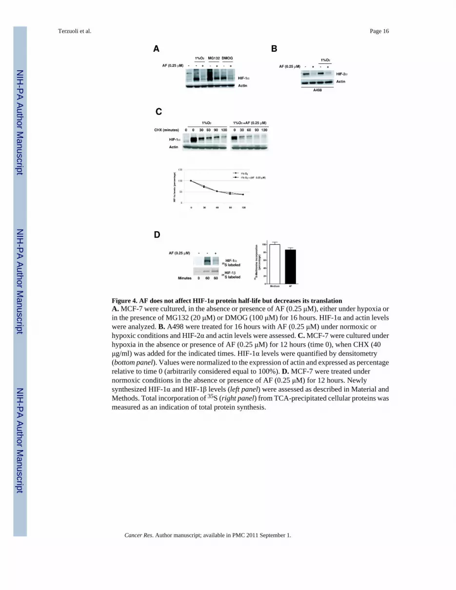
Figure 4. AF does not affect HIF-1α protein half-life but decreases its translationA. MCF-7 were cultured, in the absence or presence of AF (0.25 μM), either under hypoxia orin the presence of MG132 (20 μM) or DMOG (100 μM) for 16 hours. HIF-1α and actin levelswere analyzed. B. A498 were treated for 16 hours with AF (0.25 μM) under normoxic orhypoxic conditions and HIF-2α and actin levels were assessed. C. MCF-7 were cultured underhypoxia in the absence or presence of AF (0.25 μM) for 12 hours (time 0), when CHX (40μg/ml) was added for the indicated times. HIF-1α levels were quantified by densitometry(bottom panel). Values were normalized to the expression of actin and expressed as percentagerelative to time 0 (arbitrarily considered equal to 100%). D. MCF-7 were treated undernormoxic conditions in the absence or presence of AF (0.25 μM) for 12 hours. Newlysynthesized HIF-1α and HIF-1β levels (left panel) were assessed as described in Material andMethods. Total incorporation of 35S (right panel) from TCA-precipitated cellular proteins wasmeasured as an indication of total protein synthesis.
Terzuoli et al. Page 16
Cancer Res. Author manuscript; available in PMC 2011 September 1.
NIH
-PA Author Manuscript
NIH
-PA Author Manuscript
NIH
-PA Author Manuscript

Figure 5. Active transcription is required for inhibition of HIF-1α mRNA expression by AFA–B. MCF-7 were cultured under normoxia or hypoxia for 16 hours in the absence or presenceof AF (0.25 μM). HIF-1α mRNA (A) and HIF-1α pre-mRNA (B) expression was analyzed.** = p<0.005, * = p<0.01, relative to medium. C–D. MCF-7 were cultured under normoxia orhypoxia for 16 hours in the absence or presence of AF (0.25 μM) and/or actinomycin D (5μg/ml). HIF-1α mRNA (C) and HIF-1α pre-mRNA (D) expression was assessed. *** = p<0.001,** = p<0.005, relative to medium.
Terzuoli et al. Page 17
Cancer Res. Author manuscript; available in PMC 2011 September 1.
NIH
-PA Author Manuscript
NIH
-PA Author Manuscript
NIH
-PA Author Manuscript

Figure 6. AF inhibits HIF-1α expression and transcriptional activity in MCF-7 xenograftsA. MCF-7 were implanted into nude mice (n = 5/group) and allowed to grow up to ~ 200mg,when treatment with AF (60 mg/kg daily × 4 days i.p.) was started. Tumor weight was measuredas described in Materials and Methods, (Mann-Whitney test; *, p < 0.01). B. Quantitativedetermination of HIF-1α protein levels (**, p <0.05). C. mRNA expression of HIF-1-targetgenes (VEGF, CA9 and PDK-1) in tumor lysates from mice treated with vehicle or AF wereassessed (p < 0.05). Values are expressed as fold change relative to levels detected in mRNAharvested from normoxic MCF-7.
Terzuoli et al. Page 18
Cancer Res. Author manuscript; available in PMC 2011 September 1.
NIH
-PA Author Manuscript
NIH
-PA Author Manuscript
NIH
-PA Author Manuscript

NIH
-PA Author Manuscript
NIH
-PA Author Manuscript
NIH
-PA Author Manuscript
Terzuoli et al. Page 19
Table 1
Primers used for Real time PCR.
Human CYP1A1 Forward 5`GATTGGGCACATGCTGACG-3`
Reverse 5`-TGCTGGCTCATCCTTGACAG-3`
Human p21 Forward 5`ACGCGACTGTGATGCGC-3`
Reverse 5`-AAGTTCCATCGCTCACGGG-3`
Human CA 9 Forward 5`-GAGGCCTGGCCGTGTTG-3`
Reverse 5`-AACTGCTCATAGGCACTGTTTTCTT-3`
Human LOX Forward 5`-TGCTTGGTGGAGACTGAGATACC-3`
Reverse 5`-AATCACGTGAGGGAAGGAGAAA-3`
Human HIF-1α (intron 5-exon 6) Forward 5`-TGCTTTTTTTTTCCCTAGCATTGT-3`
Reverse 5`-TGGTTACTGTTGGTATCATATACGTGAA-3`
Human HIF-1α (exon5-exon 6) Forward 5`-TAGCCGAGGAAGAACTATGAACATAA-3`
Reverse 5`-TGAGGTTGGTTACTGTTGGTATCATATA-3`
Probe 5'-TTGCACTGCACAGGCCACATTCAC-3'
Cancer Res. Author manuscript; available in PMC 2011 September 1.


