Embed Size (px)
Citation preview

Modeling of the Aryl Hydrocarbon Receptor (AhR) ligand bindingdomain and its utility in virtual ligand screening to predict newAhR ligands
William Bisson#, Daniel Koch#, Edmond O’Donnell, Sammy M. Khalil, Nancy Kerkvliet,Robert Tanguay, Ruben Abagyan, and Siva Kumar Kolluri*Cancer Biology Laboratory, Department of Environmental and Molecular Toxicology,Environmental Health Sciences Center, Oregon State University, Corvallis, OR 97331‡Department of Molecular Biology, The Scripps Research Institute, 10550 North Torrey PinesRd., La Jolla, CA 92037
AbstractThe Aryl Hydrocarbon Receptor (AhR) is a ligand-activated transcription factor; the AhR Per-AhR/Arnt-Sim (PAS) domain binds ligands. We developed homology models of the AhR PASdomain to characterize previously observed intra- and inter-species differences in ligand bindingusing Molecular Docking. In silico structure-based virtual ligand screening using our modelresulted in the identification of pinocembrin and 5-hydroxy-7-methoxyflavone, which promotednuclear translocation and transcriptional activation of AhR and AhR-dependent induction ofendogenous target genes.
The aryl hydrocarbon receptor (AhR) is a ligand activated member of the basic-helix-loop-helix (bHLH) family of transcription factors.1–3 The AhR is activated by a variety ofcompounds, both synthetic and natural, including halogenated aromatic hydrocarbons suchas 2,3,7,8-tetrachlorodibenzo-p-dioxin (TCDD), and mediates their biological activity.1–3
The AhR is a cytosolic transcription factor bound to several co-chaperones. Upon ligandbinding, the AhR translocates from the cytoplasm to the nucleus and regulates genes,including several drug metabolizing enzymes that can influence the therapeutic activity of anumber of compounds.1–3 The AhR regulates proliferation and differentiation of cells.4–6
The AhR also induces immunosuppressive regulatory T cells with therapeutic implicationsin hyperimmune disorders.7–9 The PAS (Per-AhR/Arnt-Sim) domain of the AhR is theligand binding domain (LBD).1–3 In the present study, homology models of the AhR-LBDwere built to study the inter and intra-species differences in ligand binding and to identifyAhR ligands by virtual ligand screening.
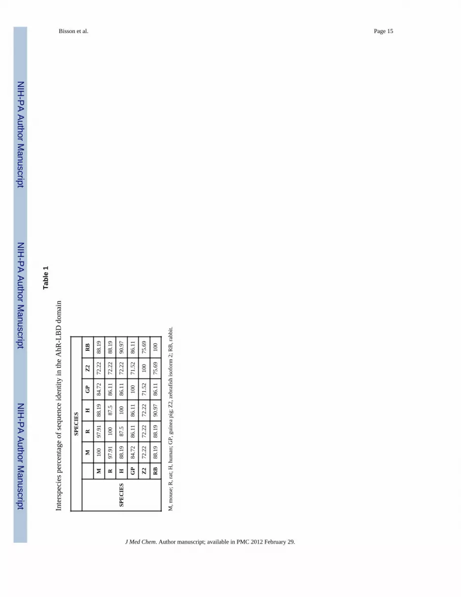
To construct the homology models, a multiple sequence alignment of the AhR-LBD domainfrom mouse, human, rat, guinea pig, rabbit and zebrafish (Figure 1a) was performed. Thesequence alignment produced sequence identities of greater than 85% between the differentspecies, with the exception of zebrafish isoform 2, which shared a consensus sequenceidentity of approximately 70% (Table 1). The AhR-LBD of mouse and rat had the highestsequence identity at nearly 98% (Table 1), suggesting a common evolutionary path and arecent species splitting as depicted by the evolutionary tree in Figure 1b. The mouse AhR-
*To whom correspondence should be addressed: Cancer Biology Laboratory, Department of Environmental and MolecularToxicology, Oregon State University, Corvallis, OR 97331, USA Phone: 001-541-737-1799, [email protected].#Equally contributed to this workSupporting Information is available on Journal of Medicinal Chemistry web site.
NIH Public AccessAuthor ManuscriptJ Med Chem. Author manuscript; available in PMC 2012 February 29.
Published in final edited form as:J Med Chem. 2009 September 24; 52(18): 5635–5641. doi:10.1021/jm900199u.
NIH
-PA Author Manuscript
NIH
-PA Author Manuscript
NIH
-PA Author Manuscript

LBD residues His285, Cys327, Ile332, Met334, Ala375 and Gln377, which have beencharacterized by mutagenesis studies and are known to influence TCDD binding,10 arehighlighted in red in Figure 1a. These residues are well conserved with the exception ofMet337 in guinea pig and Val381 in human AhR. These amino acid residues are replacedwith Ile and Ala in all other species (Figure 1a).
Several PAS domain structures are currently available in the Protein Data Bank (Pdb). Thesequence similarity between the structures of the PAS domain containing-protein family islow; however, within the family there is a high structural conservation of the α and β folds.We selected the PAS domain structure of hypoxia-inducible factor 2α (Hif-2α) (pdb 1p97)11
for generating a homology model of the mouse AhR-LBD based on its closest similarity inthis region. The model was built using the primary amino acid sequence [241–384] of theAhR-PASB domain, producing a sequence identity of 30% from the alignment with Hif-2α.The backbone geometry was preserved while loops were rebuilt using a database search formatching loop geometries.12 The side chain conformations of amino acids different in thealignment were globally optimized using the biased probability monte carlo procedure.13
The human, rat, zebrafish 1 and 2 AhR-LBD homology models were built starting from themouse model and using the respective sequences of the other species homologous to that ofthe mouse primary sequence [241–384].
Each of the AhR ligand binding domains were built using the homology modeling protocoldescribed in detail by Cardozo et al.14 The initial model was built by using the templatebackbone and side-chains in their most likely conformation. Next, the side chains wereglobally optimized using the modified ECEPP/3 energy function described by Abagyan etal.15 This energy function includes the following energy terms: van der Waals, hydrogenbonding, torsion energy, distance dependent electrostatic term, disufide bond constraints,side-chain entropy, and the implicit solvent accessible surface dependent solvation term.14
After the side chain placement the whole model was energy optimized using all the aboveenergy terms but with strong positional restraints. The refined homology models were thengeometrically corrected such that all of the residues were in sterically allowed regions of theRamachandran plot. Following development and refinement of the model, we first analyzedthe mouse AhR-LBD homology model to characterize the ligand binding pocket. Using ICMPocket Finder (Molsoft ICM v3.5-1p),16 a binding pocket was identified (volume of 201.5Å3, area of 178.3 Å2, and a radius of 3.637 Å) in the region of the protein surrounded by β-sheets Hβ and Iβ (Ala375 and Gln377) at the bottom, by the helix-connector Fα (Cys327andMet334) on the left, by Aβ on the right (His285), and at the top by the loop connecting Aβand Bβ (Figure 2a). In the helix connector Fα, the backbone NH of Ile332 established anintra H-bond with the backbone carbonyl oxygen of Ala328, keeping the helical secondarystructure stable. Replacing Ile332 with a Proline in our mouse AhR-LBD homology modelresulted in a loss of intra H-bond interactions. These results suggested that the decrease ofTCDD binding in the I332P mutant10 is due to an indirect effect caused by the disruption ofthe Fα helix connector in the vicinity of the binding pocket region. Thus, our AhR-LBDhomology model provides a structural explanation for the decreased TCDD bindingobserved with the I332P substitution in the AhR.10
We should note that while other groups have attempted modeling of the AhR-LBD,10,17
there are noteworthy differences between these models and our characterized bindingpocket. The ligand binding pocket in our model is more compact, surrounded mainly by theresidues Cys327, Gln377, His285, Met334 and Ala375. The side chains of these amino acidsare all pointing towards the binding pocket, indicating structurally their direct involvementin TCDD binding (Figure 2a). The model by Pandini et al10 is more spread, involving theregion between residues Gln377 and His285 surrounded by Dα and Eα helices in addition tothe area characterized in our model. Similar to Pandini et al., we observed that the side chain
Bisson et al. Page 2
J Med Chem. Author manuscript; available in PMC 2012 February 29.
NIH
-PA Author Manuscript
NIH
-PA Author Manuscript
NIH
-PA Author Manuscript

of residues Thr311 and Lys350 point outside the modeled LBD and do not affect AhRligand binding.10 The side chain of residues Ile319 and His320 also pointed towards thepocket cavity, confirming their key role in ligand binding specificity.17
To study TCDD binding capacity across species and to provide a structural explanation forthe biological data reported in the literature,1–3 TCDD was docked into mouse, human andzebrafish isoform 2 AhR-LBD models. TCDD docked similarly in all three species bindingpockets (Figure 2b–d) and the binding energy calculations (see methodology) were inagreement with previous inter-species biological characterizations.1–3 Lower binding energyvalues indicate a higher binding affinity of a particular ligand in our model. The valuesobtained for TCDD binding in mouse and zebrafish isoform 2 were −3.796 kcal/mol and−3.972 kcal/mol, respectively, confirming that TCDD has similarly high binding affinity inmice and zebrafish.18 In contrast, the value obtained with the human model was −2.33 kcal/mol, which was significantly higher than for the mouse and zebrafish models, indicating adecreased binding affinity compared with the other two species investigated. TCDD dockingorientation was similar in all three species. Further, the residues that were important forligand binding and facing the ligand pocket cavity were conserved in mouse and zebrafishisoform 2. In the human AhR, one of the residues, corresponding to mouse Ala375 wasreplaced by a Valine; the presence of this bulkier residue would likely sterically impairTCDD binding ability.
The mouse AhR-LBD homology model was used to structurally characterize some of theintraspecies experimental biological data available for the AhR. The C57BL/6J mouse strainexhibits a 10-fold higher TCDD susceptibility compared to the DBA/2 mouse strain.1–3
Both the mouse DBA/2 AhRLBD and human AhR-LBD share the A375V substitution(Figure 1a). Hence, the effect of this amino acid substitution on TCDD docking wasanalyzed in our mouse homology model by mutating A375 into a Valine or a Leucine. In theWT and A375V model TCDD was able to dock into the binding pocket, whereas in theA375L model TCDD failed to dock completely. Thus, the residue at position 375 located inthe middle of the ligand binding area appears to directly impact TCDD binding. Thereplacement of the amino acid Alanine with bulkier residues like Valine (as present inmouse DBA/2 and human sequences) or Leucine decreased the volume of the bindingpocket (195 Å3 (WT), 186.1 Å3 (A375V) and 148.6 Å3 (A375L)) (Figure 2e) with theconsequence of partially (Valine) or totally (Leucine) sterically blocking TCDD binding. Bychanging the tautomerization of one of the key residue for binding His285 in both WT(194.4 Å3) and the A375V mutant (185.9 Å3) models and by re-docking TCDD, the bindingenergy calculations generated values of −3.617 kcal/mol and −3.413 kcal/mol, respectively.Decreased volume of the binding pocket might result in weaker binding energy thatcontributes to decreased affinity of TCDD to AhR from human or mouse DBA/2 strain.Indeed, substitution of Alanine at 375 with Leucine has been reported to affect TCDDbinding.10
We next used homology modeling to study AhR in zebrafish. Interestingly, zebrafish havethree AhR isoforms, namely, AhR1a, AhR1b and AhR2, with AhR2 being the dominantisoform that binds TCDD, whereas AhR1a (zfAhR1a) exhibits impaired TCDDbinding. 18,19 The sequence identity between zfAhR1a-and zfAhR2-LBD is 65%. The aminoacid residues His285, Ala375 and Gln377 (Figure 2f), which are highly conserved in mostspecies (Figure 1a) and contribute to ligand binding are present in zfAhR2; however, inzfAhR1a, these amino acids are substituted with Threonine, Histidine, and Tyrosine,respectively (Figure 2g). Thus, the difference between zfAhR1a and zfAhR2 in TCDDbinding may be attributed to these dissimilarities in the ligand binding pocket region. Tounderstand the differences in the ligand binding pockets of zfAhR1a and zfAhR2, homologymodels for the zfAhR1aand zfAhR2-LBD were built and energetically minimized using the
Bisson et al. Page 3
J Med Chem. Author manuscript; available in PMC 2012 February 29.
NIH
-PA Author Manuscript
NIH
-PA Author Manuscript
NIH
-PA Author Manuscript

ICM method. TCDD docking was attempted with both zebrafish AhR isoforms models;however, TCDD was able to dock only into zfAhR2-LBD. By analyzing the binding pocketsof zfAhR1a and zfAhR2 identified with ICM Pocket Finder,16 (Figure 2f and 2g) zfAhR1awas found to have a decreased binding area that may contribute to its lower affinity forTCDD. Substitution of zfAhR2 residues Gln388 and His296 with a Histidine and Tyrosinerespectively (as present in zfAhR1a) modified the type of electrostatic interactionsestablished between the protein and TCDD, resulting in decreased binding affinity. Thus,these specific amino acid replacements (Ala386Thr, Gln388His and His296Tyr) in thezfAhR1a-LBD appear to play a direct role in the considerable decrease of TCDD binding bymodifying the electrostatic environment of the binding pocket region and the volume of thebinding pocket itself. Indeed, electrostatic and van der Waals forces are the driving force inthe total binding energy of many halogenated aromatic hydrocarbons towards the AhR.20
In addition to characterization of the prototypical AhR ligand TCDD, we also used ourhomology models to study the binding of structurally diverse molecules to the AhR.Recently, formylindolo [3,2-b]carbazole (FICZ) and 2-(1’H-indole-3’-carbonyl)-thiazole-4-carboxylic acid methyl ester (ITE) have been described as potent AhR agonists both in vitroand in vivo.8,9 These two ligands were docked into our human and mouse AhR-LBD modelsto investigate any inter-species differences in their respective binding modes. The twomolecules docked into both the human and mouse binding pockets in a similar orientation(Figure 3a–d), establishing H-bond interactions with protein residues facing the bindingcavity. FICZ made two H-bonds between the formyl carbonyl oxygen and the side chain ofSer359 (mouse) and Ser 365 (human) and between the nitrogen of the carbazole group andthe side chain of Gln377 (mouse) and Gln383 (human) (Figure 3b and 3d). On the otherhand, ITE made a single H-bond between its 3’-carbonyl group and the side chain of Ser359(mouse) and Ser365 (human) (Figure 3a and 3c). The binding affinity of FICZ and ITE ishigher for mAhR than hAhR as estimated by the binding energy calculations of the ligandand protein complexes. The binding energies for the ligand-mAhRLBD complex formationfor FICZ and ITE were −4.714 kcal/mol and −4.214 kcal/mol respectively. The estimatedvalues for hAhR-LBD-ligand binding were −4.12 kcal/mol for FICZ and −3.46 kcal/mol forITE. The H-bond pattern established between FICZ and ITE with mouse Ser359 and humanSer365, which are not present for TCDD, may play an important role in increasing AhRbinding of these two ligands.
To further validate and apply our AhR-LBD homology model, a database of 498 naturalcompounds was docked into mouse AhR-LBD model binding pocket for the purpose ofidentifying new ligands of the AhR by virtual ligand screening (VLS).21 After the dockingresults were obtained we arbitrarily set a binding energy cutoff of δ −2.5 kcal/mol, whichresulted in the selection of 25 compounds that docked into the binding pocket in anallowable orientation; these 25 compounds were then tested in cell based experiments. Thelist of the 25 docked structures and their respective AhR transcriptional activation results areprovided in Supplemental Table 1. Six of the 25 compounds were able to activate AhRtranscription and at least three of the non-activating compounds strongly antagonizedTCDD-induced AhR transcription (Table 2 and data not shown). The top two AhRactivators that we identified by VLS were 5-hydroxy-7-methoxyflavone and pinocembrin,both of which are flavonoids. Both compounds strongly activated the AhR transcription in adose-dependent manner (Figure 4a). The docking orientation of the two potent AhRactivators identified was similar, with the 2-phenyl ring allocated in the small hydrophobiccleft surrounded by residues Phe281, Leu309 and Leu347 (Figure 4b). The 1,4-benzopyronestructure of these two flavonoids bound instead in the middle of the binding pocketsurrounded by residues Cys327, His285, Met334 and Ala375 (Figure 4b). Both ligandsshared H-bond interaction between the carbonyl group of the benzopyrone template and theside chain of residue Ser359 (Figure 4b). Furthermore, 5-hydroxy-7-methoxyflavone
Bisson et al. Page 4
J Med Chem. Author manuscript; available in PMC 2012 February 29.
NIH
-PA Author Manuscript
NIH
-PA Author Manuscript
NIH
-PA Author Manuscript

established a second Hbond between its hydroxyl group adjacent to the carbonyl group andthe side chain of Ser359 (Figure 4b). We further tested the ability of 5-hydroxy-7-methoxyflavone and pinocembrin to activate endogenous target genes of AhR.22 Both thecompounds activated CYP1A1 and NADPH quinone reductase in an AhR dependent manner(Figure 4c). Furthermore, both the compounds promoted nuclear translocation ofendogenous AhR from cytosol after one hour of exposure, confirming their status as AhRagonists (Figure 5). Out of the different flavones predicted to bind to AhR ligand bindingpocket by the virtual ligand screen, 5-OH-7-methoxyflavone strongly induced AhRtranscription, whereas 3-OH-7-methoxyflavone modestly activated AhR transcription. 6-OH-7-methoxyflavone did not activate AhR, suggesting that 6-OH-7-methoxyflavone bindsto AhR, but may not bring AhR in to a transcriptionally active form. We tested whether thiscompound can act as an AhR transcriptional antagonist. Indeed, co-treatment with 6-OH-7-methoxyflavone strongly inhibited both TCDD- and 5-OH-7-methoxyflavone-induced AhRtranscription (Supplemental Figure 1). There is precedence for differential effects ofdifferent flavones on AhR activity.23,24,25 β-naphthoflavene is a strong AhR agonist,whereas α-naphtoflavone is a partial agonist/antagonist of AhR transcription.26 Bycomparing the binding pattern of 7-methoxyflavone derivatives to AhR ligand bindingpocket, we were unable to distinguish agonists from potential antagonists.
In summary, homology models of AhR-LBD in the agonist bound conformation were builtusing the ICM method.14,15 Using these models, differences in ligand binding across variousspecies was structurally characterized. The mouse AhR-LBD homology model was alsoutilized successfully to identify new AhR agonists by insilico structure based VLS.21
Experimental SectionMultiple Sequence Alignment
The AhR-LBD sequences in FASTA format for mouse, human, rat, guinea pig, rabbit andzebrafish were retrieved from NCBI database. Multiple sequence alignment was performedonline with the ClustalW program.27
Homology ModelingWe used the Nuclear Magnetic Resonance (NMR) structure of the human PAS domain ofthe hypoxia-inducible factor 2α (HIF-2α)11 available in the Protein Data Bank (Pdb) 1P97 asthe 3D coordinate template for the homology modeling of mouse, human, and zebrafishisoforms 1 and 2 AhR-LBD. All models were energetically refined using the internalcoordinate space with Molsoft ICM v3.5-1p.14,15
Molecular DockingThe receptor model is represented by five types of interaction potentials, namely, (i) the vander Waals potential for a hydrogen atom probe; (ii) the van der Waals potential for a heavy-atom probe (generic carbon of 1.7 Å radius; (iii) an optimized electrostatic term; (iv)hydrophobic terms; and (v) loan-pair-based potential, which reflects directional preferencesin hydrogen bonding. The energy terms were based on the all-atom vacuum force fieldECEPP/3 with appended terms from the Merck Molecular Force Field to account forsolvation free energy and entropic contribution.15 Modified inter-molecular terms such assoft van der Waals and hydrogen-bonding as well as a hydrophobic term are added.Conformational sampling was based on the biased probability Monte Carlo (BPMC)procedure, which randomly selects a conformation in the internal coordinate space and thenmakes a step to a new random position independent of the previous one according to apredefined continuous probability distribution. It has also been shown previously that aftereach random step, full local minimization greatly improves the efficiency of the procedure.
Bisson et al. Page 5
J Med Chem. Author manuscript; available in PMC 2012 February 29.
NIH
-PA Author Manuscript
NIH
-PA Author Manuscript
NIH
-PA Author Manuscript

In the ICM-VLS (Molsoft ICM v3.5-1p) screening procedure the ligand scoring wasoptimized to obtain maximal separation between binders and non-binders.28,29 Eachcompound was assigned a score according to its fit within the receptor, which accounts forcontinuum and discreet electrostatics, hydrophobicity, and entropy parameters.15,28,29
Binding energy calculationsThe free binding energy of a ligand was estimated according to the protocol described.30 Inthis method, the estimate consists of three essential terms and an additional constant term:ΔGbinding = ΔGH +ΔGEL + ΔGS + C. Every term (except for the constant) is a differencebetween the corresponding energy in bound and unbound states. The hydrophobic, or cavityterm ΔGH, accounts for the change in solvent accessible area upon complexation iscalculated as the solvent accessible area, calculated with a 1.4Å water probe, and multipliedby 30cal/Å2. The electrostatic term ΔGel, was composed of the coulombic interactions andelectrostatic energy of solvation estimated with the continuum dielectric model using theboundary element method31 with MMFF94 partial charges, a protein dielectric constant ofeight, water dielectric constant of 80 and probe radius of 1.4Å. The entropic term was a sumof the side chain and ligand entropic terms. The contribution of each rotatable bond frozenupon binding was estimated as 0.5 kcal/mole. The constant value of 3kcal/mole was used.
Library of compoundsA database of 498 natural compounds from TimTec (Newark, DE) was used for in silicoVLS. Twenty five compounds identified by VLS from TimTec were dissolved in DMSOand tested at a final concentration of 20 µM. The structures and ≥ 95% purity of the testednatural compounds were confirmed by TimTec using NMR.
Cell CultureAll cells were cultured in Dulbecco's Modified Eagle Medium (DMEM) with L-glutamine(Mediatech Inc., VA) supplemented with 10% FBS (Tissue Culture Biologicals, CA), 100IU/mL penicillin, and 100 µg/ml streptomycin (Mediatech Inc., VA) in a humidified 5%CO2 atmosphere. Cells were typically passaged every three days at a dilution of 1:5.
Semi-quantitative Polymerase Chain Reaction (PCR)For analysis of AhR target gene induction, a pair of Hepa1c1c7 derived cell lines were used,one with significantly reduced AhR expression (C12) and the same cell line stablyexpressing a WT AhR construct (Hepa1 C12+AhR)3, as described in the supplementalinformation.
Supplementary MaterialRefer to Web version on PubMed Central for supplementary material.
Abbreviations
AhR Aryl Hydrocarbon Receptor
TCDD 2,3,7,8-tetrachlorodibenzo-p-dioxin
PAS Per-AhR/Arnt-Sim
LBD ligand binding domain
bHLH basic-helix-loop-helix
ICM Internal Coordinate Mechanism
Bisson et al. Page 6
J Med Chem. Author manuscript; available in PMC 2012 February 29.
NIH
-PA Author Manuscript
NIH
-PA Author Manuscript
NIH
-PA Author Manuscript

HIF-2α hypoxia-inducible factor 2α
FICZ 6-formylindolo [3,2-b] carbazole
ITE 2-(1'H-indole-3'-carbonyl)-thiazole-4-carboxylic acid methyl ester
Pdb Protein Data Bank
NMR Nuclear Magnetic Resonance
DMEM Dulbecco's Modified Eagle Medium
PCR Polymerase Chain Reaction.
AcknowledgmentsWe are grateful to Dr. Michael Denison for providing the Hepa1 cells. We thank Christopher Sullivan, Center forGenome Research and Biocomputing at Oregon State University (OSU) for excellent software and hardwaresupport and Duc Nguyen for laboratory assistance. This work was supported in part by the startup funds to SKKfrom the Department of Environmental and Molecular Toxicology, OSU Research office and grants from theDepartment of Defense-Breast Cancer research Program, Medical Research Foundation of Oregon and NationalInstitute of Environmental Health Sciences to the Environmental Health Sciences Center at OSU and its corefacilities. DK and EOD were supported by the NIEHS predoctoral training grant.
References1. Beischlag TV, Morales JL, Hollingshead BD, Perdew GH. The Aryl Hydrocarbon Receptor
Complex and the control of gene expression. Crit Rev Eukaryot Gene Expr. 2008; 18:207–250.[PubMed: 18540824]
2. Nguyen LP, Bradfield CA. The search for endogenous activators of the aryl hydrocarbon receptor.Chem Res Toxicol. 2008; 21:102–116. [PubMed: 18076143]
3. Hankinson O. The aryl hydrocarbon receptor complex. Annu Rev Pharmacol Toxicol. 1995;35:307–340. [PubMed: 7598497]
4. Kolluri SK, Weiss C, Koff A, Goettlicher M. Cell Cycle control by dioxins is mediated by theinduction of the p27Kip1 inhibitor. Genes Dev. 1999; 13:1742–1753. [PubMed: 10398686]
5. Mitchell KA, Lockhart CA, Huang G, Elferink CJ. Sustained aryl hydrocarbon receptor activityattenuates liver regeneration. Mol Pharmacol. 2006; 70:163–170. [PubMed: 16636136]
6. Ma C, Marlowe JL, Puga A. The aryl hydrocarbon receptor at the crossroads of multiple signalingpathways. EXS. 2009; 99:231–257. [PubMed: 19157064]
7. Funatake CJ, Marshall NB, Steppan LB, Mourich DV, Kerkvliet NI. Cutting edge: activation of thearyl hydrocarbon receptor by 2,3,7,8-tetrachlorodibenzo-p-dioxin generates a population of CD4+CD25+ cells with characteristics of regulatory T cells. J Immunol. 2005; 175:4184–4188. [PubMed:16177056]
8. Quintana FJ, Basso AS, Iglesias AH, Korn T, Farez MF, Bettelli E, Caccamo M, Oukka M, WeinerHL. Control of Treg and TH17 cell differentiation by the aryl hydrocarbon receptor. Nature. 2008;453:65–71. [PubMed: 18362915]
9. Veldhoen M, Hirota K, Westendorf AM, Buer J, Dumoutier L, Renauld J-C, Stockinger B. The arylhydrocarbon receptor links TH17-cell-mediated autoimmunity to environmental toxins. Nature.2008; 453:106–109. [PubMed: 18362914]
10. Pandini A, Denison MS, Song Y, Soshilov AA, Bonati L. Structural and functionalcharacterization of the Aryl Hydrocarbon Receptor Ligand Binding Domain by HomologyModeling and Mutational Analysis. Biochemistry. 2007; 46:696–708. [PubMed: 17223691]
11. Erbel PJ, Card PB, Karakazu O, Bruick RK, Gardner KH. Structural basis for PAS domainheterodimerization in the basic helix-loop-helix-PAS transcription factor hypoxia-inducible factor.Proc. Natl. Acad. Sci. U.S.A. 2003; 100:15504–15509. [PubMed: 14668441]
Bisson et al. Page 7
J Med Chem. Author manuscript; available in PMC 2012 February 29.
NIH
-PA Author Manuscript
NIH
-PA Author Manuscript
NIH
-PA Author Manuscript

12. Abagyan R, Batalov S, Cardozo T, Totrov M, Webber J, Zhou Y. Homology modeling withinternal coordinate mechanics: deformation zone mapping and improvements of models viaconformational search. Proteins. 1997 Suppl 1:29–37. [PubMed: 9485492]
13. Abagyan RA, Totrov MM. Biased probability Monte Carlo conformational searches andelectrostatic calculations for peptides and proteins. J. Mol. Biol. 1994; 235:983–1002. [PubMed:8289329]
14. Cardozo T, Totrov M, Abagyan R. Homology modeling by the ICM method. Proteins. 1995;23:403–414. [PubMed: 8710833]
15. Abagyan RA, Totrov MM, Kuznetsov DA. ICM: A New Method For Protein Modeling andDesign: Applications to Docking and Structure Prediction From The Distorted NativeConformation. J. Comp. Chem. 1994; 15:488–506.
16. An J, Totrov M, Abagyan R. Pocketome via comprehensive identification and classification ofligand binding envelopes. Mol Cell Proteomics. 2005; 4:752–761. [PubMed: 15757999]
17. Goryo K, Suzuki A, Del Carpio C, Siizaki K, Kuriyama E, Mikami Y, Kinoshita K, Yasumoto K-I,Rannug A, Miyamoto A, Fujii-Kuriyama Y, Sogawa K. Identification of amino acid residues inthe Ah receptor involved in ligand binding. Biochem Biophys Res Commun. 2007; 354:396–402.[PubMed: 17227672]
18. Tanguay RL, Abnet CC, Heidemann W, Peterson RE. Cloning and characterization of thezebrafish (Danio rerio) aryl hydrocarbon receptor. Biochim. Biophys. Acta. 1999; 1444:35–48.[PubMed: 9931422]
19. Andreasen EA, Hahn ME, Heidemann W, Peterson RE, Tanguay RL. The zebrafish (Danio rerio)aryl hydrocarbon receptor type 1 is a novel vertebrate receptor. Mol. Pharmacol. 2002; 62:234–249. [PubMed: 12130674]
20. Zhao Y-Y, Tao F-M, Zeng EY. Theoretical study of the quantitative structure-activity relationshipsfor the toxicity of dibenzo-p-dioxins. Chemosphere. 2008; 73:86–91. [PubMed: 18589475]
21. Schapira M, Abagyan R, Totrov M. Nuclear hormone receptor targeted virtual screening. J MedChem. 2003; 46:3045–3059. [PubMed: 12825943]
22. Kolluri SK, Balduf C, Hofmann M, Goettlicher M. Novel target genes of the Ah (Dioxin) receptor:transcriptional induction of N-myristoyltransferase 2. Cancer Res. 2001; 61:8534–8539. [PubMed:11731439]
23. Gasiewicz TA, Kende AS, Rucci G, Whitney B, Willey JJ. Analysis of structural requirements forAh receptor antagonist activity: Ellipticines, flavones, and related compounds. BiochemPharmacol. 1996; 52:1787–1803. [PubMed: 8986142]
24. Lu YF, Santostefano M, Cunningham BD, Threadgill MD, Safe S. Substituted flavones as arylhydrocarbon (Ah) receptor agonists and antagonists. Biochem Pharmacol. 1996; 51:1077–1087.[PubMed: 8866830]
25. Puppala D, Gairola CG, Swanson HI. Identification of kaempferol as an inhibitor of cigarettesmoke-induced activation of the aryl hydrocarbon receptor and cell transformation.Carcinogenesis. 2007; 28:639–647. [PubMed: 17012224]
26. Wilhelmsson A, Whitelaw ML, Gustafsson JA, Poellinger L. Agonistic and antagonistic effects ofalpha-naphthoflavone on dioxin receptor function. Role of the basic region helix-loop-helix dioxinreceptor partner factor Arnt. J Biol Chem. 1994; 269:19028–19033. [PubMed: 8034660]
27. http://align.genome.jp/28. Totrov, M.; Abagyan, R. Protein-Ligand Docking as an Energy Optimization Problem. In: Raffa,
RB., editor. Drug-Receptor Thermodynamics: Introduction and Experimental Applications. NewYork: John Wiley & Sons; 2001. p. 603-624.
29. Totrov M, Abagyan R. Flexible protien-ligand docking by global energy optimization in internalcoordinates. Proteins. 1997 Suppl. 1:215–220. [PubMed: 9485515]
30. Schapira M, Totrov M, Abagyan R. Prediction of the binding energy for small molecules, peptidesand proteins. J. Mol. Recognition. 1999; 12:177–190.
31. Totrov M, Abagyan R. Rapid boundary element solvation electrostatics calculations in foldingsimulations: Successful folding of a 23-residue peptide. Biopolymers. 2001; 60:124–133.[PubMed: 11455546]
Bisson et al. Page 8
J Med Chem. Author manuscript; available in PMC 2012 February 29.
NIH
-PA Author Manuscript
NIH
-PA Author Manuscript
NIH
-PA Author Manuscript

Figure 1.(A) Interspecies multiple sequence alignment of the AhR-LBD performed with the programClustalW28. (B) Evolutionary tree of different species.
Bisson et al. Page 9
J Med Chem. Author manuscript; available in PMC 2012 February 29.
NIH
-PA Author Manuscript
NIH
-PA Author Manuscript
NIH
-PA Author Manuscript

Figure 2.(A) Homology model of the mouse AhR-LBD with the protein backbone displayed asribbon and colored by secondary structure (ICM v3.5-1n, Molsoft). The residues aredisplayed as sticks and colored by atom type with the carbon atoms in white. The bindingpocket volume is colored in green and was calculated using the program ICM Pocket Finder(ICM v3.5-1n, Molsoft).16 (B–D) TCDD docking orientation into mouse (B), human (C),zebrafish isoform 2 (D) AhR-LBD binding pocket. The protein backbone is displayed asribbon and colored by secondary structure. The residues are displayed as sticks and coloredby atom type with the carbon atoms in white. TCDD is displayed as sticks and colored byatom type (carbon atoms in yellow). (E) mouse AhR-LBD binding pocket volume is colored
Bisson et al. Page 10
J Med Chem. Author manuscript; available in PMC 2012 February 29.
NIH
-PA Author Manuscript
NIH
-PA Author Manuscript
NIH
-PA Author Manuscript

in green and was calculated using the program ICM Pocket Finder.16 The protein backboneis displayed as ribbon and colored by secondary structure. The residues are displayed assticks and colored by atom type with the carbon atoms in orange (wild type) and white(A375L mutant). (F–G) binding pocket region identified by ICM Pocket Finder of thezebrafish AhR-LBD of isoform 219 (F) and isoform 1a (G) colored in translucent blue anddark blue respectively. The protein backbone is displayed as ribbon and colored bysecondary structure. Residues are displayed as sticks and colored by atom type with carbonatoms in white. Docked TCDD is displayed as sticks and colored in red.
Bisson et al. Page 11
J Med Chem. Author manuscript; available in PMC 2012 February 29.
NIH
-PA Author Manuscript
NIH
-PA Author Manuscript
NIH
-PA Author Manuscript

Figure 3.ITE (A, C) and FICZ (B, D) docking orientation respectively into mouse (A, B), human (C,D) AhR-LBD homology model. The protein backbone is displayed as ribbon and colored bysecondary structure. The residues are displayed as sticks and colored by atom type with thecarbon atoms in white. FICZ and ITE are displayed as sticks and colored by atom type withcarbon atoms in yellow. H-bonds are represented by white dashed lines.
Bisson et al. Page 12
J Med Chem. Author manuscript; available in PMC 2012 February 29.
NIH
-PA Author Manuscript
NIH
-PA Author Manuscript
NIH
-PA Author Manuscript

Figure 4.(A) Mouse Hepa1 cells transfected with the AhR response element (AhRE)/ xenobioticresponse element (XRE)-luciferase reporter were treated with compounds at the indicatedconcentrations for 6 hours and assayed for reporter gene activity. (B) Docking orientation of5-hydroxy-7-methoxyflavone and pinocembrin into mouse AhR-LBD binding pocket (ICMv3.5-1n, Molsoft). The protein backbone is displayed as ribbon and colored by secondarystructure. The residues are displayed as sticks and colored by atom type with the carbonatoms in green. The ligands are displayed as sticks, colored by atom type with carbon atomsin yellow. H-bonds are represented as black dashed lines. (C) Western blot showing theexpression of AhR in the mouse Hepa1 derivative cell line C12, which has a significantlydecreased abundance of AhR, and the C12+AhR, wildtype AhR re-expressing cells. The blotshows 30 µg of whole cell lysate from each line (upper panel). Semi-quantitative RT-PCRfor AhR target genes22 following exposure to vehicle (0.1% DMSO), TCDD (1 nM), 5-hydroxy-7-methoxyflavone (20 µM) and pinocembrin (20 µM) for 18 hours. GAPDHexpression control was used to indicate similar amount of RNA usage (lower panel).CYP1A1 (Cytochrome p450 1A1), and NADPH-QO (NADPH quinone-oxidoreductase).PCR cycle numbers are indicated.
Bisson et al. Page 13
J Med Chem. Author manuscript; available in PMC 2012 February 29.
NIH
-PA Author Manuscript
NIH
-PA Author Manuscript
NIH
-PA Author Manuscript

Figure 5.Nuclear localization of AhR in Hepa1 cells. Cells were treated with the indicatedcompounds for 60 minutes, fixed and then immunostained with AhR followed byfluoresceinisothiocyanate(FITC)-conjugated secondary antibody. Cells were stained by 4,6-diamino-2-phenylindole (DAPI) to visualize nucleus and were imaged on a Zeiss AxiovertS100TV microscope.
Bisson et al. Page 14
J Med Chem. Author manuscript; available in PMC 2012 February 29.
NIH
-PA Author Manuscript
NIH
-PA Author Manuscript
NIH
-PA Author Manuscript
NIH
-PA Author Manuscript
NIH
-PA Author Manuscript
NIH
-PA Author Manuscript
Bisson et al. Page 15
Tabl
e 1
Inte
rspe
cies
per
cent
age
of se
quen
ce id
entit
y in
the
AhR
-LB
D d
omai
n
SPE
CIE
S
SPE
CIE
S
MR
HG
PZ
2R
B
M10
097
.91
88.1
984
.72
72.2
288
.19
R97
.91
100
87.5
86.1
172
.22
88.1
9
H88
.19
87.5
100
86.1
172
.22
90.9
7
GP
84.7
286
.11
86.1
110
071
.52
86.1
1
Z2
72.2
272
.22
72.2
271
.52
100
75.6
9
RB
88.1
988
.19
90.9
786
.11
75.6
910
0
M, m
ouse
; R, r
at; H
, hum
an; G
P, g
uine
a pi
g; Z
2, z
ebra
fish
isof
orm
2; R
B, r
abbi
t.
J Med Chem. Author manuscript; available in PMC 2012 February 29.

NIH
-PA Author Manuscript
NIH
-PA Author Manuscript
NIH
-PA Author Manuscript
Bisson et al. Page 16
Table 2
Activation of AhR transcription by compounds identified by virtual docking into AhR LBD
Compound Name Structure BindingEnergy
Induction of AhRTranscriptional
Activity
2,3,7,8-Tetrachlorodibenzo-p-dioxin −3.80 +++21 fold
5-Hydroxy-7-methoxyflavone −4.3 +++11.3 fold
7-Methoxyisoflavone −3.75 +2.7 fold
6-Methylflavone −3.52 ++4.8 fold
3-Hydroxy-6-methylflavone −3.37 +2.5 fold
Pinocembrin (5,7-Dihydroxyflavanone, R-form) −2.97 +++8.0 fold
7,8,2'-Trihydroxyflavone −2.74 +2.8 fold
+++Very Strong activation (≥ 8 fold);
++Strong activation (>4 fold);
+Activation (<3 fold)
J Med Chem. Author manuscript; available in PMC 2012 February 29.


