Embed Size (px)
Citation preview

Biochemical Pharmacology 82 (2011) 1234–1249
Amiodarone impairs trafficking through late endosomes inducing a Niemann-PickC-like phenotype
Elena Piccoli a,1, Matteo Nadai a,1, Carla Mucignat Caretta b, Valeria Bergonzini a, Claudia Del Vecchio a,Huy Riem Ha c, Laurent Bigler d, Daniele Dal Zoppo e, Elisabetta Faggin f, Andrea Pettenazzo g,Rocco Orlando h, Cristiano Salata a, Arianna Calistri a, Giorgio Palu a, Aldo Baritussio h,*a Department of Histology, Microbiology and Medical Biotechnologies, University of Padova, via A. Gabelli 63, 35121, Italyb Department of Human Anatomy and Physiology, University of Padova, via F. Marzolo 3, 35131, Italyc Cardiovascular Therapy Research Laboratory, Clinical Research Center, University Hospital, Ramistrasse 100, 8091 Zurich, Switzerlandd Institute of Organic Chemistry, University of Zurich, Winterthurerstrasse 190, 8057 Zurich, Switzerlande Department of Pharmaceutical Sciences, University of Padova, via F. Marzolo 5, 35131, Italyf Department of Clinical and Experimental Medicine, University of Padova, via Giustiniani 2, 35128, Italyg Department of Pediatrics, University of Padova, via Giustiniani 3, 35128, Italyh Department of Medical and Surgical Sciences, University of Padova, via Giustiniani 2, 35128 Padova, Italy
A R T I C L E I N F O
Article history:
Received 5 May 2011
Accepted 20 July 2011
Available online 23 August 2011
Keywords:
Amiodarone
Dronedarone
Late endosomes
Niemann-Pick C
Phospholipidosis
A B S T R A C T
Patients treated with amiodarone accumulate lysobisphosphatidic acid (LBPA), also known as
bis(monoacylglycero)phosphate, in airway secretions and develop in different tissues vacuoles and
inclusion bodies thought to originate from endosomes. To clarify the origin of these changes, we studied
in vitro the effects of amiodarone on endosomal activities like transferrin recycling, Shiga toxin
processing, ESCRT-dependent lentivirus budding, fluid phase endocytosis, proteolysis and exosome
secretion. Furthermore, since the accumulation of LBPA might point to a broader disturbance in lipid
homeostasis, we studied the effect of amiodarone on the distribution of LBPA, unesterified cholesterol,
sphingomyelin and glycosphyngolipids. Amiodarone analogues were also studied, including the recently
developed derivative dronedarone. We found that amiodarone does not affect early endosomal activities,
like transferrin recycling, Shiga toxin processing and lentivirus budding. Amiodarone, instead, interferes
with late compartments of the endocytic pathway, blocking the progression of fluid phase endocytosis
and causing fusion of organelles, collapse of lumenal structures, accumulation of undegraded substrates
and amassing of different types of lipids. Not all late endocytic compartments are affected, since exosome
secretion is spared. These changes recall the Niemann-Pick type-C phenotype (NPC), but originate by a
different mechanism, since, differently from NPC, they are not alleviated by cholesterol removal. Studies
with analogues indicate that basic pKa and high water-solubility at acidic pH are crucial requirements for
the interference with late endosomes/lysosomes and that, in this respect, dronedarone is at least as
potent as amiodarone. These findings may have relevance in fields unrelated to rhythm control.
� 2011 Elsevier Inc. All rights reserved.
Contents lists available at ScienceDirect
Biochemical Pharmacology
jo u rn al h om epag e: ww w.els evier .c o m/lo cat e/bio c hem p har m
1. Introduction
Amiodarone is a cationic antiarrhythmic drug extensively usedfor the control of supraventricular and ventricular arrhythmias. Itsaction mechanism includes blockade of K- and Na channels andinterference with b-adrenoreceptors and Ca currents [1,2].
Abbreviations: LBPA, lysobisphosphatidic acid; ESCRT, endosomal sorting complex
required for transport; VLP, virus like particles.
* Corresponding author. Tel.: +39 49 8212149; fax: +39 49 8212149.
E-mail address: [email protected] (A. Baritussio).1 These authors contributed equally to this work.
0006-2952/$ – see front matter � 2011 Elsevier Inc. All rights reserved.
doi:10.1016/j.bcp.2011.07.090
Amiodarone has a large volume of distribution (66 l/kg), steadystate serum levels between 0.7 and 3.7 mM, an elimination half-lifeof several weeks and a propension to accumulate in differenttissues, like adipose tissue, skeletal muscle and the liver, but not inthe brain [3–5]. Recent evidence suggests that accumulation intissues might happen by at least two mechanisms, that is theassociation with cell membranes, due to the high lypophilicity ofamiodarone, and trapping in the lumen of acidic organelles afterprotonation of the amine function present in the lateral groupdiethylamino-b-ethoxy [6,7]. Amiodarone catabolism includesstepwise dealkylation and deamination of the lateral groupdiethylamino-ethoxy, major metabolite being N-desethylamiodar-one (MDEA) whose activity and serum levels are similar to those of

E. Piccoli et al. / Biochemical Pharmacology 82 (2011) 1234–1249 1235
the parent drug [8]. Chronic exposure to amiodarone induces theformation of vacuoles and inclusion bodies in blood leukocytes,cells of the corneal epithelium, skin cells, alveolar macrophages,liver cells and cardiomyocytes [7]. Although there is agreementthat these structures derive from interference of amiodarone withthe endocytic pathway, there is uncertainty about the set ofendocytic organelles involved, the mechanism of formation ofvacuoles and the origin of materials, both amorphous andmembrane-like, accumulating in the lumen of inclusion bodies.
Regarding the section of the endocytic pathway involved, it hasbeen claimed that amiodarone does not interfere with earlycompartments of the endocytic pathway, since the drug does notmodify the distribution of the early endosomal marker EEA1 [6].However this issue remains undecided, since some found nointerference of amiodarone with the activity of diphtheria toxin,whose active moiety enters the cytoplasm from early endosomes[9], while others found that amiodarone inhibits the toxin [10]. Onthe other side several lines of evidence suggest that amiodaroneinterferes with late compartments of the endocytic pathway. Infact, (i) vacuoles and inclusion bodies bear the markers of lateendosomes/lysosomes, like Rab7 and CD63 [6,7], (ii) patientstreated with amiodarone accumulate in the airways lysobispho-sphatidic acid (LBPA), also known as bis(monoacylglycero)pho-sphate [11], which is a component of late endosome lumenalmembranes [12], (iii) amiodarone inhibits in vitro the activity ofthe anthrax toxin, whose subunits (edema factor and lethal factor)enter the cytoplasm from late endosomes/multivesicular bodies[10,13]. Thus far it is unknown whether amiodarone interfereswith recycling endosomes or with the pathway connecting directlyearly endosomes with the Golgi. Nor is it known if amiodaroneinterferes with the ESCRT complex, a highly conserved set ofproteins associated with a specialized portion of the earlyendosomes, necessary for the formation of lumenal vesicles andthe eventual maturation of this compartment into multivesicularbodies [14]. The ESCRT system is also involved in membranedeformations topologically equivalent to endovesiculation, likecytokinesis and the budding of membrane bound viruses [14].Interestingly mutations of the ESCRT system lead to phenotypescharacterized by the presence of vacuoles and inclusion bodiesbearing markers of both early and late endosomes [14].
The observation that patients treated with amiodaroneaccumulate LBPA, which is only detected in late endosomes andis intimately connected with unesterified cholesterol [12,15],suggests that the drug might alter the distribution of other lipidstoo, as found in Niemann-Pick type C disease, where mutations ofNPC1 or NPC2 proteins cause the accumulation of free cholesterol,glycosphyngolipids, sphingosine, LBPA and sphingomyelin in lateendosomes/lysosomes [16–20]. Interestingly in Niemann-Picktype C cells accumulating lipids not only are a manifestation ofdisturbed traffic, but also play a pathogenetic role, since alleviationof the phenotype can be obtained by decreasing cholesterol levels[21], by inhibiting glycosphingolipid synthesis [22] or by increas-ing lipid degradation [23]. Very recently it has been shown thatNiemann-Pick type C cells try to combat cholesterol accumulationby increasing the secretion of exosomes[24], which are vesicleslocated in the lumen of late endosomes/multivesicular bodiesdestined to be secreted rather than to be destroyed [25]. Wespeculated that, as it happens in Niemann-Pick type C disease,increased LBPA levels observed in patients treated with amiodar-one might point to a more complex disturbance in the homeostasisof cell lipids.
In order to clarify some of these issues, we examined the effectsof amiodarone on different endosomal activities. These includedtransferrin recycling, ESCRT functionality (by evaluation oflentiviruses budding) and the pathway connecting early endo-somes with the Golgi complex probed with Shiga toxin 1, which
after processing within early endosomes, moves to the Golgithrough vesicular intermediates [26]. Furthermore we analyzedthe effect of amiodarone on the secretion of exosomes and on thedistribution of lipids trafficking through late endosomes/lyso-somes, like LBPA, cholesterol, sphingomyelin and glycosphyngo-lipids. Besides this, since vacuoles may reflect a disorder in fluidphase endocytosis, we studied the effect of amiodarone on thetraffic of a non-degradable sugar. Finally we correlated thephysico-chemical properties of amiodarone analogues with effectson the distribution of LBPA and on the ability to degrade importedproteins. In particular the analogues studied included dronedar-one, an amiodarone derivative recently introduced in clinicalpractice [27].
We found that amiodarone, at concentrations close to patientserum levels, interferes with late compartments of the endocyticpathway blocking the progression of fluid phase endocytosis andcausing the inappropriate fusion of organelles, the collapse oflumenal structures, the accumulation of undegraded substratesand the amassing of lipids. These changes recall the Niemann-Picktype C (NPC) phenotype, but originate through a differentmechanism, since they are not alleviated by cholesterol removal,while cells treated with NPC-phenotype inducer U18666A revert tothe normal after reduction of the cholesterol load. Studies withanalogues indicate that high water-solubility at acidic pH is acrucial requirement for the interference with late endosomes/lysosomes and that, in this respect, dronedarone is at least aspotent as amiodarone.
2. Materials and methods
2.1. Cells
BHK and Vero cells, obtained from the European Collection ofCell Cultures (ECACC) and human fibroblasts obtained from normalskin biopsies, were grown in Dulbecco’s Modified Eagle’s medium(DMEM) supplemented with 10% fetal calf serum, 50 U/mlpenicillin, 50 mg/ml streptomycin (all from Sigma–Aldrich, SaintLouis, MO). K562 cells, obtained from ECACC, were grown in RPMImedium, 10% fetal calf serum, 50 mg/ml streptomycin, 50 U/mlpenicillin(all from Sigma–Aldrich). Rabbit alveolar macrophagesobtained by bronchoalveolar lavage and circulating monocytesobtained from healthy blood donors were cultured in Ringer buffer(145 mM NaCl, 5 mM KCl, 1 mM MgCl2, 2 mM Na2HPO4, 10 mMglucose, 10 mM HEPES, pH 7.4) plus 0.1% bovine serum albumin(Sigma–Aldrich) as previously reported [28]. Human embryonickidney cells stably transduced to express the simian virus 40 Tantigen (293T), were kindly provided by Prof. D. Baltimore(Rockefeller University, New York, NY, USA). Cell viability wastested with trypan blue exclusion or with an assay based on thereduction of a tetrazolium salt (Cell Proliferation Kit 1[MTT], RocheDiagnostics, Mannheim, Germany). Data presented are from cells>95% viable with respect to control cells.
2.2. Plasmids
pEGFP-LC3, source Dr Taamotsu Yoshimori, was from Addgene(Cambridge, MA). eEGF-Rab7 was a kind gift from Dr RobertLodge(Laval University, Quebec City, Canada). eGFP-Rab5 was fromAddgene. pDenv1 plasmid contains Gag/Pol and Rev encodingsequences of the feline immunodeficiency virus (FIV), p34TF10strain (GenBank accession no. NC_001482) [29]. pSVC21 constructcontains a complete HIV-1 provirus, molecular clone HXBc2 [30].Transfections were done using Attractene Transfection Reagentfrom Qiagen (Milan, Italy) according to a protocol provided by thesupplier or the calcium phosphate procedure.

Table 1Physicochemical properties of amiodarone and analogues
.
Compound MW N R1 R2 R3 R4 R5 pKaa Aqueous solubility of hydrochloride
salt
At pH 5.0 (mM) At pH 7.4 (mM)
AMI 645.3 2 C2H5 C2H5 H I I 8.47 2.04b 0.016b
MDEA 617.3 2 H C2H5 H I I 9.40 12.0b 0.11b
DIPAM 673.4 2 CH(CH3)2 CH(CH3)2 H I I 9.04 0.23b 0.014b
MeAMI 617.3 2 CH3 CH3 H I I 7.92 2.21b 0.051b
MOPAM 659.3 2 H I I 3.29 0.17b 0.015b
PIPAM 657.3 2 H I I 5.35 2.22b 0.023b
PYRAM 643.3 2 H I I 4.96 1.86b 0.049b
DRO 556.7 3 (CH2)3CH3 (CH2)3CH3 NHSO2CH3 H H 9.79 900 <0.001c
a Calculated pKa value of amino group using Marvin 5.0.7 software available at http://www.chemaxon.com.b Measured at 22 8C in 5 mM K2PO4 adjusted to pH 5.0 and 7.4 with diluted HCl. Methanol used as cosolvent.c Values available at http://www.patentstorm.us/patents/6939865/description.html.
E. Piccoli et al. / Biochemical Pharmacology 82 (2011) 1234–12491236
2.3. Antibodies
Mouse antibody anti LBPA (6C4) was from Echelon Biosciences(Logan, UT). Mouse anti a-tubulin was from Sigma–Aldrich. Rabbitanti NPC1 from Novus Biologicals (Littleton, CO). Rabbit antigiantin, and anti EEA1 were from Abcam (Cambridge, UK). Mouseanti LAMP-1 was from Developmental Studies Hybridoma Bank(University of Iowa, IA). Secondary antibodies conjugated withAlexa Fluor 488 or FITC were from Molecular Probes (Eugene, OR)or from Santa Cruz Biotechnology (Santa Cruz, CA).
2.4. Amiodarone analogues
Amiodarone was from Sigma–Aldrich. Dronedarone wasextracted from Multaq commercial pills (Sanofi-Aventis, Munchen-stein, Switzerland). Briefly one Multitaq tablet was dissolved in25 ml of 1 M NH3 and extracted 3 times with 10 ml of methylenechloride. The organic phase was collected, dried under reducedpressure, dissolved in DMSO and stored at �26 8C. Other analoguesused in this study, whose synthesis has been reported previously[31], are presented in Table 1. They included MeAM (2-butyl-benzofuran-3-yl)-4-[2-(dimethylamino-ethoxy)-3,5-diiodopheny-l]methanone, DIPAM (2-butyl-benzofuran-3-yl)-4-[2-(diisopropyla-mino)ethoxy]-3,5-diiodophenyl-methanone�hydrochloride, PYRAM[4-(2-butyl-benzofuran-3-yl)-[3,5-diiodophenyl-4-(2-pyrrolidin-1-yl-ethoxy)phenyl]-methanone�hydrochloride], PIPAM [4-(2-butyl-benzofuran-3-yl)-[3,5-diiodophenyl-4-(2-piperidinoethoxy)pheny-l] methanone�hydrochloride], MOPAM [2-n-butyl-3-(3,5-diiodo-4-b-Nmorpholinoethoxybenzoyl)benzofuran]. MDEA�hydrochloride[2-n-butyl-3-(3,5-diiodo-4-ethylaminoethoxybenzoyl)-benzo-furan] was a gift from Sanofi-Aventis (Muchenstein, Switzerland).
2.5. Other materials and reagents
Shiga toxin 1was a generous gift from Dr. Maurizio Brigotti(Department of Experimental Pathology, University of Bologna,Italy). Surfactant protein A (SP-A) was isolated from thetherapeutic lung lavage fluid of patients suffering from alveolar
proteinosis and labelled with 125I to a specific activity of 400–600 cpm/ng protein, as previously reported [9].
8-Hydroxypyrene-1,3,6-trisulfonic acid (HPTS) and tetra-methylrhodamine dextran (10 kDa MW) were from MolecularProbes (Eugene, OR). P-xylene-bis-pyridiniumbromide (DPX) wasfrom Chemie Brunschwig (Basel, Switzerland). DRAQ5TM was fromBiostatus Limited (Shepshed, UK). [3H]leucine (specific activity151.0 Ci/mmol) was from Amersham Pharmacia Biotech (littleChalfont, UK). Hyonic Fluor was from Packard (Groningen, TheNetherlands). Benzonase was from Novagen (San Diego, CA).Complete protease inhibitor cocktail was from Roche Diagnostics(Mannheim, Germany). Creatine phosphokinase was from Boeh-ringer (Mannheim, Germany). Lysenin and lysenin antiserum werefrom Peptide Institute (Osaka, Japan). All other reagents were fromSigma–Aldrich.
2.6. Immunofluorescence
Cells adhering to glass coverslips (Falcon, Milan, Italy), afterincubation for 1–18 h with different drugs, were fixed with 3%paraformaldehyde (PFA) in PBS for 20 min, quenched with 50 mMNH4Cl for 15 min, permeabilized with 50 mg digitonin/ml PBS for5 min, blocked with 0.2% gelatin in PBS for 30 min, incubated withprimary antibody for 1 h and then with the appropriate secondaryantibody for 30 min. Stained cells were mounted in Vectashield(Vector Laboratories, Burlingame, CA) and analyzed with a LeicaTCS SP2 laser-scanning confocal microscope. Dilutions of primaryantibodies were: anti LBPA 1/10, anti LAMP-1 1/50, anti giantin 1/50, anti NPC1 1/50. Pictures presented are from at least duplicateexperiments.
2.7. Morphometry
After staining for LBPA, cells were counterstained withpropidium iodide and, from randomly selected cell profiles,LBPA-positive structures were counted and their diameter wasmeasured. Counting and diameter measurement were done in ablind fashion by two independent observers on images obtained

E. Piccoli et al. / Biochemical Pharmacology 82 (2011) 1234–1249 1237
with the confocal resident software. Data presented are from 50 to150 cells profiles and 250 to 800 LBPA-positive structures for eachtreatment.
2.8. Free cholesterol visualization with filipin
Cells were fixed with 3% PFA for 30 min, quenched with 50 mMNH4Cl for 7 min, incubated for 2 h with 250 mg filipin/ml 0.2% BSAin PBS, mounted with 10% glycerol/PBS and then analyzed with aLeica DMR epifluorescence microscope.
2.9. Staining of sphingomyelin
Sphingomyelin was stained using lysenin, a 41 kDa pore-forming toxin from Eisenia foetida which binds sphingomyelin[32,33]. Briefly, cells adhering to glass coverslips, were fixed,quenched and permeabilized as reported in Section 2.6 and thenwere blocked with 0.2% gelatin/PBS for 30 min. Afterwards cellswere incubated on ice for 1 h with 1 mg/ml lysenin in 0.2% gelatin/PBS, rinsed extensively with PBS at room temperature, incubatedfor 1 h with antibody anti-lysenin (1/1000) and anti LBPA (1/10) in0.2% gelatin/PBS, exposed to the appropriate secondary antibodiesfor 30 min and then were mounted and analyzed by confocalmicroscopy.
2.10. BODIPY-lactosylceramide transport
Trafficking of glycosphingolipids was analyzed using boron-dipyrromethenelactosylceramide (BODIPY-LacCer, MolecularProbes, Eugene, OR) as reported [34]. Briefly, after rinsing withDMEM, BHK cells were incubated at 37 8C for 45 min with 8 mMBODIPY-LacCer/albumin complex in 1% FBS/DMEM to allowinternalization and trafficking to late endosomes. Subsequently,after extensive rinsing, cells were incubated at 37 8C for 1 morehour with 1% FBS/DMEM to allow accumulation of BODIPY-LacCerin the Golgi. Afterwards cells were back-exchanged 6 times with5% fatty acid free albumin/DMEM at 10 8C and then were analyzedwith a Leica DMR epifluorescence microscope or with a confocalmicroscope.
2.11. Dextran uptake
Adhering BHK cells were incubated for 12 h with tetramethylr-hodamine dextran (3 mg/ml in DMEM/10%FBS/antibiotics) in theabsence or in the presence of 10 mM amiodarone. Afterwards, afterextensive rinsing, cells were chased with plain medium (controls)or 10 mM amiodarone for 5 h, a time sufficient, in control cells, forthe transfer of uptaken dextran to the lysosomes [35]. At the end,after staining of the nuclei with 5 mM DRAQ5TM, LBPA wasevidenced as reported in ‘‘Immunofluorescence’’ and cells wereanalyzed by confocal microscopy.
2.12. Effect of amiodarone and analogues on the degradation of
surfactant protein A (SP-A)
SP-A, a component of lung surfactant, besides regulatingsurfactant turnover, binds viruses and bacteria present in thealveoli and presents them to alveolar macrophages that thendestroy both infectious agent and SP-A into lysosomes [36]. Tocompare the effect of amiodarone and analogues on the degrada-tion of SP-A, 2 � 106 rabbit alveolar macrophages in 1 ml Ringerbuffer, 1 mg/ml bovine serum albumin, 50 U/ml penicillin, 50 mg/ml streptomycin, were incubated for 1 h with 0–50 mM amiodar-one or analogue and then for one more hour with 1 mg/ml 125I-SP-Ain the continuous presence of amiodarone or amiodaroneanalogue. At the end the radioactivity soluble in 20% cold
trichloroacetic acid was measured and results were expressed aspercent of degradation by control cells. ID50 (concentrationresulting in 50% inhibition) was calculated, using only data fromcells over 95% viable by trypan blue exclusion.
2.13. Transferrin recycling
Transferrin (Tf) recycling was measured as reported [37].Adhering blood monocytes in RBA (2 � 106 cells/well) wereincubated for 1 h at 37 8C with 4 mg/ml 125I-Tf in the absence orin the presence of 10 mM amiodarone, to reach equilibriumlabelling. Cells were then rinsed with ice-cold PBS and surfacebound Tf was removed first by incubation for 15 min at 4 8C with50 mM deferoxaminemesylate in 150 mM NaCl, 2 mM CaCl2,20 mM Na acetate pH 5.0 and then by incubation for 20 min at4 8C with 50 mM deferoxaminemesylate and 125 nM humanapotransferrin in PBS. Subsequently cells were rinsed again withice-cold PBS and triplicate cultures were incubated at 37 8C for 0, 5or 10 min with prewarmed RBA containing 50 mg/ml unlabelledhuman Tf, and 0 or 10 mM amiodarone. At the end cells were rinsedwith ice-cold PBS, lysed with 1 N NaOH and on the lysateradioactivity was measured using Hyonic Fluor (Packard) andproteins were measured with the Bradford method. Tf recycledwas expressed as % of radioactivity lost from cells with respect totime 0.
2.14. Shiga toxin 1 cytotoxicity assay
Vero cells in DMEM/10%FBS adhering to 24-well plates (Falcon,Milan, Italy) at a density of 105 cells/well were incubated for 3 hwith Shiga toxin 1 (0–50 ng/ml) in the presence of 0–20 mMamiodarone and then for 30 min with [3H]leucine in PBS (200 nCi/well). At the end cells were washed 1 time with 50% methanol,fixed for 30 min with cold 100% methanol and then washed 3 timeswith 10% cold trichloroacetic acid (TCA). The final precipitate wasdissolved with 1 N NaOH and the lysate was used for protein assayand radioactivity counting. The activity of Shiga toxin 1 wasexpressed as the decrease in the incorporation of [3H]leucine intoacid-precipitable material.
2.15. Amiodarone effect on lentiviral particle release
293T cells (1.5 � 106) were seeded into 25 cm2 flasks (Falcon,Milan, Italy) and, twenty-four hours later, were transfected bycalcium phosphate method with 1.25 mg of the appropriate viralconstruct (pDenv1 or pSVC21). The total amount of DNA wasbrought to a total of 8 mg by adding an empty vector (pBlue-scriptKSþ, Stratagene, La Jolla, CA). Two hours prior transfection thecells were treated with 0 or 10 mM amiodarone that was maintainedtill VLP harvesting. Twenty-four hours post-transfection the culturesupernatants were collected and cells were lysed in radioimmuno-precipitation assay (RIPA) buffer [140 mM NaCl, 8 mM Na2HPO4,2 mM NaH2PO4, 1% Nonidet P-40, 0.5% sodium deoxycholate, 0.05%sodium dodecyl sulfate (SDS) pH 8.0]. Culture supernatants wereclarified by low speed centrifugation and passaged through0.46 mm-pore-size filters. Released VLPs, brought to 4 8C, weredeposited over a 20% sucrose cushion and centrifuged at 27,000 rpmfor 2 h in a Beckman SW41 rotor. Pelleted VLPs were then lysed inRIPA buffer. In order to examine intracellular GAG expression levelsand virus-like particles (VLP) release, cell and VLP lysated weresubjected to SDS-polyacrylamide gel electrophoresis and resolvedproteins were electroblotted onto Hybond-C-Extra membranes(Amersham Pharmacia Biotech, Milan, Italy). Membranes were thenincubated with the appropriate antibody, namely a monoclonal anti-FIV capsid antiserum (anti FIV-p24 Gag, Serotec, Dusseldorf,Germany) or a rabbit anti CA-polyclonal antiserum (Advanced

E. Piccoli et al. / Biochemical Pharmacology 82 (2011) 1234–12491238
Biotechnologies, Columbia, Maryland, USA) followed by theappropriate peroxidase-conjugated secondary antibody. The blotswere developed with enhanced chemiluminescence reagents(Amersham Pharmacia Biotech, Milan, Italy).
2.16. Exosome release
Exosome secretion was estimated by measuring acetylcholin-esterase activity released by K562 cells, an erytroleukemia cell lineof human origin [38]. Briefly, 20–25 � 106 K562 cells wereincubated for 8 h in 10 ml RPMI medium/10% FBS/antibioticsand 0, 2 or 10 mM amiodarone. At the end the medium wascollected on ice, centrifuged at 800 � g for 10 min to sediment cellsand then at 12,000 � g for 30 min to sediment debris. Exosomeswere sedimented from the remaining supernatant by centrifuga-tion at 100,000 � g for 2 h. The exosome pellet was washed withPBS and then suspended in 100 mL PBS, 0.1% Triton X 100. Fifty mLof that suspension were then incubated for 45 min at 37 8C with1.25 mM acetylthiocholine, 0.1 mM 5,50-dithiobis(2-nitrobenzoicacid), 120 mM NaCl, 5 mM KCl, 4 mM KH2PO4, 16 mM Na2HPO4,0.5 mM CaCl2, 1.0 mM MgCl2, pH 7.4 (final volume 1 ml) and thechange in absorbance was followed continuously. After correctionfor spontaneous degradation of substrate, enzymatic activity wasnormalized to the number of cells used and expressed as % ofactivity liberated by control cells. Viability at end incubation,analyzed by trypan blue exclusion, was >97% both for control andamiodarone treated cells.
2.17. Statistical analysis
Data are presented as means � SE. Paired ‘‘t’’ test was used toanalyze differences between means of paired samples. Single factorANOVA was used to analyze differences between several groups,using the Newman–Keuls test for data normally distributed and theDunn test for data not normally distributed. 0.05 was the level ofsignificance accepted.
3. Results
3.1. Amiodarone and its main metabolite MDEA cause fusion of LBPA-
rich organelles; the change is cytoskeleton-dependent and reversible
Clinical experience indicates that patients treated withamiodarone display inclusion bodies filled with membranousstructures in different cell types [7,9,28] and accumulate in theairways LBPA, a poorly degradable phospholipid present in theinternal membranes of late endosomes [12,15]. Since the effect ofamiodarone on the distribution of LBPA at the cell level has neverbeen studied, BHK cells were incubated with 0–10 mM amiodaronefor 16 h and the distribution of LBPA was analyzed by confocalmicroscopy. As shown in Fig. 1A, in control cells LBPA presented aspuncta with a juxtanuclear distribution, consistent with itsassociation with late endosomes. In the presence of amiodarone,LBPA-positive structures decreased in number, increased indiameter and changed distribution from juxtanuclear to peripheral(Fig. 1A). Quantitative analysis indicated that the change innumber and diameter of LBPA-positive structures was concentra-tion dependent and was significant at 2 mM amiodarone, aconcentration commonly found in the serum of chronically treatedpatients (Fig. 1B). MDEA, the main metabolite of amiodarone,which reaches serum concentrations close to those of the parentdrug [8], was more powerful than amiodarone and the combina-tion 2 mM amiodarone plus 2 mM MDEA, affected LBPA distribu-tion similarly to 10 mM amiodarone (Fig. 1A, B). The effect on thedistribution of LBPA was reproducible. In 5 independent experi-ments in which cells were incubated with 10 mM amiodarone for
16 h, the number of LBPA-rich structures per cell profile decreasedby 69.0 � 4.5% and their diameter increased by 42.6 � 5.2% (p < 0.01for both differences).
As shown in Fig. 2, the effect of amiodarone was reversible,since BHK cells incubated for 16 h with 10 mM amiodarone andthen with plain medium for further 16 h, regained their normalLBPA distribution. Fig. 2 also shows that changes in thedistribution of LBPA were attenuated by inhibiting microtubulepolymerization with nocodazole and could be prevented bytreatment with ammonium chloride. Since alkalinization inhibitsthe accumulation of amiodarone, while nocodazole has no effecton the association of amiodarone with target cells [6,7], weconclude that: (a) amiodarone induces fusion of organelles rich inLBPA, (b) the process is cytoskeleton-dependent and reversible,(c) alkalinization prevents fusion by inhibiting the accumulationof amiodarone within target cells, (d) although amiodaroneincreases the pH of acidic organelles [6], lumenal alkalinization byitself has no role in the re-distribution of LBPA, (f) the effect onLBPA distribution suggests that amiodarone interferes with latecompartments of the endocytic pathway. Since 10 mM amiodar-one had effects similar to those of 2 mM amiodarone plus 2 mMMDEA, typically found in patient sera, in most of the followingexperiments cells were incubated with 10 mM amiodarone for16 h, a time sufficient for the development of the amiodarone-induced phenotype.
3.2. Structural determinants of amiodarone interference with the
endocytic pathway
To clarify the structural basis of the interference of amiodaronewith late endosomes/lysosomes, we analyzed the effects ofamiodarone analogues (Table 1) on the distribution of LBPA andon the degradation of imported proteins. The analogues, obtainedby modification of the lateral group diethylamino-b-ethoxy, wereused under conditions that did not influence cell viability.
To study the effects on the distribution of LBPA, BHK cells wereincubated for 16 h with 10 mM amiodarone or analogue and thennumber and diameter of LBPA-positive structures were analyzedby confocal microscopy. To study the effects on the degradation ofimported proteins, rabbit alveolar macrophages were incubatedfor 1 h with 0–50 mM amiodarone or analogue and then, for afurther hour, with 1 mg/ml 125I-Surfactant Protein A (SP-A) in thecontinuous presence of amiodarone or analogue. At the end water-soluble radioactivity was measured and ID50 (concentration ofanalogue resulting in 50% inhibition with respect to degradation bycontrol cells) was calculated. SP-A is a calcium-dependent lectinsecreted in the alveoli by lung type 2 cells, which, besidesregulating the life-cycle of lung surfactant phospholipids, bindsviruses and bacteria and mediates their uptake and destruction byalveolar macrophages [39]. We previously found that amiodaroneinhibits the degradation of SP-A by lung cells both in vitro and in
vivo [9].As shown in Table 2 the effects on the degradation of SP-A and
on the distribution of LBPA were to a good extent concordant, sincecompounds with the greatest ability to inhibit the degradation ofSP-A (amiodarone or MeAMI) also had the greatest impact onnumber and size of LBPA-positive structures. On the opposite,compounds with the lowest activity on SP-A degradation (DIPAMor MOPAM), had the weakest effect on the distribution of LBPA.Trying to correlate chemico-physical properties and activity, thebest results were obtained by arranging compounds according tothe product pKa � water-solubility at pH 5.0. In fact, as shown inTable 2, compounds with the highest value of this product (MeAMIor amiodarone) had the strongest effect on the degradation of SP-Aand on the distribution of LBPA, while compounds with the lowestvalue (DIPAM or MOPAM) had little effect. From Table 2 it also
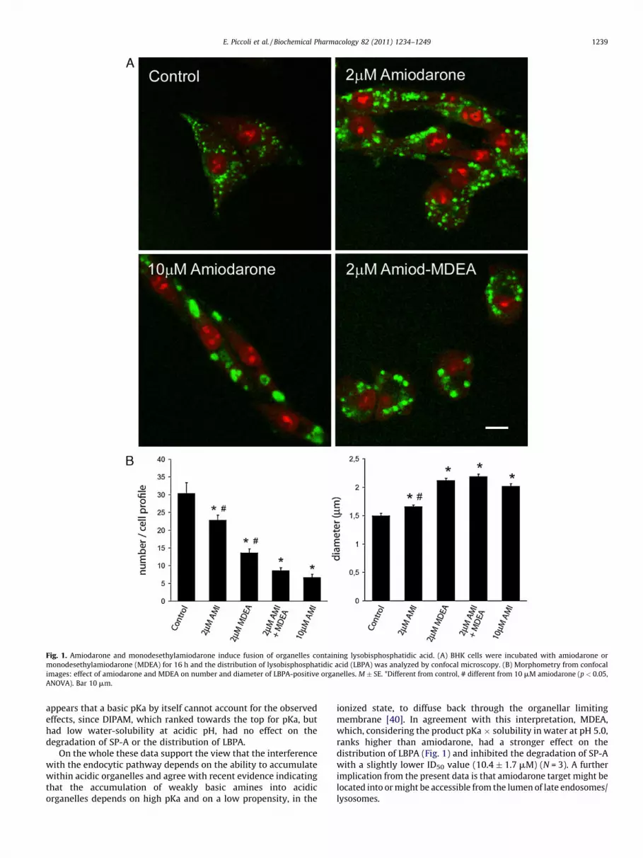
Fig. 1. Amiodarone and monodesethylamiodarone induce fusion of organelles containing lysobisphosphatidic acid. (A) BHK cells were incubated with amiodarone or
monodesethylamiodarone (MDEA) for 16 h and the distribution of lysobisphosphatidic acid (LBPA) was analyzed by confocal microscopy. (B) Morphometry from confocal
images: effect of amiodarone and MDEA on number and diameter of LBPA-positive organelles. M � SE. *Different from control, # different from 10 mM amiodarone (p < 0.05,
ANOVA). Bar 10 mm.
E. Piccoli et al. / Biochemical Pharmacology 82 (2011) 1234–1249 1239
appears that a basic pKa by itself cannot account for the observedeffects, since DIPAM, which ranked towards the top for pKa, buthad low water-solubility at acidic pH, had no effect on thedegradation of SP-A or the distribution of LBPA.
On the whole these data support the view that the interferencewith the endocytic pathway depends on the ability to accumulatewithin acidic organelles and agree with recent evidence indicatingthat the accumulation of weakly basic amines into acidicorganelles depends on high pKa and on a low propensity, in the
ionized state, to diffuse back through the organellar limitingmembrane [40]. In agreement with this interpretation, MDEA,which, considering the product pKa � solubility in water at pH 5.0,ranks higher than amiodarone, had a stronger effect on thedistribution of LBPA (Fig. 1) and inhibited the degradation of SP-Awith a slightly lower ID50 value (10.4 � 1.7 mM) (N = 3). A furtherimplication from the present data is that amiodarone target might belocated into or might be accessible from the lumen of late endosomes/lysosomes.

Fig. 2. Fusion of LBPA-rich organelles induced by amiodarone is reversible, cytoskeleton-dependent and inhibited by alkalinization. (A) BHK cells incubated for 16 h with
10 mM amiodarone and then with plain medium (wash out) or for 16 h with 10 mM amiodarone in the presence of nocodazole. (B) BHK cells incubated for 16 h with 10 mM
amiodarone without or with ammonium chloride. In both experiments number and diameter of LBPA-positive organelles were derived from confocal images. M � SE.
*Different from control, # different from 10 mM amiodarone (p < 0.05, ANOVA).
Table 2Relationship between physicochemical properties of amiodarone analogues and effects on the degradation of surfactant protein A (SP-A) and on cell distribution of LBPA.
Compounda pKa � solubility in water at pH 5.0 SP-A degradation ID50b (mM) LBPA-positive structuresc,d
Diameter (mm) N8/cell profile
MeAMI 17.50 11.2 � 1.0 2.45 � 0.08 4.5 � 0.6
Amiodarone 17.28 13.4 � 2.1 2.01 � 0.03 5.2 � 0.4
PIPAM 11.88 22.8 � 7.2 1.73 � 0.03 14.7 � 2.1
PYRAM 9.22 23.8 � 10.2 2.08 � 0.05 7.6 � 0.6
DIPAM 2.07 >50.0 1.64 � 0.04 24.6 � 3.0
MOPAM 0.55 >50.0 1.45 � 0.02 29.3 � 0.6
a Compounds arranged in order of decreasing value of the product (pKa � solubility in water at pH 5.0).b Degradation of SP-A measured as TCA-soluble radioactivity released by rabbit alveolar macrophages during 1 h incubation with 1 mg/ml 125I-SPA. ID50 = analogue
concentration needed to decrease degradation to 50% of control. M � SE, data from 3 to 13 experiments.c BHK cells were incubated for 16 h with 10 mM analogues and distribution of LBPA was analyzed by confocal microscopy. Data from 50 to 130 cells and 200 to 800
organelles.d In control cells LBPA-positive structures were 28.6 � 1.1/cell profile, and diameter was 1.46 � 0.02 mm. M � SE.
E. Piccoli et al. / Biochemical Pharmacology 82 (2011) 1234–12491240
3.3. Amiodarone changes the distribution of unesterified cholesterol,
glycosphingolipids and sphingomyelin
In Niemann-Pick type C disease, the accumulation of LBPA is afacet of a more general disturbance in the traffic of cell lipids[20,41]. We speculated that this might hold true also for cellstreated with amiodarone. To explore this possibility, we incubatedBHK cells with 0–10 mM amiodarone for 16 h and then analyzedthe distribution of unesterified cholesterol, glycosphingolipids andsphingomyelin.
Unesterified cholesterol was detected by staining fixed cellswith the fluorescent polyene antibiotic filipin [42]. As shown inFig. 3, in control BHK cells unesterified cholesterol was associatedwith small vesicles mostly distributed around the nucleus, while inthe presence of amiodarone it associated with larger structures.The change, clearly discernible in the presence of 2 mM amiodar-one, reached full expression in the presence of 10 mM amiodarone,giving to cells an appearance similar to that induced by 10 mMU18666A, a known inducer of the NPC phenotype (Fig. 3) [18].Besides changing the distribution of unesterified cholesterol,

Fig. 3. Amiodarone and U18666A alter the distribution of unesterified cholesterol. BHK cells were incubated for 16 h with plain medium, 2 or 10 mM amiodarone or 10 mM
U18666A, stained with filipin and analyzed by epifluorescence microscopy. Bar 10 mm.
E. Piccoli et al. / Biochemical Pharmacology 82 (2011) 1234–1249 1241
amiodarone also changed cell cholesterol content. In fact, after16 h incubation with 10 mM amiodarone, cell cholesterol in-creased by 17.3 � 5.2% (p = 0.03, paired ‘‘t’’ test, N = 5), while the ratiobetween total and unesterified cholesterol did not change.
To study the distribution of sphingomyelin BHK cells wereexposed to 0–10 mM amiodarone, fixed and then incubated firstwith lysenin, a sphingomyelin-binding protein isolated from thecoelomic fluid of the earthworm Eisenia foetida [32,33] and thenwith an anti-lisenin antiserum, followed by a fluorescent second-ary antibody. As shown in Fig. 4, in control cells sphingomyelin wasassociated with the plasma membrane and with organelles close tothe plasma membrane and was rigorously separated from LBPA.Cells treated with 2 mM amiodarone and 10 mM U18666Adisplayed a more intense staining with respect to control cells.Besides this, in cells treated with 10 mM amiodarone, sphingo-myelin tended to distribute as clumps, associated with complexinternal structures and, at some places, co-localized with LBPA. Thereason of the increased staining intensity in treated cells remainsunclear. Possibly amiodarone and U18666A rendered plasmamembrane sphingomyelin more accessible to lysenin.
Distribution and trafficking of glycosphingolipids was analyzedby incubation of BHK cells with BODIPY-LacCer, a sphingolipidanalogue in which the naturally occurring fatty acid moiety isreplaced with N-[5-(5,7 dimethyl boron dipyrromethanedifluor-ide)-1-pentanoic acid]. Cells were allowed to internalize BODIPY-LacCer for 45 min to allow loading of late endosomes, followed by1 h chase to accumulate the fluorescent probe in the Golgi and thenwere analyzed by confocal microscopy [34]. As shown in Fig. 5A, incontrol cells BODIPY-LacCer accumulated in juxtanuclear struc-tures compatible with the Golgi, while in amiodarone treated cellsit accumulated in round structures lacking the staining patterntypical of the Golgi system. Fig. 5A also shows that amiodarone didnot perturb the Golgi organization, since it did not change thedistribution of the specific marker giantin. Furthermore it appears
that the change in BODIPY-LacCer distribution induced byamiodarone (formation of large cytoplasmic aggregates) wasdifferent from the distribution as scattered cytoplasmic dotsobserved in the presence of 10 mM U18666A (Fig. 5B). Co-localization studies between BODIPY-LacCer and specific Golgimarkers were hindered by the fact that BODIPY-LacCer exhibits aconcentration dependent shift in its fluorescence emission fromgreen to red wavelengths as a result of excimer formation at highconcentrations [36]. From these data we conclude that amiodaronealters the cell distribution of sterols, phospholipids, sphingolipidsand glycosphingolipids, generating a phenotype with partialresemblance with Niemann-Pick type C disease [19,20,43].
3.4. Cholesterol removal alleviates the change in LBPA distribution
induced by U18666A, but not that induced by amiodarone
In NPC disease accumulating lipids are connected both spatiallyand functionally and alleviation of the phenotype can be obtainedby decreasing cholesterol levels with sterol-binding agents likemethyl-b-cyclodextrin [21], by inhibiting glycosphingolipid syn-thesis with 1-deoxynojirimycin [22] or by increasing lipiddegradation through overexpression of acid sphingomyelinase[23]. In particular LBPA accumulation can be attenuated bytreatment with methyl-b-cyclodextrin [21,41,42,44–46].
To clarify the mechanism of lipid accumulation due toamiodarone, BHK cells were loaded for 24 h with 1 mM methyl-b-cyclodextrin, rinsed extensively, incubated for further 16 h withplain medium, 10 mM amiodarone or 10 mM U186662 and thenprocessed for confocal microscopy and analysis of LBPA distribu-tion. Results were compared with those of cells not treated withmethyl-b-cyclodextrin. As shown in Fig. 6, in cells not treated withmethyl-b-cyclodextrin amiodarone and U18666A had similareffects, decreasing the number and increasing the diameter ofLBPA-positive structures. On the opposite, pre-treatment with

Fig. 4. Amiodarone changes the distribution of sphingomyelin. BHK cells, exposed for 16 h to control medium, 2 or 10 mM amiodarone or 10 mM U18666A, were fixed,
incubated first with the sphingomyelin-binding protein lysenin and then with antibodies to lysenin and LBPA. After incubation with secondary antibody cells were analyzed
by confocal microscopy. Red, sphingomyelin; green, LBPA. Arrows indicate sphingomyelin in intracellular structures. Arrowheads indicate co-localization of sphingomyelin
and LBPA (yellow). Bar 10 mm.
E. Piccoli et al. / Biochemical Pharmacology 82 (2011) 1234–12491242
methyl-b-cyclodextrin increased the diameter of the aggregates ofLBPA induced by amiodarone, worsening the phenotype, butdecreased the diameter and increased the number of aggregates ofLBPA induced by U18666A, alleviating the phenotype (Fig. 6). Fromthese data we conclude that changes induced by amiodarone donot strictly recapitulate the NPC phenotype.
3.5. Vacuoles induced by amiodarone are caused by interference with
fluid phase endocytosis
Dextrans are polysaccharides with good water solubility, lowtoxicity and resistance to cleavage by most endogenous cellglycosidases and thus represent ideal tracers to study the progressof membrane-impermeable cargo along the endocytic pathway.After uptake, dextrans move through the endocytic pathwayending up into lysosomes, from where they are returned to theextracellular milieu. Wild type fibroblasts in one day may disposeof up to 80% of uptaken dextran [18]. To study the effect ofamiodarone on the trafficking of dextran along the endocyticpathway, BHK cells were loaded with tetramethylrhodaminedextran in the absence or in the presence of 10 mM amiodaroneand then were chased for 5 h with plain medium or in the presenceof 10 mM amiodarone, a time sufficient in control cells to move allvisible dextran to the lysosomes [35]. As shown in Fig. 7, in control
cells uptaken dextran had a point-like distribution. On theopposite, cells loaded with TMR-dextran and then chased in thecontinuous presence of amiodarone, retained a greater fraction ofuptaken dextran, which was associated with swollen organelles.At some places these organelles also contained LBPA. From thesedata we conclude that amiodarone may cause the formation ofvacuoles by interfering with the progression of fluid phaseendocytosis. Furthermore, the co-localization of LBPA and dextranin swollen organelles suggests that accumulation of water-soluble materials may contribute also to the widening of inclusionbodies.
3.6. Amiodarone affects diverse but not all subsets of organelles
pertaining to late endosomes/lysosomes
In agreement with previous observations that amiodaroneaffects late compartments of the endocytic pathway [6,7], wefound that the abnormal structures induced by amiodarone exhibitlate endosomal markers like LAMP1, Rab7 and NPC1, but notmarkers of early endosomes. Indeed, the distribution of transientlyexpressed GFP-Rab5 did not change after exposure to amiodarone(Fig. 1SA). On the opposite, as shown in Figs. 8 and 9 and 1SB, incontrol cells LAMP1 presented as puncta distributed around thenucleus, while after incubation with 2 or 10 mM amiodarone it

Fig. 5. Amiodarone alters the traffic of glycosphingolipids. (A) BHK cells, exposed for 16 h to control medium or 10 mM amiodarone were incubated for 45 min with BODIPY-
LacCer, chased for one further hour and then analyzed by confocal microscopy. Inset: distribution of the Golgi marker giantin (green) in cells whose nuclei were stained red
with propidium. Arrows: abnormal collections of BODIPY-LacCer in amiodarone-treated cells. (B) Trafficking of BODIPY-LacCer in BHK cells exposed for 16 h to control
medium, 10 mM amiodarone or 10 mM U18666A and then analyzed by epifluorescence microscopy. Bar 10 mm.
Fig. 6. Cholesterol removal alleviates the change in LBPA distribution induced by U18666A, but not that induced by amiodarone. BHK cells were incubated for 24 h with
control medium or 1 mM methyl-b-cyclodextrin and then for further 16 h with control medium, 10 mM amiodarone or 10 mM U18666A. At the end the distribution of LBPA
was analyzed by confocal microscopy and number and diameter of LBPA-positive structures were measured. M � SE. *Different from control, # different from same treatment
without methyl-b-cyclodextrin (p < 0.05, ANOVA).
E. Piccoli et al. / Biochemical Pharmacology 82 (2011) 1234–1249 1243
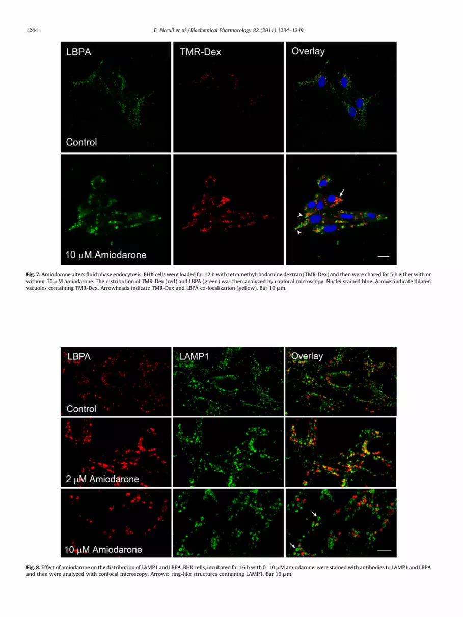
Fig. 7. Amiodarone alters fluid phase endocytosis. BHK cells were loaded for 12 h with tetramethylrhodamine dextran (TMR-Dex) and then were chased for 5 h either with or
without 10 mM amiodarone. The distribution of TMR-Dex (red) and LBPA (green) was then analyzed by confocal microscopy. Nuclei stained blue. Arrows indicate dilated
vacuoles containing TMR-Dex. Arrowheads indicate TMR-Dex and LBPA co-localization (yellow). Bar 10 mm.
Fig. 8. Effect of amiodarone on the distribution of LAMP1 and LBPA. BHK cells, incubated for 16 h with 0–10 mM amiodarone, were stained with antibodies to LAMP1 and LBPA
and then were analyzed with confocal microscopy. Arrows: ring-like structures containing LAMP1. Bar 10 mm.
E. Piccoli et al. / Biochemical Pharmacology 82 (2011) 1234–12491244

Fig. 9. Effect of amiodarone on the distribution of LAMP1 and tubulin. BHK cells, incubated for 16 h with 0–10 mM amiodarone, were stained with antibodies to LAMP1 and
tubulin and then were analyzed with confocal microscopy. Arrows: aggregates of LAMP1 surrounded by a rim of tubulin. Bar 10 mm.
E. Piccoli et al. / Biochemical Pharmacology 82 (2011) 1234–1249 1245
concentrated into less numerous but larger structures, as seen forLBPA. Aggregates of LAMP1 induced by amiodarone had differentmorphology, presenting as clumps or tiny rings (Fig. 8), as spotsarranged along the rim of dilated structures (Fig. 1S) or ascomponents of structures surrounded by a rim of tubulin (Fig. 9).Interestingly co-localization between LAMP1 and LBPA, which wasmoderate in control cells, increased after incubation with 2 mMamiodarone but was almost nil after incubation with 10 mMamiodarone, suggesting that low concentrations of the drug mightstimulate heterotypic fusion of organelles, while high concentra-tions could induce homotypic fusion.
Fig. 10. Amiodarone induces aggregates of NPC1 protein spatially separated from aggrega
with antibodies to NPC1 and LBPA and then were analyzed with confocal microscopy. A
(green). Yellow indicates co-localization.
The changes in the distribution of LAMP1 induced byamiodarone agree with changes in the distribution of NPC1protein, a multispanning protein inserted in the limiting mem-brane of late endosomes/lysosomes which, when mutated, is themost frequent cause of Niemann-Pick type C disease [16,47–49].Indeed, as shown in Fig. 10, in control cells NPC1 had a point-likedistribution and co-localized partially with LBPA, while in cellsincubated with 10 mM amiodarone it coalesced into largeaggregates spatially separated from LBPA.
To further characterize the effects of amiodarone on lateendosomes we studied its effects on the secretion of exosomes,
tes of LBPA. BHK cells incubated for 16 h with 0 or 10 mM amiodarone were stained
rrowheads indicate aggregates of NPC1 (red). Arrows indicate aggregates of LBPA

E. Piccoli et al. / Biochemical Pharmacology 82 (2011) 1234–12491246
which are vesicles located in the lumen of late endosomes/multivesicular bodies destined to be secreted in the extracellularmilieu [24]. To that end K562 cells were incubated with or without10 mM amiodarone for 16 h, secreted exosomes were then isolatedby differential centrifugation and acetylcholinesterase activityassociated with them was measured. We found that amiodaronedecreased secreted acetylcholinesterase by 12.0 � 8.7% withrespect to control cells (N = 7, change not significant), indicatingthat interference with exosome release is unlikely to contributesignificantly to the accumulation of membranes induced byamiodarone. These findings also indicate that amiodarone does notperturb the whole population of late endosomes/lysosomes.
3.7. Amiodarone has no effect on transferrin recycling, Shiga toxin
activity and lentivirus budding
The available evidence indicates that amiodarone does notinfluence the distribution of markers of early endosomes. Tocorroborate the lack of impact on this compartment, we studiedthe effects of amiodarone on early endosome-dependent activities
Fig. 11. Dronedarone alters the distribution of LBPA and unesterified cholesterol and sti
dronedarone, stained for LBPA (green) and then counterstained with propidium iodide (r
for 16 h with control medium or with 2 mM dronedarone. The distribution of LBPA (red
membranes surrounding LBPA aggregates. (C) Epifluorescence image of BHK cells incuba
stain unesterified cholesterol.
like transferrin recycling, Shiga toxin trafficking and ESCRT-dependent lentivirus budding.
The effect on transferrin recycling was analyzed by loadinghuman monocytes with labelled transferrin and then measuring itsrelease in the presence of 0 or 10 mM amiodarone. As shown inFig. 2S, amiodarone had no effect on transferrin recycling.
The effect on Shiga toxin activity was analyzed by incubatingVero cells with Shiga toxin 1 in the presence or in the absence of 0–20 mM amiodarone for a time sufficient for the toxin to be takenup, processed in early endosomes, transferred to the Golgi and thenmoved to the cytoplasm, where it inhibits protein synthesis[26,49]. As shown in Fig. 2S the activity of Shiga toxin was notdecreased by amiodarone, suggesting a correct processing andtrafficking of the toxin.
The human immunodeficiency virus type 1 (HIV-1) and otherlentiviruses, such as the feline immunodeficiency virus (FIV) exitinfected cells by budding from the plasma membrane, a processrequiring membrane fission. The only viral component involved inbudding is the structural protein Gag, which executes budding byrecruiting host proteins belonging to the ESCRT complexes, such as
mulates autophagy. (A) Confocal images of BHK cells incubated for 16 h with 5 mM
ed). (B) BHK cells, transiently transfected with pEGFP-LC3 plasmid, were incubated
) and LC3 (green) was then analyzed by confocal microscopy. Arrow: LC3 positive
ted for 16 h with 0 (control) or 5 mM dronedarone and then incubated with filipin to

E. Piccoli et al. / Biochemical Pharmacology 82 (2011) 1234–1249 1247
Tsg101 and AIP1 [50]. To understand whether amiodaroneinterferes with the ESCRT complex, 293T cells were transfectedwith constructs expressing either the feline immunodeficiencyvirus (pDenv1) or the human immunodeficiency type 1 (pSVC21)Gag/Pol. Twenty four hours post-transfection, cells were incubatedwith 0 or 10 mM amiodarone for additional 24 h. Virus likeparticles (VLP) released in the supernatant and cells were thenanalyzed by SDS-PAGE electrophoresis followed by Westernblotting using antibodies specific for either FIV or HIV-1 capsidproteins. As shown in Fig. 2S, amiodarone did not affect synthesisand processing of the intracellular Gag protein and did not inhibitthe release of mature VLPs, suggesting lack of interference withbudding. Collectively these experiments support the view thatamiodarone affects only late compartments of the endocyticpathway.
3.8. Dronedarone alters the distribution of cell lipids and stimulates
autophagy
Dronedarone is a non-iodinated benzofuran derivative, recentlyintroduced in clinical practice to get around some of amiodaronenot desirable effects like slow pharmacokinetics and iodine-relatedthyroid damage [27]. Previous experiments with rabbit alveolarmacrophages have shown that amiodarone induces the formationof vacuoles and inclusion bodies [28], but the mechanism offormation of these structures has not been studied. To clarify this,BHK cells were incubated with 0–5 mM dronedarone for 16 h(higher concentrations were toxic to cells) and then the distribu-tion of LBPA and unesterified cholesterol were analyzed. As shownin Fig. 11 cells treated with dronedarone had severe disturbancesin the distribution LBPA and unesterified cholesterol. Sincedronedarone has relatively high pKa and a very high solubilityin water at pH 5, the strong effect on LBPA distribution agrees withthe view that these physico-chemical properties play an importantrole in the interference with late compartments of the endocyticpathway (Tables 1 and 2).
Since autophagy is up-regulated by amiodarone [7], we studiedthe effect of dronedarone on the distribution of transientlyexpressed eGFP-LC3, a protein present at all steps along theautophagic pathway [51]. We found that in control cells LBPA andLC3 had a point-like distribution, whereas in cells treated with2 mM dronedarone (the highest concentrations tolerated bytransfected cells) or 10 mM amiodarone, LBPA and LC3 changedfrom a point-like to a clump-like distribution (Fig. 11 and 3S). Atsome places aggregates of LBPA were enclosed by ring-likestructures decorated with LC3, as expected during the formationof amphisomes, which are intermediate organelles formed duringautophagy. Thus, like amiodarone, dronedarone also alters thedistribution of cell lipids and induces autophagy.
4. Discussion
4.1. Vacuoles and inclusion bodies induced by amiodarone
This study shows that vacuoles and inclusion bodies forming inthe presence of amiodarone originate from late compartments ofthe endocytic pathway, since they display markers normallyassociates with late endosomes/lysosomes, like Rab7, LAMP1,NPC1, LBPA. It also shows that separate populations of organellespertaining to late endocytic compartments are affected, asindicated by the lack of co-localization of aggregates of LBPA,LAMP1 and NPC1 at high amiodarone concentrations. Finally itshows that amiodarone does not affect the whole of late endosomeactivities, since the secretion of exosomes does not change.
Vacuoles and inclusion bodies appear to form by severalmechanisms: block of fluid phase endocytosis, inappropriate
fusion of organelles, collapse of lumenal structures, accumulationof undegraded substrates and accumulation of wrongly traffickedlipids. Other mechanisms could also be at play. For example innormal cells late endosomes fuse with lysosomes, forming hybridorganelles from which lysosomes reform when the degradation ofsubstrates is complete [52]. We posit that decreased reformation oflysosomes from hybrid intermediates could contribute to thevacuolar phenotype induced by amiodarone. Such a mechanismcould explain the decreased lysosome yield detected in alveolarmacrophages treated with amiodarone [9] and the finding thatincubation of BHK cells for 16 h with 10 mM amiodarone does notmodify the total cell content of LAMP-1, a protein inserted in thelimiting membrane of lysosomes (not shown).
This study also shows that amiodarone does not directly affectearly endosomes, since: (a) it does not alter the distribution ofearly endosome markers; (b) does not inhibit binary toxinsrequiring functional early endosomes for activation, like Shigatoxin 1; (c) does not disturb transferrin recycling; (d) does notinterfere with the budding of lentiviruses depending upon therecruitment of a highly conserved set of proteins, dubbed theendosomal sorting complexes required for transport (ESCRTs), alsorequired for the formation of multivesicular bodies in earlyendosomes [14,53]. To directly confirm the lack of interferencewith the ESCRTs, we studied the effect of amiodarone on theformation of lumenal vesicles by isolated early endosomes [54]. Tothat end the post-nuclear supernatant obtained from BHK cellscultured for 3 h in the presence of 0–20 mM amiodarone, wasincubated at 37 8C with the non permeant fluorescent probe 8-hydroxypyrene-1,3,6-trisulfonic acid in the presence of an energydonor system and trapping of the probe in the interior of vesiclesforming by inward budding of endosome limiting membrane wasallowed to take place for 20 min. Subsequently, after quenchingthe extra-organellar fluorescence with p-xylene-bis-pyridiniumbromide, endosomes were isolated by flotation and the fluores-cence associated with them was measured. In 3 separatedexperiments we found no change in endosome-associatedfluorescence after incubation with amiodarone. Thus several linesof evidence indicate that amiodarone has no direct effect on earlyendosomes. It is possible, however, that chronic exposure to thedrug might reverberate on early endosomes also, perhaps bymodifying the global trafficking of membranes or through thesequestration of critical lipids.
Cell changes induced by amiodarone have thus far beenincluded under the term ‘‘phospholipidosis’’, a term based onelectron microscopy, which describes the formation of multi-laminar inclusion vesicles [1,2]. The accumulation of LBPA,cholesterol and glycosphyngolipids in cells exposed to amiodar-one is compatible with the accumulation of membranesobserved at the electron microscope. It appears, however, thatthe term phospholipidosis does not fully portray the cellchanges induced by amiodarone since it does not reflect theaccumulation of other lipid species besides phospholipids anddoes not include other effects, like disturbances in fluid phaseendocytosis.
4.2. The target of amiodarone activity
Data obtained with amiodarone analogues (Table 2) indicatethat high pKa and especially high water-solubility at acidic pHare important determinants of the interference of amiodaronewith late endosomes/lysosomes. Since basic pKa and lowmembrane permeability, favour the sequestration of weaklybasic amines in the lumen of acidic organelles [55], we infer thatamiodarone target resides into or is accessible from the lumen oflate endosomes/lysosomes. The nature of amiodarone targetremains to be clarified since the present results are compatible

E. Piccoli et al. / Biochemical Pharmacology 82 (2011) 1234–12491248
both with the inhibition of specific component(s) accessiblefrom the luminal side and with an aspecific effect, like thecompetition of amiodarone with polycationic lipid hydrolases oractivator proteins for the negative surface of late endosome/lysosome internal membranes [56]. The last mechanism,recently proposed for other cationic amphiphilic drugs, wouldexplain the accumulation of different classes of undegradedlipids and the membrane traffic jam within late endosomes/lysosomes [57].
The propensity to be trapped in the lumen of acidic organellescould also explain the preferential effect of amiodarone on lateendosomes/lysosomes, whose interior is more acidic than that ofearly endosomes. On the other side it remains at present unclearhow amiodarone may target specific sets of organelles pertainingto late endosomes/lysosomes.
4.3. Amiodarone induces a phenotype reminiscent of Niemann-Pick
type C disease
The phenotype induced by amiodarone recalls Niemann-Picktype C disease, a complex lysosomal storage disorder characterizedby the accumulation of different sorts of lipids in late endosomes/lysosomes, by defective lysosomal calcium homeostasis andunique trafficking defects [20]. NPC disease is caused by mutationsin the NPC1 or NPC2 genes, which encode respectively NPC1, amultispanning trans-membrane domain protein located on thelimiting membrane of late endosomes/lysosomes and NPC2, asoluble lysosomal protein [58].
There are differences, however, between changes induced byamiodarone and the NPC phenotype. In fact on one side methyl-b-cyclodextrin worsens the effects of amiodarone on the distributionof LBPA, while, on the contrary, it alleviates the redistribution ofLBPA caused by U18666A. On the other side budding of HIV-1 virus isinhibited in NPC cells or by treatment with U18666A [58], but is notaffected by amiodarone. Thus, rather than recapitulating the wholepathogenetic cascade of NPC disease, changes induced by amiodar-one might exemplify a common theme observed when damage tolate endosomes/lysosomes disturbs pathways intersecting at thiscrucial spot. As an example, cells lacking prosaposin, a proteincofactor necessary for glycosphingolipid degradation, presentenlarged electron-lucent structures of late endosomal origin,accumulate unesterified cholesterol and display altered traffickingof BODIPY-LacCer [59].
4.4. Clinical implications
Atrial fibrillation predisposes patients to stroke, heart failureand death and affects nearly 1 every 10 persons aged 80 years andolder [60]. Among conventional antiarrhythmic drugs amiodar-one has proved by far the most effective at maintaining sinusrhythm, but effectiveness is diminished by long elimination half-life, iodine-induced thyroid toxicity and acute lung damage. In anattempt to relieve some of these problems, dronedarone, a non-iodinated benzofuran derivative, has been recently introduced inclinical practice [27]. Dronedarone has multichannel blockingproperties comparable to those of amiodarone but is more potentat blocking peak sodium currents and acetylcholine activatedpotassium currents and has stronger antiadrenergic effects [27].On the basis of the present evidence it appears that, on a molarbasis, dronedarone is at least as potent as amiodarone withrespect to interference with late compartments of the endocyticpathway. Dronedarone, however, is metabolized rapidly andreaches serum levels over 10 times lower than in patients treatedwith amiodarone [27], thus the clinical relevance of its interfer-ence with late endocytic compartments remains to be deter-mined.
4.5. Future directions
The interference of amiodarone and its derivatives with theendocytic pathway, besides its obvious clinical interest in the fieldof cardiology, might represent the subject of future investigationsin unrelated areas, in particular for the development of novelstrategies against infectious agents. Indeed amiodarone interferesin vitro with the life cycle of the SARS coronavirus [6], mitigates inan in vivo model the effects of the encephalomyocarditis virus [61]and has been shown to be of value in the treatment of theTrypanosomacruzi infection [62]. In addition, it could be of intereststudying the effect of amiodarone on Dengue infection, because ithas recently been shown that LBPA (whose distribution may bemodified by amiodarone) is an obligatory cofactor for the entry ofthe Dengue virus into target cells [63].
4.6. Conclusion
Amiodarone induces the formation of vacuoles and inclusionbodies by interfering with late compartments of the endocyticpathway. Mechanisms of formation of vacuoles and inclusionbodies include disturbances in the progression of fluid phaseendocytosis, inappropriate fusion of organelles, collapse oflumenal structures, accumulation of undegraded substrates andaccumulation of wrongly trafficking lipids. High water-solubility atacidic pH is a crucial requirement for the interference ofamiodarone with late endosomes/lysosomes. Dronedarone is atleast as potent as amiodarone with respect to interference withlate compartments of the endocytic pathway. The interference ofamiodarone with late endosomes/lysosomes may be of value inareas outside the field of arrhythmology.
Acknowledgements
We thank Prof. E. Papini and Dr F. Tonello, University of Padova,Italy, Prof. C. Bucci, University of Lecce, Italy and Dr J.P. Krise,University of Kansas, KA, for help and advice. We also thank Prof.M. Brigotti, University of Bologna, Italy, for preparing Shiga toxin 1.This work was supported in part by Progetti di Ateneo, Universityof Padova, 2008 to A.B.
Appendix A. Supplementary data
Supplementary data associated with this article can be found, inthe online version, at doi:10.1016/j.bcp.2011.07.090.
References
[1] Vassallo P, Trohman RG. Prescribing amiodarone: an evidence-based review ofclinical indications. J Am Med Assoc 2007;298:1312–22.
[2] Zimetbaum P. Amiodarone for atrial fibrillation. N Engl J Med 2007;356:935–41.
[3] Pollak PT, Bouillon T, Shafer SL. Population pharmacokinetics of long-term oralamiodarone therapy. Clin Pharmacol Ther 2000;67:642–52.
[4] Adams PC, Holt DW, Storey GC, Morley AR, Callaghan J, Campbell RW. Amio-darone and its desethyl metabolite: tissue distribution and morphologicchanges during long-term therapy. Circulation 1985;72:1064–75.
[5] Brien JF, Jimmo S, Brennan FJ, Ford SE, Armstrong PW. Distribution of amio-darone and its metabolite, desethylamiodarone, in human tissues. Can JPhysiol Pharmacol 1987;65:360–4.
[6] Stadler K, Ha HR, Ciminale V, Spirli C, Saletti G, Schiavon M, et al. Amiodaronealters late endosomes and inhibits SARS coronavirus infection at a post-endosomal level. Am J Respir Cell Mol Biol 2008;39:142–9.
[7] Morissette G, Ammoury A, Rusu D, Marguery MC, Lodge R, Poubelle PE, et al.Intracellular sequestration of amiodarone: role of vacuolar ATPase and macro-autophagic transition of the resulting vacuolar cytopathology. Br J Pharmacol2009;157:1531–40.
[8] Ha HR, Bigler L, Wendt B, Maggiorini M, Follath F. Identification and quantita-tion of novel metabolites of amiodarone in plasma of treated patients. Eur JPharm Sci 2005;24:271–9.

E. Piccoli et al. / Biochemical Pharmacology 82 (2011) 1234–1249 1249
[9] Baritussio A, Marzini S, Agostini M, Alberti A, Cimenti C, Bruttomesso D, et al.Amiodarone inhibits lung degradation of SP-A and perturbs the distribution oflysosomal enzymes. Am J Physiol Lung Cell Mol Physiol 2001;281:L1189–9.
[10] Sanchez AM, Thomas D, Gillespie EJ, Damoiseaux R, Rogers J, Saxe JP, et al.Amiodarone and bepridil inhibit anthrax toxin entry into host cells. Antimi-crob Agents Chemother 2007;51:2403–11.
[11] Mortuza GB, Neville WA, Delaney J, Waterfield CJ, Camilleri P. Characterisationof a potential biomarker of phospholipidosis from amiodarone-treated rats.Biochim Biophys Acta 2003;1631:136–46.
[12] Kobayashi T, Beuchat MH, Lindsay M, Frias S, Palmiter RD, Sakuraba H, et al.Late endosomal membranes rich in lysobisphosphatidic acid regulate choles-terol transport. Nat Cell Biol 1999;1:113–8.
[13] Puhar A, Montecucco C. Where and how do anthrax toxins exit endosomes tointoxicate host cells? Trends Microbiol 2007;15:477–82.
[14] Raiborg C, Stenmark H. The ESCRT machinery in endosomal sorting of ubi-quitylated membrane proteins. Nature 2009;458:445–52.
[15] Chevallier J, Chamoun Z, Jiang G, Prestwich G, Sakai N, Matile S, et al. Lysobi-sphosphatidic acid controls endosomal cholesterol levels. J Biol Chem2008;283:27871–80.
[16] Brown MS, Goldstein JL. A receptor-mediated pathway for cholesterol homeo-stasis. Science 1986;232:34–47.
[17] Xie C, Turley SD, Pentchev PG, Dietschy JM. Cholesterol balance and metabo-lism in mice with loss of function of Niemann-Pick C protein. Am J Physiol1999;276:E336–44.
[18] Goldman SD, Krise JP. Niemann-Pick C1 functions independently of Niemann-Pick C2 in the initial stage of retrograde transport of membrane-impermeablelysosomal cargo. J Biol Chem 2010;285:4983–94.
[19] Liscum L, Ruggiero RM, Faust JR. The intracellular transport of low densitylipoprotein-derived cholesterol is defective in Niemann-Pick type C fibro-blasts. J Cell Biol 1989;108:1625–36.
[20] Lloyd-Evans E, Platt FM. Lipids on trial: the search for the offending metabolitein Niemann-Pick type C disease. Traffic 2010;11:419–28.
[21] Liu B, Turley SD, Burns DK, Miller AM, Repa JJ, Dietschy JM. Reversal ofdefective lysosomal transport in NPC disease ameliorates liver dysfunctionand neurodegeneration in the npc1�/� mouse. Proc Natl Acad Sci USA2009;106:2377–82.
[22] Lachmann RH, te Vruchte D, Lloyd-Evans E, Reinkensmeier G, Sillence DJ,Fernandez-Guillen L, et al. Treatment with miglustat reverses the lipid-traf-ficking defect in Niemann-Pick disease type C. Neurobiol Dis 2004;16:654–8.
[23] Devlin C, Pipalia NH, Liao X, Schuchman EH, Maxfield FR, Tabas I. Improvementin lipid and protein trafficking in Niemann-Pick C1 cells by correction of asecondary enzyme defect. Traffic 2010;11:601–15.
[24] Strauss K, Goebel C, Runz H, Mobius W, Weiss S, Feussner I, et al. Exosomesecretion ameliorates lysosomal storage of cholesterol in Niemann-Pick type Cdisease. J Biol Chem 2010;285:26279–88.
[25] Simons M, Raposo G. Exosomes—vesicular carriers for intercellular commu-nication. Curr Opin Cell Biol 2009;21:575–81.
[26] Warnier M, Romer W, Geelen J, Lesieur J, Amessou M, van den Heuvel L, et al.Trafficking of Shiga toxin/Shiga-like toxin-1 in human glomerular microvas-cular endothelial cells and human mesangial cells. Kidney Int 2006;70:2085–91.
[27] Patel C, Yan GX, Kowey PR. Dronedarone. Circulation 2009;120:636–44.[28] Quaglino D, Ha HR, Duner E, Bruttomesso D, Bigler L, Follath F, et al. Effects of
metabolites and analogs of amiodarone on alveolar macrophages: structure–activity relationship. Am J Physiol Lung Cell Mol Physiol 2004;287:L438–47.
[29] Calistri A, Del Vecchio C, Salata C, Celestino M, Celegato M, Gottlinger H, et al.Role of the feline immunodeficiency virus L-domain in the presence or absenceof Gag processing: involvement of ubiquitin and Nedd4-2s ligase in viralegress. J Cell Physiol 2009;218:175–82.
[30] Ratner L, Haseltine W, Patarca R, Livak KJ, Starcich B, Josephs SF, et al. Completenucleotide sequence of the AIDS virus, HTLV-III. Nature 1985;313:277–84.
[31] Bigler L, Spirli C, Fiorotto R, Pettenazzo A, Duner E, Baritussio A, et al. Synthesisand cytotoxicity properties of amiodarone analogues. Eur J Med Chem2007;42:861–7.
[32] Ishitsuka R, Sato SB, Kobayashi T. Imaging lipid rafts. J Biochem 2005;137:249–54.
[33] Yamaji A, Sekizawa Y, Emoto K, Sakuraba H, Inoue K, Kobayashi H, et al.Lysenin, a novel sphingomyelin-specific binding protein. J Biol Chem1998;273:5300–6.
[34] Martin OC, Pagano RE. Internalization and sorting of a fluorescent analogue ofglucosylceramide to the Golgi apparatus of human skin fibroblasts: utilizationof endocytic and nonendocytic transport mechanisms. J Cell Biol1994;125:769–81.
[35] Miedel MT, Rbaibi Y, Guerriero CJ, Colletti G, Weixel KM, Weisz OA, et al.Membrane traffic and turnover in TRP-ML1-deficient cells: a revised model formucolipidosis type IV pathogenesis. J Exp Med 2008;205:1477–90.
[36] Baritussio A, Alberti A, Armanini D, Meloni F, Bruttomesso D. Different path-ways of degradation of SP-A and saturated phosphatidylcholine by alveolarmacrophages. Am J Physiol Lung Cell Mol Physiol 2000;279:L91–9.
[37] Tanner LI, Lienhard GE. Insulin elicits a redistribution of transferrin receptorsin 3T3-L1 adipocytes through an increase in the rate constant for receptorexternalization. J Biol Chem 1987;262:8975–80.
[38] Fader CM, Sanchez D, Furlan M, Colombo MI. Induction of autophagy promotesfusion of multivesicular bodies with autophagic vacuoles in k562 cells. Traffic2008;9:230–50.
[39] Wright JR. Immunoregulatory functions of surfactant proteins. Nat Rev Immu-nol 2005;5:58–68.
[40] Duvvuri M, Konkar S, Funk RS, Krise JM, Krise JP. A chemical strategy tomanipulate the intracellular localization of drugs in resistant cancer cells.Biochemistry 2005;44:15743–9.
[41] Lloyd-Evans E, Morgan AJ, He X, Smith DA, Elliot-Smith E, Sillence DJ, et al.Niemann-Pick disease type C1 is a sphingosine storage disease that causesderegulation of lysosomal calcium. Nat Med 2008;14:1247–55.
[42] Liu B, Li H, Repa JJ, Turley SD, Dietschy JM. Genetic variations and treatmentsthat affect the lifespan of the NPC1 mouse. J Lipid Res 2008;49:663–9.
[43] Vanier MT. Lipid changes in Niemann-Pick disease type C brain: personalexperience and review of the literature. Neurochem Res 1999;24:481–9.
[44] Castanho MA, Coutinho A, Prieto MJ. Absorption and fluorescence spectra ofpolyene antibiotics in the presence of cholesterol. J Biol Chem 1992;267:204–9.
[45] Abi-Mosleh L, Infante RE, Radhakrishnan A, Goldstein JL, Brown MS. Cyclo-dextrin overcomes deficient lysosome-to-endoplasmic reticulum transport ofcholesterol in Niemann-Pick type C cells. Proc Natl Acad Sci USA2009;106:19316–21.
[46] Rosenbaum AI, Zhang G, Warren JD, Maxfield FR. Endocytosis of beta-cyclo-dextrins is responsible for cholesterol reduction in Niemann-Pick type Cmutant cells. Proc Natl Acad Sci USA 2010;107:5477–82.
[47] Infante RE, Abi-Mosleh L, Radhakrishnan A, Dale JD, Brown MS, Goldstein JL.Purified NPC1 protein. I. Binding of cholesterol and oxysterols to a 1278-aminoacid membrane protein. J Biol Chem 2008;283:1052–63.
[48] Infante RE, Radhakrishnan A, Abi-Mosleh L, Kinch LN, Wang ML, Grishin NV,et al. Purified NPC1 protein: II. Localization of sterol binding to a 240-aminoacid soluble luminal loop. J Biol Chem 2008;283:1064–75.
[49] Johannes L, Romer W. Shiga toxins—from cell biology to biomedical applica-tions. Nat Rev Microbiol 2010;8:105–16.
[50] Strack B, Calistri A, Craig S, Popova E, Gottlinger HG. AIP1/ALIX is a bindingpartner for HIV-1 p6 and EIAV p9 functioning in virus budding. Cell2003;114:689–99.
[51] Yang Z, Klionsky DJ. Eaten alive: a history of macroautophagy. Nat Cell Biol2010;12:814–22.
[52] Luzio JP, Pryor PR, Bright NA. Lysosomes: fusion and function. Nat Rev Mol CellBiol 2007;8:622–32.
[53] Hurley JH, Hanson PI. Membrane budding and scission by the ESCRT machin-ery: it’s all in the neck. Nat Rev Mol Cell Biol 2010;11:556–66.
[54] Falguieres T, Luyet PP, Bissig C, Scott CC, Velluz MC, Gruenberg J. In vitrobudding of intralumenal vesicles into late endosomes is regulated by Alix andTsg101. Mol Biol Cell 2008;19:4942–55.
[55] Kaufmann AM, Krise JP. Niemann-Pick C1 functions in regulating lysosomalamine content. J Biol Chem 2008;283:24584–93.
[56] Wilkening G, Linke T, Uhlhorn-Dierks G, Sandhoff K. Degradation of mem-brane-bound ganglioside GM1. Stimulation by bis(monoacylglycero)pho-sphate and the activator proteins SAP-B and GM2-AP. J Biol Chem2000;275:35814–9.
[57] Wilkening G, Linke T, Sandhoff K. Lysosomal degradation on vesicular mem-brane surfaces. Enhanced glucosylceramide degradation by lysosomal anioniclipids and activators. J Biol Chem 1998;273:30271–8.
[58] Tang Y, Leao IC, Coleman EM, Broughton RS, Hildreth JE. Deficiency of Nie-mann-Pick type C-1 protein impairs release of human immunodeficiency virustype 1 and results in Gag accumulation in late endosomal/lysosomal compart-ments. J Virol 2009;83:7982–95.
[59] Kiss RS, Ma Z, Nakada-Tsukui K, Brugnera E, Vassiliou G, McBride HM, et al. Thelipoprotein receptor-related protein-1 (LRP) adapter protein GULP mediatestrafficking of the LRP ligand prosaposin, leading to sphingolipid and freecholesterol accumulation in late endosomes and impaired efflux. J Biol Chem2006;281:12081–92.
[60] Furberg CD, Psaty BM, Manolio TA, Gardin JM, Smith VE, Rautaharju PM.Prevalence of atrial fibrillation in elderly subjects (the Cardiovascular HealthStudy). Am J Cardiol 1994;74:236–41.
[61] Ito H, Ono K, Nishio R, Sasayama S, Matsumori A. Amiodarone inhibitsinterleukin 6 production and attenuates myocardial injury induced by viralmyocarditis in mice. Cytokine 2002;17:197–202.
[62] Adesse D, Azzam EM, Meirelles Mde N, Urbina JA, Garzoni LR. Amiodaroneinhibits Trypanosoma cruzi infection and promotes cardiac cell recovery withgap junction and cytoskeleton reassembly in vitro. Antimicrob Agents Che-mother 2011;55:203–10.
[63] Zaitseva E, Yang ST, Melikov K, Pourmal S, Chernomordik LV. Dengue virusensures its fusion in late endosomes using compartment-specific lipids. PLoSPathog 2010;6.


