Embed Size (px)
Citation preview

ORIGINAL ARTICLE
An ErbB-3 antibody, MP-RM-1, inhibits tumor growth by blocking
ligand-dependent and independent activation of ErbB-3/Akt signaling
G Sala1,2, S Traini1,2, M D’Egidio1,2, G Vianale2, C Rossi2, E Piccolo1,2, R Lattanzio2,M Piantelli1,2, N Tinari1,2, PG Natali1, R Muraro1,2 and S Iacobelli1,2 on behalf of CINBO(Consorzio Interuniversitario Nazionale per la Bio-Oncologia)
1MediaPharma s.r.l., Chieti, Italy and 2Departement of Oncology and Experimental Medicine, University Foundation ‘G. D’Annunzio’Chieti-Pescara, Chieti, Italy
The ErbB receptors, such as ErbB-1 and ErbB-2, havebeen intensely pursued as targets for cancer therapeutics.Although initially efficacious in a subset of patients, drugstargeting these receptors led invariably to resistance,which is often associated with reactivation of the ErbB-3-PI3K-Akt signaling. This may be overcome by an ErbB-3ligand that abrogates receptor-mediated signaling. To-ward this end, we have generated a mouse monoclonalantibody, MP-RM-1, against the extracellular domain(ECD) of ErbB-3 receptor. Assessment of human tumorcell lines, as well as early passage tumor cells revealedthat MP-RM-1 effectively inhibited both NRG-1b-depen-dent and -independent ErbB-3 activation. The antagoniz-ing effect of MP-RM-1 was of non-competitive type, asbinding of [125I]-labeled NRG-1b to ErbB-3 was notinfluenced by the antibody. MP-RM-1 treatment led, inmost instances, to decreased ErbB-3 expression. Inaddition, MP-RM-1 was able to inhibit the colonyformation ability of tumor cells and tumor growth intwo human tumor xenograft nude mouse models. Treat-ment with the antibody was associated with a decreasedErbB-3 and Akt phosphorylation and ErbB-3 expressionin the excised tumor tissue. Collectively, these resultsindicate that MP-RM-1 has the potential to interfere withsignaling by ErbB-3 and reinforce the notion that ErbB-3could be a key target in cancer-drug design.Oncogene (2012) 31, 1275–1286; doi:10.1038/onc.2011.322;published online 8 August 2011
Keywords: ErbB-3 receptor; ErbB-3/Akt pathway;monoclonal antibody; targeted therapy
Introduction
The human epidermal growth factor receptor (EGFR;HER; ErbB) family of receptor tyrosine kinasesregulates a large variety of biological processes including
cell proliferation, migration, invasion and survival(Gschwind et al., 2004). The family includes fourmembers: ErbB-1 (EGFR, or HER-1), ErbB-2 (HER-2),ErbB-3 (HER-3) and ErbB-4 (HER-4). To date, 11ligands have been reported including EGF, heparin-binding EGF-like growth factor, transforming growthfactor-a, amphiregulin, epiregulin, betacellulin and theheregulins (also known as neuroregulins). These ligandsbind directly to their cognate receptors, which leads tothe formation of receptor homodimers or heterodimersthat trigger the activation of multiple signaling path-ways (Citri and Yarden 2006), among which the Ras–Raf–MAPK and PI3K–Akt pathways are prevalent. Inthe last decade, preclinical and clinical studies havedemonstrated that a fraction of epithelial cancers areaddicted to the signals propagated by ErbB receptors(Hynes and MacDonald 2009). As a consequence, thesereceptors are nowadays among the most targetedoncoproteins in cancer. Treatment of cancers drivenby the HER family of proteins are currently eithermonoclonal antibodies such as trastuzumab (directed atErbB-2), cetuximab (directed at ErbB-1), pertuzumab(preventing ErbB-2/ErbB-3 dimerization), or small-molecule tyrosine kinase inhibitors such as gefitiniband erlotinib (ErbB-1 kinase inhibitors) and lapatinib(dual ErbB-2/ErbB-1 kinase inhibitor) (Mendelsohn andBaselga 2006; Hynes 2007; Giampaglia et al., 2010;Saxena and Dwivedi 2010). Several other monoclonalantibodies and tyrosine kinase inhibitors directedtoward the ErbB family proteins are currently in clinicaltrials (Hynes and Lane 2005). Although efficacious in asubset of patients, monoclonal antibodies such astrastuzumab or tyrosine kinase inhibitors such asgefitinib, impairing only one ErbB family member,often encounter endogenous or acquired resistance(Romond et al., 2005; Mendelsohn and Baselga 2006).This resistance is often associated with compensation byor upregulation of other ErbB family members such asErbB-3 and ErbB-4 (Bianchi et al., 2006; Engelmanet al., 2007; Karamouzis et al., 2007) or increasedproduction of ErbB-1 or ErbB-3 ligands by tumor cells(Ishikawa et al., 2005; Zhou et al., 2006).
ErbB-3 overexpression has been documented inbreast, ovarian and lung cancer and this genetic featurehas been correlated with poor prognosis (Yi et al., 1997;
Received 6 February 2011; revised 22 June 2011; accepted 23 June 2011;published online 8 August 2011
Correspondence: Dr G Sala, Departement of Oncology and Experi-mental Medicine, University Foundation ‘G. D’Annunzio’ Chieti-Pescara, Via Colle dell’Ara 1, Chieti 66100, Italy.E-mail: [email protected]
Oncogene (2012) 31, 1275–1286& 2012 Macmillan Publishers Limited All rights reserved 0950-9232/12
www.nature.com/onc
Witton et al., 2003; Tanner et al., 2006). On activationby heregulins, ErbB-3 dimerizes with ErbB-2 and ErbB-1 to generate potent oncogenic receptor heterodimers(Alimandi et al., 1995; Wallasch et al., 1995). Within thiscomplex, ErbB-3 preferentially recruits phosphatidyli-nositol 3-kinase (PI3K) to its cytoplasmic dockingsites thereby regulating cell proliferation and survival(Prigent and Gullick 1994; Sithanandam et al., 2005).Interestingly, it has recently been shown that cancer cellsescape ErbB inhibition therapy by upregulation ofErbB-3 signaling (Sergina et al., 2007; Baselga andSwain, 2009) and that ErbB-3 inhibition abrogatesErbB-2-driven tamoxifen resistance in breast cancercells (Liu et al., 2007). Of interest, recent data havedocumented that ErbB-3 can be transactivated by METin lung cancer cells resistant to the ErbB-1 inhibitor,gefitinib (Engelman et al., 2007). Thus in view of thecrucial role of ErbB-3 in tumor growth and in the deve-lopment of drug resistance, it is of outmost importanceto develop drugs targeting this receptor. As the intrinsickinase activity of ErbB-3 has been shown to be B1000-fold weaker than that exhibited by ErbB-1 (Shi et al.,2010), thus hampering the use of synthetic tyrosinekinase inhibitors, antibodies directed against ErbB-3may represent the molecules of choice to disrupt itsfunction. In the present work, we report the functionalactivity in vitro and in vivo of a novel murine mono-clonal antibody, MP-RM-1, directed toward the extra-cellular domain (ECD) of ErbB-3. We showed thatMP-RM-1 (a) interferes with ligand-dependent and-independent ErbB-3 signaling (b) promotes ErbB-3downregulation and (c) inhibits the growth of xeno-grafts of tumors with activated ErbB-3–Akt pathway.
Results
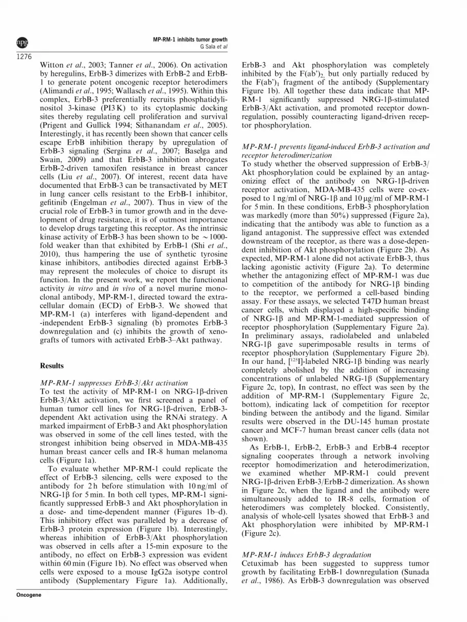
MP-RM-1 suppresses ErbB-3/Akt activationTo test the activity of MP-RM-1 on NRG-1b-drivenErbB-3/Akt activation, we first screened a panel ofhuman tumor cell lines for NRG-1b-driven, ErbB-3-dependent Akt activation using the RNAi strategy. Amarked impairment of ErbB-3 and Akt phosphorylationwas observed in some of the cell lines tested, with thestrongest inhibition being observed in MDA-MB-435human breast cancer cells and IR-8 human melanomacells (Figure 1a).
To evaluate whether MP-RM-1 could replicate theeffect of ErbB-3 silencing, cells were exposed to theantibody for 2 h before stimulation with 10 ng/ml ofNRG-1b for 5min. In both cell types, MP-RM-1 signi-ficantly suppressed ErbB-3 and Akt phosphorylation ina dose- and time-dependent manner (Figures 1b–d).This inhibitory effect was paralleled by a decrease ofErbB-3 protein expression (Figure 1b). Interestingly,whereas inhibition of ErbB-3/Akt phosphorylationwas observed in cells after a 15-min exposure to theantibody, no effect on ErbB-3 expression was evidentwithin 60min (Figure 1b). No effect was observed whencells were exposed to a mouse IgG2a isotype controlantibody (Supplementary Figure 1a). Additionally,
ErbB-3 and Akt phosphorylation was completelyinhibited by the F(ab’)2, but only partially reduced bythe F(ab’)1 fragment of the antibody (SupplementaryFigure 1b). All together these data indicate that MP-RM-1 significantly suppressed NRG-1b-stimulatedErbB-3/Akt activation, and promoted receptor down-regulation, possibly counteracting ligand-driven recep-tor phosphorylation.
MP-RM-1 prevents ligand-induced ErbB-3 activation andreceptor heterodimerizationTo study whether the observed suppression of ErbB-3/Akt phosphorylation could be explained by an antag-onizing effect of the antibody on NRG-1b-drivenreceptor activation, MDA-MB-435 cells were co-ex-posed to 1 ng/ml of NRG-1b and 10 mg/ml of MP-RM-1for 5min. In these conditions, ErbB-3 phosphorylationwas markedly (more than 50%) suppressed (Figure 2a),indicating that the antibody was able to function as aligand antagonist. The suppressive effect was extendeddownstream of the receptor, as there was a dose-depen-dent inhibition of Akt phosphorylation (Figure 2b). Asexpected, MP-RM-1 alone did not activate ErbB-3, thuslacking agonistic activity (Figure 2a). To determinewhether the antagonizing effect of MP-RM-1 was dueto competition of the antibody for NRG-1b bindingto the receptor, we performed a cell-based bindingassay. For these assays, we selected T47D human breastcancer cells, which displayed a high-specific bindingof NRG-1b and MP-RM-1-mediated suppression ofreceptor phosphorylation (Supplementary Figure 2a).In preliminary assays, radiolabeled and unlabeledNRG-1b gave superimposable results in terms ofreceptor phosphorylation (Supplementary Figure 2b).In our hand, [125I]-labeled NRG-1b binding was nearlycompletely abolished by the addition of increasingconcentrations of unlabeled NRG-1b (SupplementaryFigure 2c, top), In contrast, no effect was seen by theaddition of MP-RM-1 (Supplementary Figure 2c,bottom), indicating lack of competition for receptorbinding between the antibody and the ligand. Similarresults were observed in the DU-145 human prostatecancer and MCF-7 human breast cancer cells (data notshown).
As ErbB-1, ErbB-2, ErbB-3 and ErbB-4 receptorsignaling cooperates through a network involvingreceptor homodimerization and heterodimerization,we examined whether MP-RM-1 could preventNRG-1b-driven ErbB-3/ErbB-2 dimerization. As shownin Figure 2c, when the ligand and the antibody weresimultaneously added to IR-8 cells, formation ofheterodimers was completely blocked. Consistently,analysis of whole-cell lysates showed that ErbB-3 andAkt phosphorylation were inhibited by MP-RM-1(Figure 2c).
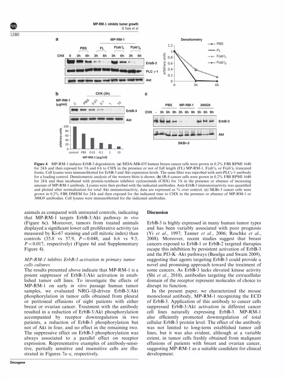
MP-RM-1 induces ErbB-3 degradationCetuximab has been suggested to suppress tumorgrowth by facilitating ErbB-1 downregulation (Sunadaet al., 1986). As ErbB-3 downregulation was observed
MP-RM-1 inhibits tumor growthG Sala et al
1276
Oncogene

after 60min of exposure to MP-RM-1 (Figure 1b), weinvestigated whether this effect could reflect the anti-body-mediated degradation of the receptor. To this end,MDA-MB-435 cells were analyzed for surface expres-sion of ErbB-3 after exposure to the antibody fordifferent times. As shown (Figure 3a), a time-dependentreduction of the membrane expression of ErbB-3 wasobserved, suggesting receptor internalization. Cell ima-ging studies showed that MP-RM-1 internalized, asrevealed by its co-localization with the early endosomemarker, EEA1 (Figure 3b). To clarify whether theinternalized MP-RM-1 antibody was still bound to thereceptor, an anti-ErbB-3 antibody (C-17) recognizingthe C-terminal residue, not competing with MP-RM-1,was used to track the receptor. As shown in Figure 3c,tracking ErbB-3 with C-17, while confirming MP-RM-1-induced receptor internalization, documented intra-cellular co-localization of the two antibodies, indicatingthat MP-RM-1 was still bound to the receptor.
To verify whether the decreased expression/internali-zation of the receptor was followed by protein degrada-tion, cells were chased with the protein-synthesisinhibitor cycloheximide (CHX) for 3 or 6 h in thepresence or absence of the antibody. As shown in
Figure 4a, receptor expression was reduced by approxi-mately eightfold as compared with control (5 vs 40%)after 3 h, whereas it was abrogated after 6 h. The effectof the antibody on ErbB-3 degradation was dosedependent (Figure 4b). Similar results were obtainedwhen intact antibody molecule was replaced by F(ab’)2,but not by F(ab’)1 (Figure 4a). Notably, cells treatedwith MP-RM-1, in the absence of NRG-1b and CHX,showed a time-dependent downregulation of ErbB-3,which represents the ‘net’ effect of the antibody onErbB-3 protein level (Supplementary Figure 2d).
The ability of MP-RM-1 to induce ErbB-3 degrada-tion was confirmed in SKBr-3 human breast cancercells (Figure 4c). The effect was specific as it was notobserved upon exposure of the cells to the unrelatedantibody, that is, the anti-ErbB2 antibody 300G9(Digiesi et al., 1992).
MP-RM-1 suppresses ligand-independent activationof ErbB-3ErbB-3 has recently been shown to associate with and tobe trans-activated by the tyrosine kinase receptor METin a ligand-independent manner (Engelman et al., 2007).
p-ErbB-3
p-Akt
ErbB-3
Akt
4mut siErbB-3NRG-1�
NRG-1�4mut
p-ErbB-3
ErbB-3
Akt
p-Akt
IR-8
ErbB-3
p-ErbB-3
p-Akt
Akt
MP-RM-1(min)
5 min NRG-1�C
MDA-MB-435
MDA-MB-435
5 min NRG-1�
MP-RM-1(�g/ml)
p-ErbB-3
ErbB-3
p-Akt
Akt
MDA-MB-435
Car
bit
rary
un
its
MP-RM-1 (�g/ml)
arb
itra
ry u
nit
s
p-ErbB-3/ErbB-3
pAkt/Akt ErbB-3
p-ErbB-3
p-Akt
Akt
IR-8
+-+-+-+-
siErbB-3
1206015- -
1010.10.050.01PBS
C
MP-RM-1 (�g/ml)1010.10.050.01PBS
Figure 1 NRG-1b-dependent ErbB-3/Akt activation is inhibited by MP-RM-1. (a) pSuper 4Mut and shErbB-3 infected MDA-MB-435 and IR-8 cells were grown in 0.2% FBS RPMI 1640 for 24 h and then stimulated with 10 ng/ml of NRG-1b for 5min before lysis.Cell lysates where then probed with the indicated antibodies. (b) MDA-MB-435 cells were grown in 0.2% FBS RPMI 1640 for 24 h andthen incubated with 10 mg/ml of MP-RM-1 for the indicated time before NRG-1b stimulation. Cell lysates were immunoblotted forphosphorylated ErbB-3 and Akt expression levels. The same filter was reprobed with anti-ErbB-3 and anti-Akt antibodies for a loadingcontrol. (c) MDA-MB-435 cells were grown in 0.2% FBS RPMI 1640 for 24 h and then incubated with the indicated amount of MP-RM-1 for 2 h before NRG-1b stimulation. Cell lysates were immunoblotted for phosphorylated ErbB-3 and Akt expression levels. Thesame filter was reprobed with anti-ErbB-3 and anti-Akt antibodies for a loading control. Anti-p-ErbB-3 and anti-p-Aktimmunoreactivity was quantified and plotted after normalization for total ErbB-3 and Akt immunoreactiviy (right panels). (d) IR-8human melanoma cancer cells were grown in 0.2% FBS RPMI 1640 for 24 h and then incubated with the 10 mg/ml of MP-RM-1 for 2 hbefore NRG-1b stimulation. Cell lysates were immunoblotted for phosphorylated ErbB-3 and Akt expression levels. The same filterwas reprobed with anti-ErbB-3 and anti-Akt antibodies for a loading control.
MP-RM-1 inhibits tumor growthG Sala et al
1277
Oncogene

To evaluate whether MP-RM-1 could disrupt MET/ErbB-3 signaling axis, we used the MKN-45 humangastric cancer cells, which exhibit constitutive highlevels of MET, ErbB-3 and Akt phosphorylation(Figures 5a–c). Moreover, MKN-45 cells do not expressthe ErbB-3 ligands, NRG-1b and Betacellulin (Wuet al., 2009), indicating the absence of an autocrine loopactivating ErbB-3 in these cells. Silencing of ErbB-3, inthese cells, markedly inhibited ligand-independentErbB-3 and Akt phosphorylation (Figure 5a), as didMP-RM-1 in a time-dependent manner (Figure 5b).Interestingly, the MET inhibitor SU11274 paralleled theeffect of MP-RM-1, indicating that the MET/ErbB-3/Akt signaling cascade is functionally active in these cells.Notably, MP-RM-1 was as effective as SU11274 orErbB-3 silencing in inhibiting ErbB-3 and Akt activa-tion (Figure 5c), whereas no effect was seen by treatingcells with an antibody to ErbB-2 (trastuzumab) or toErbB-1 (cetuximab) (Figure 5c and data not shown).Next, we tested the ability of MP-RM-1 to disruptErbB-3/MET association in MKN-45 cells. As shown inFigure 5d, exposure of the cells to the antibody caused anearly complete disappearance of ErbB-3 after immu-noprecipitation with an anti-phospho MET antibody.Interestingly, despite the presence of the MET inhibitor,SU1174, exogenous NRG-1b was still able to re-activatethe ErbB-3/Akt axis, but not in the presence of MP-RM-1 (Figure 5e). The ability of MP-RM-1 to inhibitligand-independent ErbB-3/Akt phosphorylation wasconfirmed in ErbB-2 overexpressing SKBr-3 human
breast cancer cells (Supplementary Figure 3a). Takentogether these data suggest that MP-RM-1 targets bothMET-dependent and ligand-independent ErbB-3/Aktsignaling axis.
MP-RM-1 inhibits colony formation and growth of tumorxenografts in nude miceHaving demonstrated that MP-RM-1 is able to inhibitErbB-3/Akt activation, we wondered whether thisinhibition could translate into tumor growth inhibition.As illustrated (Supplementary Figure 3b), MP-RM-1at concentrations of 10 mg/ml significantly inhibitedcolony formation ability of IR-8 cells as compared withuntreated control cultures. Untreated cells produced26±47 colonies/field, and the colony numbers weresuppressed to 15±28 (Po0.001).
To assess the antitumor activity of MP-RM-1 as asingle agent in vivo, we used IR-8 human melanoma cellsand DU-145 human prostate cancer cells, both expres-sing ErbB-3 and sensitive in vitro to the inhibitory effectof the antibody on NRG-1b stimulated ErbB-3/Aktactivation (Supplementary Figure 3c), as xenografttumor models in nude mice. Treatment of the animalswith MP-RM-1 at 20mg/kg resulted in a significanttumor growth delay in both tumor models (Figures 6aand b). In separate experiments, animals harboring IR-8xenografts (300mm3) were treated with a single injectionof the antibody (20mg/kg) and killed 4 h thereafter.A significant reduction of ErbB-3 expression and Aktphosphorylation was seen in tumors from treated
p-ErbB-3
ErbB-3
-
Akt
p-Akt
ErbB-3 phosphorylation
NRG-1� ng/ml
MP-RM-1 �g/ml
ErbB-3
IP �-ErbB-2
ErbB-2
IgG
p-Akt
Lysates
Akt
p-ErbB-3
ErbB-3
+-
-
-
-
-
- ++
+
10101010
101-- 100
NRG-1�
MP-RM-1
Figure 2 MP-RM-1 impairs ligand-driven ErbB-3 activation and receptor heterodimerization. (a) MDA-MB-435 cancer cells weregrown in 0.2% FBS RPMI 1640 for 24 h and then simultaneously incubated with 1 ng/ml of NRG-1b and 10 mg/ml of MP-RM-1 for5min. Cells were then lysed and analyzed for phosphorylated ErbB-3. The same filter was reprobed with anti-ErbB-3 for a loadingcontrol. Densytometric analysis of the western blots is shown. (b) MDA-MB-435 cancer cells were grown in 0.2% FBS RPMI 1640 for24 h and then simultaneously incubated with 10 ng/ml of NRG-1b and the indicated amount of MP-RM-1 for 5min. Cells were thenlysed and analyzed for phosphorylated Akt. The same filter was reprobed with anti-Akt antibody for a loading control. (c) IR-8 humanmelanoma cancer cells were grown in 0.2% FBS RPMI 1640 for 24 h and then simultaneously incubated with 1 ng/ml of NRG-1b and10 mg/ml of MP-RM-1 for 5min. Lysates were analysed by immunoblot with the indicated antibodies (right panel) or lysates wereimmunoprecipitated with an anti-ErbB-2 antibody (300G9) and then probed with an anti-ErbB-3 antibody (left panel). Ponceaustaining of 300G9 is shown.
MP-RM-1 inhibits tumor growthG Sala et al
1278
Oncogene

Figure 3 MP-RM-1 reduces surface expression of the receptor and induces ErbB-3 internalization. (a) FACS analysis of MDA-MB-435 cells incubated with 10 mg/ml of MP-RM-1 for 30min on ice and placed back at 37 1C for 30, 60 and 120min. The histogramsrepresent the percentage of mean fluorescence referred to control cells (maintained 30min on ice). (b) Confocal images of IR-8 cells.After 24 h of serum deprivation, cells were maintained on ice in the presence of 10mg/ml of MP-RM-1 for 30min and placed back at37 1C for 60min. Cell nuclei are shown in blue. Bars: 10mm. (c) Serum-straved IR-8 cells were maintained in the presence or absence of10mg/ml of MP-RM-1 for 30min on ice and placed back at 37 1C for 60min. ErbB-3 localization was tracked using a specific rabbitantibody raised against the C-terminal residue of the receptor (C-17). MP-RM-1 and C-17 were visualized with an anti-mouse (green)and an anti-rabbit antibody (red), respectively. Cell nuclei are shown in blue. Bars: 10 mm.
MP-RM-1 inhibits tumor growthG Sala et al
1279
Oncogene
animals as compared with untreated controls, indicatingthat MP-RM-1 targets ErbB-3/Akt pathway in vivo(Figure 6c). Moreover, tumors from treated animalsdisplayed a significant lower cell proliferative activity (asmeasured by Ki-67 staining and cell mitotic index) thancontrols (35.8 vs 57.9, P¼ 0.048, and 6.6 vs 9.5,P¼ 0.017, respectively) (Figure 6d and SupplementaryFigure 4).
MP-RM-1 inhibits ErbB-3 activation in primary tumorcells culturesThe results presented above indicate that MP-RM-1 is apotent suppressor of ErbB-3/Akt activation in estab-lished tumor cell lines. To investigate the effects ofMP-RM-1 on early in vitro passage human tumorsamples, we evaluated NRG-1b-driven ErbB-3/Aktphosphorylation in tumor cells obtained from pleuralor peritoneal effusions of eight patients with eitherbreast or ovarian cancer. Treatment with the antibodyresulted in a reduction of ErbB-3/Akt phosphorylationaccompanied by receptor downregulation in twopatients, a reduction of ErbB-3 phosphorylation butnot of Akt in four, and no effect in the remaining two.The suppressive effect on ErbB-3 phosphorylation wasalways associated to a parallel effect on receptorexpression. Representative examples of antibody-sensi-tive, partially sensitive and insensitive cells are illu-strated in Figures 7a–c, respectively.
Discussion
ErbB-3 is highly expressed in many human tumor typesand has been variably associated with poor prognosis(Yi et al., 1997; Tanner et al., 2006; Reschke et al.,2008). Moreover, recent studies suggest that breastcancers exposed to ErbB-1 or ErbB-2 targeted therapiesescape this inhibition by persistent activation of ErbB-3and the PI3-K–Akt pathways (Baselga and Swain 2009),suggesting that agents targeting ErbB-3 could provide anovel and promising approach toward the treatment ofsome cancers. As ErbB-3 lacks elevated kinase activity(Shi et al., 2010), antibodies targeting the extracellulardomain of the receptor represent molecules of choice todisrupt its function.
In the present paper, we characterized the mousemonoclonal antibody, MP-RM-1 recognizing the ECDof ErbB-3. Application of this antibody to cancer cellssuppressed ErbB-3/Akt activation in different cancercell lines naturally expressing ErbB-3. MP-RM-1also efficiently promoted downregulation of totalcellular ErbB-3 protein level. The effect of the antibodywas not limited to long-term established tumor celllines, but it was also evident, although at a variableextent, in tumor cells freshly obtained from malignanteffusions of patients with breast and ovarian cancer,suggesting MP-RM-1 as a suitable candidate for clinicaldevelopment.
PBS
FL
F(ab')1
F(ab')2
PLC �-1
FL F(ab')2PBS
ErbB-3
Akt
MP-RM-1
CHX
Densitometry
CHX
ErbB-3
Akt
PBS MP-RM-1 300G9
SKBr-3
MP-RM-1(�g/ml)
CHX (3h)
ErbB-3
Akt
6h3h6h3h6h3h6h3h0
- 3h 6h 9h 3h 6h 9h 3h 6h 9h
F(ab')1
Figure 4 MP-RM-1 induces ErbB-3 degradation. (a) MDA-MB-435 human breast cancer cells were grown in 0.2% FBS RPMI 1640for 24 h and then exposed for 3 h and 6 h to CHX in the presence or not of full length (FL) MP-RM-1, F(ab’)1 or F(ab’)2 truncatedforms. Cell lysates were immunoblotted for ErbB-3 and Akt expression levels. The same filter was reprobed with anti-PLCg-1 antibodyfor a loading control. Densitometric analysis of the western blots is shown. (b) IR-8 cancer cells were grown in 0.2% FBS RPMI 1640for 24 h and then incubated with protein-synthesis inhibitor cycloeximide (CHX) for 3 h in the presence or absence of increasingamount of MP-RM-1 antibody. Lysates were then probed with the indicated antibodies. Anti-ErbB-3 immunoreactivity was quantifiedand plotted after normalization for total Akt immunoreactiviy, data are expressed as % over control. (c) SKBr-3 cancer cells weregrown in 0.2% FBS DMEM for 24 h and then exposed for the indicated time to CHX in the presence or absence of MP-RM-1 or300G9 antibodies. Cell lysates were immunoblotted for the indicated antibodies.
MP-RM-1 inhibits tumor growthG Sala et al
1280
Oncogene

Suppression of receptor activation and promotion ofits downregulation have previously been demonstratedto mediate the antitumor activity of therapeuticantibodies targeting ErbB receptors. The anti-ErbB-1antibody cetuximab inhibits ligand-mediated phos-phorylation of ErbB-1 by directly competing with ligandbinding to ErbB-1 (Prewett et al., 1998). The anti-ErbB-2 monoclonal antibody pertuzumab stericallyhinders recruitment of ErbB-2 into ErbB/ligandcomplexes impairing the formation and activation ofErbB-2-containing dimers (Franklin et al., 2004). In thiscontext, our in vitro experiments (Figure 2) suggest thatMP-RM-1 does antagonize ligand-driven activation ofErbB-3 and prevents ErbB-2/ErbB-3 dimerization. Themechanism underlying these antagonizing effects ofMP-RM-1 is not yet understood, but it does notinvolve competition with receptor ligand, as the anti-body was unable to compete with NRG-1b for bindingto ErbB-3. Another interesting feature of MP-RM-1lies in its ability to block the activation of MET-dependent ErbB-3/Akt signaling (Figure 5). This isparticularly important at the light of evidence that cellsbecoming resistant to the ErbB-1 inhibitors displayconstitutive activation/amplification of MET, whichin turn re-activates ErbB-3/Akt axis (Engelmanet al., 2007).
The effect of MP-RM-1 on tumor growth was alsoassessed in this study. The antibody exerted a significanttumor growth inhibitory activity in vitro by decreasingthe anchorage-independent growth of IR-8 humanmelanoma cells. Moreover, when given as a single-agenttherapy, it led to a significant growth delay of xenograftsof melanoma (IR-8) and hormone refractory prostatecancer (DU-145). Of note, tumors from MP-RM-1-treated animals displayed a decreased ErbB-3/Aktphosphorylation and ErbB-3 expression, suggesting thatthe antibody targets ErbB-3/Akt signaling pathway alsoin vivo. The therapeutic activity on melanoma is ofparticularly relevance, as this cancer which is oftenresistant to currently available therapies, frequentlyoverexpresses ErbB-3 (Reschke et al., 2008) and MET(Natali et al., 1993). An increased ErbB-3 expression hasalso been documented in prostate cancer as comparedwith normal prostate, and its induction has beenobserved after progression to hormone independency(Chen et al., 2010). Thus, potential future therapeuticapplication of MP-RM-1 includes both these neoplasms.In this respect, it is noteworthy that the F(ab’)2 form ofMP-RM-1 is sufficient to inhibit ligand-driven activa-tion of ErbB-3 and to promote receptor downregula-tion, implicating that the Fc region of the antibody isnot necessary for its activity (Figure 4a and Supplemen-
Figure 5 MP-RM-1inhibits ligand-indipendent activation of ErbB-3. (a) pSuper 4Mut and shErbB-3 infected MKN-45 cancer cellswere grown in serum-free DMEM for 24 h and then lysed for immunoblot analysis. (b) MKN-45 cells were grown in serum-freeDMEM for 24 h and then exposed to MP-RM-1 for the indicated time. Lysates were analyzed by immunoblot for phosphorylatedErbB-3 and phosphorylated Akt. The same filter was reprobed with the indicated antibodies for a loading control. (c) MKN-45 cellswere grown in serum free DMEM for 24 h and then incubated for 2 h with trastuzumab (10mg/ml), MP-RM-1 (10mg/ml) or increasingamount of MET inhibitor SU11274. Lysates were analyzed by immunoblot with the indicated antibodies. (d) MKN-45 cells weregrown in serum-free DMEM for 24 h and then incubated with 10mg/ml of MP-RM-1 for 15 and 120min. Lysates were analysed byimmunoblot with the indicated antibodies (lower panel) or immunoprecipitated with an anti-phosphorylated MET antibody and thenprobed with an anti-ErbB-3 antibody (upper panel). (e) MKN-45 cells were grown in serum free DMEM for 24 h and then incubatedwith 1 mg/ml of SU1174 alone or together with 10mg/ml of MP-RM-1. After 2 h, NRG-1b (10 ng/ml) was added, as indicated.
MP-RM-1 inhibits tumor growthG Sala et al
1281
Oncogene

tary Figure 1b). Because of the delivery advantage andlarger volume of distribution of F(ab’)2 compared withwhole IgG molecule, and these data showing antibodyinternalization (Figure 3 and Supplementary Figure 1c),MP-RM-1 could be considered as an excellent vehicle todeliver toxins or drugs to cancer cells expressing ErbB-3.Finally, differently from what observed in transformedcells, ErbB-3 membrane expression as detected withMP-RM-1 was never observed in normal human tissues.
On these substrates, the antibody produced a cytoplas-mic staining pattern confined to few cell lineages andnotably absent from the cardiac muscle (Natali et al., inpreparation), thus suggesting low levels of toxicity oncethe antibody is administered to humans.
The therapeutic potential of a fully human mono-clonal antibody targeting ErbB-3, MM-121, hasrecently been reported (Schoeberl et al., 2009, 2010).The antibody proved to effectively inhibits ErbB-3
Figure 6 MP-RM-1 reduces the expression of ErbB-3 and tumor cell division. Exponentially growing IR-8 (a) and DU-145 (b) cellswere injected into the right flank of the recipient mice. Tumor growth was assessed, as described in Materials and methods section.(c) Western blotting analysis of tumor lysates of IR-8 xenografts excised after 4 h of treatment with 20mg/kg MP-RM-1 or PBS.(d) The mitotic activity, as assessed by Ki-67 immunohistochemical staining and mitotic figure counts, is lower in tumors from micereceiving MP-RM-1 than in controls.
p-ErbB-3
ErbB-3
p-ErbB-3
a b c
ErbB-3
p-ErbB-3
ErbB-3
p-Akt
Akt
p-Akt
Akt
p-Akt
Akt
Figure 7 MP-RM-1inhibits NRG-1b -induced ErbB-3/Akt axis in patient-derived cancer cells. Short-term cultured patient-derivedcarcinoma cells were serum starved for 24 h and then incubated with 10 mg/ml of MP-RM-1 for 2 h before NRG-1b stimulation. Cellswere then lysed and analyzed for phosphorylated ErbB-3 and Akt. The same filter was reprobed with anti-ErbB-3 and anti-Akt and fora loading control. Examples of antibody-sensitive (a), partially sensitive (b) and insensitive (c) cells are shown.
MP-RM-1 inhibits tumor growthG Sala et al
1282
Oncogene

phosphorylation across multiple cancer cell lines includ-ing lung, ovarian, prostate, renal, breast and colon, andit is currently in clinical development. Although bothantibodies appear to exert inhibitory functions in vitroand in vivo, MM-121 displays a remarkable higheraffinity (Kd, 0.76� 10�9m vs 3.27� 10�8m). Thissuggests that a higher affinity may not be a necessarilydesirable feature of antibodies to be used as antitumoragents in vivo. In fact, according to the ‘binding-sitebarrier’ hypothesis, macromolecular ligands could beprevented from penetrating tumors by the very fact oftheir successful strong binding to the target receptor(Weinstein and van Osdol 1992). Furthermore differ-ently from MM-121, MP-RM-1 is capable of inhibitingboth ligand-dependent and -independent activation ofErbB-3–Akt pathway, which may potentially expand thetherapeutic use of this antibody. Finally, no F(ab’)2activity has been documented for MM-121 antibody.
In conclusion, our data indicate that MP-RM-1 hasthe potential to interfere with ErbB-3/Akt activa-tion and point out new possibilities for therapeuticintervention.
Materials and methods
MP-RM-1 generationFour-weeks-old Balb/c mice were immunized by intraperito-neal injection of live NIH3T3 cells transfected with the humanErbB-3 receptor-coding sequence. After 7 days, mice weregiven an additional intraperitoneal injection of the immuno-gen. After additional 7 days, mice were boosted intravenouslywith the immunogen, and spleens were removed for cell fusion3 days later. Somatic cell hybrids were prepared by fusion ofimmune splenocytes with the murine non-secreting myelomacell line NS-1. Hybridoma supernatants were screened byELISA and selected on the basis of differential reactivity withthe cell surface LTR-ErbB3 NIH/3T3 transfected cells, but notwith LTR-neo NIH/3T3. All ErbB3-positive hybridoma cellcultures were cloned twice by limiting dilution and furthercharacterized. Potential cross-reactivity of hybridoma super-natants with other ErbB receptors were evaluated by a cell-based ELISA and immunoprecipitation using NIH3T3 cellstransfected with individual human ErbB receptor-codingsequences as recombinant antigen source. NIH3T3 cellstransfected with expression vectors for human codingsequences of ErbB family receptors have previously beendescribed (Di Fiore et al., 1987a, 1987b; Kraus et al., 1993;Baulida et al., 1996). Of B1000 hybridoma clones screened,one, MP-RM-1, recognized the ECD of ErbB3 but not that ofthe other ErbB family members (ErbB-1, ErbB-2 and ErbB-4),when used in a cell-based ELISA and immunoprecipitation.Using surface plasmon resonance, the dissociation constant(Kd) for MP-RM-1 was determined to be 3.27� 10�8m.The hybridoma murine cell line producing MP-RM-1
antibody was deposited at the DSMZ (Deutsche Sammlungvon Mikroorganismen und Zellkulturen) and designated DSMACC3018.
Materials and constructsSequence targeting the human ErbB-3 mRNA (Lee-Hoeflichet al., 2008) was subcloned into pSuper vectors. Control vectorpSuper 4Mut contains a four-point mutated sequence unable totarget the human ErbB-3 mRNA (Sala et al., 2008). Silencing of
ErbB-3 was obtained by using pSuper retro-based vectors toexpress stable RNA (shRNA). Retroviral stocks were generatedas described (Anastasi et al., 2005). Antibodies were as follows:phosphorylated ErbB-3 (Tyr1289), phosphorylated Akt (Ser473),Akt, PLCg1, phosphorylated MET (Tyr1234�1235) from CellSignaling (Boston, MA, USA); C-17 ErbB-3 and MET fromSanta Cruz Biotechnology (Santa Cruz, CA, USA); Actin fromSigma-Aldrich (St Louis, MO, USA); anti-ErbB-2 antibody300G9 was obtained as described in Digiesi et al. (1992)rhodamine[125I]-labeled phalloidin from Sigma; Draq5 fromAlexis; Yo-Pro dye from Invitrogen (Carlsbad, CA, USA).MET inhibitor SU11274 was purchased from Biaffin GmbH &Co KG (http://www.proteinkinase.de).
Cell linesMDA-MB-435, T47D and SKBr-3 human breast cancer cells,DU-145 human prostate cancer cells were purchased fromATCC. The cutaneous melanoma cell line IR-8 (Leonetti et al.,1996) was kindly provided by Dr Carlo Leonetti (‘Regina Elena’National Cancer Institute, Rome, Italy). Gastric carcinoma cellline MKN-45 (Rege-Cambrin et al., 1992) was kindly providedby Dr Sergio Anastasi (‘Regina Elena’ National CancerInstitute, Rome, Italy). MDA MB 435, DU-145 and IR-8 celllines were grown in RPMI 1640 medium, SKBr-3 and MKN-45were grown in DMEM medium supplemented with 10% fetalbovine serum (FBS), penicillin, and streptomycin and incubatedin 5% CO2 at 37 1C.
ImmunochemistryProteins were solubilized in HNTG buffer (50mM HEPES pH7.5, 150mM NaCl, 10% glycerol, 1% Triton X-100, 5mM
EGTA). Immunoblotting was performed as described inFiorini et al. (2002). Proteins from blots were quantified usingImage J software program (http://rsbweb.nih.gov/ij/).
Fluorescence-activated cell sorting analysisFor ErbB-3 surface expression analysis MDA-MB-435 cells havebeen incubated on ice in the presence of 1mg/ml of MP-RM-1 for30min and then returned to 37 1C for the indicated time,harvested and stained with a fluorescent goat anti-mousesecondary antibody. After staining procedures, samples wereanalyzed by flow cytometry using a FACSCalibur cytometer(Becton Dickinson, Franklin Lakes, NJ, USA). Finally, data wereanalyzed using CELLQuest 3.2.1.f1 software (Becton Dickinson).
Confocal microscopyMicroscopy was performed as described in Piccolo et al. (2002)using a Bio-Rad MRC-1024 confocal system (BioRad Labora-tories, Hercules, CA, USA). Briefly, cells has been plated in22� 22 coverslips and grown in 0.2% FBS RPMI 1640 for 24h.Thereafter, cells were incubated or not with 10mg/ml of MP-RM-1 for 30min on ice and placed back at 37 1C for 60min. Atthe end of the experiment, cells were fixed in 4% paraformalde-hyde, permeabilized with 0.2% Triton phosphate-buffered saline(PBS) and then stained with fluorescently labeled secondaryantibodies as indicated. Alexa-Fluor-568 monoclonal antibodylabeling kit was purchased from Molecular Probes (Invitrogen).Rhodamine-labeled phalloidin and Draq5 (or Yo-Pro dye) wereused to visualize actin cytoskeleton and nuclei, respectively. Nofurther processing of the images was done except for changes inbrightness/contrast to better visualize the data.
[125I]-labeled NRG-1b-binding studiesNRG-1b zwas [125I]-labeled using the IODO-GEN kit accord-ing to the supplier’s protocol (Thermo Fisher Scientific,
MP-RM-1 inhibits tumor growthG Sala et al
1283
Oncogene
Illkirch Cedex, France). Specific activity was about 15mCi/mg.Cells were plate at 5� 104 in 12-well plates, serum starved for24 h and then incubated with 10 ng/ml of [125I]-labeled NRG-1bin binding medium (BM; that is, DME containing 0.2%bovine serum albumin and 20mM Hepes, pH 7.5) at 4 1C for60min in the absence or presence of NRG-1b and MP-RM-1.The free [125I]-labeled NRG-1b was eliminated washing threetimes with ice-cold PBS. The cells were lysed in 0.3ml of lysisbuffer (1% sodium dodecyl sulfate and 10mM NaOH), and theradioactivity was measured by a gamma counter.
Soft-agar colony formation assayFor the anchorage-independent growth assay, 15� 103 IR-8cells were suspended in 0.3% agarose medium containingRPMI 1640 10% FBS and layered onto a 2ml bed of 0.5%agarose in a six-well plate dish. Dishes were incubated at 37 1Cin a humidified atmosphere containing 5% CO2. A measure of10 mg/ml MP-RM-1 or PBS was added to cells every alternateday and the number of colonies was determined (under lightmicroscopy) after 10 days.
In vivo tumor growthAthymic CD-1 nu/nu mice (7-weeks old) and BALB/c micewere purchased from Charles River Laboratories and main-tained under specific pathogen-free conditions with food andwater provided ad libitum. The animals health status wasmonitored daily. Procedure involving animals and their carewere conducted according to the institutional guidelinesin compliance with national and international laws andpolicies.IR-8 and DU-145 xenografts were generated by s.c. into the
right flank of 3� 106 IR-8 cells and 5� 106 DU-145 cells in200 ml PBS, respectively. When xenografts were palpable,animals were divided into two groups (10 animals each)selected to provide a similar range in tumor size in each group.One group received i.p. injection twice weekly of 20mg/kgMP-RM-1 in PBS, whereas the other received PBS only(control group). Tumor volume was monitored every day by acaliper and volumes were calculated using the followingformula: tumor volume (mm3)¼ (length�width2)/2. At theend of the study, mice were anesthetized with isofluraneinhalation before cervical dislocation. Tumors were excisedand snap frozen for immunohistochemical analysis or analyzedfor western blotting as previously described (Sala et al.,2008).
Tumor mitotic index and Ki-67 expression analysisFormalin-fixed and paraffin-embedded tissues from xenograftsDU-145 human prostate cancer cells of untreated mice (n¼ 7)and treated mice (n¼ 8) were sectioned at 5mm and stainedwith Hematoxylin and Eosin (H&E). For the evaluation of themitotic figures, the sections were scanned at low magnification(� 5). Four high-power fields (� 40; 0.17mm2 field) of tumorwith the greatest number of mitotic figures were then selectedand used for the statistical analysis. Tissue microarrays (TMA)were also constructed by extracting 2-mm diameter core ofhystologically confirmed DU-145 human prostate cancer cellsareas from each original mouse paraffin block and re-embedding these cores into gridded paraffin blocks using aTMA precision instrument (Beecher Instruments, Sun Prairie,WI, USA). TMA sections were incubated—after antigenretrieval by thermostatic bath at 96 1C in 10mM/l citratebuffer, pH 6 for 40min—with the anti-Ki-67 monoclonalantibody MIB-1 (Dako, Glostrup, Denmark) for 30min atroom temperature. The immunoreactions were revealed by a
streptavidin-biotin-enhanced peroxidase system (Super Sensi-tive Link-Label IHC DetectionSystem; BioGenex, Space,Milan, Italy). Positive and negative controls were includedfor each antibody and in each batch of staining. Meandifferences in mitotic count and Ki-67 expression werecompared with the use of the paired t test. SPSS Version15.0 (SPSS, Chicago, IL, USA) was used and Po0.05 wasconsidered statistically significant.
ImmunohistochemistryFrozen normal human adult tissues belonged to the archivialbank of CINBO and were collected and used according to theEthic Committee guidelines. Sections (4 mm) were fixed inabsolute acetone for 10min and after air drying eitherimmediately processed or stored at �20 1C with no loss ofimmune reactivity for at least 6 months. For indirectimmunoperoxidase stain, sections were incubated overnightwith MP-RM-1 antibody (50 mg/ml). The immune reactivitywas detected by using the EnVision Peroxidase DetectionSystem. Specimens using aminoethylcarbazole as chromogenand finally counterstained with Mayer’s hematoxylin fornuclear stain. The experiments included appropriate negativeand positive controls. Immunohistochemical findings werescored as follows: negative, when no stain or dubious weakreactivity was observed, 1þ when the stain was homo-genous of moderate intensity, 2þ when the stain was homo-genously intense. Scores were carried out by two independentreaders.
Patient-derived carcinoma cellsFreshly collected tumor cells from pleural and peritonealeffusions appearing in patients free from therapy werepropagated in vitro for a limited number of passages usingRPMI 1640 medium supplemented with 10 % decomplemen-ted FBS, 2mm L-glutamine at 37 1C in a 5% C02/95% airatmosphere. Tumor cells harvesting and processing beforein vitro growth as well immuno phenotyping, were carried outas described (Mottolese et al., 1988).
Statitstical analysisMean volumes of treated and untreated xenografts werecompared using an unpaired t test (Student’s t test) consideringas statistically significant a P-value o0.05 (CI 95%). Meandifferences in mitotic count and Ki-67 expression werecompared with the use of the paired t test. SPSS Version15.0 (SPSS) was used and Po0.05 was considered statisticallysignificant.
Conflict of interest
The authors declare no conflict of interest.
Acknowledgements
We thank Dr F Petronzelli (Sigma-Tau Pomezia, Italy) for themeasurement of MP-RM-1 affinity by surface plasmonicresonance; Dr O Segatto, Dr S Anastasi and Dr M Sallesefor advice and reagents, R La Sorda and MR Nicotra forthe immunohistochemical analysis, Dr A Sala and ProfessorV De Laurenzi for discussion. PGN and MP partiallysupported by AIRC.
MP-RM-1 inhibits tumor growthG Sala et al
1284
Oncogene

References
Alimandi M, Romano A, Curia MC, Muraro R, Fedi P, Aaronson SAet al. (1995). Cooperative signaling of ErbB3 and ErbB2 inneoplastic transformation and human mammary carcinomas.Oncogene 10: 1813–1821.
Anastasi S, Sala G, Huiping C, Caprini E, Russo G, Iacovelli S et al.(2005). Loss of RALT/MIG-6 expression in ERBB2-amplifiedbreast carcinomas enhances ErbB-2 oncogenic potency and favorsresistance to Herceptin. Oncogene 24: 4540–4548.
Baselga J, Swain SM. (2009). Novel anticancer targets: revisitingERBB2 and discovering ERBB3. Nat Rev Cancer 9: 463–475.
Baulida J, Kraus MH, Alimandi M, Di Fiore PP, Carpenter G. (1996).All ErbB receptors other than the epidermal growth factor receptorare endocytosis impaired. J Biol Chem 271: 5251–5257.
Bianchi S, Palli D, Falchetti M, Saieva C, Masala G, Mancini B et al.(2006). ErbB-receptors expression and survival in breast carcinoma:a 15-year follow-up study. J Cell Physiol 206: 702–708.
Chen L, Siddiqui S, Bose S, Mooso B, Asuncion A, Bedolla RG et al.(2010). Nrdp1-mediated regulation of ErbB3 expression by theandrogen receptor in androgen-dependent but not castrate-resistantprostate cancer cells. Cancer Res 70: 5994–6003.
Citri A, Yarden Y. (2006). EGF-ERBB signalling: towards the systemslevel. Nat Rev Mol Cell Biol 7: 505–516.
Di Fiore PP, Pierce JH, Fleming TP, Hazan R, Ullrich A, King CRet al. (1987a). Overexpression of the human EGF receptor confersan EGF-dependent transformed phenotype to NIH 3T3 cells. Cell
51: 1063–1070.Di Fiore PP, Pierce JH, Kraus MH, Segatto O, King CR, Aaronson
SA. (1987b). erbB-2 is a potent oncogene when overexpressed inNIH/3T3 cells. Science 237: 178–182.
Digiesi G, Giacomini P, Fraioli R, Mariani M, Nicotra MR, Segatto Oet al. (1992). Production and characterization of murine mAbs tothe extracellular domain of human neu oncogene productGP185HER2. Hybridoma 11: 519–527.
Engelman JA, Zejnullahu K, Mitsudomi T, Song Y, Hyland C, ParkJO et al. (2007). MET amplification leads to gefitinib resistance inlung cancer by activating ERBB3 signaling. Science 316: 1039–1043.
Fiorini M, Ballaro C, Sala G, Falcone G, Alema S, Segatto O. (2002).Expression of RALT, a feedback inhibitor of ErbB receptors, issubjected to an integrated transcriptional and post-translationalcontrol. Oncogene 21: 6530–6539.
Franklin MC, Carey KD, Vajdos FF, Leahy DJ, de Vos AM,Sliwkowski MX. (2004). Insights into ErbB signaling from thestructure of the ErbB2-pertuzumab complex. Cancer Cell 5:317–328.
Giampaglia M, Chiuri VE, Tinelli A, De Laurentiis M, Silvestris N,Lorusso V. (2010). Lapatinib in breast cancer: clinical experiencesand future perspectives. Cancer Treat Rev 36(Suppl 3): S72–S79.
Gschwind A, Fischer OM, Ullrich A. (2004). The discovery of receptortyrosine kinases: targets for cancer therapy. Nat Rev Cancer 4:361–370.
Hynes NE, Lane HA. (2005). ERBB receptors and cancer: thecomplexity of targeted inhibitors. Nat Rev Cancer 5: 341–354.
Hynes NE. (2007). Targeting ERBB receptors in cancer. Recent Results
Cancer Res 172: 45–57.Hynes NE, MacDonald G. (2009). ErbB receptors and signaling
pathways in cancer. Curr Opin Cell Biol 21: 177–184.Ishikawa N, Daigo Y, Takano A, Taniwaki M, Kato T, Hayama S
et al. (2005). Increases of amphiregulin and transforming growthfactor-alpha in serum as predictors of poor response to gefitinibamong patients with advanced non-small cell lung cancers. Cancer
Res 65: 9176–9184.Karamouzis MV, Badra FA, Papavassiliou AG. (2007). Breast cancer:
the upgraded role of HER-3 and HER-4. Int J Biochem Cell Biol 39:851–856.
Kraus MH, Fedi P, Starks V, Muraro R, Aaronson SA. (1993).Demonstration of ligand-dependent signaling by the erbB-3 tyrosinekinase and its constitutive activation in human breast tumor cells.Proc Natl Acad Sci USA 90: 2900–2904.
Lee-Hoeflich ST, Crocker L, Yao E, Pham T, Munroe X, Hoeflich KPet al. (2008). A central role for HER3 in HER2-amplifiedbreast cancer: implications for targeted therapy. Cancer Res 68:5878–5887.
Leonetti C, D0Agnano I, Lozupone F, Valentini A, Geiser T, Zon Get al. (1996). Antitumor effect of c-myc antisense phosphorothioateoligodeoxynucleotides on human melanoma cells in vitro and and inmice. J Natl Cancer Inst 88: 419–429.
Liu B, Ordonez-Ercan D, Fan Z, Edgerton SM, Yang X, Thor AD.(2007). Downregulation of erbB3 abrogates erbB2-mediated tamox-ifen resistance in breast cancer cells. Int J Cancer 120: 1874–1882.
Mendelsohn J, Baselga J. (2006). Epidermal growth factor receptortargeting in cancer. Semin Oncol 33: 369–385.
Mottolese M, Venturo I, Donnorso RP, Curcio CG, Rinaldi M, NataliPG. (1988). Use of selected combinations of monoclonal antibodiesto tumor associated antigens in the diagnosis of neoplastic effusionsof unknown origin. Eur J Cancer Clin Oncol 24: 1277–1284.
Natali PG, Nicotra MR, Di Renzo MF, Prat M, Bigotti A, CavaliereR et al. (1993). Expression of the c-Met/HGF receptor in humanmelanocytic neoplasms: demonstration of the relationship tomalignant melanoma tumour progression. Br J Cancer 68: 746–750.
Piccolo E, Innominato PF, Mariggio MA, Maffucci T, Iacobelli S,Falasca M. (2002). The mechanism involved in the regulation ofphospholipase Cgamma1 activity in cell migration. Oncogene 21:6520–6529.
Prewett M, Rothman M, Waksal H, Feldman M, Bander NH,Hicklin DJ. (1998). Mouse-human chimeric anti-epidermal growthfactor receptor antibody C225 inhibits the growth of humanrenal cell carcinoma xenografts in nude mice. Clin Cancer Res 4:2957–2966.
Prigent SA, Gullick WJ. (1994). Identification of c-erbB-3 binding sitesfor phosphatidylinositol 30-kinase and SHC using an EGF receptor/c-erbB-3 chimera. EMBO J 13: 2831–2841.
Rege-Cambrin G, Scaravaglio P, Carozzi F, Giordano S, Ponzetto C,Comoglio PM et al. (1992). Karyotypic analysis of gastriccarcinoma cell lines carrying an amplified c-met oncogene. Cancer
Genet Cytogenet 64: 170–173.Reschke M, Mihic-Probst D, van der Horst EH, Knyazev P, Wild PJ,
Hutterer M et al. (2008). HER3 is a determinant for poor prognosisin melanoma. Clin Cancer Res 14: 5188–5197.
Romond EH, Perez EA, Bryant J, Suman VJ, Geyer Jr CE, DavidsonNE et al. (2005). Trastuzumab plus adjuvant chemotherapyfor operable HER2-positive breast cancer. N Engl J Med 353:1673–1684.
Sala G, Dituri F, Raimondi C, Previdi S, Maffucci T, Mazzoletti Met al. (2008). Phospholipase Cgamma1 is required for metastasisdevelopment and progression. Cancer Res 68: 10187–10196.
Saxena R, Dwivedi A. (2010). ErbB family receptor inhibitors astherapeutic agents in breast cancer: Current status and futureclinical perspective. Med Res Rev (e-pub ahead of print).
Schoeberl B, Faber AC, Li D, Liang MC, Crosby K, Onsum M et al.(2010). An ErbB3 antibody, MM-121, is active in cancers withligand-dependent activation. Cancer Res 70: 2485–2494.
Schoeberl B, Pace EA, Fitzgerald JB, Harms BD, Xu L, Nie L et al.(2009). Therapeutically targeting ErbB3: a key node in ligand-induced activation of the ErbB receptor-PI3K axis. Sci Signal 2:ra31.
Sergina NV, Rausch M, Wang D, Blair J, Hann B, Shokat KM et al.(2007). Escape from HER-family tyrosine kinase inhibitor therapyby the kinase-inactive HER3. Nature 445: 437–441.
Shi F, Telesco SE, Liu Y, Radhakrishnan R, Lemmon MA. (2010).ErbB3/HER3 intracellular domain is competent to bind ATPand catalyze autophosphorylation. Proc Natl Acad Sci USA 107:7692–7697.
Sithanandam G, Fornwald LW, Fields J, Anderson LM. (2005).Inactivation of ErbB3 by siRNA promotes apoptosis and attenuatesgrowth and invasiveness of human lung adenocarcinoma cell lineA549. Oncogene 24: 1847–1859.
MP-RM-1 inhibits tumor growthG Sala et al
1285
Oncogene

Sunada H, Magun BE, Mendelsohn J, MacLeod CL. (1986).Monoclonal antibody against epidermal growth factor receptor isinternalized without stimulating receptor phosphorylation. Proc
Natl Acad Sci USA 83: 3825–3829.Tanner B, Hasenclever D, Stern K, Schormann W, Bezler M, Hermes
M et al. (2006). ErbB-3 predicts survival in ovarian cancer. J Clin
Oncol 24: 4317–4323.Wallasch C, Weiss FU, Niederfellner G, Jallal B, Issing W, Ullrich A.
(1995). Heregulin-dependent regulation of HER2/neu oncogenicsignaling by heterodimerization with HER3. EMBO J 14: 4267–4275.
Weinstein JN, van Osdol W. (1992). Early intervention in cancerusing monoclonal antibodies and other biological ligands: micro-pharmacology and the ‘binding site barrier’. Cancer Res 52:2747s–2751s.
Witton CJ, Reeves JR, Going JJ, Cooke TG, Bartlett JM. (2003).Expression of the HER1-4 family of receptor tyrosine kinases inbreast cancer. J Pathol 200: 290–297.
WuWK, Tse TT, Sung JJ, Li ZJ, Yu L, Cho CH. (2009). Expression ofErbB receptors and their cognate ligands in gastric and colon cancercell lines. Anticancer Res 29: 229–234.
Yi ES, Harclerode D, Gondo M, Stephenson M, Brown RW, YounesM et al. (1997). High c-erbB-3 protein expression is associated withshorter survival in advanced non-small cell lung carcinomas. ModPathol 10: 142–148.
Zhou BB, Peyton M, He B, Liu C, Girard L, Caudler E et al. (2006).Targeting ADAM-mediated ligand cleavage to inhibit HER3and EGFR pathways in non-small cell lung cancer. Cancer Cell
10: 39–50.
Supplementary Information accompanies the paper on the Oncogene website (http://www.nature.com/onc)
MP-RM-1 inhibits tumor growthG Sala et al
1286
Oncogene


