Embed Size (px)
Citation preview

An Immunological Approach Reveals Biological Differences betweenthe Two NDF/Heregulin Receptors, ErbB-3 and ErbB-4*
(Received for publication, September 25, 1995, and in revised form, January 3, 1996)
Xiaomei Chen‡§, Gil Levkowitz‡, Eldad Tzahar‡, Devarajan Karunagaran‡¶, Sara Lavi‡,Noa Ben-Baruch‡, Orith Leitner‡, Barry J. Ratzkini, Sarah S. Bacus**, and Yosef Yarden‡ ‡‡
From the ‡Department of Chemical Immunology, The Weizmann Institute of Science, Rehovot 76100, Israel,iAmgen Center, Thousand Oaks, California 91320, and **Advanced Cellular Diagnostics, Elmhurst, Illinois 60126
The group of subtype I transmembrane tyrosine ki-nases includes the epidermal growth factor (EGF) recep-tor (ErbB-1), an orphan receptor (ErbB-2), and two re-ceptors for the Neu differentiation factor (NDF/heregulin), namely: ErbB-3 and ErbB-4. Here weaddressed the distinct functions of the two NDF recep-tors by using an immunological approach. Two sets ofmonoclonal antibodies (mAbs) to ErbB-3 and ErbB-4were generated through immunization with recombi-nant ectodomains of the corresponding receptors thatwere fused to immunoglobulin. We found that theshared ligand binds to highly immunogenic, but immu-nologically distinct sites of ErbB-3 and ErbB-4. NDF re-ceptors differed also in their kinase activities; whereasthe catalytic activity of ErbB-4 was activable by mAbs,ErbB-3 underwent no activation by mAbs in living cells.Likewise, down-regulation of ErbB-4, but not ErbB-3,was induced by certain mAbs. By using the generatedmAbs, we found that the major NDF receptor on mam-mary epithelial cells is a heterodimer of ErbB-3 withErbB-2, whereas an ErbB-1/ErbB-2 heterodimer, or anErbB-1 homodimer, is the predominant species thatbinds EGF. Consistent with ErbB-2 being a shared re-ceptor subunit, its tyrosine phosphorylation was in-creased by both heterologous ligands and it mediated atrans-inhibitory effect of NDF on EGF binding. Last, weshow that the effect of NDF on differentiation of breasttumor cells can be mimicked by anti-ErbB-4 antibodies,but not by mAbs to ErbB-3. Nevertheless, an ErbB-3-specific mAb partially inhibited the effect of NDF oncellular differentiation. These results suggest that ho-modimers of ErbB-4 are biologically active, but het-erodimerization of the kinase-defective ErbB-3, proba-bly with ErbB-2, is essential for transmission of NDFsignals through ErbB-3.
Signals for growth and differentiation are mediated by bind-ing of soluble growth factors to transmembrane receptors, that
carry an intrinsic tyrosine kinase activity (1). The group ofsubtype I receptor tyrosine kinases includes four members thatare characterized by ectodomains with two cysteine-rich se-quences. Despite extensive structural homology, these recep-tors differ in their ligand specificities. Thus, ErbB-1 (also calledHER-1) binds several distinct ligands whose prototype is theepidermal growth factor (EGF),1 whereas ErbB-3 and ErbB-4are the respective low and high affinity receptors for more thandozen isoforms of the Neu differentiation factor (NDF/heregu-lin) (2–4). The fourth member of the family, ErbB-2/Neu re-mains an orphan receptor because no fully characterized ligandof this receptor has been reported (5). Besides the interest inErbB proteins as mediators of signal transduction, these recep-tors attracted attention due to their involvement in cancerdevelopment (6). Both ErbB-1 and ErbB-2 are oncogenic whenoverexpressed in murine fibroblasts (7, 8), and their overex-pression in human adenocarcinomas is associated with poorprognosis (9, 10). Likewise, ErbB-3 is overexpressed in someadenocarcinomas, but its prognostic significance is still unclear(11–13).Unlike ErbB-1 and ErbB-2, whose expression patterns in-
clude many mesenchymal tissues, both ErbB-3 and ErbB-4 arenot expressed in fibroblasts and their expression in epithelialcells is limited to specific organs. On the other hand, mesen-chymal cells are the major producers of the ligands for ErbB-3and ErbB-4, implying that these receptors may play a role inmesenchyme-epithelium interactions (14, 15). However,ErbB-3 differs from ErbB-4, as well as from other receptortyrosine kinases, in certain structural motifs of the catalyticportion (13, 16). These differences are probably responsible forthe severely impaired kinase activity of ErbB-3 (17). Neverthe-less, ErbB-3 contains many tyrosine autophosphorylation sitesthat are potential docking residues for signaling proteins thatinclude a phosphotyrosine-specific binding cleft, called Src ho-mology 2 (SH-2) domain. For example, ErbB-3 appears to allowcoupling of ErbB-1 to phosphatidylinositol 39-kinase (PI3K)(18, 19). In addition, the relatively low ligand binding affinity ofErbB-3 is augmented by co-expression of ErbB-2 (20). Consist-ently, prevention of ErbB-2 expression at the cell surface, byusing intracellular antibodies, significantly impaired signalingby NDF (21), due to acceleration of ligand dissociation rate (22).These and other observations led to the possibility that ErbB-3functions as a kinase-defective docking protein analogous tothe IRS-1 substrate of insulin receptor (23). However, experi-
* This work was supported by grants from the Lynne and WilliamFrankel Fund for the Diagnosis and Treatment of Ovarian and BreastCancer, the National Cancer Institute, and the Wolfson ResearchAwards administered by the Israel Academy of Sciences and Humani-ties. The costs of publication of this article were defrayed in part by thepayment of page charges. This article must therefore be hereby marked“advertisement” in accordance with 18 U.S.C. Section 1734 solely toindicate this fact.§ Present address: Dept. of Membrane Research, The Weizmann In-
stitute of Science, Rehovot 76100, Israel.¶ Present address: Dept. of Tumor Biology, M. D. Anderson Cancer
Center, Houston, TX 77030.‡‡ To whom correspondence should be addressed: Dept. of Chemical
Immunology, The Weizmann Institute of Science, Rehovot, Israel. Tel.:972-8-343974; Fax: 972-8-344141.
1 The abbreviations used are: EGF, epidermal growth factor; BSA,bovine serum albumin; BS3, bis(sulfosuccinimidyl)suberate; CHO, Chi-nese hamster ovary; DMEM, Dulbecco’s modified Eagle’s medium;ICAM-1, intercellular cell adhesion molecule 1; IgB, immunoglobulinErbB fusion protein; mAb, monoclonal antibody; NDF, Neu differenti-ation factor; PBS, phosphate-buffered saline; PAGE, polyacrylamide gelelectrophoresis; WAF-1, wild-type p53 activated fragment.
THE JOURNAL OF BIOLOGICAL CHEMISTRY Vol. 271, No. 13, Issue of March 29, pp. 7620–7629, 1996© 1996 by The American Society for Biochemistry and Molecular Biology, Inc. Printed in U.S.A.
7620

ments that made use of chimeric proteins comprised of theextracellular domain of ErbB-1 fused to the cytoplasmic portionof ErbB-3, implied that ErbB-3 is a ligand-activable kinase thattransmits proliferative signals through interactions with sev-eral SH-2 proteins, including phosphatidylinositol 3-kinaseand SHC (24, 25). To which extent these signaling events aremediated by ErbB-2, which forms heterodimers with ErbB-3(26), is currently unknown. Another open question relates tothe dual effect of NDF as a mitogen (26, 27) or as a growth-arresting and differentiation-inducing factor (28, 29). Poten-tially, this duality may correlate with the fact that NDF bindsto two distinct receptors.The present study addressed the biological rational behind
the existence of two different receptors for NDF. To approachthis question, we undertook an immunological strategy andattempted to detect differences between the two NDF recep-tors. Monoclonal antibodies have been previously shown to beefficient research tools for the study of ErbB proteins. Forexample, anti-ErbB-1 mAbs enabled discrimination betweentwo types of ligand binding sites and resolved the necessity ofreceptor dimerization for biological actions (30). Likewise, cer-tain antibodies to ErbB-2 probably mimic the putative ligand ofthis orphan receptor (31), and other mAbs were able to inhibitits transforming action (32), probably because they inducegrowth arrest and differentiation (33). In the case of NDFreceptors, mAbs may be especially useful, because unlike theshared ligand, they may discriminate between the two differentreceptors. By generating two sets of mAbs to ErbB-3 andErbB-4, here we demonstrate that certain mAbs to ErbB-4, butnot mAbs to the kinase-defective NDF receptor, namelyErbB-3, are biologically active. Nevertheless, ErbB-3 can me-diate NDF signals through heterodimer formation. We showthat ErbB-2 is the predominant partner of ErbB-3 in epithelialcells and this combination of receptors, like ErbB-4 alone, isable to generate differentiation signals in certain mammarycancer cells.
EXPERIMENTAL PROCEDURES
Cell Lines and Materials—All cell lines were cultured in DMEM(Biological Industries, Beit Haemek, Israel). CB1, CB2, CB3, and CB4cells were derived from the CHO cell line by transfection with thefull-length cDNA of erbB-1, erbB-2, erbB-3, and erbB-4, respectively,and clonal selection for overexpression of the corresponding protein byusing a ligand binding assay. Full description of the establishment ofthe CB series of cell lines will be described elsewhere.2 The parentalCHO cell line expresses no ErbB protein, except for low level of ErbB-2.The respective transfected cells express 2 3 105 ErbB-1 molecules percell, 0.3 3 105 ErbB-3, 1.0 3 105 ErbB-4, and approximately 0.8 3 105
ErbB-2 molecules per cell. CB23 cells were established by transfectingerbB-2 cDNA into CB3 cells. For immunoprecipitation and immunoblot-ting experiments, cells were grown to 90% confluence and starved for12–16 h in medium that contained 0.1% bovine serum. A monoclonalantibody against the extracellular part of human ErbB-2 (mAb N24)has been previously described (34). mAb 528 directed to the extracel-lular domain of the EGF receptor was a gift from JohnMendelsohn. Thepolyclonal antibody to ErbB-3 was purchased from Transduction Lab-oratories (Lexington, KY), and was directed to a recombinant fragmentof the extracellular domain of the receptor. The monoclonal anti-phos-photyrosine antibody (PY-20) was purchased from ICN (Costa-Mesa,CA). Full-length erbB-3 and erbB-4 cDNA were cloned into the pJT-2eukaryotic expression vector (35). Recombinant NDF was from Amgen(Thousand Oaks, CA) and recombinant EGF was from Toyobo.Construction and Expression of Soluble ErbB-3 and ErbB-4 Recep-
tors—To construct soluble ErbB-3 and ErbB-4 receptors, we used animmunoglobulin chimeric DNA inserted in the expression vector CDM7(Invitrogen) that codes for the extracellular portion of ErbB-1 fusedin-frame to an Fc portion (hinge, CH2, and CH3 domains) of humanimmunoglobulin G1 (denoted CDM7-IgB-1, kindly provided by G. Plow-
man). Construction of IgB-3 and IgB-4 was performed as follows: theCDM7-IgB-1 plasmid was digested with BamHI and EcoRV or BamHIand EcoRI to allow fusion of the Fc portion with the extracellulardomain of ErbB-3 or ErbB-4, respectively. The extracellular domains ofErbB-3 and ErbB-4 were then amplified by the polymerase chain reac-tion (30 cycles of 1.5 min at 96 °C, 2 min at 52 °C, and 3 min at 72 °C).The amplified DNA that codes for the extracellular domains of ErbB-3or ErbB-4 were purified and digested with BamHI and EcoRV or withBamHI and EcoRI, respectively, and inserted into the appropriate sitesin the expression vector. The upstream and downstream oligonucleotideprimers of ErbB-3 had the following sequences: 59-CTCTTGCCTC-GATATCCTAGCCTAG-39 and CTTGAAGCTCTGGATCCTTACACC-39, respectively. The upstream and downstream oligonucleotide primersof ErbB-4 had the following sequences: 59-CGCCGGGAATTC-CAAAAAATGAAGCCGGCGAC-39 and 59-CCCGGGGATCCCTAGCAT-GTTGTGGTAAAGTGG-39, respectively. The different cloning sites areunderlined. Nucleotide sequencing confirmed the integrity of the openreading frames of the chimeric cDNAs and partially verified correctsequences. For electroporation of HEK-293 cells, 10 mg of IgB-3 or IgB-4plasmids, together with 0.5 mg of pSV2/neo, were mixed with 2 3 106
cells in 0.8 ml of DMEM. For transfection we used the Bio-Rad genepulser with voltage and capacitance setting of 270 V and 960 microfar-ads. Individual clones were selected with G418 (800 mg/ml) and main-tained in DMEMwith 10% fetal calf serum. The conditioned media wereassayed for the presence of secreted recombinant proteins by using agoat anti-human antibody (Sigma). The serum-free conditioned me-dium containing IgB-3 or IgB-4 was cleared by centrifugation, andloaded on a Sepharose-protein A column (3 ml). After extensive wash-ing, the IgB proteins were eluted from the column with 0.1 M citric acid(pH 3.0).Generation of mAbs—Balb/c mice were injected subcutaneously with
30–50 mg of purified IgB-3 or IgB-4 protein at 2 weekly intervals, firstwith complete Freund’s adjuvant and on the second and third boostswith incomplete Freund’s adjuvant. The fourth dose was given withoutadjuvant, intraperitoneally. The mouse used for the fusion was boosted1 month after the fourth dose, with 50 mg of the protein given intra-peritoneally, and the spleen was removed 4 days later. Splenocyteswere obtained from the immunized mouse and fused with NSO my-eloma cells at a ratio of 10:1, with polyethylene glycol 1500. The hybridswere plated out into 96-well plates in DMEM containing 20% horseserum and 2 3 oxaloacetate/pyruvate/insulin (Sigma), and hypoxan-thine/aminopterin/thymidine selection was begun. On day 8, 100 ml ofDMEM containing 20% horse serum was added to all the wells. Super-natants of the hybrids were screened by using either an immunopre-cipitation assay with [35S]methionine-labeled CB3 cells for anti-ErbB-3mAbs, or immunoprecipitation followed by autophosphorylation at 4 °Cin the presence of MnCl2 (10 mM) and [g-32P]ATP (2 mCi), for anti-ErbB-4 mAbs. Positive hybridomas were cloned twice by limiting dilu-tion. Determination of antibody class was done with class-specific sec-ond antibodies. Large quantities of specific mAbs were produced bypreparation of ascites fluid in Balb/c mice. The mAbs were purified byaffinity chromatography on Sepharose-protein A, using elution condi-tions specific for each immunoglobulin subclass.Radiolabeling of Ligands—Human recombinant EGF and human
recombinant NDF-b1177–246 were labeled with IODO-GEN (Pierce) asfollows: 5 mg of protein in PBS was mixed in an IODO-GEN-coated (1 mgof reagent) tube with Na125I (1 mCi). Following 10 min at 23 °C, tyro-sine was added to a final concentration of 0.1 mg/ml, and the mixturewas separated on a column of Excellulose GF-5 (Pierce). The range ofspecific activity varied between 2 and 5 3 105 counts/min/ng.Chemical Cross-linking—Monolayers of cells were incubated on ice
for 2 h with either 125I-EGF or 125I-NDF-b1177–246. The chemical cross-linking reagent BS3 was then added to 1 mM final concentration, andthe cells transferred to 22 °C. After 45 min of incubation, the cells wereput on ice, washed with PBS, and cell lysates were prepared for immu-noprecipitation analysis.Immunoprecipitation—Solubilization buffer was directly added to
the monolayer of cells on ice. The proteins in the lysate supernatantswere immunoprecipitated with aliquots of the protein A-Sepharose-antibody complex for 1 h at 4 °C. Immunoprecipitates were then washedthree times with HNTG (1 ml each wash). The contents of solubilizationbuffer and HNTG solution were as described before (36).Ligand Binding Assays—Monolayers of CB3 or CB4 cells were plated
in 48-well plates and assayed at confluence. The cells were incubatedfor 2 h at 4 °C with increasing concentrations of 125I-NDF in the pres-ence of anti-ErbB-3 or anti-ErbB-4 mAbs, and cells were washed threetimes with ice-cold binding buffer. Labeled cells were lysed in 0.5 ml of0.1 N NaOH, 0.1% SDS for 30 min at 37 °C and radioactivity was
2 E. Tzahar, H. Waterman, X. Chen, D. Karunagaran, S. Lavi, B.Ratzkin, and Y. Yarden, submitted for publication.
NDF Receptors 7621

determined using a g-counter. Nonspecific binding was determined inthe presence of 100-fold excess of unlabeled ligand. Scatchard analysiswas performed by using the computerized program LIGAND (37).Assay of Tyrosine Phosphorylation in Living Cells—Confluent mono-
layers of cells were grown in 6-well tissue culture plates at 37 °C for18–24 h and then incubated in serum-free medium for 14 h prior toaddition of the indicated concentrations of ligands or mAbs. After treat-ment with ligand or mAbs for 10 min at room temperature, cells weresolubilized and the cleared supernatants were immunoprecipitated bymAbs to individual ErbB proteins. The samples were subjected toSDS-PAGE followed by Western blotting and probing with anti-phos-photyrosine as we previously described (4).Determination of the Effect of mAbs on Receptor Turnover—CB3 or
CB4 cells were grown in 24-well plates to 80% confluence and thenlabeled for 16 h at 37 °C with [35S]methionine (50 mCi/ml). After wash-ing with PBS, the cells were incubated for 8 h with fresh medium in theabsence or presence of the antibodies, at a concentration of 10 mg/ml.The cells were then washed, and cell lysates were subjected to immu-noprecipitation with mAbs to ErbB-3 or to ErbB-4 as we described (38).Lipid Staining—A modified “Oil Red O in propylene glycol” method
was used to visualize neutral lipids as described previously (39). Thepercentage of lipid-stained cells was determined by examination of 10microscope fields.Immunohistochemical Staining of WAF-1 and ICAM-1—Cells were
fixed for 10 min in 10% neutral buffered formalin, followed by 3 min inmethanol that was kept at 220 °C, and finally the slides were incubatedfor 2 min in formalin (220 °C). Rabbit serum (10%) was used forblocking in the presence of Triton X-100 (1%). The primary anti-p21/WAF-1 monoclonal antibody (Oncogene Science, Cambridge, MA), or ananti-ICAM-1 monoclonal antibody (Becton Dickinson, San Jose, CA),was incubated for 30 min at 37 °C with the fixed cells. This was followedby a secondary rabbit anti-mouse IgG (Jackson Labs, West Grove, PA)and a tertiary ABC complex (Vector Labs, Burlington, CA), that wereincubated with the fixed cells at 37 °C for 20 and 15 min, respectively.Washing with PBS was performed between incubations. Detection wasachieved by using DAB in citrate buffer with sodium perborate. Coun-terstaining was performed with either CAS Ethyl Green (Becton Dick-inson Cellular Imaging Systems, San Jose, CA) or with CAS Red chro-mogen. Quantitation of the percent of cells expressing WAF-1 orICAM-1 was performed on a dual channel image analysis system.
RESULTS
Generation of mAbs to ErbB-3 and ErbB-4—In order to gen-erate a large repertoire of mAbs to NDF receptors, it wasessential to immunize mice with large quantities of the anti-gens in their native and glycosylated form. To this end weconstructed fusion proteins between the extracellular domainof either ErbB-3 or ErbB-4 and the Fc portion of human IgG.The complete ectodomain-coding DNA sequences of the twohuman receptors were inserted into a mammalian expressionvector and expressed in cultured HEK-293 human embryonickidney cells. Due to the inclusion of the endogenous signalpeptides, the resulting fusion proteins, denoted IgB-3 andIgB-4, were secreted from the cells and could be convenientlypurified by using Sepharose beads to which protein A wasimmobilized (Fig. 1A). Due to the presence of disulfide bondsbetween the Fc portions of the IgG molecule, the expressedproteins formed homodimers under nonreducing conditions(Fig. 1A). Because we were interested in antibodies that recog-nize natural epitopes, and especially the ligand binding sites,we examined the ability of the expressed proteins to bind NDF.Radiolabeled NDF-b1177–246, that contains only the EGF-likedomain (40), was incubated with the soluble extracellular do-mains and this was followed by covalent cross-linking of theresulting ligand-receptor complexes. The results of this exper-iment are shown in Fig. 1B. Evidently, both monomers anddimers of NDF-IgB were stabilized by the covalent cross-link-ing reagent. The specificity of interactions was indicated by twocontrol reactions. First, the inclusion of high concentrations ofunlabeled NDF abolished labeling of IgB-4 and significantlyreduced labeling of IgB-3 (Fig. 1B). Second, no cross-linking ofradiolabeled EGF to either soluble form of NDF receptor wasobserved (Fig. 1B).
In agreement with conservation of the functional conforma-tion by the soluble receptors, mice that were repeatedly immu-nized with the purified IgB proteins developed high titer anti-sera that reacted with CHO cells expressing thetransmembrane ErbB-3 or ErbB-4, but not with untransfectedCHO cells (data not shown). The respective CHO-derived celllines, denoted CB3 and CB4 cell lines, expressed 0.3 and 1.0 3105 receptors/cell and will be described elsewhere. Spleens fromthe immunized mice were therefore used for hybridoma gener-ation. To select hybridomas producing anti-ErbB-3 or anti-ErbB-4 mAbs, we screened their supernatants for the ability toimmunoprecipitate the corresponding protein from lysates ofeither CB3 or CB4 cells. Detection of the immunoprecipitatedErbB-4 was performed by an in vitro kinase assay (Fig. 2A).However, because very faint, if any, signals were obtained inthe in vitro kinase assays of ErbB-3 (data not shown), we used[35S]methionine biosynthetic labeling of CB3 cells in order to
FIG. 1. Expression and ligand specificity of Ig-ErbB fusionproteins. A, stable transfectants were established by cotransfectingIgB constructs (10 mg of DNA) together with the pSV2/Neo plasmid (0.5mg of DNA) into HEK-293 cells and this was followed by G418 selection.The growth media of cells that stably express human Ig-ErbB-3 or-ErbB-4 fusion proteins (denoted IgB-3 and IgB-4, respectively) werecollected. IgBs were purified from conditioned media by using Sepha-rose beads coupled to protein A. Purified proteins were resolved bySDS-PAGE (7.5% acrylamide) in the absence or presence of the reduc-ing agent b-mercaptoethanol (bME), followed by immunoblotting withanti-human IgG antibody (Fc specific), and chemiluminescence-baseddetection (ECL, Amersham). The locations of molecular weight markerproteins are indicated in kilodaltons (kDa) and the presumed mono-meric (M) and dimeric (D) forms of the fusion proteins are indicated byarrows. B, covalent cross-linking of EGF or NDF to soluble ErbB pro-teins. Media of HEK-293 cells that secrete IgB-3 or IgB-4 were reactedwith protein A-Sepharose beads. After washing, the beads were sus-pended in 0.1 ml of PBS that contained BS3 and 10 ng/ml of 125I-NDFb1177–246 (
125I-N), 125I-EGF (125I-E), or 100-fold excess of unlabeledNDF (Ex.N). Following 30 min of incubation at 22 °C, the beads werewashed, heated for 5 min at 95 °C in gel loading buffer, and subjected toSDS-PAGE. The gel was dried and exposed to an x-ray film for 12 h at270 °C.
NDF Receptors7622
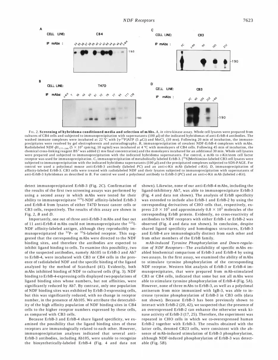
detect immunoprecipitated ErbB-3 (Fig. 2C). Confirmation ofthe results of the first two screening assays was performed byusing a second assay in which mAbs were tested for theirability to immunoprecipitate 125I-NDF affinity-labeled ErbB-3and ErbB-4 from lysates of either T47D breast cancer cells orCB3 cells, respectively. The results of this assay are shown inFig. 2, B and D.Importantly, one out of three anti-ErbB-3 mAbs and four out
of 11 anti-ErbB-4 mAbs could not immunoprecipitate the 125I-NDF affinity-labeled antigen, although they reproducibly im-munoprecipitated the 32P- or 35S-labeled receptor. This sug-gested that the corresponding mAbs were directed to the NDFbinding sites, and therefore the antibodies are expected toinhibit ligand binding to cells. To examine this possibility, twoof the suspected antibodies, namely Ab105 to ErbB-3 and Ab7to ErbB-4, were incubated with CB3 or CB4 cells in the pres-ence of radiolabeled NDF and the specific binding of the ligandanalyzed by the method of Scatchard (41). Evidently, bothmAbs inhibited binding of NDF to cultured cells (Fig. 3). NDFbinding to ErbB-4-expressing cells displayed two populations ofligand binding sites whose numbers, but not affinities, weresignificantly reduced by Ab7. By contrast, only one populationof NDF binding sites was exhibited by ErbB-3-expressing cells,but this was significantly reduced, with no change in receptornumber, in the presence of Ab105. We attribute the detectabil-ity of the high affinity population of NDF binding sites in CB4cells to the higher receptor numbers expressed by these cells,as compared with CB3 cells.Because ErbB-3 and ErbB-4 share ligand specificity, we ex-
amined the possibility that the ligand binding sites of thesereceptors are immunologically related to each other. However,immunoprecipitation analyses indicated that several anti-ErbB-3 antibodies, including Ab105, were unable to recognizethe biosynthetically-labeled ErbB-4 (Fig. 4 and data not
shown). Likewise, none of our anti-ErbB-4 mAbs, including theligand-inhibitory Ab7, was able to immunoprecipitate ErbB-3(Fig. 4 and data not shown). The analysis of ErbB specificitywas extended to include also ErbB-1 and ErbB-2 by using thecorresponding derivatives of CHO cells that, respectively, ex-press 2.0 3 105 and approximately 0.8 3 105 molecules of thecorresponding ErbB protein. Evidently, no cross-reactivity ofantibodies to NDF receptors with either ErbB-1 or ErbB-2 wasobserved (Fig. 4 and data not shown). In conclusion, despiteshared ligand specificity and homologous structures, ErbB-3and ErbB-4 are immunologically distinct from each other andfrom other members of the ErbB family.mAb-induced Tyrosine Phosphorylation and Down-regula-
tion of NDF Receptors—The availability of specific mAbs en-abled biochemical comparison of ErbB-3 and ErbB-4 by usingtwo assays. In the first assay, we examined the ability of mAbsto stimulate tyrosine phosphorylation of the correspondingNDF receptor. Western blot analysis of ErbB-3 or ErbB-4 im-munoprecipitates, that were prepared from mAb-stimulatedCB3 or CB4 cells, indicated that some but not all mAbs wereable to stimulate tyrosine phosphorylation of ErbB-4 (Fig. 5A).However, none of three mAbs to ErbB-3, as well as a polyclonalantiserum from mice immunized with IgB-3, was able to in-crease tyrosine phosphorylation of ErbB-3 in CB3 cells (datanot shown). Because ErbB-3 has been previously shown tointeract with ErbB-2 (20, 42), we suspected that the presence ofan overexpressed ErbB-2 can enhance the otherwise weak ki-nase activity of ErbB-3 (17, 25). Therefore, the experiment wasrepeated in CHO cells in which we co-overexpressed humanErbB-2 together with ErbB-3. The results obtained with thelatter cells, denoted CB23 cells, were consistent with the ab-sence of mAb-induced stimulation of ErbB-3 phosphorylation,although NDF-induced phosphorylation of ErbB-3 was detect-able (Fig. 5B).
FIG. 2. Screening of hybridoma conditioned media and selection of mAbs. A, in vitro kinase assay. Whole cell lysates were prepared fromcultures of CB4 cells and subjected to immunoprecipitation with supernatants (100 ml) of the indicated hybridomas of anti-ErbB-4 antibodies. Thewashed immune complexes were incubated at 22 °C with [g-32P]ATP (5 mCi) and MnCl2 (10 mM). Following 20 min of incubation, the immuno-precipitates were resolved by gel electrophoresis and autoradiography. B, immunoprecipitation of covalent NDF-ErbB-4 complexes with mAbs.Radiolabeled NDF-b1177–246 (5 3 105 cpm/ng; 10 ng/ml) was incubated at 4 °C with monolayers of CB4 cells. Following 45 min of incubation, thechemical cross-linking reagent BS3 was added (1 mM final concentration) and the monolayers incubated for an additional 30 min. Whole cell lysateswere prepared and subjected to immunoprecipitation with the indicated hybridoma supernatants. For control, a mAb to c-Kit/stem cell factorreceptor was used for immunoprecipitation. C, immunoprecipitation of metabolically labeled ErbB-3. [35S]Methionine-labeled CB3 cell lysates weresubjected to immunoprecipitation with the indicated hybridoma supernatants (100 ml) and the precipitated complexes subjected to SDS-PAGE. Forcontrol we used a polyclonal mouse anti-ErbB-3 antibody (labeled PC) and an anti-c-Kit mAb (labeled c-Kit). D, immunoprecipitation ofaffinity-labeled ErbB-3. CB3 cells were treated with radiolabeled NDF and their lysates subjected to immunoprecipitation with supernatants ofanti-ErbB-3 hybridomas as described in B. For control we used a polyclonal antibody to ErbB-3 (PC) and an anti-c-Kit mAb (labeled c-Kit).
NDF Receptors 7623

The second assay examined the ability of the various mAbs toinduce accelerated degradation of NDF receptors. TransfectedCHO cells were prelabeled with [35S]methionine and thenchased for 8 h in the absence or presence of either mAbs orNDF. Interestingly, whereas NDF was unable to down-regu-late ErbB-4, certain mAbs significantly accelerated disappear-ance of the receptor. It is worth noting that Abs 77 and 50 werethe most active antibodies in both receptor down-regulationand kinase stimulation, implying that these two activities arefunctionally coupled. In line with this conclusion, the rate ofErbB-3 degradation was not significantly affected by twomAbs, and a third mAb, namely Ab252, like NDF, deceleratedreceptor degradation (Fig. 6B). The observation that NDF isunable to down-regulate its own receptors, either ErbB-3 orErbB-4, in CHO cells is interesting as it differs from the effectof ligands to ErbB-1. Indeed, in experiments that are not pre-sented we found that the rates of NDF and EGF internalization
remarkably differ in CHO and in myeloid cells that ectopicallyexpress ErbB proteins.A Heterodimer of ErbB-3 with ErbB-2 Is the Predominant
NDF Receptor in Human Carcinoma Cells and Is Involved inTrans-phosphorylation and Trans-inhibition of Ligand Bind-ing—In the next step we used the anti-NDF receptor mAbs toanalyze the status of this receptor in several human cancercells from epithelial origin. Various tumor cells were incubatedwith radiolabeled NDF and then the ligand was covalentlycross-linked to the cells. This was followed by immunoprecipi-tation of the affinity-labeled receptors with antibodies to thefour ErbB proteins. In all tumor cell lines tested by this assay,including breast carcinomas (SKBR-3, MCF-7, T47D, AU-565,MCF-10, MDA-MB453), gastric cancer (CACO-2, N-87, LS180),transformed human keratinocytes (HACAT), and hepatomacells (PLC/RPF/5) we observed very high signals in anti-ErbB-3immunoprecipitates. The results obtained with some of thesetumor cells are shown in Fig. 7. In addition to labeling ofErbB-3, ErbB-2-specific mAbs also precipitated NDF that wascross-linked to its direct receptor, as we previously reported(43). However, in contrast with the appearance of both mono-meric and dimeric complexes in ErbB-3 immunoprecipitates,mostly the dimeric species was present in immunoprecipitatesof ErbB-2. The simplest interpretation of these results impliesthat the labeled receptor in ErbB-2 immunoprecipitates is aco-precipitated ErbB-3, that is either covalently cross-linked toErbB-2, and therefore it appears as a dimer, or noncovalentlyassociated with ErbB-2, and therefore it appears as a monomerin gel. It is interesting to note that no co-immunoprecipitationof ErbB-1 with ErbB-3 was detectable, although functionalinteraction between these receptors has been reported (18, 19).In addition, the affinity-labeled ErbB-4 was detectable in somecell lines (e.g. MCF-7, MDA-MB453, and T47D cells) but itrequired very long exposure of the films. In experiments that
FIG. 3. Inhibition of NDF binding by mAbs to ErbB-3 andErbB-4. Monolayers of CHO cells (2 3 104 cells/well) overexpressingeither ErbB-4 (CB4 cells, panel A) or ErbB-3 (CB3 cells, panel B) wereincubated for 2 h at 4 °C with different concentrations of 125I-NDF-b1177–246 in the presence of different mAbs to ErbB-4 or to ErbB-3.Nonspecific binding was determined by the addition of 100-fold excessof the unlabeled ligand and it was subtracted from the total amount ofbound radioactivity. Scatchard analysis was performed by using thecomputerized program LIGAND. The data are presented also as satu-ration curves (insets). The following symbols were used: Panel A: closedsquares (control), closed triangles (Ab7), open triangles (Ab94), and opencircles (Ab43). Panel B: closed squares (control), closed triangles(Ab105), and open triangles (Ab252). Each data point represents theaverage of a duplicate determination and each experiment was re-peated twice.
FIG. 4. Lack of cross-reactivity of anti-NDF-receptor mAbs.CB1, CB2, CB3, and CB4 cells were metabolically labeled with [35S]me-thionine, and the cell lysates were separately subjected to an immuno-precipitation assay with either mAbs to ErbB-4 (antibodies 7 and 72) orto ErbB-3 (antibodies 90 and 105). Proteins were separated by SDS-PAGE (7.5% polyacrylamide). An autoradiogram of the dried gel isshown and the locations of molecular weight marker proteins areindicated.
NDF Receptors7624

are not presented here we found that this was due to relativelylow expression of ErbB-4. On the basis of these results, weconcluded that in many epithelial cell lines the predominantreceptor for NDF is a heterodimer of ErbB-3 with ErbB-2.In order to analyze the functional consequences of the exten-
sive interaction between ErbB-3 and ErbB-2, we examinedtyrosine phosphorylation of the latter protein in SKBR-3 breastcancer cells, that overexpress ErbB-2. The results of this anal-ysis are shown in Fig. 8. Evidently, both NDF and EGF causedphosphorylation of their direct receptors, namely ErbB-3 andErbB-1, respectively, but these ligands also elevated tyrosinephosphorylation of ErbB-2. However, no evidence for trans-phosphorylation between ErbB-1 and ErbB-3 was obtained. Itis worth noting that heterodimers containing ErbB-1 andErbB-2 can be induced by EGF (36, 44), in analogy to ErbB-3/ErbB-2 heterodimers that are stabilized by NDF (Figs. 7 and9). Therefore, it is conceivable that tyrosine phosphorylation ofErbB-2 is activated in trans, either by NDF binding to ErbB-3or by EGF binding to ErbB-1.The observation that ErbB-2 is a common phosphorylation
partner of NDF and EGF receptors, together with reports onthe ability of ErbB-2 to enhance binding affinities of NDF andEGF (20, 22, 44), imply that NDF receptors and EGF receptorsmay compete for interaction with ErbB-2. To test this predic-tion we performed affinity labeling experiments with radiola-beled NDF or EGF and analyzed reciprocal binding effects of
the two ligands. Fig. 9 depicts the results of this experiment,that was performed on T47D breast cancer cells. In agreementwith the observed trans-phosphorylation between ErbB-1 andErbB-2, it appeared that the major species that binds EGF inT47D cells was a heterodimer of ErbB-1 with ErbB-2, whereasthe major NDF receptor was a heterodimer of ErbB-3 with
FIG. 5. mAb-induced tyrosine phosphorylation of ErbB-4 butnot ErbB-3. Monolayers of CB4 or CB23 cells were incubated for 10min at 37 °C with the indicated antibodies at 10 mg/ml or with NDF (10ng/ml). Cell lysates were prepared and subjected to immunoprecipita-tion (I.P.) with mAbs to either ErbB-4 (A), ErbB-3, and ErbB-2 (B).After gel electrophoresis, the immunoprecipitated proteins were elec-trophoretically transferred onto nitrocellulose and immunoblotted (I.B.)with a mAb to phosphotyrosine (P-TYR). The results of chemilumines-cence-based detection are shown, along with the locations of molecularweight standard marker proteins.
FIG. 6. Effects of mAbs on receptor turnover. Monolayers (2 3106 cells per lane) of either CB4 cells or CB3 cells were biosyntheticallylabeled with [35S]methionine. After a brief wash, cells were chased for8 h with unlabeled methionine-containing medium, in the presence ofthe indicated mAbs (at 10 mg/ml) to ErbB-4 (A), or to ErbB-3 (B) or withNDF (50 ng/ml). For control, cells were incubated with medium alone(lanes labeledNone). The cells were then washed, lysed, and cell lysateswere subjected to immunoprecipitation with mAbs to either ErbB-4 (A)or to ErbB-3 (B). The resulting autoradiograms (16 h exposure with anintensifying screen) are shown.
FIG. 7. Covalent cross-linking of radiolabeled NDF to the sur-face of various human tumor cell lines. 125I-Labeled NDF-b1177–246(10 ng/ml) was incubated for 2 h at 4 °C with 5 3 106 cells of thefollowing human tumor cell lines: SKBR-3 breast cancer, MCF-7 breastcancer, CACO2 colon cancer, and the PLC/PRF/5 hepatoma line. Thecell monolayers were washed with PBS and then cross-linked for 30 minwith BS3 (1 mM, Pierce) followed by cell lysis. After clearance of celldebris, the detergent-solubilized lysates were subjected to separateimmunoprecipitation reactions with antibodies to the indicated fourErbB proteins. Immune complexes were resolved by gel electrophoresisand autoradiography.
NDF Receptors 7625

ErbB-2. In addition, comparison of the affinity labeling pat-terns reflected the apparent exclusive nature of these inter-receptor interactions, as no ErbB-1/ErbB-3 heterodimers wereobserved. Remarkably, when the affinity labeling of EGF re-ceptors was performed in the presence of NDF, a significantreduction in both ErbB-1 and ErbB-2 labeling was observed.This implied that NDF can inhibit EGF binding to ErbB-1 andthat ErbB-2 is involved in this trans-regulatory effect. Thereciprocal experiment, that examined the effect of unlabeledEGF on binding of radiolabeled NDF, revealed a lower trans-inhibitory effect. These results are consistent with our previousreport that NDF can accelerate the rate of EGF release fromErbB-1 (45), and they attribute the effect to a competitionbetween ligand-bound ErbB-3 and ErbB-1 for the availableErbB-2. Presumably, ErbB-3/ErbB-2 heterodimers are favoredover ErbB-1/ErbB-2 heterodimers, and therefore the trans-inhibitory effect of NDF is stronger than that of EGF.Both ErbB-4 and ErbB-3 Mediate Cellular Differentiation—
Although ErbB-4 appears to act as a minor NDF receptor in the
epithelial cells that we examined, this receptor, unlike ErbB-3,possesses an active kinase and can undergo down-regulation inresponse to specific mAbs. On the other hand, the observedextensive interaction between ErbB-2 and ErbB-3 probablycompensates for the defective kinase of ErbB-3 and allowssignaling through NDF-induced heterodimers. In order to se-lectively examine the biological activities of ErbB-4 and ErbB-3we employed specific mAbs and tested their agonist function ina mammary cell differentiation assay (39). By using this assayit has been previously demonstrated that both NDF and certainmAbs to ErbB-2 can induce growth arrest of breast cancer cells,which is accompanied by secretion of milk components (caseinand lipids) and an elevated expression of the intracellularadhesion molecule 1 (ICAM-1) (29, 33). In the present study weextended these analyses to include WAF-1/p21cip, an inhibitorof cyclin-dependent kinases (46), that is involved in the induc-tion of differentiation in certain cellular systems (47). Fig. 10depicts the results of differentiation assays that were per-formed with MCF-7 breast cancer cells, which express all fourErbB proteins (22). Evidently, Ab36, an anti-ErbB-4 mAb thathas partial kinase stimulatory effect, was able to mimic NDF inthat it induced differentiation of cultured breast cancer cells.Other agonist mAbs, such as Ab77, were also stimulatory, butin general the effect of monoclonal antibodies was less exten-sive than the response to NDF. In addition, anti-ErbB-4 anti-bodies induced the appearance of other landmarks of the dif-ferentiated phenotype, including up-regulation of WAF-1expression in the nuclei of treated cells (Fig. 10, 3.5-fold) andelevated expression of ICAM-1 at the cell surface (data notshown). Screening of several mAbs to ErbB-4 identified Ab179as an antibody that inhibits NDF binding (Fig. 2B), but has aminimal effect on cell differentiation. Therefore, we used thisantibody as an antagonist of NDF to test the possibility thatErbB-4 is the sole receptor that transmits the differentiationsignal of NDF. However, even at oversaturating concentra-tions, Ab179 only partially inhibited the effect of NDF ondifferentiation of AU-565 breast cancer cells (Table I). Appar-ently, although ErbB-4 can transmit differentiation signals, itis not the only functional NDF receptor on AU-565 cells.In order to examine the role of ErbB-3 in transmission of the
differentiation effect of NDF, we tested the ability of anti-ErbB-3 mAbs to mimic the action of NDF on two breast cancercell lines. Unlike most anti-ErbB-4 mAbs, that were capable ofinducing some cellular differentiation, none of three mAbs toErbB-3 was effective (Table I, and data not shown). This resultis consistent with the inability of the mAbs to stimulate tyro-sine phosphorylation and down-regulation of ErbB-3 (Figs. 5Band 6B). We next analyzed the ability of Ab105, which antag-onizes NDF binding (Fig. 3B), to inhibit the induction of cellu-lar differentiation by this ligand. Indeed, like in the case of aligand-inhibitory mAb to ErbB-4, Ab105 reduced the effect of
FIG. 8. Ligand-induced tyrosinephosphorylation of ErbB proteins.Confluent monolayers of SKBR-3 humanbreast cancer cells were incubated in se-rum-free medium for 12 h and then incu-bated for 10 min at 37 °C in PBS in thepresence or absence of ligands (eitherNDF-b1 or EGF, each at 20 ng/ml), asindicated. The corresponding ErbB pro-teins were immunoprecipitated (IP) fromwhole cell lysates with specific antibodies,resolved by SDS-PAGE, and their tyro-sine phosphorylation detected by immu-noblotting (IB) with an antibody to phos-photyrosine (P-TYR). Control immuno-precipitation was performed with a non-relevant antibody (lanes labeled c-Ab)
FIG. 9. Trans-inhibitory effects of ErbB ligands.Monolayers of 53 106 T47D human breast cancer cells were incubated with radiola-beled EGF or NDF (each at 10 ng/ml) in the presence or absence of theother ligand in its unlabeled form, as indicated. Covalent cross-linkingof the radiolabeled ligands to cell surfaces was performed as describedin the legend to Fig. 7, and this was followed by immunoprecipitation ofindividual ErbB proteins and SDS-PAGE (6.5% acrylamide) of theprecipitated complexes. The autoradiograms show monomers anddimers of the affinity-labeled receptors. Note that NDF decreased la-beling of both ErbB-1 and ErbB-2 by EGF, but the latter only slightlyreduced binding of NDF to ErbB-3 and ErbB-2.
NDF Receptors7626

NDF on lipids and ICAM-1 (Table I). Although the inhibitoryeffect of mAb105 was incomplete, it was reproducibly largerthan the effect of an ErbB-4-blocking antibody. Remarkably,even at over-saturating concentrations, that completely abolishligand binding to CB3 cells, Ab105 was unable to completelyabolish the effect of NDF. Therefore, we concluded that ErbB-3mediates the differentiation action of NDF in a non-exclusivemanner. Thus, although ErbB-3 is biologically inactive, thisreceptor mediates part of the effect of NDF, conceivably byheterodimer formation with ErbB-2. This proposition is sup-ported by the finding that certain anti-ErbB-2 mAbs are effec-tive inducers of differentiation (33).
DISCUSSION
Most mammalian receptor tyrosine kinases belong to smallgroups of 2–9 highly related proteins that bind to homologousgrowth factors. Examples include the Trk family of receptorsfor neurotrophic factors and the relatively large family of Eph-like receptors (48). Because in insect cells each subgroup isrepresented by a single receptor, it is reasonable that receptormultiplicity evolved in order to provide physiological answers.The group of type I receptor tyrosine kinases is unique in thatit contains a receptor with no known ligand, namely ErbB-2,and another receptor, ErbB-3, whose tyrosine kinase domainincludes several unusual sequence motifs. In this respectErbB-3 resembles several other receptor-like tyrosine kinases,such as Klg (49) and Vik/Ryk (50), whose biological and bio-chemical functions are unknown. The present study addressedthe multiplicity of type I receptor tyrosine kinases, and espe-
cially the two distinct receptors for NDF. By expressing recom-binant forms of the extracellular domains of ErbB-3 andErbB-4 in their native forms, we were able to obtain a rela-tively large repertoire of mAbs to these NDF receptors. On thebasis of experiments that were performed with the new mAbs,we reached three major conclusions that are summarizedbelow.1) The Two NDF Receptors Are Immunologically Distinct,
Including Differences in the Structures of Their Ligand BindingSites—Considering the fact that no known mAb to ErbB-2 cancross-react with ErbB-1, the structural heterogeneity of the twoNDF receptors may not be surprising. Because the mAbs wegenerated displayed no cross-reactivity with ErbB-1 andErbB-2 (Fig. 4), it is likely that each ErbB protein is distinctfrom the other family members. Another common immunolog-ical feature of ErbB proteins emerged from our results andprevious analyses of mAbs to ErbB-1 (30, 51–53), namely: theligand binding sites of all ErbB proteins are apparently themost immunogenic sites on these molecules. Nevertheless, it ispossible that some of the ligand-inhibitory mAbs that we gen-erated sterically inhibit ligand binding without directly inter-acting with the binding cleft.2) The Two NDF Receptors Remarkably Differ in Their Bio-
logical Actions—Because anti-ErbB-4 mAbs are biologically ac-tive, whereas anti-ErbB-3 antibodies are inactive in severalassays, it is conceivable that ErbB-4 homodimers can transmitbiological signals, but homodimers of ErbB-3 are non-func-tional. We attribute the impaired biological function of ErbB-3to its defective kinase domain. The conclusion that ErbB-3 ispractically an inactive kinase is based on our failure to detectautophosphorylation of ErbB-3 in assays that were performedin vitro (data not shown), and the observation that none of ourmAbs to ErbB-3 stimulated tyrosine phosphorylation of thisreceptor in living cells (Fig. 5B). This conclusion is consistentwith the lack of mAb-induced down-regulation of ErbB-3 inliving cells (Fig. 6B), and it is consistent with the observationthat an insect cell-expressed ErbB-3 possessed an impairedtyrosine kinase activity (17). However, previous analyses thatwere performed with murine fibroblasts that expressed chime-ras of ErbB-1 and ErbB-3 reported ligand-induced tyrosineautophosphorylation of the ErbB-3 kinase domain (24, 25).Possibly, the observed phosphorylation was mediated by het-erodimers between ErbB-3 and the kinase intact partners,either ErbB-1 or ErbB-2, that are present in fibroblasts. Thispossibility is supported by our preliminary experiments with32D myeloid cells, that express no endogenous ErbB protein.Using this cellular system, we observed no NDF-induced tyro-sine phosphorylation in cells that ectopically express only
FIG. 10. An anti-ErbB-4 mAb in-duces differentiation of breast can-cer cells and up-regulates the WAF-1protein. MCF-7 cells were incubated for4 days in the absence or presence of 10mg/ml mAb36 to human ErbB-4. The cellmonolayers were then processed for stain-ing of neutral lipids with Oil Red O (redperinuclear droplets) or immunohisto-chemical localization of WAF-1 (brownnuclear staining).
TABLE IThe effect of mAbs to ErbB-3 and ErbB-4 on NDF-induced
differentiation of breast cancer cells in vitroSubconfluent monolayers of the indicated breast cancer cell lines
were treated for 4 days with NDF or mAbs. The cells were then stainedfor ICAM-1 by using specific antibodies. Lipid visualization was carriedout according to the Oil Red O in propylene glycol method, and theresults are expressed as the percentage of positively-stained cells in 10microscope fields. Quantification of ICAM-1 immunostaining was de-termined by using an image analysis system and it is expressed inarbitrary units. The experiment was repeated three times.
AU-565 cells MCF-7 cells, Lipids(% of cells)Lipids (% of cells) ICAM-1 (units)
Control 32 6 12NDF-b1 (10 ng/ml) .90 40 98Ab105 (20 mg/ml) 28 2 11NDF 1 Ab105 44 10 45Ab179 (20 mg/ml) 44 NDa NDNDF 1 Ab179 71 ND ND
a ND, not determined.
NDF Receptors 7627

ErbB-3, but it was readily detectable in cells that co-expressedErbB-3 and other ErbB proteins.3 The ability of NDF to in-crease tyrosine phosphorylation of the kinase-impaired ErbB-3is probably mediated by the catalytic activity of ErbB-2, be-cause a kinase-defective mutant of ErbB-2 failed to mediateErbB-3 phosphorylation (54). It is noteworthy that in contrastwith our inability to mimic the differentiation effect of NDF byusing anti-ErbB-3 mAbs, it was reported that another mAb toErbB-3 moderately stimulated anchorage-independent growthof breast cancer cell lines (55), although this mAb was devoid ofa kinase-stimulatory activity.3) The Predominant Form of NDF Receptor in Epithelial
Cells Is a Heterodimer of ErbB-3 with ErbB-2—Despite theimpaired activity of ErbB-3 homodimers, our covalent cross-linking analyses indicated that ErbB-3 is the predominantNDF receptor in mammary and in other epithelial cells (Fig. 7).In addition, because a ligand-inhibitory mAb to ErbB-3 blockedmost of the biological effect of NDF (Table I), it is conceivablethat ErbB-3 is the major receptor that mediates cellular differ-entiation by NDF. However, our results imply that an alterna-tive route of NDF signaling involves ErbB-4 (Fig. 10 and TableI), whose expression level in most epithelial cells is at least10-fold lower than that of ErbB-3.4 The observed predominantoccurrence of ErbB-3/ErbB-2 heterodimers may explain theobservation that co-overexpression of the partners of this het-erodimer caused transformation of fibroblasts (42, 54). It mayalso explain why ErbB-2 strongly increases NDF affinity toErbB-3 (20). Evidently, the formation of this heterodimer po-tently stimulates tyrosine phosphorylation of both partners(Fig. 8). This very efficient trans-phosphorylation is probablyresponsible for the initial erroneous identification of NDF/heregulin as a direct ligand of ErbB-2 (27, 28). Taken together,the present and previous results identify ErbB-2 as a commonpartner of EGF and NDF receptors. Moreover, the data pre-sented in Fig. 9 and in our previous report (45) suggest thatthese receptors compete for recruitment of ErbB-2, becauseeach ligand exerts a trans-inhibitory effect on binding of theother ligand. Apparently, recruitment of ErbB-2 by NDF-occu-pied ErbB-3 is more efficient than the formation of ErbB-1/ErbB-2 heterodimers, as the trans-inhibitory effect of NDF onEGF binding (45) and on affinity labeling of ErbB-2 (Fig. 9,upper panel) is more prominent than the reciprocal interaction.Taken together, our results are consistent with the notion
that the multiplicity of ErbB proteins confers diversification ofsignal transduction by the corresponding ligands (5, 23). Ac-cording to the emerging model, the two NDF receptors differ ina major aspect: ErbB-4 can generate biological signals uponhomodimerization, but ErbB-3 homodimers are signaling-de-fective. Heterodimerization apparently reconstitutes signalingby ErbB-3, and the preferred partner of this major NDF recep-tor is ErbB-2. However, the latter protein forms heterodimersalso with ErbB-1, so that it probably functions as a commonsignaling subunit of NDF and EGF receptors. This propositionis consistent with the observation that abolishment of ErbB-2expression severely impairs signaling by both growth factors(21, 22), and it may imply that ErbB-2 can function without aligand of its own. The existence and role of ErbB-4/ErbB-2heterodimers remain unclear, but ErbB-4 appears to be theminor NDF receptor, at least in epithelial cells. Presumably,besides reconstitution of ErbB-3 activity, the process of recep-tor heterodimerization confers additional levels of complexity
and regulation to the mechanism of signaling by growthfactors.
Acknowledgments—We thank Esther Hurwitz and Leah Klapper forhelpful discussions and Anat Bromberg for technical assistance.
REFERENCES
1. Fantl, W. J., Johnson, D. E., and Williams, L. T. (1993) Annu. Rev. Biochem.62, 453–481
2. Plowman, G. D., Green, J. M., Culouscou, J.-M., Carlton, G. W., Rothwell, V.M., and Sharon, B. (1993) Nature 366, 473–475
3. Carraway, K. L., III, Sliwkowski, M. X., Akita, R., Platko, J. V., Guy, P. M.,Nuijens, A., Diamonti, A. J., Vandlen, R. L., Cantley, L. C., and Cerione, R.A. (1994) J. Biol. Chem. 269, 14303–14306
4. Tzahar, E., Levkowitz, G., Karunagaran, D., Yi, L., Peles, E., Lavi, S., Chang,D., Liu, N., Yayon, A., Wen, D., and Yarden, Y. (1994) J. Biol. Chem. 269,25226–25233
5. Peles, E., and Yarden, Y. (1993) BioEssays 15, 815–8246. Aaronson, S. A. (1991) Science 254, 1146–11537. Di Fiore, P. P., Pierce, J., Kraus, M. H., Segatto, O., King, C. R., and Aaronson,
S. A. (1987) Science 237, 178–1828. Hudziak, R. M., Schlessinger, J., and Ullrich, A. (1987) Proc. Natl. Acad. Sci.
U. S. A. 84, 7159–71639. Gullick, W. J. (1990) Int. J. Cancer Suppl. 5, 55–6110. Hynes, N. E., and Stern, D. F. (1994) Biochim. Biophys. Acta 1198, 165–18411. Lemoine, N. R., Barnes, D. M., Hollywood, D. P., Hughes, C. M., Smith, P.,
Dublin, E., Prigent, S. A., Gullick, W. J., and Hurst, H. C. (1992) Br. J.Cancer 66, 1116–1121
12. Lemoine, N. R., Lobresco, M., Leung, H., Barton, C., Hughes, C. M., Prigent, S.A., Gullick, W. J., and Kloppel, G. (1992) J. Pathol. 168, 269–273
13. Kraus, M. H., Issing, M., Popescu, N. C., and Aaronson, S. A. (1989) Proc. Natl.Acad. Sci. U. S. A. 86, 9193–9197
14. Orr-Urtreger, A., Trakhtenbrot, L., Ben-Levi, R., Wen, D., Rechavi, G., Lonai,P., and Yarden, Y. (1993) Proc. Natl. Acad. Sci. U. S. A. 90, 1867–1871
15. Ben-Baruch, N., and Yarden, Y. (1994) Proc. Soc. Exp. Biol. Med. 206, 221–22716. Plowman, G. D., Whitney, G. S., Neubauer, M. G., Green, J. M., McDonald, V.
I., Todaro, G. J., and Shoyab, M. (1990) Proc. Natl. Acad. Sci. U. S. A. 87,4905–4909
17. Guy, P. M., Platko, J. V., Cantley, L. C., Cerione, R. A., and Carraway, K. L.(1994) Proc. Natl. Acad. Sci. U. S. A. 91, 8132–8136
18. Soltoff, S. P., Carraway, K. L., Prigent, S. A., Gullick, W. G., and Cantley, L.C. (1994) Mol. Cell. Biol. 14, 3550–3558
19. Kim, H.-H., Sierke, S. L., and Koland, J. G. (1994) J. Biol. Chem. 269,24747–24755
20. Sliwkowski, M. X., Schaefer, G., Akita, R. W., Lofgren, J. A., Fitzpatrick, V. D.,Nuijens, A., Fendly, B. M., Cerione, R. A., Vandlen, R. L., and Carraway, K.L., III (1994) J. Biol. Chem. 269, 14661–14665
21. Graus-Porta, D., Beerli, R. R., and Hynes, N. E. (1995) Mol. Cell. Biol. 15,1182–1191
22. Karunagaran, D., Tzahar, E., Beerli, R. R., Chen, X., Graus-Porta, D., Ratzkin,B., Seger, R., Hynes, N. E., and Yarden, Y. (1996) EMBO J. 15, 254–264
23. Carraway, K. L., III, and Cantley, L. C. (1994) Cell 78, 5–824. Kraus, M. H., Fedi, P., Starks, V., Muraro, R., and Aaronson, S. A. (1993) Proc.
Natl. Acad. Sci. U. S. A. 90, 2900–290425. Prigent, S. A., and Gullick, W. J. (1994) EMBO J. 13, 2831–284126. Marikovsky, M., Lavi, S., Pinkas-Kramarski, R., Karunagaran, D., Liu, N.,
Wen, D., and Yarden, Y. (1995) Oncogene 10, 1403–141127. Holmes, W. E., Sliwkowski, M. X., Akita, R. W., Henzel, W. J., Lee, J., Park, J.
W., Yansura, D., Abadi, N., Raab, H., Lewis, G. D., Shepard, M., Wood, W.I., Goeddel, D. V., and Vandlen, R. L. (1992) Science 256, 1205–1210
28. Peles, E., Bacus, S. S., Koski, R. A., Lu, H. S., Wen, D., Ogden, S. G., Ben Levy,R., and Yarden, Y. (1992) Cell 69, 205–216
29. Bacus, S. S., Gudkov, A. V., Zelnick, C. R., Chin, D., Stern, R., Stancovski, I.,Peles, E., Ben-Baruch, N., Farbstein, H., Lupu, R., Wen, D., Sela, M., andYarden, Y. (1993) Cancer Res. 53, 5251–5261
30. Defize, L. H. K., Boonstra, J., Meisenhelder, J., Kruijer, W., Tertoolen, L. G. J.,Tilly, B. C., Hunter, T., van Bergen en Henegouwen, P. M. P., Moolenaar,W. H., and de Laat, S. W. (1989) J. Cell Biol. 109, 2495–2507
31. Yarden, Y. (1990) Proc. Natl. Acad. Sci. U. S. A. 87, 2569–257332. Drebin, J. A., Link, V. C., Weinberg, R. A., and Greene, M. I. (1986) Proc. Natl.
Acad. Sci. U. S. A. 83, 9129–913333. Bacus, S. S., Stancovski, I., Huberman, E., Chin, D., Hurwitz, E., Mills, G. B.,
Ullrich, A., Sela, M., and Yarden, Y. (1992) Cancer Res. 52, 2580–258934. Stancovski, I., Hurwitz, E., Leitner, O., Ullrich, A., Yarden, Y., and Sela, M.
(1991) Proc. Natl. Acad. Sci. U. S. A. 88, 8691–869535. Wen, D., Peles, E., Cupples, R., Suggs, S. V., Bacus, S. S., Luo, Y., Trail, G., Hu,
S., Silbiger, S. M., Ben Levy, R., Koski, R. A., Lu, H. S., and Yarden, Y.(1992) Cell 69, 559–572
36. Goldman, R., Ben-Levy, R., Peles, E., and Yarden, Y. (1990) Biochemistry 29,11024–11028
37. Munson, P. J., and Rodbard, D. (1980) Anal. Biochem. 107, 220–23938. Peles, E., Ben Levy, R., Or, E., Ullrich, A., and Yarden, Y. (1991) EMBO J. 10,
2077–208639. Bacus, S. S., Kiguchi, K., Chin, D., King, C. R., and Huberman, E. (1990) Mol.
Carcinog. 3, 350–36240. Wen, D., Suggs, S. V., Karunagaran, D., Liu, N., Cupples, R. L., Luo, Y.,
Jansen, A. M., Ben-Baruch, N., Trollinger, D. B., Jacobson, V. L., Meng, T.,Lu, H. S., Hu, S., Chang, D., Yanigahara, D., Koski, R. A., and Yarden, Y.(1994) Mol. Cell. Biol. 14, 1909–1919
41. Scatchard, G. (1949) Ann. N. Y. Acad. Sci. 51, 660–67242. Alimandi, M., Romano, A., Curia, M. C., Muraro, R., Fedi, P., Aaronson, S. A.,
Di Fiore, P. P., and Kraus, M. H. (1995) Oncogene 10, 1813–1821
3 R. Pinkas-Kramarski, L. Soussan, H. Waterman, G. Levkowitz, I.Alroy, L. Klapper, S. Lavi, B. Ratzkin, M. Sela, and Y. Yarden (1996)EMBO J. 15, in press.
4 X. Chen and Y. Yarden, unpublished results.
NDF Receptors7628

43. Peles, E., Ben-Levy, R., Tzahar, E., Naili, L., Wen, D., and Yarden, Y. (1993)EMBO J. 12, 961–971
44. Wada, T., Qian, X., and Greene, M. I. (1990) Cell 61, 1339–134745. Karunagaran, D., Tzahar, E., Liu, N., Wen, D., and Yarden, Y. (1995) J. Biol.
Chem. 270, 9982–999046. Luo, Y., Hurwitz, J., and Massague, J. (1995) Nature 375, 159–16147. Halevy, O., Novitch, B. G., Spicer, D. B., Skapek, S. X., Rhee, J., Hannon, G. J.,
Beach, D., and Lassar, A. B. (1995) Science 267, 1018–102148. van der Geer, P., Hunter, T., and Lindberg, R. A. (1994) Annu. Rev. Cell Biol.
10, 251–33749. Chou, Y.-H., and Hayman, M. J. (1991) Proc. Natl. Acad. Sci. U. S. A. 88,
4897–4901
50. Kelman, Z., Simon-Chazottes, D., Guenet, J.-L., and Yarden, Y. (1993) Onco-gene 8, 37–44
51. Waterfield, M. D., Mayes, E. L., Stroobant, P., Bennet, P. L. P., Young, S.,Goodfellow, P. N., Banting, G. S., and Ozanne, B. (1982) J. Cell. Biochem.20, 149–161
52. Kawamoto, T., Sato, J. D., Le, A., Poliakoff, J., Sato, G. H., and Mendelsohn, J.(1983) Proc. Natl. Acad. Sci. U. S. A. 80, 1337–1341
53. Fernandez-Pol, J. A. (1985) J. Biol. Chem. 260, 5003–501154. Wallasch, C., Weiss, F. U., Niederfellner, G., Jallal, B., Issing, W., and Ullrich,
A. (1995) EMBO J. 14, 4267–427555. Rajkumar, T., and Gullick, W. J. (1994) Br. J. Cancer 70, 459–465
NDF Receptors 7629


