Embed Size (px)
Citation preview

J O U R N A L O F P R O T E O M I C S 7 4 ( 2 0 1 1 ) 6 8 3 – 6 9 7
ava i l ab l e a t www.sc i enced i r ec t . com
www.e l sev i e r . com/ loca te / j p ro t
Analysis of proteome changes in doxorubicin-treated adult ratcardiomyocyte
Suresh N. Kumara, Eugene A. Konorevb,1,Deepika Aggarwalc,1, Balaraman Kalyanaramand,⁎aDepartment of Pathology, Medical College of Wisconsin, Milwaukee, WI, USAbDepartment of Pharmaceutical Sciences, University of Hawaii at Hilo, Hilo, HI, USAcDr. Reddy's Laboratories Ltd., Hyderabad, IndiadDepartment of Biophysics and Free Radical Research Center, Medical College of Wisconsin, Milwaukee, WI, USA
A R T I C L E I N F O
Abbreviations: ACTH, adrenocorticotropictriphosphate; DOX, doxorubicin; ETC, electrprotein dehydrogenase; HSP27, heat shock pMMSDH, methylmalonate semialdehyde dehalpha.⁎ Corresponding author at: Department of Bioph
+1 414 456 4000; fax: +1 414 456 6512.E-mail address: [email protected] (B. K
1 Present address.
1874-3919/$ – see front matter © 2011 Elsevidoi:10.1016/j.jprot.2011.02.013
A B S T R A C T
Article history:Received 3 November 2010Accepted 12 February 2011Available online 19 February 2011
Doxorubicin-induced cardiomyopathy in cancer patients is well established. The proposedmechanism of cardiac damage includes generation of reactive oxygen species,mitochondrial dysfunction and cardiomyocyte apoptosis. Exposure of adult ratcardiomyocytes to low levels of DOX for 48 h induced apoptosis. Analysis of proteinexpression showed a differential regulation of several key proteins including the voltagedependent anion selective channel protein 2 and methylmalonate semialdehydedehydrogenase. In comparison, proteomic evaluation of DOX-treated rat heart showed aslightly different set of protein changes that suggests nuclear accumulation of DOX. Using anew solubilization technique, changes in low abundant protein profiles were monitored.Altered protein expression, modification and function related to oxidative stress responsemay play an important role in DOX cardiotoxicity.
© 2011 Elsevier B.V. All rights reserved.
Keywords:DoxorubicinCardiomyopathyProteomics
1. Introduction
The anthracycline antibiotic, doxorubicin (DOX), is an anti-cancer drug that is used widely in treating leukemias,lymphomas, lung, breast, ovary and uterine cancers. However,DOX chemotherapy causes a cumulative and delayed cardio-myopathy in cancer patients (2% in 2-year versus 5% in 15-yearsurvivors) as a major side effect [1,2]. Human case studyreports and preclinical data in animal models indicate thatDOX treatment causedmitochondrial damage and decrease in
hormone; ADP, adenosinon transport chain; ETF,rotein 27 kDa; MALDI-TOydrogenase; PBS, phospha
ysics, Medical College of Wi
alyanaraman).
er B.V. All rights reserved
ATP/ADP ratio [3]. The proposed mechanism of cardiotoxicityinvolves free radical generation [4], lipid peroxidation [5],cardiomyocyte apoptosis [6,7] and somatic mitochondrialalteration and dysfunction [8–10]. DOX undergoes a one-electron redox cycling at the complex I site of the mitochon-drial electron transport chain (ETC), forming a semiquinonethat reduces molecular oxygen to superoxide that dismutatesto form hydrogen peroxide [11–13]. DOX can be reduced by avariety of flavoproteins including cytochrome P450 reductase,mitochondrial NADH (nicotinamide adenine dinucleotide —
e diphosphate; AMP, adenosine monophosphate; ATP, adenosineelectron transfer flavoprotein; ETF-QO/ETFDH, electron transferF, matrix assisted laser desorption and ionization time of flight;te buffered saline; p38 alpha, Mitogen activated protein kinase p38
sconsin, 8701 Watertown Plank Road, Milwaukee, WI, 53226, USA. Tel.:
.

684 J O U R N A L O F P R O T E O M I C S 7 4 ( 2 0 1 1 ) 6 8 3 – 6 9 7
reduced) dehydrogenase and nitric oxide synthase (reductasedomain) [12,14]. In the presence of redox-active metal ions(iron and copper), highly reactive hydroxyl radicals or perferryliron are formed from DOX redox activation [15]. Theseoxidants target lipid, protein, and DNA causing extensivemodifications [16–18]. Reactive oxygen species formed fromDOX redox cycling also affect iron uptake and iron homeosta-sis [19,20], inhibit aconitase and iron regulatory protein (IRP)function and alter the transferrin/ferritin mRNA (messengerribonucleic acid) levels. Another mechanism by which aconi-tase was inactivated is via the interaction with the secondaryalcohol derivative of DOX [21,22].
Doxorubicin caused a dose-dependent cell death in isolat-ed adult rat cardiomyocyte [23,24]. Studies with antioxidants[25] and iron chelators [26] suggest that mechanisms otherthan peroxidative stress contribute to cardiac impairment inDOX chemotherapy [27,28]. In addition, DOX accumulates inthe nucleus causing DNA damage. DOX also selectivelyinhibits gene expression and interferes with the transcriptionfactor's ability to bind to DNA due to formation of GC adductsat the 5′-UTRs (untranslated regions). We have previouslydemonstrated the critical role of activated transcription factorkappa-B in DOX-mediated apoptosis in endothelial cells andin isolated adult rat cardiomyocyte [29–33]. The DNA repairprocess in DOX-treated cells is different from that of hydrogenperoxide-treated cells [34–36]. Overall, published data suggestthat oxidative stress appears to play a key role in DOX-mediated cardiotoxicity and apoptotic myocyte death. In thisstudy, we report the protein expression changes observed inisolated cardiomyocyte and adult rat heart during DOXtreatment and also provide a methodological improvementthat identifies changes in non-abundant protein levels.
2. Materials and methods
2.1. Reagents
All reagents of the highest quality commercially available werepurchased. TRIS base, glycine, sodium dodecyl sulfate (SDS),doxorubicin-HCl, dithiotheritol (DTT), Urea, 3-[(3-Cholamido-propyl) dimethylammonio]-1-propanesulfonate (CHAPS), am-monium bicarbonate, α-cyano-4-hyroxycinnamic acid (CHCA),deoxyribonuclease (DNAse), bovine serum albumin (fraction V),80 mesh screen filter, medium 199, iodoacetamide, creatine,taurine, insulin, cytosineβ-D-arabinofuranoside, vitaminsolution(100×), sodium deoxycholate, Triton x-100, glycerol, urea,thiourea, ethylenediaminetetraacetic acid (EDTA), ethylenegly-coltetraacetic acid (EGTA), diethylene triamine pentaacetic acid(DTPA), phenylmethylsulphonyl fluoride (PMSF), aprotinin,leupeptin, β-glycerophosphate, sodium fluoride, sodiumorthovanadate, sodium pyrophosphate, 4-(2-Hydroxyethyl)-1-piperazineethane sulfonic acid (HEPES), sucrose, mannitol,calcium chloride (CaCl2), tween-20, acetonitrile (ACN), di-methyl sulfoxide (DMSO), acetone and HPLC pure water werepurchased from Sigma-Aldrich Co. USA. DMEM, fetal bovineserum (FBS), collagenase (type II, the preparation with lowcollagenase activity of 200–250 U/mg) and Dulbecco phos-phate buffered saline (DPBS), complete amino acid mixture(50×) and MEM non-essential amino acid solution (100×) were
from Gibco, USA. Immobilized IPG (3–10) strips, Bio-lyte 3–10ampholytes and prestained protein molecular weightmarker were purchased from Biorad, USA. 2D-quant kit andadvanced ECL kit were purchased from GE Life Sciences.Syringe filters (5 μm) and Zip-Tip C18 was purchased fromMillipore, USA. All other general chemicals such as potassi-um dihydrogen phosphate (KH2PO4), sodium bicarbonate(NaHCO3), sodium chloride (NaCl), potassium chloride (KCl),and magnesium sulfate (MgSO4) were purchased from FisherScientific, USA.
2.2. Buffers
All buffers involved in cardiomyocyte isolation were pre-warmed to 37 °C before use. Perfusion buffer contains 25 mMHEPES buffer, 110 mM NaCl, 11 mM glucose, 1 mg/mL BSA,5 mM creatine, 20 mM taurine, 2.6 mM KCl, 1.2 mM MgSO4,20 μl/ml of 50× complete amino acid mixture, 10 μl of 100×stock non-essential amino acid mixture, 10 μl/mL of 100×stock of vitamin solution and 1.2 mM KH2PO4 and 5 mMpyruvate sodium (pH 7.4). The pH of the perfusion buffer wasadjusted to 7.4 and sterile-filtered using 0.2 μm celluloseacetate filter. Digestion buffer contain perfusion buffer,25 μM CaCl2 and 200 U/mL of collagenase [37]. Isolation buffercontains digestion buffer, 20 μg/mL of DNAse and 1% BSA.Wash buffers I, II and III are made up of perfusion buffercontaining 200 μM, 500 μM and 1 mM CaCl2, respectively.Isolated cardiomyocytes were cultured in M-199 mediumcontaining 25 mM NaHCO3, 25 mM HEPES, 2 mg/mL BSA,0.5 mM creatine, 5 mM taurine, 0.1 μM insulin, 10 μM cytosineβ-D-arabinofuranoside, 10% FBS, 100 U/mL penicillin and100 μg/mL streptomycin. Protease inhibitor cocktail contains5 mM EDTA, 2 mM EGTA, 5 mM DTPA, 2 mM PMSF, 5 μg/mLaprotinin, 5 μg/mL leupeptin, 1 mM β-glycerophosphate, 1 mMsodium fluoride, 1 mM sodiumorthovanadate, 2.5 mM sodiumpyrophosphate and 5 mM DTT. Radio-immunoprecipitationassay (RIPA) buffer was made with 50 mM Tris pH 7.6, 150 mMNaCl, 1% triton x-100, 1% sodium deoxycholate, 0.1% SDS andprotease inhibitor cocktail. Rehydration buffer is made up of7 M urea, 2 M thiourea, 4% CHAPS, 0.2% ampholytes (3–10) and100 mM DTT. Equilibration buffer I is made up of 6 M urea,375 mM Tris–HCl, pH 8.8, 2% SDS, 20% glycerol and 2% DTT.Equilibration buffer II is made up of 6 M urea, 375 mM Tris–HCl, pH 8.8, 20% glycerol and 2.5% idoacetamide. Cytoplasmicextraction buffer is made up of 1× PBS pH 7.0 containing 0.1%digitonin and protease inhibitor cocktail. Membrane fraction Iextraction buffer is made up of 1× PBS pH 7.0 containing 0.3%triton x-100 and protease inhibitor cocktail. Membranefraction II extraction buffer is made up of 1x PBS pH 7.0containing, 1% triton x-100 and protease inhibitor cocktail.Filamentous protein extraction buffer is made up of 1× PBS pH7.0 containing 0.5% triton x-100 and 0.6 M KCl and proteaseinhibitor cocktail.
2.3. Adult rat cardiomyocyte isolation and culture
The ventricular cardiomyocytes were isolated according to apreviously described method [38,39] that was modified in ourlaboratory. Adult male Sprague–Dawley rats weighing approx-imately 180–220 g were anesthetized with intraperitoneal

685J O U R N A L O F P R O T E O M I C S 7 4 ( 2 0 1 1 ) 6 8 3 – 6 9 7
injections of pentobarbital (60 mg/kg body weight) followed byintraperitoneal injection of 250 U/kg of heparin. The heart wasexcised, mounted on aortic cannula and perfused (in aLangendorff non-recirculating mode) with the oxygenatedperfusion buffer supplemented with 1 mM CaCl2. After 15 minof perfusion, it was switched to calcium-free oxygenatedbuffer in a non-recirculating mode for 5 min followed byrecirculation at a constant flow rate for 30 min. At the end ofperfusion the ventricular tissue was dissected, minced andincubated with gentle shaking in the isolation buffer at 37 °Cfor 10 min. The single cells released were dispersed from theremaining undigested tissue by mixing the suspension with a5 ml serological pipette, and the cells were collected using 80mesh screen filter. The cells were washed by centrifugation at150 rpm in succession with wash buffers I, II, and III andresuspended in a small volume of wash buffer III. This cellsuspension was layered on a 4% BSA — 1 mM CaCl2 cushionand centrifuged at 200 rpm for 2–3 min. Ventricular myocytessettled down and the resulting cell pellet was resuspended ingrowth medium and plated. Cardiomyocytes were separatedfrom other cells by differential plating at 1.5×10⁶ cells/mL onto laminin coated 150 mm dishes. This isolation proceduregenerally yields 75–80% rod-shaped cardiomyocyte [40] thatexclude trypan blue.
2.4. Doxorubicin treatment of cardiomyocyte and isolationof cell lysate
The isolated cardiomyocytes were placed in the incubator at37 °C with 5% CO2 for overnight incubation. DOX was added tothe medium at 0.5 μM concentration and the cells were lysedin RIPA buffer at 0, 24, or 48 h after treatment. At theseconcentrations the cells did not release significant lactatedehydrogenase (marker for cellular damage) [40] but exhibitapoptosis as seen by a large amount of floating cells andpositive TUNEL assay [41]. The cell lysate was left to stand onice for 30 min and centrifuged at 4 °C at 13,000 rpm for 30 min.The clear supernatant was transferred to a fresh tube andaliquots in triplicate were analyzed for protein concentrationusing 2D-quant kit (bicinchoninic acid method) using BSA.Protein concentration equivalent to 600 μg was precipitated byincubating with 8 volumes of ice cold acetone and left to standat −20 °C overnight. The precipitated protein was collected bycentrifuging at 4 °C at 13,000 rpm for 30 min, and the pelletwashed with ice cold acetone twice. The final pellet was driedunder vacuum and was stored at −80 °C until use.
2.5. Fractionation of cell lysate
For fractionation experiments, DOX-treated (0.5 μM for 48 hand 20 μM for 24 h) and untreated control cells (vehicle for48 h) were collected in 1× DPBS and washed twice in the samebuffer. The cell pellet was resuspended in 2 mL of cytoplasmicextraction buffer, incubated on ice for 1 h and spun at16,000 rpm for 15 min at 4 °C. The resultant supernatant wasspun at 100,000×g for 20 min at 4 °C (supernatant collected).The pellets from 16,000 rpm and 100,000×g were pooled andreextracted with membrane fraction I extraction buffer andspun at 16,000 rpm for 20 min at 4 °C (supernatant collected).The resultant pellet was extracted with membrane fraction II
extraction buffer and spun at 16,000 rpm for 20 min at 4 °C(supernatant collected). The final pellet was again extractedwith filamentous protein extraction buffer and spun at16,000 rpm for 20 min at 4 °C (supernatant collected). Proteinestimation of the extracts was performed as above.
2.6. DOX-treated adult rat
Animal experiments were performed in accordance with theanimal protocol approved by the Medical College ofWisconsinAnimal Care and Use Committee. Adult male Sprague–Dawleyrats were segregated into groups as control (n=16) and DOX-treated (n=11). The control groupwas injected through the tailveinwith saline and the DOX-treated group received 2.5 mg/kgof DOX in saline. Echocardiographic monitoring of cardiacfunction was performed at various time points and theanimals were sacrificed at 8 wkswith intraperitoneal injectionof sodium pentabarbitol (100 mg/kg body weight). The hearttissue was collected, flash frozen and stored at −80 °C forfurther analysis. Approximately 500 mg of tissue was homog-enized in RIPA buffer, protein estimated, and sample proces-sing for 2D electrophoresis was performed as described above.
2.7. 2D gel electrophoresis and silver staining
To each 600 μg of the dried precipitated protein extract, 600 μlof rehydration buffer was added to solubilize the protein, and300 μl (equivalent to 300 μg protein sample) of this sample wasloaded on 17 cm IPG strips (pH 3–10, 0.5 mm thick and 4% T/3%C composition). Isoelectric focusing was performed usingProtean IEF system (Biorad). After initial rehydration at 20 °Cfor 16 h and at 50 V constant, isoelectric focusing was done inthree stages. The settings were 20 °C, 250 V for 0.5 h (rapidramping— conditioning step), 10,000 V for 3 h (linear ramping— voltage ramping step) and 10,000 V for 60,000 V h (rapidramping — final focusing step) with maximum current set at50 μA/gel strip. After the first dimensional run the gels werewashed to remove the excess mineral oil, and reduced usingequilibration buffer I for 15 min at room temperature, followedby alkylation using equilibration buffer II for 15 min at roomtemperature in the dark. Finally the gels were washed andequilibrated in the Laemmli SDS-running buffer (Tris/glycine/SDS) and layered on a 12% SDS-PAGE gel. The gel was sealedwith 1% agarose containing 0.001% Bromophenol blue dye andran at 35 mA/gel constant and at 4 °C using precooled (4 °C)running buffer. Gelswere stainedwithmodified silver stainingmethod [42] that is compatible with mass spectroscopicanalysis. Briefly, after running the gels, they were markedand fixed overnight at 4 °C with shaking in fixative solution.The gels were sensitized with sensitizer containing 30%methanol, 4% sodium thiosulphate solution (stock — 50 mg/mL) and 68 g/L sodium acetate for 30 min at room tempera-ture. The gels were washed with water thrice for 10 min each,followed by silver solution (2.5 g/L silver nitrate) treatment for25 min at room temperature. This was followed bywater washthrice and developer (25 g/L sodium carbonate and 0.04%formaldehyde solution) until all spotswere developed, and thereaction was terminated using 14.6 g/L EDTA for 10 minfollowed by water wash. The stained gels were documentedusing a Kodak gel documentation system.

686 J O U R N A L O F P R O T E O M I C S 7 4 ( 2 0 1 1 ) 6 8 3 – 6 9 7
2.8. Mass spectrometry — MALDI-TOF detection
2.8.1. In-gel digestionSpots of interest were punched out, transferred to fresh tubesand trypsindigestedaccording to [43]withmodifications. Thegelpieces were destained using 15mg/mL of sodium thiosulphateand 10mg/mL of potassium ferricyanide (1:1 ratio) for 30 min atroom temperature. The destained sliceswerewashed twicewith50mM ammonium bicarbonate, reduced, and S-alkylated with20mMDTT (20minat 60 °C) and 100mM iodoacetic acid (20 minat room temperature in the dark) in 50 mM ammoniumbicarbonate. The gel slices were washed and dehydrated withACN and dried under vacuum. The dried gels were rehydratedwith 12.5 ng/μl trypsin and allowed to swell for 1 h on ice. Finallytheexcessbufferwas removedandtrypsin-freebufferwasaddedand allowed to digest overnight at 37 °C. The digested peptideswere elutedwith 0.1%TFA followed by 50%ACN in 0.1%TFA anddried down using vacuum. The dried material was resuspendedin 0.1% TFA and used for the desalting procedure.
2.8.2. Sample preparationMatrix solution was prepared using recrystallized α-CHCA at10 mM to prepare saturated solution in 50% ACN/0.1% TFA(1:1) by vortexing and centrifuging at 13,000 rpm for 5 min atroom temperature. The clear supernatant was transferred to anew amber tube and used for co-crystallizing the peptides.The in-gel digested peptides were desalted using C18 reversephase Zip-Tips according tomanufacturer's protocol. After thefinal wash, the bound peptides were eluted using the matrixsolution and directly deposited onto the MALDI target plate.The plate was dried and immediately used for data collection.
Recrystallization of CHCA was done by dissolving approx-imately 5 mg/mL in ultrapure ethanol and warmed to 40 °C for2 h with stirring. The bright yellow liquid was filtered usingWhatman # 1 paper and diluted to 50% with ultrapure waterand left overnight at 4 °C. CHCA precipitates out of thesolution. The precipitated material was collected by filteringand vacuum dried.
2.8.3. Data collectionMass spectroscopic data collection was done using AppliedBiosystem's PerSeptive voyager DE-PRO mass spectrometer(MALDI-TOF). Data was collected at 3 GHz laser repetition rate,in reflector mode, positive polarity, delayed extraction (reso-lution=8000) mode at 25 kV accelerating voltage. The ionsignals were recorded under delay time, and grid voltages setdepending on the standards for accuratem/z values (routinelythe extraction delay time is at 150–180 ns, grid voltage at 70–77%, and grid wire voltage set at 0.01–0.02%). Nitrogen laserpulse at 337 nm was used for sample ionization and 200 lasershots/spectrum were averaged. After correcting for back-ground and noise levels the m/z values of the collectedspectrum were externally calibrated with bradykinin (mono-isotopic [M+H]+— 1060.57 Da), angiotensin I (monoisotopic [M+H]+ — 1296.69 Da), ACTH (1–17) – (monoisotopic [M+H]+ —2093.08 Da), ACTH (18–39) – (monoisotopic [M+H]+ —2465.20 Da) and internally calibrated using known trypsinauto digest peptides (3 point calibration, m/z 842.51, m/z1045.564 and m/z 2211.105). The trypsin that is used in our in-gel digestion protocol is TPCK (N-p-tosyl-L-phenylalanine
chloromethyl ketone) treated and reductively methylated tolimit auto digestion. An empty gel spot was always digestedwith excess of trypsin in order to identify the trypsin autodigest peptides under our experimental conditions.
2.8.4. Database searchDatabase search was done with MASCOT (Matrix Science)peptide mass fingerprint software with peptides in the mono-isotopic [M+H]+ mass range of 800–3500 Da. The target proteinwas identified using NCBI database, taxonomy restricted toRattus, missed cleavage set at maximum of four (in-gel diges-tions are often incomplete), enzyme chosen, cysteine modifica-tionwith iodoacetamide andmonoisotopic tolerance at 50 ppm.This protocol had consistently given us correct identification ofknown standards, and therefore was applied for identifyingunknown peptides. Any identification with more than 6peptides matching, and at least 3 overlapping peptides, wereconsidered positive identification in order to eliminate bias andmaintain stringency in data analysis. The masses of thosefragmentswere not included in the search if the isotopic patterndid not follow the typical mass range pattern. Each fragmentmass pattern was individually analyzed manually prior toaccepting for inclusion in database search, and the knowntrypsin peptides were manually eliminated from the search.
3. Results
3.1. Proteome changes in DOX-treated adult ratcardiomyocytes
The changes in protein levels in doxorubicin-treated cardio-myocytes were documented by using the 2D gel analysis(Fig. 1). We have previously shown that treatment with DOX atclinically relevant concentrations (0.5 μM) induced ROS pro-duction and subsequently apoptosis in a caspase-3-dependentmanner.We analyzed protein expression changes using 2D gelresolution in isolated adult rat cardiomyocytes under thesame conditions. Significant changes occurred after a 48 hDOX treatment, as compared with untreated control and 24 hDOX treatment. Proteins identified include those that areinvolved in apoptosis, energy metabolism, stress responseand signaling.We identified four types of protein changes thatwere either upregulated, differentially upregulated, differen-tially downregulated, or downregulated after DOX treatment.The upregulated proteins were detected only in DOX-treatedcells but not in control cells, representing treatment responseproteins and modified proteins. Differentially upregulatedproteins were those that were present at higher quantities inDOX-treated cells; the differentially downregulated werequantitatively less in DOX-treated cells. The differentiallyregulated proteins could also be representing a group of stressresponse proteins that are different from the upregulated ordownregulated proteins. Finally, the downregulated proteins(minor) were lost due to DOX treatment. These changes werepronounced at 48 h treatment as shown in Fig. 1 and to thebest of our knowledge, this represents the first documentedDOX induced changes in proteome in adult cardiomyocytes.
Fig. 2 demonstrates differentially downregulated anddownregulated proteins. Therewere several spots that yielded

Fig. 1 – Upregulated and differentially upregulated proteins in DOX-treated adult rat cardiomyocytes. Silver stained 12% SDS-PAGE (first dimension pI 3–10) analysis of adult ratcardiomyocyte cultured for 48 h without treatment (control), 0.5 μM DOX treatment for 24 h and 0.5 μM DOX treatment for 48 h. Spots are marked as upregulated anddifferentially upregulated (DUR) protein. Upregulated proteins are those protein spots that are present in the 48 h DOX treatment and not present in the untreated control gel andDUR are the proteins present in both untreated and treated cells but present quantitativelymore in the DOX treatment. Some of the spotswere represented in bold to indicate thepI/molecular weight matches across the gel.
687JO
UR
NA
LO
FPR
OT
EO
MIC
S74
(2011)
683–697

Fig. 2 – Downregulated/differentially downregulated proteins in DOX-treated adult rat cardiomyocytes. Silver stained 12% SDS-PAGE demonstrates the differentiallydownregulated (DDR) and the downregulated (DR) protein changes in DOX-treated cardiomyocytes. DR proteins are proteins present only in the untreated control but not in 48 hDOX treatment and DDR are the proteins present in both untreated and treated cells but present quantitatively more in the control gels.
688JO
UR
NA
LO
FPR
OT
EO
MIC
S74
(2011)
683–697

Table 1 – Protein changes in DOX-treated adult rat cardiomyocytes. Proteins identified to undergo changes due to 48 h DOX treatment. Some of the unknown proteins areincluded since their mass spectrum is very good yet the data base search does not yield a significant ID.
Accession # Molecular function Matched/measuredpeptides
Score
Upregulated proteins (DOX treated cardiomyocytes)Spot # 1 Alpha B-crystallin (19.9 kDa, pI 6.8) CAA42911 Structural protein/molecular chaperone 11/46–22% coverage 68Spot # 2 Alpha B-crystallin (19.9 kDa, pI 6.8) AAP31996 Structural protein/Molecular chaperone 11/46–22% coverage 68Spot # 3 Potassium dependent sodium–calcium exchanger (NCKX2) (19.7 kDa, pI 6.5) NP_113931 Transporter 26/44–54% coverage 212Spot # 4 DNA damage inducible transcript 4-like/SMHS1 (Ddit4) (21.9 kDa, pI 6.8) NP_536324 Energy metabolism/apoptosis regulator 9/33–12% coverage 64Spot # 9 Troponin-T (TnnT2) (35.7 kDa, pI 5) NP_036808 Actin binding motor protein 16/46–36% coverage 116Spot # 11 Protease (prosome, macropain) 28 subunit, alpha (PSME1) (28.8 kDa, pI 5.6) AAH60574 Proteosome activator 6/42–17% coverage 74Spot # 19 Translocation protein 1/AB2-292 (Tloc1, 45.9 kDa, pI 6.9) NP_001029301 Protein transporter activity 8/28–16% coverage 71Spot # 22 MTSG1 346 amino acid isoform (Mtus1, 40.2 kDa, pI 5.8) AAO60218 Mitochondrial tumor suppressor gene may
be involved in regulating cell proliferation17/42–34% coverage 124
Spot # 23 MTSG1 346 amino acid isoform (Mtus1, 40.2 kDa, pI 5.8) AAO60218 Mitochondrial tumor suppressor gene maybe involved in regulating cell proliferation
17/43–34% coverage 124
Spot # 24 Lamin C2 (Lmna, 52.6 kDa, pI 6.2) CAA67641 Intermediate filament (structural protein) 9/29–20% coverage 194Spot # 25 Electron transfer flavoprotein ubiquinone oxidoreductase precursor
(ETFDH, 68.1 kDa, pI 7.4)AAQ67364 Oxidoreductase 8/42–16% coverage 71
Spot # 26 Stress induced phosphoprotein I (Stip1, 63.2 kDa, pI 6.4) AAH61529 Chaperone 14/63–22% coverage 92Spot # 29 MAP kinase 14/p38 alpha (MAPK14, 40 kDa, pI 5.9) NP_112282 Non receptor serine threonine protein kinase 12/36–28% coverage 107Spot # 31 F1-ATPase beta subunit (ATP5b, 38.8 kDa, pI 5.1) AAA40778 Ion channel hydrogen transporter 15/29–45% coverage 170Spot # 34 Lactate dehydrogenase B (Ldhb, 36.9 kDa, pI 5.7) AAH59149 Dehydrogenase (oxidoreductase) 10/44–26% coverage 64
Differentially upregulated proteins (DOX treated cardiomyocytes)DUR # 11 Beta enolase (ENO3, 47 kDa, pI 7.53) CAA68788 Lyase 19/48–40% coverage 173DUR # 14 F1-ATPase beta subunit (ATP5b, 56.3 kDa, pI 4.9) AAA40778 ATP synthase 9/48–24% coverage 104DUR # 15 Actin gamma-2 (Actg2, pI 5.2) NP_037025 Actin and actin related protein 7/38–17% coverage 97DUR # 16 Enolase alpha (Eno1, pI 6.5) AAH78896 Lyase 18/38–43% coverage 176DUR # 17 Lipocortin V (Anxa5, pI 4.65) AAC06290 Transfer/carrier protein 10/32–26% coverage 158DUR # 18 Creatine kinase (Ckm, 43 kDa, pI 7.1) AAA40936 Transferase 7/46–14% coverage 148DUR # 20 Lactate dehydrogenase B (Ldhb, pI 5.92) AAH59149 Dehydrogenase 8/44–22% coverage 159
Differentially downregulated proteins (DOX treated cardiomyocytes)DDR # 21 ATP synthase, H+ transporting, mitochondrial F1 complex,
beta precursor (Atp5b, 56.3 kDa, pI 4.92)P10719 ATP synthase 21/45–60% coverage 209
DDR # 22 Electron transfer flavoprotein beta subunit (ETF-β, pI 7.9) NP_001004220 Hydroxylase 11/29–44% coverage 110DDR # 25 Methylmalonate semialdehyde dehydrogenase
(MMSDH, ALDH6A1, 57.8 kDa, pI 7.9)Q02253 Aldehyde dehydrogenase member 16/30–34% coverage 163
DDR # 26 Voltage dependent anion selective channel protein 1 (Vdac1, pI 8.6) AAH72484 Anion channel/Voltage gated ion channel 10/29–54% coverage 201
Downregulated proteins (DOX treated cardiomyocytes)DR # 27 Voltage dependent anion selective channel protein 2 (Vdac2, 31.8 kDa, pI 7.5) P81155 Anion channel/Voltage gated ion channel 8/77–34% coverage 74DR # 28 Pleckstrin homology domain containing family A
(PI binding specific ) member 8 (Plekha8, pI 4.7)NP_001102705 Nucleic acid binding/Transfer protein 6/41–28% coverage 81
Protein score is −10 ⁎ log(P) where P is the probability that the observed match is a random event.Protein scores greater than 61 are significant (P<0.05).
689JO
UR
NA
LO
FPR
OT
EO
MIC
S74
(2011)
683–697

690 J O U R N A L O F P R O T E O M I C S 7 4 ( 2 0 1 1 ) 6 8 3 – 6 9 7
very good mass spectral data (not shown) but did not yieldaccurate identification. It is likely that the spot represents acomplex mixture of modified proteins. Table 1 indicates thatthe proteins identified are stress responsive (ATP synthase,enolase alpha, Alpha B-crystallin, translocation protein 1,stress induced phosphoprotein 1) or apoptotic/cell damagemarkers (p38 alpha, lipocortin, voltage dependent anionselective channel protein 2, creatine kinase, MTUS1). Also, F1-ATP synthase upon DOX treatment was observed in differen-tially downregulated (P10719), differentially upregulated(AAA40778) and upregulated (AAA40778) list. This is due tothe fact that protein P10719 is the precursor of proteinAAA40778 that probably undergoes modification (differentiallyupregulated) and cleavage (differentially downregulated).The amount of the observed protein that in the modified orcleaved form were quantitatively very small as compared tothe P10719, indicating the occurrence of a dynamic process.Another differentially downregulated protein, ETF-β (Spot 22,Table 1), might also be downregulated through a putativenitrative modification on the single tyrosine residue (data notshown).
Fig. 3 shows differentially downregulated protein displayedon 2D gel, along with the densitometric scan to focus on thelevel of expression changes (due to the limitation of silverstaining technique, the data is to be considered representationof the gels shown here). It is evident from this data that theeffect of DOX induced proteome changes were profound at48 h treatment under the conditions described. The largestdrop in the expression is MMSDH involved in valine andpyrimidine catabolic pathway.
3.2. Proteome changes in DOX-treated adult rat model
In order to better understand the proteome changes observedin the DOX-treated cardiomyocyte and DOX-treated adultrats, adult male rats were injected with DOX over a period of8 wks. The analysis of the heart proteome revealed thatalthough the protein profile are strikingly similar on a 2Dgel, the protein changes occurring are somewhat differentthan those recorded in the cellular model (Fig. 4). MALDI-TOFanalyses of the proteins indicated that major protein changesinvolve stress response proteins (Table 2). The upregulatedproteins in the animal model included molecular chaperone,hydroxylase, oxidoreductases, cytoskeletal proteins, kinases,phospholipid transporter, endopeptidase and transcriptioncoactivator. The common protein changes between the in vitrocellular model and the in vivo animal model are overexpres-sion of beta enolase, troponin and alpha B-crystallin. Othermajor protein changes were quite different from thoseidentified in the cell culture model.
3.3. Proteome analysis of DOX-treated cardiomyocyteswith prefractionation of the cell lysate
The above models indicate that there are significant differ-ences in proteome changes in cellular and in vivo models ofDOX cardiomyopathy. We investigated fractionation of pro-tein by solubilization technique in order to enrich lessabundant proteins which might be difficult to identify bytraditional methods. In our solubilization methods and 2D gel
analysis of our cellular model, we were able to resolve manymore proteins that were not easily detected before (Fig. 5). Thisis the result of enrichment of the protein; we are able to loadsufficient quantities of those proteins that were previouslybelow the detection limit. In addition, the changes on proteinprofile are quite dramatic for 0.5 μMDOX treatment for 48 h ascompared to untreated control; however, DOX treatment at20 μM concentration for 24 h had quantitatively higher level ofa small number of proteins and this difference is morepronounced in the cytoplasmic fraction than in otherfractions.
4. Discussion
The cytotoxic drugs such as DOX are capable of causingcardiotoxicity due to generation of free radicals, lipid peroxida-tion, immune modulation and apoptosis. DOX also binds toseveral cellular and plasma proteins, modulating their functions[44,45]. Several models including isolated cardiomyocyte culture[40,46],mitochondrialpreparation [47] andanimalmodel [9]havebeen used to study themechanism of DOX-mediated cardiotoxi-city. Protein changes in any model may depend on the kind ofsecondary metabolites it generates [48]. Also, DOX accumulatesin the nucleus besides mitochondria, thus interfering withnuclear functions [49]. The excessive amount of oxidantgenerated caused oxidative stress that is beyond the cellularantioxidant capacity, and therefore damages cell membranesdue to lipid peroxidation and alters the signaling pathways andprotein expression. As a consequence of this process, severalproteins undergo changes that are critical for understandingDOX-induced damage. This study, using proteomic analysis,attempts to give insight into this process.
Among the proteins identified, lipocortin V (annexin V) isan annexin family protein that is found on the cytosolic side ofthe plasma membrane, and it is known to exhibit anti-inflammatory effects by inhibiting the cytosolic phospholi-pase A2 activity [50]. Annexin V (along with other isoforms IIand VI) is overexpressed in end stage heart failure [51], and itbinds to phosphatidylserine (PS) exposed on the apoptotic cellsurface [52]. Alpha B-crystallin, a 22 kDa protein found in ratheart, is a stress inducible molecular chaperone and has beenshown to salvage cardiomyocyte apoptosis [53]. It is actually afunctional small heat-shock protein induced by heat and otherphysiological stress and hyper-induced in neurodegenerativedisease such as Alzheimer's, Creutzfeldt–Jacob, and Parkin-son's diseases [54].
The ETF-QO/ETFDH couple accepts electrons (2×1e−) fromfive acyl Co-A dehydrogenases and four amino acid catabo-lism products through ETF-β. DOX treatment is known toinhibit long chain fatty acid oxidation and transport acrossmitochondrial membrane [55,56]. Loss of ETF-β in the DOX-treated samples and low levels of ETFDH overexpression/modification detected in the 48 h sample could be a defensemechanism, as ETFDH is a nuclear encoded mitochondrialprotein and is regulated by signaling from AMP-activatedprotein kinase that detect the low level ATP/AMP involved infatty acid oxidation and amino acid oxidation. Both of theseare catabolic processes that result in generation of energy tocompensate for the loss of mitochondrial function [57].

Fig. 3 – Quantitative evaluation of DOX-treated adult rat cardiomyocytes. Representative changes in protein levels with DOX treatment for 24 h and 48 h as compared to control.The changes are also expressed as densitometric quantification values (average of two independent assays). The spots were quantified from 300 dpi images from the Kodak geldocumentation system. Prior to staining, the gels were run in duplicate and stained in an identical manner.
691JO
UR
NA
LO
FPR
OT
EO
MIC
S74
(2011)
683–697

Fig. 4 – Proteome changes in DOX-treated adult rat. Adult rats were treated with weekly injections of saline or DOX for 8 wks asdescribed in Materials and methods. The heart tissue was collected at the end of experiment and whole tissue extract wasprepared and resolved on a 2D gel and silver stained. Protein concentration equivalent to 300 μg was used for each gel.
692 J O U R N A L O F P R O T E O M I C S 7 4 ( 2 0 1 1 ) 6 8 3 – 6 9 7
MMSDH is an aldehyde dehydrogenase in the valine andpyrimidine catabolic pathway. Fatty acid acylation via myr-istate (C14:0) is a covalent modification of the active sitecysteine that inhibits the enzyme activity, and the level ofinactivity varies with the metabolic state of the mitochondria[58]. MMSDH has been identified as a marker for agingheart where it is nitrated [59]. Deregulation of this proteinwas not previously reported, and thus the large extentof downregulation reported here by DOX is a novel finding.The significance of its downregulation might be consistentwith the problems with fatty acid metabolism involving ETF-β/ETFDH, deregulation of ETC, and loss of mitochondria thatis damaged with DOX treatment. The alternative explanationis that the modified (acylated/nitrated) protein migrateddifferently than the original protein. The role of nitrativestress in DOX-induced cardiotoxicity has been reported[60,61]. Taken together, protein modification by nitrationappears to play a crucial role in DOX mediated cardiomyocytecell death.
The cardiomyopathy results also from a loss of F-actinnetwork (due to depolymerization) which coordinates tropo-nin (F-actin binding protein) involved in controlling contrac-tion. Thus the modifications of troponin T such asphosphorylation could be a manifestation of DOX-inducedinjury. DOX treatment suppresses alpha actin, troponin I(resulting in myofibrillar loss) [29], sarcoplasmic reticulum Ca2+
ATPase, calcium gated Ca2+ release channel (resulting in
impaired Ca2+ handling and perturbation of contraction andrelaxation cycle) [62], Rieske Fe–S protein, ADP/ATP translocase,phosphofructokinase, and creatine kinase M isoform (resultingin energy impairment) [31]. It is worth noting that the animalstreated for 10 wks with DOX did not survive well with thetreatment plan. In this animalmodel, the anti-apoptotic proteinHSP27 that confer resistance to DOX and other anticancer drugswas downregulated [63,64]. Suppression of this protein inthe animal model in the presence of DOX is of interestwith respect to the apoptoticmechanism. In our cardiomyocytemodel, p38 is highlighted in the overexpressed protein list,and the, blot analysis shows that this protein gets phosphory-lated with time upon DOX treatment (data not shown).Phosphorylation activates p38 kinase, and it in turn phosphor-ylates many substrates including HSP27 thus regulatingapoptosis [65,66].
Doxorubicin-treated cardiomyocyte mitochondrial prepa-ration showed a small but significant decrease in the complexIII activity, while the complex I activity remained unaffected(data not shown). It is possible that under our experimentalconditions, superoxide generated could be at both complexes Iand III, but complex III is also affected by DOX effects onETFDH and ETF-β. We attempted to confirm if the change inETFDH levels are true overexpression of ETFDH or somemodification by testing the untreated control and DOX-treatedsample on a Western blot (data not shown) using an ETFDHantibody (kindly donated by Rikke K.J. Olsen, Aarhus
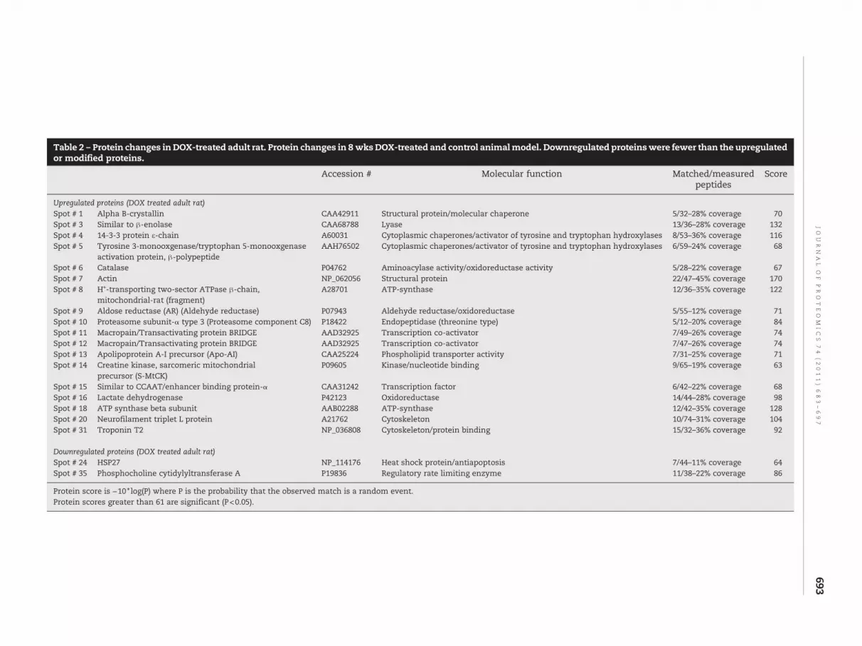
Table 2 – Protein changes in DOX-treated adult rat. Protein changes in 8 wksDOX-treated and control animalmodel. Downregulated proteinswere fewer than the upregulatedor modified proteins.
Accession # Molecular function Matched/measuredpeptides
Score
Upregulated proteins (DOX treated adult rat)Spot # 1 Alpha B-crystallin CAA42911 Structural protein/molecular chaperone 5/32–28% coverage 70Spot # 3 Similar to β-enolase CAA68788 Lyase 13/36–28% coverage 132Spot # 4 14-3-3 protein ε-chain A60031 Cytoplasmic chaperones/activator of tyrosine and tryptophan hydroxylases 8/53–36% coverage 116Spot # 5 Tyrosine 3-monooxgenase/tryptophan 5-monooxgenase
activation protein, β-polypeptideAAH76502 Cytoplasmic chaperones/activator of tyrosine and tryptophan hydroxylases 6/59–24% coverage 68
Spot # 6 Catalase P04762 Aminoacylase activity/oxidoreductase activity 5/28–22% coverage 67Spot # 7 Actin NP_062056 Structural protein 22/47–45% coverage 170Spot # 8 H+-transporting two-sector ATPase β-chain,
mitochondrial-rat (fragment)A28701 ATP-synthase 12/36–35% coverage 122
Spot # 9 Aldose reductase (AR) (Aldehyde reductase) P07943 Aldehyde reductase/oxidoreductase 5/55–12% coverage 71Spot # 10 Proteasome subunit-α type 3 (Proteasome component C8) P18422 Endopeptidase (threonine type) 5/12–20% coverage 84Spot # 11 Macropain/Transactivating protein BRIDGE AAD32925 Transcription co-activator 7/49–26% coverage 74Spot # 12 Macropain/Transactivating protein BRIDGE AAD32925 Transcription co-activator 7/47–26% coverage 74Spot # 13 Apolipoprotein A-I precursor (Apo-AI) CAA25224 Phospholipid transporter activity 7/31–25% coverage 71Spot # 14 Creatine kinase, sarcomeric mitochondrial
precursor (S-MtCK)P09605 Kinase/nucleotide binding 9/65–19% coverage 63
Spot # 15 Similar to CCAAT/enhancer binding protein-α CAA31242 Transcription factor 6/42–22% coverage 68Spot # 16 Lactate dehydrogenase P42123 Oxidoreductase 14/44–28% coverage 98Spot # 18 ATP synthase beta subunit AAB02288 ATP-synthase 12/42–35% coverage 128Spot # 20 Neurofilament triplet L protein A21762 Cytoskeleton 10/74–31% coverage 104Spot # 31 Troponin T2 NP_036808 Cytoskeleton/protein binding 15/32–36% coverage 92
Downregulated proteins (DOX treated adult rat)Spot # 24 HSP27 NP_114176 Heat shock protein/antiapoptosis 7/44–11% coverage 64Spot # 35 Phosphocholine cytidylyltransferase A P19836 Regulatory rate limiting enzyme 11/38–22% coverage 86
Protein score is −10 ⁎ log(P) where P is the probability that the observed match is a random event.Protein scores greater than 61 are significant (P<0.05).
693JO
UR
NA
LO
FPR
OT
EO
MIC
S74
(2011)
683–697

Fig. 5 – Prefractionated samples of DOX-treated adult rat cardiomyocytes — 2D gel analysis. Two dimensional gel analysis ofdifferent solubilization extracts. Protein extract of 300 μg was resolved and silver stained. The controls were 48 h treated withDMSO and DOX treatment were for the indicated period.
694 J O U R N A L O F P R O T E O M I C S 7 4 ( 2 0 1 1 ) 6 8 3 – 6 9 7
University Hospital and Faculty of Health Sciences, Denmark).We could not detect any marked modification. There is,however, a quantitative increase in ETFDH levels in DOX-treated samples. Therefore it is possible that the ETF-QOprotein levels and loss of ETF-β protein levels drastically affectcomplex III ability to function properly, thus increasing thechances of superoxide generated at this site. In addition,generation of superoxide at complex I site could be due toredox cycling of DOX. It would be interesting to obtain a directevidence to prove this point, as superoxide generated atcomplex I and complex III (Qi center) is released towards thematrix where glutathione and other oxidant protectionmachinery are present, while the superoxide generated fromcomplex III (Qo center — quinol oxidase) are released to theinner membrane side [67,68]. DOX also affects highly unsat-
urated fatty acids, desaturating and elongating enzymes thatare involved in the biosynthesis of essential fatty acids [69].Increased ROS formation in the mitochondria triggers theintrinsic pathway that leads to the opening of transition pores,and the process is favored by oxidation of glutathione andother sulfhydryls [70]. Events such as activation of BAK (BCL-2antagonist/Killer 2) by BCL2-Homology (BH3)-only proteinstranslocate to mitochondrial outer membrane. This results inthe translocation of cytochrome C to cytoplasm where ittriggers the formation of apoptosome in association withApaf1 (apoptotic protease activating factor 1) protein. Apopto-some activates caspase 9 which then activates the effectorcaspase 3 resulting in apoptosis.
Another important downregulated protein in response toDOX treatmentwas VDAC2. It iswell known that VDAC2 plays a

695J O U R N A L O F P R O T E O M I C S 7 4 ( 2 0 1 1 ) 6 8 3 – 6 9 7
critical role in mitochondrial apoptosis. VDAC2 is a member ofanion channel proteins (porins) residing in the outermitochon-drial membrane. These proteins are involved in regulation ofmetabolic interactions and solutes exchange between mito-chondria and cytosol, and in regulating the permeabilization ofouter mitochondrial membrane during apoptosis. Permeabili-zation of the membrane is known to initiate the release ofcytochrome c into the cytosol and initiate mitochondrialapoptotic pathway. VDAC2 is an isoform that is present in lowabundance in the outer mitochondrial membrane and specifi-cally interacts with amultidomain proapoptoticmember of Bcl-2 family of proteins BAK [71]. This interaction preventsoligomerization of BAK, the event that leads to the formationof outer mitochondrial membrane pores and leaking ofcytochrome c into the cytosol [71]. Genetic depletion ofVDAC2 resulted in excessive BAK oligomerization andapoptotic cell death, while overexpression of this proteinprevented BAK activation and inhibited mitochondrial path-way [71,72]. On the other hand, other studies performedon mouse embryonic fibroblasts and other cell types,have shown that VDAC2 recruits BAK to the mitochondrialmembrane and is required for BAX- and BID-induced apopto-sis [73,74]. The role of VDAC2 in the regulation of mitochon-drial apoptotic pathway is cardiomyocytes is not known.Interestingly, another proteomic study reported upregulationof VDAC2 abundance in hepatoma cells as a result of theaction of hepatocarcinogenic dioxin compound [75]. The fateof the regulation of VDAC2 expression in cardiomyocyteshas not been described previously. It has been known fromour previous publication that doxorubicin induces anabundant release of cytochrome c into the cytosol in bothadult and neonatal cardiomyocytes [76]. Changed expressionof this protein by doxorubicin could clearly be an importantfactor regulating the mitochondrial apoptotic pathway incardiomyocytes.
It is interesting to note that the proteomic changes in DOX-treated cardiomyocyte and rat heart are not similar. The exactreasons for this difference are not clear. However, the culturedmyocytes were treated with DOX for 24 or 48 h, whereas in vivocardiac tissues were harvested 7 days after the last injection.Under these conditions, no DOX was present in the cardiactissues isolated from rats. In contrast, DOX was continuouslypresent during in vitro cardiomyocyte cell culture experiments(thus generating oxy-radicals continuously). Thus, the differ-ence between DOX-induced effects in cardiomyocyte cultureand in vivo animals could be due to differences in the amountand duration of DOX exposure. Another reason for theobserved difference might also be possibly due to the factthat the adult rat heart tissue has more protein in the wholetissue extract. Therefore, we investigated other methods ofsample preparation to improve protein identification. Thesubcellular fractionation is a useful technique, but isolatedfractions are often contaminated with other subcellularorganelles. Charge fractionations have protein overlaps, andchromatographic fractionation requires a large amount ofsample. On the contrary, solubilization techniques are simple,highly reproducible, and enrich low abundant proteins in aninexpensive and reproducible manner. It is also advantageousto further separate them on narrow-range pH strips to furtherresolve the proteins. Our experimental results in Fig. 5 clearly
document the advantage of such a technique which will helpin identifying the changes occurring in the low abundantproteins.
5. Conclusions
The global proteomic analyses of DOX-treated cardiomyocytesand heart tissues isolated from DOX-treated adult rat indicatedifferent changes in proteome. The common protein changesbetween the in vitro cellular model and the in vivo animalmodel areoverexpressionof beta enolase, troponin andalphaB-crystallin. Othermajor protein changes detected in tissuesweremuch different from those identified in the cell culture model.
Acknowledgements
This work was made possible with the help of NIH grantsR01CA152810 and 1UL1RR031973.
R E F E R E N C E S
[1] Singal PK, Iliskovic N. Doxorubicin induced cardiomyopathy.N Engl J Med 1998;339:900–5.
[2] Kremer LC, Van Dalen EC, Offringa M, Ottenkamp J, Voute PA.Anthracycline induced clinical heart failure in a cohort of 607children: long term follow up study. J Clin Oncol 2001;19:191–6.
[3] Kawasaki N, Lee JD, Shimizu H, Ueda T. Long term L-carnitinetreatment prolongs the survival in rats with adriamycininduced heart failure. J Card Fail 1996;2:293–9.
[4] Powis G. Free radical formation by anti tumor quinones. FreeRadic Biol Med 1989;6:63–101.
[5] Myers CE, McGuire RH, Liss RH, Ifrim I, Grotzinger K, YoungRC. Adriamycin: the role of lipid peroxidation in cardiactoxicity and tumor response. Science 1977;197:165–7.
[6] Andrieu-Abadie N, Jaffrezou J, Hatem S, Laurent G, Levade T,Mercadier J. L-carnitine prevents doxorubicin inducedapoptosis of cardiac myocytes: role of inhibition of ceramidegeneration. FASEB J 1999;13:1501–10.
[7] Konorev EA, Kotamraju S, Zhao H, Kalivendi S, Joseph J,Kalyanaraman B. Paradoxical effects of metalloporphyrins ondoxorubicin induced apoptosis. scavenging of reactive oxygenspecies versus induction of heme oxygenase-1. Free Radic BiolMed 2002;33:988–97.
[8] Adachi K, Fujiura Y, Mayumi F, Nozuhara A, Sugiu Y,Sakanashi T, et al. A deletion ofmitochondrial DNA inmurinedoxorubicin induced cardiotoxicity. Biochem Biophys ResCommun 1993;195:945–51.
[9] Zhou S, Starkov A, Froberg MK, Leino RL, Wallace KB.Cumulative and irreversible cardiac mitochondrialdysfunction induced by doxorubicin. Cancer Res 2001;61:771–7.
[10] Lebrecht D, Setzer B, Ketelsen UP, Haberstroh J, Walker UA.Time dependent and tissue specific accumulation of mtDNAand respiratory chain defects in chronic doxorubicincardiomyopathy. Circulation 2003;108:2423–9.
[11] Davies KJ, Doroshow JH. Redox cycling of anthracyclines bycardiac mitochondria. I. Anthracycline radical formation byNADH dehydrogenase. J Biol Chem 1986;261:3060–7.
[12] Jung K, Reszka R. Mitochondria as sub cellular targets forclinically useful anthracyclines. Adv Drug Deliv Rev 2001;49:87–105.

696 J O U R N A L O F P R O T E O M I C S 7 4 ( 2 0 1 1 ) 6 8 3 – 6 9 7
[13] Vasquez-Vivar J, Martasek P, Hogg N, Masters BS, Pritchard KA,Kalyanaraman B. Endothelial nitric oxide synthase-dependentsuperoxide generation from adriamycine. Biochemistry1997;36:11293–7.
[14] Minotti G, Cairo G, Monti E. Role of iron in anthracyclinecardiotoxicity : new tunes for an old song? FASEB J 1999;13:199–212.
[15] Kalyanaraman B, Joseph J, Kalivendi S, Wang S, Konorev E,Kotamraju S. Doxorubicin induced apoptosis: implications incardiotoxicity. Mol Cell Biochem 2002;234–235:119–24.
[16] Doroshow JH, Davis KJ. Redox cycling of anthracyclines bycardiac mitochondria. II. Formation of superoxide anion,hydrogen peroxide and hydroxyl radical. J Biol Chem1986;261:3068–74.
[17] Winterbourn CC, Gutteridge JMC, Halliwell B.Doxorubicin-dependent lipid peroxidation at low partialpressure of O2. J Free Radic Biol Med 1985;1:43–9.
[18] Gille L, Nohl H. Analyses of the molecular mechanism ofadriamycin-induced cardiotoxicity. Free Radic Biol Med1997;23:775–82.
[19] Torti FM, Torti SV. Regulation of ferritin genes and protein.Blood 2002;99:3505–16.
[20] Brazzolotto X, Gaillard J, Pantopoulos K, Hentze MW, MoulisJM. Human cytoplasmic aconitase (Iron regulatory protein 1)is converted into its [3Fe–4S] form by hydrogen peroxide invitro but is not activated for iron responsive element binding.J Biol Chem 1999;274:21625–30.
[21] Minotti G, Recalcati S, Liberi G, Calafiore AM, Mancuso C,Preziosi P, et al. The secondary alcohol metabolite ofdoxorubicin irreversibly inactivates aconitase/iron regulatoryprotein-1 in cytosolic fractions from human myocardium.FASEB J 1998;12:541–52.
[22] Minotti G, Ronchi R, Salvatorelli E, Menna P, Cairo G.Doxorubicin irreversibly inactivates iron regulatory protein 1and 2 in cardiomyocytes. Evidence for distinct metabolicpathways and implications for iron mediated cardiotoxicityof antitumor therapy. Cancer Res 2001;61:8422–8.
[23] Tobin TP, Abbot BC. A stereological analysis of the effect ofadriamycin on the ultrastructure of rat myocardial cells inculture. J Mol Cell Cardiol 1980;12:1207–25.
[24] Kumar D, Kirshenbaum L, Li T, Danelisen I, Singal P.Apoptosis in isolated adult cardiomyocytes exposed toadriamycin. Ann N Y Acad Sci 1999;874:156–68.
[25] Li T, Danelisen I, Bello-Klein A, Singal PK. Effect of probucol onchanges of antioxidant enzymes in adriamycin inducedcardiomyopathy in rats. Cardiovasc Res 2000;46:523–30.
[26] Heon S, Bernier M, Servant N, Dostanic S, Wang C, Kirby GM,et al. Dexrazoxane does not protect against doxorubicininduced damage in the young rat. Am J Physiol Heart CircPhysiol 2003;285:H499–506.
[27] Lawenda BD, Kelly KM, Ladas EJ, Sagar SM, Vickers A, BlumbergJB. Should supplemental antioxidant administration beavoided during chemotherapy and radiation therapy? J NatlCancer Inst 2008;100:773–83.
[28] Block KI, Koch AC, Mead MN, Tothy PK, Newman RA,Gyllenhaal C. Impact of antioxidant supplementation onchemotherapeutic toxicity: a systematic review of theevidence from randomized controlled trials. Int J Cancer2008;123:1227–39.
[29] Ito H, Miller SC, Billingham ME, Akimoto H, Torti SV, Wade R,et al. Doxorubicin selectivity inhibits muscle gene expressionin cardiac muscle cells in vivo and in vitro. Proc Natl Acad Sci1990;87:4275–9.
[30] Jeyaseelan R, Poizat C, Baker RK, Abdishoo S, Isterabadi LB,Lyons GE, et al. A novel cardiac-restricted target fordoxorubicin. CARP, a nuclear modulator of gene expression incardiac progenitor cells and cardiomyocytes. J Biol Chem1997;272:22800–8.
[31] Jeyaseelan R, Poizat C,WuHY, Kedes L. Molecularmechanismsof doxorubicin induced cardiomyopathy. Selective suppressionof Reiske iron sulfur protein, ADP/ATP translocase andphosphofructokinase genes is associatedwithATP depletion inrat cardiomyocytes. J Biol Chem 1997;272:5828–32.
[32] Cutts SM, Parsons PG, Strum RA, Phillips DR. Adriamycininduced DNA adducts inhibit DNA interactions of transcriptionfactors and RNA polymerase. J Biol Chem 1996;271:5422–9.
[33] Wang S, Kotamraju S, Konorev E, Kalivendi S, Joseph J,Kalyanaraman B. Activation of nuclear factor-kappaB duringdoxorubicin induced apoptosis in endothelial cells andmyocytes is proapoptotic: the role of hydrogen peroxide.Biochem J 2002;367:729–40.
[34] GigliM,Doglia SM,Millot JM,Valentini L,ManfaitM.Quantitativestudy of doxorubicin in living cell nuclei bymicrospectrofluorometry. BiochimBiophysActa 1988;950:13–20.
[35] L'Ecuyer T, Sanjeev S, Thomas R, Novak R, Das L, Cambell W,et al. DNA damage is an early event in doxorubicin inducedcardiac myocyte death. Am J Physiol Heart Circ Physiol2006;291:H1273–80.
[36] Ellis CN, Ellis MB, Blakemore WS. Effect of adriamycin onheart mitochondrial DNA. Biochem J 1987;245:309–12.
[37] Piper HM, Volz A, Schwartz P. Adult ventricular rat heart cells.In: Piper HM, editor. Cell Culture Techniques in Heart andVessel Research. Heidelberg: Springer Verlag; 1990. p. 36–60.
[38] Hohl CM, Altschuld RA, Brierley GP. Effects of calcium on thepermeability of isolated adult rat heart cells to sodium. ArchBiochem Biophys 1982;221:197–205.
[39] Armstrong SC, Ganote CE. Effects of 2,3-butanedionemonoxime (BDM) on contracture and injury of isolated ratmyocytes following metabolic inhibition and ischemia. J MolCell Cardiol 1991;23:1001–14.
[40] Konorev EA, Kennedy MC, Kalyanaraman B. Cell-permeablesuperoxide dismutase and glutathione peroxidase mimeticsafford superior protection against doxorubicin inducedcardiotoxicity. The role of reactive oxygen and nitrogenintermediates. Arch Biochem Biophys 1999;368:421–8.
[41] Kotamraju S, Konorev EA, Joseph J, Kalyanaraman B.Doxorubicin induced apoptosis in endothelial cells andcardiomyocytes is ameliorated by nitrone spin traps andebselen. Role of reactive oxygen and nitrogen species. J BiolChem 2000;275:33585–92.
[42] Gharahdaghi F, Weinberg CR, Meagher DA, Imai BS, MischeSM. Mass spectrometric identification of proteins from silverstained polyacrylamide gels: a method for the removal ofsilver ions to enhance sensitivity. Electrophoresis 1999;20:601–5.
[43] Shevchenko A,WilmM, VormO, MannM.Mass spectrometricsequencing of proteins silver stained polyacrylamide gels.Anal Chem 1996;68:850–8.
[44] Nagi MN, Mansour MA. Protective effect of thymoquinoneagainst doxorubicin-induced cardiotoxicity in rats: a possiblemechanism of protection. Pharmacol Res 2000;41:283–9.
[45] Kim YK, Lee WK, Jin Y, Lee KJH, Yu YG. Doxorubicin binds toun-phosphorylated form of hNopp 140 and reduces proteinkinase CK2-dependent phosphorylation of hNopp140.J Biochem Mol Biol 2006;39:774–81.
[46] Green PS, Leeuwenburgh C. Mitochondrial dysfunction is anearly indicator of doxorubicin induced apoptosis. BiochimBiophys Acta 2002;1588:94–101.
[47] Tokarska-SchlattnerM, DolderM, Gerber I, Speer O,WallimannT, Schlattner U. Reduced creatine stimulated respiration indoxorubicin challenged mitochondria: particular sensitivity ofthe heart. Biochim Biophys Acta 2007;1767:1276–84.
[48] Salvatorelli E, Guarnieri S, Menna P, Liberi G, Calafiore AM,Mariggio MA, et al. Defective one- and two-electron reductionof the anticancer anthracycline epirubicin in human heart.Relative importance of vesicular sequestration and impaired

697J O U R N A L O F P R O T E O M I C S 7 4 ( 2 0 1 1 ) 6 8 3 – 6 9 7
efficiency of electron addition. J Biol Chem 2006;281:10990–1001.
[49] Zhou S, Palmeira CM, Wallace KB. Doxorubicin inducedpersistent oxidative stress to cardiac myocytes. Toxicol Lett2001;121:151–7.
[50] Romisch J, Grote M, Weithmann KU, Heimburger N, Amann E.Annexin protein PP4 and PP4-X: comparative characterizationof biological activities of placental and recombinant proteins.Biochem J 1990;272:223–9.
[51] Strauss HW, Narula J, Blankenberg F. Radioimaging to identifymyocardial cell deathandprobably injury. Lancet 2000;356:180–1.
[52] Andree HA, Reutelingsperger CP, Hauptmann R, Hemker HC,Hermens WT, Willems GM. Binding of vascular anticoagulant(VAC) to planar phospholipid bilayers. J Biol Chem 1990;265:4923–8.
[53] Ray PS, Martin JL, Swanson EA, Otani H, Dillmann WH, DasDK. Transgene over expression of alpha B crystallin conferssimultaneous protection against cardiomyocyte apoptosisand necrosis during myocardial ischemia and reperfusion.FASEB J 2001;15:393–402.
[54] Klemenz R, Frohli E, Steiger RH, Schafer R, Aoyama A. AlphaB-crystallin is a small heat shock protein. Proc Natl Acad Sci1991;88:3652–6.
[55] Abdel-aleem S, el-Merzabani MM, Syed-ahmed M, Taylor DA,Lowe JE. Acute and chronic effects of adriamycine on fattyacid oxidation in isolated cardiac myocytes. J Mol Cell Cardiol1997;29:789–97.
[56] Hong YM, Kim HS, Yoon HR. Serum lipid and fatty acidprofiles in adriamycine treated rats after administration ofL-carnitine. Pediatr Res 2002;51:249–55.
[57] Zong H, Ren JM, Young LH, Pypaert M, Mu J, Birnbaum J, et al.AMP kinase is required for mitochondrial biogenesis inskeletal muscle in response to chronic energy deprivation.Proc Natl Acad Sci 2002;99:15983–7.
[58] Berthiaume L, Deichaite I, Peseckis S, Resh MD. Regulation ofenzymatic activity by active site fatty acylation. A new role forlong chain fatty acid acylation of proteins. J Biol Chem1994;269:6498–505.
[59] Kanski J, Behring A, Pelling J, Schoeneich C. Proteomicidentification of 3-nitrotyrosine containing rat cardiacproteins. Effects of biological aging. Am J Physiol Heart CircPhysiol 2005;288:H371–81.
[60] Pacher P, Liaudet L, Bai P, Virag L, Mabley JG, Hasko G, et al.Activation of poly(ADP-ribose) polymerase contributes todevelopment of doxorubicin-induced heart failure.J Pharmacol Exp Ther 2002;300:862–7.
[61] Mukhopadhyay P, Rajesh M, Batkai S, Kashiwaya Y, Hasko G,Liaudet L, et al. Role of superoxide, nitric oxide, andperoxynitrite in doxorubicin-induced cell death in vivo andinvitro. Am J Physiol Heart Circ Physiol 2009;296:H1466–83.
[62] Arai M, Tomaru K, Takizawa T, Sekiguchi K, Yokoyama T,Suzuki T, et al. Sarcoplasmic reticulum genes are selectivelydownregulated in cardiomyopathy produced by doxorubicinin rabbits. J Mol Cell Cardiol 1998;30:243–54.
[63] Turakhia S, Venkatakrishnan CD, Dunsmore K, Wong H,Periannan K, Zweier JL, et al. Doxorubicin-inducedcardiotoxicity: direct correlation of cardiac fibroblasts andH9c2cells survival and aconitase activitywith heat shock protein 27.Am J Physiol Heart Circ Physiol 2007;293:H3111–21.
[64] O'Callaghan-Sunol C, Gabai VL, ShermanMY.Hsp27modulatesp53 signaling and suppresses cellular senescence. Cancer Res2007;67:11779–88.
[65] Arrigo AP, Landry J. Expression and function of the lowmolecular weight heat shock proteins. In: Morimoto RI,Tissieres A, Georgopoulos C, editors. The biology of heat shockproteinsandmolecular chaperones,Vol. 26.ColdSpringHarbor,NY: Cold Spring Harbor Laboratory Press; 1994. p. 335–73.
[66] Venkatakrishnan CD, Tewari AT, Moldovan L, Cardounel AJ,Zweier JL, Kuppuswamy P, et al. Heat shock protects cardiaccells from doxorubicin induced toxicity by activating p38MAPK and phosphorylation of small heat shock protein 27.Am J Physiol Heart Circ Physiol 2006;291:H2680–91.
[67] Chen Q, Vazquez EJ, Moghaddas S, Hoppel CL, Lesnefsky EJ.Production of reactive oxygen species by mitochondria.Central role of complex III. J Biol Chem 2003;278:36027–31.
[68] Young TA, Cunningham CC, Bailey SM. Reactive oxygenspecies production by the mitochondrial respiratory chain inisolated rat hepatocytes and liver mitochondria : studiesusing myxothiazol. Arch Biochem Biophys 2002;405:65–72.
[69] Bordoni A, Biagi P, Hrelia S. The impairment of essential fattyacid metabolism as a key factor in doxorubicin induceddamage in cultured rat cardiomyocytes. Biochim BiophysActa 1999;1440:100–6.
[70] Chernyak BV. Redox regulation of the mitochondrialpermeability transition pore. Biosci Rep 1997;17:293–302.
[71] Cheng EH, Sheiko TV, Fisher JK, Craigen WJ, Korsmeyer SJ.VDAC2 inhibits BAK activation and mitochondrial apoptosis.Science 2003;301:513–7.
[72] Ren D, Kim H, Tu HC, Westergard TD, Fisher JK, Rubens JA,et al. The VDAC2-BAK rheostat controls thymocyte survival.Sci Signal 2009;2:ra48.
[73] Roy SS, Ehrlich AM, Craigen WJ, Hajnoczky G. VDAC2 isrequired for truncated BID-inducedmitochondrial apoptosisby recruiting BAK to mitochondria. EMBO Rep 2009;10:1341–7.
[74] Yamagata H, Shimizu S, Nishida Y, Watanabe Y, Craigen WJ,Tsujimoto Y. Requirement of voltage-dependent anionchannel 2 for pro-apoptotic activity of Bax. Oncogene 2009;28:3563–72.
[75] Sarioglu H, Brandner S, HabergerM, Jacobsen C, LichtmanneggerJ, Wormke M, et al. Analysis of 2,3,7,8-tetrachlorodibenzo-p-dioxin-induced proteome changes in 5L rat hepatoma cellsrevealsnovel targetsofdioxinaction including themitochondrialapoptosis regulator VDAC2. Mol Cell Proteomics 2008;7:394–410.
[76] Konorev EA, Vanamala S, Kalyanaraman B. Differences indoxorubicin-induced apoptotic signaling in adult andimmature cardiomyocytes. Free Radic Biol Med 2008;45:1723–8.


