Embed Size (px)
Citation preview
The
Jour
nal o
f G
ener
al P
hysi
olo
gy
A RT I C L E
The Rockefeller University Press $30.00J. Gen. Physiol. Vol. 132 No. 4 397–405www.jgp.org/cgi/doi/10.1085/jgp.200809960 397
I N T R O D U C T I O N
Adult ventricular cardiomyocytes are not spontaneously
active on their own, but instead are specialized to form
an electrically excitable meshwork, where excitation re-
lies on highly developed cell-to-cell contacts and spe-
cialized pacemaker cells. Compared with adult myocytes,
embryonic cardiomyocytes show remarkable functional
plasticity. They must be able to maintain their activity
during transition periods without physical connections
to other cells, enabling cell migration in the developing
heart ( Buckingham et al., 2005 ). In addition, these cardio-
myocytes have the ability to synchronize their electrical ac-
tivity and contraction with other cells to form coordinated
contraction ( Kamino, 1991 ), just like adult cardiomyo-
cytes. This functional versatility of embryonic cardiomyo-
cytes is essential to ensure the continuous beating of the
heart muscle cells, a prerequisite for the survival of the
embryo and development of the heart itself ( Copp, 1995 ;
Puceat and Jaconi, 2005 ).
The mechanisms behind the induction and mainte-
nance of the initial activity of the cardiomyocytes have
not been identifi ed, but two separate, seemingly contra-
dictory mechanisms have been proposed on the basis of
experiments with isolated cells and genetically manipu-
lated animals and cells. One line of evidence suggests
that upon spontaneous depolarization of membrane
voltage (V m ), calcium infl ux through voltage-activated
calcium channels (VACCs) triggers myocyte contraction,
Correspondence to Pasi Tavi: p a s i . t a v i @ o u l u . fi
Abbreviations used in this paper: AM, acetoxymethyl; AP, action poten-
tial; E – C, excitation – contraction; I f , hyperpolarization-activated current;
IP 3 , inositol-3-phosphate; IP 3 R, IP 3 receptor; NCX, NA + /Ca 2+ exchanger;
RyR, ryanodine receptor; SL, sarcolemmal; VACC, voltage-activated cal-
cium channel; V m , membrane voltage.
The online version of this article contains supplemental material.
with little or no contribution of calcium release from
intracellular stores inside the SR ( Nakanishi et al., 1988 ;
Takeshima et al., 1998 ). Opposing data indicates that
spontaneous SR calcium oscillations in fact drive the
contractions and electrical activity ( Viatchenko-Karpinski
et al., 1999 ; Mery et al., 2005 ; Sasse et al., 2007 ). From
the early stages of cardiogenesis, mammalian cardio-
myocytes have both an excitable membrane with a vari-
ety of voltage-activated ion channels ( Doevendans et al.,
2000 ; Seisenberger et al., 2000 ; Cribbs et al., 2001 ) and
a functional SR ( Moorman et al., 2000 ; Seki et al., 2003 ),
suggesting that the components for V m oscillation-
driven calcium infl ux and SR-induced calcium release
may coexist in the myocytes during early embryonic de-
velopment. However, it is not known whether all embry-
onic cardiomyocytes possess both of these features or
if specialized cell types are needed to fulfi ll these dif-
ferent tasks. We identify the mechanism by which spon-
taneous beating of solitary embryonic cardiomyocytes
originates from oscillatory calcium releases from the
SR. We further report that the same cells have a mecha-
nism for action potential (AP)-induced calcium infl ux
and subsequent CICR from the SR. These results ex-
plain the mechanisms of how the cardiac myocytes are
autonomous in producing their basic activity during the
early phases of heart development, yet are able to syn-
chronize their activity with other myocytes. Further, we
explain how these mechanisms are regulated to modify
the beating rate of the developing heart. These charac-
terized mechanisms could provide the explanation for
Excitation – Contraction Coupling of the Mouse Embryonic Cardiomyocyte
Risto Rapila , Topi Korhonen , and Pasi Tavi
Institute of Biomedicine, Department of Physiology and Biocenter Oulu, University of Oulu, 90014 Oulu, Finland
In the mammalian embryo, the primitive tubular heart starts beating during the fi rst trimester of gestation. These early heartbeats originate from calcium-induced contractions of the developing heart muscle cells. To explain the initiation of this activity, two ideas have been presented. One hypothesis supports the role of spontaneously acti-vated voltage-gated calcium channels, whereas the other emphasizes the role of Ca 2+ release from intracellular stores initiating spontaneous intracellular calcium oscillations. We show with experiments that both of these mech-anisms coexist and operate in mouse cardiomyocytes during embryonic days 9 – 11. Further, we characterize how inositol-3-phosphate receptors regulate the frequency of the sarcoplasmic reticulum calcium oscillations and thus the heartbeats. This study provides a novel view of the regulation of embryonic cardiomyocyte activity, explaining the functional versatility of developing cardiomyocytes and the origin and regulation of the embryonic heartbeat.
© 2008 Rapila et al. This article is distributed under the terms of an Attribution–Noncom-mercial–Share Alike–No Mirror Sites license for the fi rst six months after the publication date (see http://www.jgp.org/misc/terms.shtml). After six months it is available under a Cre-ative Commons License (Attribution–Noncommercial–Share Alike 3.0 Unported license, as described at http://creativecommons.org/licenses/by-nc-sa/3.0/).
on June 13, 2016jgp.rupress.org
Dow
nloaded from
Published September 15, 2008
http://jgp.rupress.org/content/suppl/2008/09/15/jgp.200809960.DC1.html Supplemental Material can be found at:
398 Embryonic E – C Coupling
(pH 7.4, bubbled with 95% O 2 /5% CO 2 ). To measure myocyte calcium signals, Fluo-4 – loaded myocytes were excited at 488 nm and the emitted light was collected with a spectral detector from 520 to 620 nm through a 20 × objective lens. To excite the cells, myocytes were stimulated with 1-ms voltage pulses 50% over the excitation threshold through two platinum wires located on both sides of the Petri dish. At the time of electrical stimulation or spontaneous activity, cells were line scanned at 400 – 600 Hz de-pending on the length of the scanning line, with a fi xed pixel time of 10 μ s. The frame scan images (256 × 256 pixels) were ac-quired at 23.7 Hz through a 20 × objective and converted into a movie fi le (FluoView 1000; Olympus). The line scan images were analyzed with the ImageJ 1.36b (http://rsb.info.nih.gov/ij/) and Origin 7.5 (OriginLab Co.) programs. Fluo-4 fl uorescence inten-sity is expressed as an F/F 0 -ratio, where F is the background sub-tracted fl uorescence intensity and F 0 is background subtracted minimum fl uorescence value measured from each cell at rest.
Electrophysiological Recordings Primary isolated E9-11 embryonic cardiomyocytes were placed sparsely on laminin-coated glass-bottom Petri dishes and cultured for 12 – 20 h. The whole cell patch-clamp method was used to re-cord whole cell currents and APs. Electrode resistances were 1 – 5 M Ω in whole cell current recordings and 10 – 15 M Ω in AP record-ings. The membrane capacitances were measured by applying a 5-mV pulse to the holding potential. The hyperpolarization-acti-vated current (I f ) was measured by applying voltage clamps rang-ing from � 50 to � 120 mV with 10-mV steps from a 0-mV holding potential. I f was defi ned as the difference between the initial cur-rent at the beginning of the voltage clamp and the current at the end of the voltage clamp. The solutions and the protocol were ad-opted from a previous report ( Yasui et al., 2001 ). The external so-lution contained 137 mM NaCl, 5.4 mM KCl, 0.5 mM MgCl, 1 mM CaCl 2 , 11.8 mM HEPES, 10 mM glucose, 2 mM BaCl 2 , 2 mM NiCl 2 , and 0.5 mM 4-AP, pH 7.40 (NaOH), and the pipette solution con-tained 80 mM KCl, 60 mM K-aspartic acid, 5 mM Na 2 -phosphocre-atine, 5 mM Mg-ATP, 0.65 mM CaCl 2 , 10 mM EGTA, and 5 mM HEPES, pH 7.20 (KOH). APs were measured using the current clamp mode (I = 0). In some experiments when APs were re-corded together with [Ca 2+ ] i , cells were preloaded with Fluo-4 and the cells were continuously line-scanned (see above) at the time of V m recordings. The intracellular solution used was the same as described previously ( Yang et al., 2005 ) and contained 120 mM K-aspartate, 25 mM KCl, 1 mM MgCl 2 , 2 mM Na 2 phos-phocreatine, 4 mM Na 2 ATP, 2 mM NaGTP, 10 mM EGTA, and 5 mM HEPES, pH 7.2 (KOH), and the bath solution was Dulbecco ’ s modifi ed Eagle medium plus glutamax I (Invitrogen). The Na + /Ca 2+ current (I NCX ) and [Ca 2+ ] i were recorded simultaneously us-ing the Ca 2+ imaging methods and perforated-patch whole cell voltage clamp (120 μ g/ml amphotericin-B). Solutions and proto-col were as described previously ( Ginsburg and Bers, 2005 ), ex-cept that the V m was clamped to � 70 mV in I NCX recordings. To block I NCX , 10 mM Ni 2+ was added. The bath solution contained 140 mM NaCl, 4 mM CsCl, 1 mM MgCl 2 , 2 mM CaCl 2 , and 10 mM HEPES, pH 7.4 (NaOH) plus 10 mM NiCl 2 , and the pipette con-tained 40 mM CsCl, 80 mM Cs-methanesulfonate, 1 mM MgCl 2 , 1 mM KCl, and 10 mM HEPES, pH 7.2 (CsOH). Whole cell cur-rents were fi ltered at 2 kHz and acquired at 10 kHz. Clampex 9.2 software and Axopatch-1D amplifi er and Digidata 1322A A/D-D/A (Axon Instruments) were used to control the command poten-tials and data acquisition. All electrophysiological measurements were done at +34 ° C. Data analysis was made using the Clampfi t 9.2 (Axon Instruments), Origin 7.5 (OriginLab Corporation), and Matlab 6.5 (The Mathworks) software. Clampfi t ’ s leak resis-tance subtraction and software fi lters were used when necessary. The membrane currents were scaled by dividing them with the cell membrane capacitance.
the origin and the regulation of the heartbeats before
the differentiation of the functional pacemaker cells.
M AT E R I A L S A N D M E T H O D S
Cell Isolation and Culturing A previously described method was modifi ed to isolate and cul-ture embryonic days 9 – 11 (E9 – 11) embryonic cardiomyocytes ( Sturm and Tam, 1993 ; Liu et al., 1999 ; Doevendans et al., 2000 ). Shortly, pregnant mice were killed by cervical dislocation. Em-bryos were excised and transferred to +2 ° C isolation buffer con-taining 100 mM NaCl, 10 mM KCl, 1.2 mM KH 2 PO 4 , 4 mM MgSO 4 , 50 mM taurine, 20 mM glucose, and 10 mM HEPES (pH 6.9 with NaOH). Ventricles were cut from the embryos under a stereo-microscope. Dissected ventricles were incubated in the same solution containing 2 mg/ml pancreatin (Sigma-Aldrich) and 2 mg/ml collagenase type II (Worthington) for 50 min at 37 ° C on a rotating Ferris wheel. The tissue was mechanically dissociated by gentle trituration with a fi re-polished glass pipette. The cell suspension was centrifuged at 750 g for 5 min followed by resus-pension in Dulbecco ’ s modifi ed Eagle medium plus glutamax I (Invitrogen) with 10% FBS and 1% penicillin/streptomycin. Cells were plated on laminin-coated glass coverslips and incubated for 12 – 20 h at 37 ° C in a humidifi ed 5% CO 2 incubator. Pregnant CD-1 mice from the Center for Experimental Animals at the Uni-versity of Oulu were used. The experimental designs were ap-proved by the Animal Use and Care Committee of the University of Oulu.
Immunofl uorescence Labeling and Microscopy E10 cardiomyocytes were fi rst cultured for 16 h on glass-bottom Petri dishes, and then rinsed with 0.1 M Tris-HCl, pH 7.3, fi xed with 3% paraformaldehyde for 2 min, and permeabilized for 10 min with 0.5% Triton X-100. After washing with 0.1 M Tris-HCl, pH 7.3, twice for 5 min, the primary general ryanodine receptor (RyR; goat anti-RyR; Santa Cruz Biotechnology, Inc.) and general anti – inositol-3-phosphate (IP 3 ) receptor (IP 3 R; rabbit anti-IP 3 R I/II/III; Santa Cruz Biotechnology, Inc.) antibodies were incubated for 1 h in 0.1 M Tris-HCl, pH 7.3, containing 10% FBS and 0.05% Triton X-100. Again, the specimens were washed twice and the secondary antibody (Alexa Fluor 488 chicken anti – goat and chicken anti – rabbit; Invitrogen) was incubated (pH 7.3) for 1 h. Dilutions for primary antibodies were 1:500, and the secondary antibody dilution was 1:750. After labeling, images were taken freshly with a confocal microscope (excitation 488 nm, emission 505 – 600, and 60 × objective; FV1000; Olympus) at 0.2 μ m/pixel spatial resolution.
Confocal Ca 2+ Imaging Primary isolated E9 – 11 embryonic cardiomyocytes were grown on laminin-coated glass-bottom Petri dishes for 12 – 20 h. Because car-diomyocytes are known to differentiate during long culture peri-ods ( Husse and Wussling, 1996 ; Zimmermann et al., 2002 ), the culturing times of the cells before starting the experiments were kept to a minimum. 12 – 20 h was considered suffi cient time for the cells to recover from the isolation, but short enough to pre-vent them from further differentiating in culture. Cardiomyocytes were loaded with Fluo-4-acetoxymethyl (AM)-ester (1 μ M dis-solved in pluronic DMSO; Invitrogen) in culture medium for 30 min at 37 ° C in an incubator. The solution was changed twice, and cells were incubated at 37 ° C for at least 30 min for the dye to de-esterify. After this, the culturing dishes were placed in a cus-tom-made perfusion system built into a confocal inverted micro-scope (FluoView 1000; Olympus). Cells were held at a steady 34 ° C by continuous superfusion with preheated Dulbecco ’ s modifi ed Eagle medium plus glutamax I (Invitrogen) culturing medium
on June 13, 2016jgp.rupress.org
Dow
nloaded from
Published September 15, 2008
Rapila et al. 399
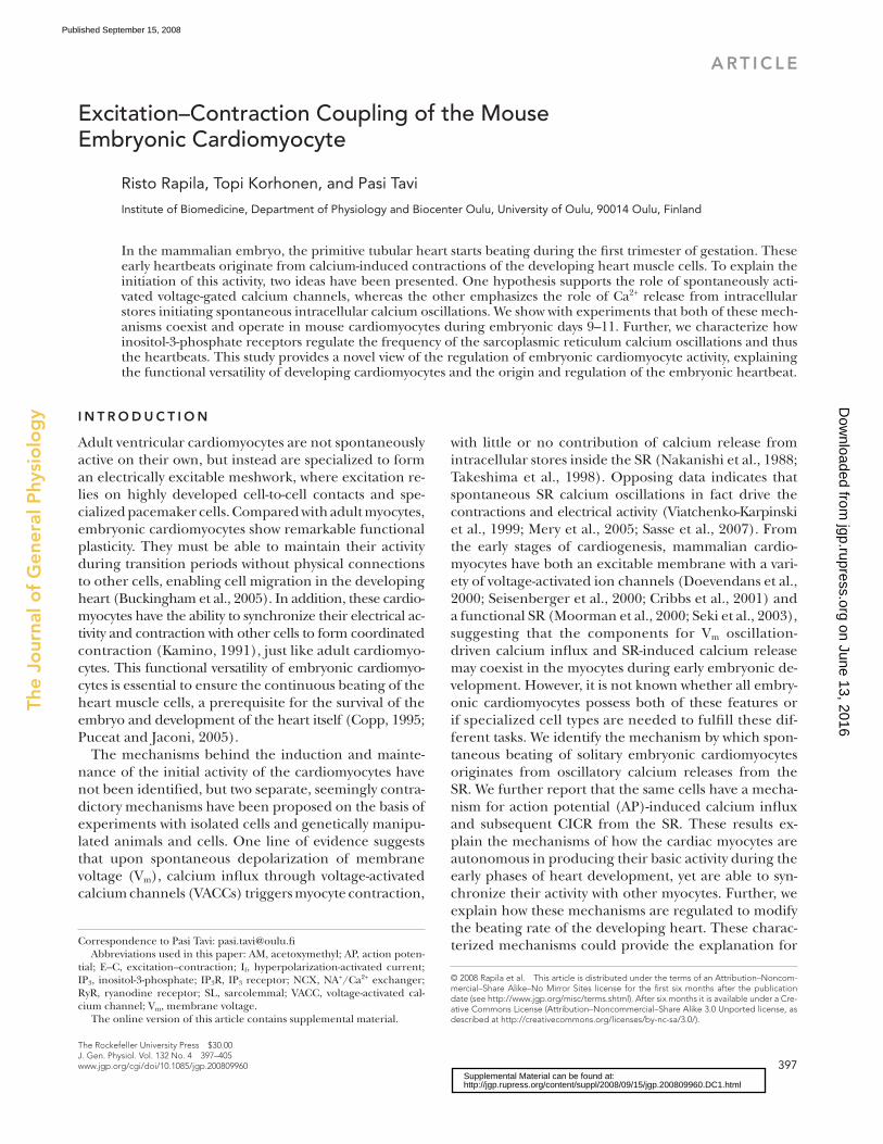
change was observed in three out of seven. Because this
effect was not consistent, the effect of I f block on the
frequency or the amplitude of the spontaneous calcium
signals was not statistically signifi cant ( Fig. 1, B – D ). We
next asked if the suggested spontaneous SR calcium os-
cillations ( Sasse et al., 2007 ) could provide an alternative
explanation for this activity. Therefore, we did confocal
calcium imaging of the E9 – 11 cardiomyocytes to char-
acterize the spatio-temporal properties of the spontane-
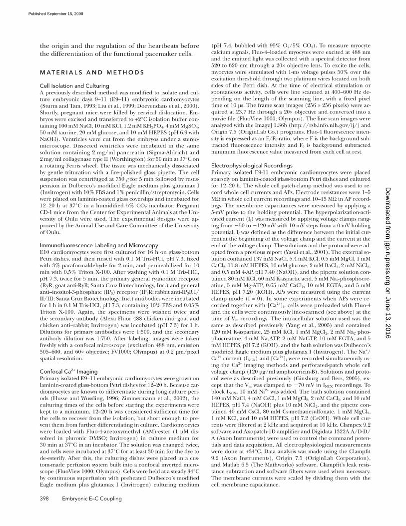
ous calcium signals. We noticed that calcium signals are
initiated at the vicinity of the thin perinuclear area ( Fig.
2 A ) corresponding to the location of the SR in develop-
ing cardiomyocytes ( Mesaeli et al., 1999 ). After the ini-
tial release from the SR, calcium diffuses in the cytosol
and the [Ca 2+ ] i increases near the plasma membrane after
a delay ( Fig. 2 A and Video 1, which is available at http://
www.jgp.org/cgi/content/full/jgp.200809960/DC1).
The delay between [Ca 2+ ] i rise near the SR and near the
sarcolemmal (SL) varied from 8 to 68 ms, with 28.5 ±
4.9 ms ( n = 12) average delay. This SR calcium release
appeared to be relatively well developed; the amplitude of
the spontaneous global calcium signal was 56.5 ± 0.5%
( n = 23) of the amplitude of the caffeine-induced cal-
cium transient, indicating that 56.5% of the available
Statistics Results are expressed as mean ± SEM. Statistical signifi cance of difference was analyzed using the paired t test and one-way ANOVA. The r 2 and p-values for linear fi t were determined using unweighted linear regression.
Online Supplemental Material The online supplemental material includes a video from a spontane-ously active embryonic cardiomyocyte, which is loaded with Fluo-4 and frame scanned with a confocal microscope. Video 1 is available at http://www.jgp.org/cgi/content/full/jgp.200809960/DC1.
R E S U LT S
Origin of the Intracellular [Ca 2+ ] Oscillations To elucidate the mechanisms triggering cardiomyocyte
activity we did confocal [Ca 2+ ] i imaging and electrophys-
iological measurements from spontaneously beating
isolated single embryonic (E9 – 11) myocytes. Without
physical connections to other cells, isolated embryonic
myocytes contract spontaneously at a rate of 0.40 ± 0.03
Hz ( n = 94), accompanied by oscillations in [Ca 2+ ] i and
V m ( Table I ). It was suggested earlier that spontaneous
plasmalemmal APs triggered by the I f initiate this activ-
ity ( Nakanishi et al., 1988 ; Takeshima et al., 1998 ). If so,
inhibition of this current should stop the spontaneous
activity of these cells. Zeneca ZD7288 has been shown to
be an effi cient blocker of I f in adult guinea pig sinoatrial
node cells ( BoSmith et al., 1993 ), and we wanted to test
if it could also be used to block the I f of the embryonic
cardiomyocytes. Therefore, we measured I f with whole
cell patch clamp from isolated E10 cardiomyocytes by
applying voltage clamps ranging from � 50 to � 130 mV,
with 10-mV steps from a 0-mV holding potential as de-
scribed previously ( Yasui et al., 2001 ). In these condi-
tions, Zeneca was found to be a very effective blocker of
embryonic I f . 10 μ M Zeneca reduced I f by � 86% ( Fig.
1 A ), from � 2.50 ± 0.26 pA/pF to � 0.35 ± 0.40 pA/pF
(at � 130 mV; P = 0.002; n = 7). When 10 μ M Zeneca
was applied to spontaneously active E9 – 11 cardiomyo-
cytes, the frequency of the spontaneous oscillations was
slightly reduced in four out of seven myocytes and no
TA B L E I
Characteristics of Embryonic Cardiomyocyte Calcium Signals and APs
Parameter (unit) Value (mean ± SEM)
n
Frequency of activity (Hz) 0.40 ± 0.03 94
[Ca 2+ ] transient amplitude (Fluo-4, F/F 0 ) 0.90 ± 0.05 94
Decay ( � ) of the [Ca 2+ ] transient (ms) 1,191 ± 95 94
r.p. (mV) � 57.2 ± 0.9 27
dV/dt max (mV/ms) 77.9 ± 19.7 27
AP amplitude (mV) 95.6 ± 2.7 27
APD 90 (ms) 253.5 ± 27.5 27
Maximum hyperpolarization
after AP (mV)
11.1 ± 1.1 27
Figure 1. Inhibition of pacemaker (I f ) current does not inhibit spontaneous calcium signals of embryonic cardiomyocytes. (A) Representative recording showing the effect of 10 μ M Zeneca on the I f (bottom left) elicted by hyperpolarization of the embry-onic cardiomyocyte membrane from � 50 to � 120 mV in whole cell voltage clamp (top left) and the current – voltage relationship of the I f current with and without Zeneca (right). (B) Sponta-neous calcium signals recorded from E10 cardiomyocytes before (left) and after application of the pacemaker current inhibitor Zeneca ZD7288 (right), and the effect on the frequency (C) and amplitude (D) of the calcium signals.
on June 13, 2016jgp.rupress.org
Dow
nloaded from
Published September 15, 2008
400 Embryonic E – C Coupling
spontaneous activity until the cells show no calcium sig-
nals ( Fig. 2 B ).
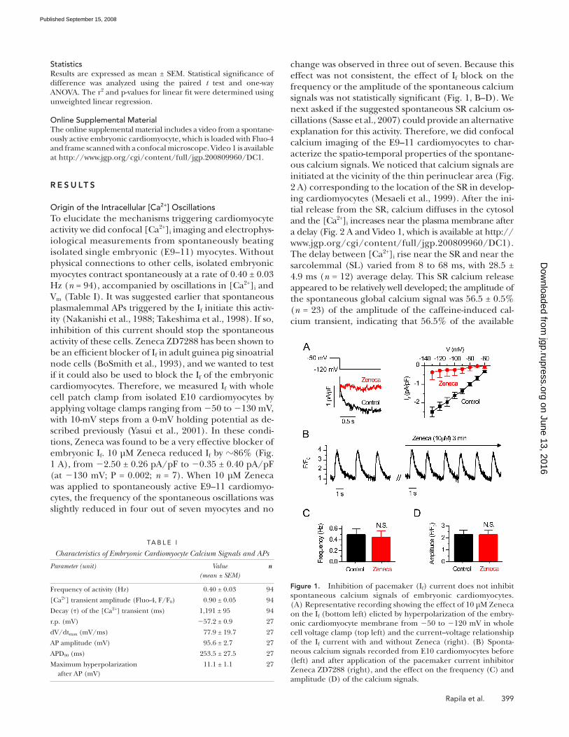
Regulation of the Rate of the Spontaneous SR Ca 2+ Oscillations It seems that both types of SR calcium release channels
(RyRs and IP 3 Rs) are needed for spontaneous calcium
SR calcium is released during each spontaneous calcium
release. To occur, SR calcium release seems to require
intact calcium uptake as well as two types of SR calcium
release channels because blocking either IP 3 Rs (with
2-APB) ( Maruyama et al., 1997 ) ( n = 14) or RyRs (with
ryanodine; n = 27), as well as inhibiting SR calcium up-
take (with thapsigargin; n = 11), initially slows down the
Figure 2. Spontaneous calcium release initiates cytosolic calcium oscillations in embryonic cardiac myocytes. (A) Laser scanning con-focal image from a Fluo-4 – loaded, isolated E10 spontaneously active cardiomyocyte (left) with corresponding line scan (yellow line) through the cytosol surrounding the dark nuclear area (red n). Red and blue arrowheads in the line scan image denote near SR area and near SL area, respectively. Graphs above the line scan image show relative calcium signals (Fluo-4 emission, F/F 0 ) from near SR (red line) and near SL (blue line). Insert (gray background) shows both graphs in an expanded timescale from selected area below (gray). (B) Effects of inhibitors of SR Ca 2+ -ATPase (10 μ M thapsigargin; top), RyRs (50 μ M ryanodine; middle), and IP 3 Rs (50 μ M 2-APB; bot-tom) on spontaneous calcium signals from isolated E10 cardiomyocytes.
Figure 3. IP 3 Rs regulate the frequency of the spon-taneous SR calcium releases. (A) RyR (top) and IP 3 R (bottom) immunolabeling in isolated E10 embryonic cardiomyocytes. Red arrowheads indicate specifi c stain-ing pattern around nuclei (red n). (B) When IP 3 Rs are inhibited with 2-APB, rapid application of 10 mM caf-feine activates RyRs to release calcium ( n = 10), and slower application of caffeine ( n = 5) induces frequent oscillatory calcium releases (insert). In an identical ex-perimental arrangement, when RyRs are inhibited with 50 μ M ryanodine and IP 3 Rs are stimulated with IP 3 AM, a sustained calcium leak is triggered that is proportional to the applied [IP 3- AM] (bottom). (C) Application of membrane-permeable IP 3 (IP 3- AM) increases the fre-quency of spontaneous calcium oscillations in isolated E10 cardiac myocytes.
on June 13, 2016jgp.rupress.org
Dow
nloaded from
Published September 15, 2008
Rapila et al. 401
brane-permeable IP 3 and noticed that the spontaneous
rate of calcium release events and consequently the
beating frequency in myocyte cultures increased by 27.3 ±
9.2% ( n = 7; P < 0.05; Fig. 3 C ).
The Mechanisms for Plasmalemmal Electrical Activity Embryonic myocytes have several depolarizing currents,
including a sodium current ( Doevendans et al., 2000 ),
T- and L-type calcium currents ( Seisenberger et al., 2000 ;
Cribbs et al., 2001 ), and a pacemaker current (I f ) ( Yasui
et al., 2001 ) that together constitute an electrically excit-
able membrane, which might in theory generate spon-
taneous V m oscillations and pacemaker-type APs ( Yasui
et al., 2001 ). However, because embryonic myocytes
also have a prominent Na + /Ca 2+ exchanger (NCX) cur-
rent ( Shepherd et al., 2007 ), it was suggested ( Janowski
et al., 2006 ; Sasse et al., 2007 ) that the spontaneous SR
calcium release might induce a depolarizing current
by activating the NCX and thereby trigger APs. Several
independent original fi ndings of the present study sup-
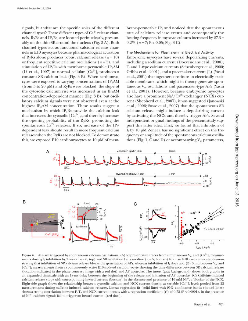
port this latter idea. First, we found that inhibition of
I f by 10 μ M Zeneca has no signifi cant effect on the fre-
quency or amplitude of the spontaneous calcium oscilla-
tions ( Fig. 1, C and D ) or accompanying V m parameters,
signals, but what are the specifi c roles of the different
channel types? These different types of Ca 2+ release chan-
nels, RyRs and IP 3 Rs, are located perinuclearly, presum-
ably on the thin SR around the nucleus ( Fig. 3 A ). Both
channel types act as functional calcium release chan-
nels in E10 myocytes because pharmacological activation
of RyRs alone produces robust calcium release ( n = 10)
or frequent repetitive calcium oscillations ( n = 5), and
stimulation of IP 3 Rs with membrane-permeable IP 3 AM
( Li et al., 1997 ) at normal cellular [Ca 2+ ] i produces a
constant SR calcium leak ( Fig. 3 B ). When cardiomyo-
cytes were exposed to varying concentrations of IP 3 AM
(from 5 to 20 μ M) and RyRs were blocked, the slope of
the cytosolic calcium rise was increased in an IP 3 AM
concentration – dependent manner ( Fig. 3 B ), but oscil-
latory calcium signals were not observed even at the
highest IP 3 AM concentration. These results suggest a
mechanism by which IP 3 Rs provide the calcium leak
that increases the cytosolic [Ca 2+ ] i and thereby increases
the opening probability of the RyRs, promoting the
spontaneous Ca 2+ releases. If so, increase of the IP 3 -
dependent leak should result in more frequent calcium
releases when the RyRs are not blocked. To demonstrate
this, we exposed E10 cardiomyocytes to 10 μ M of mem-
Figure 4. APs are triggered by spontaneous calcium oscillations. (A) Representative traces from simultaneous V m and [Ca 2+ ] i measure-ments during I f inhibition by Zeneca ( n = 6; top) and SR inhibition by ryanodine ( n = 5; bottom) from an E10 cardiomyocyte, demon-strating that inhibition of SR calcium release blocks the generation of APs, whereas inhibition of I f does not. (B) Simultaneous V m and [Ca 2+ ] i measurements from a spontaneously active E10-isolated cardiomyocyte showing the time difference between SR calcium release (location indicated in the phase contrast image with a red dot) and AP upstroke. The insert (gray background) shows both graphs in an expanded timescale with an 18-ms delay between the beginning of the release and initiation of AP upstroke. (C) Caffeine-induced calcium release (top) with corresponding inward current (bottom) in the absence and presence of 10 mM Ni 2+ , a blocker of the NCX. Right-side graph shows the relationship between cytosolic calcium and NCX current density at variable [Ca 2+ ] i levels pooled from 22 measurements during caffeine-induced calcium releases. Linear regression fi t (solid line) with 95% confi dence bands (dotted lines) shows a strong correlation between F/F 0 and NCX current density with a regression coeffi cient (r 2 ) of 0.72 (P < 0.0001). In the presence of Ni 2+ , calcium signals fail to trigger an inward current (red dots).
on June 13, 2016jgp.rupress.org
Dow
nloaded from
Published September 15, 2008
402 Embryonic E – C Coupling
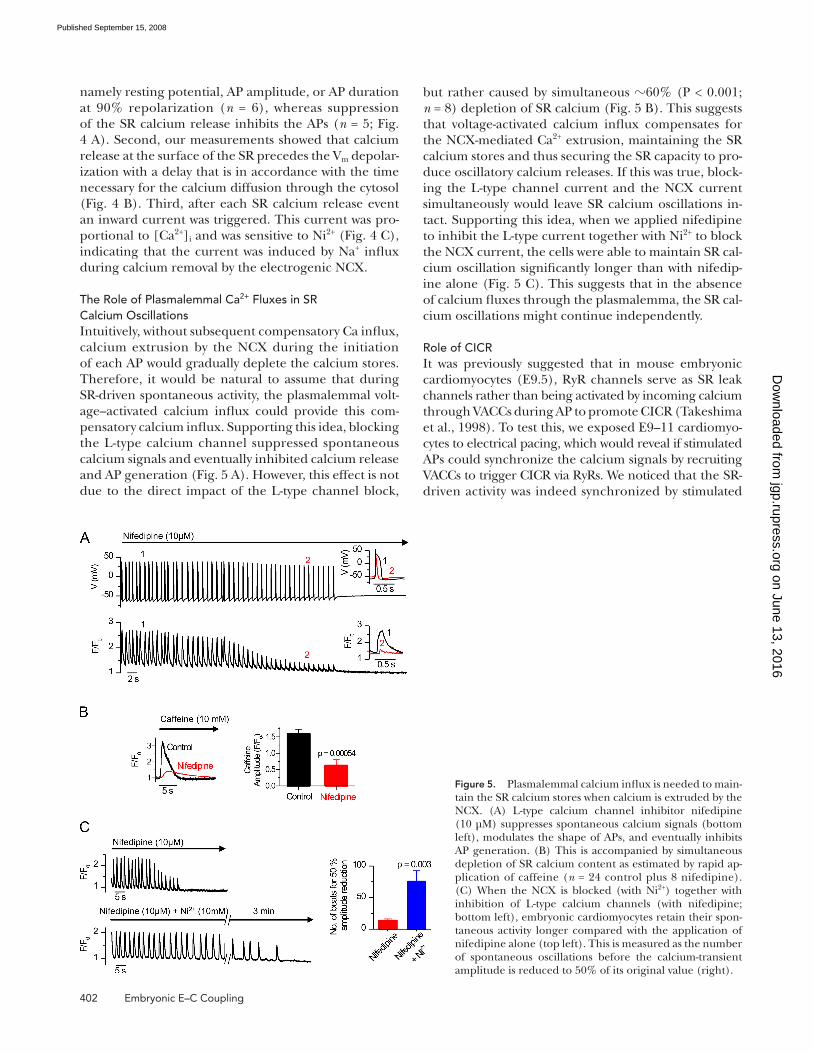
but rather caused by simultaneous � 60% (P < 0.001;
n = 8) depletion of SR calcium ( Fig. 5 B ). This suggests
that voltage-activated calcium infl ux compensates for
the NCX-mediated Ca 2+ extrusion, maintaining the SR
calcium stores and thus securing the SR capacity to pro-
duce oscillatory calcium releases. If this was true, block-
ing the L-type channel current and the NCX current
simultaneously would leave SR calcium oscillations in-
tact. Supporting this idea, when we applied nifedipine
to inhibit the L-type current together with Ni 2+ to block
the NCX current, the cells were able to maintain SR cal-
cium oscillation signifi cantly longer than with nifedip-
ine alone ( Fig. 5 C ). This suggests that in the absence
of calcium fl uxes through the plasmalemma, the SR cal-
cium oscillations might continue independently.
Role of CICR It was previously suggested that in mouse embryonic
cardiomyocytes (E9.5), RyR channels serve as SR leak
channels rather than being activated by incoming calcium
through VACCs during AP to promote CICR ( Takeshima
et al., 1998 ). To test this, we exposed E9 – 11 cardiomyo-
cytes to electrical pacing, which would reveal if stimulated
APs could synchronize the calcium signals by recruiting
VACCs to trigger CICR via RyRs. We noticed that the SR-
driven activity was indeed synchronized by stimulated
namely resting potential, AP amplitude, or AP duration
at 90% repolarization ( n = 6), whereas suppression
of the SR calcium release inhibits the APs ( n = 5; Fig.
4 A ). Second, our measurements showed that calcium
release at the surface of the SR precedes the V m depolar-
ization with a delay that is in accordance with the time
necessary for the calcium diffusion through the cytosol
( Fig. 4 B ). Third, after each SR calcium release event
an inward current was triggered. This current was pro-
portional to [Ca 2+ ] i and was sensitive to Ni 2+ ( Fig. 4 C ),
indicating that the current was induced by Na + infl ux
during calcium removal by the electrogenic NCX.
The Role of Plasmalemmal Ca 2+ Fluxes in SR Calcium Oscillations Intuitively, without subsequent compensatory Ca infl ux,
calcium extrusion by the NCX during the initiation
of each AP would gradually deplete the calcium stores.
Therefore, it would be natural to assume that during
SR-driven spontaneous activity, the plasmalemmal volt-
age – activated calcium infl ux could provide this com-
pensatory calcium infl ux. Supporting this idea, blocking
the L-type calcium channel suppressed spontaneous
calcium signals and eventually inhibited calcium release
and AP generation ( Fig. 5 A ). However, this effect is not
due to the direct impact of the L-type channel block,
Figure 5. Plasmalemmal calcium infl ux is needed to main-tain the SR calcium stores when calcium is extruded by the NCX. (A) L-type calcium channel inhibitor nifedipine (10 μ M) suppresses spontaneous calcium signals (bottom left), modulates the shape of APs, and eventually inhibits AP generation. (B) This is accompanied by simultaneous depletion of SR calcium content as estimated by rapid ap-plication of caffeine ( n = 24 control plus 8 nifedipine). (C) When the NCX is blocked (with Ni 2+ ) together with inhibition of L-type calcium channels (with nifedipine; bottom left), embryonic cardiomyocytes retain their spon-taneous activity longer compared with the application of nifedipine alone (top left). This is measured as the number of spontaneous oscillations before the calcium-transient amplitude is reduced to 50% of its original value (right).
on June 13, 2016jgp.rupress.org
Dow
nloaded from
Published September 15, 2008
Rapila et al. 403
lin, show embryonic malformations, impaired calcium
signaling, and compromised calcium-dependent tran-
scription ( Mesaeli et al., 1999 ). We showed that block-
ing of any of the SR Ca 2+ release channels, IP 3 R or RyR,
or the SR Ca 2+ pump SERCA diminished and fi nally
stopped the Ca 2+ signals and APs ( Fig. 2 B ). We found
that stimulation of IP 3 R only increased SR Ca 2+ leak but
did not induce global cytosolic [Ca 2+ ] i transients, and
that for global spontaneous cytosolic [Ca 2+ ] i transients,
RyR activation was a prerequisite ( Fig. 3 ). Accordingly,
mice defi cient in the cardiac isoform of the RyR (RyR-2)
have impaired calcium signals, and the development
of SR and mitochondria are consequently disturbed
( Takeshima et al., 1998 ). However, genetic ablation of
any of the individual IP 3 R subtypes alone does not pro-
duce any evident cardiac phenotype in mouse. Lack of
IP 3 R type 1 results in the most dramatic phenotype, with
increased embryonic lethality and severe neurologi-
cal malformations ( Matsumoto et al., 1996 ). Both IP 3 R
type 2 and 3 knockout mice are viable and have no ob-
vious defects, whereas double knockout (IP 3 R2 � / � plus
IP 3 R3 � / � ) mice die within 4 wk after birth due to defects
in the intracellular calcium handling of endocrine cells
and consequent metabolic disturbances ( Futatsugi et al.,
2005 ). This suggests that none of the IP 3 R types is indis-
pensable in heart development, but it does not rule out
the possibility that total lack of IP 3 R activity might induce
more drastic changes. In embryonic stem cell – derived
cardiomyocytes, IP 3 R antisense against the predominant
IP 3 R isoform in these cells, as well as pharmacological
inhibition of IP 3 R activity, abolishes the spontaneous ac-
tivity of the cells ( Mery et al., 2005 ), just as in this study
( Fig. 2 B ). In theory, RyRs are capable of producing os-
cillatory Ca 2+ releases on their own, but the frequency
of these oscillations would be an order of magnitude
slower than what we observed in E9 – 11 cardiomyocytes
( Keizer and Levine, 1996 ). We found that increases in
the spontaneous RyR openings are dependent on the
Ca 2+ leak via IP 3 R ( Fig. 3 C ), which stimulates the RyRs
to open and subsequently increases the frequency of the
global [Ca 2+ ] i transients in E9 – 11 cardiomyocytes. This
study illuminates how the interaction between multiple
factors modulating cytosolic [IP 3 ] levels will also regu-
late the embryonic heart rate.
Recently, it was shown that in some of the early em-
bryonic cardiomyocytes, the activity originates from
local high frequency ( � 4 Hz) Ca 2+ oscillations ( Sasse
et al., 2007 ). Similar calcium signals were also seen in
this study ( Fig. 2 A ), suggesting that SR also produces
“ subthreshold ” calcium events, spark-like local signals
that are not large enough to propagate over the whole
SR surface and recruit the majority of the Ca 2+ -release
units. However, it is not clear whether these signals serve
a true physiological function in triggering force-produc-
ing contractions or if they only represent the noise of
the system.
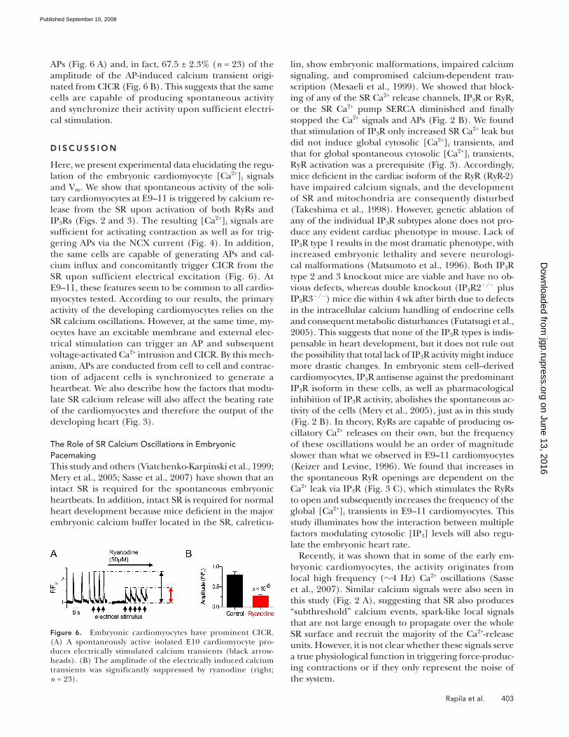
APs ( Fig. 6 A ) and, in fact, 67.5 ± 2.3% ( n = 23) of the
amplitude of the AP-induced calcium transient origi-
nated from CICR ( Fig. 6 B ). This suggests that the same
cells are capable of producing spontaneous activity
and synchronize their activity upon sufficient electri-
cal stimulation.
D I S C U S S I O N
Here, we present experimental data elucidating the regu-
lation of the embryonic cardiomyocyte [Ca 2+ ] i signals
and V m . We show that spontaneous activity of the soli-
tary cardiomyocytes at E9 – 11 is triggered by calcium re-
lease from the SR upon activation of both RyRs and
IP 3 Rs ( Figs. 2 and 3 ). The resulting [Ca 2+ ] i signals are
suffi cient for activating contraction as well as for trig-
gering APs via the NCX current ( Fig. 4 ). In addition,
the same cells are capable of generating APs and cal-
cium infl ux and concomitantly trigger CICR from the
SR upon sufficient electrical excitation ( Fig. 6 ). At
E9 – 11, these features seem to be common to all cardio-
myocytes tested. According to our results, the primary
activity of the developing cardiomyocytes relies on the
SR calcium oscillations. However, at the same time, my-
ocytes have an excitable membrane and external elec-
trical stimulation can trigger an AP and subsequent
voltage-activated Ca 2+ intrusion and CICR. By this mech-
anism, APs are conducted from cell to cell and contrac-
tion of adjacent cells is synchronized to generate a
heartbeat. We also describe how the factors that modu-
late SR calcium release will also affect the beating rate
of the cardiomyocytes and therefore the output of the
developing heart ( Fig. 3 ).
The Role of SR Calcium Oscillations in Embryonic Pacemaking This study and others ( Viatchenko-Karpinski et al., 1999 ;
Mery et al., 2005 ; Sasse et al., 2007 ) have shown that an
intact SR is required for the spontaneous embryonic
heartbeats. In addition, intact SR is required for normal
heart development because mice defi cient in the major
embryonic calcium buffer located in the SR, calreticu-
Figure 6. Embryonic cardiomyocytes have prominent CICR. (A) A spontaneously active isolated E10 cardiomyocyte pro-duces electrically stimulated calcium transients (black arrow-heads). (B) The amplitude of the electrically induced calcium transients was significantly suppressed by ryanodine (right; n = 23).
on June 13, 2016jgp.rupress.org
Dow
nloaded from
Published September 15, 2008
404 Embryonic E – C Coupling
generation of electrically evoked APs, embryonic cardio-
myocytes produce [Ca 2+ ] i signals that are mainly based
on SR calcium release (CICR) ( Fig. 6 ). However, in the
absence of SR calcium release, myocytes are still able to
produce global whole cell [Ca 2+ ] i signals based only on
voltage-activated SL calcium intrusion ( Fig. 6 ).
Coexistence of the pacemaking and E – C coupling mech-
anism in the same cells explains how embryonic cardio-
myocytes maintain their activity during transition periods
without physical connections to other cells, enabling cell
migration in the developing heart ( Buckingham et al.,
2005 ), and also how they are able to synchronize their
electrical activity and contraction with other cells to form
coordinated contraction ( Kamino, 1991 ) of the heart.
This study provides a novel view of the regulation of
embryonic cardiomyocyte activity, characterizing the
functional versatility of developing cardiomyocytes and
further explaining fundamental phenomena in the ori-
gin of the heartbeat. In addition, although these results
open interesting perspectives in the fi eld of mammalian
cardiac development, they also elucidate mechanisms that
may be exploited in the development of cellular excit-
ability and calcium signaling in general.
We thank S.L. H ä nninen for valuable comments on the manu-script and A. Rautio for technical assistance.
This study was supported by the Finnish Heart Research Foun-dation, Academy of Finland, and Sigrid Juselius Foundation.
Olaf S. Andersen served as editor.
Submitted: 11 January 2008 Accepted: 19 August 2008
R E F E R E N C E S BoSmith , R.E. , I. Briggs , and N.C. Sturgess . 1993 . Inhibitory actions
of ZENECA ZD7288 on whole-cell hyperpolarization activated in-
ward current (I f ) in guinea-pig dissociated sinoatrial node cells.
Br. J. Pharmacol. 110 : 343 – 349 .
Buckingham , M. , S. Meilhac , and S. Zaffran . 2005 . Building the
mammalian heart from two sources of myocardial cells. Nat. Rev. Genet. 6 : 826 – 835 .
Copp , A.J. 1995 . Death before birth: clues from gene knockouts and
mutations. Trends Genet. 11 : 87 – 93 .
Cribbs , L.L. , B.L. Martin , E.A. Schroder , B.B. Keller , B.P. Delisle ,
and J. Satin . 2001 . Identifi cation of the T-type calcium channel
(Ca(V)3.1d) in developing mouse heart. Circ. Res. 88 : 403 – 407 .
Doevendans , P.A. , S.W. Kubalak , R.H. An , K.D. Becker , K.R. Chien ,
and R.S. Kass . 2000 . Differentiation of cardiomyocytes in fl oating
embryoid bodies is comparable to fetal cardiomyocytes. J. Mol. Cell. Cardiol. 32 : 839 – 851 .
Ebert , S.N. , and D.G. Taylor . 2006 . Catecholamines and develop-
ment of cardiac pacemaking: an intrinsically intimate relation-
ship. Cardiovasc. Res. 72 : 364 – 374 .
Futatsugi , A. , T. Nakamura , M.K. Yamada , E. Ebisui , K. Nakamura , K.
Uchida , T. Kitaguchi , H. Takahashi-Iwanaga , T. Noda , J. Aruga , and
K. Mikoshiba . 2005 . IP 3 receptor types 2 and 3 mediate exocrine
secretion underlying energy metabolism. Science . 309 : 2232 – 2234 .
Ginsburg , K.S. , and D.M. Bers . 2005 . Isoproterenol does not en-
hance Ca-dependent Na/Ca exchange current in intact rabbit
ventricular myocytes. J. Mol. Cell. Cardiol. 39 : 972 – 981 .
According to our results, isolated embryonic cardiomyo-
cytes have a lower spontaneous beating rate ( � 0.4 Hz)
than the developing heart in utero ( � 2 Hz; Gui et al.,
1996 ). This might be due to the fact that in the intact em-
bryonic heart in utero, the myocytes are subjected to a va-
riety of diffuse hormonal and local hormonal stimuli,
which modify the cellular components involved in the
pacemaking of the cells and are not present in embryonic
cardiomyocyte cultures. In addition to the known stimula-
tory effects of several hormones and neurotransmitters on
the embryonic heartbeat, such as catecholamines ( Ebert
and Taylor, 2006 ), we actually report an IP 3 -dependent
mechanism that speeds up the embryonic heart rate ( Fig.
3 C ), suggesting that all of the hormones able to regulate
IP 3 production (e.g., AngII, ET-1, and � -adenergic stimuli)
also affect the heart rate. In addition, it is likely that the
heart rate of the intact heart is defi ned by the myocyte with
the highest frequency of spontaneous activity. Therefore,
the average frequencies of the isolated myocytes and the
frequency of the intact heart are not strictly comparable.
Interplay between SL Ion Currents and SR Calcium Release Most of the recent knowledge of embryonic excitation –
contraction (E – C) coupling originates from studies with
genetically engineered mouse models, which collectively
suggest that several components are involved in the reg-
ulation of the embryonic heartbeat. So far, genetic dele-
tion or suppression of any of the key components of E – C
coupling has produced phenotypes with severe heart
malformations and functional impairments that induce
early embryonic lethality. Knockout of the HCH4 gene,
coding the pacemaker current I f channel, slows down
the heart rate at E9.5 and abolishes the formation of ma-
ture pacemaker cells ( Stieber et al., 2003 ). Similarly, ge-
netic suppression of L-type calcium channels slows down
the heart rate at E10.5, but it also reduces the cytosolic
calcium signals ( Weissgerber et al., 2005 ). Both adult
and embryonic cardiomyocytes express the electrogenic
plasmalemmal Na + -Ca 2+ exchanger (NCX) for calcium
extrusion ( Koban et al., 1998 ). Targeted inactivation of
cardiac NCX leads to embryonic lethality before E11,
and the hearts of NCX-defi cient (NCX � / � ) mice lack
spontaneous heartbeats and organized contractile myo-
fi brils ( Koushik et al., 2001 ).
Our results suggest that in embryonic cardiomyocytes,
two different mechanisms (SR- and AP-driven) can be
separately recruited to generate global calcium signals
( Figs. 2 and 6 ). During SR calcium oscillations, when
the NCX extrudes calcium and triggers APs, SL calcium
infl ux is required to maintain SR calcium content and
secure global spontaneous [Ca 2+ ] i oscillations ( Fig. 5 A ).
However, if [Ca 2+ ] i signals and V m are uncoupled by
blocking the NCX, the SR calcium oscillations can be
produced independently of the SL calcium fl uxes ( Fig.
5 C ). Similarly, the generation of AP-induced calcium
signals can be dissected into two separate parts. Upon
on June 13, 2016jgp.rupress.org
Dow
nloaded from
Published September 15, 2008
Rapila et al. 405
Nakanishi , T. , M. Seguchi , and A. Takao . 1988 . Development of the
myocardial contractile system. Experientia . 44 : 936 – 944 .
Puceat , M. , and M. Jaconi . 2005 . Ca 2+ signalling in cardiogenesis.
Cell Calcium . 38 : 383 – 389 .
Sasse , P. , J. Zhang , L. Cleemann , M. Morad , J. Hescheler , and B.K.
Fleischmann . 2007 . Intracellular Ca 2+ oscillations, a potential pace-
making mechanism in early embryonic heart cells. J. Gen. Physiol. 130 : 133 – 144 .
Seisenberger , C. , V. Specht , A. Welling , J. Platzer , A. Pfeifer , S.
Kuhbandner , J. Striessnig , N. Klugbauer , R. Feil , and F. Hofmann .
2000 . Functional embryonic cardiomyocytes after disruption of
the L-type alpha(1C) (Ca(v)1.2) calcium channel gene in the
mouse. J. Biol. Chem. 275 : 39193 – 39199 .
Seki , S. , M. Nagashima , Y. Yamada , M. Tsutsuura , T. Kobayashi , A.
Namiki , and N. Tohse . 2003 . Fetal and postnatal development of
Ca 2+ transients and Ca 2+ sparks in rat cardiomyocytes. Cardiovasc. Res. 58 : 535 – 548 .
Shepherd , N. , V. Graham , B. Trevedi , and T. Creazzo . 2007 . Changes
in regulation of sodium/calcium exchanger of avian ventricular
heart cells during embryonic development. Am. J. Physiol. Cell Physiol. 292:C1942 – C1950.
Stieber , J. , S. Herrmann , S. Feil , J. Loster , R. Feil , M. Biel , F.
Hofmann , and A. Ludwig . 2003 . The hyperpolarization-activated
channel HCN4 is required for the generation of pacemaker ac-
tion potentials in the embryonic heart. Proc. Natl. Acad. Sci. USA . 100 : 15235 – 15240 .
Sturm , K. , and P.P.L. Tam . 1993 . Isolation and culture of whole
postimplantation embryos and germ layer derivatives. Methods Enzymol . 225 : 164 – 190 .
Takeshima , H. , S. Komazaki , K. Hirose , M. Nishi , T. Noda , and M.
Lino . 1998 . Embryonic lethality and abnormal cardiac myocytes in
mice lacking ryanodine receptor type 2. EMBO J. 17 : 3309 – 3316 .
Weissgerber , P. , W. Bloch , B. Held , L. Kastner , P. Lipp , K. Chien , V.
Flockerzi , and M. Freichel . 2005 . Embryonic heart failure after dis-
ruption of the Ca 2+ channel subunit beta(2) gene (Ca-v beta(2))
leads to defects in vascular remodeling. Naunyn Schmiedebergs Arch. Pharmacol. 371 : R55 .
Viatchenko-Karpinski , S. , B.K. Fleischmann , Q. Liu , H. Sauer , O.
Gryshchenko , G.J. Ji , and J. Hescheler . 1999 . Intracellular Ca 2+ os-
cillations drive spontaneous contractions in cardiomyocytes dur-
ing early development. Proc. Natl. Acad. Sci. USA . 96 : 8259 – 8264 .
Yang , Z. , W. Shen , J.N. Rottman , J.P. Wikswo , and K.T. Murray . 2005 .
Rapid stimulation causes electrical remodeling in cultured atrial
myocytes. J. Mol. Cell. Cardiol. 38 : 299 – 308 .
Yasui , K. , W. Liu , T. Opthof , K. Kada , J.K. Lee , K. Kamiya , and I.
Kodama . 2001 . I f current and spontaneous activity in mouse em-
bryonic ventricular myocytes. Circ. Res. 88 : 536 – 542 .
Zimmermann , W.H. , K. Schneiderbanger , P. Schubert , M. Didie ,
F. Munzel , J.F. Heubach , S. Kostin , W.L. Neuhuber , and T.
Eschenhagen . 2002 . Tissue engineering of a differentiated car-
diac muscle construct. Circ. Res. 90 : 223 – 230 .
Gui , Y.H. , K.K. Linask , P. Khowsathit , and J.C. Huhta . 1996 . Doppler
echocardiography of normal and abnormal embryonic mouse
heart. Pediatr. Res. 40 : 633 – 642 .
Husse , B. , and M. Wussling . 1996 . Developmental changes of cal-
cium transients and contractility during the cultivation of rat neo-
natal cardiomyocytes. Mol. Cell. Biochem. 164 : 13 – 21 .
Janowski , E. , L. Cleemann , P. Sasse , and M. Morad . 2006 . Diversity
of Ca 2+ signaling in developing cardiac cells. Ann. NY Acad. Sci. 1080 : 154 – 164 .
Kamino , K. 1991 . Optical approaches to ontogeny of electrical-activ-
ity and related functional-organization during early heart devel-
opment. Physiol. Rev. 71 : 53 – 91 .
Keizer , J. , and L. Levine . 1996 . Ryanodine receptor adaptation and
Ca 2+ -induced Ca 2+ release-dependent Ca 2+ oscillations. Biophys. J. 71 : 3477 – 3487 .
Koban , M.U. , A.F.M. Moorman , J. Holtz , M.H. Yacoub , and K.R.
Boheler . 1998 . Expressional analysis of the cardiac Na-Ca ex-
changer in rat development and senescence. Cardiovasc. Res. 37 : 405 – 423 .
Koushik , S.V. , J. Wang , R. Rogers , D. Moskophidis , N.A. Lambert ,
T.L. Creazzo , and S.J. Conway . 2001 . Targeted inactivation of the
sodium-calcium exchanger (Ncx1) results in the lack of a heartbeat
and abnormal myofi brillar organization. FASEB J. 15 : 1209 – 1211 .
Li , W.H. , C. Schultz , J. Llopis , and R.Y. Tsien . 1997 . Membrane-per-
meant esters of inositol polyphosphates, chemical syntheses and
biological applications. Tetrahedron . 53 : 12017 – 12040 .
Liu , W. , K. Yasui , A. Arai , K. Kamiya , J. Cheng , I. Kodama , and J.
Toyama . 1999 . beta-adrenergic modulation of L-type Ca 2+ -chan-
nel currents in early-stage embryonic mouse heart. Am. J. Physiol. 276 : H608 – H613 .
Maruyama , T. , T. Kanaji , S. Nakade , T. Kanno , and K. Mikoshiba .
1997 . 2APB, 2-aminoethoxydiphenyl borate, a membrane-pene-
trable modulator of Ins(1,4,5)P 3 -induced Ca 2+ release. J. Biochem. (Tokyo) . 122 : 498 – 505 .
Matsumoto , M. , T. Nakagawa , T. Inoue , E. Nagata , K. Tanaka , H.
Takano , O. Minowa , J. Kuno , S. Sakakibara , M. Yamada , et al .
1996 . Ataxia and epileptic seizures in mice lacking type 1 inositol
1,4,5-trisphosphate receptor. Nature . 379 : 168 – 171 .
Mery , A. , F. Aimond , C. Menard , K. Mikoshiba , M. Michalak , and M.
Puceat . 2005 . Initiation of embryonic cardiac pacemaker activity
by inositol 1,4,5-trisphosphate-dependent calcium signaling. Mol. Biol. Cell . 16 : 2414 – 2423 .
Mesaeli , N. , K. Nakamura , E. Zvaritch , P. Dickie , E. Dziak , K.H.
Krause , M. Opas , D.H. MacLennan , and M. Michalak . 1999 .
Calreticulin is essential for cardiac development. J. Cell Biol. 144 : 857 – 868 .
Moorman , A.F.M. , C.A. Schumacher , P.A.J. de Boer , J. Hagoort , K.
Bezstarosti , M.J.B. van den Hoff , G.T.M. Wagenaar , J.M.J. Lamers ,
F. Wuytack , V.M. Christoffels , and J.W.T. Fiolet . 2000 . Presence of
functional sarcoplasmic reticulum in the developing heart and its
confi nement to chamber myocardium. Dev. Biol. 223 : 279 – 290 .
on June 13, 2016jgp.rupress.org
Dow
nloaded from
Published September 15, 2008


