Embed Size (px)
Citation preview

Introduction
Osseous tumours of the skull are rare entities characterized by alarge variety of histological findings, as described by Arana et al.[1, 2], Ciray et al. [3], and Hunt et al. [4]. The majority of such tu−mours encountered in the field of neurosurgery are either me−ningiomas or metastases [5, 6]. Symptoms include changes inthe patient’s appearance and neurological deficits due to intra−cranial expansion of the tumour. Although resection of the lesionis always the primary objective of surgical treatment, increasedattention has also been devoted recently to restoration of theconcomitant defect leading to a cosmetically satisfying outcome[7± 10].
Drilling of the skull by means of a robotic system has proved tobe a valuable surgical technique for precise insertion of cochleaimplants [11,12] or has been evaluated intraoperatively for thedevelopment of a system for craniotomy [13]. The craniectomycould be a new application for a robotic system in patients with
Analysis of Surgical Management of Calvarial Tumoursand First Results of a Newly Designed RoboticTrepanation System
M. Engelhardt1, 5
P. Bast2, 5
N. Jeblink3
W. Lauer4
A. Popovic4
H. Eufinger2
M. Scholz1
A. Christmann3
A. Harders1
K. Radermacher4
K. Schmieder1
Affiliation1 Department of Neurosurgery, Ruhr University Bochum, Bochum, Germany2 Department of Oral and Maxillofacial Plastic Surgery, Knappschaftskrankenhaus, Recklinghausen, Germany3 Department of Statistics, University of Dortmund, Dortmund, Germany4 Institute for Biomedical Technologies, Aachen University, Aachen, Germany5 These authors contributed equally to the work of this paper.
CorrespondenceDr. med. Martin Engelhardt ´ Neurochirurgische Universitätsklinik ´ Ruhr−Universität Bochum ´Knappschaftskrankenhaus ´ In der Schornau 23 ±25 ´ 44892 Bochum ´ Germany ´Tel.: +49/234/299/3602 ´ Fax: +49/234/299/3609 ´ Fax: martin.engelhardt@ruhr−uni−bochum.de
BibliographyMinim Invas Neurosurg 2006; 49: 98±103 � Georg Thieme Verlag KG Stuttgart ´ New YorkDOI 10.1055/s−2006−932173ISSN 0946−7211
Abstract
This study was performed to evaluate the surgical strategy in pa−tients with calvarial tumours, in order to design and modify a ro−bot−assisted trepanation system. A total of 75 patients under−went craniectomy for the treatment of calvarial tumours duringthe 10−year period from 1993 to 2002. The patients’ complaints,the size, location and histology of the tumour, and the variouscranioplasty techniques used were analysed retrospectively. In asecond procedure several craniectomies at typical locations ac−cording to the study’s results were performed in a laboratory set−ting using a hexapod robotic tool, constructed at the Helmholtz−Institute, RWTH Aachen University, and plastic model heads. Theworkflow was documented and the reproducibility and the accu−racy of the procedure were registered. A total of 83 surgical pro−cedures were performed on 75 patients. The majority (87 %) of le−sions treated surgically were located in the frontal, temporal andanterior parts of the parietal region. Histological examination re−vealed benign lesions in 66 % of the patients and dural involve−ment in 46 %. According to these results craniectomies were per−formed using the robotic system. Mean positioning accuracy ofthe robotic system while milling was 0.24 mm, with a standarddeviation of 0.04 mm, and maximum error under 1 mm. Craniec−tomies leaving a 1−mm layer of the tabula interna intact to en−sure a healthy dura were performed in several regions success−fully. The majority of calvarial tumours, requiring surgical treat−ment in our patients, were located in cosmetically relevant areasin which drilling can be carried out with the robotic trepanation
system. Consequently, the surgical approach had to be plannedcarefully in order to achieve a good cosmetic outcome.
Key wordsTrepanation ´ robotic assisted surgery ´ calvarial tumours ´ neuro−surgery
Orig
inalA
rticle
98

osseous tumours, especially in cosmetically important areas ofthe skull.
The aim of this study, which is supported by the German Re−search Foundation (DFG) within the SPP 1124, was to evaluateosseous lesions with regard to location, histological findingsand surgical management. Furthermore, first drilling tests witha plastic model head were carried out in a laboratory setting, ac−cording to the evaluated workspace area.
Patients, Materials and Methods
A total of 75 patients (54 females and 21−males) with a mean ageof 57 years (range: 4 ± 95 years) who had been treated surgicallyfor calvarial tumours during the 10−year period from 1993 to2002 were included in the study. The demographic data, clinicaland pathological findings, and information on the preoperativediagnostic procedures used for surgical planning were obtainedfrom the files. Lesions were classified according to their location,size and histology; cosmetic impairments were documented.The surgical management was analysed, with special emphasison dural involvement and repair and the type of cranioplastyused. Fisher’s exact test [14] was used to test for dependence incontingency tables; the level of significance was set at 0.05.
The data on location and size were also analysed to identify re−quirements for a hexapod robot system and to perform the firstcraniectomies in a plastic model skull. The robotic system usedwas a hexapod parallel robot with platforms of 440 and200 mm in diameter; the legs could be varied in length from430 mm to 580 mm. Preliminary tests have been conducted toevaluate the robotic drilling parameters and the accuracy of cra−niectomy in a laboratory setting with a plastic model head.
Several craniectomies were performed in typical locations, ac−cording to the statistical evaluation of calvarial tumours, in orderto acquire data on accuracy and reproducibility. Furthermore,several robot settings differing in parameters such as speed ofdrilling and movement were evaluated and compared.
Results
A total of 83 surgical procedures were performed in 75 patients.Cosmetic changes of the cranial vault were detected in 46 % of thepatients, whereas neurological deficits were diagnosed in only13 %. Twenty−two percent of the patients complained of head−aches at admission (Table 1). Forty−five percent of the lesionswere located in the frontal region of the skull.
The preoperative diagnostic work−up included CT or MRI in allpatients; both were carried out in 39 patients. Starting in 1997,the technique of neuronavigation was used in 33 % of cases(n = 14). A total of 13 of the navigated lesions were located in thefrontal and/or parietal region; half of them were small tumoursbelow 2 cm in size. The mean size of the craniotomy performedwas 4.5 cm overall (range: 1 ± 17 cm). Infiltration of the dura waspresent in 46 % of the lesions. Sixty−three percent of the lesionswith dural infiltration were meningiomas; the remainder consist−
ed of metastatic lesions of the skull. Infiltration of venous sinuseswas verified during surgery in 13 %. The correlation between his−tological classification of the tumour and the presence of dural orsinus infiltration was statistically significant (p = 0.047). Asshown in Table 2, a broad spectrum of pathology was encoun−tered. The majority (66 %) of the lesions treated were benignlesions and tumours of the skull.
In 43 % of the patients with benign lesions, craniotomy and resec−tion of the infiltrated part of the bone followed by reinsertion ofthe non−affected part was possible; acrylic bone cement was
Table 2 Histological findings in resected lesions of the skull(n = 83)
Numberof lesions
%
Benignlesions
Fibrous dysplasia 10 18 22
Encephalocele, cyst 3
Hyperostosis 2
Growth fracture 1
Bone cysts 1
Tuberculosis 1
Benigntumours
Meningioma, benign 30 36 43
Haemangioma 3
Astrocytoma 1
Multiple myeloma 1
Osteoma 1
Malignanttumours
Adenocarcinoma metastasis 14 28 34
Meningioma, malignant 6
Basal cell carcinoma 3
Breast cancer metastasis 2
Angiosarcoma 1
Metastasis, primary unknown 1
Malignant histiocytoma 1
No differen−tiation
Malignant 1 1 1
Total 83 83 100
Table 1 Symptoms and clinical findings in patients with osseoustumours on admission (n = 75)
Patients %
Cosmeticdisfigurement
Localised swelling 38 46
Palpable bone defect 3 4
Skin infiltration 4 5
No visible signs 38 46
Neurologicalimpairment
Exophthalmos 7 8
Headache 18 22
Neurological deficits 11 13
No neurological impairment 47 57
Engelhardt M et al. Analysis of Surgical Management of ¼ Minim Invas Neurosurg 2006; 49: 98 ± 103
Orig
inalA
rticle
99
used in 46 % of cases. In three patients, individually prefabricatedtitanium implants were inserted after resection of the tumour. Inpatients with small defects (i.e., less than 2 cm in diameter)without cosmetic relevance, cranioplasty was not performed.
Analysing the location of tumours, we were able to define aworking space covering 87 % of all lesions including the frontal,temporal and the anterior part of the parietal skull (Table 3).Tumour diameter was greater than 6 cm in 16 % of the patients,partly with several differently oriented bony surfaces to bedrilled. Supine positioning intraoperatively was used in 86 % ofthe lesions.
Owing to the cosmetic relevance of these tumours, navigationalcontrol and prefabricated cranioplasty were indicated.
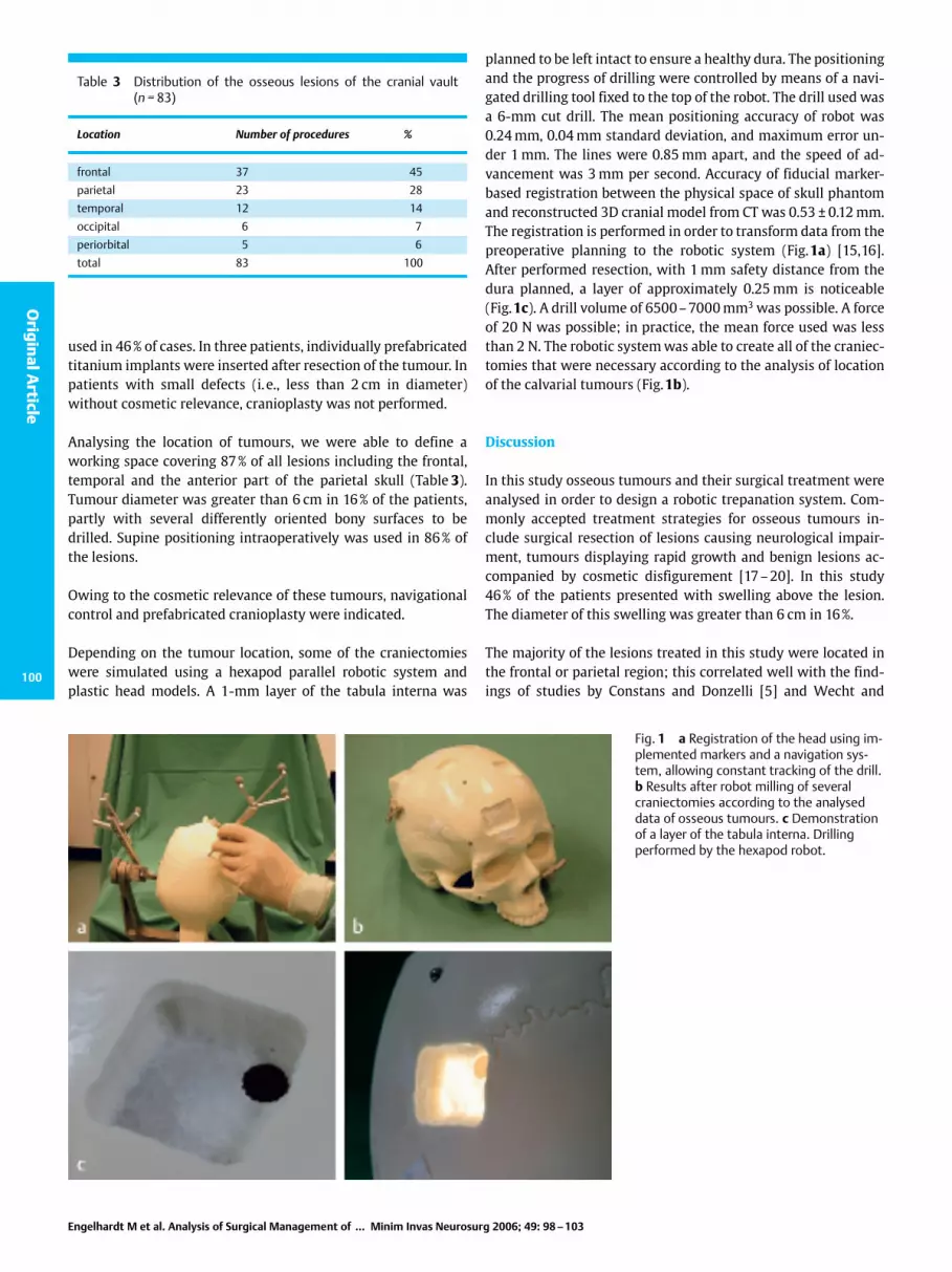
Depending on the tumour location, some of the craniectomieswere simulated using a hexapod parallel robotic system andplastic head models. A 1−mm layer of the tabula interna was
planned to be left intact to ensure a healthy dura. The positioningand the progress of drilling were controlled by means of a navi−gated drilling tool fixed to the top of the robot. The drill used wasa 6−mm cut drill. The mean positioning accuracy of robot was0.24 mm, 0.04 mm standard deviation, and maximum error un−der 1 mm. The lines were 0.85 mm apart, and the speed of ad−vancement was 3 mm per second. Accuracy of fiducial marker−based registration between the physical space of skull phantomand reconstructed 3D cranial model from CT was 0.53 � 0.12 mm.The registration is performed in order to transform data from thepreoperative planning to the robotic system (Fig.1a) [15,16].After performed resection, with 1 mm safety distance from thedura planned, a layer of approximately 0.25 mm is noticeable(Fig.1c). A drill volume of 6500 ± 7000 mm3 was possible. A forceof 20 N was possible; in practice, the mean force used was lessthan 2 N. The robotic system was able to create all of the craniec−tomies that were necessary according to the analysis of locationof the calvarial tumours (Fig.1b).
Discussion
In this study osseous tumours and their surgical treatment wereanalysed in order to design a robotic trepanation system. Com−monly accepted treatment strategies for osseous tumours in−clude surgical resection of lesions causing neurological impair−ment, tumours displaying rapid growth and benign lesions ac−companied by cosmetic disfigurement [17 ± 20]. In this study46 % of the patients presented with swelling above the lesion.The diameter of this swelling was greater than 6 cm in 16 %.
The majority of the lesions treated in this study were located inthe frontal or parietal region; this correlated well with the find−ings of studies by Constans and Donzelli [5] and Wecht and
Fig. 1 a Registration of the head using im−plemented markers and a navigation sys−tem, allowing constant tracking of the drill.b Results after robot milling of severalcraniectomies according to the analyseddata of osseous tumours. c Demonstrationof a layer of the tabula interna. Drillingperformed by the hexapod robot.
Table 3 Distribution of the osseous lesions of the cranial vault(n = 83)
Location Number of procedures %
frontal 37 45
parietal 23 28
temporal 12 14
occipital 6 7
periorbital 5 6
total 83 100
Engelhardt M et al. Analysis of Surgical Management of ¼ Minim Invas Neurosurg 2006; 49: 98 ± 103
Orig
inalA
rticle
100

Sawaya [10]. In contrast, Artico et al. [7] found no preferred re−gion of the skull for tumours in their study population.
The variety of histological entities found in our study is consis−tent with the findings of other authors [5, 7,10]. In one case thepathologist could not classify the lesion definitively. This wasconsistent with the findings of Wecht and Sawaya [10], who re−ported five cases in which histological classification was impos−sible.
In contrast to Sekhar [9], who used fluoroscopy and preoperativescintigraphic scanning, in this study CT and MRI were employed,since the interpretation of the information in navigation is wellestablished and necessary to control the robot intraoperatively.
Infiltration of the dura and/or the sinus was present in nearlytwice as many patients as in other studies (46 % vs. 20±26%)[5, 7,10], mainly due to the large number of meningiomas foundin our series. Sixty−three percent of the meningiomas and 31 % ofthe metastases had already infiltrated into the dura. Of the pa−tients with dural involvement, 19 % exhibited infiltration of thesinus. Involvement of these structures required special surgicalplanning to prevent laceration of the dura and brain tissue. Fur−thermore, dural infiltration has important technical implicationsfor the design of the robotic trepanation system, especially thedistance from the healthy brain tissue.
Following resection of the infiltrated dura, the resulting defectswere repaired. In our study autologous material was used forthis purpose in the vast majority of cases. A similar managementstrategy was used in the studies of Artico et al. [7] and Constansand Donzelli [5]. In this study Lyodura� or Teflon� implants were
employed in only two cases. We preferred to use autologous ma−terials such as galea periosteum or muscle fascia, taken eitherfrom the temporalis muscle or the fascia lata, because thesenatural materials resemble normal dura more closely.
In patients with small lesions we performed only soft tissue re−placement without cranioplasty, in common with Wecht and Sa−waya [10]. Constans and Donzelli [5], however, never performedcranioplasty; they and others used acrylic bone cement to repairdefects resulting from the excision of larger malignant lesions. Adifferent method was described by Saringer et al. [20], who usedindividual carbon fibre polymer. The relationship between defectsize and cranioplasty was statistically significant (p = 0.043).
In a large number of cases, therefore, cranioplasty is mandatoryfor cosmetic reasons, with the insertion of a prefabricated im−plant usually being the restoration modality of choice. Moreover,enhanced circulation and improved cognitive ability have beenreported by Agner et al. [21] following the performance of cra−nioplasty to repair a large osseous defect.
In patients with larger benign lesions involving cosmetically re−levant structures of the forehead and the face, we have foundprefabricated titanium implants to be suitable, especially incases where exact preoperative planning of the resection waspossible as described by Eufinger et al. [22]. Fig. 2 shows a pa−tient with a large meningioma extending into the frontal area ofthe face. Resection was followed by cranioplasty with a titaniumimplant; this resulted in a perfect cosmetic outcome. Since tita−nium implants are expensive, several authors have advocated theuse of bone cement during elective cranioplasty.
Fig. 2 a ± c A 47−year−old male patientwith enlargement of the skull and loca−lised pain. No neurological deficits werenoted at admission. MRI revealed a largetumour with enlargement of bone anddural infiltration. The tumour was resect−ed in toto including a dural repair withfascia lata and cranioplasty with a pre−fabricated titanium implant. The lesionwas classified histologically as a menin−gioma.
Engelhardt M et al. Analysis of Surgical Management of ¼ Minim Invas Neurosurg 2006; 49: 98 ± 103
Orig
inalA
rticle
101

In patients with skin involvement, a rotational skin flap or split−skin graft was put in place during the initial operation or during asecond surgical procedure, as also mentioned by Constans andDonzelli [5] and Wecht and Sawaya [10]. Four patients with skininvolvement were treated with a rotational skin flap.
Other authors [11 ± 13] reported about drilling of the skull,whereby they used different robotic platforms. The hexapod ro−botic system we used demonstrated the ability to drill the calvar−ia accurately. Furthermore, it was possible to reach all necessarylocations, based on the analysis of the lesions in this study. In thistest setting the robotic system, with six degrees of freedom, per−formed a craniectomy by drilling the skull to a thickness of 1 mmto ensure a healthy dura in natural circumstances (Figs. 3 and 4).
The potential advantages of robot−assisted resection are: precisedrilling at different bone surface angles and the possibility ofperforming a planned resection and inserting a prefabricated al−ternative. Further drilling tests have to be done to refine parame−ters of the robotic system such as speed of advancement andtemperature increase, with the danger of bone necrosis and adisturbed healing process of the bone, possibly influencing thecosmetic outcome.
Apart from these refinements we would state the following threepreconditions:a) Preoperative planning including an adequate diagnostic
work−up is mandatory.b) Navigational support has to be provided for the localisation
and surveillance of the resection area.c) The working range of the robotic system has to cover the en−
tire region in question.
Acknowledgements
We thank Mr. Roseveare for editing this manuscript and the Ger−man Research Foundation for the support of this project withinthe SPP 1124 (Medizinische Navigation und Robotik).
References
1 Arana E, Marti−Bonmati L, Paredes R, Bautista D. Focal calvarial bonelesions: comparison of logistic regression and neural network models.Invest Radiol 1999; 33: 738 ± 745
2 Arana E, Marti−Bonmati L, Bautista D, Paredes R. Qualitative diagnosisof calvarial metastasis by neural network and logistic regression. AcadRadiol 2004; 11: 45± 52
3 Ciray I, Astrom G, Sundstrom C, Hagberg H, Ahlstrom H. Assessment ofsuspected bone metastases. CT with and without clinical informationcompared to CT−guided bone biopsy. Acta Radiol 1997; 38: 890 ± 895
4 Hunt JA, Hobar PC. Common craniofacial anomalies: conditions of cra−niofacial atrophy/hypoplasia and neoplasia. Plast Reconstr Surg 2003;111: 1497 ± 1508
5 Constans JP, Donzelli R. Surgical features of cranial metastases. SurgNeurol 1981; 15: 35± 38
6 Couldwell WT, Fankhauser H, de Tribolet N. Osseous metastases froma benign intraventricular meningioma. Case report. Acta Neurochir(Wien) 1992; 117: 195 ±199
7 Artico M, de Garo GMF, Salvati M, Carloja S, Rastelli E, Wierzbicki V,Manni M. Solitary metastases of the cranial vault ± report of ten cases.J Neurosurg 2000; 44: 33± 38
Fig. 3 Hexapod robotic system in laboratory surrounding, placed be−neath the patient’s head with connected high−speed drill and naviga−tional array (enlarged in Fig. 4).
Fig. 4 High−speeddrill and navigationalarray of hexapodrobotic system.
Engelhardt M et al. Analysis of Surgical Management of ¼ Minim Invas Neurosurg 2006; 49: 98 ± 103
Orig
inalA
rticle
102

8 Bogaev CA. Cosmetic considerations in cranial base surgery. Neuro−surg Clin North Am 2002; 13: 421 ±441
9 Sekhar LN. The cosmetic aspects of neurosurgery. Neurosurg ClinNorth Am 2002; 13: 401 ±403
10 Wecht D, Sawaya R. Lesions of the calvaria: surgical experience with42 patients. Ann Surg Oncol 1997; 4: 28 ± 36
11 Federspil PA, Geisthoff UW, Henrich D, Plinkert PK. Development ofthe first force−controlled robot for otoneurosurgery. Laryngoscope2003; 113: 465 ± 471
12 Plinkert PK, Federspil PA, Plinkert B, Henrich D. Force−based local na−vigation in robot−assisted implantation in the lateral skull base. An ex−perimental study. HNO 2002; 50: 233± 239
13 Eggers G, Wirtz C, Korb W, Engel D, Schorr O, Kotrikova B, RaczkowskyJ, Wörn H, Mühling J, Hassfeld S, Marmulla R. Robot−assisted craniot−omy. Minim Invas Neurosurg 2005; 48: 154 ± 158
14 Campbell MJ, Machin D. Medical statistics. A commonsense approach.New York: Wiley, 1990
15 Popovic A, Engelhardt M, Heger S, Radermacher K. Efficient non−inva−sive registration with A−mode ultrasound in skull surgery: Methodand first clinical trials. International Congress Series (2005). In: LemkeHU et al. (eds.). CARS 2005; Elsevier Science: 821 ±826
16 Bast P, Engelhardt M, Popovic A, Schmieder K, Radermacher K. CRANIO± Entwicklung eines Systems zur computer− und roboterunterstütztenKraniotomie. Biomedizinische Technik 2002; 47 (Suppl 1): 9 ± 11
17 Clauser L, Vinci R, Curioni C. Dismantling and reassembling of the fa−cial skeleton in tumor surgery of the craniomaxillofacial area. History,surgical anatomy, and notes of surgical technique: Part 1. J CraniofacSurg 2000; 11: 318± 325
18 Hefti F, Jundt G. Langerhans cell histiocytosis. Orthopaede 1995; 24:73±81
19 Howarth DM, Gilchrist GS, Mullan BP, Wiseman GA, Edmonson JH,Schomberg PJ. Langerhans cell histiocytosis: diagnosis, natural his−tory, management and outcome. Cancer 1999; 85: 2278 ±2290
20 Saringer W, Nobauer−Huhmann I, Knosp E. Cranioplasty with indivi−dual carbon fibre reinforced polymer (CFRP) medical grade implantsbased on CAD/CAM technique. Acta Neurochir (Wien) 2002; 144:1193 ± 1203
21 Agner C, Dujovny M, Gaviria M. Neurocognitive assessment before andafter cranioplasty. Acta Neurochir (Wien) 2000; 144: 1033± 1040
22 Eufinger H, Wehmoeller M, Machtens E, Heuser L, Harders A, Kruse D.Reconstruction of craniofacial bone defects with individual alloplasticimplants based on CAD/CAM ± manipulated CT−data. J Craniomaxillo−fac Surg 1995; 23: 175± 181
Engelhardt M et al. Analysis of Surgical Management of ¼ Minim Invas Neurosurg 2006; 49: 98 ± 103
Orig
inalA
rticle
103


